Note: Descriptions are shown in the official language in which they were submitted.
CA 02917938 2016-04-22
SURGICAL TRAINING AND IMAGING BRAIN PHANTOM
FIELD
The present disclosure relates to models of the mammalian head and
brain. More particularly, the present disclosure relates to models or
phantoms of the mammalian head and brain for training and/or simulation of
medical procedures, such as training with different types of imaging
modalities and training for invasive surgical procedures to mention just a
few.
BACKGROUND
In the field of medicine, imaging and image guidance are a significant
component of clinical care. From diagnosis and monitoring of disease, to
planning of the surgical approach, to guidance during procedures and follow-
up after the procedure is complete, imaging and image guidance provides
effective and multifaceted treatment approaches, for a variety of procedures,
CA 02917938 2016-01-08
WO 2015/003271
PCT/CA2014/050659
including surgery and radiation therapy. Targeted stem cell delivery,
adaptive chemotherapy regimes, and radiation therapy are only a few
examples of procedures utilizing imaging guidance in the medical field.
Advanced imaging modalities such as Magnetic Resonance Imaging
("MRI") have led to improved rates and accuracy of detection, diagnosis and
staging in several fields of medicine including neurology, where imaging of
diseases such as brain cancer, stroke, Intra-Cerebral Hemorrhage ("ICH"),
and neurodegenerative diseases, such as Parkinson's and Alzheimer's, are
performed. As an imaging modality, MRI enables three-dimensional
visualization of tissue with high contrast in soft tissue without the use of
ionizing radiation. This modality is often used in conjunction with other
modalities such as Ultrasound ("US"), Positron Emission Tomography
("PET") and Computed X-ray Tomography ("CT"), by examining the same
tissue using the different physical principals available with each modality.
CT
is often used to visualize boney structures, and blood vessels when used in
conjunction with an intra-venous agent such as an iodinated contrast agent.
MRI may also be performed using a similar contrast agent, such as an intra-
venous gadolinium based contrast agent which has pharmaco-kinetic
properties that enable visualization of tumors, and break-down of the blood
brain barrier. These multi-modality solutions can provide varying degrees of
contrast between different tissue types, tissue function, and disease states.
Imaging modalities can be used in isolation, or in combination to better
differentiate and diagnose disease.
In neurosurgery, for example, brain tumors are typically excised
through an open craniotomy approach guided by imaging. The data collected
2
CA 02917938 2016-01-08
WO 2015/003271
PCT/CA2014/050659
in these solutions typically consists of CT scans with an associated contrast
agent, such as iodinated contrast agent, as well as MRI scans with an
associated contrast agent, such as gadolinium contrast agent. Also, optical
imaging is often used in the form of a microscope to differentiate the
boundaries of the tumor from healthy tissue, known as the peripheral zone.
Tracking of instruments relative to the patient and the associated imaging
data is also often achieved by way of external hardware systems such as
mechanical arms, or radiofrequency or optical tracking devices. As a set,
these devices are commonly referred to as surgical navigation systems.
Since image-guided surgical procedures are complex in nature and
the risk associated with use of such procedures in the brain is very high, the
surgical staff must often resort to performing a simulated rehearsal of the
entire procedure. Unfortunately, the tools and models that are currently
available for such simulated rehearsal and training exercises typically fail
to
provide a sufficiently accurate simulation of the procedure.
SUMMARY
An embodiment provides a complimentary head phantom kit,
comprising:
complimentary head phantom kit, comprising:
a) a first imaging head phantom including mammalian brain
anatomical mimics constructed of materials selected on the basis of being
imageable with one or more imaging technique;
b) at least a second biomechanical head phantom including
mammalian brain anatomical mimics constructed of one or more materials
3
CA 02917938 2016-01-08
WO 2015/003271
PCT/CA2014/050659
selected on the basis that said one or more materials mimic one or more
biomechanical properties of a mammalian head; and
c) said first imaging head phantom and said at least a second
biomechanical head phantom being registered together, wherein one or
more acquired images taken of said first imaging head phantom using said at
least one imaging technique are registered with said at least a second
biomechanical phantom, by ensuring that features in the one or more
acquired images from the imaging phantom are geometrically correlated to
corresponding features in the biomechanical head phantom, for providing
navigation of said second biomechanical head phantom during surgical
training procedures.
There is also provided a method of producing a brain phantom
including deep sulci, comprising:
acquiring an image of a human brain;
using said image to 3D-print an anatomically accurate model of the
brain with deep sulci emulating the human brain;
applying a flexible mold material to an outer surface of the model of
the brain and after the mold material has set to form a brain mold, releasing
the brain mold from the model of the brain;
placing the brain mold into a rigid outer shell and filing the mold with a
liquid precursor of a brain material mimic, optionally embedding in the liquid
precursor one or more mimics for one or more structural brain features;
inducing the liquid precursor to set to form an anatomically correct
brain phantom in one piece with deep sulci; and
releasing the brain phantom from the brain mold.
4
CA 02917938 2016-01-08
WO 2015/003271
PCT/CA2014/050659
There is also provided a mammalian brain phantom, comprising:
a simulated mammalian brain including sulci topographical structure
on an outer surface thereof, said a simulated mammalian brain having a
composition which,
upon being imaged by an imaging technique, one or more
structural features of the simulated mammalian brain are discernable
in an image taken by the imaging technique; and
exhibits one or more biomechanical properties comparable to
one or more associated biomechanical properties of a real mammalian
brain.
BRIEF DESCRIPTION OF THE DRAWINGS
Embodiments will now be described, by way of example only, with
reference to the drawings, in which:
FIG. 1 is an illustration of an example port-based surgical approach. A
port is inserted along the sulci to approach a tumor located deep in the
brain.
FIG. 2 is an illustration of an example training model in an exploded
view, illustrating parts of the base component and the training component.
FIG. 3 is an illustration of an example base component of the training
model illustrating the tray, the head and the skull.
FIG. 4 is an illustration of an example base component of the training
model without the skull section, illustrating fiducials that are important for
registration of images acquired using different modalities.
FIG. 5 is an illustration of an example base component of the training
model, shown containing the training component.
5
CA 02917938 2016-01-08
WO 2015/003271
PCT/CA2014/050659
FIG. 6 is an illustration providing a detail view of an example training
component, illustrating various clinically relevant example components that
may be emulated in the model.
FIG. 7 is an image shown an example model of a mammalian brain
that is contained within the training component. This model illustrates the
sulci and the lobes of the brain.
FIGS. 8A and 8B show photographs of different example
embodiments of the training model, illustrating the base component and the
brain component. Facial features are not shown in this example
implementation of the base component.
FIG. 9 shows a display presenting MR images of an example brain
phantom, illustrating visibility of surface structures (sulci), embedded
target
tumor and fiducials.
FIG. 10 is a CT image obtained using the same training model
illustrating the brain region and embedded tumors.
FIG. 11 shows a 3D reconstruction of the CT image, illustrating
reference markers or fiducials and surface structures (sulci).
FIG. 12 shows the use of MR images acquired at the time of
manufacturing (left portion of the figure) for fine tuning data acquisition
protocols and parameters in an iterative manner. The improvement is
achieved using an effectiveness measure or metric.
FIG. 13 shows a picture of a brain phantom produced in accordance
with the methods disclosed herein.
FIG. 14 shows a diffusion image acquired with MRI in which the grid is
a reconstruction of the fiber tracts within the image.
6
CA 02917938 2016-01-08
WO 2015/003271
PCT/CA2014/050659
DETAILED DESCRIPTION
Various embodiments and aspects of the disclosure will be described
with reference to details discussed below. The following description and
drawings are illustrative of the disclosure and are not to be construed as
limiting the disclosure. Numerous specific details are described to provide a
thorough understanding of various embodiments of the present disclosure.
However, in certain instances, well-known or conventional details are not
described in order to provide a concise discussion of embodiments of the
present disclosure.
As used herein, the terms "comprises" and "comprising" are to be
construed as being inclusive and open ended, and not exclusive.
Specifically, when used in the specification and claims, the terms
"comprises" and "comprising" and variations thereof mean the specified
features, steps or components are included. These terms are not to be
interpreted to exclude the presence of other features, steps or components.
As used herein, the term "exemplary" means "serving as an example,
instance, or illustration," and should not be construed as preferred or
advantageous over other configurations disclosed herein.
As used herein, the terms "about" and "approximately" are meant to
cover variations that may exist in the upper and lower limits of the ranges of
values, such as variations in properties, parameters, and dimensions. In one
non-limiting example, the terms "about" and "approximately" mean plus or
minus 10 percent or less.
When performing surgical and/or diagnostic procedures that involve
the brain, neurosurgical techniques such as a craniotomy, or a minimally
7
CA 02917938 2016-01-08
WO 2015/003271
PCT/CA2014/050659
invasive procedure such as an endo-nasal surgery or a port based surgical
method, may be performed to provide access to the brain. In such
procedures, as indicated, the medical procedure is invasive of the
mammalian head. For example, in the port-based surgical method illustrated
in FIG. 1, a port (100) is inserted along the sulci (110) of the brain (120)
to
access a tumor (130) located deep in the brain.
According to embodiments provided herein, the simulation of such
procedures may be achieved by providing a brain model that is suitable for
simulating the surgical procedure through one or more layers of the head.
Such a procedure may involve perforating, drilling, boring, punching,
piercing, or any other suitable methods, as necessary for an endo-nasal,
port-based, or traditional craniotomy approach. For example, some
embodiments of the present disclosure provide brain models comprising an
artificial skull layer that is suitable for simulating the process of
penetrating a
mammalian skull. As described in further detail below, once the skull layer is
penetrated, the medical procedure to be simulated using the training model
may include further steps in the diagnosis and/or treatment of various
medical conditions. Such conditions may involve normally occurring
structures, aberrant or anomalous structures, and/or anatomical features
underlying the skull and possibly embedded within the brain material.
In some example embodiments, the brain model is suitable for
simulating a medical procedure involving a brain tumor that has been
selected for resection. In such an example embodiment, the brain model is
comprised of a brain material having a simulated brain tumor provided
therein. This brain material simulates, mimics, or imitates at least a portion
8
CA 02917938 2016-01-08
WO 2015/003271
PCT/CA2014/050659
of the brain at which the medical procedure is directed or focused.
The simulation of the above described medical procedure is achieved
through simulation of both the surgical procedure and the associated imaging
steps that are performed prior to surgery (pre-operative imaging) and during
surgery (intra-operative imaging). Pre-operative imaging simulation is used to
train surgical teams on co-registration of images obtained through more than
one imaging methodology such as MR, CT and PET. Appropriate co-
registration geometrically aligns images from different modalities and, hence,
aids in surgical planning step where affected regions in the human body are
identified and suitable route to access the affected region is selected.
Another use of pre-operative imaging is to train the surgical team and
radiologists on optimizing the imaging parameters so that clinically relevant
images are acquired prior to the surgical procedure. For example, pre-
operative MR images need to be acquired in a specific manner to ensure that
the acquired data can be used to generate tractography information, such as
Diffusion Tensor Imaging (DTI), which shows the location and direction of the
brain tracks which are not visually observable by the surgeon. Intra-operative
imaging is used to guide the surgeon through accurate surgical intervention
while avoiding damaging the brain tracks if possible. Surgical intervention
includes accessing a previously identified affected region in the human body
and subsequent resection of affected tissue.
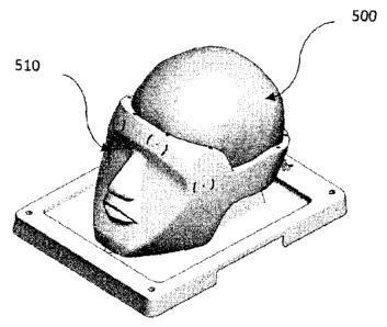
Referring to FIGS. 2-6, an exploded view of an example model or
phantom shown generally at 100 is provided that is suitable for use in
training or simulation of a medical procedure which is invasive of a
mammalian head. The training model 100 may be adapted or designed to
9
CA 02917938 2016-01-08
WO 2015/003271
PCT/CA2014/050659
simulate any mammalian head or a portion thereof. It is to be understood
that the person to be trained may be selected from a wide variety of roles,
including, but not limited to, a medical doctor, resident, student,
researcher,
equipment technician, or other practitioner, professionals, or personnel. In
other embodiments, the models provided herein may be employed in
simulations involving the use of automated equipment, such as robotic
surgical and/or diagnostic systems.
Referring now to FIG. 2, an exploded view of an example
implementation of training model (100) is shown that includes a base
component and a training component. The base component is comprised of
a tray component (200) and a head component. The head component is
comprised of a bowl component (210) and a skull component (220). The
training component may be comprised of a brain (230) with the following
layers: dura, CSF (cerebro spinal fluid), vessels, white matter, grey matter,
fiber bundles or tracks, target tumors, or other anomalous structures. The
training component may also include the aforementioned skull component
(220) when crafted in a skull mimicking material. Optionally, the training
model (100) may be also comprised of a covering skin layer (not shown).
Further, the base component may include a holder (240) provided on the tray
(200) to facilitate easy mounting of fiducials or reference points for
navigation.
Referring to FIGS. 3-5, the tray component (200) forming part of the
base component defines a training receptacle which includes a pedestal
section (242) which is sized and configured for receipt of the bowl
component (210) therein. Thus the training component is sized, configured
CA 02917938 2016-01-08
WO 2015/003271
PCT/CA2014/050659
or otherwise adapted to be compatible with, or complementary to the base
component, and particularly the training component receptacle, such that the
base component and the training component may be assembled to provide
the assembled training model (100).
The base component may have any size, shape and configuration
capable of maintaining the training component, mounted within the training
component receptacle, in a position suitable for performing the medical
procedure to be trained. This base component comprises features that
enable registration, such as fiducials, touchpoint locations, and facial
contours for 3D surface scanning, MR, CT, OCT, US, PET, optical
registration or facial registration. Furthermore, the base component is
adapted or configured to maintain the training component in a relatively
stable or fixed position throughout the performance of the medical procedure
to be simulated during the training procedure. The base component provides
both mechanical support during the training procedure and aids in the proper
orientation of the training components to mimic actual positioning of a
patient's head during the surgical procedure.
Referring to FIGS. 2 and 3, as noted above, the base component may
be comprised of a head component (210) and a tray component (200). The
tray component (200) is sized, configured or otherwise adapted to be
compatible with, or complementary to the head component (210). The tray
component (200) is adapted or configured to maintain the head component
(210) in a relatively stable or fixed position throughout the performance of
the
imaging or medical procedure to be simulated. This may be accomplished
with the use of a mechanical feature such as a snap mechanism that exists
11
CA 02917938 2016-01-08
WO 2015/003271
PCT/CA2014/050659
to affix the head component (210) to the tray component (200). The tray
component (200) may contain a trough (244) to catch liquids, and insertion
points to affix hardware to aid with image registration and/or the medical
procedure to be trained.
The head component (210) is sized, configured or otherwise adapted
to be compatible with, or complementary to the tray component (200) and the
training component. The head component (210) is adapted or configured to
maintain the training component (230) (located under skull component 300)
in a relatively stable or fixed position throughout the performance of the
medical procedure to be simulated. This head component (210) is adapted
or configured to enable anatomically correct surgical positioning. This may
include affixing the head component (210) with a surgical skull clamp or
headrest, for example a Mayfield skull clamp. This head component (210) is
also adapted or configured to enable anatomically correct imaging
positioning for any contemplated imaging modality including, but not limited
to, MR, CT, OCT, US, PET, optical registration or facial registration. For
example the head component (210) may be positioned in a supine position
within an MRI apparatus to enable anatomically accurate coronal image
acquisition.
In some embodiments, the head component (210) is shaped or
configured to simulate a complete or full skull. In other words, the training
component comprises bowl section (210) and skull section (220), while the
bowl section (210) comprises a further portion of a complete skull and head.
In some embodiments, as shown in FIG. 2, the head component i.e., bowl
section (210) and skull section (220), and training component (230) together
12
CA 02917938 2016-01-08
WO 2015/003271
PCT/CA2014/050659
provide a complete simulated skull or together provide a simulated head
including skull (220) and brain (230). The simulated head provided by the
training model (100) enhances the reality of the overall simulation training
experience.
In addition, the base and training components of the training model
(100), and particularly the head component, may also include one or more
external anatomic landmarks or fiducial locations 400, as shown in FIG. 4,
such as those likely to be relied upon by the medical practitioner for image
registration for example, touchpoints, the orbital surface, nasal bone, middle
nasal concha, inferior nasal concha, occipital bone, nape, and nasal
passage. These features will aid in registering the training component with
the preoperative images, such as MR, CT, OCT, US, PET, so that the
surgical tools can be navigated appropriately.
In this regard, navigation to establish the location of the hole or
passage through the skull of the patient during the craniotomy procedure is
often critical for the success of the medical procedure. Accordingly, external
anatomic landmarks and/or touchpoints are provided by the simulated head
in order to provide training on the correct registration of the training model
with the acquired images. These anatomic landmarks and touchpoints may
be utilized for attaching registration hardware, for example a facial
registration mask or fiducial landmark. Thus, the training model, and
particularly the simulated head, are sized, configured and shaped to
approximate and closely resemble the size, configuration and shape of the
head of a patient on which the medical procedure is to be performed. In
other words, the head component may be both life-like' and life-sized'.
13
CA 02917938 2016-01-08
WO 2015/003271
PCT/CA2014/050659
The base component may be comprised of any cornposition or
material suitable for providing the training component receptacle, and may
be suitable for being cast, molded or otherwise configured to provide or
support the simulated head when assembled with the training component.
For instance, the base component may be comprised of any suitable casting
compound, casting composition or plaster. The base component may be
comprised of a material that is rigid, non-reflective, non-ferrous, non-
porous,
cleanable, and lightweight, for example a urethane or acrylonitrile butadiene
styrene (ABS). In addition, the bowl (210) and skull (220) components of the
base component may be comprised of a material that is visible by the
imaging procedure of interest to enable registration. The material for the
bowl (210) and skull (220) components of the base may therefore be
selected to be visible by MR, CT, and/or PET. Suitable properties for
mimicking the skull component (220) for various imaging modalities are
illustrated in Tables 1,2 and 3.
In another embodiment, the base component may be manufactured
from a material that is not visible in MR, CT and PET. This is particularly of
value when the scope of training does not include facial registration and
craniotomy. For example, it is widely known that Teflon TM may be chosen
when the base component needs to be transparent in MRI. This further
eliminates subsequent software processing steps where the skull structure of
the head needs to be removed prior to visualizing the brain structure. This
step is commonly known as skull stripping and it can be computationally
costly.
The three simulation steps described previously (namely, pre-
14
CA 02917938 2016-01-08
WO 2015/003271
PCT/CA2014/050659
operative imaging simulation, surgical simulation and intra-operative imaging
simulation) can be realized using models or phantoms that share some
properties in common. Properties of tissue mimicking materials that are
suitable for imaging using various modalities are presented next.
PET imaging requires the injection of radioactive contrast agent prior
to the imaging step. The half-life of the contrast agents will limit the shelf-
life
of the training phantom. This can be overcome by manufacturing the
phantom with micro-capillaries so that contrast agents can be introduced via
the capillaries just prior to PET imaging. Alternatively, contrast agents may
be injected to selected regions of the brain component.
Densities for Brain mimicking for imaging in CT and US
Physical properties of the brain that are preferred for CT and US
imaging are illustrated in Table 1 (Barber, Ted W., Judith A. Brockway, and
Lawrence S. Higgins. "The Density Of Tissues In And About The Head." Acta
Neurologica Scandinavica 46.1 (1970): 85-92. Web.).
Human Brain Density (q/cmA3)
Frontal White 1.073
Frontal Gray 1.090
Parietal White 1.026
Parietal Gray 1.109
Occipital White 1.073
Occipital Gray 1.103
Corpus-callosum 1.093
Thalamus 1.052
Caudate Nucleus 1.075
Putamen 1.081
Global Pallidus 1.084
Brachium Pontis 1.116
CA 02917938 2016-01-08
WO 2015/003271
PCT/CA2014/050659
Medulla 1.057
Pons 1.069
Cerebellum 1.058
Table 1: Properties suitable for CT and US imaging
Optical Properties for Brain mimicking for imaging using OCT are
presented in Table 2 (Cheong, W.f., S.a. Prahl, and A.j. Welch. "A Review of
the Optical Properties of Biological Tissues." IEEE Journal of Quantum
Electronics 26.12 (1990): 2166-185. Web.).
Brain Transmission Absorption Scattering Wavelength
Matter Coefficient Coefficient Coefficient
White 52.6 1.58 51 633
Grey 62.8 2.63 60.2 633
Table 2: Properties of the brain that are preferred for imaging using
OCT.
Alternately, the properties of materials suitable for mimicking intra-
operative
ultrasound (i-US) may be established using operating frequency of typical
ultrasound transducers.
(reference: http://www.bkmed.com/Intraoperative US en.htm).
For intra-operative US, frequency range is from 4 to 10 MHz. This is based
on such instruments as BK medical transducers, intraoperative 8815, T-
shaped intraoperative 8816, Intraoperative biplane 8814, Hockey Stick 8809,
and the, Intraoperative biplane 8824. At 5 Mhz the average propagation
speed through mixed tissue is 1565m/s. Another property of the tissue that is
preferred for ultrasound imaging is attenuation coefficient. This is
illustrated
in Table 3.
16
CA 02917938 2016-01-08
WO 2015/003271
PCT/CA2014/050659
(Kremkau, Frederick W. "Ultrasonic Attenuation and Propagation Speed in
Normal Human Brain." The Journal of the Acoustical Society of America70.1
(1981): 29. Web.)
Table 3: Attenuation coefficients for 5MHz frequency ultrasound
through the brain
Brain Matter Attenuation Coefficient
White Matter ==7db/s
Grey Matter ==4db/s
Mixed Matter ==3db/s
Parameters of the brain tissue that are essential for mimicking MR
images are Ti, T2, and Spin Densities. This is illustrated in Table 4 for
various components of the brain.
Table 4: Ti, T2 and spin densities of various parts of the brain
Type of Matter Relative Spin 1.5T 3.0T
Densities
Ti(ms) T2(ms) Ti(ms) T2(ms)
Gray Matter 83 1000 100(T2), 1331 85(T2)
65(T2*) 42(T2*)
White Matter 71 710 80(T2) 4000 70(T2)
78(T2*) 49(T2*)
Cerebral Spinal 100 4000 2200 4000 2200
Fluid
Venous Blood 82 1435 150 1584 80
Arterial Blood 82 1435 235 1664 165
Fat 90 300 165 380 133
Finally, PS-OCT is used to visualize birefringence property of living
17
CA 02917938 2016-01-08
WO 2015/003271
PCT/CA2014/050659
tissue. Birefringence is directionality dependent and would therefore vary
quantitatively depending on the particular brain being imaged; however, the
visually invisible brain tracts could be reproduced and optimized using a
particular material that may be organized in such a way to give the desired
brain tract orientation.
As shown in FIG. 5, the training component (230) and the base
component (210) are complementary or compatible such that when the
training component (230) is mounted on the pedestal (242) in the training
component receptacle in tray (200), together they provide the training model.
Furthermore, the configuration and dimensions of the training component
(230) and the base component (210) are complimentary or compatible such
that the training component (230) may be received and fixedly or releasably
mounted in the base component (210).
In some embodiments, in order to permit the replacement or
substitution of the training component (230), the training component is
detachably or releasably mounted in the base component (210). Any
detachable or releasable fastener or fastening mechanism may be used
which is capable of securing the training component (230) in the receptacle,
while also permitting the training component (230) to be readily detached,
released or removed as desired or required. In one embodiment, the training
component (230) is releasably or detachably mounted within the base
component (210), specifically the training component is held within the base
component (210) to emulate the mechanical fixation of the brain in the skull.
Thus, in the present example embodiment, the training component
(230) may be removed from the base component (210) and replaced with an
18
CA 02917938 2016-01-08
WO 2015/003271
PCT/CA2014/050659
alternate, replacement or substitute training component as desired or
required by the user of the training model. For instance, a replacement
training component (230) may be required where the previous training
component (230) is damaged or modified during the training of the
procedure. An alternate training component (230) may be adapted or
designed for use in the training of the performance of a specific medical
procedure or condition of the patient, allowing for the reuse of the base
component (210).
Alternatively, as indicated, the training model (100) may not include
the base component (210). In this instance, the other components
comprising the training model (100), such as the training component (230) in
isolation, may be supported directly by a supporting structure or a support
mechanism (not shown) that does not look like a mammalian head.
Specifically, the supporting structure may securely maintain the training
component (230), without the other components of the training model, in the
desired orientation. In such an embodiment, the training component (230)
may be releasably attached or fastened with the supporting structure such
that the training component (230) may be removed from the supporting
structure and replaced with an alternate, replacement or substitute training
component (230) as desired or required by the user of the training model.
Referring to FIG. 6, the training component (230) may be comprised
of a simulated mammalian head. The simulated head may be comprised of
skull (220), dural layer (610) (or dura), CSF layer (620), blood vessels
(630),
a brain section including grey matter (640), white matter (650), diffusion or
brain fibers (660), and a tumor target (670). This training component (230)
19
CA 02917938 2016-01-08
WO 2015/003271
PCT/CA2014/050659
may be customized as desired to train on the medical procedure of interest,
for example the training component (230) may include all of these layers or a
subset such as the dura (610), white matter (650), and tumor target (670).
In one embodiment, the skull layer ( 220) is included as an element of
the training component (230). The skull layer (220) is formed from osseous
type material as described herein. This skull layer (220) is constructed of a
skull material which simulates osseous tissue when penetrated. Hence, this
layer (220) is intended to simulate surgical resection. Thus, the skull
material of the skull section (220) mimics or imitates osseous tissue when
penetrated, pierced or passed into or through. In an embodiment described
herein, the medical procedure is comprised of drilling into or through a
portion of the skull, which is simulated by the skull section (600). In order
for
the skull section ( 220) of the present phantom to mimic or imitate osseous
tissue, properties illustrated in Table 5 need to be met by the material
simulating skull section (220).
Property Mean
Skull Thickness .272in
Diploe Thickness .108in
Dry Weight Density .051/inA3
Modulus Compression Radial 350000psi
Secondary Modulus Compression 53000
Radial
Modulus compression 810000
tangential direction
Poisson's ratio 0.19
compression radial
Poisson's ratio 0.22
CA 02917938 2016-01-08
WO 2015/003271
PCT/CA2014/050659
compression tangential
Ultimate strength 10700psi
compression radial
Ultimate strength 14000psi
compression tangential
Ultimate strain 97000in/in
compression radial
Ultimate strain 0.051in/in
compression tangential
Energy absorption 1200/^3
compression radii
Energy absorption 480/inA3
compression tangential
Microhardness Vickers DPH 31.6
inner table
Microhardness Vickers DPH 34.2
outer table
Ultimate strength 3100psi
diploe direct shear
Index of isotropy 2.5
Ultimate strength 3200psi
diploe torsion
Modulus torsion diploe 200000psi
Ultimate strength 6300psi
tension tangential
Modulus tension 780000psi
composite psi
Ultimate strength 11500psi
tension tangential
compact tables
Table 5: Properties of the Skull for surgical mimicking
21
CA 02917938 2016-01-08
WO 2015/003271
PCT/CA2014/050659
Thus, in such embodiments, the skull material particularly simulates,
mimics or imitates the "feel" and resistance of osseous tissue when it is
being penetrated by drilling. For example, the osseous tissue mimic may be
formed from an acrylonitrile butadiene styrene (ABS) material texturized and
patterned to resemble skull tissue. ABS is a terpolymer of acrylonitrile,
butadiene and styrene and typical or usual compositions are about half
styrene with the balance divided between butadiene and acrylonitrile. An
advantage of using ABS material is that considerable compositional variation
is possible, resulting in many different grades of acrylonitrile butadiene
styrene with a range of properties so if material tuning is required to
achieve
the properties noted in Table 5 there are considerable options. In addition,
many blends with other materials such as polyvinylchloride, polycarbonates
and polysulf ones may be used. Acrylonitrile butadiene styrene materials can
be processed by any of the standard thermoplastic processing methods.
In addition, in order to more closely simulate the skull, the skull layer
(220) may have a thickness which approximates that of the mammalian skull.
In one embodiment, the skull layer (220) has a thickness which particularly
approximates that of the portion or area of the neurocranium typically
penetrated in performance of the medical procedure to be trained. Thus, the
skull section of the training model, may have a total thickness in a range of
about 5 to about 10 millimeters.
However, depending upon the medical procedure to be trained, the
training model need not include the skull layer (220). Specifically, in some
medical procedures, the medical procedure is directed at structures
underlying the dural layer (610). In these instances, the emulation of the
22
CA 02917938 2016-01-08
WO 2015/003271
PCT/CA2014/050659
skull layer (220) is not critical. Thus the skull section (600) will not be
required in the training model for that procedure. Further, as shown in FIG.
6,
in some embodiments, a dural layer (610) may be provided which underlies
the skull section (220). The dural layer (610) may be positioned to abut or
lie
adjacent to the innermost surface of the skull section (220). In other words,
the dural layer (610) underlies the skull (600). The dural layer (610)
material
may be comprised of any material or substance capable of simulating dural
tissue as described when applying surgical instruments or when imaged.
Thus, the dural material of the dura section (610) also mimics or
imitates dural tissue visually, or when imaged with MR, CT, OCT, US, and/or
PET, or when penetrated, pierced, stitched, or passed into or through. Thus,
in the embodiments, as indicated above, the dural material particularly
simulates, mimics or imitates the "feel" and resistance of dural tissue when
it
is being cut by a scalpel or surgical scissors. In some embodiments, the
dural layer (610) also simulates the non-absorbent and liquid tightness
exemplified by dural tissue. Thus creating a water-tight enclosure for the
liquid surrounding the brain and preventing absorption of the CSF-type liquid
used in the training model (230).
For instance, in some embodiments, the dural layer (610) material
may be comprised of urethane or silicone brushed fibers. Although any
suitable silicone or urethane may be used for the purpose of mimicking
biomechanical properties of the dural layer (610), it may be beneficial to
select a silicone or urethane that is opaque in nature, in order to obscure
the
view of the brain and its sulci below. Furthermore, it may be beneficial to
select a silicon or urethane that forms or takes on the shape of the
23
CA 02917938 2016-01-08
WO 2015/003271
PCT/CA2014/050659
underlying sulci.
In addition, the biomechanical property of the dural layer (610) may be
mimicked using a layer that may have a thickness which approximates that
of the dura matter underlying the skull. In some embodiments, the dural
layer (610) has a thickness which particularly approximates that of the dura
mater underlying the inner portion or area of the neurocranium typically
penetrated in performance of the medical procedure to be trained. In an
embodiment, the dural layer (610) has a thickness of less than approximately
1mm, for example, between about 0.5 to about 0.8mm which is typical of the
human dura.
Furthermore, as shown in FIG. 6, in some embodiments, a vessel
layer (630) may be provided which underlies the dura section (610). The
vessel layer (630) may abut or lie adjacent to the outermost surface of the
brain section. The biomechanical property of the vessels may be simulated
using material that may be comprised of any material or substance capable
of simulating vessel tissue as described when applying surgical instruments
or when imaged. Also, the vessel material of the vessel section (630) may
also mimic or imitate vessel tissue visually, when imaged, for example with
MR, CT, OCT, US, and/or PET or when penetrated, pierced, stitched, or
passed into or through. Thus, in the embodiments, as indicated above, the
vessel material particularly simulates, mimics or imitates the "feel" and
resistance of vessel tissue when it is being cut by a scalpel or other
surgical
instruments. For instance, in some embodiments, the vessel layer (630)
material may be comprised of a silicone material or a polyvinyl alcohol
cryogel (PVA-C) mixture. This material is suitable for mimicking
24
CA 02917938 2016-01-08
WO 2015/003271
PCT/CA2014/050659
biomechanical properties and for producing appropriate MR and CT images.
Biomechanical properties are mimicked through appropriate control of
stiffness of the material using controlled cooling and heating cycles.
Pigmentation may be applied to the vessel layer (630) material to represent a
lifelike vessel coloring.
In addition, the vessel layer (630) may have a tubular shape with a
diameter which approximates that of the vessels within the skull. In some
embodiments, the vessel layer (630) is hollow to allow for the routing of
fluids
within, for example, a blood-like liquid mimic. In an embodiment, the vessel
layer (630) has a thickness of between about 0.2mm and 3mm, and more
preferably has a thickness of about 1 mm, a typical range for the human
brain. Further, as shown in FIG. 6, in some embodiments, CSF layer (620)
may be provided which underlies the dural layer (610). The CSF layer (620)
may lie between the water-tight dural layer (610) and the non-liquid
absorbent brain layer including grey matter layer (640), white matter layer
(650), possibly surrounding the vessel layer (630), and within the brain
ventricles if provided.
The CSF liquid in the CSF layer (620) may be comprised of any liquid
or substance capable of simulating CSF as described when passing through
or imaging the training component. Thus, the CSF liquid mimics or imitates
CSF visually and when imaged, for example with MR, CT, OCT, US, and/or
PET, or when passed into or through. Thus, in the embodiments, as
indicated above, the CSF material in CSF layer (620) particularly simulates,
mimics or imitates the "feel" and viscosity of CSF liquid when it is being
passed through by surgical instruments. For instance, in some embodiments,
CA 02917938 2016-01-08
WO 2015/003271
PCT/CA2014/050659
the CSF liquid mimic may be comprised of a mineral oil or saline solution.
The above stated CSF liquid mimicking material primarily simulates
biomechanical property of the brain. In an embodiment, this liquid may be
used to hydrate the fibrous structures included within the brain layer
including grey matter layer (640), white matter layer (650).
In addition, the dural layer (610) may enclose a volume which
approximates that of the CSF in the mammalian brain. In an embodiment,
the CSF section (620) has a volume of between about 100m1 and 200m1,
such as, approximately 150m1.
Further, as shown in FIG. 6 and 7, a brain layer may be provided
which underlies the dural layer and the skull section. Where both the dural
layer and the brain layer are provided, the brain layer may abut or lie
adjacent to the dural layer. In other words, the dural layer (610) is
underlying
the skull section, while the brain layer is underlying the dural layer (610).
Thus, the dural layer (610) is interposed between the skull layer and the
brain layer.
In some embodiments, the brain layer is constructed of brain layer
material which simulates or mimics brain tissue, including grey matter layer
(640) and white matter layer (650), when physically penetrated and/or when
imaged, for example with MR, CT, OCT, US, and/or PET. As noted above,
this brain layer may be divided into grey (640) and white matter (650) so that
the brain layer material may be configured to mimic or imitate grey and white
matter tissue when penetrated, pierced, or passed into or through and/or
when imaged. For example, following the cutting or incising of the dura
mater, the medical procedure may comprise penetration of the brain layer by
26
CA 02917938 2016-01-08
WO 2015/003271
PCT/CA2014/050659
inserting or passing surgical instruments, for example, a trocar, catheter,
drain, port, obturator, MyriadTM, into or through a portion of the brain. In
such
a case, the brain layer material mimic may have a composition that responds
to these instruments in a manner that mimics that of real brain tissue, for
example, the brain tissue will not clog up the MyriadTM.
Thus, the brain layer material may particularly simulate, mimic and/or
imitate the "feel" and resistance of brain tissue when it is being penetrated
in
this manner. However, the specific nature and composition of the grey and
white matter (640) and (650) being penetrated may vary depending upon the
particular medical condition of the patient and the procedure to be trained
for.
Accordingly, the grey and white matter material mimic (640) and (650) may
be specifically selected to simulate, mimic or imitate the "feel" resistance,
and imaging properties of the brain tissue likely to be encountered in the
context of the patient's medical condition and the performance of the medical
procedure being trained.
Accordingly, the brain layer mimic material may be comprised of any
material or substance capable of simulating brain tissue as described. For
instance, in some embodiments, the brain layer mimic material may be
comprised of a polyurethane MCG-1 or PVA-C material. In an embodiment,
the brain layer mimic material is comprised of a polyurethane material mixed
with an additive such as glass bubbles or mineral oil to achieve the desired
consistency of brain material.
In one example implementation, the brain layer material is comprised
of a mixture of the polyurethane and glass bubbles. The glass bubbles may
be incorporated so that they do not exceed 5% of the total volume, to mimic
27
CA 02917938 2016-01-08
WO 2015/003271
PCT/CA2014/050659
the tear strength and tensile properties of brain tissue. In another
embodiment, the brain layer material mimic is comprised of 6% PVA-C mixed
with water and one freeze-thaw cycle.
In some embodiments, the brain layer mimic material may have a
thickness, dimensions, and anatomically accurate sulci and ventricles, which
approximates the brain tissue likely to be encountered in the performance of
the medical procedure to be trained. As indicated, in some medical
procedures, the medical procedure is directed at the brain or structures lying
within the brain. Thus the thickness of the brain layer mimic material will be
selected to simulate the location within the brain, or the location of a
structure within the brain, at which the medical procedure is directed.
Furthermore, as shown in FIG. 6, in some embodiments, fiber bundles
or brain tracks (660) may be embedded within the brain matter layer mimic
material. These fiber tracts (660) are intended to simulate the fiber tracts
that are found within the brain matter, for instance the major white matter
fiber tracts. The fiber bundles (660) may are positioned within the brain
phantom to emulate the white matter tracts within brain tissue. The fiber
bundles (660) may be comprised of any material or substance capable of
simulating the mechanical and imaging characteristics of white matter fibers
when imaged, for example with MR, CT, OCT, US, and/or PET. For instance,
the fiber bundles (660) will embody the diffusion and mechanical properties
of the white matter fibers when imaged with DWI and/or DTI or when
applying surgical instruments. Thus, the white matter fibers (660) of the
fiber
section also mimics or imitates white matter fibers visually, when imaged, or
when penetrated, pierced, stitched, or passed into or through. Thus, in the
28
CA 02917938 2016-01-08
WO 2015/003271
PCT/CA2014/050659
embodiments, as indicated above, the diffusion fibers (660) particularly
simulate, mimic or imitate the "feel" and resistance of human brain diffusion
fibers when it is being cut by a scalpel. Further, the diffusion fibers (660)
provide a structured channel for water molecules to diffuse through. This
structured diffusion results in the generation of diffusion tensor images
(DTI)
that resemble DTI obtained on living organs such as brain and heart. In
some embodiments, the diffusion fibers (660) may be comprised of a fibrous
structure within a sheath or tube, for instance polyester, nylon,
poloypropelene or Dyneema fibers packed within a plastic tube or heat-
shrink. In another embodiment, the fibers (660) may be directly embedded
within the white matter mimic material (650). The surrounding brain mimic
material hydrates the fibers and provides the liquid that diffuses.
In addition, the diffusion fibers (660) may have a tubular shape with a
diameter which approximates that of the white matter tracts within the brain
section. In some embodiments, the diffusion fibers (660) are threaded
through the brain matter mimic and protrude from the brain matter where
they are exposed and hydrated by the surrounding CSF fluid mimic layer
(620). This CSF fluid layer (620) provides the hydration which diffuses
through the tubes and is visible in the acquired images. These diffusion fiber
mimics (660) provide an enhanced training experience for the surgeon as
during the training procedure they are encouraged to avoid tearing or
harming the diffusion fibers during the operation.
Furthermore, as shown in FIG. 6, depending upon the specific medical
procedure to be trained for, in some embodiment of the training model, a
target may be provided which underlies the skull layer, and underlies the
29
CA 02917938 2016-01-08
WO 2015/003271
PCT/CA2014/050659
dural layer, within the brain layer material. However, the specific location
of
the target underlying the skull section may vary depending upon the nature
of the target and the nature of the medical procedure to be trained which is
directed at the target.
In general terms, the target is intended to simulate, mimic or imitate a
specific structure embedded within the brain layer material which is the focus
of the medical procedure to be trained or at which the medical procedure is
directed. The specific structure or focus of the medical procedure may be
normal or aberrant anatomical structure, clot, lesion, structure resulting
from
a pathological condition or other structure desired to be acted upon by the
medical practitioner.
In one embodiment, the specific structure to be acted upon, or at
which the medical procedure is directed, is a target tumor (670). This target
tumor (670) may be comprised of any material or substance capable of
simulating tumor tissue as described when applying surgical instruments or
when imaged.
Thus, the target tumor (670) material mimics or imitates tumor tissue
visually, when imaged, for example with MR, CT, OCT, US, and/or PET, or
when penetrated, pierced, resected, or passed into or through. Thus, in the
embodiments, as indicated above, the target tumor (670) material particularly
simulates, mimics or imitates the "feel" and resistance of tumor tissue when
it
is being cut by a scalpel or MyriadTM. For instance, in some embodiments,
the target tumor (670) material may be comprised of a hydrocolloid material,
a rubber-glass mixture, or a PVA-C mixture. These materials may be doped
with contrast agents to simulate the imaging characteristics of tumor tissue.
CA 02917938 2016-01-08
WO 2015/003271
PCT/CA2014/050659
Exemplary, non limiting examples of contrast agents include any one of a
fluoride, a chloride, or sulfate. Non-limiting examples include chromium
fluoride, gadolinium chloride, copper sulfate, barium sulfate, manganese
chloride. In addition, agarose may be used as well.
Hence, the simulated tumor (670) is suitable for mimicking
biomechanical properties and for generating images that resemble tumor
regions in living tissue. In an embodiment, pigmentation may be applied to
the target tumor (670) material to represent a lifelike tumor coloring.
Referring to FIG. 6, in an embodiment, the target (such as, but not
limited to tumor (670)) may be located or positioned a spaced distance from
the innermost surface of the brain layer sulci (clearly visible in FIG. 7). In
this
instance, the brain layer is provided underlying the dural layer (610),
wherein
at least a portion of the brain layer is interposed between the dural layer
(610) and the tumor target (670) in order to simulate the anomalous structure
to be trained upon. Accordingly, the brain layer, or a portion thereof, will
be
required to be penetrated in order to access the tumor target (670). The
specific location of the tumor target (670) within or underlying the brain
layer
will be selected to closely approximate the location of tumors within the
human brain.
The imaging and biomechanical brain phantoms may be constructed
to have specific dedicated sites, anywhere under the skull layer ( 220), sized
to receive specifically sized target tumors (670) which match known types of
tumors. The various parts of the brain phantom may have a modular
construction, for example the skull (220), dura (610), CSF (620), vessels
(630), brain section including grey matter (640), white matter (650), and
31
CA 02917938 2016-01-08
WO 2015/003271
PCT/CA2014/050659
diffusion fibers (660), may be constructed in a lego style so that sections
can
be readily removed to allow insertion of the target tumor (670) in pre-
selected
locations in the head below skull layer (220). The brain phantom kit disclosed
herein may come with a plurality of different sized and shaped tumor targets
(670) in order to be able to reconfigure the brain phantoms to allow training
to be conducted for multiple types of tumors and multiple locations within the
skull as well as for differently sized head phantoms emulating or mimicking
differently aged patients.
In addition, one or both of the imaging and biomechanical phantoms
may be constructed to include strategically placed sensors within the
different anatomical mimics for the purpose of, but not limited to, assisting
in
navigation. The sensors may be coded for pre-selected locations in the brain
phantom.
As noted elsewhere, to produce a lifelike brain phantom with
biomechanical properties to allow a practitioner to train on various brain
invasive surgical procedures, using the port based method involving insertion
of the surgical port into the brain via the sulci, requires the model to be
life
size and with the deep sulci morphology.
An exemplary, non-limiting method of producing a one piece brain
phantom including deep sulci includes acquiring an image of a human brain
using MRI, using this image to 3D-print an anatomically accurate one piece
model of the brain with deep sulci emulating the human brain. Once the
model is produced, applying a flexible mold material to an outer surface of
the model of the brain and after the mold material has set to form a brain
mold, releasing the brain mold from the model of the brain. The brain mold is
32
CA 02917938 2016-01-08
WO 2015/003271
PCT/CA2014/050659
placed into a rigid outer shell to prevent swelling. The mold is filed with a
liquid precursor of a brain material mimic. Depending on the ultimate purpose
of the particular phantom being produced, there may be embedded in the
liquid precursor one or more mimics for one or more structural brain features,
such as a dural layer, blood vessel layer, and brain matter tracks to mention
a few. The liquid precursor is then induced to set to form an anatomically
correct brain phantom in one piece with deep sulci and after the liquid
precursor has set, the set brain mold is released from the brain mold.
The gyri and sulci may be produced to exhibit any one or combination
of elastic modulus, shear modulus, tensile strength and nonlinear elastic
properties comparable to a mammalian sulci.
It is noted that the MR image used to make the model may be that of
the patient to be operated on, so that the outer sulci morphology closely
resembles that of the patient. Non patient specific, or generic brain phantoms
may be produced for general training procedures, but the advantage of using
the patient's brain phantom allows the practitioner to practise on a brain
phantom closely matching that of the patient in question.
An example shape for the brain component of the training component
is illustrated in FIG. 7 which as mentioned above shows the outer topography
of the brain phantom. The shape is such that sulci and the two lobes are
accurately represented. A manufactured example of the training model is
illustrated in FIGS. 8A and 8B which shows the brain section (800) wrapped
in the dura component (810) positioned on the base component (820) with
fiducial or reference markers (830) placed at specific locations to facilitate
image registration for surgical navigation.
33
CA 02917938 2016-01-08
WO 2015/003271
PCT/CA2014/050659
FIG. 9 illustrates the MR image obtained for this same training model
or phantom shown in FIGS. 8A and 8B which was constructed using a
polyurethane material as the brain matter mimic. As evident in the image, the
surface profile (920), fiducials or reference (910) and the embedded tumors
(900) are clearly visible in the acquired image.
The CT image of the same training model is illustrated in FIG. 10,
which is shown as a reconstructed 3D image in FIG. 11. These figures
further illustrate the location of the tumor (1010) in the brain tissue (1000)
seen in FIG. 10, the surface profile of the gray matter (1110) and location of
fiducial or reference markers (1100), seen in FIG. 11. The visibility of
fiducials (1100) in images acquired using multiple imaging modalities
facilitates registration of different images and their subsequent using in
image guided navigation during the surgical procedure described previously.
The tumor target may have any shape, configuration and dimensions,
capable of and suitable for simulating, mimicking or imitating the specific
structure underlying the skull of a patient which is the focus of the medical
procedure to be trained on or at which the medical procedure is directed. An
example size range may be from 1mm to 3cm, and the consistency may
range from gelatinous to rigid. In one embodiment a tumor is made with a
size of 1cm of hydrocolloid material with a concentration of 0.2% copper
sulfate as contrast agent. In another embodiment, rubber glass may be used
as a tumor target with a rubber glass to slacker ratio of up to 1:4 or only
composed of rubber glass, see:
http://www.smooth-on.com/tb/files/Slacker Tactile Mutator.pdf
In an embodiment, the training model or phantom may be provided
34
CA 02917938 2016-01-08
WO 2015/003271
PCT/CA2014/050659
with a simulated skin. Specifically, a skin layer may be provided for
overlying
at least the outer-most surface of the skull section. In other words, the skin
layer overlies the skull section (230). In addition to overlying the skull
section
(230), which comprises the outer surface of the training component in some
embodiments, a skin layer may also be provided for overlying all or a portion
of the outer surface of the head component. Thus, for instance, the head
component may include the skin layer in order to provide a more realistic
simulation of the medical procedure.
The skin layer is constructed of a skin layer mimic material which
simulates skin tissue when penetrated. Thus, the skin layer material mimics
or imitates skin tissue when penetrated, pierced or passed into or through.
In an embodiment described herein, prior to penetrating the skull, the
medical procedure may further require the penetration of the skin in order to
provide access to the skull for the subsequent procedure. In this instance,
the skin is typically cut or incised. Thus, in such embodiments, the skin
layer
material particularly simulates, mimics or imitates the "feel" and resistance
of
skin tissue being penetrated by cutting or incising.
The skin layer mimic material may be comprised of any material or
substance capable of simulating skin tissue as described. For instance, in
some embodiments, the skin layer mimic material is comprised of a silicone
rubber or a flexible silicone elastomer. This material is intended to simulate
biomechanical and imaging properties of living skin layer. Further, the skin
layer material may provide a surface enabling image registration and/or facial
registration, for example with MR, CT, US, and/or PET. In an embodiment,
the skin layer material is comprised of a platinum cure silicone rubber which
CA 02917938 2016-01-08
WO 2015/003271
PCT/CA2014/050659
may be tinted with flesh colored dye or pigment, and which is commercially
available under the name Dragon Skin, Dragon Skin is a trade-mark of
Smooth-On Inc. This is suitable for mimicking the biomechanical and visual
properties of the skin.
In addition, the skin layer may have a thickness which approximates
that of the skin of the human head. In some embodiments, the skin layer has
a thickness which particularly approximates that of the skin covering the
portion or area of the neurocranium typically penetrated in performance of
the medical procedure to be trained. In an embodiment, the skin layer has a
thickness of about 2 millimeters.
In one embodiment of this training model, the imaging phantom and
the biomechanical phantom are constructed independently however are
identical in form and reference each other identically In other words, the
imaging and biomechanical phantoms are anatomical analogues of each
other or they are anatomically correlated. Therefore, the imaging phantom
can be used for imaging purposes, for example with MR, CT, OCT, US,
and/or PET. These images can be registered with the biomechanical
phantom and used for navigation for the surgical procedure to be trained. In
other words the imaging phantom and the biomechanical phantom both
embody the characteristics of the training model, however the imaging
phantom targets the imaging characteristics specifically and is directly
correlated with the biomechanical phantom. While the biomechanical
phantom embodies the biomechanical and physical characteristics of the
phantom, for instance mimicking the tactile and tensile properties associated
with the various layers within the training component.
36
CA 02917938 2016-01-08
WO 2015/003271
PCT/CA2014/050659
The above described independent construction of two phantoms that
are identical in form and reference each other may be extended to more than
two phantoms where third and subsequent phantoms may be constructed to
be optimal for third and different imaging modalities. In one such
embodiment, a quality assurance/control phantom is constructed which may
be a deformable or non-deformable phantom that generates known and
consistent Diffusion Tensor Images (DTI), so that these images are obtained
with a phantom containing diffusion bundles (660) as shown in FIG. 6. This is
illustrated in FIG. 12. A reference image (1210) acquired at the time of
construction of the phantom may be included with the phantom so that
imaging and surgical practitioners may iteratively improve their MR imaging
protocols until the DTI output generated by the practitioners (1220) closely
matches the DTI produced at the time of manufacturing. In other words, the
phantom and its associated DTI, which may be stored on a CD or other
medium, are shipped together to the end user as a brain phantom kit.
Alternatively, the entity producing the brain phantom may keep the DTI
stored at a location of its choosing but make it available online to the users
of
the brain phantom.
Provided along with the DTI are all the imaging parameters that were
used to obtain the DTI so that the practitioner may reproduce these imaging
parameters when they are practicing on the phantom. The quality control
phantom will include known truths to be tested, such as being imaged with
optimal pulse sequences that may be recorded and included with the
phantom. For example, an optimal DTI scan may be used to image the
diffusion fiber mimics whereupon at the practitioner's scanner, they can tune
37
CA 02917938 2016-01-08
WO 2015/003271
PCT/CA2014/050659
their diffusion sequence to optimize the pulse sequence for the practitioners
scanner in order to recreate the quality control DTI sequence, knowing the
reference level that they can and should achieve.
A software package containing appropriate algorithms may be used
by the practitioner to run the analysis. For example, the practitioner scans
the phantom and runs the analysis with the software analyzing the acquired
image for quality and comparing it with the factory produced reference
image. The software package is programmed with instructions to provide
appropriate feedback to the practitioner, for example with respect to the
resolution used by the practitioner, the feedback may be "your resolution is
too low thus resulting in a noisy image, please try increasing your slice
thickness to x". Alternatively, if it is a research site they can use the
quality
control phantom to develop new pulse sequences knowing what they should
achieve based on the quality control DTI sequence.
The software package may also contain appropriate algorithms
programmed to analyze patient images for quality. For example, once an
operator or technician performs a scan, the software may be programmed
with algorithms to detect movement causing a reduction the quality of the
image and then prompt the technician to repeat the scan given the artifact. If
the artifact does not disappear, the operator/technician has the opportunity
to
scan the quality control phantom to try and troubleshoot the problem.
It is noted that pulse sequences are continually evolving, so that the
software can be updated to reflect these new pulse sequences. For example
due to continual improvements in MRI hardware, an optimal DTI scan today
will unlikely be what is optimal is a year's time.
38
CA 02917938 2016-01-08
WO 2015/003271
PCT/CA2014/050659
The development of optimal imaging parameters by the practitioner
training on the phantom such as optimal MR pulse sequences may be further
guided through the development of a scoring system (1230) that may be
implemented as software or dedicated hardware. The scoring system (1230)
will compare the MR image (1220) generated by the practitioner training on
the phantom against the reference image (1210) generated at the time just
after manufacturing the phantom where the latter image is considered to be
the golden standard to the truth. Parameters for score may include aspects
of the image that are relevant to development of optimal DTI output. Such
parameters may include, but not are not limited to, resolution, scan time,
contrast, signal-to-noise ratio, correct representation of direction of fiber
bundles via DTI, raw dimensions of the acquired image etc. Hence, the
imaging phantom teaches capture of optimal MR images with clinically useful
DTI information that are of value for safely approaching and resecting tumor
targets from the biomechanical phantom.
It will be understood that above described DTI phantom for
qualification and optimization of data acquisition parameters need not be
anatomically identical to a human organ such as brain or heart. Instead, it
could be a fixed geometrical shape such as a sphere or a cylinder. Hence,
total set or kit of training phantoms may include a brain-like biomechanical
phantom, a brain-like imaging phantom and a third rigid phantom of fixed
geometric shape for optimizing MR data acquisition parameters. Some of the
hardware features of the phantom used to optimize MRI acquisition are
illustrated in Table 6. These features may be compared with optimal values
established a priori. Hence, the latter phantom can be used qualify
39
CA 02917938 2016-01-08
WO 2015/003271
PCT/CA2014/050659
acquisition protocols and used to perform root-cause analysis when failure of
the MR equipment is suspected.
Hardware feature Purpose
Geometrically shaped Spherical or cylindrical shapes are
easier to insert into standard MR
bores.
Chambers with known Ti and T2 Their values at varying field strengths
values are known apriori.
Water chamber To check water signal suppression
functionality/ determine water peak
reference
Chamber with known contrast To optimize MRI contrast level for Ti,
T2, T2*
Resolution plate with geometric To establish x, y, z resolution
feature (possibly curved, square, pyramid or
combination)
Fits in head coil Multiple size and shape variations of
phantom for customization of various
MRI machines
Has perfusion structures To assure ability to detect signals at
scales essential for effective brain
imaging (ex. capillary scale)
Has diffusion fibers arranged in To optimize and test DTI ability of
geometric formation scanner. To determine effectiveness
of DTI in multiple directions
Set area to measure SNR To measure SNR
Material possible acrylic MRI compatible material
Features to demonstrate correct Features to line up with landmark
orientation in the magnet
Pressure expansion screw for To detect possible deformations of
shipping purpose phantom caused by pressure
CA 02917938 2016-01-08
WO 2015/003271
PCT/CA2014/050659
changes during delivery
Temperature sensor To plot temperature with respect to
time during scan (using MRI
compatible electronics). To
determine instantaneous temperature
of Machine
Choline chamber For MRS optimization (correlates
with cell density)
Built-in registration points For registration with reference
images
Ultrasmall super-paramagnetic iron To optimize and determine detection
particles (USPIO) sensitivity
Chambers with different nuclei Possibly Carbon 13, Xenon 129
Larmor frequencies To optimize gradient and RF coils
Table 6: Salient features of phantom used to qualify and optimize
MR data acquisition
Some of the parameters analyzed using the latter phantom and their
role are presented in Table 7. These parameters are intended to reduce the
need for repeat scans for patients due to lack of insufficient data or quality
in
pre-operative scans, diagnostic scans, post-operative scans and functional
MRI scans. The analysis result can be used to automatically deduce faults in
the data acquisition process and suggest ways of improving the scans.
Parameter Purpose
Completion status of protocol To assure all tests are completed
Presence of area of interest To test if F.O.V. is correct
Resolution [6] To assure minimal resolution level is
achieved
Contrast agent uptake (gadolinium) To test detection and optimize
imaging parameters with respect to
41
CA 02917938 2016-01-08
WO 2015/003271
PCT/CA2014/050659
contrast agent detection
Signal to noise ratio (SNR) [10] To test detection and optimize
imaging parameters with respect to
SNR
CNR [5] To test detection and optimize
imaging parameters with respect to
CNR
Confirm placement of fiducial To test detector orientation
markers output/warping of images/other
factors
Checking for gating on/off Assuring that gating is turned on
when needed (example when
imaging the heart), if gating is off
when needed the software suggests
it be turned on and vice versa
Check for presence of spikes in K- All artefacts in test scans will be
space (image artefact check detected using software and
Presence of motion artefacts suggestions to improve the image will
Identify presence of wrapping be provided. After implementation of
artefact suggestions the image will be
Identify presence of ghosting artefact recaptured and the process will
Identify flow artefact repeat until artefacts are removed
Detect signal dropout Use signal tracking
software/reference signal to detect
errors in signal
acquisition/processesing and suggest
alterations in imaging parameter to
reduce artefacts
RF zippers Detection of RF Zipper artefacts and
removal thereof by suggestions to
alter imaging parameters (caused by
external interference from EM waves
(i.e. electronic devices) with the RF
42
CA 02917938 2016-01-08
WO 2015/003271
PCT/CA2014/050659
suppression signal)
Gibbs ringing Detection of ringing artefacts and
removal thereof by suggestions to
alter imaging parameters (caused by
Gibbs phenomenon occurring when
using the Fourier transform on the
detected signal)
Chemical shift Detection of ringing artefacts and
removal thereof by suggestions to
alter imaging parameters (caused by
the machine mistaking a shift in
frequency to be a shift in position
which results in a ghosting effect
around fat-organ boundaries)
Moire fringes In MRI, the appearance of moire
fringes can be caused by a variety of
reasons e.g., inhomogeneity of the
main magnetic field caused by a
defect shielding (interference with RF
pulses), interferences produced by
aliasing, and interferences of echoes
from different excitation modes (with
different echo times).
Black boundary Detection of black boundary artefacts
and removal thereof by suggestions
to alter imaging parameters (caused
by choice of echo time which
coincides with the phase shift
between Fat and Water spins causing
the 2 signals to cancel during
detection)
Table 7: Parameters analyzed for optimizing MR data acquisition
43
CA 02917938 2016-01-08
WO 2015/003271
PCT/CA2014/050659
parameters using an MR phantom
It will be understood that mammalian (human or animal) brain and
head models disclosed herein may be employed for a wide variety of
applications, for example, involving simulation, training, demonstration,
education, research, and/or calibration of instruments and systems. In some
example applications, embodiments provided herein may be employed for
the simulation of medical procedures including brain tumor resection,
deployment of deep brain stimulation devices, clot removal, craniotomy and
installation of shunts. Furthermore, the procedures may be image guided
where imaging modalities may include MR, DWI, CT, OCT, PET and
ultrasound.
The above discussion has listed various materials that may be used
for head and brain phantoms. The following give examples of imaging and
biomechanical phantoms.
Example Imaging Phantom and Method of Making
An image obtained through MRI was used to 3D-print an anatomically
accurate shape of the brain with deep sulci emulating the human brain.
While MRI was used in this example, it will be appreciated that images
obtained using other modalities may also be used, including, but not limited
to, MRI, CT, and PET. This brain was then used to form a mold by painting it
with a flexible mold material such as a silicon plastic, rubbers, or latex.
This
mold was then released from the printed brain by scoring a large X on the
underside of the brain, allowing the underside of the mold to be folded back
to release the printed brain within. The flexible mold may then be used to
44
CA 02917938 2016-01-08
WO 2015/003271
PCT/CA2014/050659
mold an anatomically correct brain in one piece with deep sulci that may be
released from the mold via the crosshairs on the underside. The brain
material to be molded may by a firm or soft material, such as agar, gelatin,
polyurethane, soybean gel, or a PVA formulation between 1 and 15% with
between 1 and 8 freeze/thaw cycles. During the molding process, the brain
mold is situated within a tough outer shell that prevents the mold from
expanding during the process. A PVA hydrogel was constructed by
emulsifying 4% PVA and 0.1% biocide in water. This formulation was poured
into the mold and processed with 2 freeze/thaw cycles to achieve the
appropriate biomechanical properties of the brain. FIG. 13 shows a picture
of the brain phantom produced this way.
As discussed previously, this brain phantom may contain targets for
resection, such as for example tumor targets, blood clots, abnormal
anatomical features and the like. These targets are designed to emulate the
biomechanical and imaging (MRI, CT, US) properties of the particular
target(s). For example, in the case of brain tumors, ICH/Abscess,
Metastatic/Cavernoma, High grade glioma, or low grade glioma mimics are
provided. These targets are shaped by forming a mold with a size
approximating a lesion, from 0.1cm to 5cm. This mold may be spherical in
nature or erratically shaped as brain lesions may be. These molds may also
contain leads which serve to tether the tumor within the brain. For example,
one target may model a 3 cm metastatic tumor in the frontal-left portion of
the brain, 2-4cm from the surface of the sulci. These tumors are formed from
between 1-15% PVA concentration dissolved in water and sent through
between 1 and 8 freeze/thaw cycles to achieve the desired biomechanical
CA 02917938 2016-01-08
WO 2015/003271
PCT/CA2014/050659
and imaging properties. The targets may be situated within the brain
phantom using a series of intersecting wires for support. These wires serve
to position and suspend the targets in desired and repeatable locations. The
tumor target(s) are positioned on the wires prior to pouring the brain
formulation precursor within. Once the mold has set, the wires are pulled out
from the mold and the targets remain within.
The imaging phantom will be composed of the same PVA-C
concentration as the resection phantom, although enclosed in a skull for
preservation as this imaging phantom is non-disposable. This imaging
phantom may contain other brain features that are not present within the
resection (biomechanical) phantom and do not disrupt the relationship
between the two phantoms. For example, a target within the imaging brain
phantom will correspond directly to the target within the resection or
biomechanical phantom. Although, it will be understood that the imaging
phantom may be produced to differentiate white matter from gray matter,
contain a cerebellum, ventricles, CSF, and diffusion fibers. The gray matter
layer may be over-molded on the white matter layer and both may be
comprised of a PVA-C hydrogel mixture of between 2% and 8% with one or
more freeze/thaw cycles and doped with appropriate concentrations of a
suitable contrast agent to achieve the Ti, T2, and T2* properties of the
human brain. This will be achieve by mixing the appropriate chemical into
the PVA formulation a suitable material. As noted above, these contrast
agents may be any one of a fluoride, a chloride, or sulfate. Non-limiting
examples include chromium fluoride, gadolinium chloride, copper sulfate,
barium sulfate, manganese chloride. In addition, agarose may be used as
46
CA 02917938 2016-01-08
WO 2015/003271
PCT/CA2014/050659
well.
Diffusion fibers or fiber bundle mimics may be constructed by
arranging and immersing strands of wicking material such as thread, twine,
cloth, or rope within a hydrogel. The phantoms maybe produced replicating
various fractional anisotropy (FA) and apparent diffusion coefficient (ADC)
characteristics. Fractional anisotropy (FA) is a scalar constant with a value
between zero and one that indicates the strength of directionality, where zero
is isotropic and one indicates strong diffusion in only one direction. The
apparent diffusion coefficient (ADC), indicates how diffusive the fiber is,
where a large value indicates lots of diffusion and a low value indicates very
little.
Within the diffusion phantom, these ADC and FA values are achieved
by varying fiber parameters, such as the material of the fibers, diameter of
the fibers (0.001mm - 5mm), and the amount of fibers within a bunch or rope
formation. Fiber organization may include individual threads or tube like
structures, these may be braided or bunched. Sample fiber materials include
wire, organic, and/or synthetic fibers, for instance nylon, cotton, polyester,
polyethylene, animal hair, wool, silk, teflon, bamboo, rayon, fiberglass,
silica
and microfibers. These fibers may be coated in a material such as wax
which will also determine the FA and ADC properties of the diffusion.
These strands may be individual or formed together in a bundle. The
individual strand or bundle may be less than 5mm in diameter or be thinner
to approximate the diameter of fiber tracts within the human brain. These
fibers may be constructed from material such as wood, bamboo, silk,
polypropylene, or nylon. The hydrogel serves to hydrate while the wicking
47
CA 02917938 2016-01-08
WO 2015/003271
PCT/CA2014/050659
and fibrous nature of the strand provides direction. These fibers may be
arranged to emulate the pattern of diffusion fibers or tracks within the
brain.
Alternatively, these fibers may be arranged within a quality assurance or
quality control phantom to develop diffusion pulse sequences, or alternatively
to assess the quality of imaging. A diffusion tensor image phantom was
constructed from a 4mm diameter nylon kernmantle rope that has been
suspended in a grid formation within a plastic container. This container was
then filled with a hydrogel formed from 8% PVA that was processed with two
freeze-thaw cycles. A diffusion weighted image (DWI) was acquired with this
phantom and is included in FIG. 14. The grid seen is that formed by the
diffusion fiber mimics within the phantom.
Alternatively, diffusion fibers or fiber bundle mimics may be
constructed by immersing strands of the wicking material within a tube filled
with water and sealed. For example, a nylon kernmantle rope with a
diameter of 4mm is threaded through a Teflon tube with a 6mm diameter,
filled with water and then sealed with a heat sealing plastic. These fibers,
when immersed within an imaging liquid, such as a PVA hydrogel, can be
used to replicate the diffusion fibers or diffusion tracks within the brain or
alternatively to calibrate or qualify diffusion weighted imaging scans with
MRI.
While the preceding disclosure relates to a brain phantom exhibiting both
imaging properties and biomechanical properties (either in a single phantom
or in a pair of phantoms in which one is specifically optimized for use as an
imaging phantom and the other is optimized for use as a biomechanical
phantom), it will be appreciated that the same principles above may be
48
CA 02917938 2016-04-22
applied to produce phantoms of other anatomical parts, organs, joints, spine,
etc. Mimic materials are chosen to give imaging properties and
biomeohanical properties which closely mimic the actual anatomical parts
based on the same principles as disclosed above for the brain phantom or
brain simulator.
49