Note: Descriptions are shown in the official language in which they were submitted.
CA 02918363 2016-01-14
WO 2015/007903
PCT/EP2014/065554
TARGETED MODIFIED TNF FAMILY MEMBERS
The present invention relates to a modified cytokine of the TNF superfamily,
with reduced
activity to its receptor, wherein said modified cytokine is specifically
delivered to target cells.
Preferably, said modified cytokine is a single chain variant of a member of
the TNF
superfamily, even more preferably, one or more of the chains carry one or more
mutations,
resulting in a low affinity to the receptor, wherein said mutant cytokine is
specifically delivered
to target cells. The targeting is realized by fusion of the modified cytokine
of the TNF
superfamily to a targeting moiety, preferably an antibody or antibody-like
molecule. The
invention relates further to the use of such targeted modified cytokine of the
TNF superfamily
to treat diseases.
The TNF superfamily consists of pro-inflammatory cytokines with crucial
functions in the
immune system by regulating cell death, proliferation and differentiation. In
addition, members
of the family were described to exert functions on bone metabolism, the
nervous system, on
neo-vasculature and carcinogenesis. It contains 19 ligands, type ll
(intracellular N terminus
and extracellular C terminus) transmembrane proteins, which are biologically
active as self-
assembling, non-covalent bound homotrimers. Although most TNF superfamily
ligands are
synthesized as membrane-bound proteins, soluble forms can be generated by
proteolytic
cleavage. All of them bind to one or more molecules from the TNF receptor
superfamily
through their C-terminal TNF homology domain, which exhibits ¨20-30% sequence
homology
between family members. So far, 29 TNF superfamily receptors have been
identified in
humans. These are primarily type I (extracellular N terminus, intracellular C
terminus)
transmembrane glycoproteins with a cystein-rich motif in the ligand-binding
extracellular
domain. However, there are some exceptions like TRAIL-R3 that is attached to
the membrane
by a covalently linked C-terminal glycolipid. Soluble receptors can be
generated by proteolytic
cleavage (e.g. TNF-R1 and TNF-R2) or by alternative splicing of the exon
encoding the
transmembrane domain. The receptors of this superfamily can be divided in 3
groups based on
their signaling properties: receptors with a cytoplasmic death domain that
induce apoptosis;
receptors with a TRAF-interacting motif that induce several signaling pathways
such as NF-K13,
JNK, p38, ERK and PI3K; and the decoy receptors that lack intracellular
signaling domains.
TNF induces apoptosis through interaction with TNF-R1 (p55), while binding to
TNF-R2 (p75,
primarily expressed on immune cells) promotes proliferation. TRAIL signaling
is more complex
as it can bind to two death receptors (TRAIL-R1 (DR4) and TRAIL-R2 (DR5)), to
two decoy
receptors (TRAIL-R3 (DCR1) and TRAIL-R4 (DCR2)) and to the soluble
osteoprotegerin
(OPG). Binding to one of the latter three receptors inhibits TRAIL-mediated
apoptosis as it
tethers TRAIL away from the death receptors (Gaur and Aggerwal, 2003; Hehlgans
and
Pfeffer, 2005; Huang and Sheikh, 2007).
1
CA 02918363 2016-01-14
WO 2015/007903
PCT/EP2014/065554
The death-inducing TNF superfamily members TNF, CD95L (FasL) and TRAIL are
potential
therapeutics for cancers that express their respective receptor TNF-R1, CD95,
TRAIL-R1 and
TRAIL-R2. In fact, TNF was originally discovered more than 25 years ago as a
factor with
extraordinary antitumor activity, by causing hemorrhagic necrosis of certain
tumors in vivo.
Later it became clear that the selective damage attributed by TNF to tumor
neovasculature
also defines its anti-tumor potential (Lejeune et al., 2006; van Horssen et
al., 2006).
Unfortunately, systemic use of TNF in cancer treatment is still hampered by
its shock-inducing
properties. It is currently only clinically used in the setting of isolated
limb perfusion in
combination with chemotherapy to treat soft tissue sarcomas and in-transit
melanoma (Roberts
et al., 2011). Also CD95L is toxic when administered systemically as it causes
lethal
hepatotoxicity due to massive hepatocyte apoptosis (Galle et al., 1995).
TRAIL, however, has
been shown to induce apoptosis in cancer cells with little or no cytotoxicity
against non-
transformed cells, and clinical trials in various advanced cancers report
stable disease in many
cases. Still, to obtain sufficient overall therapeutic activity combined
treatment is required,
which implies possible side effects due to sensitization of normal cells to
TRAIL-induced
apoptosis (Ashkenazi and Herbst, 2008; Falschlehner et al., 2009). Different
approaches have
been undertaken to minimize the toxicity upon systemic administration of death-
inducing TNF
superfamily members, such as mutant TNF with lower toxicity and higher
efficiency (Li et al.,
2012), delivery of TNF or TRAIL, normally as a single chain construct, by
tumor-specific
moieties (de Bruyn et al., 2013; Gregorc et al., 2009; Liu et al., 2006;
Siegemund et al., 2012;
Wang et al., 2006), chimeric soluble CD95L (Daburon et al., 2013) or agonistic
TRAIL-R1-,
TRAIL-R2 or CD95-specific antibodies (Johnstone et al., 2008; Ogasawara et
al., 1993; Fox et
al., 2010). Some of them can increase the therapeutic index but never to such
an extent that it
dramatically improves clinical outcome.
Surprisingly, we found that it is possible to make a construct comprising a
cytokine of the TNF
superfamily, wherein the cytokine is modified to lower the affinity towards
the receptor, wherein
said cytokine is linked to a targeting moiety, and wherein said construct has
a strongly reduced
systemic toxicity, and only shows significant biological activity towards the
cells that are
targeted by the targeting moiety.
A first aspect of the invention is a construct, comprising (i) three copies of
a cytokine chain of
the TNF superfamily, wherein the resulting cytokine is modified (referred to
as "modified
cytokine") so that the affinity towards its receptor is lowered, (ii) a linker
sequence and (iii) a
targeting moiety, wherein said linker sequence is linking the cytokine copies
to the targeting
moiety. A construct, as used here, can be any proteinaceous construct known to
the person
skilled in the art, including, but not limited to chemically modified
proteins, protein complexes
and fusion proteins. In one preferred embodiment, individual, self-trimerizing
cytokine chains
2
CA 02918363 2016-01-14
WO 2015/007903
PCT/EP2014/065554
are used, wherein one or more of the chains may be linked to the targeting
moiety. In another
preferred embodiment, the three copies are presented as a single chain
cytokine; in a
preferred embodiment, the copies are separated by a linker sequence to
facilitate the
presentation of the cytokine in a trimeric form. It is clear for the person
skilled in the art that
mixed forms, with one free cytokine chain and two cytokine copies linked to
each other, are
also possible. The resulting trimeric cytokine carries a modification that
lowers its biological
activity, compared to the wild type cytokine. Such a modification can be a
modification that
decreases the activity of the normal wild type cytokine, or it can be a
modification that
increases the activity of a homologous, non-endogenous TNF family cytokine
(such as, but not
limited to, a TNF family cytokine of another species that is not binding to a
human TNF family
cytokine receptor). Modifications can be any modification reducing or
increasing the activity,
known to the person skilled in the art, including but not limited to chemical
and/or enzymatic
modifications such as pegylation and glycosylation, fusion to other proteins,
and mutations. In
case two or more copies of the cytokine are presented as a single chain, the
length of the
linker may be adapted to disturb the normal trimeric structure, resulting in a
lower activity
toward the receptor. Alternatively, special amino acids may be incorporated in
the linker to
modify the structure; said amino acids may further be modified. As a non-
limiting example, a
lysine may be incorporated in the linker to allow pegylation. Preferably said
modification is a
mutation, even more preferably it is a mutation decreasing the affinity of
cytokine towards its
receptor. A reduced affinity and a consequent reduced biological activity as
used here means
that the modified cytokine has a biological activity of less than 70% of the
biological activity of
the wild type cytokine, even more preferably less than 60% of the biological
activity of wild type
cytokine, more preferably less than 50% of the biological activity of the wild
type cytokine,
more preferably less than 40% of the biological activity of wild type
cytokine, more preferably
less than 30% of the biological activity of the wild type cytokine, more
preferably less than 20%
of the biological activity of the wild type cytokine, most preferably less
than 10% of the
biological activity of the wild type cytokine as compared to wild type
cytokine that normally
binds to the receptor. Preferably, the modified cytokine of the TNF
superfamily is a mutant of
the wild type cytokine of the TNF superfamily and the activity is compared
with the wild type
cytokine of the TNF superfamily. The affinity and/or the activity can be
measured by any
method known to the person skilled in the art. The affinity of the mutant TNF
to the receptor, in
comparison to the affinity of the wild type TNF to the receptor can be
measured by Scatchard
plot analysis and computer-fitting of binding data (e.g. Scatchard, 1949) or
by reflectometric
interference spectroscopy under flow through conditions, as described by
Brecht et al. (1993).
Preferably, said cytokine is selected from the group, consisting of FasL,
TRAIL, TNF, CD3OL,
CD4OL, OX4OL, RANKL, TVVEAKL, LTalpha, LTbeta, LIGHT, CD27L, 41BBL, GITRL,
APRIL,
EDA, VEGI, and BAFF. Preferably, said cytokine is presented as a single chain,
wherein the
3
CA 02918363 2016-01-14
WO 2015/007903
PCT/EP2014/065554
chains are connected by a linker sequence.
The modified cytokine is linked to a targeting moiety. "Linked" as used here
may be by a
covalent binding, or it may be by an affinity binding. In a non-limiting
example, said targeting
moiety may be a bispecific antibody, directed to a binding site on the target
cell for one
specificity, and to the modified cytokine, or to a tag fused to said cytokine
for the other
specificity. In another non-limiting example, the targeting moiety may be
chemically linked to
the modified cytokine, or it may be a recombinant fusion protein. Preferably,
said targeting
construct is a recombinant fusion protein. Preferably, said targeting moiety
is targeting the
cytokine to a tumor environment, particularly to the tumor vasculature (e.g.
to endothelial cells
of the tumor, typically neo-endothelial cells). Even more preferably, a
targeting moiety is a
binding molecule that can direct the fusion protein towards a binding site on
a cell that is
expressing a receptor for the cytokine of the TNF superfamily, preferably a
receptor capable of
interacting with the modified cytokine, by specific interaction between the
binding site and the
binding molecule. In one preferred embodiment, said binding molecule is a
small compound,
specifically binding to a molecule situated on the outside of the cell. In
another preferred
embodiment, said binding molecule is a sugar structure, directed towards a
lectin-like molecule
expressed on the cell wall. In another preferred embodiment said binding
molecule is a
peptide, targeting the tumor environment, preferably to the tumor vasculature.
Such peptides
are known to the person skilled in the art, and include, but are not limited
to, NGR (targeting
CD13 isoforms expressed in tumor vessels) and RGD peptides (Yang et al., 2011;
W02005054293). Preferably, said peptide is an RGD-4C peptide (Arap et al.,
1998) which
targets the oiv83 integrin. In still another preferred embodiment, said
binding molecule is a
protein comprising a binding domain. Binding domains are known to the person
skilled in the
art. Non-limiting examples of such binding domains are carbohydrate binding
domains (CBD)
(Blake et al, 2006), heavy chain antibodies (hcAb), single domain antibodies
(sdAb),
minibodies (Tramontano et al., 1994), the variable domain of camelid heavy
chain antibodies
(VHH), the variable domain of the new antigen receptors (VNAR), affibodies
(Nygren et al.,
2008), alphabodies (W02010066740), designed ankyrin-repeat domains (DARPins)
(Stumpp
et al., 2008), anticalins (Skerra et al., 2008), knottins (Kolmar et al.,
2008) and engineered CH2
domains (nanoantibodies; Dimitrov, 2009). Preferably, said targeting moiety is
a nanobody.
In a preferred embodiment, the targeting moiety is linked to the modified
cytokine in a
recombinant fusion protein. The targeting moiety may be fused directly to the
mutant cytokine,
or it may be fused with the help of a linker fragment. Preferably, said linker
is a GGS linker.
Even more preferably, said linker consists of at least 5 GGS repeats, more
preferably of at
least 10 GGS repeats, more preferably of at least 15 GGS repeats, more
preferably said linker
is a linker consisting of 17-23 GGS repeats, most preferably said linker
consists of 20 GGS
4
CA 02918363 2016-01-14
WO 2015/007903
PCT/EP2014/065554
repeats. The targeting moiety may be fused at the amino-terminal or at the
carboxy-terminal
end of the mutated cytokine; preferably said targeting moiety is fused at the
amino-terminal
extremity of the mutated cytokine molecule. Apart from the mutant cytokine and
the targeting
moiety, the construct may further comprise other domains such as, but not
limited to, a tag
sequence, a signal sequence, another cytokine or an antibody.
Preferably, the targeting moiety is directed towards a target selected from
the group consisting
of CD20, CD33, CD47, CD70, PSCA, PSMA, Her2, c-Met, EGFR, Axl, tenascin C,
oiv133
integrin, fibronectin EDA end EDB domains, fibronectin type III (FNIII)
repeats (Al - D),
tenascin-C, and CD13, and tumor cell-specific splice variants thereof. When
the targeting
moiety targets to the tumor vasculature, particularly envisaged targets
include, but are not
limited to, CD13, oiv[33 integrin, fibronectin EDA end EDB domains,
fibronectin type III (FNIII)
repeats (Al - D), and tenascin-C. According to alternative particular
embodiments, said
targeting moiety is directed to CD20. It is particularly envisaged that the
targeting moiety is an
antibody (or a nanobody), as antibodies against these targets are readily
available and/or can
easily be generated.
The mutation in the cytokine chain can be any mutation known to the person
skilled in the art,
such as a deletion, an insertion, or a point mutation. At least one chain has
at least one
mutation; however, several mutations may be combined in one chain. The three
chains may
carry the same mutations, or different mutations. Preferably said mutations
are lowering the
affinity of the cytokine to its receptor or one of its receptors. Preferably
said mutation is a point
mutation.
In one preferred embodiment, the cytokine is TNF, and the construct is
targeted towards a
marker, expressed on a TNFR1 and/or TNFR2 expressing cell. In another
preferred
embodiment, said marker is a tissue specific marker, even more preferably said
marker is a
neo-vasculature tissue or cancer tissue specific marker. Most preferably, said
marker is
selected from the group consisting of CD20, Her2, c-Met, EGFR, tenascin C, 03
integrin, and
CD13.
Preferably, the modified cytokine is a mutant TNF wherein the mutation is
selected from the
group consisting of mutations on position R32, N34, Q67, H73, L75, T77, S86,
Y87, V91, 197,
T105, P106, A109, P113, Y115, E127, N137, D143, A145. Even more preferably,
said
mutation is selected from the group consisting of TNF R32G, N34G, Q67G, H73G,
L75G,
L75A, L75S, T77A, S86G, Y87Q, Y87L, Y87A, Y87F, V91G, V91A, 197A, 197Q, 197S,
T105G,
P106G, A109Y, P113G, Y115G, Y115A, E127G, N137G, D143N, A145G and A145T. Even
more preferably, said mutation is selected from the group consisting of Y87X,
197X and Y1 15X.
Most preferably, said mutation is selected from the group consisting of TNF
Y87Q, Y87F, 197A,
5
CA 02918363 2016-01-14
WO 2015/007903
PCT/EP2014/065554
I97S, Y115A and Y115G (numbering according to the human TNF sequence, genbank
accession number BAG70306, version BAG70306.1 GI: 197692685). The mutation may
be
present in one, two or all three copies of the trimeric TNF. Different copies
within the trimeric
construct may carry different mutations; several mutations may be combined in
one or more of
the chains. Apart from the cited mutations, other mutations may be present in
one or more
chains.
Preferred regions for mutation in TRAIL are T127-R132, E144-R149, E155-H161,
Y189-Y209,
T214-1220,K224-A226, W231, E236-L239, E249-K251, T261-H264 and H270-E271
(Numbering based on the human sequence, genbank accession number NP_003801,
version
NP_003801.1, GI: 4507593).
Another aspect of the invention is a fusion protein according to the invention
for use as a
medicament. In one preferred embodiment, the fusion protein according to the
invention is for
use in treatment of cancer.
This is equivalent as stating that methods of treating cancer in a subject in
need thereof are
provided, comprising administering a fusion protein as described herein to
said subject. The
cancer is thereby treated. This can for instance be evaluated by evaluating
tumor size, as
shown in the Examples (see also Fig. 16).
Subjects suitable for treatment are typically mammals, most typically humans.
However,
treatment of non-human animals is also envisaged herein. Examples of non-human
animals
that can be treated include, but are not limited to, horses, cows, sheep,
pigs, goats, cats, dogs,
and other domesticated animals. If non-human animals are envisaged for
treatment, it is
particularly envisaged that the modified cytokine is from the species to be
treated.
Modifications by mutation are then modifications of the residues in homolog
positions
compared to the human sequence. By way of non-limiting example, as shown in
the Examples
section, in mouse TNF, the residue that is a homolog of Y87 in human TNF is at
position 86
(Y86). This can be mutated as detailed above (e.g. Y86F or Y86Q).
Different forms of cancer can be treated using this strategy. Essentially, any
tumor that can be
targeted (directly or indirectly, through the tumor environment) with a
targeting moiety, thereby
reactivating the modified TNF family cytokine, and thus inducing tumor cell
death, is suitable
for treatment.
Particularly envisaged cancers thus are those that can be readily targeted.
According to
particular embodiments, the targeting is to the tumor vasculature.
Accordingly, highly
vascularized tumors are particularly envisaged. Examples of such tumors are
those that can
be treated with anti-angiogenic approaches, such as anti-VEGF drugs or anti-
angiopoietin/Tie2
6
CA 02918363 2016-01-14
WO 2015/007903
PCT/EP2014/065554
agents. These include, but are not limited to, breast cancer, renal cell
carcinoma, colorectal
cancer, non-small cell lung cancer (NSCLC), hepatocellular carcinoma,
pancreatic cancer,
glioblastoma, ovarian cancer, gastric cancer, prostate cancer, melanoma,
gastrointestinal
stromal tumor (GIST), neuroendocrine tumors, soft tissue sarcoma, medullary
thyroid cancer,
and endometrial cancer (see e.g. Welti et al., 2013, particularly Table 1 and
Supplemental
Table 1 therein).
According to particular embodiments, the cancer is a solid tumor. However, it
should be noted
that also hematological cancers such as leukemias (e.g. CML, AML), multiple
myeloma and
lymphomas can be treated with anti-angiogenic agents (Schmidt and Carmeliet,
2011; Roccaro
et al., 2006). Thus, according to alternative embodiments, the cancer is a
tumor of the
hematopoietic and/or lymphoid tissues (see also Example 12).
As angiogenesis plays a major role in tumor metastasis, and in activating
metastatic lestions,
according to particular embodiments, the cancer is a metastatic cancer. The
increased
presence of neo-endothelial cells will make these cancers more susceptible to
molecules
targeted to markers of these cells (the tumor vasculature markers described
above).
BRIEF DESCRIPTION OF THE FIGURES
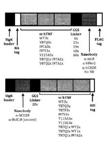
Figure 1: Representation of the structural elements of the different set-ups
of the sc hTNF-
nanobody fusion protein.
Figure 2: Firefly luciferase activity induced by the indicated sc hTNF
preparations, as
compared to WT hTNF, on HekT cells (panel A) or Hek-mLR cells (panel B). Both
were
transiently transfected with the NF-KB luciferase reporter.
Figure 3: Firefly luciferase activity induced by the indicated sc hTNF
preparations carrying a
linker with 6 GGS repeats on HekT cells (panel A) or Hek-mLR cells (panel B).
Both were
transiently transfected with the NF-KB luciferase reporter.
Figure 4: Firefly luciferase activity induced by the indicated sc hTNF
preparations carrying a
linker with 13 GGS repeats on HekT cells (panel A) or Hek-mLR cells (panel B).
Both were
transiently transfected with the NF-KB luciferase reporter.
Figure 5: Firefly luciferase activity induced by the indicated sc hTNF
preparations carrying a
linker with 19 GGS repeats on HekT cells (panel A) or Hek-mLR cells (panel B).
Both were
7
CA 02918363 2016-01-14
WO 2015/007903
PCT/EP2014/065554
transiently transfected with the NF-KB luciferase reporter.
Figure 6: Fold induction of IL-6 mRNA levels upon treatment of SK-BR-3 cells
with 500 ng/ml
of the indicated sc hTNF preparations compared to the levels in untreated
cells and cells
stimulated with the Her2 nanobody. Data represents the mean SD of 2
independent
experiments (n=4).
Figure 7: % activity of hTNF mutants compared to WT hTNF as measured by
toxicity on
MCF7 cells (a breast cancer cell line).
Figure 8: Toxicity on MCF7 (panel A) or MCF7-mLR (panel B) cells of targeted
modified TNFs
coupled to mLR NB (NB C-terminally of TNF).
Figure 9 Toxicity on MCF7 (panel A) or MCF7-hCD20 (panel B) cells of targeted
modified
TNFs coupled to hCD20 NB (NB C-terminally of TNF).
Figure 10: Toxicity on MCF7 (panel A) or MCF7-hCD20 (panel B) cells of
targeted modified
TNFs coupled to hCD20 or control Bc1110 NB (NB N-terminally of TNF).
Figure 11: Toxicity on MCF7 (panel A) or MCF7-hCD20 (panel B) cells of
targeted modified
TNFs containing sc hTNF with combined mutations (NB N-terminally of TNF).
Figure 12: Toxicity on MCF7 (panel A) or MCF7-mLR (panel B) cells of targeted
modified TNF
with individual trimerizing chains coupled to mLR NB (NB C-terminally of TNF).
Figure 13: Toxicity on MCF7 (panel A) or MCF7-hCD20 (panel B) cells of
targeted modified
TNF with individual trimerizing chains coupled to hCD20 NB (NB N-terminally of
TNF).
Figure 14: Comparison of the in vivo toxicity of WT hTNF versus targeted WT
and modified sc
hTNFs coupled to hCD20 or control Boni() NB (NB N-terminally of TNF). (A)
Hypothermia (B)
Mortality.
Figure 15: In vivo toxicity of WT or modified (Y86F3x) sc mouse (m)TNF coupled
to control
Boni() NB (NB N-terminally of TNF). (A) Hypothermia (B) Mortality.
Figure 16: In vivo anti-tumor effect of WT or modified (Y86F3x) sc mouse
(m)TNF coupled to
mCD20 or control Boni() NB (NB N-terminally of TNF). (A) Tumor growth (B)
Mortality.
8
CA 02918363 2016-01-14
WO 2015/007903
PCT/EP2014/065554
EXAMPLES
Materials and methods to the examples
Nanobodies
The nanobody 4-10 directed against the murine leptin receptor (mLR) was
described in
Zabeau et al. (2012). Its coding sequence is cloned into the mammalian
expression vector
pMET7 (Takebe et al., 1988) in fusion with the SIgK leader peptide, the HA tag
and albumin.
Plasmid name: pMET7 SIgK-HA-4.11-Albumin. The anti-Her2 nanobody 1R59B was
described
in Vaneycken et al. (2011). The NB 2HCD25 directed against the human CD20
(hCD20) and
the 2MC57 NB against mouse CD20 (mCD20) were generated using standard
techniques
(Gharouhdi et al., 1997; Pardon et al., 2014). The control NB Bc1I10 was
described in De
Groeve et al. (2010).
scTNF
scTNF that consists of three hTNF monomers coupled via GGGGS-linkers (SEQ ID
NO: 1) has
been described by Boschert et al. (2010). The Y87Q mutation in hTNF was shown
to
completely abrogate the binding to both receptors, TNF-R1 and TNF-R2. Mutating
197 results
in reduced binding of hTNF to both receptors (Loetscher et al., 1993). A whole
range of
residues within hTNF were mutated (QuikChange Site-Directed Mutagenesis Kit,
Stratagene
Cat# 200518) and tested for their toxic effects on MCF7 cells (figure 7). We
selected the
following mutations for the targeted constructs: Y87Q, Y87F, I97S, I97A, Y1
15A, Y1 15G. The
coding sequences of sc hTNF WT-6xGGS, sc hTNF Y87Q3x-6xGGS, sc hTNF I97A3x-
6xGGS, sc hTNF Y87Q1x I97A2x-6xGGS, sc hTNF Y87Q2x I97A1x-6xGGS, sc hTNF WT1x
Y87Q2x-6xGGS, sc hTNF WT2x Y87Q1x-6xGGS, sc hTNF I9753x-6xGGS, sc hTNF
Y115A3x-6xGGS, sc hTNF Y87F3x, sc hTNF Y115G3x, sc mTNF WT and sc mTNF Y86F3x
were generated by gene synthesis (GeneArt). The individual chains are
separated by a
GGGGS (SEQ ID NO: 1) linker.
scTNF-nanobody fusion construction
The coding sequence of the 1R59B Her2 nanobody was synthesized by PCR from the
plasmid
pHEN6-1R59B with the following primers: forward
5'-
GTCAAGATCTGGCGGTTCGGCGGCCGCAATGG000AGGTGCAGCTGCAG-3' (SEQ ID
NO: 2), reverse
5'-
CAGTTCTAGATTACTTATCGTCGTCATCCTTGTAATCCGAACCGCCGTCCGGAGAGGAGA
CGGTGAC-3' (SEQ ID NO: 3). This PCR introduces a GGS in between a BglIl and
Notl site at
the amino terminus and a FLAG tag at the carboxy terminus of the 1R59B
nanobody. The PCR
9
CA 02918363 2016-01-14
WO 2015/007903
PCT/EP2014/065554
product was digested with BglIl and Xbal. The pMK-RQ-sc hTNF WT, pMK-RQ-sc
hTNF
Y87Q3x and pMK-RQ-sc hTNF I97A3x were digested with Ndel and BgIII. The
digested PCR
product and synthetic gene fragments were cloned into Ndel-Xbal digested pMET7
SIgK-HA-
leptin vector to obtain pMET7 SIgK-HA-sc hTNF WT-6xGGS-1R59B-FLAG, pMET7 SIgK-
HA-
sc hTNF Y87Q3x-6xGGS-1R59B-FLAG and pMET7 SIgK-HA-sc hTNF 197A3x-6xGGS-
1R59B-FLAG. The control vectors without the 1R59B nanobody were obtained by
inserting the
following annealed oligos containing the GGS and the FLAG tag in between BglIl
and Xbal
instead of the PCR product: forward:
5'GATCTGGCGGTTCGGCGGCCGCAGATTACAAGGATGACGACGATAAGTAAT3' (SEQ ID
NO: 4),
reverse:
5'CTAGATTACTTATCGTCGTCATCCTTGTAATCTGCGGCCGCCGAACCGCCA3' (SEQ ID
NO: 5). The control vector with only the 1R59B nanobody was obtained by
inserting the
following annealed oligos instead of the Ndel-sc hTNF-BglIl fragment: forward:
5'-
TATGATGTG000GACTACGCTGGCGGCAGCA-3' (SEQ ID NO: 6), reverse 5'-
GATCTGCTGCCGCCAGCGTAGTCGGGCACATCA-3' (SEQ ID NO: 7). The length of the
GGS linker was adjusted to a GGS linker of 13 repeats and 19 repeats by adding
7xGGS or
13xGGS repeats (made by gene synthesis, GeneArt) to the original 6xGGS in
between the
BglIl and Notl site.
A similar approach was used to obtain pMET7 SIgK-HA-sc hTNF WT-6x/13x/19xGGS-
4.10-
FLAG, pMET7 SIgK-HA-sc hTNF Y87Q3x-6xGGS-4.10-FLAG, pMET7 SIgK-HA-sc hTNF
197A3x-6x113x/19xGGS-4.10-FLAG, pMET7 SIgK-HA-sc hTNF Y87Q1x 197A2x-
6x/13x/19xGGS-4.10-FLAG, pMET7 SIgK-HA-sc hTNF Y87Q2x 197A1x-6x/13x/19xGGS-
4.10-
FLAG, pMET7 SIgK-HA-sc hTNF WT-6x/13x/19xGGS-2HCD25-FLAG, pMET7 SIgK-HA-sc
hTNF 19753x-6x113x/19xGGS-2HCD25-FLAG, pMET7 SIgK-HA-sc hTNF I97A3x-
6x/13x/19xGGS-2HCD25-FLAG and pMET7 SIgK-HA-sc hTNF Y115A3x-6x113x/19xGGS-
2HCD25-FLAG.
To obtain the individual trimerizing hTNF constructs, sc hTNF in pMet7-SIgK-HA-
sc hTNF WT-
GGS-4.10-Flag was replaced by Ndel-Sall digest of the PCR product obtained
with the forward
primer
5'-
CATATGATGTGCCCGACTACGCTGGCGGCAGCAGCTCTAGAACCCCCAGCGATAAGCCT
GTG-3' (SEQ ID NO: 8) and the reverse primer 5'-GTCGACCAGGGCAATGATGCCGAAGT-3'
(SEQ ID NO: 9) on the plasmids pMet7-SIgK-His-hTNF WT or pMet7-SIgK-His-hTNF
I97A.
This resulted in the following vectors: pMet7-SIgK-HA-hTNF WT-6xGGS-4.10-Flag
and pMet7-
SIgK-HA-hTNF 197A-6xGGS-4.10-Flag.
The nanobody-TNF fusion expression constructs with the NB N-terminally of
individual
trimerizing or single chain, human or mouse TNF were made in pMet7 and
designed as such
that each subunit is interchangeable through unique restriction sites: Agel-
nanobody-Sall-GGS
CA 02918363 2016-01-14
WO 2015/007903
PCT/EP2014/065554
linker-Notl-TNF-Xhol-His-Xbal.
pGL3-(1L6-kB)3-fireflyluciferase was kindly provided by W. Vanden Berghe
(Vanden Berghe et
al., 1998).
Production of the nanobody-TNF fusion proteins for in vitro studies
HekT cells were transfected with the protein fusion constructs using the
standard calcium
phosphate precipitation method. 48 hours after the transfection culture
mediums were
harvested and stored at -20 C. The concentration was determined with a
commercial hTNF
ELISA (DY210, R1D systems).
Production of the nanobody-scTNF fusion proteins for in vivo studies
FreeStyle TM 293-F cells were transfected with the protein fusion constructs
using the PEIpr0TM
transfection reagent (PolyPlus, Cat# 115-375) according to the manufacturer's
guidelines. The
endotoxin content was in all preparations under the detection limit as
assessed by a
chromogenic Limulus Amebocyte Lysate Assay (Lonza, Cat# 50-647U).
Cell lines
Hek, HekT, Hek-mLR, MCF7, MCF7-hCD20, MCF7-mLR and B161316-mCD20 cells were
grown in DMEM supplemented with 10% FCS. The FreeStyleTM 293-F cell line was
obtained
from Invitrogen, Life Technologies (Cat# R790-07) and maintained in
FreeStyleTM 293
Expression Medium from Gibco, Life Technologies (Cat# 12338). The human breast
cancer
SK-BR-3 (ATCC: HTB-30) cell line was obtained from ATCC and maintained in
McCoy's 5A
medium supplemented with 10% FCS.
The Hek-mLR cell line was generated as follows: Flp-In-293 cells (Invitrogen)
were stably co-
transfected with a plasmid containing the expression cassettes for mEcoR and
neomycin
resistance and with a pXP2d2-rPAP1-luci reporter construct (Eyckerman et al.
2001). Stable
transfected clones were isolated in G418 (400 ug/mI)-containing medium and a
clone was
selected with high LIF (1ng/mI)-induced luciferase activity. The expression
vector pcDNA5/FRT
containing the mLR was stably integrated in this cell line using the Flp-In
recombinase reaction
(Invitrogen) and after selection on hygromycin (100 ug/m1) for 10 days.
The human breast cancer MCF7 (ATCC: HTB-22) cell line was obtained from ATCC.
The
MCF7-hCD20 and MCF7-mLR cell lines were generated as follows: MCF7 cells were
stably
co-transfected with a plasmid containing the expression cassette for hCD20 or
mLR, and with
a plasmid containing the neomycin resistance gene. Stable transfected cells
were selected
with G418 (1 mg/ml)-containing medium, followed by FACS sorting of hCD20- or
mLR-
expressing cells.
The B161316-mCD20 cell line was generated as follows: B16B16 cells were stably
co-
transfected with a plasmid containing the expression cassette for mCD20 and
with a plasmid
11
CA 02918363 2016-01-14
WO 2015/007903
PCT/EP2014/065554
containing the neomycin resistance gene. Stable transfected cells were
selected with G418 (2
mg/ml)-containing medium.
The human breast cancer SK-BR-3 (ATCC: HTB-30) cell line was obtained from
ATCC and
maintained in McCoy's 5A medium supplemented with 10% FCS.
Measurement of the luciferase activities
TNF specific activities were measured by quantifying the luciferase activity
under the control of
the NF-KB promoter. Two days after transfection of the NF-KB luciferase
reporter (pGL3-(IL6-
KB)3-fireflyluciferase) by standard calcium phosphate precipitation method,
cells were
stimulated for 6h with targeted or control sc hTNF. Lysates were prepared
(lysis buffer: 25 mM
Tris, pH 7.8, 2 mM EDTA, 2 mM dithiothreitol, 10% glycerol, 1% Triton X-100),
and 35 pl of
luciferase substrate buffer (20 mM Tricine, 1.07 mM (MgCO3)4Mg(OH)2.5H20, 2.67
mM
Mg504=7H20, 0.1 mM EDTA, 33.3 mM dithiothreitol, 270 pM coenzyme A, 470 pM
luciferin,
530 pM ATP, final pH 7.8) was added per 50 pl of lysate. Light emission was
measured for 5 s
in a TopCount chemiluminescence counter (Packard).
Quantitative RT-PCR
The expression of the TNF inducible gene IL-6 was quantified by RT-PCR
relatively to HPRT
in SK-BR-3 cells treated for 6 hours with 500 ng/ml of targeted or control sc
hTNF. Total RNA
was purified with RNeasy columns (Qiagen) and equal amounts of RNA (0.5 pg)
were used for
reverse transcription using the Primescript RT Reagent kit (Takara Bio, Shiga,
Japan),
following the manufacturer's instructions. The 10-fold diluted cDNA was added
to an RT-QPCR
mixture containing lx SYBR Green I master mix (04 887 352 001, Roche) and 1 nM
gene-
specific primers. Assays were performed in triplicate on a LightCycler 480
Real-Time PCR
System thermocycler (Roche Applied Science), and the results were analyzed
using the LACT
method. The following primers were used:
HPRT forward: 5'TGACACTGGCAAAACAATGCA3' (SEQ ID NO: 10);
HPRT reverse: 5'GGTCCTTTTCACCAGCAAGCT3' (SEQ ID NO: 11);
IL-6 forward: 5'GACAGCCACTCACCTCTTCA3' (SEQ ID NO: 12);
IL-6 reverse: 5'AGTGCCTCTTTGCTGCTTTC3' (SEQ ID NO: 13).
Toxicity analysis on MCF7 cells
TNF-specific activities were also measured by assessing the cellular toxicity
on MCF7 cells.
1000 cells were plated in a black 96-well plate and 24 hours later stimulated
with the different
TNF constructs. After 48-72 hours, the number of viable cells was determined
using the
CellTiter-Glo Luminescent Cell Viability Assay (Promega Cat# G7570) according
to the
manufacturer's guidelines.
12
CA 02918363 2016-01-14
WO 2015/007903
PCT/EP2014/065554
In vivo toxicity analysis
To assess hTNF toxicity in vivo, female 8 weeks old C57BL/6J mice (purchased
from Charles
River, France) were injected intraperitoneally with 500 ng rhTNF or sc hTNF-
nanobody fusion
proteins in combination with 10 mg D-Galactosamine (diluted in LPS-free PBS,
injected in a
volume of 500 pl). Morbidity was monitored by measurement of peripheral
(rectal) body
temperature. n=2-4 per fusion protein.
To evaluate mTNF toxicity in vivo, mice were injected intravenously with 10,
35, 100 or 200 p.g
sc mTNF-nanobody fusion proteins (injected volume 200 pl, dilution in LPS-free
PBS).
Morbidity was monitored by measurement of peripheral (rectal) body
temperature. n=2 per
dose, per fusion protein, except for 200 pg (n=1).
In vivo anti-tumor studies
Female C57BL/6J mice of 8 weeks old were shaved and inoculated with 6x10⁵
B161316-mCD20
tumor cells subcutaneously in the back (day 0). Treatment was started when the
product of the
largest perpendicular diameters was approximately 50 mm2 (on day 10). PBS or
35 p.g
nanobody-sc mTNF fusion proteins were administered for 8 consecutive days (day
10-17,
indicated in Fig. 16A as a grey bar) via paralesional injection (subcutaneous
injection near the
tumor site but outside the tumor nodule). Tumors were measured daily with a
caliper and are
shown as mean SEM. Morbidity was monitored by daily measurement of body
weight and
temperature. n=5 per treatment.
Example 1: The sc hTNF-nanobody fusion proteins
Figure 1 shows a schematic representation of the sc hTNF-nanobody fusion
proteins either
with the nanobody N- or C-terminally of sc hTNF.
Example 2: Targeting TNF activity on mLR-expressing Hek cells
The induction of NF-KB luciferase reporter activity upon TNF stimulation was
tested in HekT
cells and in Hek cells that express the murine leptin receptor (Hek-mLR). As
shown in figure
2A, WT sc hTNF-induced NF-KB induction is completely (>1000-fold) or partly
(100-fold)
abrogated by the Y87Q3x or the I97A3x mutation, respectively. Moreover, in
HekT cells that do
not express the mLR, all sc hTNF constructs (WT, Y87Q3x and I97A3x) induce
similar NF-KB
activity independently of the fusion to the mLR nanobody (Fig. 2A). In
contrast, coupling to the
mLR nanobody is able to restore NF-KB induction of sc hTNF I97A3x in Hek cells
that express
the mLR to a similar extent as WT sc hTNF (Fig. 2B). We estimated that cells
expressing the
mLR are 100-fold more sensitive than parental HekT cells to the nanobody-
coupled sc hTNF
I97A3x. In contrast, the triple Y87Q mutation did not show any rescue effect
of TNF
13
CA 02918363 2016-01-14
WO 2015/007903
PCT/EP2014/065554
responsiveness in Hek-mLR cells compared to HekT cells (Fig. 2B).
Example 3: Comparison of different mutant combinations and different linker
lengths
In order to optimize the constructs, sc hTNF constructs with different
mutations in the individual
chains were tested, as well as different linker lengths between the sc hTNF
and the targeting
moiety. The results are summarized in figure 3, 4 and 5. sc hTNF I97A3x and sc
hTNF
Y87Q1x I97A2x do not show activity on Hek cells that do not express the leptin
receptor, but
have a clear dose dependent activity when targeted to the leptin receptor.
Example 4: Targeting TNF activity on Her2-expressing Hek cells
We generated fusions protein using the a-Her2 nanobody 1R59B and sc hTNF WT,
sc hTNF
Y87Q3x or sc hTNF I97A3x. The linker between the nanobody and sc hTNF was
either 6xGGS
or 19xGGS. These molecules were tested on the Her2-overexpressing SK-BR-3
breast cancer
cell line for the induction of the IL-6 TNF-inducible gene as determined
relatively to HPRT by
quantitative RT-PCR.
Figure 6 shows the fold induction of IL-6 mRNA upon sc hTNF treatment (500
ng/ml)
compared to IL-6 mRNA levels in untreated cells and cells stimulated with the
Her2 nanobody.
In correspondence to the transcriptional activation of NF-KB, we observe that
Y87Q3x mutation
completely prevents TNF-induced IL-6 production while sc hTNF I97A3x can still
induce IL-6
production but to a lesser extent than WT sc hTNF. When sc hTNF is fused to
the nanobody
less IL-6 mRNA is produced. This could be due to steric hindrance as the
effect is more
pronounced with the 6xGGS linker compared to the longer 19xGGS linker where
there is likely
more flexibility. By coupling sc hTNF I97A3x to the Her2 nanobody the
induction of IL-6 can be
restored to similar levels as WT sc hTNF coupled to the nanobody through the
corresponding
linker. In contrast, specific targeting of the more severe Y87Q3x sc hTNF
mutant to Her2-
expressing cells cannot restore the IL-6 inducing property of sc hTNF.
Example 5: comparing the toxicity of hTNF mutants on MCF7 cells
Because of the relatively high residual activity of I97A3x mutant sc hTNF, we
searched for
further mutations by measuring the toxicity of different individual
trimerizing hTNF mutants as
luciferase activity in MCF7 cells. The activity of the mutants relative to WT
individual trimerizing
TNF is shown in figure 7. Most mutations do not affect the TNF activity
drastically (>1% of WT)
and might be less promising for the development of targeted constructs because
of their
possibly remaining substantial toxicity. We are more interested in mutations
that (almost)
completely abrogate TNF function. The use of null mutations (<0,1% of WT)
results in targeted
constructs that do not have side effects but that have as a possible drawback
that reactivation
upon targeting is less easily accomplished. The mutations that have some
residual activity
14
CA 02918363 2016-01-14
WO 2015/007903
PCT/EP2014/065554
(0,02 ¨ 5% of WT, particularly 0,1% - 1% of WT) have a better chance of being
reactivated
whilst not being toxic. 6 different mutations covering an activity range
between 0,02 and 5 % of
individual trimerizing WT TNF were selected for the development of the
targeted modified
cytokines: Y87Q (0,02 %), I97S and Y1 15A (0,2 %), Y87F (0,5-1 %), Y1 15G (1-2
%) and I97A
(2-5 %).
Example 6: Targeting TNF activity on mLR-expressing MCF7 cells
The toxicity of mLR NB-targeted TNF was assessed on MCF7 and MCF7-mLR cells.
Different
mutations (197A3x, I97S3x and Y115A3x) were tested as well as different
linkers between sc
hTNF and the mLR NB (6xGGS, 13xGGS, 19xGGS). As shown in figure 8A, toxicity
is reduced
20-fold by the I97A3x mutation and 500-fold by the I97S3x and Y115A3x
mutation, which is
similar to what we observed for individual trimerizing TNF (Fig. 7). Moreover,
in MCF7 cells
that do not express the mLR, fusion to the mLR NB does not alter the activity
of WT or mutant
sc hTNF (Fig 8A), while this fusion reactivates all sc hTNF mutants on MCF7-
mLR cells (Fig.
8B). We estimated that cells expressing the mLR are 100-fold more sensitive
than parental
MCF7 cells to the NB-coupled I97S3x and Y115A3x sc hTNF. However, these
targeted
modified TNFs are still about 20-fold less active than WT sc hTNF. In
contrast, the I97A3x
targeted modified TNF is restored to WT activity levels on MCF7-mLR cells,
which
corresponds to a 20-fold reactivation (Fig. 8B).
Example 7: Targeting TNF activity on hCD20-expressing MCF7 cells
To assess the effect of other targeting moieties for the targeting of modified
TNF, we replaced
the mLR NB in the constructs of Example 6 with the hCD20 NB and tested their
toxicity on
MCF7 cells and MCF7 cells that express hCD20 (MCF7-hCD20). The results are
shown in
figure 9. As expected, mLR NB and hCD20 NB targeted modified TNFs behave
similarly on
parental MCF7 cells (Fig. 8A & 9A).
Example 8: Targeting TNF activity on hCD20-expressing cells with a different
hCD20 NB
fusion set-up.
We tried to improve the hCD20 NB-TNF constructs by placing the NB in front
instead of after
sc hTNF. We also tested 2 additional, less drastic mutations (Y87F3x and
Y115G3x, Fig. 7).
The MCF7 and MCF7-hCD20 toxicity studies with these constructs are shown in
figure 10. Sc
hTNF coupled to hCD20 NB exerts the same toxicity on MCF7 cells as the
corresponding
mutant coupled to the control Bc1110 NB (Fig. 10A), and the level of activity
is similar as to
what we observed for the individual trimerizing TNF mutants (Fig. 7). This
reduced toxicity of
the mutants is (partially) reverted upon hCD20 targeting on the MCF7-hCD20
cells: hCD20
NB-coupled modified TNF give a 10-fold (Y115G3x), 15-fold (Y87F3x), 100-fold
(19753x,
CA 02918363 2016-01-14
WO 2015/007903
PCT/EP2014/065554
Y115A3x) or even higher (Y87Q3x) increased activity compared to the
corresponding Bc1110
control NB-coupled sc hTNFs (Fig. 10B). In this experiment, when the hCD20 NB
is placed at
the carboxy-terminal end instead of the amino-terminal end of the sc hTNF the
reactivation is
less (Fig. 9B).
Example 9: Comparison of different mutant combinations
Despite the fact that the difference of targeted modified TNF versus non-
targeted modified
TNF is at least a 100-fold, some mutations show lower rescued activity than WT
activity levels
(Y87Q3x) which might affect its anti-tumor effects. Alternatively, some
mutations still have
some residual activity (197S3x and Y115A3x) which might lead to some
(systemic) toxicity
when used in vivo. To overcome these potential drawbacks, we tested additional
constructs by
mutating different residues in the individual chains of sc hTNF in order to
see whether the
activity levels could thus be further modulated. As shown in figure 11,
combining different
mutations in the single chain can alter the residual activity on non-targeted
cells and the level
of reactivation upon targeting.
Example 10: Comparison of targeted individual trimerizing TNF versus single
chain
modified hTNF.
To compare the efficiency of targeted individual trimerizing versus single
chain TNF, WT or
I97A hTNF was coupled C-terminally to the mLR NB as a monomer. Their toxicity
was tested
on MCF7 cells and on MCF7 cells that express the mLR (MCF7-mLR), and is shown
in Figure
12. Also in the individual trimerizing form, the I97A mutation is toxic on
MCF7 cells but to a
lesser extent than WT hTNF (Fig 8A & Fig 12A). Moreover, when coupled C-
terminally to the
mLR nanobody, individual trimerizing -but not single chain- TNF becomes less
toxic on MCF7
cells, and this is the case both for WT and I97A (Fig. 8A & Fig 12A). Most
probably, the 3
nanobodies present in the hTNF trimer formed with the individual trimerizing
TNF-mLR NB
constructs are sterically hindering the binding of hTNF to its receptor. This
reduced activity
can, however, be reverted by targeting to the mLR on MCF7-mLR cells (Fig.
12B).
Interestingly, this offers a further level of modulation of activity: one can
combine different
mutations, as well as use the sterical hindrance to influence residual
activity and level of
reactivation upon targeting.
To address whether this is a general phenomenon, we coupled individual
trimerizing WT and
Y115A hTNF N-terminally to Bc1110 or hCD20 nanobody and tested their toxicity
on MCF7 and
MCF7-hCD20 cells. As shown in figure 13A, this coupling does not affect the
toxicity of
individual trimerizing WT or Y115A3x hTNF. Moreover, upon targeting,
individual trimerizing
Y115A3x hTNF becomes as active as non-targeted individual trimerizing WT hTNF
(Fig 13B).
16
CA 02918363 2016-01-14
WO 2015/007903
PCT/EP2014/065554
Example 11: Assessment of in vivo toxicity of targeted modified hTNF
To evaluate the toxicity of hTNF mutants preclinically is not evident, since
TNF displays a
remarkable species specificity in mice. In contrast to mTNF, hTNF only induces
lethality at
extremely high doses (Brouckaert et al. 1992). Although the reason for this
species specificity
was long thought to be caused by hTNF not interacting with the murine TNF-R2,
pharmacokinetic studies have shown that hTNF is cleared much faster than mTNF
in mice and
that the consequential limited hTNF exposure is responsible for its lack of
morbidity (Ameloot
et al. 2002).
Nevertheless, when treated with a sensitizing agent such as D-galactosamine,
species
specificity is abolished and extremely low doses 500 ng) of hTNF are
equally lethal as
mTNF (Broeckaert et al., 1992). To assess the in vivo toxicity of the various
targeted modified
hTNFs, we therefore injected mice intraperitoneally with 500 ng of either
recombinant (r) hTNF,
sc hTNF WT or sc mutant hTNF (Y87Q3x or Y115A3x). The sc WT and modified hTNF
were
coupled N-terminally to either Bc1110 or to hCD20 NB. As shown in figure 14,
sc WT hTNF is at
least as toxic as rhTNF, causing severe hypothermia and mortality within 10 h
after injection.
Targeted modified hTNF Y87Q3x and Y115A3x did not cause any signs of morbidity
(pilo-
erection, tremor, lethargy, loss of grooming or drop in body temperature; see
figure 14A for the
latter).
Example 12: Assessment of in vivo toxicity and anti-tumor effect of targeted
modified
mTNF.
As already mentioned, in vivo toxicity of hTNF cannot be easily studied in
mice. Therefore, as
well as because of anticipated anti-tumor experiments in immunocompetent
syngeneic mice,
we decided to mutate residues of mTNF homologous to the ones we selected for
hTNF (see
example 5). As illustrated in figure 15, Bc1110NB-sc mTNF WT caused severe
morbidity (Fig 15
A) and 100% mortality (Fig 15B) when injected intravenously in doses as low as
10 g. In
contrast, Bc1110NB-sc mTNF Y86F3x did not induce mortality (Fig 15B) nor cause
any signs of
toxicity (pilo-erection, tremor, lethargy, loss of grooming or drop in body
temperature; see
figure 15A for the latter), not even when injected as an intravenous bolus of
200 g.
Nevertheless, when injected daily paralesionally in a dose of 35 g in B161316-
mCD20-tumor
bearing mice, nanobody-coupled sc mTNF Y86F3x could still reduce/prevent tumor
growth,
especially when targeted to mCD20 (Fig. 16A). The effect of non-targeted
mutant TNF on
tumor growth (Fig. 16A) is due to the high dose (35pg) used, as lower doses
more closely
mimic PBS-treated animals (data not shown). Daily treatment with the NB-sc
mTNF Y86F3x
did not cause any signs of morbidity or mortality, while tumor-bearing mice
treated with NB-sc
mTNF WT succumbed after 1 or 2 injections (Fig 16B).
17
CA 02918363 2016-01-14
WO 2015/007903
PCT/EP2014/065554
REFERENCES
- Ameloot, P., Takahashi N., Everaerdt, B., Hostens, J., Eugster, H.P.,
Fiers, W., and
Brouckaert, P. (2002). Bioavailability of recombinant tumor necrosis factor
determines its
lethality in mice. Eur J lmmunol, 32, 2759-65.
- Arap, W., Pasqualini, R. and Ruoslahti, E. 51998). Cancer treatment by
targeted drug
delivery to tumor vasculature in a mouse model. Science, 279, 377-380.
- Ashkenazi, A. & Herbst, R.S. To kill a tumor cell: the potential of
proapoptotic receptor
agonists. J Clin Invest 118, 1979-1990 (2008).
- Blake, A.W., McCartney, L., Flint, J., Bolam, D.N., Boraston, A.B.,
Gilbert, H.J. and Knox,
J.P. (2006) Understanding the biological rationale for the diversity of
cellulose-directed
carbohydrate-binding molecules in prokaryotic enzymes. J. Biol. Chem. 281,
29321-29329.
- Boschert, V. et al. Single chain TNF derivatives with individually
mutated receptor binding
sites reveal differential stoichiometry of ligand receptor complex formation
for TNFR1 and
TNFR2. Cell Signal 22, 1088-1096 (2010).
- Brecht et al., Peptide immobilization and characterization of binding
specificity. J Biol
Chem 268: 15425-15434
- Brouckaert, P., Libert, C., Everaerdt, B. and Fiers W (1992). Selective
species specificity of
tumor necrosis factor for toxicity in the mouse. Lymphokine Cytokine Res, 11,
193-6.
- Brown, K.C. (2010). Peptidic tumor targeting agents; the road from phagfe
display
selections to clinical applications. Curr. Pharm. Des. 16, 1040-1054.
- Daburon, S. et al. Functional characterization of a chimeric soluble Fas
ligand polymer with
in vivo anti-tumor activity. PLoS One 8, e54000 (2013).
- de Bruyn, M., Bremer, E. & Helfrich, W. Antibody-based fusion proteins to
target death
receptors in cancer. Cancer Lett 332, 175-183 (2013).
- Dimitrov, D.S. (2009) Engineered CH2 domains (nanoantibodies). mAbs 1,26-28.
- Eyckerman, S., Waelput, W., Verhee, A., Broekaert, D., Vandekerckhove,
J., and
Tavernier, J. (1999). Eur. Cytok. Netw. 10, 549-559.
- Falschlehner, C., Ganten, T.M., Koschny, R., Schaefer, U. & Walczak, H.
TRAIL and other
TRAIL receptor agonists as novel cancer therapeutics. Adv Exp Med Biol 647,
195-206
(2009).
- Fox, N.L., Humphreys, R., Luster, T.A., Klein, J. & Gallant, G. Tumor
Necrosis Factor-
related apoptosis-inducing ligand (TRAIL) Receptor-1 and Receptor-2 agonists
for cancer
therapy. Expert Opin Biol Ther 10, 1-18 (2010).
- Galle, P.R. et al. Involvement of the CD95 (APO-1/Fas) receptor and
ligand in liver
damage. J Exp Med 182, 1223-1230 (1995).
- Gaur, U. & Aggarwal, B.B. Regulation of proliferation, survival and
apoptosis by members
of the TNF superfamily. Biochem Pharmacol 66, 1403-1408 (2003).
18
CA 02918363 2016-01-14
WO 2015/007903
PCT/EP2014/065554
- Ghahroudi A.M., Desmyter, A., Wyns, L., Hamers, R., Muyldermans, S.
(1997). Selection
and identification of single domain antibody fragments from camel heavy-chain
antibodies.
FEBS Lett. 414, 521-6
- Gregorc, V. et al. Phase lb study of NGR-hTNF, a selective vascular
targeting agent,
administered at low doses in combination with doxorubicin to patients with
advanced solid
tumours. Br J Cancer 101, 219-224 (2009).
- Hehlgans, T. & Pfeffer, K. The intriguing biology of the tumour necrosis
factor/tumour
necrosis factor receptor superfamily: players, rules and the games. Immunology
115, 1-20
(2005).
- Huang, Y. & Sheikh, M.S. TRAIL death receptors and cancer therapeutics.
Toxicol Appl
Pharmacol 224, 284-289 (2007).
- Johnstone, R.W., Frew, A.J. & Smyth, M.J. The TRAIL apoptotic pathway in
cancer onset,
progression and therapy. Nat Rev Cancer 8, 782-798 (2008).
- Koivunen, E., Wang, B. and Ruoslahti, E. (1994). Isolation of a highly
specific ligand for the
a5131 integrin from a phage library. J. Cell. Biol. 124, 373-380.
- Kolmar, H. (2008) Alternative binding proteins: biological activity and
therapeutic potential
of cysteine-knot miniproteins. FEBS J. 275, 2684-2690.
- Lejeune, F.J., Lienard, D., Matter, M. & Ruegg, C. Efficiency of
recombinant human TNF in
human cancer therapy. Cancer lmmun 6, 6 (2006).
- Li, M. et al. Phase II multicenter, randomized, double-blind study of
recombinant mutated
human tumor necrosis factor-alpha in combination with chemotherapies in cancer
patients.
Cancer Sci 103, 288-295 (2012).
- Liu, Y. et al. The antimelanoma immunocytokine scFvMEL/TNF shows reduced
toxicity and
potent antitumor activity against human tumor xenografts. Neoplasia 8, 384-393
(2006).
- Loetscher, H., Stueber, D., Banner, D., Mackay, F. & Lesslauer, W. Human
tumor necrosis
factor alpha (TNF alpha) mutants with exclusive specificity for the 55-kDa or
75-kDa TNF
receptors. J Biol Chem 268, 26350-26357 (1993).
- Nygren, P-A. (2008) Alternative binding proteins: affibody binding
proteins developed from
a small three-helix bundle scaffold. FEBS J. 275, 2668-2676.
- Ogasawara, J. et al. Lethal effect of the anti-Fas antibody in mice. Nature
364, 806-809
(1993).
- Pardon,E., Laeremans, T., Triest, S., Rasmussen, S.G.F., Wohlkonig, A.,
Ruf, A.,
Muyldermans, S., Hol, W.G.J, Kobilka, B.K. and Steyaert, J. (2014). A general
protocol for
the generation of Nanobodies for structural biology. Nature Protocols 9, 674-
693
- Roberts, N.J., Zhou, S., Diaz, L.A., Jr. & Holdhoff, M. Systemic use of
tumor necrosis factor
alpha as an anticancer agent. Oncotarget 2, 739-751 (2011).
19
CA 02918363 2016-01-14
WO 2015/007903
PCT/EP2014/065554
- Roccaro AM, Hideshima T, Raje N, Kumar S, lshitsuka K, Yasui H, Shiraishi
N, Ribatti D,
Nico B, Vacca A, Dammacco F, Richardson PG, Anderson KC. Bortezomib mediates
antiangiogenesis in multiple myeloma via direct and indirect effects on
endothelial cells.
Cancer Res. 2006; 66(1):184-91.
- Scatchard G. Ann New York Acad Sci 1949; 51, 660-72.
- Schmidt T, Carmeliet P. Angiogenesis: a target in solid tumors, also in
leukemia?
Hematology Am Soc Hematol Educ Program. 2011; 2011:1-8.
- Siegemund, M. et al. Superior antitumoral activity of dimerized targeted
single-chain TRAIL
fusion proteins under retention of tumor selectivity. Cell Death Dis 3, e295
(2012).
- Skerra, A. (2008) Alternative binding proteins: anticalins ¨ harnessing the
structural
plasticity of the lipocalin ligand pocket to engineer novel binding
activities. FEBS J. 275,
2677-2683.
- Stump, M.T., Binz, H.K., Amstutz, P. (2008) DARPins: a new generation of
protein
therapeutics. Drug iscov. Today 13, 695-701.
- Tramontano, A., Bianchi, E., Venturini, S., Martin, F., Pessi, A and
Sollazzo, M. (1994) The
making of the minibody: an engineered beta-protein for the display of
confromationally
constrained peptides. J. Mol. Recognition 7, 9-24.
- Vanden Berghe, W. et al. p38 and extracellular signal-regulated kinase
mitogen-activated
protein kinase pathways are required for nuclear factor-kappaB p65
transactivation
mediated by tumor necrosis factor. J Biol Chem 273, 3285-3290 (1998).
- Vaneycken, I. et al. Preclinical screening of anti-HER2 nanobodies for
molecular imaging
of breast cancer. FASEB J 25, 2433-2446 (2011).
- van Horssen, R., Ten Hagen, T.L. & Eggermont, A.M. TNF-alpha in cancer
treatment:
molecular insights, antitumor effects, and clinical utility. Oncologist 11,
397-408 (2006).
- Wang, H., Yan, Z., Shi, J., Han, W. & Zhang, Y. Expression, purification,
and
characterization of a neovasculature targeted rmhTNF-alpha in Escherichia
coli. Protein
Expr Purif 45, 60-65 (2006).
- Welti J, Loges S, Dimmeler S, Carmeliet P. Recent molecular discoveries
in angiogenesis
and antiangiogenic therapies in cancer. J Clin Invest. 2013; 123(8):3190-200.
- Yang, Y.H., Rajaiah, R., Ruoslahti, E. and Moudgil, K.D. (2011). Peptides
targeting
inflamed synovial vasulature attenuate autoimmune arthritis. PBNAS 108, 12857-
12862.
- Zabeau, L. et al. Selection of non-competitive leptin antagonists using a
random nanobody-
based approach. Biochem J 441, 425-434 (2012).