Note: Descriptions are shown in the official language in which they were submitted.
METHODS FOR PRODUCING CARTILAGE AND BONE
[0001]
SEQUENCE LISTING PROVIDED AS A TEXT FILE
[0002] A Sequence Listing is provided herewith as a text file, "UCSF-
454W0_SeqList_5T25.txt" created on July 15, 2013 and having a size of 29 KB.
INTRODUCTION
[0003] Articular cartilage injuries are common, lead to pain and
disability, and contribute to
the development of osteoarthritis. While various cell-based methods are in
development for
tissue repair, such methods have significant limitations, including lack of an
optimal cell
source.
[0004] Articular chondrocytes and mesenchymal stem cells (MSCs) are
commonly used
cells for cartilage regeneration. However, articular chondrocytes are
available in limited
quantities, and MSCs form cartilaginous tissues that do not closely resemble
articular
cartilage.
[0005] There is a need in the art for methods of generating cartilage and
bone.
Literature
[0006] Fischer et al. (2010) Arthritis and Rheumatism 62:2696; Iwata et
al. (2012)
Biomaterials 444:454; Jiang et al. (2010) Tissue Engineering 16:1621; Shum et
al. (2003)
Int. J. Dev. Biol. 47:423; Steintert et al. (2009) Arthr. Res. Ther. 11:R148;
U.S. Patent No.
6,150,163; U.S. Patent No. 7,169,610; U.S. Patent No. 8,017,394; U.S. Patent
Publication
No. 2003/0175257; Scotti et al. (2010) Proc. Natl. Acad. Sci. USA 107:7251;
Jukes et al.
(2008) Proc. Natl. Acad. Sci. USA 105:6840; Gawlitta et al. (2010) Tissue
Engineering
16:385; Oliviera et al. (2009) Tissue Engineering 15:635; U.S. Patent
Publication No.
2011/0256109; Brittberg et al. (1994) N Engl J Med. 331(14):889-95; Schulze-
Tanzil
(2009) Ann Anat. 191(4):325-38; Safran et al .(2010) J Am Acad Orthop Surg.
18(5):259-
66; Pelttari et al. (2008) Injury 39 Suppl 1:S58-65; Pelttari et al. (2006)
Arthritis Rheum.
54(10):3254-66.
1
CA 2918486 2019-10-07
CA 02918486 2016-01-15
WO 2014/015109 PCT/US2013/051022
SUMMARY
[0007] The present disclosure provides methods of producing cartilage in
vitro. The present
disclosure provides treatment methods, involving introducing in vitro-produced
cartilage
into a treatment site in vivo. The present disclosure provides methods of
enhancing bone
formation, the method involving introducing in vitro-produced hypertrophic
cartilage into a
treatment site in vivo.
BRIEF DESCRIPTION OF THE DRAWINGS
[0008] Figure 1 depicts a comparison of chondrocyte expansion in serum-free
medium
versus serum-containing medium.
[0009] Figure 2 depicts gene expression in chondrocytes expanded in serum-
free culture
conditions.
[0010] Figure 3 depicts chondrogenic gene expression in chondrocytes
cultured in medium
including transforming growth factor-beta 1 (TGF-pl) or bone morphogenic
protein-4
(BMP4).
[0011] Figure 4 depicts passaged chondrocytes cultured in medium including
TGF-I31 or
BMP-4.
[0012] Figure 5 depicts stimulation of bone formation by hypertrophic
cartilage pellets
implanted in vivo.
[0013] Figure 6 provides amino acid sequences of TGF-131 polypeptides. Homo
sapiens
(SEQ ID NO: 1); Mus muscu/us (SEQ ID NO: 2).
[0014] Figure 7 provides amino acid sequences of TGF-02 polypeptides. Homo
sapiens
(SEQ ID NO: 3); Mus muscu/us (SEQ ID NO: 4).
[0015] Figure 8 provides amino acid sequences of BMP-4 polypeptides. Homo
sapiens
(SEQ ID NO: 5); Mus muscu/us (SEQ ID NO: 6).
[0016] Figures 9A and 9B provide the composition of an exemplary basal
medium.
[0017] Figure 10 provides an amino acid sequence of parathyroid hormone
(PTH). (SEQ ID
NO: 7).
[0018] Figure 11 provides an amino acid sequence of parathyroid hormone
related protein
(PTHrP or PTHRP) (SEQ ID NO: 8). An additional isoform is SEQ ID NO: 10.
[0019] Figure 12 provides an aminoa acid sequence of TGF-I33 (SEQ ID NO:
9).
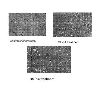
[0020] Figure 13 depicts primary and passaged bovine articular chondrocytes
that have
been grown with the indicated treatments in vitro. Staining is with safranin-O
for sulfated
glycosaminoglycans.
2
CA 02918486 2016-01-15
WO 2014/015109 PCT/US2013/051022
[0021] Figure 14 depicts bovine articular chondrocytes grown in vitro with
BMP and TGFI3
and stained as indicated.
[0022] Figure 15 depicts primary and passaged bovine articular chondrocytes
grown in vitro
under the indicated conditions and stained for mineralization using Von Kossa
stain.
[0023] Figure 16 depicts gross vascularization of primary and passaged
bovine articular
chondrocytes grown in vitro under the indicated conditions and then grown in
vivo in
murine subcutaneous pouches.
[0024] Figure 17 depicts primary and passaged bovine articular chondrocytes
grown in vitro
under the indicated conditions and then grown in vivo in murine subcutaneous
pouches.
Samples are stained with safranin-O for glycosaminoglycan accumulation.
[0025] Figure 18 depicts primary and passaged bovine articular chondrocytes
grown in vitro
under the indicated conditions and then grown in vivo in murine subcutaneous
pouches.
Samples are stained with Von Kossa stain to show mineralization.
[0026] Figure 19 depicts micro computed tomography (microCT) images of bone
formation
resulting from implantation of human induced hypertrophic cartilage.
[0027] Figure 20 depicts Trichrome staining of new bone formation resulting
from induced
human hypertrophic cartilage.
DEFINITIONS
[0028] The term "chondrocyte" refers to a cartilage-specific cell that
gives rise to normal
cartilage tissue growth in vivo; chondrocytes synthesize and deposit the
supportive matrix
(composed principally of collagen and proteoglycan) of cartilage.
[0029] The terms "individual," "subject," "host," and "patient," used
interchangeably
herein, refer to a mammal, including, but not limited to, murines (rats,
mice), non-human
primates, humans, canines, felines, ungulates (e.g., equines, bovines, ovines,
porcines,
caprines), etc. In some embodiments, the individual is a human. In some
embodiments, the
individual is a murine.
[0030] The terms "treat," "treatment," "treating." and the like are used
herein to generally
mean obtaining a desired pharmacologic and/or physiologic effect, e.g.,
increased bone
formation. The effect may be prophylactic in terms of completely or partially
preventing a
disease or symptom thereof and/or may be therapeutic in terms of a partial or
complete cure
for a disease and/or adverse effect attributable to the disease. "Treatment"
as used herein
covers any treatment of a disease in a mammal, particularly a human, and
includes: (a)
preventing a disease or condition (e.g., preventing the loss of cartilage)
from occurring in a
3
subject who may be predisposed to the disease but has not yet been diagnosed
as having it;
(b) inhibiting the disease, e.g., arresting loss of cartilage; or (c)
relieving the disease (e.g.,
enhancing the development of cartilage).
[0031] A "therapeutically effective amount" or "efficacious amount" means
the number of
cells that, when administered to a mammal or other subject for treating a
disease, is
sufficient to effect such treatment for the disease, and/or to replace damaged
or missing
tissue. The "therapeutically effective amount" will vary depending on the
cell, the disease
and its severity and the age, weight, etc., of the subject to be treated.
[0032] As used herein the term "isolated" with reference to a cell,
refers to a cell that is in
an environment different from that in which the cell naturally occurs, e.g.,
where the cell
naturally occurs in a multicellular organism, and the cell is removed from the
multicellular
organism, the cell is "isolated."
[0033] Before the present invention is further described, it is to be
understood that this
invention is not limited to particular embodiments described, as such may, of
course, vary.
It is also to be understood that the terminology used herein is for the
purpose of describing
particular embodiments only, and is not intended to be limiting, since the
scope of the
present invention will be limited only by the appended claims.
[0034] Where a range of values is provided, it is understood that each
intervening value, to
the tenth of the unit of the lower limit unless the context clearly dictates
otherwise, between
the upper and lower limit of that range and any other stated or intervening
value in that
stated range, is encompassed within the invention. The upper and lower limits
of these
smaller ranges may independently be included in the smaller ranges, and are
also
encompassed within the invention, subject to any specifically excluded limit
in the stated
range. Where the stated range includes one or both of the limits, ranges
excluding either or
both of those included limits are also included in the invention.
[0035] Unless defined otherwise, all technical and scientific terms used
herein have the
same meaning as commonly understood by one of ordinary skill in the art to
which this
invention belongs. Although any methods and materials similar or equivalent to
those
described herein can also be used in the practice or testing of the present
invention, the
preferred methods and materials are now described.
4
CA 2918486 2019-10-07
CA 02918486 2016-01-15
WO 2014/015109 PCT/US2013/051022
[0036] It must be noted that as used herein and in the appended claims, the
singular forms
"a," "an," and "the" include plural referents unless the context clearly
dictates otherwise.
Thus, for example, reference to "a chondrocyte" includes a plurality of such
chondrocytes
and reference to "the composition" includes reference to one or more
compositions and
equivalents thereof known to those skilled in the art, and so forth. It is
further noted that the
claims may be drafted to exclude any optional element. As such, this statement
is intended
to serve as antecedent basis for use of such exclusive terminology as
"solely," "only" and
the like in connection with the recitation of claim elements, or use of a
"negative"
limitation.
[0037] It is appreciated that certain features of the invention, which are,
for clarity,
described in the context of separate embodiments, may also be provided in
combination in a
single embodiment. Conversely, various features of the invention, which are,
for brevity,
described in the context of a single embodiment, may also be provided
separately or in any
suitable sub-combination. All combinations of the embodiments pertaining to
the invention
are specifically embraced by the present invention and are disclosed herein
just as if each
and every combination was individually and explicitly disclosed. In addition,
all sub-
combinations of the various embodiments and elements thereof are also
specifically
embraced by the present invention and are disclosed herein just as if each and
every such
sub-combination was individually and explicitly disclosed herein.
[0038] The publications discussed herein are provided solely for their
disclosure prior to the
filing date of the present application. Nothing herein is to be construed as
an admission that
the present invention is not entitled to antedate such publication by virtue
of prior invention.
Further, the dates of publication provided may be different from the actual
publication dates
which may need to be independently confirmed.
DETAILED DESCRIPTION
[0039] The present disclosure provides methods of producing cartilage in
vitro. The present
disclosure provides treatment methods, involving introducing in vitro-produced
cartilage
into a treatment site in vivo. The present disclosure provides methods of
enhancing bone
formation, the method involving introducing in vitro-produced hypertrophic
cartilage into a
treatment site in vivo.
[0040] A method of the present disclosure provides for in vitro
amplification of
chondrocytes to provide cell numbers sufficient for implantation into a
treatment site of an
individual. A method of the present disclosure provides for formation, in
vitro, of
CA 02918486 2016-01-15
WO 2014/015109 PCT/US2013/051022
hypertrophic cartilage, which can stimulate the formation of blood vessels
when implanted
into a treatment site of a mammalian subject. Since bone regeneration requires
blood vessel
formation. hypertrophic cartilage offers an advantage over bone grafts that do
not stimulate
formation of blood vessels. Hypertrophic cartilage produced using a subject
method can be
implanted into a treatment site for tissue repair, and for formation of
mineralized bone. A
method of the present disclosure can provide for formation, in vitro, of
permanent cartilage.
Permanent cartilage produced using a subject method can be implanted into a
treatment site
in an individual, to replace missing cartilage, or to replace diseased or
damaged cartilage.
IN VITRO PRODUCTION OF HYPERTROPHIC CARTILAGE
[0041] The present disclosure provides methods of producing hypertrophic
cartilage in
vitro. In carrying out a subject method for producing hypertrophic cartilage,
a two-stage
procedure is carried out in which chondrocytes are first expanded (first
stage), then
differentiated (second stage) into hypertrophic cartilage. In some cases,
chondrocytes are
cultured in a chemically defined, serum-free liquid culture medium in vitro.
[0042] The present disclosure thus provides method for producing
hypertrophic cartilage in
vitro, the method comprising:
[0043] a) culturing in a first liquid medium in vitro a starting population
of chondrocytes,
where the first liquid medium is serum free. Culturing the starting population
of
chondrocytes provides for an at least 50-fold increase in the number of
chondrocytes over
the number of chondrocytes in the starting population, thereby generating an
expanded
chondrocyte population; and
[0044] b) culturing the expanded chondrocyte in vitro in a second liquid
medium, where the
second liquid medium is serum free and comprises a transforming growth factor-
13 (TGF13)
superfamily polypeptide and/or a bone morphogenic protein (BMP). Culturing the
expanded
chondrocyte population in the second liquid medium results in production of
hypertrophic
cartilage.
[0045] The hypertrophic cartilage thus generated can be directly implanted
into a treatment
site in an individual. The hypertrophic cartilage thus generated can also be
associated with a
matrix (scaffold), to generate a hypertrophic cartilage matrix composition;
and the
hypertrophic cartilage matrix composition can be implanted into a treatment
site in an
individual.
First stage
[0046] A starting population of chondrocytes is cultured in vitro to expand
cell numbers in
a first culture medium. This expansion can occur under any conditions. In some
cases, the
6
CA 02918486 2016-01-15
WO 2014/015109 PCT/US2013/051022
first culture medium is a chemically defined, serum-free liquid culture
medium. The first
culture medium can include a TGF-I3 superfamily member (e.g., TGF-I31; TGF-2;
TGF-P3;
and the like). The starting population of chondrocytes is expanded in the
first step (stage),
generating an in vitro-expanded population of chondrocytes.
First culture medium
[0047] The first culture medium includes a basal medium, where any known
basal medium
suitable for culturing mammalian cells can be used. Suitable basal media
include, but are
not limited to, Dulbecco's modified Eagle's medium (DMEM), DMEM/F12, Iscove's
modified Dulbecco's medium, Minimum Essential Medium, RPMI 1640, and the like.
As
one non-limiting example, the basal medium is DMEM/Fl 2; the composition of
DMEM/F12 is provided in Figures 9A and 9B.
[0048] The basal medium can be supplemented with, e.g., 50 g/ml ascorbic
acid 2-
phosphate, 0.1% albumin, 100 1..ig/m1 sodium pyruvate, 100 units/ml
penicillin, 100 ig/m1
streptomycin, and insulin/transferrin/selenium (ITS; e.g., where the final
concentration in
the culture medium is 10 mg/L insulin, 5.5 mg/L transferrin, and 6.7 pg/L
sodium selenite).
[0049] In some cases, the first culture medium is serum-free. In other
cases, the first culture
medium is not serum-free. In some cases, the first culture medium comprises
basal medium
supplemented with a TGF-I3 superfamily protein, such as TGF-131 (TGFI31), TGF-
132
(TGF1332), or TGF-133 (TGF133). TGF-I3 superfamily proteins are known in the
art and are
described in, e.g., Patil et al. (2011) J. Cell. Physiol. 226:3094. A TGF-I3
superfamily
protein can have a mature peptide length of from about 90 amino acids to about
150 amino
acids, e.g., from about 100 amino acids to about 110 amino acids, from about
100 amino
acids to about 120 amino acids, etc. In some cases, the first culture medium
does not
comprise a TGF-p superfamily protein.
[0050] TGF-131 suitable for use in the first culture medium can comprise an
amino acid
sequence having at least about 85%, at least about 90%, at least about 95%. at
least about
98%, at least about 99%, or 100%, amino acid sequence identity to a contiguous
stretch of
from about 100 amino acids to about 120 amino acids of amino acids 279-390 of
a TGF-P 1
amino acid sequence depicted in Figure 6 (e.g., set forth in any one of SEQ ID
NOs: 1 and
2)..
[0051] TGF-132 suitable for use in the first culture medium can comprise an
amino acid
sequence having at least about 85%, at least about 90%, at least about 95%. at
least about
98%, at least about 99%, or 100%, amino acid sequence identity to a contiguous
stretch of
from about 100 amino acids to about 110 amino acids of amino acids 303-414 of
a TGF-P2
7
CA 02918486 2016-01-15
WO 2014/015109 PCT/US2013/051022
amino acid sequence depicted in Figure 7 (e.g., set forth in any one of SEQ ID
NOs: 3 and
4).
[0052] TGF-I33 suitable for use in the first culture medium can comprise an
amino acid
sequence having at least about 85%, at least about 90%, at least about 95%, at
least about
98%, at least about 99%, or 100%, amino acid sequence identity to a contiguous
stretch of
from about 100 amino acids to about 112 amino acids of amino acids 301-412 of
a TGF-P3
amino acid sequence depicted in Figure 12 (e.g., as set forth in SEQ ID NO:
9).
[0053] A TGF-I3 superfamily protein (e.g., TGF-01, TGB-02, TGF-I33, etc.)
is present in the
first culture medium in a concentration of from about 0.1 ng/ml to about 100
ng/ml, e.g.,
from about 0.1 ng/ml to about 0.5 ng/ml, from about 0.5 ng/ml to about I
ng/ml, from about
1 ng/ml to about 5 ng/ml, from about 5 ng/ml to about 10 ng/ml, from about 10
ng/ml to
about 25 ng/ml, from about 25 ng/ml to about 50 ng/ml, or from about 50 ng/ml
to about
100 ng/ml. In an exemplary embodiment, a TGF-p superfamily protein is present
in the
culture medium in a concentration of 1 ng/ml.
[0054] As one non-limiting example, the first culture medium includes
DMEM/F12; 50
g/ml ascorbic acid 2-phosphate; 0.1% albumin; 100 [tg/m1 sodium pyruvate; 100
units/ml
penicillin; 100 ug/m1 streptomycin; ITS; and 1 ng/ml TGF-I31.
[0055] In some cases, the chemically-defined, serum-free liquid culture
medium does not
include one or more of the following components: PDGF; lipids stearic acid,
myristic
acid, oleic acid, linoleic acid, palmitic acid, palmitoleic acid, arachidonic
acid, linolenic
acid, cholesterol, and alpha-tocopherol acetate); parathyroid hormone-related
protein
(PTHRP); and parathyroid hormone (PTH). For example, in some cases, the
chemically-
defined, serum-free liquid culture medium does not include PTHRP. In some
cases, the
chemically-defined, serum-free liquid culture medium does not include PTHRP or
PDGF.
In some cases, the chemically-defined, serum-free liquid culture medium does
not include
PTHRP, PDGF, or a lipid. In some cases, the chemically-defined, serum-free
liquid culture
medium does not include PTH. In some cases, the chemically-defined, serum-free
liquid
culture medium does not include PTH, PDGF, or PTHRP.
[0056] In the first stage, the starting population of chondrocytes are
cultured over a period
of time of from about 5 days to about 180 days, e.g., from about 5 days to
about 150 days,
from about 5 days to about 100 days, from about 5 days to about 75 days, from
about 5 days
to about 60 days, from about 5 days to about 45 days, from about 5 days to
about 30 days,
from about 5 days to about 25 days, from about 10 days to about 180 days, from
about 10
days to about 150 days, from about 10 days to about 100 days, from about 10
days to about
8
CA 02918486 2016-01-15
WO 2014/015109 PCT/US2013/051022
75 days, from about 10 days to about 60 days, from about 10 days to about 45
days, from
about 10 days to about 30 days, from about 10 days to about 25 days, from
about 5 days to
about 10 days, from about 10 days to about 15 days, from about 15 days to
about 20 days,
from about 20 days to about 25 days, or from about 25 days to about 30 days.
Cell numbers
[0057] Culturing chondrocytes cultured in vitro as described above results
in an increase in
the number of chondrocytes, thereby generating an expanded chondrocyte
population. For
example, culturing a starting population of chondrocytes in a serum-free
liquid culture
medium, in the presence of a TGFP superfamily protein and/or a BMP, results in
an at least
about 50-fold, at least about 75-fold, at least about 100-fold, at least about
250-fold, at least
about 500-fold, at least about 1,000-fold, at least about 5,000-fold, at least
about 10,000-
fold, at least about 15,000-fold, at least about 20,000-fold, or greater than
20.000-fold,
increase in the number of chondrocytes relative to the number of chondrocytes
in the
starting population, where the increase can be over a period of from about 5
days to about
20 days, or from about 10 days to about 15 days, e.g., about 5, 6, 7, 8, 9,
10, 11, 12, 13, 14,
15, 16, 17, 18, 19. or 20 days.
Second stage
[0058] The in vitro expanded chondrocytes generated by the first step are
cultured in vitro
in a second liquid medium, and includes any process that stimulates
hypertrophic
differentiation of chondrocytes. In some cases, the second liquid medium is
serum free and
comprises any molecule that stimulates TGFP superfamily signaling (a TGFP
polypeptide,
e.g., TGF31, TGF32, TGF03; and/or a BMP. Culturing the expanded chondrocyte
population in the second liquid medium results in production of hypertrophic
cartilage.
[0059] During the second stage, the in vitro expanded chondrocytes
generated by the first
step are cultured in vitro in a second serum-free liquid medium comprising any
molecule
that stimulates TGFp superfamily signaling (e.g., a TGFP polypeptide, e.g..
TGFP1, TGFP2,
TGF133; a BMP, e.g., BMP-4; etc.) for a period of time of from about 5 days to
about 180
days, e.g., from about 5 days to about 150 days, from about 5 days to about
100 days, from
about 5 days to about 75 days, from about 5 days to about 60 days, from about
5 days to
about 45 days, from about 5 days to about 30 days, from about 5 days to about
25 days,
from about 10 days to about 180 days, from about 10 days to about 150 days,
from about 10
days to about 100 days, from about 10 days to about 75 days, from about 10
days to about
60 days, from about 10 days to about 45 days, from about 10 days to about 30
days, from
about 10 days to about 25 days, from about 5 days to about 10 days, from about
10 days to
9
CA 02918486 2016-01-15
WO 2014/015109 PCT/US2013/051022
about 15 days, from about 15 days to about 20 days, from about 20 days to
about 25 days,
or from about 25 days to about 30 days.
Second culture medium
[0060] The second culture medium includes a basal medium, where any known
basal
medium suitable for culturing mammalian cells can be used. Suitable basal
media include,
but are not limited to. DMEM, DMEM/F12, Iscove's modified Dulbecco's medium,
Minimum Essential Medium, RPMI 1640, and the like. As one non-limiting
example, the
basal medium is DMEM/F12; the composition of DMEM/F12 is provided in Figures
9A
and 9B.
[0061] The basal medium can be supplemented with. e.g., 50 g/m1 ascorbic
acid 2-
phosphate, 0.1% albumin, 100 [tg/m1 sodium pyruvate, 100 units/ml penicillin,
100 lag/m1
streptomycin, and ITS.
[0062] As noted above, the second culture medium can comprise basal medium
supplemented with any molecule that stimulates TGFI3 superfamily signaling
(e.g., a TGF13
polypeptide, e.g., TGFI31, TGFI32, TGFI33; and/or a BMP, e.g., BMP-4).
[0063] TGF-I31 suitable for use in the second culture medium can comprise
an amino acid
sequence having at least about 85%, at least about 90%, at least about 95%. at
least about
98%, at least about 99%, or 100%, amino acid sequence identity to a contiguous
stretch of
from about 100 amino acids to about 120 amino acids of amino acids 279-390 of
a TGF-I31
amino acid sequence depicted in Figure 6 (e.g., set forth in any one of SEQ ID
NOs: 1 and
2)..
[0064] TGF-I32 suitable for use in the second culture medium can comprise
an amino acid
sequence having at least about 85%, at least about 90%, at least about 95%, at
least about
98%, at least about 99%, or 100%, amino acid sequence identity to a contiguous
stretch of
from about 100 amino acids to about 110 amino acids of amino acids 303-414 of
a TGF-I32
amino acid sequence depicted in Figure 7 (e.g., set forth in any one of SEQ ID
NOs: 3 and
4).
[0065] TGF-133 suitable for use in the first culture medium can comprise an
amino acid
sequence having at least about 85%, at least about 90%, at least about 95%, at
least about
98%, at least about 99%, or 100%, amino acid sequence identity to a contiguous
stretch of
from about 100 amino acids to about 112 amino acids of amino acids 301-412 of
a TGF-I33
amino acid sequence depicted in Figure 12 (e.g., as set forth in SEQ ID NO:
9).
[0066] A TGF-I3 superfamily protein (e.g., TGF-I31, TGB-I32, TGF-I33, etc.)
is present in the
second culture medium in a concentration of from about 0.5 ng/ml to about 100
ng/ml, e.g.,
CA 02918486 2016-01-15
WO 2014/015109 PCT/US2013/051022
from about 0.5 ng/ml to about 1 ng/ml, from about 1 ng/ml to about 5 ng/ml,
from about 5
ng/ml to about 10 ng/ml, from about 10 ng/ml to about 25 ng/ml, from about 25
ng/ml to
about 50 ng/ml, or from about 50 ng/ml to about 100 ng/ml. In an exemplary
embodiment, a
TGF-13 superfamily protein is present in the second culture medium in a
concentration of 10
ng/ml.
[0067] A bone morphogenic protein suitable for use can be any BMP that
provides the
desired effect. A variety of BMP are known in the art. See, e.g., Rider and
Mulloy (2010)
Biochem. 429:1. For example, any of the BMP depicted in Figure 1 of Rider and
Mulloy
((2010) Biochem. 429:1), or an active variant thereof, can be used. For
example, a BMP
suitable for use can comprise an amino acid sequence having at least about
85%, at least
about 90%, at least about 95%. at least about 98%, at least about 99%. or
100%, amino acid
sequence identity over a contiguous stretch of at least 100 amino acids to a
BMP amino acid
sequence depicted in Rider and Mulloy ((2010) Biochem. J. 429:1). UniProt
accession
numbers for the BMP amino acid sequences depicted in Rider and Mulloy ((2010)
Biochem.
J. 429:1) are: BMP-2: P21274; BMP-3: P97737; BMP-4: P21275; BMP-5: P49003; BMP-
7:
P23359; and BMP-8: P34821. An exemplary BMP-6 amino acid sequence is found
under
GenBank Accession No. AAB18235. Suitable BMP include, e.g., BMP-1, BMP-2, BMP-
3,
BMP-4, BMP-5, BMP-6, BMP-7, BMP-8, BMP-9, BMP-10, BMP-11, BMP-12, BMP-13,
BMP-14, and BMP-15. In some cases, the BMP included in the second culture
medium is
BMP-4.
[0068] BMP-4 suitable for use in the second culture medium can comprise an
amino acid
sequence having at least about 85%, at least about 90%, at least about 95%, at
least about
98%, at least about 99%, or 100%, amino acid sequence identity to a contiguous
stretch of
from about 100 amino acids to about 120 amino acids of amino acids 293-408 of
a BMP-4
amino acid sequence depicted in Figure 8 (e.g., set forth in any one of SEQ ID
NOs: 5 and
6).
[0069] A BMP (e.g., BMP-4) is present in the second culture medium in a
concentration of
from about 20 ng/ml to about 1000 ng/ml, e.g., from about 20 ng/ml to about 25
ng/ml,
from about 25 ng/ml to about 50 ng/ml. from about 50 ng/ml to about 75 ng/ml,
from about
75 ng/ml to about 100 ng/ml, from about 100 ng/ml to about 200 ng/ml, from
about 200
ng/ml to about 250 ng/ml, from about 250 ng/ml to about 500 ng/ml, from about
500 ng/ml
to about 750 ng/ml, or from about 750 ng/ml to about 1000 ng/ml. In an
exemplary
embodiment, a BMP is present in the second culture medium in a concentration
of 200
ng/ml.
11
CA 02918486 2016-01-15
WO 2014/015109 PCT/US2013/051022
[0070] In some cases, the chemically-defined, serum-free liquid culture
medium does not
include one or more of the following components: PDGF; lipids (e.g., stearic
acid, myristic
acid, oleic acid, linoleic acid. palmitic acid, palmitoleic acid, arachidonic
acid, linolenic
acid, cholesterol, and alpha-tocopherol acetate); PTHRP; and PTH. For example,
in some
cases, the chemically-defined, serum-free liquid culture medium does not
include PTHRP.
In some cases, the chemically-defined, serum-free liquid culture medium does
not include
PTHRP or PDGF. In some cases, the chemically-defined, serum-free liquid
culture medium
does not include PTHRP, PDGF, or a lipid. In some cases, the chemically-
defined, serum-
free liquid culture medium does not include PTH. In some cases, the chemically-
defined,
serum-free liquid culture medium does not include PTH, PDGF, or PTHRP.
[0071] As one non-limiting example, the second culture medium includes
DMEM/F12; 50
g/ml ascorbic acid 2-phosphate: 0.1% albumin; 100 [tg/m1 sodium pyruvate; 100
units/ml
penicillin; 100 .i,g/m1 streptomycin; ITS; and 10 ng/ml TGF-131.
[0072] As a further non-limiting example, the second culture medium
includes
DMEM/F12; 50 g/ml ascorbic acid 2-phosphate; 0.1% albumin; 100 [tg/m1 sodium
pyruvate; 100 units/ml penicillin; 100 mg/m1 streptomycin; ITS; and 200 ng/ml
BMP-4.
[0073] As a further non-limiting example, the second culture medium
includes
DMEM/F12; 50 g/ml ascorbic acid 2-phosphate; 0.1% albumin; 100 [tg/m1 sodium
pyruvate; 100 units/ml penicillin; 100 mg/m1 streptomycin; ITS; 10 ng/ml TGF-
I31; and 200
ng/ml BMP-4.
[0074] As discussed above, the second culture medium does not include
PTHrP. Where the
second culture medium does not include PTHrP, hypertrophic cartilage is
formed.
Hypertrophic cartilage can provide for formation of bone.
Further culturing
[0075] Cells cultured for 5-10 days as described above for the second stage
can be further
cultured in vitro in serum-free culture medium without a TGB-13 superfamily
protein and
without a BMP. Such further culturing can be carried out over a period of from
about 5 days
to about 3 months, e.g., from about 5 days to about 10 days, from about 10
days to about 2
weeks, from about 2 weeks to about 4 weeks, or from about 1 month to about 3
months.
[0076] The serum-free culture medium can be as described above for the
second stage,
without a TGB-I3 superfamily protein and without a BMP. For example, the serum-
free
culture medium can include DMEM/F12; 0.1% albumin; 50 g/ml ascorbic acid 2-
phosphate;
100 [tg/m1 sodium pyruvate; 100 units/ml penicillin; 100 [tg/m1 streptomycin;
and ITS.
12
CA 02918486 2016-01-15
WO 2014/015109 PCT/US2013/051022
Source of chondrocytes
[0077] Chondrocytes can be obtained from any of a variety of tissue
sources. For example,
a starting population of chondrocytes can be obtained from hyaline cartilage,
elastic
cartilage, and fibrocartilage. Chondrocytes can be isolated from bone marrow
(e.g., human
bone marrow), human bone marrow mesenchymal stromal cells, cartilage (e.g.,
hyaline
cartilage, fibrocartilage, articular cartilage, non-articular cartilage,
elastic cartilage, etc.),
and the like. Suitable chondrocytes include, but are not limited to, articular
chondrocytes
(e.g., juvenile articular chondrocytes, adult articular chondrocytes, and the
like),
nonarticular chondrocytes, synovial capsule chondrocytes, and periosteum
chondrocytes.
[0078] Chondrocytes can be obtained from any age, species, and health. For
example,
chondrocytes can be obtained from any of a variety of mammals, including, but
not limited
to, humans, non-human primates, porcines, murines (e.g., mice), bovine, and
the like. In
some cases, the source of the chondrocytes will be the same species as the
prospective
recipient of hypertrophic cartilage generated from the chondrocytes. For
example, in some
embodiments, chondrocytes will be obtained from a human; the chondrocytes will
be
cultured in vitro to generate hypertrophic cartilage; and the hypertrophic
cartilage thus
generated will be implanted into a treatment site in a human. In other cases,
the source of
the chondrocyte will be a different species from the prospective recipient of
hypertrophic
cartilage.
[0079] In some instances, the individual from whom chondrocytes are
obtained is the same
as the prospective recipient of hypertrophic cartilage generated from the
chondrocytes; i.e.,
the chondrocytes will be autologous to the prospective recipient. In other
instances, the
individual from whom chondrocytes are obtained is the same species, but
different from the
prospective recipient of hypertrophic cartilage generated from the
chondrocytes; i.e., the
chondrocytes will be allogeneic to the prospective recipient.
[0080] Thus, relative to an intended recipient of a subject cell
composition, hypertrophic
cartilage-producing chondrocytes can be autologous, allogeneic, or xenogeneic.
For
example, where the intended or prospective recipient of a subject hypertrophic
cartilage
composition is a human, the cells present in the hypertrophic cartilage
composition can be
human cells. Where the intended or prospective recipient of a subject
hypertrophic cartilage
composition is a human, the cells present in a subject hypertrophic cartilage
composition
can be autologous or allogeneic. Where the intended or prospective recipient
of a subject
hypertrophic cartilage composition is a human, the cells present in a subject
hypertrophic
cartilage composition can in some cases be xenogeneic.
13
CA 02918486 2016-01-15
WO 2014/015109 PCT/US2013/051022
[0081] Chondrocytes can be obtained from tissue of any age and/or health,
including, but
not limited to fetal tissue, neonatal tissue, post-natal tissue, juvenile
tissue, and adult tissue,
etc (e.g., chondrocytes can be articular chondrocytes from an osteoarthritic
human joint,
also known as human osteoarthritic articular chondrocytes).
[0082] Chondrocytes can be isolated from a tissue source using any well-
known method. As
one non-limiting example, articular cartilage can be harvested from femoral
condyles of
human donors, and chondrocytes can be released from the cartilage by overnight
digestion
in 0.1% collagenase.
Purity
[0083] Generally, chondrocytes that are cultured in vitro according to a
method of the
present disclosure are isolated, e.g., purified. For example, chondrocytes
present in a
population of chondrocytes are at least about 85%, at least about 90%, at
least about 95%, at
least about 98%, at least about 99%, or greater than 99% (e.g., 99.5%, 99.8%,
99.9%, etc.),
pure, where "pure" indicates that a population of chondrocytes is
substantially free of cells
other than chondrocytes. For example, a "pure" population of chondrocytes is a
population
of chondrocytes that is substantially free of mesenchymal stem cells (MSCs).
For example,
the starting population of chondrocytes is pure; and the expanded population
of
chondrocytes is pure.
Gene expression
[0084] Chondrocytes cultured in vitro as described above express one or
more of the
following (as mRNA and/or protein): aggrecan (ACAN); type II collagen (Co12);
Sox9. See,
e.g., Sive et al. ((2002) Mol. Pathol. 55:91) for a discussion of chondrocyte
markers.
[0085] Chondrocytes express one or more of the following markers: 11-
fibrau; aggrecan;
annexin VI; beta-1 integrin (CD29); cartilage oligomeric matrix protein
(COMP); cathepsin
B; CD44, CD151, and CD49c; chondrocyte expressed protein-68 (CEP-68);
cartilage matrix
protein (CMP; matrilin-1); collagen II (type II collagen); collagen IX; Sox9;
and collagen X
(type X collagen). Chondrocytes can be identified as, e.g., CD29 , CD90 ,
CD166+, CD49 ,
CD44+, CD54+, CD14-, CD34-, CD24-, and CD31-.
[0086] Chondrocytes can be characterized by secretion of one or more of the
following:
type II collagen; type X collagen; and a proteoglycan such as aggrecan.
Aggrecan is a
proteoglycan comprising a protein core that is modified with
glycosaminoglycans (GAG)
such as chondroitin sulfate and keratan sulfate. Whether a chondrocyte
secretes aggrecan
can be determined by detecting the presence of GAG. GAG can be detected using
any
known assay, including, e.g., a 1,9-dimethylmethylene blue (DMMB) assay (see,
e.g., Oke
14
CA 02918486 2016-01-15
WO 2014/015109 PCT/US2013/051022
et al. (2003) Am. J. Vet. Res. 64:894); and a safranin-O staining method (see,
e.g.,
Rosenberg (1971) J. Bone Joint Surg. 53:69)
[0087] In some cases, a subject in vitro culture method increases Col2 gene
expression,
relative to beta-2 microglobulin (I32M) by at least about 25%, at least about
50%, at least
about 2-fold, at least about 2.5-fold, at least about 3-fold, at least about 4-
fold, at least about
5-fold, at least about 10-fold, or more than 10-fold, compared with Col2 gene
expression in
chondrocytes cultured in serum-free culture medium in the absence of a TGF13
superfamily
protein and/or a BMP.
[0088] In some cases, a subject in vitro culture method increases ACAN gene
expression,
relative to 132M by at least about 25%, at least about 50%, at least about 2-
fold, at least
about 2.5-fold, at least about 3-fold, at least about 4-fold, at least about 5-
fold, at least about
10-fold, or more than 10-fold, compared with ACAN gene expression in
chondrocytes
cultured in serum-free culture medium in the absence of a TGFI3 superfamily
protein and/or
a BMP.
[0089] Gene expression can be determined using any of a variety of well-
known methods,
which include, e.g., quantitative polymerase chain reaction (qPCR) to
determine the level of
an mRNA product in a cell. Such methods can entail the use of nucleic acid
primer pairs
that specifically amplify a particular mRNA (or a cDNA copy of a particular
mRNA), such
as an aggrecan mRNA, a type 2 collagen mRNA, a 5ox9 mRNA, and the like. Gene
expression can also be determined by detecting a polypeptide product, using
any of a variety
of well-known methods, such as immunological methods, including, e.g., enzyme-
linked
immunosorbent assay (EL1SA), immunoprecipitation assay, a Western blot assay,
and the
like. Antibody specific for the polypeptide product (e.g., aggrecan, collagen
type 2, etc.) can
be used.
Morphology
[0090] Chondrocytes cultured in vitro as described above assume a
hypertrophic
morphology. Chondrocytes cultured in vitro as described above resemble native
articular
cartilage., e.g., when cultured over a period of from about 5 days to about 20
days, or from
about 10 days to about 15 days.
IN VITRO PRODUCTION OF PERMANENT CARTILAGE
[0091] The present disclosure provides methods of producing permanent
cartilage in vitro.
In carrying out a subject method for producing permanent cartilage, a two-
stage procedure
is carried out in which chondrocytes are first expanded (first stage), then
differentiated
(second stage) into permanent cartilage. In some cases, chondrocytes are
cultured in a
CA 02918486 2016-01-15
WO 2014/015109 PCT/US2013/051022
chemically defined, serum-free liquid culture medium in vitro. In these
methods, in some
cases, the second culture medium includes PTHrP and/or PTH in addition to any
molecule
that stimulates TGFP superfamily signaling (a TGFP polypeptide, e.g., TGFP1,
TGFP2,
TGFP3; and/or a BMP, e.g., BMP-4). Permanent cartilage does not provide for
formation of
bone. Permanent cartilages include articular cartilage. Thus, inclusion of
PTHrP in the
second culture medium (in addition to a TGF-P superfamily protein and/or a
BMP), leads to
formation of permanent cartilage.
[0092] Permanent cartilage includes articular cartilage, fibrocartilage,
and elastic cartilage.
Articular cartilage covers the articulating surfaces of the portions of bones
in joints.
Intraarticular fibrocartilages are found in those joints which are most
exposed to violent
concussion and subject to frequent movement, e.g., the meniscus of the knee.
Examples of
such joints include the temporo-mandibular, sterno-clavicular, acromio-
clavicular joints.
Elastic cartilage contains collagen fibers that are histologically similar to
elastin fibers. Such
cartilage is found in the human body in the auricle of the external ear, the
Eustachian tubes,
the comicula laryngis, and the epiglottis.
[0093] Thus, in some embodiments, the present disclosure provides a method
of generating
permanent cartilage, the method comprising:
[0094] a) culturing in a first liquid medium in vitro a starting population
of chondrocytes,
where the first liquid medium is serum free. Culturing the starting population
of
chondrocytes provides for an at least 50-fold increase in the number of
chondrocytes over
the number of chondrocytes in the starting population, thereby generating an
expanded
chondrocyte population; and
[0095] b) culturing the expanded chondrocyte in vitro in a second liquid
medium, where the
second liquid medium is serum free and comprises any molecule that stimulates
TGFp
superfamily signaling (a TGFp polypeptide, e.g., TGFP1, TGFP2, TGFI33; and/or
a BMP,
e.g., BMP-4), and further comprises PTHrP. Culturing the expanded chondrocyte
population in the second liquid medium results in production of permanent
cartilage.
[0096] The first stage, the first stage culture medium, the TGF-p
superfamily protein, and
the BMP are as described for in vitro production of hypertrophic cartilage.
First stage
[0097] A starting population of chondrocytes is cultured in vitro to expand
cell numbers in
a first culture medium. This expansion can occur under any conditions. In some
cases, the
first culture medium is a chemically defined, serum-free liquid culture
medium. The first
culture medium can include a TGF-I3 superfamily member (e.g., TGF-I31; TGF-2;
TGF-P3;
16
CA 02918486 2016-01-15
WO 2014/015109 PCT/US2013/051022
and the like). The starting population of chondrocytes is expanded in the
first step (stage),
generating an in vitro-expanded population of chondrocytes.
First culture medium
[0098] The first culture medium includes a basal medium, where any known
basal medium
suitable for culturing mammalian cells can be used. Suitable basal media
include, but are
not limited to, DMEM, DMEM/F12, Iscove's modified Dulbecco's medium, Minimum
Essential Medium, RPMI 1640, and the like. As one non-limiting example, the
basal
medium is DMEM/F12; the composition of DMEM/F12 is provided in Figures 9A and
9B.
[0099] The basal medium can be supplemented with, e.g., 50 g/m1 ascorbic
acid 2-
phosphate, 0.1% albumin, 100 pg/m1 sodium pyruvate, 100 units/ml penicillin,
100 [Tim]
streptomycin, and insulin/transferrin/selenium (ITS; e.g., where the final
concentration in
the culture medium is 10 mg/L insulin, 5.5 mg/L transferrin, and 6.7 .i,g/L
sodium selenite).
[00100] In some cases, the first culture medium is serum-free. In other
cases, the first culture
medium is not serum-free. In some cases, the first culture medium comprises
basal medium
supplemented with a TGF-I3 superfamily protein, such as TGF-I31 (TGFI31), TGF-
I32
(TGF1332), or TGF-I33 (TGFI33). In some cases, the first culture medium does
not comprise
a TGF-I3 superfamily protein.
[00101] TGF-I31 suitable for use in the first culture medium can comprise
an amino acid
sequence having at least about 85%, at least about 90%, at least about 95%, at
least about
98%, at least about 99%, or 100%, amino acid sequence identity to a contiguous
stretch of
from about 100 amino acids to about 120 amino acids of amino acids 279-390 of
a TGF-I31
amino acid sequence depicted in Figure 6 (e.g., set forth in any one of SEQ ID
NOs: 1 and
2)..
[00102] TGF-f32 suitable for use in the first culture medium can comprise
an amino acid
sequence having at least about 85%, at least about 90%, at least about 95%, at
least about
98%, at least about 99%, or 100%, amino acid sequence identity to a contiguous
stretch of
from about 100 amino acids to about 110 amino acids of amino acids 303-414 of
a TGF-132
amino acid sequence depicted in Figure 7 (e.g., set forth in any one of SEQ ID
NOs: 3 and
4).
[00103] TGF-I33 suitable for use in the first culture medium can comprise
an amino acid
sequence having at least about 85%, at least about 90%, at least about 95%, at
least about
98%, at least about 99%, or 100%, amino acid sequence identity to a contiguous
stretch of
from about 100 amino acids to about 112 amino acids of amino acids 301-412 of
a TGF-I33
amino acid sequence depicted in Figure 12 (e.g., as set forth in SEQ ID NO:
9).
17
CA 02918486 2016-01-15
WO 2014/015109 PCT/US2013/051022
[00104] A TGF-I3 superfamily protein (e.g., TGF-I31, TGB-I32, etc.) is
present in the first
culture medium in a concentration of from about 0.1 ng/ml to about 100 ng/ml,
e.g., from
about 0.1 ng/ml to about 0.5 ng/ml, from about 0.5 ng/ml to about 1 ng/ml,
from about 1
ng/ml to about 5 ng/ml, from about 5 ng/ml to about 10 ng/ml, from about 10
ng/ml to about
25 ng/ml, from about 25 ng/ml to about 50 ng/ml, or from about 50 ng/ml to
about 100
ng/ml. In an exemplary embodiment, a TGF-I3 superfamily protein is present in
the culture
medium in a concentration of 1 ng/ml.
[00105] As one non-limiting example, the first culture medium includes
DMEM/F12; 50
g/ml ascorbic acid 2-phosphate; 0.1% albumin; 100 jig/m1 sodium pyruvate; 100
units/ml
penicillin; 100 [ig/m1 streptomycin; ITS; and 1 ng/ml TGF-131.
[00106] In some cases, the chemically-defined, serum-free liquid culture
medium does not
include one or more of the following components: PDGF; lipids (e.g., stearic
acid, myristic
acid, oleic acid, linoleic acid, palmitic acid, palmitoleic acid, arachidonic
acid, linolenic
acid, cholesterol, and alpha-tocopherol acetate); parathyroid hormone-related
protein
(PTHrP); and parathyroid hormone (PTH). For example, in some cases, the
chemically-
defined, serum-free liquid culture medium does not include PTHrP. In some
cases, the
chemically-defined, serum-free liquid culture medium does not include PTHrP or
PDGF. In
some cases, the chemically-defined, serum-free liquid culture medium does not
include
PTHRP, PDGF, or a lipid. In some cases, the chemically-defined, serum-free
liquid culture
medium does not include PTH. In some cases, the chemically-defined, serum-free
liquid
culture medium does not include PTH, PDGF, or PTHRP.
[00107] In the first stage, the starting population of chondrocytes are
cultured over a period
of time of from about 5 days to about 180 days, e.g., from about 5 days to
about 150 days,
from about 5 days to about 100 days, from about 5 days to about 75 days, from
about 5 days
to about 60 days, from about 5 days to about 45 days, from about 5 days to
about 30 days,
from about 5 days to about 25 days, from about 10 days to about 180 days, from
about 10
days to about 150 days, from about 10 days to about 100 days, from about 10
days to about
75 days, from about 10 days to about 60 days, from about 10 days to about 45
days, from
about 10 days to about 30 days, from about 10 days to about 25 days, from
about 5 days to
about 10 days, from about 10 days to about 15 days, from about 15 days to
about 20 days,
from about 20 days to about 25 days, or from about 25 days to about 30 days.
Cell numbers
[00108] Culturing chondrocytes cultured in vitro as described above results
in an increase in
the number of chondrocytes, thereby generating an expanded chondrocyte
population. For
18
CA 02918486 2016-01-15
WO 2014/015109 PCT/US2013/051022
example, culturing a starting population of chondrocytes in a serum-free
liquid culture
medium, in the presence of any molecule that stimulates TGFI3 superfamily
signaling (a
TGFP polypeptide, e.g., TGFI31, TGFI32, TGFI33; and/or a BMP, e.g., BMP-4),
results in an
at least about 50-fold, at least about 75-fold, at least about 100-fold, at
least about 250-fold,
at least about 500-fold, at least about 1,000-fold, at least about 5,000-fold,
at least about
10,000-fold, at least about 15,000-fold, at least about 20,000-fold, or
greater than 20.000-
fold, increase in the number of chondrocytes relative to the number of
chondrocytes in the
starting population, where the increase can be over a period of from about 5
days to about
20 days, or from about 10 days to about 15 days, e.g., about 5, 6, 7, 8, 9,
10, 11, 12, 13, 14,
15, 16, 17, 18, 19. or 20 days.
Second stage
[00109] The in vitro expanded chondrocytes generated by the first step are
cultured in vitro
in a second liquid medium, and includes any process that stimulates
hypertrophic
differentiation of chondrocytes. In some cases, the second liquid medium is
serum free and
comprises any molecule that stimulates TGFI3 superfamily signaling (a TGFI3
polypeptide,
e.g., TGFI31, TGFI32, TGFI33; and/or a BMP, e.g., BMP-4), and can further
comprise PTHrP
and/or PTH. Culturing the expanded chondrocyte population in the second liquid
medium
results in production of permanent cartilage.
[00110] During the second stage, the in vitro expanded chondrocytes
generated by the first
step are cultured in vitro in a second serum-free liquid medium comprising any
molecule
that stimulates TGFI3 superfamily signaling (e.g., a TGFI3 polypeptide, e.g.,
TGFI31, TGFI32,
TGF133; and/or a BMP, e.g., BMP-4; etc) for a period of time of from about 5
days to about
180 days, e.g., from about 5 days to about 150 days, from about 5 days to
about 100 days,
from about 5 days to about 75 days, from about 5 days to about 60 days, from
about 5 days
to about 45 days, from about 5 days to about 30 days, from about 5 days to
about 25 days,
from about 10 days to about 180 days, from about 10 days to about 150 days,
from about 10
days to about 100 days, from about 10 days to about 75 days, from about 10
days to about
60 days, from about 10 days to about 45 days, from about 10 days to about 30
days, from
about 10 days to about 25 days, from about 5 days to about 10 days, from about
10 days to
about 15 days, from about 15 days to about 20 days, from about 20 days to
about 25 days,
or from about 25 days to about 30 days.
Second culture medium
[00111] The second culture medium includes a basal medium, where any known
basal
medium suitable for culturing mammalian cells can be used. Suitable basal
media include,
19
CA 02918486 2016-01-15
WO 2014/015109 PCT/US2013/051022
but are not limited to. DMEM, DMEM/F12, Iscove's modified Dulbecco's medium,
Minimum Essential Medium, RPMI 1640, and the like. As one non-limiting
example, the
basal medium is DMEM/F12; the composition of DMEM/F12 is provided in Figures
9A
and 9B.
[00112] The basal medium can be supplemented with, e.g., 50 g/ml ascorbic
acid 2-
phosphate, 0.1% albumin, 100 1..ig/m1 sodium pyruvate, 100 units/ml
penicillin, 100 112/m1
streptomycin, and ITS.
[00113] In some cases, the chemically-defined, serum-free liquid culture
medium does not
include one or more of the following components: PDGF; lipids (e.g., stearic
acid, myristic
acid, oleic acid, linoleic acid, palmitic acid, palmitoleic acid, arachidonic
acid, linolenic
acid, cholesterol, and alpha-tocopherol acetate). In some cases, the
chemically-defined,
serum-free liquid culture medium does not include PDGF. In some cases, the
chemically-
defined, serum-free liquid culture medium does not include PDGF, or a lipid.
In some cases,
the chemically-defined, serum-free liquid culture medium does not include PTH.
[00114] As noted above, the second culture medium can comprise basal medium
supplemented with any molecule that stimulates TGFP superfamily signaling
(e.g., a TGFP
polypeptide, e.g., TGFI31, TGFI32, TGFI33; and/or a BMP, e.g., BMP-4); and
further
includes PTHrP.
[00115] TGF-131 suitable for use in the second culture medium can comprise
an amino acid
sequence having at least about 85%, at least about 90%, at least about 95%, at
least about
98%, at least about 99%, or 100%, amino acid sequence identity to a contiguous
stretch of
from about 100 amino acids to about 120 amino acids of amino acids 279-390 of
a TGF-131
amino acid sequence depicted in Figure 6 (e.g., set forth in any one of SEQ ID
NOs: l and
2).
[00116] TGF-132 suitable for use in the second culture medium can comprise
an amino acid
sequence having at least about 85%, at least about 90%, at least about 95%. at
least about
98%, at least about 99%, or 100%, amino acid sequence identity to a contiguous
stretch of
from about 100 amino acids to about 110 amino acids of amino acids 303-414 of
a TGF-P2
amino acid sequence depicted in Figure 7 (e.g., set forth in any one of SEQ ID
NOs: 3 and
4).
[00117] A TGF-I3 superfamily protein (e.g., TGF-I31, TGB-132, etc.) is
present in the second
culture medium in a concentration of from about 0.5 ng/ml to about 100 ng/ml,
e.g., from
about 0.5 ng/ml to about 1 ng/ml, from about 1 ng/ml to about 5 ng/ml, from
about 5 ng/ml
to about 10 ng/ml, from about 10 ng/ml to about 25 ng/ml, from about 25 ng/ml
to about 50
CA 02918486 2016-01-15
WO 2014/015109 PCT/US2013/051022
ng/ml, or from about 50 ng/ml to about 100 ng/ml. In an exemplary embodiment,
a TGF-I3
superfamily protein is present in the second culture medium in a concentration
of 10 ng/ml.
[00118] A bone morphogenic protein suitable for use can be any BMP that
provides the
desired effect. A variety of BMP are known in the art. See, e.g., Rider and
Mulloy (2010)
Biochem. J. 429:1. For example, any of the BMP depicted in Figure 1 of Rider
and Mulloy
((2010) Biochem. J. 429:1), or an active variant thereof, can be used. For
example, a BMP
suitable for use can comprise an amino acid sequence having at least about
85%, at least
about 90%, at least about 95%, at least about 98%, at least about 99%. or
100%, amino acid
sequence identity over a contiguous stretch of at least 100 amino acids to a
BMP amino acid
sequence depicted in Rider and Mulloy ((2010) Biochem. J. 429:1). UniProt
accession
numbers for the BMP amino acid sequences depicted in Rider and Mulloy ((2010)
Biochem.
J. 429:1) are: BMP-2: P21274; BMP-3: P97737; BMP-4: P21275; BMP-5: P49003; BMP-
7:
P23359; and BMP-8: P34821. An exemplary BMP-6 amino acid sequence is found
under
GenBank Accession No. AAB18235. Suitable BMP include, e.g., BMP-1, BMP-2, BMP-
3,
BMP-4, BMP-5, BMP-6, BMP-7, BMP-8, BMP-9, BMP-10, BMP-11, BMP-12, BMP-13,
BMP-14, and BMP-15. In some cases, the BMP included in the second culture
medium is
BMP-4.
[00119] BMP-4 suitable for use in the second culture medium can comprise an
amino acid
sequence having at least about 85%, at least about 90%, at least about 95%, at
least about
98%, at least about 99%, or 100%, amino acid sequence identity to a contiguous
stretch of
from about 100 amino acids to about 120 amino acids of amino acids 293-408 of
a BMP-4
amino acid sequence depicted in Figure 8 (e.g., set forth in any one of SEQ ID
NOs: 5 and
6).
[00120] A BMP (e.g., BMP-4) is present in the second culture medium in a
concentration of
from about 20 ng/ml to about 1000 ng/ml, e.g., from about 20 ng/ml to about 25
ng/ml,
from about 25 ng/ml to about 50 ng/ml, from about 50 ng/ml to about 75 ng/ml,
from about
75 ng/ml to about 100 ng/ml, from about 100 ng/ml to about 200 ng/ml, from
about 200
ng/ml to about 250 ng/ml, from about 250 ng/ml to about 500 ng/ml, from about
500 ng/ml
to about 750 ng/ml, or from about 750 ng/ml to about 1000 ng/ml. In an
exemplary
embodiment, a BMP is present in the second culture medium in a concentration
of 200
ng/ml.
[00121] PTHrP suitable for use in the second culture medium can comprise an
amino acid
sequence having at least about 85%, at least about 90%, at least about 95%, at
least about
98%, at least about 99%, or 100%, amino acid sequence identity to a contiguous
stretch of
21
CA 02918486 2016-01-15
WO 2014/015109 PCT/US2013/051022
from about 50 amino acids to about 139 amino acids of amino acids 37-175 of a
PTHrP
amino acid sequence depicted in Figure 11 (e.g., set forth in any of SEQ ID
NOs: 8 and 10)
(SEQ ID NO: 10 is an isoform of SEQ NO: 8).
[00122] PTHrP can be present in the second culture medium in a
concentration range of from
about 10 nM to about 1000 nM, e.g., from about 10 nM to about 50 nM, from
about 50 nM
to about 100 nM, from about 100 nM to about 250 nM, from about 250 nM to about
500
nM, from about 500 nM to about 750 nM, or from about 750 nM to about 1000 nM.
In
some cases, PTHrP is present in the second culture medium in a concentration
of 100 nM.
[00123] As one non-limiting example, the second culture medium includes
DMEM/F12; 50
g/ml ascorbic acid 2-phosphate; 0.1% albumin; 100 jig/m1 sodium pyruvate; 100
units/ml
penicillin; 100 jig/m1 streptomycin; ITS; 10 ng/ml TGF-I31; and 100 nM PTHrP.
[00124] As a further non-limiting example, the second culture medium
includes
DMEM/F12; 50 g/m1 ascorbic acid 2-phosphate; 0.1% albumin; 100 jig/m1 sodium
pyruvate; 100 units/ml penicillin; 100 jig/ml streptomycin; ITS; 200 ng/ml BMP-
4; and 100
nM PTHrP.
[00125] As a further non-limiting example, the second culture medium
includes
DMEM/F12; 50 g/m1 ascorbic acid 2-phosphate; 0.1% albumin; 100 jig/m1 sodium
pyruvate; 100 units/ml penicillin; 100 jig/ml streptomycin; ITS; 10 ng/ml TGF-
I31; 200
ng/ml BMP-4; and 100 nM PTHrP.
[00126] In some cases, PTH is used instead of PTHrP. PTH suitable for use
in the second
culture medium can comprise an amino acid sequence having at least about 85%,
at least
about 90%, at least about 95%, at least about 98%, at least about 99%, or
100%, amino acid
sequence identity to a contiguous stretch of from about 50 amino acids to
about 84 amino
acids of amino acids 32-115 of a PTH amino acid sequence depicted in Figure 10
(e.g., as
set forth in SEQ ID NO: 7).
Further culturing
[00127] Cells cultured for 5-10 days as described above for the second
stage can be further
cultured in vitro in serum-free culture medium without a TGB-13 superfamily
protein,
without a BMP, and without PTHrP. Such further culturing can be carried out
over a period
of from about 5 days to about 3 months, e.g., from about 5 days to about 10
days, from
about 10 days to about 2 weeks, from about 2 weeks to about 4 weeks, or from
about 1
month to about 3 months.
[00128] The serum-free culture medium can be as described above for the
second stage,
without a TGB-I3 superfamily protein, without a BMP, and without PTHrP. For
example,
22
CA 02918486 2016-01-15
WO 2014/015109 PCT/US2013/051022
the serum-free culture medium can include DMEM/F12; 0.1% albumin; 50 g/ml
ascorbic
acid 2-phosphate; 1001..tg/m1 sodium pyruvate; 100 units/ml penicillin; 100
jig/m1
streptomycin; and ITS.
Source of chondrocytes
[00129] Chondrocytes can be obtained from any of a variety of tissue
sources. For example,
a starting population of chondrocytes can be obtained from hyaline cartilage,
elastic
cartilage, and fibrocartilage. Chondrocytes can be isolated from bone marrow
(e.g., human
bone marrow), human bone marrow mesenchymal stromal cells. cartilage (e.g.,
hyaline
cartilage, fibrocartilage, articular cartilage, non-articular cartilage,
elastic cartilage, etc.),
and the like. Suitable chondrocytes include, but are not limited to, articular
chondrocytes
(e.g., juvenile articular chondrocytes, adult articular chondrocytes, and the
like),
nonarticular chondrocytes, synovial capsule chondrocytes, and periosteum
chondrocytes.
[00130] Chondrocytes can be obtained from any age, species, and health. For
example,
chondrocytes can be obtained from any of a variety of mammals, including, but
not limited
to, humans, non-human primates, porcines, murines (e.g., mice), bovine, and
the like. In
some cases, the source of the chondrocytes will be the same species as the
prospective
recipient of hypertrophic cartilage generated from the chondrocytes. For
example, in some
embodiments, chondrocytes will be obtained from a human; the chondrocytes will
be
cultured in vitro to generate hypertrophic cartilage; and the hypertrophic
cartilage thus
generated will be implanted into a treatment site in a human. In other cases,
the source of
the chondrocyte will be a different species from the prospective recipient of
hypertrophic
cartilage.
[00131] In some instances, the individual from whom chondrocytes are
obtained is the same
as the prospective recipient of permanent cartilage generated from the
chondrocytes; i.e., the
chondrocytes will be autologous to the prospective recipient. In other
instances, the
individual from whom chondrocytes are obtained is the same species, but
different from the
prospective recipient of permanent cartilage generated from the chondrocytes;
i.e., the
chondrocytes will be allogeneic to the prospective recipient.
[00132] Thus, relative to an intended recipient of a subject cell
composition, permanent
cartilage-producing chondrocytes can be autologous, allogeneic, or xenogeneic.
For
example, where the intended or prospective recipient of a subject permanent
cartilage
composition is a human, the cells present in the hypertrophic cartilage
composition can be
human cells. Where the intended or prospective recipient of a subject
permanent cartilage
composition is a human, the cells present in a subject permanent cartilage
composition can
23
CA 02918486 2016-01-15
WO 2014/015109 PCT/US2013/051022
be autologous or alio aeneic. Where the intended or prospective recipient of a
subject
permanent cartilage composition is a human, the cells present in a subject
hypertrophic
cartilage composition can in some cases be xenogeneic.
[00133] Chondrocytes can be obtained from tissue of any age and/or health,
including, but
not limited to fetal tissue, neonatal tissue, post-natal tissue, juvenile
tissue, and adult tissue,
etc (e.g., chondrocytes can be articular chondrocytes from an osteoarthritic
human joint,
also known as human osteoarthritic articular chondrocytes).
[00134] Chondrocytes can be isolated from a tissue source using any well-
known method. As
one non-limiting example, articular cartilage can be harvested from femoral
condyles of
human donors, and chondrocytes can be released from the cartilage by overnight
digestion
in 0.1% collagenase.
Purity
[00135] Generally, chondrocytes that are cultured in vitro according to a
method of the
present disclosure are isolated, e.g., purified. For example, chondrocytes
present in a
population of chondrocytes are at least about 85%, at least about 90%, at
least about 95%, at
least about 98%, at least about 99%, or greater than 99% (e.g., 99.5%, 99.8%,
99.9%, etc.),
pure, where "pure" indicates that a population of chondrocytes is
substantially free of cells
other than chondrocytes. For example, a "pure" population of chondrocytes is a
population
of chondrocytes that is substantially free of mesenchymal stem cells (MSCs).
For example,
the starting population of chondrocytes is pure; and the expanded population
of
chondrocytes is pure.
Gene expression
[00136] Chondrocytes cultured in vitro as described above express one or
more of the
following (as mRNA and/or protein): aggrecan (ACAN); type II collagen (Co12);
Sox9. See,
e.g., Sive et al. ((2002) Moi. Pathol. 55:91) for a discussion of chondrocyte
markers.
[00137] Chondrocytes express one or more of the following markers: 11-
fibrau; aggrecan;
annexin VI; beta-1 integrin (CD29): cartilage oligomeric matrix protein
(COMP); cathepsin
B; CD44, CD151, and CD49c; chondrocyte expressed protein-68 (CEP-68);
cartilage matrix
protein (CMP; matrilin-1); collagen II (type II collagen); collagen IX; Sox9;
and collagen X
(type X collagen). Chondrocytes can be identified as. e.g., CD29'-, CD90+,
CD166-',
CD44+, CD54 , CD14-, CD34-, CD24-, and CD31-.
[00138] Chondrocytes can be characterized by secretion of one or more of
the following:
type II collagen; type X collagen; and a proteoglycan such as aggrecan.
Aggrecan is a
proteoglycan comprising a protein core that is modified with
glycosaminoglycans (GAG)
24
CA 02918486 2016-01-15
WO 2014/015109 PCT/US2013/051022
such as chondroitin sulfate and keratan sulfate. Whether a chondrocyte
secretes aggrecan
can be determined by detecting the presence of GAG. GAG can be detected using
any
known assay, including, e.g., a 1,9-dimethylmethylene blue (DMMB) assay (see,
e.g., Oke
et al. (2003) Am. J. Vet. Res. 64:894); and a safranin-O staining method (see,
e.g.,
Rosenberg (1971) J. Bone Joint Surg. 53:69)
[00139] In some cases, a subject in vitro culture method increases Col2
gene expression,
relative to beta-2 microglobulin (f32M) by at least about 25%, at least about
50%, at least
about 2-fold, at least about 2.5-fold, at least about 3-fold, at least about 4-
fold, at least about
5-fold, at least about 10-fold, or more than 10-fold, compared with Co12 gene
expression in
chondrocytes cultured in serum-free culture medium in the absence of a TGFf3
superfamily
protein and/or a BMP.
[00140] In some cases, a subject in vitro culture method increases ACAN
gene expression,
relative to I32M by at least about 25%, at least about 50%, at least about 2-
fold, at least
about 2.5-fold, at least about 3-fold, at least about 4-fold, at least about 5-
fold, at least about
10-fold, or more than 10-fold, compared with ACAN gene expression in
chondrocytes
cultured in serum-free culture medium in the absence of a TGFI3 superfamily
protein and/or
a BMP.
[00141] Gene expression can be determined using any of a variety of well-
known methods,
which include, e.g., quantitative polymerase chain reaction (qPCR) to
determine the level of
an mRNA product in a cell. Such methods can entail the use of nucleic acid
primer pairs
that specifically amplify a particular mRNA (or a cDNA copy of a particular
mRNA), such
as an aggrecan mRNA, a type 2 collagen mRNA, a Sox9 mRNA, and the like. Gene
expression can also be determined by detecting a polypeptide product, using
any of a variety
of well-known methods, such as immunological methods, including, e.g., enzyme-
linked
irnmunosorbent assay (ELISA), immunoprecipitation assay, a Western blot assay,
and the
like. Antibody specific for the polypeptide product (e.g., aggrecan, collagen
type 2, etc.) can
be used.
MATRICES
[00142] Chondrocytes generated as described above can be grown (cultured in
vitro) without
scaffold support to create a three-dimensional tissue for cartilage repair
and/or bone
formation. Alternatively, chondrocytes generated using a method of the present
disclosure
can be implanted in vivo in combination with suitable biodegradable, polymeric
matrix or
hydrogel to form new cartilage tissue and/or induce bone formation. Thus, the
present
disclosure provides a chondrocyte/matrix composition (or a hypertrophic
cartilage/matrix
CA 02918486 2016-01-15
WO 2014/015109 PCT/US2013/051022
composition; or a permanent cartilage/matrix composition), which composition
is suitable
for in vivo implantation into a treatment site in an individual. The
composition can also be
referred to as an "implant." The matrix can be provided with a binding agent
that enables
the implant to form a gel-like implant, a semi-solid implant, or a solid
implant.
Macromolecules included in the matrix (also referred to herein as a
"scaffold") can include
polypeptides, proteoglycans, polysaccharides, glycosaminoglycans, synthetic
polymers, and
the like. In certain embodiments, the matrix is a hydrogel. In certain
embodiments, the
matrix is a semi-interpenetrating network hydrogel.
[00143] For example, a matrix (also referred to as a "biocompatible
substrate") is a material
that is suitable for implantation into a subject. A biocompatible substrate
does not cause
toxic or injurious effects once implanted in the subject. In one embodiment,
the
biocompatible substrate is a polymer with a surface that can be shaped into
the desired
structure that requires repairing or replacing. The polymer can also be shaped
into a part of
a structure that requires repairing or replacing. The biocompatible substrate
can provide the
supportive framework that allows cells to attach to it and grow on it. Cells
can also be
suspended within the matrix.
[00144] Suitable matrix components include, e.g., collagen; gelatin;
fibrin; fibrinogen;
laminin; a glycosaminoglycan; elastin; hyaluronic acid; a proteoglycan; a
glycan;
poly(lactic acid); poly(vinyl alcohol); poly(vinyl pyrrolidone); poly(ethylene
oxide);
cellulose; a cellulose derivative; starch; a starch derivative;
poly(caprolactone);
poly(hydroxy butyric acid); mucin; and the like. In some embodiments, the
matrix
comprises one or more of collagen, gelatin, fibrin, fibrinogen, laminin, and
elastin; and can
further comprise a non-proteinaceous polymer, e.g., can further comprise one
or more of
poly(lactic acid), poly(vinyl alcohol), poly(vinyl pyrrolidone), poly(ethylene
oxide),
poly(caprolactone), poly(hydroxy butyric acid), cellulose, a cellulose
derivative, starch, and
a starch derivative. In some embodiments, the matrix comprises one or more of
collagen,
gelatin, fibrin, fibrinogen. laminin, and elastin; and can further comprise
hyaluronic acid, a
proteoglycan, a glycosaminoglycan, or a glycan. Where the matrix comprises
collagen, the
collagen can comprise type I collagen, type II collagen, type III collagen,
type V collagen,
type XI collagen, and combinations thereof.
[00145] A subject chondrocyte/matrix composition can further comprise one
or more
additional components, where suitable additional components include, e.g., a
growth factor;
an antioxidant; an angiogenic factor; a nutritional transporter (e.g.,
transferrin); a polyamine
(e.g., glutathione, spermidine, etc.); an antibiotic; and the like.
26
CA 02918486 2016-01-15
WO 2014/015109 PCT/US2013/051022
[00146] The cell density in a subject chondrocyte/matrix composition can
range from about
102 cells/mm3 to about 109 cells/mm3, e.g., from about 102 cells/mm3 to about
104
cells/mm3, from about 104 cells/mm3 to about 106 cells/mm3, from about 106
cells/mm3 to
about 107 cells/mm3, from about 107 cells/mm3 to about 108 cells/mm3, or from
about 108
cells/mm3 to about 109 cells/mm3.
[00147] The matrix can take any of a variety of forms, or can be relatively
amorphous, or can
be molded to a shape that is suitable for a particular implantation site. The
matrix can be in
the form of a sheet, a cylinder, a sphere, etc. A matrix can also be provided
in a shape that
provides natural contours of a body part, e.g., a nose or nose part, an ear or
ear part, a
meniscus, etc.
[00148] Suitable matrices (scaffolds) include, but are not limited to,
matrices comprising
photopolymerizable components; matrices comprising fibrin glue components
(e.g.,
thrombin and fibrinogen); alginates, including modified alginates; agarose;
and collagen
matrices.
[00149] Suitable matrices include those that form a hydrogel. The term
"hydrogel" as used
herein refers to a hydrophilic cross-linked polymer capable of containing a
large volume
fraction of water. For example, a hydrogel can contain more than about 70%,
more than
about 75%, more than about 80%, more than about 85%, or more than about 90%
water on
a volume/volume basis. When a hydrophilic polymer is formed in situ (e.g., in
vivo), it can
acquire water from its environment or from solutions used to create the
hydrogel.
[00150] The matrix can be a hydrogel. A suitable hydrogel is a polymer of
two or more
monomers, e.g., a homopolymer or a heteropolymer comprising multiple monomers.
Suitable hydrogel monomers include the following: lactic acid, glycolic acid,
acrylic acid,
1-hydroxyethyl methacrylate (HEMA), ethyl methacrylate (EMA), propylene glycol
methacrylate (PEMA), acrylamide (AAM), N-vinylpyrrolidone, methyl methacrylate
(MMA), glycidyl methacrylate (GDMA), glycol methacrylate (GMA), ethylene
glycol,
fumaric acid, and the like. Common cross linking agents include tetraethylene
glycol
dimethacrylate (TEGDMA) and N,N'-methylenebisacrylarnide. The hydrogel can be
homopolymeric, or can comprise co-polymers of two or more of the
aforementioned
polymers. Exemplary hydrogels include, but are not limited to, a copolymer of
poly(ethylene oxide) (PEO) and poly(propylene oxide) (PPO); PluronicTM F-127
(a
difunctional block copolymer of PEO and PPO of the nominal formula E0100-P065-
E0100,
where EO is ethylene oxide and PO is propylene oxide); poloxamer 407 (a tri-
block
copolymer consisting of a central block of poly(propylene glycol) flanked by
two
27
CA 02918486 2016-01-15
WO 2014/015109 PCT/US2013/051022
hydrophilic blocks of poly(ethylene glycol)); a poly(ethylene oxide)-
poly(propylene oxide)-
poly(ethylene oxide) co-polymer with a nominal molecular weight of 12,500
Daltons and a
PEO:PPO ratio of 2:1); a poly(N-isopropylacrylamide)-base hydrogel (a PNIPAAm-
based
hydrogel); a PNIPAAm-acrylic acid co-polymer (PNIPAAm-co-AAc); poly(2-
hydroxyethyl
methacrylate); poly(vinyl pyrrolidone); and the like. Suitable hydrogels
include those
described in U.S. Patent No. 8,142.808.
[00151] A matrix can include a glycosaminoglycan (e.g., a polysaccharide
comprising a
basal structure containing an amino sugar and uronic acid or galactose).
Suitable
glycosaminoglycans include, but are not limited to, hyaluronic acid,
chondroitin,
chondroitin sulfate, dermatan sulfate, keratan sulfate, heparin, and heparan
sulfate.
[00152] Suitable hydrophilic polymers include synthetic polymers such as
poly(ethylene
glycol), poly(ethylene oxide), partially or fully hydrolyzed poly(vinyl
alcohol),
poly(vinylpyrrolidone), poly(ethyloxazoline), poly(ethylene oxide)-co-
poly(propylene
oxide) block copolymers (poloxamers and meroxapols), poloxamines,
carboxyrnethyl
cellulose, and hydroxyalkylated celluloses such as hydroxyethyl cellulose and
methylhydroxypropyl cellulose, and natural polymers such as polypeptides,
polysaccharides
or carbohydrates such as FicollTM, polysucrose, hyaluronic acid, dextran,
heparan sulfate,
chondroitin sulfate, heparin, or alginate, and proteins such as gelatin,
collagen, albumin, or
ovalbumin or copolymers or blends thereof. As used herein, "cellulose"
includes cellulose
and cellulose derivatives; similarly, "dextran" includes dextran and dextran
derivatives
thereof. In certain embodiments, the hydrophilic polymer is a poly(ethylene
glycol).
[00153] Examples of materials that can be used to form a hydrogel include
modified
alginates. Alginate is a carbohydrate polymer isolated from seaweed, which can
be
crosslinked to form a hydrogel by exposure to a divalent cation such as
calcium. Alginate is
ionically crosslinked in the presence of divalent cations, in water, at room
temperature, to
form a hydrogel matrix. Modified alginate derivatives may be synthesized which
have an
improved ability to form hydrogels. The use of alginate as the starting
material is
advantageous because it is available from more than one source, and is
available in good
purity and characterization. As used herein, the term "modified alginates"
refers to
chemically modified alginates with modified hydrogel properties. Naturally
occurring
alginate may be chemically modified to produce alginate polymer derivatives
that degrade
more quickly. For example, alginate may be chemically cleaved to produce
smaller blocks
of gellable oligosaccharide blocks and a linear copolymer may be formed with
another
preselected moiety, e.g. lactic acid or epsilon-caprolactone. The resulting
polymer includes
28
CA 02918486 2016-01-15
WO 2014/015109 PCT/US2013/051022
alginate blocks which permit ionically catalyzed gelling, and oligoester
blocks which
produce more rapid degradation depending on the synthetic design.
Alternatively, alginate
polymers may be used wherein the ratio of mannuronic acid to guluronic acid
does not
produce a film gel, which are derivatized with hydrophobic, water-labile
chains, e.g.,
oligomers of epsilon-caprolactone. The hydrophobic interactions induce
gelation.
[00154] In some embodiments, a matrix component comprises a moiety
comprising an
arginine-glycine-aspartic acid (RGD) peptide covalently linked to another
component. For
example, an alginate can comprise a covalently linked moiety comprising an RGD
peptide.
[00155] Additionally, polysaccharides which gel by exposure to monovalent
cations,
including bacterial polysaccharides, such as gellan gum, and plant
polysaccharides, such as
carrageenans, may be crosslinked to form a hydrogel using methods analogous to
those
available for the cros slinking of alginates. Polysaccharides which gel in the
presence of
monovalent cations form hydrogels upon exposure, for example, to a solution
comprising
physiological levels of sodium. Hydrogel precursor solutions also may be
osmotically
adjusted with a nonionic compound, such as mannitol, and then injected to form
a gel.
[00156] Polysaccharides that are very viscous liquids or are thixotropic,
and form a gel over
time by the slow evolution of structure, are also useful. For example,
hyaluronic acid, which
forms an injectable gel with a consistency like a hair gel, may be utilized.
Modified
hyaluronic acid derivatives can also be used. As used herein, the term
"hyaluronic acids"
refers to natural and chemically modified hyaluronic acids. Modified
hyaluronic acids may
be designed and synthesized with preselected chemical modifications to adjust
the rate and
degree of crosslinking and biodegradation. For example, modified hyaluronic
acids may be
designed and synthesized which are esterified with a relatively hydrophobic
group such as
propionic acid or benzylic acid to render the polymer more hydrophobic and gel-
forming, or
which are grafted with amines to promote electrostatic self-assembly. Modified
hyaluronic
acids thus may be synthesized which are injectable, in that they flow under
stress, but
maintain a gel-like structure when not under stress. Hyaluronic acid and
hyaluronic
derivatives are available from commercial sources.
[00157] Other polymeric hydrogel precursors include polyethylene oxide-
polypropylene
glycol block copolymers such as PluronicsTm or TetronicsTm, which are
crosslinked by
hydrogen bonding and/or by a temperature change, as described in Steinleitner
et al.,
Obstetrics & Gynecology, vol. 77, pp. 48-52 (1991); and Steinleitner et al.,
Fertility and
Sterility, vol. 57, pp. 305-308 (1992). Other materials which may be utilized
include
proteins such as fibrin, collagen, and gelatin. Polymer mixtures also may be
utilized. For
29
CA 02918486 2016-01-15
WO 2014/015109 PCT/US2013/051022
example, a mixture of polyethylene oxide and polyacrylic acid which gels by
hydrogen
bonding upon mixing may be utilized. In one embodiment, a mixture of a 5% w/w
solution
of polyacrylic acid with a 5% w/w polyethylene oxide (polyethylene glycol,
polyoxyethylene) 100,000 can be combined to form a gel over the course of
time, e.g., as
within a few seconds.
[00158] Water soluble polymers with charged side groups may be crosslinked
by reacting the
polymer with an aqueous solution containing ions of the opposite charge,
either cations if
the polymer has acidic side groups or anions if the polymer has basic side
groups. Examples
of cations for cross-linking of the polymers with acidic side groups to form a
hydrogel are
monovalent cations such as sodium, divalent cations such as calcium. and
multivalent
cations such as copper, calcium, aluminum, magnesium, strontium, barium, and
tin, and di-,
tri- or tetra-functional organic cations such as alkylammonium salts. Aqueous
solutions of
the salts of these cations are added to the polymers to form soft, highly
swollen hydrogels
and membranes. The higher the concentration of cation, or the higher the
valence, the
greater the degree of cross-linking of the polymer. Additionally, the polymers
may be
crosslinked enzymatically, e.g., fibrin with thrombin.
[00159] Suitable ionically crosslinkable groups include phenols, amines,
imines, amides,
carboxylic acids, sulfonic acids and phosphate groups. Negatively charged
groups, such as
carboxylate, sulfonate and phosphate ions, can be crosslinked with cations
such as calcium
ions. The crosslinking of alginate with calcium ions is an example of this
type of ionic
crosslinking. Positively charged groups, such as ammonium ions, can be
crosslinked with
negatively charged ions such as carboxylate, sulfonate and phosphate ions. In
some cases,
the negatively charged ions contain more than one carboxylate, sulfonate or
phosphate
group.
[00160] Exemplary anions for cross-linking of the polymers to form a
hydrogel are
monovalent, divalent or trivalent anions such as low molecular weight
dicarboxylic acids,
for example, terephthalic acid, sulfate ions and carbonate ions. Aqueous
solutions of the
salts of these anions are added to the polymers to form soft, highly swollen
hydrogels and
membranes, as described with respect to cations.
[00161] A variety of polycations can be used to complex and thereby
stabilize the polymer
hydrogel into a semi-permeable membrane. Examples of materials that can be
used include
polymers having basic reactive groups such as amine or imine groups, e.g.,
having a
molecular weight of between 3,000 daltons and 100,000 daltons, where exemplary
polymers
include polyethylenimine and polylysine. These are commercially available. An
exemplary
CA 02918486 2016-01-15
WO 2014/015109 PCT/US2013/051022
polycation is poly(L-lysine); examples of synthetic polyamines include
polyethyleneimine,
poly(vinylamine), and poly(ally1 amine). Also suitable for use are naturally-
occurring
polycations such as chitosan.
[00162] Polyanions that can be used to form a semi-permeable membrane by
reaction with
basic surface groups on the polymer hydrogel include polymers and copolymers
of acrylic
acid, methacrylic acid, and other derivatives of acrylic acid, polymers with
pendant SO3H
groups such as sulfonated polystyrene, and polystyrene with carboxylic acid
groups. These
polymers can be modified to contain active species polymerizable groups and/or
ionically
crosslinkable groups. Methods for modifying hydrophilic polymers to include
these groups
are well known to those of skill in the art.
[00163] Suitable polymers include natural polymers, semisynthetic polymers,
and synthetic
polymers. Suitable synthetic polymers include, but are not limited to,
polymers or
copolymers derived from polydioxane, polyphosphazene, polysulphone resins,
poly(acrylic
acid), poly(acrylic acid) butyl ester, poly(ethylene glycol), poly(propylene),
polyurethane
resins, poly(methacrylic acid), poly(methacrylic acid)-methyl ester,
poly(methacrylic acid)-
n butyl ester, poly(methacrylic acid)-t butyl ester, polytetrafluoroethylene,
polyperfluoropropylene, poly N-vinyl carbazole, poly(methyl isopropenyl
ketone), poly
alphamethyl styrene, polyvinylacetate, poly(oxymethylene), poly(ethylene-co-
vinyl
acetate), a polyurethane, a poly(vinyl alcohol), and polyethylene
terephthalate; ethylene
vinyl alcohol copolymer (commonly known by the generic name EVOH or by the
trade
name EVAL); polybutylmethacrylate; poly(hydroxyvalerate); poly(L-lactic acid)
or poly(L-
lactide); poly(e-caprolactone); poly(ethylene glycol) (PEG); a derivatized
PEG,
poly(ethylene glycol) dimethacrylate (PEGDA); poly(lactide-co-glycolide);
poly(hydroxybutyrate); poly(hydroxybutyrate-co-valerate); polydioxanone;
polyorthoester;
polyanhydride; polyethylene terephthalate (PET); polyethylene oxide (PEO),
e.g.,
crosslinkable PEO, non-crosslinkable PEO; poly(glycolic acid) (PGA); poly(D,L-
lactide)
(PDLL); poly(L-Lactide)(PLL); copolymers of PGA, PDLA, and/or PLA;
poly(glycolic
acid-co-trimethylene carbonate); polyphosphoester; polyphosphoester urethane;
poly(amino
acids); cyanoacrylates; poly(trimethylene carbonate); poly(iminocarbonate);
copoly(ether-
esters) (e.g., PEO/PLA); polyalkylene oxalates; polyphosphazenes;
polyurethanes;
silicones; polyesters; polyolefins; polyisobutylene and ethylene-alphaolefin
copolymers;
acrylic polymers and copolymers; vinyl halide polymers and copolymers, such as
polyvinyl
chloride; polyvinyl ethers, such as polyvinyl methyl ether; polyvinylidene
halides, such as
polyvinylidene fluoride and polyvinylidene chloride; polyacrylonitrile;
polyvinyl ketones;
31
polyvinyl aromatics, such as polystyrene; polyvinyl esters, such as polyvinyl
acetate;
copolymers of vinyl monomers with each other and olefins, such as ethylene-
methyl
methacrylate copolymers, acrylonitrile-styrene copolymers, ABS resins, and
ethylene-vinyl
acetate copolymers; polyamides, such as Nylon 66 and polycaprolactam; alkyd
resins;
polycarbonates; polyoxymethylenes; polyimides; polyethers; epoxy resins;
polyurethanes;
rayon; rayon-triacetate; cellulose; cellulose acetate; cellulose butyrate;
cellulose acetate
butyrate; cellophane; cellulose nitrate; cellulose propionate; cellulose
ethers; amorphous
TM
Teflon; and carboxymethyl cellulose.
[00164] Suitable hydrogel monomers include the following: lactic acid,
glycolic acid, acrylic
acid, 1-hydroxyethyl methacrylate (HEMA), ethyl methacrylate (EMA), propylene
glycol
methacrylate (PEMA), acrylamide (AAM), N-vinylpyrrolidone, methyl methacrylate
(MMA), glycidyl methacrylate (GDMA), glycol methacrylate (GMA), ethylene
glycol,
fumaric acid, and the like. Common cross linking agents include tetraethylene
glycol
dimethacrylate (TEGDMA) and N,N'-methylenebisacrylamide. The hydrogel can be
homopolymeric, or can comprise co-polymers of two or more of the
aforementioned
polymers.
[00165] Suitable polymers for inclusion in a hydrogel include, but are not
limited to, poly(N-
isopropylacrylamide); poly(N-isopropylacrylamide-co-acrylic acid); hyaluronic
acid or
hyaluronate; crosslinked hyaluronic acid or hyaluronate; PHEMA; or copolymers
p(NIPAAm)-based sIPNs and other hydrogel sIPNs (semi-interpenetrating
networks).
[00166] In some embodiments, the hydrogel is a temperature-sensitive
hydrogel. In some
embodiments, a temperature-sensitive hydrogel is a polyacrylic acid or
derivative thereof,
e.g., poly (N-isopropylacrylamide) gel, and the increase in temperature causes
the hydrogel
to contract, thereby forcing the active agent out of the hydrogel.
Alternatively, the
temperature-sensitive hydrogel is an interpenetrating hydrogel network of
poly(acrylamide)
and poly(acrylic acid), and the increase in temperature causes the hydrogel to
swell. The
temperature required for triggering release of an active agent from the
hydrogel is generally
about normal body temperature, e.g., about 37 C.
[00167] Non-limiting examples of suitable matrix materials are PEGDA and
PET, e.g., a
scaffold that includes PEGDA and PET; and a sIPN network hydrogel, e.g., a APS
network
hydrogel comprising a non-crosslinkable PEO. For example, a scaffold
comprising PEGDA
and PET at a ratio of 30:70 provides for good chondrocyte matrix synthesis
with sufficient
mechanical properties and cell viability.
32
CA 2918486 2019-10-07
CA 02918486 2016-01-15
WO 2014/015109 PCT/US2013/051022
[00168] Suitable matrix components include collagen; a collagen derivative;
a methylated
collagen; a combination of a collagen or a derivative thereof and a
fibrinogen; a
combination of a collagen or a derivative thereof and a thrombin; a
combination of (a) a
collagen or a derivative thereof; (b) a fibrinogen; and (c) a thrombin; a
combination of a
methylated collagen and a poly(ethylene glycol) or a derivative thereof;
atelopeptidic
collagen telopeptide collagen crosslinked collagen; and the like.
[00169] Suitable matrix components include fibrin glue components such as
fibrinogen and
thrombin. For example, a matrix component can include a fibrinogen component
comprising fibrinogen; and a thrombin component comprising thrombin. The
fibrinogen
component can further include aprotinin, a fibrinolysis inhibitor. The
thrombin component
can further include CaCl2. The ratio of fibrinogen to thrombin can range from
about 0.5:1 to
about 2:1, e.g., from about 0.5:1 to about 1:1, from about 1:1 to about 1.5:1,
or from about
1.5:1 to about 2:1.
[00170] Suitable matrix components include co-polymers of poly(ethylene
glycol) of
different molecular weights. For example, a matrix component can include a
first PEG
polymer of an average molecular weight in the range of from about 2,000
daltons (Da) to
about 10,000 Da; and a second PEG polymer of an average molecular weight in
the range of
from about 10.000 Da to about 50,000 Da. The first and/or the second PEG
polymer can be
modified with a glycosaminoglycan, e.g., chondroitin sulfate, heparan sulfate,
hyaluronic
acid, etc.
[00171] An exemplary PEG gel comprises a nucleophilic "8-arm" octomer (PEG-
NH), MW
20 kDa) and a "2-arm" amine-specific electrophilic dimer (SPA-PEG-SPA, MW 3.4
kDa),
and is available from Shearwater Corporation, Huntsville, Ala. The addition-
elimination
polymerization reaction results in a nitrogen-carbon peptide-like linkage,
resulting in a
stable polymer whose rate of polymerization increases with pH and gel
concentration.
[00172] Suitable polymers include synthetic polymers that comprise a
photopolymerizable
moiety. Suitable polymers include, e.g., water-soluble synthetic polymers
including, but not
limited to, poly(ethylene oxide) (PEO), poly(ethylene glycol) (PEG),
poly(vinyl alcohol)
(PVA), poly(vinylpyrrolidone) (PVP), poly(ethyloxazoline) (PEOX)
polyaminoacids,
pseudopolyamino acids, and polyethyloxazoline, as well as copolymers of these
with each
other or other water soluble polymers or water insoluble polymers, provided
that the
conjugate is water soluble. Exemplary photopolymerizable moieties are
acrylates,
diacrylates, oligoacrylates, methacrylates, dimethacrylates,
oligomethoacrylates, or other
biologically acceptable photopolymerizable groups.
33
CA 02918486 2016-01-15
WO 2014/015109 PCT/US2013/051022
[00173] A synthetic polymer comprising one or more photopolymerizable
moieties can be
crosslinked via photopolymerization to one or more polysaccharides that are
modified with
one or more suitable photopolymerization moieties. Suitable polysaccharides
include, e.g.,
alginate, hyaluronic acid, chondroitin sulfate, dextran, dextran sulfate,
heparin, heparin
sulfate, heparan sulfate, chitosan, gellan gum, xanthan gum, guar gum, water
soluble
cellulose derivatives, and carrageenan. For example, a polysaccharide can be
modified by
the addition of carbon-carbon double or triple bond-containing moieties,
including acrylate,
diacrylate, methacrylate, ethacrylate, 2-phenyl acrylate, 2-chloro acrylate, 2-
bromo acrylate,
itaconate, oliogoacryl ate, dimethacrylate, oligomethacrylate, acrylamide,
methacryl amide,
styrene groups, and other biologically acceptable photopolymerizable groups.
[00174] Initiation of polymerization is accomplished by irradiation with
light at a wavelength
of between about 200 nm-700 nm, e.g., in the long wavelength ultraviolet range
or visible
range. e.g., 320 nm or higher, or from about 376 nm to about 514 nm. This
light can be
provided by any appropriate source able to generate the desired radiation,
such as a mercury
lamp, longwave ultraviolet (UV) lamp, He-Ne laser, or an argon ion laser, or
through the
use of fiber optics.
[00175] An example of a water soluble conjugate is a block copolymer of
polyethylene
glycol and polypropylene oxide, e.g., poly(ethylene glycol) (PEG) polymers
that include
one or more photopolymerizable moieties that are polymerizable by
photoinitiation. For
example, a suitable polymer is a PEG polymer that includes one or more
polymerizable
moieties that are polymerizable by free radical generation, e.g., using
visible or long
wavelength ultraviolet radiation. One exemplary photopolymerizable PEG polymer
is PEG-
diacrylate. A suitable PEG polymer has an average molecular weight in a range
of from
about 2000 daltons (Da) to about 20,000 Da, e.g., from about 2,000 Da to about
4,000 Da,
from about 4,000 Da to about 7,000 Da, from about 7,000 Da to about 10,000 Da,
from
about 10,000 Da to about 20,000 Da, from about 20,000 Da to about 30,000 Da,
or from
about 30,000 Da to about 40,000 Da. The PEG polymer comprises one or more
photopolymerizable moieties, as described above.
[00176] A non-limiting example of a suitable polysaccharide is a
glycosaminoglycan (e.g., a
chondroitin sulfate, a heparan sulfate, a hyaluronic acid, etc.). An example
of a chondroitin
sulfate is chondroitin-4-sulfate (CS-4) and chondroitin-6-sulfate (CS-6). In
some cases, a
combination of CS-4 and CS-6 is used. The CS-4/CS-6 mixture can include 10%-
90% CS-4
and 10%-90% C56, e.g., a CS-4/C5-6 mixture can comprise 10%-20% CS-4 and 80%-
90%
CS-6; 20%-30% CS-4 and 70%-80% CS-6; 30%-40% C54 and 60%-70% CS-6; 40%-
34
CA 02918486 2016-01-15
WO 2014/015109 PCT/US2013/051022
60%CS4 and 40%-60% CS-6; 70%-80% CS-4 and 20%-30% CS-6; or 80%-90% CS4 and
10%-20% CS-6. The chondroitin sulfate is modified with a moiety such as
acrylate,
diacrylate, methacrylate, ethacrylate, 2-phenyl acrylate, 2-chloro acrylate. 2-
bromo acrylate,
itaconate, oliogoacrylate, dimethacrylate, oligomethacrylate, acrylamide,
methacrylamide,
styrene groups, and other biologically acceptable photopolymerizable groups.
For example,
the CS-4 and the CS-6 can comprise a methacrylate moiety.
[00177] Exemplary photopolymerizable polymers includes chondroitin sulfate
and a
poly(ethylene glycol) as described in, e.g., Varghese et al. (2008) Matrix
Biol. 27:12-21;
Wang et al. (2007) Nat. Mater. 6:385; Eli sseeff (2008) Nat. Mater. 7:271;
Hwang et al.
(2007) Methods Mol. Biol. 407:351.
IMPLANTATION INTO A TREATMENT SITE
[00178] The present disclosure provides treatment methods, involving
introducing in vitro-
produced hypertrophic cartilage (optionally within a matrix, as described
above) into a
treatment site in vivo. The present disclosure provides methods of enhancing
bone
formation. the method involving introducing in vitro-produced hypertrophic
cartilage
(optionally within a matrix, as described above) into a treatment site in
vivo.
[00179] The present disclosure provides treatment methods, involving
introducing in vitro-
produced permanently cartilage (optionally within a matrix, as described
above) into a
treatment site in vivo. The present disclosure provides methods of replacing
missing,
diseased, or damaged cartilage, the method involving introducing in vitro-
produced
permanent cartilage (optionally within a matrix, as described above) into a
treatment site in
vivo.
Implanting hypertrophic cartilage
[00180] Hypertrophic cartilage produced in vitro as described above can be
introduced into a
treatment site in an individual. Implanting hypertrophic cartilage into a
treatment site in an
individual in need thereof can replace missing cartilage; and/or can replace
damaged or
diseased cartilage; and/or can enhance bone formation.
[00181] In some embodiments, a subject method comprises: a) generating
hypertrophic
cartilage in vitro according to a method of the present disclosure, as
described above; and b)
implanting the in vitro-generated hypertrophic cartilage into a treatment site
in a
mammalian subject. Implanting the in vitro-generated hypertrophic cartilage
into a
treatment site provides for treatment, where treatment includes, e.g.,
repairing cartilage
defects; replacing disease or damaged cartilage; and replacing cartilage that
is missing due
to age-related degeneration, sports-related degeneration, or disease-related
degeneration.
CA 02918486 2016-01-15
WO 2014/015109 PCT/US2013/051022
[00182] Hypertrophic cartilage produced in vitro using a method of the
present disclosure
can repair cartilage defects produced as a result of injury, disease, or
aging. Defects due to
injury that can be repaired using a subject method can be sports- or accident-
related, and
may involve only the superficial cartilage layer, or may include the
underlying subchondral
bone. Defects due to disease which can be repaired using the hypertrophic
cartilage
described herein include those resulting from osteoarthritis and rheumatoid
arthritis.
Whether from injury or disease, such defects may be in either mature or growth
plate
cartilage.
[00183] A treatment site can be at or near (proximal to) a site of
cartilage defect (e.g.,
missing, diseased, or injured cartilage). In some embodiments, the defective
cartilage (e.g.,
missing, diseased, or injured cartilage) is articular cartilage. Treatment
sites include, but are
not limited to, knees, shoulders, vertebral column (e.g., at or near an
intervertebral disc),
and the like. In some cases, treatment site is a joint.
[00184] Hypertrophic cartilage produced in vitro using a method of the
present disclosure
can be introduced into (e.g., implanted into) a treatment site in an
individual using any
known method. Such methods include, e.g., injection directly into a treatment
site; and
surgical placement of the cartilage into a treatment site.
[00185] As discussed above, hypertrophic cartilage produced by a subject
method is useful
for replacing or regenerating cartilage in vivo, e.g., in an individual in
need of cartilage
replacement and/or regeneration. Such methods are useful in, for example, the
repair of
defects or lesions in cartilage tissue which is the result of degenerative
wear such as that
which results in arthritis, as well as other mechanical derangements which may
be caused
by trauma to the tissue, such as a displacement of torn meniscus tissue,
meniscectomy. a
taxation of a joint by a torn ligament, mis-alignment of joints, bone
fracture, or by
hereditary disease. Hypertrophic cartilage produced by a subject method is
also useful for
remodeling cartilage matrix, such as in plastic or reconstructive surgery, as
well as in
periodontal surgery. Hypertrophic cartilage produced by a subject method can
be used in
conjunction with a reparative procedure, e.g., surgical repair of a meniscus,
ligament, or
cartilage.
[00186] In some instances, a hypertrophic cartilage composition will
include one or more
scaffold components that are photopolymerizable. In such instances, a subject
treatment
method can involve: a) introducing a subject hypertrophic cartilage
composition into a
treatment site in an individual; and b) exposing the introduced composition to
a wavelength
of light for such a time as to effect polymerization of the scaffold
components.
36
CA 02918486 2016-01-15
WO 2014/015109 PCT/US2013/051022
[00187] In some instances a subject hypertrophic cartilage composition will
include two
components that, when mixed, will form a fibrin glue. In such instances, a
subject treatment
method can involve: a) admixing a first composition comprising thrombin with a
second
composition comprising fibrinogen, where one of the compositions also includes
a subject
hypertrophic cartilage composition, where the mixing results in a cartilage
production
admixture composition; and b) introducing the admixture composition into a
treatment site
in an individual. The time that elapses between the admixing and the
introducing steps can
be less than about 5 minutes.
[00188] A subject hypertrophic cartilage composition can be introduced into
an individual in
need thereof to regenerate cartilage of a diarthroidal joint, such as a knee,
an ankle, an
elbow, a hip, a wrist, a knuckle of a finger, a knuckle of a toe, or a
temporomandibular joint.
The treatment can be directed to the meniscus of the joint, to the articular
cartilage of the
joint, or both. As another example, a subject hypertrophic cartilage
composition can be used
to treat a degenerative disorder of a knee, e.g., where the degenerative
disorder is the result
of traumatic injury (e.g., a sports injury or excessive wear) or
osteoarthritis.
[00189] As another example, a subject hypertrophic cartilage composition is
introduced into
an intervertebral disc, to treat degeneration of an intervertebral disc, and
disorders resulting
from degeneration of an intervertebral disc.
[00190] A subject hypertrophic cartilage composition is useful to enhance
attachment of a
prosthetic device implanted in an individual. A subject hypertrophic cartilage
composition
can also form a part of a prosthetic device, to be implanted into an
individual. Prosthetic
devices include, but are not limited to, an artificial meniscus, an artificial
tendon, an
artificial ligament, etc.
[00191] A subject hypertrophic cartilage composition can be used for
remodeling cartilage
matrix, such as in plastic or reconstructive surgery. For example, a subject
hypertrophic
cartilage composition can be used for remodeling cartilage in the external
ear, in the nose,
and the like.
[00192] A subject hypertrophic cartilage composition can be used to induce
arthrodesis, e.g.,
in spinal applications. A subject hypertrophic cartilage composition can be
used to replace
damaged, diseased, or missing bone (e.g., to treat osteonecrosis). A subject
hypertrophic
cartilage composition can be used to replace the function of a growth place
(physis), e.g.,
for use in pediatric orthopedics.
37
CA 02918486 2016-01-15
WO 2014/015109 PCT/US2013/051022
Enhancing bone formation
[00193] The present disclosure provides methods of enhancing bone
formation, the method
involving introducing in vitro-produced hypertrophic cartilage into a
treatment site in vivo.
In some embodiments, the method comprises: a) generating hypertrophic
cartilage in vitro
according to a method of the present disclosure, as described above; and b)
implanting the
in vitro-generated hypertrophic cartilage into a treatment site in the
mammalian subject,
where the implanted hypertrophic cartilage enhances formation of bone at the
treatment.
[00194] A method of the present disclosure for enhancing bone formation can
provide for an
increase in bone volume and/or bone strength. A method of the present
disclosure for
enhancing bone formation is useful for repairing diseased or damaged bone; and
for
strengthening weakened bones (e.g., bones weakened due to immobility,
insufficient
calcium, aging, etc.); and for replacing missing bone. For example, a subject
hypertrophic
cartilage composition is useful for treating osteoporosis, for repairing bone
fractures, and
for carrying out bone reconstruction.
[00195] A treatment site can be at or near (proximal to) a site of bone
deficiency, damage. or
disease. Hypertrophic cartilage produced in vitro using a method of the
present disclosure
can be introduced into (e.g., implanted into) a treatment site in an
individual using any
known method. Such methods include, e.g., injection directly into a treatment
site; and
surgical placement of the cartilage into a treatment site.
Implanting permanent cartilage
[00196] Permanent cartilage produced in vitro as described above can be
introduced into a
treatment site in an individual. Implanting permanent cartilage into a
treatment site in an
individual in need thereof can replace missing cartilage, damaged cartilage,
or diseased
cartilage.
[00197] In some embodiments, a subject method comprises: a) generating
permanent
cartilage in vitro according to a method of the present disclosure, as
described above; and b)
implanting the in vitro-generated permanent cartilage into a treatment site in
a mammalian
subject. Implanting the in vitro-generated permanent cartilage into a
treatment site provides
for treatment, where treatment includes, e.g., repairing cartilage defects;
replacing disease
or damaged cartilage; and replacing cartilage that is missing due to age-
related
degeneration, sports-related degeneration, or disease-related degeneration.
[00198] Permanent cartilage produced in vitro using a method of the present
disclosure can
repair cartilage defects produced as a result of injury, disease, or aging.
Defects due to
injury that can be repaired using a subject method can be sports- or accident-
related, and
38
CA 02918486 2016-01-15
WO 2014/015109 PCT/US2013/051022
may involve only the superficial cartilage layer, or may include the
underlying subchondral
bone. Defects due to disease which can be repaired using the permanent
cartilage described
herein include those resulting from osteoarthritis and rheumatoid arthritis.
Whether from
injury or disease, such defects may be in either mature or growth plate
cartilage.
[00199] A treatment site can be at or near (proximal to) a site of
cartilage defect (e.g.,
missing, diseased, or injured cartilage). In some embodiments, the defective
cartilage (e.g.,
missing, diseased, or injured cartilage) is articular cartilage. Treatment
sites include, but are
not limited to, knees, shoulders, vertebral column (e.g., at or near an
intervertebral disc),
and the like. In some cases, treatment site is a joint.
[00200] Permanent cartilage produced in vitro using a method of the present
disclosure can
be introduced into (e.g., implanted into) a treatment site in an individual
using any known
method. Such methods include, e.g., injection directly into a treatment site;
and surgical
placement of the cartilage into a treatment site. Permanent cartilage produced
in vitro using
a method of the present disclosure can be introduced intra-articularly.
[00201] As discussed above, permanent cartilage produced by a subject
method is useful for
replacing or regenerating cartilage in vivo, e.g., in an individual in need of
cartilage
replacement and/or regeneration. Such methods are useful in, for example, the
repair of
defects or lesions in cartilage tissue which is the result of degenerative
wear such as that
which results in arthritis, as well as other mechanical derangements which may
be caused
by trauma to the tissue, such as a displacement of torn meniscus tissue,
meniscectomy, a
taxation of a joint by a torn ligament, mis-alignment of joints, bone
fracture, or by
hereditary disease. Permanent cartilage produced by a subject method is also
useful for
remodeling cartilage matrix, such as in plastic or reconstructive surgery, as
well as in
periodontal surgery. Permanent cartilage produced by a subject method can be
used in
conjunction with a reparative procedure, e.g., surgical repair of a meniscus,
ligament, or
cartilage.
[00202] In some instances, a permanent cartilage composition will include
one or more
scaffold components that are photopolymerizable. In such instances, a subject
treatment
method can involve: a) introducing a subject permanent cartilage composition
into a
treatment site in an individual; and b) exposing the introduced composition to
a wavelength
of light for such a time as to effect polymerization of the scaffold
components.
[00203] In some instances a subject permanent cartilage composition will
include two
components that, when mixed, will form a fibrin glue. In such instances, a
subject treatment
method can involve: a) admixing a first composition comprising thrombin with a
second
39
CA 02918486 2016-01-15
WO 2014/015109 PCT/US2013/051022
composition comprising fibrinogen, where one of the compositions also includes
a subject
permanent cartilage composition, where the mixing results in a cartilage
production
admixture composition; and b) introducing the admixture composition into a
treatment site
in an individual. The time that elapses between the admixing and the
introducing steps can
be less than about 5 minutes.
[00204] A subject permanent cartilage composition can be introduced into an
individual in
need thereof to regenerate cartilage of a diarthroidal joint, such as a knee,
an ankle, an
elbow, a hip, a wrist, a knuckle of a finger, a knuckle of a toe, or a
temporomandibular joint.
The treatment can be directed to the meniscus of the joint, to the articular
cartilage of the
joint, or both. As another example, a subject permanent cartilage composition
can be used to
treat a degenerative disorder of a knee, e.g., where the degenerative disorder
is the result of
traumatic injury (e.g., a sports injury or excessive wear) or osteoarthritis.
[00205] As another example, a subject permanent cartilage composition is
introduced into an
intervertebral disc, to treat degeneration of an intervertebral disc, and
disorders resulting
from degeneration of an intervertebral disc.
[00206] A subject permanent cartilage composition is useful to enhance
attachment of a
prosthetic device implanted in an individual. A subject permanent cartilage
composition can
also form a part of a prosthetic device, to be implanted into an individual.
Prosthetic devices
include, but are not limited to, an artificial meniscus, an artificial tendon,
an artificial
ligament, etc.
[00207] A subject permanent cartilage composition can be used for
remodeling cartilage
matrix, such as in plastic or reconstructive surgery. For example, a subject
permanent
cartilage composition can be used for remodeling cartilage in the external
ear, in the nose,
and the like.
EXAMPLES
[00208] The following examples are put forth so as to provide those of
ordinary skill in the
art with a complete disclosure and description of how to make and use the
present
invention, and are not intended to limit the scope of what the inventors
regard as their
invention nor are they intended to represent that the experiments below are
all or the only
experiments performed. Efforts have been made to ensure accuracy with respect
to
numbers used (e.g. amounts, temperature, etc.) but some experimental errors
and deviations
should be accounted for. Unless indicated otherwise, parts are parts by
weight, molecular
weight is weight average molecular weight, temperature is in degrees Celsius,
and pressure
CA 02918486 2016-01-15
WO 2014/015109 PCT/US2013/051022
is at or near atmospheric. Standard abbreviations may be used, e.g., bp, base
pair(s); kb,
kilobase(s); pl, picoliter(s); s or sec, second(s); min, minute(s); h or hr,
hour(s); aa, amino
acid(s); kb, kilobase(s); bp, base pair(s); nt, nucleotide(s); i.m.,
intramuscular(ly); i.p.,
intraperitoneal(ly); s.c., subcutaneous(ly); and the like.
Example 1: Generation of hypertrophic cartilage and mineralized bone
Culture in a novel, chemically-defined, serum-free media enhances chondrocyte
proliferation compared to culture in standard, serum-containing media.
[00209] Primary bovine articular chondrocytes were grown in serum-free or
standard, serum-
containing media. Cells were passaged at 90% confluence. Cumulative cell
expansion was
determined (Figure 1). Culture in the serum-free condition led to much faster
proliferation
than culture in serum-containing media (13.700 fold expansion at 16 days
compared to 40
fold expansion at 26 days).
[00210] Figure 1: Comparison of chondrocyte expansion in serum-free versus
serum-
containing media.
Expansion in serum-free media maintains cartilage-gene expression better than
expansion in serum-containing media.
[00211] Chondrocytes were cultured until passage 7 in serum-free or serum-
containing
media. They were then grown in pellet culture for an additional week, which
enhances
cartilage differentiation. Real-time PCR (qPCR) was then used to evaluate
expression of the
cartilage-related genes aggrecan (ACAN), type II collagen (Col 2). and Sox9.
Serum-free
conditions led to significantly higher expression (p <0.05) of ACAN and Col 2
(Figure 2).
[00212] Figure 2: Expansion of chondrocyte numbers in serum-free conditions
maintains
cartilaginous gene expression. Expression levels relative to the reference
gene beta-2
microglobulin are shown.
TGF-I3 superfamily members enhance chondrogenesis of chondrocytes expanded in
serum-free conditions.
[00213] Chondrocytes were expanded under serum-free conditions to passage 7
and
subsequently grown in pellet culture. Pellets were treated with 1) TGF-I31, 2)
Bone
morphogenetic protein 4 (BMP-4), or 3) no additional treatment (control or
ITS). Cells
treated with TGF-I3 superfamily members expressed significantly more (p <0.05)
ACAN,
Col 2, and Sox9 (Figure 3). Histology with safranin-O staining also showed
that TGF-I3 and
BMP treatment led to tissues that closely resembled native cartilage, with
cells in lacunae
41
CA 02918486 2016-01-15
WO 2014/015109 PCT/US2013/051022
and intense staining for proteoglycans (red staining) (Figure 4). The BMP-4
treated
chondrocytes assumed a hypertrophic morphology, with cells within large
lacunae.
[00214] Figure 3: Treatment of passaged chondrocytes with TGF-131 or BMP-4
enhances
chondrogenic gene expression. "ITS" indicates control cells.
[00215] Figure 4: Treatment of passaged chondrocytes with TGF-131 or BMP-3
leads to
tissues that resemble native articular cartilage with intense safranin-O
staining.
Implantation of hypertrophic pellets in animals (mice) leads to the formation
of bone.
[00216] Hypertrophic chondrocyte pellets derived arising from either BMP-4
treatment or
combined BMP-4 and TGF-I3 treatment were implanted into subcutaneous pouches
in mice.
After 4 weeks, the pellets were harvested and the formation of mineralized
tissue was
assessed with microCT and histology (Von Kossa Staining). Results of both of
these
showed formation of tissues with characteristics consistent with bone (Figure
5). When
pellets treated with BMP-4 and parathyroid hormone-related peptide where
implanted into
mice, they maintained an appearance consistent with articular cartilage, with
no formation
of bone.
[00217] Figure 5. Hypertrophic cartilage pellets stimulate bone formation
when implanted in
vivo. Chondrocyte pellets were grown under hypertrophic conditions and then
implanted
into mice. After 4 weeks, microCT analysis (left panel) shows formation of
abundant
mineralized tissue. Histology and Von Kossa staining shows mineralized tissue
(black
staining; right panel) with the appearance of bone.
Example 2: In vitro pre-treatment of passaged chondrocytes can be used to
control
endochondral ossification when implanted in vivo
Passaging of chondrocytes in vitro enhances the ability of these cells to form
mineralized tissues during subsequent in vivo implantation.
[00218] Passaged and primary bovine articular chondrocytes were grown in
pellet culture.
Samples were treated for three days, then maintained in insulin-transfenin-
sodium selenite
media for a total of 28 days. A portion of these were harvested for
histological analysis
(Figures 13 and 15). The remaining pellets were implanted subcutaneously into
nude mice
(immune-compromised) and grown for another 28 days (Figures 16-18). The mice
were
then sacrificed and the pellets were harvested for histological analysis.
Saffranin-O staining
(Figure 13 and Figure 17) was used to visualize proteoglycan and Von Kossa
staining
(Figure 15 and Figure 18) was used to visualize calcium.
42
CA 02918486 2016-01-15
WO 2014/015109 PCT/US2013/051022
[00219] The results indicated that after in vitro treatment (prior to in
vivo
implantation)(Figures 13 and 15), passaged chondrocytes showed hypertrophy
after
treatment with BMP and BMP + TGFI3, but minimal to no hypertrophy when PTHrP
was
added along with BMP. Primary chondrocytes showed hypertrophy only when
treated with
BMP, but minimal to no hypertrophy when either TGFI3 or PTHrP were added.
[00220] Figure 13 depicts primary and passaged bovine articular
chondrocytes that have
been grown with the indicated treatments in vitro. Staining is with safranin-O
for sulfated
glycosaminoglycans.
[00221] Figure 14 depicts bovine articular chondrocytes grown in vitro with
BMP and TGFI3
and stained as indicated. These results indicate that TGF13 superfamily
members cause
articular chondrocytes to adopt a hypertrophic phenotype.
[00222] Figure 15 depicts primary and passaged bovine articular
chondrocytes grown in
vitro under the indicated conditions and stained for mineralization using Von
Kossa stain.
[00223] After in vivo implantation (Figures 16-18), passaged chondrocytes
treated with
BMP and BMP + TOF13 showed evidence of vascularization and calcium deposition,
while
samples treated with BMP + PTHrP did not. Passaged chondrocytes treated with
BMP and
BMP + TGFI3 even lost their previous chondrocytic phenotype (it is also
possible that they
were replaced by tissues with a different phenotype after in vivo
implantation). Primary
chondrocytes treated with BMP and BMP + TGFI3 seemed to have been absorbed
into the
mouse, while samples treated BMP + PTHrP maintained chondrocytic properties.
[00224] Figure 16 depicts gross vascularization of primary and passaged
bovine articular
chondrocytes grown in vitro under the indicated conditions and then grown in
vivo in
murine subcutaneous pouches. Gross vascularization of the specimens (growth of
blood
vessels into the tissue) formed from passaged chondrocytes with TGFI3
superfamily
treatment is shown. Blood vessel formation is important for remodeling of
hypertrophic
cartilage into bone. Gross analysis of pellets harvested post implantation
showed
vascularization in passaged cells treated with BMP and BMP in conjunction with
TGFI3.
Minimal to no vascularization was observed either in passaged cells treated
with BMP in
conjunction with PTHrP; or in primary cells (i.e., cells that were not
expanded prior to
differentiation, i.e., only stage 2 was performed) regardless of treatment.
The conclusion
from this figure is that both stages are important for formation of functional
hypertrophic
cartilage.
43
CA 02918486 2016-01-15
WO 2014/015109 PCT/US2013/051022
[00225] Figure 17 depicts primary and passaged bovine articular
chondrocytes grown in
vitro under the indicated conditions and then grown in vivo in murine
subcutaneous
pouches. Samples were stained with safranin-O for glycosaminoglycan
accumulation.
[00226] Figure 18 depicts primary and passaged bovine articular
chondrocytes grown in
vitro under the indicated conditions and then grown in vivo in murine
subcutaneous
pouches. Samples were stained with Von Kossa stain to show mineralization. Von
Kossa
staining showed calcification in passaged cells treated with BMP and BMP in
conjunction
with TGFI3. Minimal to no calcification was seen either in passaged cells
treated with BMP
in conjunction with PTHrP or in primary cells regardless of treatment. One
conclusion is
that such tissue (e.g., tissue that does not calcify) can be used for
repairing permanent
cartilage such as articular cartilage.
[00227] In vitro treatment with the biochemical factors above allowed for
control of the cells
without the caveats associated with biochemical usage in vivo. The effects of
the
biochemical factors persisted not only throughout the 25 day treatment in ITS
media, but
continued to persist through in vivo implantation.
[00228] In passaged chondrocytes, BMP treatment promoted hypertrophy and
preconditioned the samples for vascularization and calcium deposition after
implantation.
The addition of TGFI3 enhanced the effect while the addition of PTHrP
suppressed it.
Different combinations of these factors will allow for precise control over
endochondral
ossification with only one treatment in vitro.
[00229] These effects were not observed when using primary chondrocytes.
The addition of
BMP and TGF13 neither promoted vascularization and calcium deposition after
implantation, nor maintained a cartilaginous phenotype. This may be due to BMP
and TGFT3
not persisting in primary chondrocytes. Addition of PTHrP did maintain the
cartilaginous
phenotype, which suggests that PTHrP plays a role in preserving the phenotype
and
preventing either ossification or integration.
[00230] It appears that passaging is not only non-detrimental, but
beneficial. Because
passaging chondrocytes causes de-differentiation, it is possible that de-
differentiated
chondrocytes become more malleable and therefore susceptible to the
biochemical
manipulation. Passaging also greatly increased the amount of usable cells
derived from a
given harvest. These data suggest that it may be possible to use chondrocytes
collected from
patients with osteoarthritis receiving arthroplasties. This allows for
otherwise waste
cartilage to be recycled, while bypassing the requirement for fresh cartilage
and donor-site
morbidity.
44
CA 02918486 2016-01-15
WO 2014/015109 PCT/US2013/051022
[00231] In conclusion, passaged chondrocytes can be conditioned to undergo
endochondral
ossification in a controlled manner. Conditioning is quick and done in vitro,
while its effects
persist and manifest in vivo.
Example 3: New bone formation resulting from induced human hypertrophic
cartilage
[00232] Human articular chondrocytes from osteoarthritic joints were grown
in vitro to
expand cell numbers (ie passaging), and then grown with BMP and TGFI3 in
vitro. Samples
were then grown in vivo in murine subcutaneous pouches. Figure 19 depicts
micro
computed tomography (microCT) images of bone formation resulting from
implantation of
human induced hypertrophic cartilage. MicroCT analysis shows bone formation.
Left
panel shows a cross sectional image. Right panel shows a three dimensional
image.
[00233] Human articular chondrocytes from the femoral resections of total
knee
replacements were isolated and subjected to the two-step process for induced
hypertrophy.
The induced hypertrophic cartilage was subsequently implanted into tibial
defects in nude
mice. After four weeks in vivo, the tissues were harvested and assessed
histologically for
bone formation. Trichrome staining shows replacement of the implanted
cartilage with
vascularized bone (dark blue regions) (Figure 20). Figure 20 depicts Trichrome
staining of
new bone formation resulting from induced human hypertrophic cartilage.
[00234] While the present invention has been described with reference to
the specific
embodiments thereof, it should be understood by those skilled in the art that
various
changes may be made and equivalents may be substituted without departing from
the true
spirit and scope of the invention. In addition, many modifications may be made
to adapt a
particular situation, material, composition of matter, process, process step
or steps, to the
objective, spirit and scope of the present invention. All such modifications
are intended to
be within the scope of the claims appended hereto.