Note: Descriptions are shown in the official language in which they were submitted.
- 1 -
COMPOSITIONS, METHODS AND KITS FOR DIAGNOSING
AND TREATING CD206 EXPRESSING CELL-RELATED DISORDERS
[0001]
BACKGROUND
[0002] Various receptor-binding compounds have been developed for use in
the
diagnosis or treatment of various medical conditions. Such receptor-binding
compounds
typically are designed to bind to one or more receptor sites on one or more
specific
proteins. Receptor-binding compounds can be used to deliver therapeutic or
diagnostic
agents to specific target cells, or even to block certain receptors for
therapeutic reasons.
[0003] By way of example, U.S. Patent No. 6,409,990 ("the '990 Patent"),
titled
"Macromolecular Carrier for Drug and Diagnostic Agent Delivery," which issued
on
June 25, 2002, discloses receptor-binding macromolecules which have been shown
to
be useful as carrier molecules for the delivery of radioisotopes for use in
sentinel
node imaging for staging breast cancer and
Date Recue/Date Received 2022-01-04
CA 02918782 2016-01-19
WO 2015/013341 PCMJS2014/047708
- 2 -
melanoma. The carrier molecules described in the '990 Patent exhibit
significant and
sustained uptake by sentinel lymph nodes, thus allowing the delivery of the
radioisotopes
attached to the carrier molecule.
[0004] By
way of a more specific example, one currently marketed diagnostic agent
produced in accordance with the '990 Patent is technetium Tc 99m tilmanocept,
which is
marketed by Navidea Biopharmaceuticals Inc. of Dublin, Ohio, under the name
LYMPHOSEEK Injection kit. The LYMPHOSEEK kit is distributed in the form of
vials containing tilmanocept powder. The tilmanocept powder is radiolabeled
with
technetium Tc 99m prior to use in order to prepare the technetium Tc 99m
tilmanocept
diagnostic agent. This diagnostic agent is formed when a technetium Tc 99m
pertechnetate solution is added to the vial containing the tilmanocept powder,
and a
reducing agent, such that the technetium Tc 99m is chelated to the
diethylenetfiaminepentaacetic acid ("DTPA") moieties of the tilmanocept
molecule. The
resulting radioactive diagnostic agent is approved for use in lymphatic
mapping using
single-photon emission computerized tomography (SPECT; with or without
computerized tomography, CT), and/or gamma-emission-based scintigraphy, and/or
using a hand-held gamma counter in order to assist in the localization of
lymph nodes
draining a primary tumor site (i.e., sentinel lymph nodes) in patients having
breast
cancer, melanoma, or squamous cell carcinoma (SCC).
[0005]
Tilmanocept, the non-radiolabeled precursor of the LYMPHOSEEK diagnostic
agent, has a dextran backbone to which a plurality of amino-terminated leashes
(--0(CH2)3S(CH2)2NH2) are attached to the core glucose elements. In addition.
mannose
moieties are conjugated to amino groups of a number of the leashes, and the
chelator
diethylenetriamine pentaacetic acid (DTPA) is conjugated to the amino group of
other
leashes not containing the mannose. Tilmanocept generally consists of dextran
3-[(2-
aminoethyl)thio]propyl 17-
carboxy-10,13,16-tris(carboxymethyl)-8-oxo-4-thia-
7,10,13,16-tetraazaheptadec-1-y1 3- [ [2- [ [1-imino-2- (D-
mannopyranosylthio)
ethyl]amino]ethyl]thiolpropyl ether complexes, and generally has the following
structure:
CA 02918782 2016-01-19
WO 2015/013341 PCMJS2014/047708
- 3 -
_
-
Hr3X,Nomogit..\ _________________________
e>
s>
?,R N
It should be noted that in some instances certain ones of the glucose moieties
may have
no attached aminothiol leash.
[0006] The
DTPA chelator portion of tilmanocept is used for attachment of the
radioactive isotope Tc 99m to the carrier molecule. After radiolabeling (e.g.,
as described
in the '990 Patent), technetium tilmanocept is formed: technetium Tc 99m,
dextran 3-[(2-
aminoethyl)thio]propyl 17-carboxy-10,13,16-
tris(carboxymethyl)-8-oxo-4-thia-
7,10,13,16-tetraazaheptadec-1-y1 3-[[2-[[1-imino-2-(D-
mannopyranosylthio)
ethyl]amino]ethyl]thio]propyl ether complexes. Technetium Tc 99m tilmanocept
has the
following structure:
CA 02918782 2016-01-19
WO 2015/013341 PCMJS2014/047708
- 4 -
H 0 --[
-()..
HO ¨Kb 140---N, ,),,,,,, OH
1Pti A
,¨ R
Hr v
R---- 41
..= CO2-H¨
f6Mc.1* 0
or
-02C õ N N -._ ,it, .....-.=..õ , S ..,_ -es,.
H
L. ,,...
s.... 02-
Or
H 0 --÷,, N ii
>----- 0
HONim.-c. "),c,
H sDH
HO
CH--
,
The molecular formula of technetium Tc 99m tilmanocept is
[Ca-110051w (Ci9H28N409S99mTc)a.(C13H241\1205S2)h- (C5H1 I NS),, wherein n is
between
about 35 and about 58, and n > (a + b + c). In the commercially marketed
version, it
contains 3-8 conjugated DTPA (diethylenetriamine pentaacetic acid) moieties
(a); 12-20
conjugated mannose moieties (b), and 0-17 unconjugated amine side chains (c).
[0007] When used to stage breast cancer, melanoma or SCC, technetium Tc 99m
labeled
tilmanocept (i.e., Lymphoseek) demonstrates rapid clearance from an injection
site, rapid
and sustained uptake by the sentinel lymph node(s), and low uptake by distal
or second-
echelon lymph nodes. While the mannose moiety on tilmanocept was known to be
responsible for receptor binding, the nature and scope of such binding was not
known.
[0008] While a variety of devices and techniques may exist for diagnosing
and/or treating
macrophage related disorders, it is believed that no one prior to the
inventor(s) has made
or used an invention as described herein.
CA 02918782 2016-01-19
WO 2015/013341 PCT/US2014/047708
- 5 -
BRIEF DESCRIPTION OF THE DRAWINGS
[0009] While the specification concludes with claims which particularly
point out and
distinctly claim the invention, it is believed the present invention will be
better
understood from the following description of certain examples taken in
conjunction with
the accompanying drawings.
DETAILED DESCRIPTION
[0010] The following description of certain examples should not be used to
limit the
scope of the present invention. Other features, aspects, and advantages of the
versions
disclosed herein will become apparent to those skilled in the art from the
following
description. As will be realized, the versions described herein are capable of
other
different and obvious aspects, all without departing from the invention.
Accordingly, the
drawings and descriptions should be regarded as illustrative in nature and not
restrictive.
[0011] The present invention is directed to compositions, methods and kits
for the
diagnosis and/or treatment of CD206 expressing cell-related disorders using
synthetic
macromolecules (e.g., about 2-30 kDa). The CD-206 expressing cell-related
disorders
include any disease, disorder or condition in which macrophages, dendritic
cells or other
CD206 expressing cells are involved or recruited, such as those in which the
number of
macrophages or other CD206 expressing cells is increased and/or such cells are
abnormally localized (e.g., in tumors, in affected joints, to vascular
endothelium, etc.).
Such disorders include, but are not limited to, immune diseases, immune-
mediated
immune diseases, autoimmune diseases, inflammatory diseases, auto-inflammatory
diseases, and infectious diseases.
[0012] As further discussed below, the compositions described herein
include synthetic,
macromolecular carrier molecules, as well as synthetic, macromolecular carrier
molecules having one or more detectable moieties and/or therapeutic agents
attached
thereto. Embodiments described herein also provide diagnostic and/or treatment
kits
containing such carrier molecules, optionally in a pharmaceutically acceptable
carrier
CA 02918782 2016-01-19
WO 2015/013341 PCMJS2014/047708
- 6 -
(e.g., one which includes a pharmaceutically acceptable vehicle) suitable for
administering the carrier molecule to a mammalian subject, or in a solution
which
facilitates ex vivo diagnostic testing. In some embodiments the kit comprises
a carrier
molecule in a form suitable for attaching one or more detectable moieties
and/or one or
more therapeutic agents to the carrier molecule, while in other embodiments
the kit
comprises the carrier molecule already having one or more detectable moieties
and/or
one or more therapeutic agents attached thereto. In one particular embodiment,
a kit
comprises the carrier molecule (e.g., in the form of a lyophilized powder) in
a container
along with one or more adjuvants for facilitating the attachment of one or
more
radioactive isotopes prior to administration to a subject.
[0013] In still further embodiments, diagnostic and/or treatment methods
are provided,
these methods comprising the administration of the carrier molecule to a
subject. In the
case of treatment methods, one or more therapeutic agents are attached to the
carrier
molecule. In diagnostic methods, one or more detectable moieties are attached
to the
carrier molecule. In additional embodiments, a combined diagnostic and
treatment
method is provided wherein one or more therapeutic agents and one or more
detectable
moieties are both attached to the carrier molecule such that the carrier
molecule can be
used for both diagnostic methods and treatment. In still further embodiments,
the
therapeutic agent and the detectable diagnostic moiety are the same compound
or
material¨i.e., the attached moiety is not only detectable but also therapeutic
(e.g.,
gallium-68). Other embodiments provide ex vivo diagnostic methods wherein a
bodily
fluid or tissue sample is collected from a subject and then contacted with a
carrier
molecule having one or more detectable moieties attached thereto.
[0014] As used herein, the term "diagnosing" includes determining the
presence or
absence of a disorder, determining the likelihood that a particular disorder
will develop in
the future, and/or determining the status of a previously confirmed disorder
in a subject.
For example, in the case of cancer, the term diagnosing encompasses
determining the
presence or absence of cancer, the stage of cancer, and/or the detection of
the presence,
absence, or stage of a precancerous condition in a patient. Determining the
status of a
CA 02918782 2016-01-19
WO 2015/013341 PCMJS2014/047708
- 7 -
previously confirmed disorder also includes determining the progress, lack of
progress,
decline or remission of the disorder (e.g., a macrophage-related disorder).
And the term
"treatment" (as well as "treating") is intended to mean the broadest
definition, including
not only curing or eliminating a disorder (e.g., a disease or medical
condition), but also
reducing, slowing the progress of, or ameliorating one or more effects of the
disorder.
[0015] Macrophage-related and other CD206 expressing cell-related disorders
for which
the compositions and methods herein may be used include, but are not limited
to:
acquired immune deficiency syndrome (AIDS), acute disseminated
encephalomyelitis
(ADEM), Addison's disease, agammaglobulinemia, allergic diseases, alopecia
areata,
Alzheimer's disease, amyotrophic lateral sclerosis, ankylosing spondylitis,
antiphospholipid syndrome, antisynthetase syndrome, arterial plaque disorder,
asthma,
atherosclerosis, atopic allergy, atopic dermatitis, autoimmune aplastic
anemia,
autoimmune cardiomyopathy, autoimmune enteropathy, autoimmune hemolytic
anemia,
autoimmune hepatitis, autoimmune hypothyroidism, autoimmune inner ear disease,
autoimmune lymphoproliferative syndrome, autoimmune peripheral neuropathy,
autoimmune pancreatitis, autoimmune polyendocrine syndrome, autoimmune
progesterone dermatitis, autoimmune thrombocytopenic purpura, autoimmune
urticarial,
autoimmune uveitis, Balo disease/Balo concentric sclerosis, Behcet's disease,
Berger's
disease, Bickerstaff s encephalitis, Blau syndrome, bullous pemphigoid,
cardiovascular
vulnerable plaque, Castleman's disease, celiac disease, Chagas disease,
chronic
inflammatory demyelinating polyneuropathy, chronic recurrent multifocal
osteomyelitis,
chronic obstructive pulmonary disease, chronic venous stasis ulcers, Churg-
Strauss
syndrome, cicatricial pemphigoid, Cogan syndrome, cold agglutinin disease,
complement
component 2 deficiency, contact dermatitis, cranial arteritis, CREST syndrome,
Crohn's
disease, Cushing's Syndrome, cutaneous leukocytoclastic angiitis, Dego's
disease,
Dercum's disease, dermatitis herpetiformis, dermatomyositis, Diabetes mellitus
type I,
Diabetes mellitus type II diffuse cutaneous systemic sclerosis, Dressler's
syndrome, drug-
induced lupus, discoid lupus erythematosus, eczema, emphysema, endometriosis,
enthesitis-related arthritis, eosinophilic fasciitis, eosinophilic
gastroenteritis. eosinophilic
CA 02918782 2016-01-19
WO 2015/013341 PCMJS2014/047708
- 8 -
pneumonia, epidermolysis bullosa acquisita, erythema nodosum, erythroblastosis
fetalis,
essential mixed cryoglobulinemia. Evan's syndrome, fibrodysplasia ossificans
progressive, fibrosing alveolitis (or idiopathic pulmonary fibrosis),
gastritis,
gastrointestinal pemphigoid. Gaucher's disease, glomerulonephritis,
Goodpasture's
syndrome, Graves disease, Guillain-Barre syndrome (GBS), Hashimoto's
encephalopathy, Hashimoto's thyroiditis, heart disease, Henoch-Schonlein
purpura,
herpes gestationis (aka gestational pemphigoid), hidradenitis suppurativa, HIV
infection,
Hughes-Stovin syndrome, hypogammaglobulinemia, infectious diseases (including
bacterial infectious diseases), idiopathic inflammatory demyelinating
diseases, idiopathic
pulmonary fibrosis, idiopathic thrombocytopenic purpura, IgA nephropathy,
inclusion
body myositis, inflammatory arthritis, inflammatory bowel disease,
inflammatory
dementia, interstitial cystitis, interstitial pneumonitis, juvenile idiopathic
arthritis (aka
juvenile rheumatoid arthritis), Kawasaki's disease, Lambert-Eaton myasthenic
syndrome,
leukocytoclastic vasculitis, lichen planus, lichen sclerosus, linear IgA
disease (LAD),
lupoid hepatitis (aka autoimmune hepatitis), lupus erythematosus, lymphomatoid
granulomatosis, Majeed syndrome, malignancies including cancers (e.g.,
sarcoma,
Kaposi's sarcoma, lymphoma, leukemia, carcinoma and melanoma), Meniere's
disease,
microscopic polyangiitis, Miller-Fisher syndrome, mixed connective tissue
disease,
morphea, Mucha-Habermann disease (aka Pityriasis lichenoides et varioliformis
acuta),
multiple sclerosis, myasthenia gravis, myositis, narcolepsy, neuromyelitis
optica (aka
Devic's disease), neuromyotonia, occular cicatricial pemphigoid, opsoclonus
myoclonus
syndrome, Ord's thyroiditis, palindromic rheumatism, PANDAS (pediatric
autoimmune
neuropsychiatric disorders associated with streptococcus), paraneoplastic
cerebellar
degeneration. Parkinsonian disorders, paroxysmal nocturnal hemoglobinuria
(PNH),
Parry Romberg syndrome, Parsonage-Turner syndrome, pars planitis, pemphigus
vulgaris, peripheral artery disease, pernicious anaemia, perivenous
encephalomyelitis,
POEMS syndrome, polyarteritis nodosa, polymyalgia rheumatic, polymyositis,
primary
biliary cirrhosis, primary sclerosing cholangitis, progressive inflammatory
neuropathy,
psoriasis, psoriatic arthritis, pyoderma gangrenosum, pure red cell aplasia,
Rasmussen's
encephalitis, Raynaud phenomenon, relapsing polychondritis, Reiter's syndrome,
CA 02918782 2016-01-19
WO 2015/013341 PCMJS2014/047708
- 9 -
restenosis, restless leg syndrome, retroperitoneal fibrosis, rheumatoid
arthritis, rheumatic
fever, sarcoidosis, schizophrenia, Schmidt syndrome, Schnitzler syndrome,
scleritis,
scleroderma, sepsis, serum Sickness, Sjogren's syndrome, spondyloarthropathy,
Still's
disease (adult onset), stiff person syndrome, stroke, subacute bacterial
endocarditis
(SBE), Susac's syndrome, Sweet's syndrome, Sydenham chorea, sympathetic
ophthalmia,
systemic lupus erythematosus, systemic rheumatic diseases, Takayasu's
arteritis,
temporal arteritis (aka "giant cell arteritis"), thin-capped fibro-atheroma,
thrombocytopenia, Tolosa-Hunt syndrome, transplant (e.g., heart/lung
transplants)
rejection reactions, transverse myelitis, tuberculosis, ulcerative colitis,
undifferentiated
connective tissue disease, undifferentiated spondyloarthropathy, urticarial
vasculitis,
vasculitis, vitiligo, and Wegener's granulomatosis.
[0016] Applicants have discovered that tilmanocept as well as other related
carrier
molecules described in the '990 Patent, as well as other carrier molecules
based on a
dextran backbone, bind exclusively to the mannose receptor protein CD206 found
on the
surface of macrophages and certain other cells (e.g., Kaposi's sarcoma spindle
cells and
dendritic cells) when administered to mammals or when contacted with CD206
expressing cells ex vivo. No other receptors are believed to specifically bind
or transduce
these carrier molecules, even though there are numerous other carbohydrate-
binding
receptors found in mammals. CD206 is a C-type lectin protein found on the
surface of
macrophages and certain other types of cells. The finding that the CD206
protein, found
for example on the surface of macrophages, appears to be the sole gateway for
tilmanocept binding in mammalian patients means that the tilmanocept carrier
molecule
(as well as related carrier molecules) can be used as the basis for preparing
a variety of
therapeutically and diagnostically effective molecular species for use in the
diagnosis
and/or treatment of macrophage related disorders and other CD206 expressing
cell-
related disorders.
[0017] The carrier molecules used in the compositions, kits and therapeutic
and
diagnostic methods described herein are used to deliver a detectable moiety
and/or a
therapeutic agent (e.g., a cytotoxic agent) to cells. These carrier molecules
include one or
CA 02918782 2016-01-19
WO 2015/013341 PCMJS2014/047708
- 10 -
more features which allow a detectable moiety and/or a therapeutic agent to be
attached
to the carrier molecule, as well as one or more receptor ligands (also
referred to as
receptor substrates) which direct the carrier molecules to bind exclusively to
CD206. In
this manner, the detectable moiety or therapeutic agent is delivered to cells
expressing
CD206 for purposes of subsequent detection (i.e., for diagnostic purposes)
and/or for
therapeutic purposes (e.g., to target a cytotoxic agent to cells expressing
CD206, or
neighboring cells to CD206-expressing cells).
[0018] It has also been discovered that the carrier molecules described
herein not only
bind to CD206 on the cell surface, but are also internalized into the cell.
Once inside
macrophages, the carrier molecules persist in what appears to be stable, non-
digesting
vesicles. This additional finding means that the amount of carrier molecules
which bind
to cell in vivo is not limited to the number of CD206 receptors on the cell
surface, since
once a carrier molecule is internalized the CD206 protein to which that
carrier molecule
attached will be available for binding an additional carrier molecule through
recycling.
This aspect allows a greater number of carrier molecules and attached
detectable and/or
therapeutic moieties to bind to (including within) the targeted cells, thus
improving
diagnostic detection and/or the amount of therapeutic agent delivered to the
targeted
cells. It should be noted that, unless the context indicates otherwise,
wherever reference
is made to carrier molecules bound to CD206 expressing cells, this will be
understood to
include carrier molecules which have attached to CD206 and then internalized
into the
cell.
[0019] In the case of ex vivo diagnostic testing, as further described
herein, in some
embodiments it may be desirable to prevent or limit the internalization of
carrier
molecules. The reason for this is that some of the ex vivo diagnostic methods
herein are
based on correlating the number of carrier molecules bound to cells to the
number of
macrophages or other CD206 expressing cells. If the carrier molecules are
internalized
into the cells, more carrier molecules are able to attach to the CD206
receptors, thus
making it more difficult in some instances to correlate the number of bound
carrier
molecules to the number of CD206 expressing cells. As described belowõ one
means of
CA 02918782 2016-01-19
WO 2015/013341 PCMJS2014/047708
- 11 -
preventing or limiting the internalization of carrier molecules is to reduce
the temperature
of a mixture of bodily fluid and carrier molecules during an incubation period
to below
the normal physiological temperature (i.e., normal body temperature) of the
mammalian
subject. The highest level of inhibition of carrier molecule internalization
occurs at
temperatures slightly above 0 C (as discussed below).
[0020] The carrier molecules used herein generally comprise a dextran
backbone of the
type described in the '990 Patent. Thus, the backbone comprises a plurality of
glucose
moieties (i.e., residues) primarily linked by a-1,6 glycosidic bonds. Other
linkages such
as a-1,4 and/or a-1,3 bonds may also be present. In some embodiments, the
dextran
backbone has a MW of between about 1 kDa and about 50 kDa, while in other
embodiments the dextran backbone has a MW of between about 5 and about 25 kDa.
still other embodiments, the dextran backbone has a MW of between about 8 and
about
15 kDa, such as about 10 kDa. While in other embodiments the dextran backbone
has a
MW of between about 1 and about 5 kDa, such as about 2 kDa. The MW of the
dextran
backbone may be selected based upon the particular disorder to be diagnosed,
evaluated,
or treated, as well as whether the macromolecular construct is to be used for
treatment,
diagnosis, or evaluation.
[0021] By way of one example, carrier molecules having smaller MW dextran
backbones
may be appropriate for instances where the molecule is desired to cross the
blood-brain
barrier, or when reduced residence time is desired (i.e., the duration of
binding to CD206
is reduced). Carrier molecules having larger MW dextran backbones may be
appropriate
for instances where increased residence time is desired (i.e., the duration of
binding to
CD206 is increased). In still other embodiments, carrier molecules having
smaller MW
dextran backbones (e.g.. about 1 to about 5 kDa) may be employed when more
efficient
receptor substrates are attached to the dextran backbone (e.g., branched
mannose
moieties, as described below). More efficient receptor substrates will bind to
CD206 for
longer durations and/or more effectively, thus allowing for the use of smaller
dextran
backbones.
CA 02918782 2016-01-19
WO 2015/013341 PCMJS2014/047708
- 12 -
[0022] In addition to the dextran backbone. the carrier molecule further
includes one or
more receptor substrates which bind to CD206, wherein the receptor substrates
are
conjugated to the dextran backbone. Each receptor substrate attached to the
dextran
backbone comprises one or more residues selected from the group consisting of
mannose,
fucose, n-acetylglucosamine, D-galactose, n-acetylgalactoseamine, sialic acid
and
neuraminic acid, attached to one or more of the glucose residues of the
dextran backbone.
In some embodiments, receptor substrates are attached to between about 10% and
about
50% of the glucose residues of the dextran backbone, or between about 20% and
about
45% of the glucose residues, or between about 25% and about 40% of the glucose
residues. (It should be noted that the MWs referenced herein, as well as the
number and
degree of conjugation of receptor substrates, leashes, and
diagnostic/therapeutic moieties
attached to the dextran backbone refer to average amounts for a given quantity
of carrier
molecules, since the synthesis techniques will result in some variability.)
[0023] In some embodiments each receptor substrate comprises a single
residue of
mannose, fucose, n-acetylglucosamine, D-galactose, n-acetylgalactoseamine,
sialic acid
or neuraminic acid attached to a separate glucose residue (i.e., each receptor
substrate is a
monosaccharide). In other embodiments two or more receptor substrates (which
may be
the same or different) are conjugated to each other and attached to the
dextran backbone
at a single glucose residue. Thus, in these embodiments each receptor
substrate comprises
a disaccharide, oligosaccharide or polysaccharide. In the case of a
polysaccharide
receptor substrate, one embodiment comprises mannan, in particular branched
mannan.
[0024] In one particular embodiment, the carrier molecule comprises a
dextran backbone
(e.g., having a MW of between about 1 and about 50 kDa) to which at least one
mannose
residue is attached, optionally along with one or more residues of fucose, n-
acetylglucosamine, D-galactose, n-acetylgalactoseamine, sialic acid and
neuraminic acid.
In still further embodiments, one or more branched mannose residues are
attached to one
glucose moiety of the dextran backbone. A branched mannose residue means a di-
, oligo-
or polysaccharide comprising a mannose residue to which, individually or in
combination, one more mannose, fucose, n-acetylglucosamine, D-galactose, n-
CA 02918782 2016-01-19
WO 2015/013341 PCMJS2014/047708
- 13 -
acetylgalactoseamine, sialic acid or neuraminic acid residues are attached,
either linearly
or as one or more branches. For example, some embodiments of the carrier
molecule
comprise a dextran backbone having at least one receptor substrate attached to
a glucose
moiety of the dextran, wherein that receptor substrate comprises three or more
mannose
residues (linear or branched). Such additional mannose residues provide
increased
binding to CD206, thereby allowing, for example, the use of smaller MW dextran
backbones.
[0025] The receptor substrates are attached to the glucose moieties of the
dextran
backbone directly or indirectly. In some embodiments, the receptor substrates
are
attached via leashes which are first attached to at least some of the glucose
residues of the
dextran backbone (e.g., leashes are attached to between about 50% and 100% of
the
glucose moieties, or between about 70% and about 95%, or even between about
80% and
90%). The same leash may be attached at all of the locations, or two or more
different
leashes may be used.
[0026] As described in the '990 Patent, in some embodiments a plurality of
amino-
terminated leashes are attached to the majority of the glucose moieties,
wherein the
amino-terminated leashes comprise --0(CH2)35(CH2)2NFI2 such that a hydroxyl
group of
the glucose residue of the dextran backbone is replaced by the amino-
terminated leash.
The leash may be attached to the dextran backbone by allylating at least some
of the
hydroxyl groups on the dextran backbone using ally' bromide. Then, the allyl
groups are
reacted with aminoethanethiol hydrochloride to produce a dextran backbone
having a
plurality of -0(CH2)3S(CH7)2NH2 leashes. To provide the CD206 binding,
receptor
substrates (as described above) are conjugated to the amino group of at least
some of the
leashes. This may be accomplished by the methods described in the '990 Patent,
or in
other ways known to those skilled in the art. By way of example, mannose
and/or
galactose is conjugated to the amino group of some of the leashes. As
discussed above,
the attached receptor substrate may be a single moiety, or a linear or
branched chain of
two or more receptor substrates.
CA 02918782 2016-01-19
WO 2015/013341 PCMJS2014/047708
- 14 -
[0027] Various other leashes known to those skilled in the art or
subsequently discovered
may be used in place of (or in addition to) --0(CH9)3S(CH2)2NH2. These
include, for
example, bifunctional leash groups such as alkylene diamines
(H2N¨(CH2),¨NF12),
where r is from 2 to 12; aminoalcohols (H0¨(CH2),¨NH2), where r is from 2 to
12;
aminothiols (HS¨(CH2)r¨NH2), where r is from 2 to 12; amino acids that are
optionally
carboxy-protected; ethylene and polyethylene glycols (H¨(0¨CH7¨CH2)11¨OH,
where n is 1-4). Suitable bifunctional diamines include ethylenediamine, 1,3-
propanediamine, 1,4-butanediamine, spermidine, 2,4-diaminobutyric acid,
lysine, 3,3'-
diaminodipropylamine, diaminopropionic acid, N-(2-aminoethyl)-1,3-
propanediamine, 2-
(4-aminophenyl)ethylamine, and similar compounds. One or more amino acids also
can
be employed as the bifunctional leash molecule, such as 13-alanine, y-
aminobutyric acid
or cysteine, or an oligopeptide, such as di- or tri- alanine.
[0028] Other bifunctional leashes include, but are not limited to:
¨NH¨(CH2),¨NH¨, where r is from 2-5,
¨0¨(CH2),¨NH¨, where r is from 2-5,
¨NH¨CH ,¨C(0)¨,
¨0¨CH2¨CH2-0¨CH2¨CH2-0¨,
¨NH¨NH¨C(0)¨CH7¨.
¨NH¨C(CH3)2C(0)¨,
¨S¨(CH2)r¨C(0)¨, where r is from 1-5,
¨S¨(CH,),¨NH¨, where r is from 2-5,
¨S¨(CH2),-0¨, where r is from 1-5,
¨S¨(CH2)¨CH(NH2)¨C(0)¨,
S _______ (CH2) __ CH(COOH) __ NH ,
CA 02918782 2016-01-19
WO 2015/013341 PCMJS2014/047708
- 15 -
¨0¨CH2¨CH(OH)¨CH2¨S¨CH(CO2H)¨NH¨,
0 _______ CH2 __ CH(OH) __ CH2¨S __ CH(NH2) __ C(0) .. ,
¨0¨CH2¨CH(OH)¨CH2¨S¨CH2¨CH2¨NH¨,
¨S¨CH2¨C(0)¨NH ______________ CH2 CH2¨NH--, and
¨NH¨O¨C(0)¨CH2¨CH2-0¨P(02H)¨.
[0029] The macromolecules used in the therapeutic and diagnostic methods
and
compositions described herein further include a detectable moiety and/or a
therapeutic
agent which is attached to the carrier molecule. In some embodiments, the
detectable
moiety and/or a therapeutic agent is attached directly to a glucose residue of
the carrier
molecule (e.g., via covalent bonding chemistry and synthesis techniques),
while in other
embodiments the detectable moiety and/or therapeutic agent is attached using
one or
more leashes (which may be the same or different leashes as those used to
attach receptor
substrates), as described below.
[0030] In still further embodiments, a chelator is attached to the carrier
molecule for use
in attaching a detectable moiety and/or therapeutic agent. In some embodiments
using
leashes attached to the carrier backbone, and as described in the '990 Patent,
a chelator is
conjugated to the amino group of some of the leashes and is used to bind the
detectable
moiety thereto. Suitable chelators include ones known to those skilled in the
art or
hereafter developed, such as, for example, tetraazacyclododecanetetraacetic
acid
(DOTA), mercaptoacetylglycylglycyl-glycine (MAG3). diethylenetriamine
pentaacetic
acid (DTPA), dimercaptosuccinic acid, diphenylethylene diamine, porphyrin,
iminodiacetic acid, and ethylenediaminetetraacetic acid (EDTA).
[0031] In one particular embodiment, the carrier molecule comprises a
dextran backbone
of between about 10 and about 15 glucose moieties, or about 11 to about 12
glucose
moieties, or about 13 glucose moieties. Receptor substrates are conjugated to
between
about 2 and about 4 of the glucose moieties, or in other embodiments two of
the glucose
moieties. The receptor substrates are attached directly to the glucose
moieties or
CA 02918782 2016-01-19
WO 2015/013341 PCMJS2014/047708
- 16 -
indirectly using leashes (e.g., one of those previously described herein, such
as
--0(01))3S(CH2)2NH2). The receptor substrates comprise branched
oligosaccharide
moieties, each comprising three or more attached moieties chosen from the
group
consisting of mannose, fucose, n-acetylglucosamine, D-galactose, n-
acetylgalactoseamine, sialic acid and neuraminic acid. In some instances, each
receptor
substrate attached to one of the glucose residues of the dextran backbone
comprises a
branched oligosaccharide comprising four or more attached moieties chosen from
the
group consisting of mannose. fucose, n-acetylglucosamine, D-galactose, n-
acetylgalactoseamine, sialic acid and neuraminic acid. In further embodiments,
each
receptor substrate attached to one of the glucose residues of the dextran
backbone
comprises a branched oligosaccharide comprising five or more, or even six or
more
attached moieties chosen from the group consisting of mannose, fucose, n-
acetylglucosamine, D-galactose, n-acetyl2alactoseamine, sialic acid and
neuraminic acid.
In still further embodiments, each receptor substrate attached to one of the
glucose
residues of the dextran backbone comprises a branched oligosaccharide
comprising four
or more, or in some instances five or more, mannose residues. In these
embodiments of a
carrier molecule comprising a dextran backbone of about 10-15, 11-12 or 13
glucose
moieties, a chelator such as DTPA and/or DOTA is conjugated to one or more of
the
glucose moieties not having a receptor substrate, either directly or via a
leash, so as to
provide attachment points for a detectable moiety and/or a therapeutic agent.
[0032] In other embodiments, the chelator is not needed, particularly when
the detectable
moiety and/or therapeutic agent can be attached directly to one of the glucose
residues of
the dextran backbone or to one of the leashes attached to a glucose residue of
the dextran
backbone. By way of example, amine reactive dyes such as various commercially
available fluorophores readily react with the amino group of the leash
--0(CH2)3S(CH2)2NH2. These dyes typically are in the form of N-
hydroxysuccinimide
(NHS) esters, and may be reacted with amino groups on carrier molecule leashes
simply
by mixing the carrier molecule and NHS ester of the dye in a cosolvent (e.g.,
DMSO or
DMF). Thus, for some applications a chelator is not necessary on the carrier
molecule.
- 17 -
[0033] In one specific embodiment, the carrier molecule comprises
tilmanocept (the
structure of which was described in the Background section herein). A
detectable moiety
such as an amine reactive dye can be readily attached to tilmanocept simply by
reacting
the dye with the amino group on the unconjugated amine side chains (i.e., the
leashes
which are not bound to a mannose residue or DTPA). A radioactive isotope also
can be
readily attached to tilmanocept in order to provide a detectable moiety and/or
a
therapeutic agent.
[0034] In one particular embodiment, the carrier molecule is tilmanocept
which, as
described previously, includes the chelator DTPA attached to the amino group
of a
portion of the leashes. A radioactive isotope such as 99mTc is bound to the
DTPA shortly
before use for diagnostic purposes (i.e., acts as a detectable moiety). By way
of specific
example, and as described in U.S. Pat. No. 8,545,808, a kit comprising
tilmanocept
powder in a vial is provided, wherein the vial contains a mixture of 250 mcg
tilmanocept, 20 mg trehalose dihydrate, 0.5 mg glycine, 0.5 mg sodium
ascorbate, and
0.075 mg stannous chloride dihydrate. The contents of the vial are lyophilized
and are
under nitrogen. Prior to administration to a subject, a sodium pertechnetate
Tc 99m
solution is aseptically added to the vial of tilmanocept powder in order to
radiolabel
the tilmanocept with Tc 99m. Thereafter, a diluent such as sterile saline or a
sterile, buffered diluent solution comprising 0.04% (w/v) potassium phosphate,
0.11% (w/v) sodium phosphate (heptahydrate), 0.5% (w/v) sodium chloride, and
0.4%
(w/v) phenol, with a pH of about 6.8 ¨ 7.2, is added to the vial. The
resulting
radiolabeled tilmanocept is then ready for administration to a patient (e.g.,
by
intravenously). Other carrier molecules described herein may be radiolabeled
in a similar
manner, with 99mTc or a variety of other radioactive isotopes. Radioactive
therapeutic
agents may be similarly attached to the carrier molecules, as desired¨either
in
combination with one or more detectable moieties or other therapeutic agents
or alone.
[0035] As used herein, the term "detectable moiety" means an atom, isotope,
or chemical
structure which is: (1) capable of attachment to the carrier molecule; (2) non-
toxic to
humans or other mammalian subjects; and (3) provides a directly or indirectly
detectable
Date Recue/Date Received 2022-01-04
CA 02918782 2016-01-19
WO 2015/013341 PCMJS2014/047708
- 18 -
signal, particularly a signal which not only can be measured but whose
intensity is related
(e.g., proportional) to the amount of the detectable moiety. The signal may be
detected by
any suitable means, including spectroscopic, electrical, optical, magnetic,
auditory, radio
signal, or palpation detection means.
[0036] Suitable detectable moieties include, but are not limited to
radioisotopes
(radionuclides), fluorophores, chemiluminescent agents, bioluminescent agents,
magnetic
moieties (including paramagnetic moieties), metals (e.g., for use as contrast
agents),
RFID moieties, enzymatic reactants, colorimetric release agents, dyes, and
particulate-
forming agents.
[0037] By way of specific example, suitable detectable moieties include,
but are not
limited to:
-contrast agents suitable for magnetic resonance imaging (MRI), such as
gadolinium
(Gd3+), paramagnetic and superparamagnetic materials such as superparamagnetic
iron
oxide;
-contrast agents suitable for computed tomographic (CT) imaging, such as
iodinated
molecules, ytterbium and dysprosium;
-radioisotopes suitable for scintigraphic imaging (or scintigraphy) such as
technetium-
99m, 210/212/213/214Bi, 131/140Duma, 11/14C, 51cr, 67/68Ga, 153Gd, 88190/91y,
123/124/125/1311, 111/115min,
195Rh, 1538m, 67Cu, 166Ho. 177Lu, "6Re and 188Re, 32/33p, 46/47sc, 72/75se,
35s, 182Ta,
123m/127/129/132M, 65Zn and 89/95Zr;
-gamma-emitting agents suitable for single-photon emission computed tomography
(SPECT), such as 99mTc. in 117mSn and 1231;
-dyes and fluorescent agents suitable for optical imaging, including but not
limited to,
dyes such as cyanine fluorophores (e.g., Cy3. Cy5, Cy5.5, Cy7), Alexa Fluor
dyes
(available from Molecular Probes, Inc.) anthracene, coumarin, fluorescein,
rhodamine,
pHrodoTM, green fluorescence protein, biarsenical¨tetracysteine, 2-(4)-
CA 02918782 2016-01-19
WO 2015/013341 PCMJS2014/047708
- 19 -
dehydroxycoelenterazine, 5-FAM-diacetate, isocyanine green, and deriviatives
thereof;
and
-agents suitable for positron emission tomography (PET) such as 18F.
[0038] In one particular embodiment, the carrier molecules used in the
therapeutic and
diagnostic methods and compositions described herein include the cyanine dye
Cy3.
Cy3-tilmanocept can be prepared, for example, by treating a dimethylsulfoxide
(DMSO)
solution of mannosyl-dextran prepared using methods described in Vera et al
JNM 2001,
42:951-9, dropwi se with a DMSO solution of the N-hydroxy succinimide ester of
Cy3.
After standing at room temperature for 1 hour, the reaction mixture was
purified to
provide Cy3-tilmanocept.
[0039] In another particular embodiment, the fluorescent agent Alexa
Fluor 488 (Alexa
Fluor 488 carboxylic acid, succinimidyl ester) is attached to the carrier
molecule in a
manner similar to Cy3.
[0040] In some embodiments, the carrier molecules used in the
therapeutic and diagnostic
methods and compositions described herein include a therapeutic agent which is
attached
to the carrier molecule¨either in place of a detectable moiety or in
conjunction
therewith. As used herein, the term "therapeutic agent" means an atom,
isotope, or
chemical structure which is effective in curing or eliminating a disease or
other
condition, as well those which are effective in reducing, slowing the progress
of, or
ameliorating the adverse effects of a disease or other condition..
[0041] In some embodiments, the therapeutic agent comprises a high
energy killing
isotope which has the ability to kill macrophages and tissue in the
surrounding
macrophage environment. Suitable radioisotopes include: 210/212/213/2=
131/140D 1_,a, 11/14C,
51 Cr, 67/68Ga, 153Gd, 99m Tc, 88/90/91y, 1231124/125/1311, 111/115m111, 18F,
105Rh, 153sm, 67cn, 166H0,
88 233 35 83m
177Lu, 186Re and 1 3/ 46/47 72/75 12 Re, P,
Sc, Se, S, 12/127/129/132 Te, 65 89195 Zr.
and Zr.
[0042] In other embodiments, the therapeutic agent comprises a non-
radioactive species
selected from, but not limited to, the group consisting of: Bi, Ba, Mg, Ni,
Au, A2, V, CO,
CA 02918782 2016-01-19
WO 2015/013341 PCMJS2014/047708
- 20 -
Pt, W, Ti, Al, Si, Os, Sn, Br, Mn, Mo, Li, Sb, F, Cr, Ga, Gd, I, Rh, Cu, Fe.
P, Se, S, Zn
and Zr.
[0043] In still further embodiments, the therapeutic agent is selected from
the group
consisting of cytostatic agents, alkylating agents, antimetabolites, anti-
proliferative
agents, tubulin binding agents, hormones and hormone antagonists,
anthracycline drugs,
vinca drugs, mitomycins, bleomycins, cytotoxic nucleosides, pteridine drugs,
diynenes,
podophyllotoxins, toxic enzymes, and radiosensitizing drugs. By way of more
specific
example, the therapeutic agent is selected from the group consisting of
mechlorethamine,
triethylenephosphoramide, cyclophosphamide, ifosfamide, chlorambucil,
busulfan,
melphalan, triaziquone, nitrosourea compounds, adriamycin, carminomycin,
daunorubicin (daunomycin), doxorubicin, isoniazid, indomethacin, gallium(III),
68gallium (III), am i nopteri n , methotrex ate, meth opteri n, mithramycin ,
streptoni grin ,
di chloromethotrex ate, mi tom yci n C, actin omycin-D, poifiromycin , 5-fl
uorouracil,
floxuridine, ftorafur, 6-mercaptopurine, cytarabine, cytosine arabinoside,
podophyllotoxin, etoposide, etoposide phosphate, melphalan, vinblastine,
vincristine,
leurosidine, vindesine, leurosine, taxol, taxane, cytochalasin B, gramicidin
D, ethidium
bromide, emetine, tenopo side, colchicin, dihydroxy anthracin dione,
mitoxantrone,
procaine, tetracaine, lidocaine, propranolol, puromycin, ricin subunit A,
abrin, diptheria
toxin. botulinum, c yangino sin s , s axitoxin, shigatoxin, tetanus,
tetrodotoxin,
trichothecene, verrucologen, corticosteroids, progestins, estrogens,
antiestrogens,
androgens, aromatase inhibitors, calicheamicin, esperamicins, and dynemicins.
[0044] In embodiments wherein the therapeutic agent is a hormone or hormone
antagonist, the therapeutic agent may be selected from the group consisting of
prednisone, hydroxyprogesterone, medroprogesterone, diethylstilbestrol,
tamoxifen,
testosterone, and aminogluthetimide.
[0045] In embodiments wherein the therapeutic agent is a prodrug, the
therapeutic agent
may be selected from the group consisting of phosphate-containing prodrugs,
thiophosphate-containing prodrugs, sulfate containing prodrugs, peptide
containing
CA 02918782 2016-01-19
WO 2015/013341 PCMJS2014/047708
- 21 -
prodrugs, (-lactam-containing prodrugs, optionally substituted
phenoxyacetamide-
containing prodrugs, optionally substituted phenylacetamide-containing
prodrugs. 5-
fluorocytosinem, and 5-fluorouridine prodrugs that can be converted to the
more active
cytotoxic free drug.
[0046] The
therapeutic agent is attached to the carrier molecule in a variety of ways. In
some embodiments, and as described in the '990 Patent, a chelator is
conjugated to the
amino group of some of the leashes and is used to bind the therapeutic agent
thereto.
Suitable chelators include ones known to those skilled in the art or hereafter
developed,
such as, for example,
tetraazacyclododecanetetraacetic acid (DOTA),
mercaptoacetylglycylglycyl-glycine (MAG3), diethylenetriamine pentaacetic acid
(DTPA), dimercaptosuccinic acid, diphenylehtylene diamine, porphyrin,
iminodiacetic
acid, and ethylenediaminetetraacetic acid (EDTA).
[0047] The
macromolecular compounds described herein may be administered in a
variety of ways, using any of a variety of pharmaceutically acceptable
carriers and
vehicles. For example, a pharmaceutical preparation comprising the carrier
molecule
having one or more detectable moieties and/or therapeutic agents attached
thereto, in
combination with a pharmaceutically acceptable carrier is administered via
intravenous
injection, subcutaneous injection, intradermal injection, parenchymal
introduction,
inhalation, pulmonary lavage, suppository, or oral, sublingual, intracranial,
intraocular,
intranasal, or intraaural introduction. The diagnostic methods of the present
invention
include not only detecting the presence of absence of a disorder, but also
tracking the
progress of treatment for a disorder such as by detecting CD206 expressing
cells at a
predetermined target location at a first time, administering treatment (by the
treatment
methods described herein or other treatment methods), and detecting CD206
expressing
cells at a predetermined target location at a later second time. A difference
in CD206
expressing cells, if sufficiently significant, can be used to demonstrate the
efficacy or
lack of efficacy of the treatment. Diagnosing also includes identifying
subjects
predisposed to a disorder or to diagnose markers indicating a disorder is
likely to become
symptomatic or develop in the future.
CA 02918782 2016-01-19
WO 2015/013341 PCT/US2014/047708
- 22 -
[0048] In addition to the in vivo methods of diagnosing and treating
various disorders,
the carrier molecules described above, particularly when one or more
detectable moieties
are attached to the carrier molecule, can be used in ex vivo diagnostic
methods and
diagnostic kits. These methods and kits are used to quantitate the number of
cells
expressing CD206 in a bodily fluid sample, which is then used for diagnostic
purposes.
For example, the determined number of cells expressing CD206 in a given
quantity of
bodily fluid is used to diagnose the presence or absence of a medical
condition, or is used
to determine the status of a previously confirmed medical condition in a
patient by
comparing the number of CD206 expressing cells to previously acquired or
compiled
data.
[0049] In one specific example, these ex vivo diagnostic methods and kits
are used to
diagnose the presence of rheumatoid arthritis ("RA") in a mammalian subject
and to
assess the stage or treatment progress of rheumatoid arthritis in a mammalian
subject
previously determined to have RA. In the case of RA, the bodily fluid
collected from the
subject is synovial fluid, extracted from a joint which is suspected or known
to be
affected by RA.
[0050] The bodily fluid is contacted with the carrier molecule having at
least one
detectable moiety attached thereto such that the carrier molecule binds to
cells expressing
CD206 which are present in the bodily fluid. This contacting step may be
accomplished
in any suitable container such as a suitably seized vial which may be capped
to allow
thorough mixing of the fluid and the carrier molecule. In one embodiment, the
fluid and
carrier molecule are combined in a centrifuge vial (also known as a centrifuge
tube).
Following mixing of the fluid and carrier molecule, the resulting mixture is
incubated for
a predetermined period of time sufficient to allow the carrier molecule to
bind to CD206
on the surface of cells in the bodily fluid.
[0051] In some embodiments. incubation is performed at a temperature below
the
subject's physiological temperature in order to inhibit the carrier molecule
from being
internalized into the cells. If carrier molecules are internalized into cells,
the CD206
CA 02918782 2016-01-19
WO 2015/013341 PCMJS2014/047708
-23 -
receptors to which the molecules attached become available once again for
attachment of
additional carrier molecules. However, by reducing the incubation temperature
the
internalization of carrier molecules is inhibited or prevented. In some
embodiments, the
mixture is incubated at a temperature of between about 0 C and about 25 C; in
other
embodiments between about 1 C and about 10 C; and in still further
embodiments
between about 1 C and about 4 C. In one particular embodiment, the mixture is
incubated at a temperature of about 4 C
[0052] In some embodiments, the mixture is incubated for a duration of
between about 1
minute to about 1 day. In some embodiments, the mixture is incubated for a
duration of
between about 1 minute to about 1 hour. In other embodiments, the mixture is
incubated
for a duration of between about 1 minute to about 5 minutes.
[0053] Following incubation, the cells of the bodily fluid are separated
from unbound
carrier molecules. Since the cells are insoluble and the carrier molecules are
water
soluble, separation can be accomplished by centrifugation. The unbound carrier
molecules will remain in the liquid phase, and thus may be easily removed
(e.g., by
decantation or using a pipette). Thereafter, the level of the detectable
moiety in the cell
portion (i.e., the solid phase following centrifugation) is measured. The
measurement
method will depend upon the nature of the detectable moiety.
[0054] By way of example, when the detectable moiety is a dye such as a
flurophore,
measuring the level of detectable moiety in the cell fraction comprises
spectroscopically
measuring the level of fluorescence of the cell fraction.
[0055] Embodiments of the present invention further include a diagnostic
kit for
quantitating the number of cells expressing CD206 in a bodily fluid sample,
which is
then used for diagnostic purposes. The kit generally comprises:
(a) a first sealed container containing a carrier molecule as described
previously
herein, with at least one spectroscopically detectable moiety attached thereto
(e.g.. a fluorophore);
CA 02918782 2016-01-19
WO 2015/013341 PCMJS2014/047708
- 24 -
(b) a second sealed container containing a diluent;
(c) at least one centrifuge vial; and
(d) at least one cuvette for use in the spectroscopic measuring device.
In one particular embodiment, the diluent is saline, sterilized water or a
buffer solution,
such as a phosphate buffer.
[0056] The diagnostic kit may be used in conjunction with, for example, a
fluorometer
adapted for use in a doctor's office or small lab Any suitable fluorometer can
be used.
Examples of fluorometers include but are not limited to, a QuantusTM
Fluorometer
(Promega Corporation) single-tube fluorometer and a GloMax0-Multi+ Multimode
Microplate Reader.
[0057] Similarly, the kit may be used in conjunction with, for example, a
centrifuge
adapted for use in a doctor's office or small lab. In one embodiment the
centrifuge is a
mini centrifuge. In another embodiment the centrifuge is a micro-centrifuge.
Examples of
centrifuges include but are not limited to MyFugeTM Mini Centrifuge, Alkali
Scientific.
[0058] In one embodiment the centrifuge tube is a Centrifugal Filter. In
another
embodiment the centrifuge tube is a Micro-Centrifugal Filter. In one
particular
embodiment the Micro-Centrifugal Filters have a volume of between about 504,
to
about 750 L. In another particular embodiment the micro-centrifugal filter
comprises a
polypropylene filter housing with tapered 2mL, capped receiver tube, Thermo
Scientific.
[0059] The next sections provide examples demonstrating carrier molecule
binding to
CD206 as well as describe various CD206 expressing cell-related disorders
which may
be diagnosed and/or treated with the carrier molecules described herein
(including data
and diagnostic/treatment methods). It will be understood, however, that the
specific
carrier molecules described in the following examples are merely exemplary of
those
which may be used in diagnosing or treating the disorders discussed below.
Thus, any of
the previously described carrier molecules may be used in place of those in
the specific
examples below. In addition, it will also be understood that the present
invention is not
CA 02918782 2016-01-19
WO 2015/013341 PCMJS2014/047708
-25 -
limited to the diagnosis and/or treatment of the specific disorders discussed
below, as
these are intended to be merely exemplary of particular embodiments.
Tilmanocept-Cy3 binding to human macrophages
[0060] Whether tilmanocept binds to lymphocytes or macrophages was
determined using
human peripheral blood mononuclear cells (PBMCs). A quantity of PBMCs
consisting of
lymphocytes and macrophages was cultured for 5 days to enable blood monocytes
to
differentiate into macrophages (human monocyte-derived macrophages, or
"MDMs"),
and then pre-treated with or without unlabeled (cold) tilmanocept. Next, the
cells were
incubated with varying concentrations (1.25, 2.5, 5.0, 10 and 20 ILI g/mL) of
Cy3-labeled
tilmanocept (Cy3-tilmanocept). Tilmanocept binding to PBMC cell populations
was
analyzed by flow cytometry by gating separately for macrophages and
lymphocytes. The
resulting data showed that tilmanocept binds specifically to the macrophage
population in
a dose-dependent manner, as shown in FIG. 1A. FIG. lA depicts fluorescence-
activated
cell sorting ("FACS") analysis of PBMCs, focusing on macrophages and
lymphocytes.
For the macrophages that were pre-treated with cold tilmanocept (100-fold
excess), the
binding of Cy3-tilmanocept was nearly abolished even at the highest
concentrations, as
shown in FIG. 1B (FACS analysis showing inhibition of Tilmanocept-Cy3 binding
to
macrophages in presence of unlabeled Tilmanocept "P <0.005).
[0061] To corroborate these findings, MDMs were treated in monolayer
culture in a
similar way, and fluorescence confocal microscopy experiments were performed.
The
binding of Cy3-tilmanocept to macrophages was readily apparent and this
binding was
nearly abolished for macrophages that were pre-treated with cold tilmanocept,
as seen in
FIG. 1C. Depicted data is representative of two independent experiments, each
performed in duplicate, and the results were consistent with receptor-mediated
binding of
tilmanocept to macrophages. The upper and lower left images in FIG. 1C depict
confocal
microscopy representative images (magnification: 120x) which show binding
(upper left)
and inhibition of binding (lower left) of tilmanocept-Cy3 to macrophages in
the absence
or presence of tilmanocept with no fluorophore, respectively. The gray regions
indicate
CA 02918782 2016-01-19
WO 2015/013341 PCMJS2014/047708
- 26 -
macrophage nuclei, and the white portions indicate tilmanocept-Cy3. The upper
and
lower right images in FIG. 1C are DIC images which show the individual cell
structure
of the adjacent fluorescent images (to the left of each DIC image). "DIC" is
Differential
Interference Contrast (phase contrast microscopy).
Co-localization of Tilmanocept with the CD206 on human macrophages
[0062] MDM monolayers were incubated with Cy3-tilmanocept for 10 minutes,
fixed
with paraformaldehyde, incubated with anti-MR primary Ab, and stained with
Alexa
Fluor 488-conjugated secondary Ab. The monolayers were then analyzed by
confocal
microscopy. FIG. 2 illustrates representative confocal images (magnification:
160x)
showing expression of CD206 (FIG. 2A), tilmanocept binding by the macrophage
(FIG.
2B), and co-localization between CD206 and tilmanocept in both confocal and
phase
contrast images (FIGS. 2C and 2D). The results shown are representative of
three
independent experiments.
[0063] Macrophages are known to be associated with several disease states,
such as
Kaposi's sarcoma (KS), rheumatoid arthritis (RA) and tuberculosis (TB),
wherein
macrophages with high CD206 expression localize to disease lesions and can be
targeted
for imaging using CD206 biomarker technology.
DIAGNOSIS AND TREATMENT OF KAPOSI'S SARCOMA
[0064] Inflammation is a necessary response to numerous disease states,
including tumor
expression. A major component of this inflammatory process is now recognized
to be
driven by macrophages, which impact tumor initiation, promotion and
progression. For
cancer tissues, tumor-associated macrophages (TAMs) have been identified that
play
important roles in tumor invasion, cancer cell proliferation and metastasis.
These M2-
type macrophages typically express high levels of CD206. A model tumor for
macrophage-dependent progression is Kaposi's sarcoma (KS), as KS is driven by
TAMS.
There is also strong evidence that KS metastasis is associated with tumor
cells that co-
CA 02918782 2016-01-19
WO 2015/013341 PCMJS2014/047708
- 27 -
express macrophage markers. Thus, macrophages are potentially an important
target to
exploit in KS pathogenesis.
[0065] HIV-associated KS is an aggressive, multi-focal, neoplasm associated
with herpes
virus (HHV8/KSHV) infection. KS involves cutaneous and visceral tissues, with
later
disease associated with organ involvement. KS is a form of cancer where
inflammation
appears to play a critical role in tumor development. KS tumor cells co-
expressing
various macrophage markers are becoming resistant to current anti-viral
approaches for
treatment of KS and AIDS. Applicants have discovered that, as tilmanocept and
related
carrier molecules described above bind to CD206, in the tumor parenchyma where
this
CD206 expression may be critical pathway in the development of a new antitumor
agents
directed against TAMs and metastatic tumor cells and tracking their metastatic
pattern,
diagnosis, and response to therapy.
[0066] KS macrophages may be a significant HIV reservoir of infected cells
resistant to
standard anti-retroviral therapy. The tumor associated forms may directly
contribute to
KS pathogenesis, although all forms of HIV within tissues in AIDS patients
with
advanced disease are macrophage tropic.
[0067] Liposomal doxorubicin (Doxil (doxorubicin HCl liposome injection),
Janssen
Products, LP) is most effective for treating KS resistant to antiretroviral
therapy (ART),
however it is generally unavailable. Treatments would benefit from a better
understanding of the immune makeup of Kaposi sarcoma especially important for
monitoring therapeutic responses in general.
[0068] Historically, there has been no imaging platform that has been able
to identify KS
specific lesions or metastatic foci in patients with KS. This has been
problematic in
delivery of clinical care as physicians are unable to appropriately stage
patients with KS,
other than the tracking of skin lesions. KS is known to involve lymph nodes
and organs,
but to date no approach has been able to confirm tumor involvement beyond
skin.
CA 02918782 2016-01-19
WO 2015/013341 PCMJS2014/047708
- 28 -
[0069] In one embodiment, the carrier molecules described above having
receptor
substrates which bind to CD206 are used to provide methods for effective
imaging of KS
involved nodes and other visceral sites of disease. In another embodiment, the
compositions of the present invention provide methods of defining tumor burden
allowing for earlier tumor specific treatment beyond the current use of anti-
retroviral
therapy alone, which is proving ineffective in growing numbers of KS patients
worldwide. In another embodiment, the compositions of the present invention
provide
methods of tracking tumor metastatic patterns by one of several external
imaging
methods, including but not limited to scintigraphy, SPECT, SPECT/CT, gamma
probing
(in vivo or ex vivo), external (ex vivo) or internal (in vivo) fluorescence.
In another
embodiment, the compositions of the present invention provide methods for
tracking
response to tumor therapy as indicated in the immediate previous methods or in
vitro
utilizing biopsy tissue and the same diagnostic agents employed in the
laboratory setting..
[0070] An elegant precision diagnostic approach to the above is macrophage-
targeted
imaging mediated via a key receptor, CD206. CD206 has been successfully
exploited as
the target for precision imaging using tilmanocept, which binds to CD206 by
interaction
of mannose moieties on the tilmanocept molecule and is taken into the
macrophage
where it persists in stable non-digesting vesicles. Detectable moieties such
as Cy3 or
Tc99m allow targeted imaging. This precision targeting mechanism provides a
novel
pathway to image key functions of the macrophage-driven disease process such
as in KS
and other macrophage-mediated diseases and disorders. Presence of CD206 allows
the
compositions of present invention to be used as tumor specific imaging agents
capable of
identifying both tumor cells as well as TAMs in patients with KS.
[0071] In the studies outlined below, a CD206-targeted tilmanocept platform
imaging
approach was evaluated in Kaposi's sarcoma (KS) derived from AIDS patients.
These
studies demonstrate that the majority of both TAMS and KS cells express the
macrophage marker CD206 that can be specifically targeted with the carrier
molecules
described herein, such as tilmanocept. This allows, for example, detectable
moieties to be
targeted to KS lesions for diagnostic purposes. This also provides treatment
compositions
CA 02918782 2016-01-19
WO 2015/013341 PCMJS2014/047708
- 29 -
and methods using the carrier molecules described herein. Applicants tested a
large
collection of both skin and visceral forms of KS to determine whether CD206
would be
present on both KS tumor cells and TAMs. Applicants tested the frequency of
macrophage antigens on HHV8/KSHV infected KS tumor cells and the frequency of
CD206+ tilmanocept binding cells within KS lesion cell subpopulations.
Over 96% of KS Lesion Cells Express the Human Mannose Receptor (MR, CD206)
[0072] Immunophenotypic analysis of KS lesion cells confirmed that over 96%
of both
tumor associated macrophages (TAMs) and KS cells express CD206 that can be
specifically targeted with the carrier molecules described herein to define
the KS lesion
or provide targeted treatment of KS. A tissue microscopic array (TMA)
containing 66
cases of AIDS KS and controls was obtained from the AIDS and Cancer Specimen
Resource (ACSR). MO antigens were identified by IHC studies and results were
standardized to the proportion of KSHV LANA+ cells (KS tumor specific marker).
The
TMA was stained for the presence of HHV8/KSHV latent antigen (LANA), and
macrophage markers MAC387 (M1), CD163 (M2), CD68 (pan macrophage). and CD206
(macrophage mannose receptor, M2) to test for prevalence of these antigens in
cases of
KS. Included in the TMA were skin as well as visceral lesions. The results of
the
immuno-histochemistry analysis of the 66 cases of KS are shown in Table 1.
Table 1
Staining MAC387 CD163 CD68 CD206
(n=66) (n=66) (n=61) (n=61)
Negative 6.0% 15.2% <1% <1%
Macrophage 19.6% 12.1% 9.8% 3.8%
only
Macrophage and 74.2% 72.7% 90.2% 95.5%
KS Tumor Cells
Mac387, CD163 and CD68 are macrophage specific markers
CA 02918782 2016-01-19
WO 2015/013341 PCMJS2014/047708
- 30 -
[0073] Table 1 summarizes the proportion of KS cases expressing macrophage
antigens
on TAMs and HHV8/KSHV LANA+ tumor cells. The immuno-histochemistry analysis
shows that macrophage antigens are highly associated within KS tumor
associated cells.
The frequency of the CD68 macrophage antigen staining within KS lesions was
highly
consistent with KS being a tumor with extensive TAM infiltration. Also, as had
been
reported in a limited number of cases, this extensive analysis confirmed that
KS spindle
cells also co expressed macrophage antigens including CD206.
[0074] Most TAMs in KS tissues were identified with the M2 specific anti-
CD163
antibody whereas the M1 anti MAC387 antibody identified a smaller subset of
cells. The
CD68 antibody also identified a large number of TAMs in more than 90% of
tumors. KS
tumor spindle cells in general expressed macrophage antigens; however the most
prevalent antigen for both KS tumor cells (LANA+) and TAMs was CD206 molecule.
Expression of MO antigens and CD206 in relation to level of LANA within tumor
tissues
was similar across all tissue forms of KS (plaque, oral, visceral). A pilot
study of KS
tissues from Africa showed the similar results. Most of LANA+ KS tumor cells
co-
expressed CD206. CD68+ tissue macrophages were also associated with CD206
antigen
in African KS tissues. The results confirmed that both TAMs and KS tumor cells
express
the CD206 macrophage mannose receptor (Uccini et al. AJP March 1997, 150: 929
938).
[0075] FIG. 3 depicts a photomicrograph of KS tumor cells showing markers
for nuclei
(blue), KS tumor cells (red) and CD206 (green), demonstrating the pan-cellular
expression of the CD206 human mannose receptor that binds to the carrier
molecules
described herein.
KS Tumor Cells and Macrophages that Express CD206 Bind and Internalize
Tilmanocept-Cy3
[0076] As seen in FIG. 4, both KS tumor cells and macrophages express
CD206 and bind
tilmanocept-Cy3 (red) on the surface (Figure 4A) and subsequently internalize
tilmanocept-Cy3 into cytoplasmic vesicles (Figure 4B). Internalization is
anticipated to
provide for stable accumulation of tilmanocept-Cy3 and potential specific KS
lesion
CA 02918782 2016-01-19
WO 2015/013341 PCMJS2014/047708
-31 -
imaging. Tilmanocept and the other carriers described herein are thus useful
diagnostic
and treatment compositions in patients with KS to, for example, stage and
quantitatively
image tumor specific response to therapy. By extension, other classes of
tumors may
contain similar, hybrid-like cells and may be imaged with tilmanocept-based
agents and
clinically addressed with macrophage-targeted therapy.
Immunofluorescence Stain and Confocal Microscopy
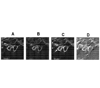
[0077] Immunofluorescence stain and confocal microscopy studies determined
rates of
co-expression of CD206 on both tissue macrophages and LANA expressing KS tumor
cells. The immunofluorescence stain and confocal microscopy studies were
performed on
a tissue microscopic array (TMA) containing 66 cases of AIDS KS and controls
obtained
from the AIDS and Cancer Specimen Resource (ACSR). The results are shown in
FIG. 5
which depicts confocal microscopy representative images showing co-
localization of
macrophage mannose receptor CD206 on both LANA expressing tumor cells and
tissue
macrophage. A. DAPI (blue); B. CD206 (green); C, LANA (red); D, CD68 (yellow);
E,
All merged (63X). Cy3-tilmanocept uptake by HHV8+ KS tumor cells was also
examined, and FIG. 6 depicts example confocal images of KS biopsy tissue
culture with
Cy3 tilmanocept. Confocal images of HHV8+ KS tumor cell biopsy. 25x (CD68,
Yellow; Cy3-tilmanocept, Red; HHV8, Green; DAPI, Blue)
[0078] Cy3-tilmanocept uptake into CD206-expressingmacrophages was also
examined.
3-day CD206+ macrophage cultures were incubated with Cy3-tilmanocept (100
g/mL)
for 4, 24 and 48 hours at 37 C. Background levels of Cy3 fluorescence were
determined
in cultures exposed to conjugates at room temperature for the same time
periods. Flow
cytometric evaluation of Cy3 and CD206 was performed at all time points
indicating
Cy3-tilmanocept uptake into CD206+ macrophages. FIG. 7 shows a flow cytometric
evaluation of Cy3 and CD206 in 3 day CD206+ macrophage cultures incubated with
Cy3-tilmanocept.
[0079] In light of the above, in further specific embodiments the carrier
molecules
described herein are used for diagnosing and/or treating KS (and similar types
of cancers
CA 02918782 2016-01-19
WO 2015/013341 PCMJS2014/047708
- 32 -
and tumors). For diagnostic purposes, a detectable moiety such as 99mTc or
68Ga is
attached to the carrier molecule (e.g. to a DTPA or DOTA chelator), and the
radiolabeled
composition administered to a subject such as by subcutaneous or intradermal
injection
proximal to (i.e., adjacent) the tumor or suspected lesion, intra-
tumorally/intra-lesionally
injected directly into the tumor or lesion, or by intravenous injection. It
will be
understood that other detectable moieties described herein, known to those
skilled in the
art, or hereafter developed may be attached to the carrier molecule for use in
diagnosing
KS, such as any of a varietyy of fluorophores. Following administration to a
patient, the
tumor or lesion site (or suspected tumor or lesion site) is imaged, such as by
scintigraphy
(e.g., using a gamma camera), single-photon emission computed tomography
(SPECT),
positron emission tomography (PET), or optical imaging (e.g., when the
detectable
moiety is a fluorescent dye such as cyanimine). It will be understood,
however, that other
diagnostic moieties other than those mentioned above may be employed, as well
as
various other imaging or diagnostic methods for detecting the presence of the
labeled
carrier molecules in the KS tumor or lesion.
[0080] In
one specific embodiment for KS diagnostic imaging, the carrier molecule is
tilmanocept: dextran 3- [(2-
aminoethyl)thio]propyl 17-carboxy-10,13,16-
tris(carboxymethyl)- 8-oxo-4-thia-7, 1 0 ,13, 16-tetraaz aheptadec- 1 - yl 3-
[ [2- [[ 1 -imino-2- (D-
mannopyranosylthio) ethyl]amino]ethyl]thio]propyl ether complexes. In this
particular
embodiment, the detectable moiety is 99mTc or 68Ga, and the detectable moiety
is attached
to a DTPA chelator just prior to use by mixing the carrier molecule with the
elute from a
99mTc generator or a gallium-68 generator, as known to those skilled in the
art. In other
embodiments, the detectable moiety is Cy-3 and is attached to a leash of
tilmanocept, as
known to those skilled in the art. For diagnostic imaging of KS using 99mTc-
tilmanocept
or 68Ga-tilmanocept, in some embodiments the radiolabeled carrier molecule has
sufficient radioisotope to provide a dose, when administered locally (e.g.,
subcutaneuously) to a subject, of between about 0.3 to about 5.0 millicuries,
or about 0.5
to about 2.0 millicuries, or about 0.5 or about 1 millicurrie. In other
embodiments, such
as for diagnostic imaging of KS using 99mTc-tilmanocept or 68Ga-tilmanocept,
the
- 33 -
radiolabeled carrier molecule has sufficient radioisotope to provide a dose,
when
administered systemically (e.g., intravenously) to a subject, of between about
2 mCi to
about 30 mCi, from about 5 mCi to about 30 mCi, and from about 10 mCi to about
25
mCi. When administered to a subject by injection, the radiolabeled carrier is,
in some
embodiments, combined with a pharmaceutically acceptable carrier containing
one or
more excipients, diluents and the like (e.g., sterile saline). For diagnostic
imaging of KS
using tilmanocept having one or more detectable moieties attached thereto,
between
about 50 and about 500 micrograms of tilmanocept is administered.
[0081] For therapeutic use of the carrier molecules described herein in
treating KS, a
suitable therapeutic agent is attached to the carrier and the resulting
composition is
combined with a pharmaceutically acceptable carrier containing one or more
excipients,
diluents and the like. As with the diagnostic imaging, the carrier molecule
with attached
therapeutic agent is administered to a patient such as by injection, or even
topically to a
tumor or lesion. Suitable therapeutic agents for treating KS include
functional
chemotherapeutic agents such as doxorubicin, daunorubicin, paclitaxel
(Taxol(D),
gemcitabine (Gemzare)), vinorelbine (Navelbinee)), bleomycin, vinblastine
(Velbane)),
vincristine (Oncovin(D), and etoposide (VP-16). In one particular embodiment,
the
therapeutic composition comprises Doxorubicin-tilmanocept, which is
administered
topically (e.g., as a 10 g dose) or intravenously (e.g., as a 5mg dose).
KS IMAGING EXAMPLE 1
[0082] Tilmanocept lyophilized powder, marketed by Navidea
Biopharmaceuticals Inc.
under the name LYMPHOSEEK Injection kit, is obtained. The tilmanocept powder
has
a mean diameter of about 7nm, and is contained in a 0.5 mL vial as a mixture
of 0.250
mg tilmanocept, 20 mg trehalose dihydrate, 0.5 mg glycine, 0.5 mg sodium
ascorbate and
0.075 mg stannous chloride dihydrate. The tilmanocept powder is then
radiolabeled with
Tc 99m using sodium 99mTc-pertechnetate eluted from a Technetium-99m
generator.
Using a sterile syringe, approximately 92.5 MBq (2.5 mCi) of sodium 99mTc-
pertechnetate in about 0.35mL is aseptically added to the vial. The vial is
gently shaken,
Date Recue/Date Received 2022-01-04
- 34 -
and the radiolabeling reaction allowed to proceed at room temperature for at
least 10-15
minutes. Normal saline is then added to the vial to bring the contents to 2.5
cc. A buffer
is optionally added, as described in U.S. Pat. Pub. No. 2010/0196272 Al,
published
August 5, 2010.
[0083] A
single patient dose is 50 mcg of tilmanocept and 0.5 mCi of technetium 99m, as
prepared above, totaling 0.5 cc. The radiolabeled tilmanocept is administered
by
subcutaneous or intradermal injection, within six hours of radiolabeling.. In
an alternative
embodiment, 100 mcg of tilmanocept and 1.0 mCi of technetium, totaling 1 cc,
is
injected intravenously within six hours of radiolabeling.
[0084]
Within 30 to 180 minutes of injection, the patient is imaged using Single
Photon
Emission Computed Tomography (SPECT). The findings of localized radioactivity
within the skin lesion(s) is presumptive evidence of mannose binding receptors
and/or
macrophage activity, which is consistent with the presence of Kaposi' s
sarcoma, and the
absence of such activity would essentially rule out Kaposi' s sarcoma.
KS IMAGING EXAMPLE 2
[0085] The
following illustrates yet another example of the evaluation of Primary
Cutaneous Kaposi's Sarcoma (KS) by SPECT and SPECT/CT Imaging using
Lymphoseek0 (also known as technetium 99mTc-tilmanocept injection) a
radiopharmaceutical that binds to mannose binding receptors (CD206) that
reside on the
surfaces of dendritic cells and macrophages. The results indicate that, in
patients with
primary cutaneous Kaposi's sarcoma, Lymphoseek aids in the detection of
Kaposi's
sarcoma lesion(s) using single photon emission computed tomography (SPECT) and
SPECT computed tomography (SPECT/CT).
[0086] An 18
year-old male patient receives a single dose of 50 lig tilmanocept
radiolabeled with 2.0 mCi 99mTc by subcutaneous injection. The total volume of
99mTc-tilmanocept injection is 0.3 to 0.5 mL.
Date Recue/Date Received 2022-01-04
CA 02918782 2016-01-19
WO 2015/013341 PCMJS2014/047708
- 35 -
[0087] The patient has a marker lesion (> lcm in diameter) with a confirmed
diagnosis of
KS (CD 206-expressing cutaneous KS) via punch biopsy. The location of the
marker KS
lesion is on the extremities: from the shoulder to the metacarpal region or
from the groin
to the metatarsal region.
[0088] The dose is administered by a syringe with a 5/8 inch, 25- or 27-
gauge, fixed
needle, or other syringe/needle combinations that are acceptable for
subcutaneous
injections. The injection is made 1.5 0.25 cm distal from the marker lesion,
4-8 cm
distal to the marker lesion, or 4-8 cm proximal to the marker lesion.
[0089] The patient undergoes a regional dynamic SPECT scan immediately post-
injection for a duration of 30 minutes. After the initial scan, the patient
undergoes whole
body SPECT/CT imaging at 1 hour and whole body SPECT imaging at 4-6 hours post
injection. The patient is permitted to leave the imaging center after the 4-6
hour SPECT
scan.
[0090] Dynamic SPECT/CT (limited CT exposure; GE Infinia Hawkeye 4) imaging
occurs immediately following injection for 30 minutes (-3 minutes/rotation).
Each dual
head spin is segmented into 32 (5.625 ) angles. SPECT images are acquired at
anterior,
45 anterior oblique and lateral positions, with each acquisition being 3-5
minutes in
duration for a total of 30-45 minutes.
[0091] Acquisition on SPECT/CT systems is performed in a sequential mode.
With
devices that have a low-dose CT component, data are typically acquired by
rotating the X
ray detector 220 around the patient, with the X ray tube operated at 140 kV
and 2.5 mA.
The CT images obtained have an in-plane spatial resolution of 2.5 mm, and of
10 mm in
the axial direction. Scan time is approximately 16 seconds per slice, for a
total duration
of 30-45 minutes for the CT. SPECT/CT systems using a diagnostic CT component
are
characterized by higher spatial resolution and faster scanning time
(approximately 30
seconds for the whole field of view), associated however with higher radiation
doses. An
attenuation map is created at the end of the CT acquisition time.
CA 02918782 2016-01-19
WO 2015/013341 PCMJS2014/047708
- 36 -
DIAGNOSIS AND TREATMENT OF TUBERCULOSIS
[0092] Tuberculosis is a respiratory infection caused by the bacteria
mycobacterium
tuberculosis. In response to TB infection, a patient's immune system forms
granulomas
which allow TB bacteria to remain within the granulomas for long periods of
time with
no apparent clinical symptoms of TB. While treatment can reduce the risk of
patient
developing active TB infection, the granulomas act as a barrier to diagnostic
and
therapeutic agents. Macrophages are part of the processes of the formation and
maintenance of the granulomas. Because of this, Applicants have deduced that
the carrier
molecules described herein can be used to target CD206 on the surface of the
macrophages associated with TB granulomas.
BINDING OF TILMANOCEPT TO MACROPHAGES INFECTED WITH TB
[0093] In order to demonstrate the ability of the carrier molecules
described herein to
bind to macrophages infected with TB and deliver diagnostic and/or therapeutic
agents
into the interior of the macrophages where the TB bacterium are located, human
monocyte-derived macrophages in monolayer culture that make up the components
of the
TB granulomas were infected with a GFP-expressing M. tuberculosis which was
internalized by macrophages (GFP = green fluorescent protein). The infected
cells were
then exposed to tilmanocept which had been labeled with cyanine (Cy3) dye, and
analyzed by confocal microscopy. FIG. 5 depicts the confocal microscopy of the
TB-
infected macrophages. Red indicates Cy3-tilmanocept, green indicates GFP M.
tuberculosis, and yellow indicates the co-localization of Cy3-tilmanocept and
GFP M.
tuberculosis. Thus, FIG. 8 demonstrates that the Cy3-tilmanocept binds to, and
is
internalized by the macrophages
[0094] In light of the above, in further specific embodiments the carrier
molecules
described herein are used for diagnosing and/or treating tuberculosis. For
diagnostic
purposes, a detectable moiety such as 99mTc or 68Ga is attached to the carrier
molecule
(e.g. to a DTPA or DOTA chelator), and the radiolabeled composition
administered to a
subject such as by inhalation, intravenous injection or pulmonary lavage. It
will be
CA 02918782 2016-01-19
WO 2015/013341 PCMJS2014/047708
- 37 -
understood that other detectable moieties described herein, known to those
skilled in the
art, or hereafter developed may be attached to the carrier molecule for use in
diagnosing
tuberculosis. Following administration to a patient, the subject's lungs are
imaged, such
as by scintigraphy (e.g., using a gamma camera), single-photon emission
computed
tomography (SPECT), or positron emission tomography (PET). It will be
understood,
however, that other diagnostic moieties other than those mentioned above may
be
employed, as well as various other imaging or diagnostic methods for detecting
the
presence of the labeled carrier molecules in the subject's lung tissue.
[0095] In one specific embodiment for tuberculosis diagnostic imaging, the
carrier
molecule is tilmanocept: dextran 3-[(2- aminoethyl)thio]propyl 17-carboxy-
10,13,16-
tris (c arb oxymethyl)- 8-oxo-4-thia-7 ,10,13,16-tetraaz aheptadec-1- yl 3- [
[2- [[1-imino-2- (D-
mannopyranosylthio) ethyl]aminolethyl]thiolpropyl ether complexes. In this
particular
embodiment, the detectable moiety is 99mTc or 68Ga, and the detectable moiety
is attached
to the DTPA chelator just prior to use by mixing the carrier molecule with the
elute from
a 99mTc generator or a gallium-68 crenerator, as known to those skilled in the
art. For
diagnostic imaging of tuberculosis using 99mTc-tilmanocept or 68Ga-
tilmanocept, in some
embodiments the radiolabeled carrier molecule has sufficient radioisotope to
provide a
dose, when administered to a subject, of between about 0.3 to about 5.0
millicuries, or
about 0.5 to about 2.0 millicuries, or about 1 millicunie.
[0096] When administered to a subject by inhalation, the radiolabeled
carrier is, in some
embodiments, combined with a pharmaceutically acceptable vehicle. By way of
specific
example, the radiolabeled carrier is delivered to the lungs of a human subject
by an
inhalation device¨e.g., a fixed dose inhaler, a dry powder in haler, a metered
dose
inhaler, or a nebulizer. In one embodiment, the radiolabeled carrier is
administered using
a metered dose inhaler containing a suspension of the radiolabeled carrier in
a vehicle
comprising a pharmaceutically acceptable inert liquid propellant such as a
chlorofluorocarbon, fluorocarbon or hydrofluroalkane. By way of more specific
example,
the metered dose inhaler is configured to deliver about 10 to about 5000
micrograms, or
about 10 to about 500 micrograms, of radiolabeled carrier per puff. In still
further
CA 02918782 2016-01-19
WO 2015/013341 PCMJS2014/047708
- 38 -
embodiments, the radiolabeled carrier is suspended in a pharmaceutically
acceptable
vehicle comprising sterilized water or saline, and administered by
nebulization. In yet
another embodiment, the radiolabeled carrier is dried to a powder and then
administered
from a pouch or other container.
[0097] For therapeutic use of the carrier molecules described herein in
treating TB, a
suitable therapeutic agent is attached to the carrier and the resulting
composition is
combined with a pharmaceutically acceptable vehicle containing one or more
excipients,
diluents and the like. As with the diagnostic imaging, the carrier molecule
with attached
therapeutic agent is administered to a patient such as by inhalation,
intravenous injection
or pulmonary lavage for treating TB (dormant or active infection).
[0098] When administered to a subject by inhalation, the therapeutic
agent+carrier is, in
some embodiments, combined with a pharmaceutically acceptable vehicle. By way
of
specific example, the therapeutic agent+carrier is delivered to the lungs of a
human
subject by an inhalation device¨e.g., a fixed dose inhaler, a dry powder in
haler, a
metered dose inhaler, or a nebulizer. In one embodiment, the therapeutic
agent+carrier is
administered using a metered dose inhaler containing a suspension of the
therapeutic
agent+carrier in a vehicle comprising a pharmaceutically acceptable inert
liquid
propellant such as a chlorofluorocarbon, fluorocarbon or hydrofluroalkane. By
way of
more specific example, the metered dose inhaler is configured to deliver about
10 to
about 5000 micrograms, or about 10 to about 500 micrograms, of therapeutic
agent+carrier per puff. In still further embodiments, the therapeutic
agent+carrier is
suspended in a pharmaceutically acceptable vehicle comprising sterilized water
or saline,
and administered by nebulization. In yet another embodiment, the therapeutic
agent+carrier is dried to a powder and then administered from a pouch or other
container.
And in yet another embodiment, the therapeutic agent+carrier is suspended in a
pharmaceutically acceptable vehicle and administered intravenously, at a
dosage of up to
mg of the therapeutic agent+carrier molecules.
CA 02918782 2016-01-19
WO 2015/013341 PCMJS2014/047708
- 39 -
[0099] Suitable therapeutic agents attached to the carrier molecule for
treating TB
include indomethacin, isoniazid, and/or Ga (optionally as 68Ga, such that the
composition
is used as both a diagnostic and therapeutic composition). In still further
embodiments, a
composition for both diagnosing and treating tuberculosis is provided wherein
both 68Ga
and Ga (i.e., non-radioactive Ga) are conjugated to the carrier molecule. In
other
embodiments, two or more of indomethacin, isoniazid and Ga (optionally as
68Ga) are
conjugated to the carrier molecule. Indomethacin, isoniazid. and Ga are known
treatment
agents for TB, however, by attaching one or more of these agents to the
carrier molecules
described herein they are better able to enter the macrophages of granulomas
associated
with TB wherein the therapeutic agents will target the TB bacterium within
those
macrophages.
[00100] Provided below are examples of compositions and methods which are
effective
for diagnosing or treating TB.
TUBERCULOSIS IMAGING EXAMPLE
[00101] Tilmanocept lyophilized powder, marketed by Navidea
Biopharmaceuticals Inc.
under the name LYMPHOSEEK Injection kit, is obtained. The tilmanocept powder
contained in a 0.5 mL vial as a mixture of 0.250 mg tilmanocept, 20 mg
trehalose
dihydrate. 0.5 mg glycine, 0.5 mg sodium ascorbate and 0.075 mg stannous
chloride
dihydrate. The tilmanocept powder is then radiolabeled with Tc 99m using
sodium
99mTc-pertechnetate eluted from a Technetium-99m generator. Using a sterile
syringe,
approximately 92.5 MBq (2.5 mCi) of sodium 99mTc-pertechnetate in about 0.35mL
is
aseptically added to the vial. The vial is gently shaken, and the
radiolabeling reaction
allowed to proceed at room temperature for at least 10-15 minutes. Normal
saline is then
added to the vial to bring the contents to 5 cc immediately prior to
administration.
[00102] A single patient dose of the radiolabeled composition is prepared
such that the
dose is 100 mcg of 99mTc-tilmanocept, 1 mCi, totaling 2 cc. The radiolabeled
tilmanocept
is administered by inhalation within six hours of radiolabeling. The
composition is
loaded into an aerosol machine and the patient then inhales the composition.
Within
- 40 -
about 30 to 180 minutes after inhalation, the patients lungs are imaged using
Single
Photon Emission Computed Tomography (SPECT). The findings of localized
radioactivity in the hilar and mediastinal areas of the thoracic cavity will
be presumptive
evidence of granuloma formation, which is a hallmark of tuberculosis.
TUBERCULOSIS TREATMENT EXAMPLE
[00103] Tilmanocept lyophilized powder, marketed by Navidea
Biopharmaceuticals Inc.
under the name LYMPHOSEEK Injection kit, is obtained. The tilmanocept powder
has
a mean diameter of about 7nm, and is contained in a 0.5 mL vial as a mixture
of 0.250
mg tilmanocept, 20 mg trehalose dihydrate, 0.5 mg glycine, 0.5 mg sodium
ascorbate and
0.075 mg stannous chloride dihydrate. The tilmanocept powder is then bound to
bound to
Isoniazid molecules. Normal saline is then added to the vial to bring the
contents to 2.5
cc. A buffer is optionally added, as described in U.S. Pat. Pub. No.
2010/0196272 Al,
published August 5, 2010.
[00104] A single patient dose of the composition prepared as described
above is about 100
to about 500 mcg of tilmanocept, depending on the patient's age and weight,
totaling 1
cc. The isoniazid-tilmanocept composition is administered to the patient by
intravenous
injection. When administered in this fashion, the isoniazid-tilmanocept
composition
would be expected to localize in the granulomas containing the intracellular
tuberculosis
bacilli and deliver the isoniazid to the intracellular space within the
macrophage where
the TB is located. This will allow for a concentrated dose of Isoniazid to be
delivered
directly to the tuberculosis bacilli, bypassing the usual barriers of drug
delivery
frequently encountered in TB treatment.
[00105] In variations of the above TB treatment composition and method,
indomethacin
and/or Ga (optionally as 68Ga) are also attached to the tilmanocept in the
manner
described previously in order to provide Ga-isoniazid-tilmanocept,
indomethacin-
isoniazid-tilmanocept, and/or indomethacin-Ga-isoniazid-tilmanocept, which is
then
formulated into suitable compositions and administered in the various manners
described
previously. As a still further variation, Ga (optionally as 68Ga) and/or Ga-
isoniazid-
Date Recue/Date Received 2022-01-04
CA 02918782 2016-01-19
WO 2015/013341 PCMJS2014/047708
- 41 -
tilmanocept is attached to the tilmanocept in place of isoniazid in the manner
described
previously in order to provide Ga- tilmanocept, indomethacin-tilmanocept
and/or Ga-
indomethacin-tilmanocept which is then formulated in to suitable compositions
and
administered in the various manners described previously.
[00106] DIAGNOSIS OF INCREASED ARTERIAL INFLAMMATION
[00107] Macrophages, in high-risk coronary atherosclerotic plaque samples
from patients
who experienced sudden cardiac death, express CD206 - along with CD163. These
high-
risk plaques have been characterized, morphologically, as thin-cap
fibroatheromas
(TCFA) and infiltrated by activated macrophages throughout the necrotic plaque
core and
thin, fibrous plaque cap. Thus, increased arterial inflammation, as evidenced
by the
presence of macrophages, is an indicator of increased risk for developing high
risk
morphology coronary plaque burden.
[00108] FIG. 9 depicts images of immunofluorescent staining of left
ventricle and aorta
from rhesus macaque. The images illustrate the Co-localization of CD163/Alex -
Fluor
488, CD206/Alexa - Fluor 568, and Cy3 tilmanocept. Alexa-Flour 568 is
fluorescent dye
readily attachable to tilmanocept in the manner previously described.
[00109] Based on their unique properties to tag activated macrophages, the
compositions
of the present invention provide a method for imaging arterial inflammation
and
identifying individuals with arterial macrophage-specific inflammation and
heightened
immune-mediated cardiovascular disease (CVD) risk.
[00110] One embodiment of the present invention provides a method of
quantifying
measurable aortic uptake of the of systemically injected CD206 targeting
compositions of
the present invention using single photon emission computed tomography
(SPECT/CT).
In another embodiment, the present invention provides a method of measuring
the
density of infiltrating activated macrophages in arterial atherosclerotic
plaque.
[00111] In another embodiment, the invention provides a method of
functional arterial
imaging to characterize the propensity of individual coronary plaques to
rupture. In
CA 02918782 2016-01-19
WO 2015/013341 PCMJS2014/047708
- 42 -
another embodiment, the invention provides a method for identifying patients
at risk for
CVD before they experience clinically significant events. In one particular
embodiment,
the method is applied to specific high-risk patients, such as HIV-infected
patients. In
another particular embodiment, the method is applied to patients with
vulnerable plaque
at risk for rupture in the general population. F In another particular
embodiment, the
invention provides functional arterial imaging for monitoring the efficacy of
anti-
inflammatory strategies that modulate accelerated atherogenesis.
Arterial Inflammation Imaging Example
[00112] The following example illustrates the evaluation of aortic and
coronary artery
99mTc-tilmanocept uptake using SPECT and SPECT/CT Imaging).
[00113] An 18 year-old male patient, with documented HIV infection with a
history of
subclinical aortic plaque and high-risk morphology coronary plaque on CCTA ,
receives
an intravenous injection via catheter of ¨10 mCi of 99mTc-tilmanocept. The
catheter is
flushed post-injection with approximately 10 mL of saline solution.
[00114] The subject is positioned on the scanner table of a Siemens
SPECT/CT scanner
(Siemens Medical Solutions, Hoffman Estates, IL). After a lag time of
approximately 60
minutes, SPECT acquisitions will be performed using 2x60 views, step and shoot
mode,
1 mm per view of the thorax and neck based on the scout CT performed prior to
SPECT.
Gated acquisitions are performed when imaging the heart. Thoracic images
include all of
the lungs. Data acquisition of SPECT and CT take approximately 70 minutes. All
projections are acquired in two energy windows, namely [90-120 keV] and [126-
154
keV], corrected for Compton scatter using a scatter window and for attenuation
using the
CTAC and reconstructed using iterative ordered subsets expectation
maximization
algorithm (OSEM). The resulting reconstructed volume are used to quantify
target to
background ration using regions of interest (ROT) drawn on areas of 99mTc-
tilmanocept
uptake of interest normalized to a reference region of interest
DIAGNOSIS OF RHEUMATOID ARTHRITIS
CA 02918782 2016-01-19
WO 2015/013341 PCMJS2014/047708
-43 -
[00115] Rheumatoid arthritis ("RA") is an autoimmune disease that is also
difficult to
diagnose and treat. The carrier molecules described herein (e.g., tilmanocept)
are
designed to bind to CD206 of reticuloendothelial cells that are invasive to
focal RA
tissue, such as is present in association with RA of the joints and/or
viscerally-involved.
Thus these carrier molecules can be used for diagnostic imaging as well as
treatment of
RA. As to tilmanocept, for example, the mannoses act as ligand moieties for
the CD206,
and the DTPA serves as a chelating moiety for radiolabeling with, for example,
Tc 99m.
When Tc 99m-tilmanocept is injected in close proximity to the suspected
diseased locus
(i.e., a joint where RA is present or suspected), scintigraphic imaging in
conjunction with
a stationary gamma camera and/or intraoperatively in conjunction with a
handheld
gamma detection probe may be used to localize involved tissue for purposes of
diagnosing RA. Tilmanocept has a mean diameter of about 7 nm, and this small
diameter
permits enhanced diffusion into tissue channels and blood capillaries,
resulting in a rapid
injection site clearance and CD206 binding in the inflammasome. By way of
example,
fluorescent and/or radioactive tilmanocept and other carrier molecules
described
previously can be used for the diagnosis and non-invasive imaging of joints
with early
forms of RA.
[00116] In order to demonstrate that tilmanocept binds to CD206 receptors
found on
macrophages in synovial fluid of subjects having RA, synovial fluid and tissue
were
acquired from patients diagnosed with frank RA. Tissues were probed with
Manocept-
Cy3, DAPI nuclear fluor, and anti CD206-cyanine green. The tissues and fluids
were
imaged by micro-fluorescence and compared to normal frozen archival tissue and
synovial tissue procured from patients with osteoarthritis (OA). MP
localization and
degree of fluorescence were compared by digital image analysis. In particular,
the micro-
fluoroescence images were analyzed using scanning quantitative fluorescence
microscopy, with integration algorithms was used to quantitate and contrast
pixel counts
of Cy3 fluorescent dye in images of tissue and synovial fluid.
[00117] The results indicated that the synovial tissue and fluid from
subjects with RA
contain large macrophage populations that express high levels of CD206.
Additionally,
CA 02918782 2016-01-19
WO 2015/013341 PCMJS2014/047708
- 44 -
these MPs strongly localize Cy3-tilmanocept on CD206. In addition, the degree
of
macrophage invasion and CD206 residence in normal and OA tissue is
significantly
lower than in RA tissues, as seen in FIG. 10. Thus, the carrier molecules of
the present
invention, when provided with a detectable moiety such as a flurophore, are
able to not
only diagnose RA from synovial fluid (either in vivo or ex vivo), but also can
distinguish
RA from OA.
Imaging of macrophages in cartilage antibody-induced arthritis in mice using
Cy3-tilmanocept
[00118] Cy3-tilmanocept was used to image macrophages in a mouse model of
early
immune-mediated arthritis and cartilage antibody-induced arthritis in Dbal
mice using
fluorescent luminescence. Arthritis was induced in mice by injection of a five
monoclonal antibody anti-cartilage cocktail followed in three days by an
injection of E.
coli lipopolysaccharide. The mice developed swollen and reddened joints in the
feet,
carpi, tarsi, elbows, and knees of variable degrees in 7-11 days, evidencing
arthritis.
[00119] Mice were imaged in vivo on days 7 or 8 and mice were euthanized on
days 9 or
11. After euthanasia, the limbs were dissected, skin was removed, and the
samples were
reimaged (epifluorescent imaging), radiographed (Faxitron MX20) and then
decalcified,
embedded. and stained with H&E.
[00120] For epifluorescent imaging, mice were injected intravenously with
Cy3-
tilmanocept, and epifluorescent imaging was conducted in vivo and ex vivo at 1-
2 hours
using an IVIS Lumina II machine (Caliper Life Sciences, Hopkinton, MA). Living
Image
software was used to visualize the visible and fluorescent images and to
quantitate the
number of photons using regions of interest ("ROI") and subtraction of
background
fluorescence. After euthanasia the limbs were dissected, skin was removed
(except for
the digits), and re-imaged. Specific fluorescence was detected in arthritic
knees and
elbows, as seen in FIG. 11. FIG. 12 depicts in vivo fluorescence of the elbows
and feet of
a mouse with immunemediated arthritis (top) and control mouse (bottom). The
mouse
with arthritis had increased fluorescence due to Cy3-Tilmanocept in the elbow
compared
CA 02918782 2016-01-19
WO 2015/013341 PCMJS2014/047708
-45 -
to the control mouse. There was background fluorescence from the skin, which
was
prominent on the feet. FIG. 13 shows ex vivo fluorescence data, and FIG. 14
depicts ex
vivo fluorescence of the knees of control mice and mice with immune-mediated
arthritis.
Although both knees in the treated mouse (lower image) had arthritis, the knee
on the
right was affected more severely and had greater fluorescence due to Cy3-
Tilmanocept
labeling.
[00121] In particular embodiments for RA diagnostic imaging, the carrier
molecule is
tilmanocept, and the detectable moiety is 99mTc or 68Ga attached to the DTPA
chelator
prior to use, or a fluorescent dye attached to the amino-terminated leash
(e.g., Cy3). In
the case of Cy3-tilmanocept, optical imaging is employed at to determine the
presence
and/or extend of RA. The above-described mouse studies have confirmed that
labeled
tilmanocept (e.g., Cy-3-tilmanocept) is useful in diagnosing RA.
[00122] In particular embodiments for RA treatment, the carrier molecule is
tilmanocept,
and the therapeutic is a therapeutic isotope. In one particular embodiment,
the therapeutic
isotope is 117mSn. In other particular embodiments for RA treatment, the
carrier molecule
is tilmanocept, and the therapeutic is a toxin. In one particular embodiment,
the toxin is
botulinum or cholera toxin. In another particular embodiment for RA treatment,
the
carrier molecule is tilmanocept, and the therapeutic is a methotrexate.
RA IMAGING EXAMPLE
[00123] Ti lm an ocept lyophilized powder, marketed by Navidea
Biopharmaceuticals Inc.
under the name LYMPHOSEEK Injection kit, is obtained. The tilmanocept powder
has
a mean diameter of about 7nm, and is contained in a 0.5 mL vial as a mixture
of 0.250
mg tilmanocept, 20 mg trehalose dihydrate, 0.5 mg glycine, 0.5 mg sodium
ascorbate and
0.075 mg stannous chloride dihydrate. The tilmanocept powder is then
radiolabeled with
Tc 99m using sodium 99mTc-pertechnetate eluted from a Technetium-99m
generator.
Using a sterile syringe, approximately 92.5 MBq (2.5 mCi) of sodium 99mTc-
pertechnetate in about 0.35mL is aseptically added to the vial. The vial is
gently shaken,
and the radiolabeling reaction allowed to proceed at room temperature for at
least 10-15
CA 02918782 2016-01-19
WO 2015/013341 PCT/US2014/047708
- 46 -
minutes. Normal saline is then added to the vial to bring the contents to 5
cc, and a buffer
may optionally be added as described previously.
[00124] A single patient dose is 100 mcg of tilmanocept and 1 mCi of
technetium 99m,
totaling 2 cc. The radiolabeled tilmanocept is administered by intravenous
injection
within six hours of radiolabeling. Within 30 to 180 minutes of injection, the
patient is
imaged using Single Photon Emission Computed Tomography (SPECT). The findings
of
localized radioactivity in the joints is presumptive evidence of an
inflammatory process
in the intrarticular area, which is a hallmark of rheumatoid arthritis (RA),
thereby ruling
out RA if it is absent and aiding in the diagnosis of early or ongoing RA if
it is present.
[00125] While several compositions and methods for the diagnosis and/or
treatment of
macrophage-related disorders have been discussed in detail above, it should be
understood that the compositions, features, configurations, and methods of
using the
compositions discussed are not limited to the contexts provided above.