Note: Descriptions are shown in the official language in which they were submitted.
COMPOUNDING SYSTEMS AND METHODS FOR
SAFE MEDICAMENT TRANSPORT
[0001] BACKGROUND
1. Technical Field
[0002] The present application relates to systems and methods for the
safe transportation
of medicaments and, more particularly, to systems and methods for the handling
and transport of
potentially hazardous medicaments, in particular, cytotoxic drugs and the
like.
2. Background of Related Art
[0003] In one instance, hazardous medicines are frequently applied in
the treatment of
certain diseases, in particular, for example, in the treatment of cancer.
Cytotoxic drugs have
generally been used to kill cancer cells. However, the use of cytotoxic drugs,
in the treatment of
cancer cells, presents specific dangers to all cells, both in the patient and
in healthcare providers.
Although the exposure to a health care provider is normally very small for
each cytotoxic drug
dose administration, evidence suggests that chronic, low-dose exposure can
produce significant
health problems. Accordingly, a system that allows the safe handling of
hazardous drugs while
significantly reducing and/or eliminating the exposure to providers would be
of great benefit.
[0004] Drugs are typically supplied in glass or plastic vials that are
capped with a gas
impermeable liquid seal or stopper. In some instances, the vial contents are a
solid powder, such
that a liquid needs to be injected for mixing (e.g.. reconstitution). The
injection of additional
contents (e.g., liquid) into the vial produces an increased pressure which
stresses the seal or
1
CA 2920199 2017-07-05
CA 02920199 2016-02-02
WO 2015/017858 PCT/US2014/049609
stopper. Although the vial is intended to be sealed to liquid and gases, drug
molecules in vapor
phase can leak or pass around the sides of the stopper or through the stopper
as the injection
needle is withdrawn, thus presenting a hazard to the provider or clinician.
[0005] Accordingly, with the potential for aerosol leakage,
leakage/spraying upon needle
withdrawal, or spills, a means with which to prevent the accidental vapor
phase drug egress is
required. The provision of a pressure gradient/differential across the seals
will ensure that any
gas will flow from high to low pressure. Establishing a negative relative
pressure between the
inside of the transfer volume and atmosphere will prohibit the egress of vapor
phase drug.
[0006] Thus, the need exists for new components and systems capable of
transferring
gases/fluids/liquids or other substances between a conventional syringe and
one of a vial, a
patient I.V. (intra-venous) set, or an I.V. bag without leaking or spilling
and without exposure of
the liquids to substances outside the closed system. As such, healthcare
personnel may more
safely use and handle fluid substances including potentially hazardous liquids
and the like.
[0007] The hazardous medicines, including Cytotoxic drugs amongst
others, are typically
prepared by a technician in a. clean room setting, or by a fully automated or
robotic system.
However, it is desirable to provide a system for the preparation of these
hazardous medicines that
is semi-automated or that is a user/technician assisted system, wherein some
portion or steps in
the preparation of these hazardous medicines is accomplished by the
user/technician and sonic
portion in the preparation of these hazardous medicines is accomplished by an
apparatus or the
like.
[0008] Additionally, these hazardous medicines must be prepared in a
clean room setting
or the like, such as, for example, in a room, under a hood, in a chamber, or
the like. A clean
room is a room in which the concentration of airborne particles is controlled
to meet a specified
airborne particulate cleanliness class. Clean rooms are classified by the
cleanliness of their air.
Accordingly, for the preparation of these hazardous medicines, it is required
that the clean room
have an ISO (International Standards Organization) class 5 rating.
CA 02920199 2016-02-02
WO 2015/017858 PCT/US2014/049609
[0009] Clean rooms are designed to maintain positive air pressure,
preventing "unclean"
(contaminated) air from flowing inside and less-clean air from flowing into
clean areas. The idea
is to ensure that filtered air always flows from cleanest to less-clean
spaces.
[0010] ISO class 5 and cleaner facilities rely on unidirectional, or
laminar, airflow.
Laminar airflow means that filtered air is uniformly supplied in one direction
(at a fixed velocity)
in parallel streams, usually vertically. Air is generally re-circulated from
the base of the walls of
the clean room back up to the filtering system.
[0011] Thus, a critical factor in clean room design is controlling air-
change per hour
(ACII), also known as the air-change rate, or ACR. This refers to the number
of times each hour
that filtered outside air replaces the existing volume in a building or
chamber.
[0012] Further, another critical factor in clean room design
controlling or reducing the
turbulence of the air flowing through the clean room, wherein lower turbulence
will increase the
cleanliness of the room.
[0013] III ISO class 5 clean rooms, the particle per cubic meter must
be no more than
3520 particles/m3 in a size of 0.5 micrometers or larger when counted at
representative locations
normally not more than 1 foot away from the work site, within the airflow, and
during
filling/closing operations.
[00141 Accordingly, improvements in systems for the handling and
transport of
potentially hazardous medicines, in particular, cytotoxic drugs and the like,
in a clean room or
chamber, is desired and warranted.
SUMMARY
[0015] The present application relates to systems and methods for the
handling and
transport of potentially hazardous medicines, in particular, cytotoxic drugs
and the like.
[0016] According to an aspect of the present disclosure, an automatic
or semi-automatic
preparation system for forming a medicament solution from a vial containing
one of a liquid and
a non-liquid material, is provided. The preparation system includes a carousel
configured to
provide three axes of motion. The carousel includes a manipulator having at
least one first rail
3
CA 02920199 2016-02-02
WO 2015/017858 PCT/US2014/049609
defining a first axis; at least one second rail defining a second axis, the
second axis being
oriented orthogonal to the first axis; at least one third rail defining a
third axis, the third axis
being oriented orthogonal to the each of the first axis and the second axis; a
first gear belt
movably supported on at least one of the first rails, the second rails or the
third rails, wherein the
first gear belt, the first gear belt is movably supported on a series of
sprockets; and a second gear
belt movably supported on at least one of the first rails, the second rails or
the third rails, wherein
the second gear belt, the first gear belt is movably supported on a series of
sprockets; the first
gear belt and the second gear belt being spaced apart from one another and
being arranged in
parallel with one another.
[0017] The carousel further includes at least one component holder
supported on at least
one of the first gearbelt and the second gear belt, each component holder
being configured to
selectively hold a syringe, a vial, a syringe adapter or a vial adapter; and
at least one of a rotation
station, a transfer station, and a weigh station disposed about the carousel.
[0018] The rotation station is configured for inverting and reverting
a syringe and vial
assembly. The transfer station is configured for transferring material from a
vial to a syringe.
The weigh station is configured for weighing at least one of the syringe, the
vial, and the syringe
and vial assembly.
[0019] The first gearbelt may be movable in a -first plane defined by
the first axis that is
defined by the first rail and the second axis that is defined by the second
rail. The second
gearbelt may be movable in a second plane defined by the first axis that is
defined by the first
rail and the second axis that is defined by the second rail. The second plane
may be parallel to
the first plane and may be spaced a distance therefrom.
[0020] At least one component holder may be movable along the third
axis, between the
first plane and the second plane.
[0021] The preparation system may further include at least one syringe
adapter
manipulatable by a component holder of the carousel. Each syringe adapter
includes a body
portion defining a lumen therethrough; and a seal member connected to a distal
end of the body
portion and extending across the lumen thereof; and at least one vial adapter
connectable to a
neck of a vial and configured to receive the body portion of the syringe
adapter.
4
=
CA 02920199 2016-02-02
WO 2015/017858 PCT/US2014/049609
[0022] The vial adapter may include a base having at least one
retainer configured to
engage the neck of the vial, with the base defining an opening having a seal
member disposed
therewithin. The vial adapter includes a stem extending from the base, with
the stem defining a
lumen therethrough and being in operative communication with the opening of
the base, and
with the stem defining an opening through a wall thereof.
10023] The vial adapter may include a needle shuttle valve slidably
disposed within the
lumen of the stem, with the needle shuttle valve forming a fluid tight seal
with the stem, and with
the needle shuttle valve supporting a transfer needle such that the transfer
needle extends from a
first and a second end thereof and supporting a vacuum needle such that the
vacuum needle
extends from the first end of the needle shuttle valve.
[0024] The vial adapter may include a vacuum cup slidably supported on
the stem, with
the vacuum cup being in fluid tight contact with the stem and with the base. A
vacuum chamber
may be defined in the space between the base, the stem and the vacuum cup. The
vacuum
chamber may be in fluid communication with the lumen of the stem through the
opening formed
in the wall of the stem.
[0025] The preparation system may further include a transfer station
having a first
condition in which the needle shuttle valve of the vial adapter is in a
retracted position such that
the transfer needle and the vacuum needle do not extend through the seal
member of the base of
the vial adapter. The vacuum cup may be in an advanced position such that the
volume of the
vacuum chamber is at a minimum.
[0026] The transfer station may have a second condition in which the
body portion of the
syringe adapter is advanced through the lumen of the stem such that the second
end of the
transfer needle penetrates through the seal member of the body portion and the
needle shuttle
valve is advanced through the lumen of the stem to penetrate the first end of
the transfer needle
and a tip of the vacuum needle through the seal member of the vial adapter.
The vacuum needle
may be brought into fluid communication with the opening formed in the wall of
the stem.
[0027] The transfer station may have a third condition in which the
vacuum cup is moved
to a proximal position thereby enlarging the vacuum chamber and drawing a
vacuum through the
vacuum needle.
5
CA 02920199 2016-02-02
WO 2015/017858 PCT/US2014/049609
[0028] The carousel may be configured to connect a syringe adapter to
a syringe, and to
transport the assembled syringe and syringe adapter to a vial having a vial
adapter connected
thereto. The carousel may be configured to connect the syringe adapter, that
is connected to the
syringe, to the vial adapter, that is connected to the vial.
[0029] A component holder of the preparation system may include a gripper
having a
first pair of fixed, spaced apart jaws, the first pair of jaws including a
first jaw and a second jaw;
and a second pair of fixed, spaced apart jaws, the second pair of jaws
including a first jaw and a
second jaw. The first pair of jaws may be translatable relative to the second
pair of jaws; and the
first jaw of the first pair of jaws may be interposed between the second pair
of jaws, and the
second jaw of the second pair of jaws may be interposed between the first pair
of jaws.
[0030j Operation of the gripper may include translation of the first
pair of jaws relative to
the second pair of jaws to grip a component at (1) a first gripping position
located between the
first jaw of the first pair of jaws and the first jaw of the second pair of
jaws; (2) a second
gripping position located between the second jaw of the first pair of jaws and
the first jaw of the
second pair of jaws; and (3) a third gripping position located between the
second jaw of the first
pair of jaws and the second jaw of the second pair of jaws.
[0031] The first pair of jaws may support a rack, and the second pair
of jaws may support
a rack, and wherein a pinion may interconnect the each rack. In use, rotation
of the pinion may
result in axial translation of the first pair of jaws and the second pair of
jaws relative to one
another.
[0032] The preparation system may further include an error trapping
protocol to check
and confirm that correct components are being manipulated about the carousel
relative to one
another. In use, for a particular stage in the process, the error trapping
protocol may compare a
known dimension of a component expected in the gripper against a real-time
dimension of a
components gripped within the gripper, and may trigger an alert when a known
expected
dimension for the component is different than a real-time measured dimension
of the component
that is present in the gripper.
[0033] According to another aspect of the present disclosure, a
component holder for an
automatic or semi-automatic preparation system for forming a medicament
solution from a vial
containing one of a liquid and a non-liquid material, is provided. The
component holder includes
6
CA 02920199 2016-02-02
WO 2015/017858 PCT/US2014/049609
a gripper having a first pair of fixed, spaced apart jaws, the first pair of
jaws including a first jaw
and a second jaw; and a second pair of fixed, spaced apart jaws, the second
pair of jaws
including a first jaw and a second jaw. The first pair of jaws is translatable
relative to the second
pair of jaws; and the first jaw of the first pair of jaws is interposed
between the second pair of
jaws, and the second jaw of the second pair ofjaws is interposed between the
first pair of jaws.
[0034]
Operation of the gripper may include translation of the first pair of jaws
relative to
the second pair of jaws to grip a component at (1) a first gripping position
located between the
first jaw of the first pair of jaws and the first jaw of the second pair of
jaws; (2) a second
gripping position located between the second jaw of the first pair of jaws and
the first jaw of the
second pair of jaws; and (3) a third gripping position located between the
second jaw of the first
pair of jaws and the second jaw of the second pair of jaws.
[0035] The
first pair of jaws may support a rack, and the second pair of jaws may support
a rack, and wherein a pinion may interconnect the each rack. In use, rotation
of the pinion
results in axial translation of the first pair of jaws and the second pair of
jaws relative to one
another.
[0036]
According to still another aspect of the present disclosure, a process of
operating
an automatic or semi-automatic preparation system for forming a medicament
solution from a
vial containing one of a liquid and a non-liquid material, is provided. The
process includes
loading a preselected vial, containing a quantity of a medicament, into a
component holder of the
preparation system; loading a vial adapter into a component holder of the
preparation system;
loading a syringe into a component holder of the preparation system; loading a
syringe adapter
into a component holder of the preparation system; and performing a medicament
extraction
process; and disengaging the syringe adapter from the vial adapter.
[0037] The medicament extraction process includes approximating the
vial and the vial
adapter; mechanically and fluidly coupling the vial and the vial adapter to
form an assembly;
approximating the syringe and the syringe adapter; mechanically and fluidly
coupling the
syringe and the syringe adapter to form an assembly; then, moving the syringe
adapter into
engagement with the vial adapter, wherein a seal of the syringe adapter makes
connection with a
seal of the vial adapter; and advancing the syringe adapter toward the vial
adapter until a stopper
of the vial is engaged by the seal of the vial adapter.
7
CA 02920199 2016-02-02
WO 2015/017858 PCT/US2014/049609
100381 The
medicament extraction process further includes withdrawing a plunger of the
syringe relative to a barrel of the syringe to begin withdrawing a fluid from
the vial; advancing
the plunger of the syringe relative to the barrel of the syringe to inject
fluid back into the vial;
and withdrawing the plunger of the syringe relative to the barrel of the
syringe to withdraw the
fluid from the vial to complete a transfer of a medicament from the vial to
the syringe.
[00391 The
process may further include connecting the syringe containing the
medicament to a container, and injecting the medicament into the container,
[0040] The
process may further include reconstituting a lyopholized medicament
contained in the vial. The reconstituting step may include injecting a
dilutent into the vial
containing the lyopholized medicament; and agitating the vial containing the
lyopholized
medicament to dissolve the lyopholized medicament.
[00411 The
reconstituting step may occur after the vial adapter is connected to the
syringe adapter.
[0042] The
reconstituting step may include inverting the syringe, the syringe adapter,
the
vial adapter and the vial after the vial adapter is connected to the syringe
adapter.
[00431 The
process may further include weighing the vial prior to the reconstituting
step;
and weighing the vial after the reconstituting step.
[0044] The
invention will be explained in greater detail below in descriptions of
preferred embodiments and referring to the attached figures.
BRIEF DESCTRIPTION OF THE DRAWINGS
[0045] In the
following, the preferred embodiments of invention will be described in
detail with reference to the following attached figures:
[0046] FIG. 1
is a schematic illustration of a closed fluid transfer system of the present
disclosure illustrating a fluid conneetability of a syringe to an I.V. Set, a
vial and an I.V. bag via
combination of a syringe adapter and one of an I.V. set adapter, a vial
adapter and an I.V. bag
adapter;
8
CA 02920199 2016-02-02
WO 2015/017858 PCT/US2014/049609
100471 FIG. 2 is a perspective view of a syringe adapter of the closed
fluid transfer
system of FIG. 1;
[0048] FIG. 3 is a perspective view, with parts separated, of the
syringe adapter of FIG.
2;
[0049] FIG. 4 is a longitudinal, cross-sectional view of the syringe
adapter of FIGS. 2
and 3;
[0050] FIG. 5 is an enlarged view, of the indicated area of detail of
FIG. 2, with the outer
side portions shown in phantom;
[0051] FIG. 6 is atop, perspective view of a collar of the syringe
adapter of FIGS. 1-5;
[0052] FIG. 7 is a longitudinal cross-sectional view of the collar of FIGS.
5 and 6;
[0053] FIG. 8 is a perspective view of a vial adapter of the closed
fluid transfer system of
FIG. 1;
[0054] FIG. 9 is a perspective view, with parts separated, of the vial
adapter of FIG. 8;
[0055] FIG. 10 is a longitudinal, cross-sectional view of the vial
adapter of FIGS. 8 and
9;
[0056] FIG. 11 is a top, perspective view of a patient push adapter of
the closed fluid
transfer system of FIG. 1;
[0057] FIG. 12 is a bottom, perspective view of a patient push adapter
of the closed fluid
transfer system of FIG. 1;
[0058] FIG. 13, is a perspective view, with parts separated, of the patient
push adapter of
FIGS. 11 and 12;
[00591 FIG. 14 is a longitudinal, cross-sectional view of the patient
push adapter of
FIGS. 11-13;
9
CA 02920199 2016-02-02
WO 2015/017858 PCT/US2014/049609
[0060] FIG. 15 is a bottom, perspective view of an 1.V. bag adapter of
the closed fluid
transfer system of FIG. 1;
[0061] FIG. 16 is a longitudinal, cross-sectional view of the I.V. bag
adapter of FIG. 15;
[0062] FIG. 17 is a distal, perspective view of a syringe adapter,
with the housing
removed, according to another embodiment of the present disclosure;
[0063J FIG. 18 is a side, elevational view of a distal end of the
syringe adapter of FIG.
17, with one housing half removed;
100641 FIG. 19 is a further side, elevational view of a distal end of
the syringe adapter of
FIG. 17;
[0065] FIG. 20 is a longitudinal, cross-sectional view of a distal end of
the syringe
adapter of FIGS. 17-19;
[0066] FIG. 21 is a further, longitudinal, cross-sectional view of a
distal end of the
syringe adapter of FIGS_ 17-19, illustrating a locking system of the syringe
adapter in a first
condition;
[0067] FIG. 22 is a cross-sectional view of the syringe adapter of FIG. 21,
as taken
through 22-22 of FIG. 21;
[00681 FIG. 23 is a further, longitudinal, cross-sectional view of a
distal end of the
syringe adapter of FIGS. 17-19, illustrating a locking system of the syringe
adapter in a second
condition;
[0069] FIG. 24 is a cross-sectional view of the syringe adapter of FIG. 23,
as taken
through 24-24 of FIG. 22;
[00701 FIG. 25 is a schematic, elcvational view of a universal vial
adapter according to
an embodiment of the present disclosure, shown connected to a vial neck having
a first diameter;
[0071] FIG. 26 is a top, plan view of a hub of the universal vial
adapter as connected to
the vial of FIG. 25;
CA 02920199 2016-02-02
WO 2015/017858 PCTXS2014/049609
[0072] FIG.
27 is a perspective view of the hub of the universal vial adapter as connected
to the vial of FIG. 25;
[0073] FIG.
28 is a schematic, elevational view of the universal vial adapter of FIG. 25,
shown connected to a vial neck having a second diameter;
[0074] FIG. 29 is a top, plan view of a hub of the universal vial adapter
as connected to
the vial of FIG. 28;
[0075] FIG.
30 is a perspective view of the hub of the universal vial adapter as connected
to the vial of FIG. 28;
[0076] FIG.
31 is a schematic, longitudinal, cross-sectional view of the universal vial
adapter of FIGS. 25-30;
[0077] FIGS.
32-38 illustrate a sequence of fluidly connecting a syringe adapter and a
patient push adapter;
[0078] FIGS.
38A-381I is a process flow diagram illustrating a method of use of the
automated system of FIGS. 26-37 together with a medicament transport system of
the present
disclosure;
[00791 FIGS.
39A-39C is a process flow diagram illustrating a further method of use of
the automated system of FIGS. 26-37 together with a medicament transport
system of the present
disclosure;
[0080] FIGS. 40A-40G are schematic, perspective views of the
preparation system 1000,
and sub-systems thereof, in accordance with the present disclosure;
[0081] FIGS. 41A-41H is an annotated process flow diagram illustrating
the method of
use of FIGS. 38A-38H, of the automated system of FIGS. 26-37 together with a
medicament
transport system of the present disclosure, as accomplished with the various
sub-systems and/or
stations of the preparation system illustrated in FIGS. 40A-40G; and
11
CA 02920199 2016-02-02
WO 2015/017858 PCT/US2014/049609
[0082] FIGS.
42A-42C is an annotated process flow diagram illustrating the further
method of FIGS. 39A-39C, of the automated system of FIGS. 26-37 together with
a medicament
transport system of the present disclosure as accomplished with the various
sub-systems and/or
stations of the preparation system illustrated in FIGS. 40A-40G.
DETAILED DESCRIPTION
[0083] The
closed fluid transfer system, in accordance with the present disclosure, is
generally designated as 100 and generally includes a module/adapter that
fluidly connects to a
syringe or any male liter lock connection point; a patient push module/adapter
that fluidly
connects directly to an I.V. line; at least a module/adapter that fluidly
connects to a vial/container
storing/containing a fluid/liquid in the form of a hazardous drug and the
like; and a
module/adapter that fluidly connects to an I.V. bag. Each
of the above-mentioned
modules/adapters will he described in greater detail below with reference to
the accompanying
figures, wherein like numbers identify like elements.
[0084] In
accordance with the present disclosure, the system is a "closed" fluid-
transfer
system capable of transferring liquids between a conventional syringe and one
of a patient I.V.
set, a vial, or an I.V. bag without leaking or spilling and without exposure
of the
gases/fluids/liquids or other substances to a location or a substance outside
the closed system.
One purpose of the closed fluid transfer system is to permit health care
personnel to safely use
and handle liquid-form medicine, including potentially hazardous liquid drugs
and/or the like.
[0085] In accordance with the present disclosure, and as will be discussed
in greater
detail below, the closed fluid transfer system 100 includes a syringe adapter
11 (see FIGS. 1-7)
that is structured to provide a closed fluid connection between a first fluid
container in the form
of a conventional needleless syringe "I" and a second fluid container/conduit
in the form of a
patient 1.V. set, a vial "V", or an 1.V. bag. The fluid transfer is
accomplished by first connecting
one of a patient push adapter 15 (see FIGS. 1 and 11-14) to an LV. set, a vial
adapter 13 (see
FIGS. 1 and 8-10) to a vial, or an I.V. bag adapter 17 (see FIGS. 1 and 15-16)
to an I.V. bag, as
necessary. Each adapter 13, IS, 17 is provided with an identical male stem 19
which defines an
internal lumen 21 closed at one end by a resilient seal 23. The syringe
adapter 11 is mated to the
12
CA 02920199 2016-02-02
WO 2015/017858 PCT/US2014/049609
male stem 19, thereby permitting fluid flow from or to the syringe "1", as
described in more
detail herein.
[0086] Referring now specifically to FIGS. 1-7, the closed fluid
transfer system 100
includes a syringe adapter 11. Syringe adapter 11 is a type of valve which can
be in an open
state to permit fluid flow therethrough or in a closed state to prevent fluid
flow. The open and
closed states occur in a specific sequence dictated by the syringe adapter 11
architecture as
described herein.
[0087] The syringe adapter 11 consists of four main parts which are a
housing 25, a
conventional hollow metal needle 27, a shuttle 29, and a collar 31. The
housing 25 is generally
cylindrical in shape having a distal end 33 and a proximal end 35, a
longitudinal axis 37, a distal
opening 39, and a female cavity 41 into which the male stem 19 is received.
Housing 25 may be
formed to have two housing side portions or halves 43, 45 and a housing base
portion 47 which
fits partially between the side portions 43, 45. Side portions 43, 45 define
opposed slots 49, 51
(see FIGS. 2 and 4) which begin at housing distal end 33 and extend within
housing 25. Slots
49, 51 which receive a respective guide pin 53, 55 and guide surface 57, 59 of
any male stem 19,
which are each keyed to a respective one of the slots 49, 51 (or a respective
one of slots 51, 49),
for the purposes described in full detail below.
100881 Hollow metal needle 27, as seen in FIGS. 3 and 4, is a
conventional needle with a
sharpened tip 61, a tip end opening 63, a proximal end opening 65, and a lumen
67 permitting
fluid flow through the conventional needle 27 between the needle openings 63,
65. It is
envisioned that needle 27 will be a conventional 18 gauge steel "pencil tip"
needle commercially
available (18 gauge refers to the outer diameter of needle 27). The
conventional pencil tip
needle 27 has an extremely sharp tip 61 with opening 63 spaced slightly away
from the
sharpened tip 61. The pencil tip needle 27 is of a type and size
conventionally used with
syringes to penetrate patient blood vessels for delivery or extraction of
fluids.
[0089] Needle 27 is mounted within housing 25, in fixed-positional
relationship, on an
inner side of base 47 with tip 61 of needle 27 pointing/extending toward
distal end 33 of housing
25. An advantage of this design is that needle 27, and specifically, the
extremely sharp needle
tip 61 of needle 27, are fully enclosed within the housing 25 and are
completely shielded from
13
CA 02920199 2016-02-02
WO 2015/017858 PCT/US2014/049609
contact with a user. In this manner, the possibility of injuries as a result
of user needle-stick, has
been significantly reduced and/or eliminated.
[0090]
Housing base 47 is rotatably supported in housing 25. Housing base 47 includes
an outer side with a conventional luer connector 69 provided to accept the
delivery end of a
conventional needless syringe. A lumen 71 extends through base 47 between luer
connector 69
and proximal opening 65 of needle 27 permitting fluid flow between the needle
tip opening 63
and the 'tier connector 69.
[0091]
Housing 25 and housing base 47 of syringe adapter 11 cooperate with one
another
to provide a ratchet mechanism by which syringe adapter 11 may not be
accidentally or
inadvertently disconnected from syringe "I". In particular, the ratchet
mechanism includes, as
seen in FIG. 3, a plurality of ribs 25a formed on an inner surface of housing
25 and at least one
resilient finger 47a supported on housing base 47, whereby housing base 47 is
held in a fixed
position relative to housing 25 when syringe adapter 11 is connected to
syringe 11 and to is free
to rotate relative to housing 25 if syringe adapter 11 is being inadvertently
or accidently
disconnected from syringe "I". In this manner, the closed system between the
syringe adapter 11
and syringe 11 is better maintained.
[0092]
Generally, in operation, when syringe adapter 11 is connected to syringe "I",
the
at least one resilient finger 47a of housing base 47 engages ribs 25a of
housing in such a manner
that rotation of housing base 47 relative to housing 25 is inhibited and
syringe adapter 11 may be
securely connected to syringe "I". Further, if there is an inadvertent or
accidental rotation of
syringe adapter 11 relative to syringe "I", tending to disconnect syringe
adapter 11 from syringe
"I", and thus destroy the closed system, each resilient finger 47a is
configured to slip over and
across ribs 25a of housing 25, allowing housing base 47 to rotate relative to
housing 25 and thus
maintain the closed system.
[0093] If it is desired to intentionally disconnect syringe "I" from
syringe adapter 11, a
user may squeeze housing 25 radially inward, in the proximity of luer
connector 69, to engage at
least one tooth (not shown) formed on an inner surface of housing 25 with a
respective notch 47b
formed in an outer surface of housing base 47. Then, with the at least one
tooth (not shown) of
housing 25 engaged with the respective notch 47b of housing base 47, the user
may rotate
14
CA 02920199 2016-02-02
WO 2015/017858 PCT/US2014/049609
syringe adapter 11 relative to syringe "I" to disconnect syringe "I" from luer
connector 69 of
housing base 47.
[00941
Shuttle 29 is provided for at least the following important purposes. First,
shuttle
29 supports shuttle distal seal 73 across distal opening 39 of housing 25 to
close cavity 41 of
housing 25 so that contaminants cannot enter the housing 25 when the syringe
adapter 11 is not
mated to one of the adapters 13, 15, 17. Second, the shuttle 29 supports
shuttle distal seal 73 at a
position across distal opening 39 of housing 25 so that distal seal 73 can be
easily swabbed with
alcohol before use to ensure that the seal 73 is sterile. In accordance with
the present disclosure,
and as is customary, a seal 23 of any male stem 19 (as seen in for example
FIG. 8 and as will be
described in greater detail below) is also swabbed with alcohol or other
microbial agent before
being mated to the syringe adapter 11, so as to ensure sterility of the
abutment between seals 23
and 73. Finally, the shuttle 29 provides a fluid-tight enclosure for needle 27
to prevent fluid flow
outside of syringe adapter 11 when in the closed state.
100951 As
illustrated in FIGS. 3 and 4, shuttle 29 includes distal and proximal annular
flanges 75, 77, respectively, and an intermediate body portion 79 between
flanges 75, 77
defining a shuttle lumen 81 therethrough. Distal flange 75 supports a distal
seal 73 and a barrel
83, seated on distal flange 75, holds distal seal 73 on distal flange 75.
Shuttle proximal flange 77
supports a proximal seal 85.
[00961 As
illustrated in FIGS. 3 and 4, tip 61 of needle 27 extends into shuttle lumen
81
and proximal seal 85 forms a fluid-tight seal around needle 27. In the closed
state, when syringe
adapter 11 is fluidly connected to syringe "I", needle tip 61 and opening 63
are within shuttle
lumen 81 and seals 73, 85 prevent fluid from exiting shuttle lumen 81.
[00971 Each
seal 23, 73 is generally disk shaped and includes a respective outward
projection 87, 89 (i.e., convex surface) which abut one another when the seals
23, 73 are held
together, as described later herein. Seals 23, 73 and 85 are made of
polyisoprene and seals 23
and 73 are designed want to retain or return to their original convex profile
when in abutment
with one another. Put another way, since seals 23, 73 are fabricated from a
resilient material and
tend to want to retain or return to their original convex profile, when seals
23, 73 are in abutment
with one another, a substantially continuous interface between seals 23, 73 is
established and
CA 02920199 2016-02-02
WO 2015/017858 PCT/US2014/049609
maintained. While it is preferred that seals 23 and 73 be made from
polyisoprenc, it is
contemplated and within the scope of the present disclosure, that seals 23, 73
may be made from
thermoplastic elastomers (TPE), silicone, more specifically, HaloButyl-
Polyisoprene,
Chlorobutyl, thermoplastic vulcanizates (TPVs), any other resilient polymer,
or any
combinations thereof
[0098]
Intermediate portion 79 of shuttle 29 rides in collar opening 91 in collar end
wall
93 of collar 31 for axial movement along axis 37 within housing 25. Barrel 83
is generally
cylindrical in shape and has an outside diameter slightly less than an inside
diameter of collar 31
to permit barrel 83 and shuttle 29 to reciprocate inside collar 31.
[0099] A spring 95 is provided and bears against end wall 93 of collar 31
and distal
flange 75, partially within barrel 83. Spring 95 biases shuttle 29 toward
distal end 33 of housing
25 so that distal seal 73 of shuttle 29 covers or extends across opening 39 of
housing 25, for the
reasons previously described. Spring-biased contact between barrel 83 and end
wall 93 of collar
31 limits inward movement of shuttle 29 toward proximal end 35 of housing 25,
and contact
between proximal flange 77 of shuttle 29 and end wall 93 of collar 31 limits
outward movement
of shuttle 29 toward distal end 33 of housing 25.
1001001 Distal
seal 73 of shuttle 29 does not contact the housing 25 and is supported
solely by shuttle 29 and travels within collar 31 spaced from housing 25.
Shuttle 29 is pushed
axially toward proximal end 35 of housing 25 when contacted by seal 23 of any
male stem 19
during use, as described more fully below.
1001011 With continued reference to FIGS. 2-7, collar 31 and housing 25
cooperate to
hold male stern 19 and seal 23 (for example, as seen in FIG. 8) thereof in
abutment with distal
seal 73 of shuttle 29 so that the abutting seals 23, 73 can subsequently be
pierced by needle tip
61 of needle 27 and so that needle 27 can enter lumen 21 of male stern 19 to
open the fluid path
through syringe adapter 11. The abutment between seals 23, 73 established that
distal seal 73 of
shuttle 29 is the closure for distal opening 39 of housing 25 and also places
distal seal 73 of
shuttle 29 in a position convenient for swabbing with alcohol before use. The
abutment between
seals 23, 73 ensures that the two seals 23, 73 function as one and can be
pierced together by
needle 27. If the seals 23, 73 were to separate with needle tip opening 63
extended outside of
16
=
CA 02920199 2016-02-02
WO 2015/017858 PCT/US2014/049609
lumen 81 of shuttle 29, liquids could leak into cavity 41 of housing 25, which
is contrary to the
purpose of providing a closed system.
100102] Referring now to FIGS. 3-7, collar 31 is generally cylindrical
in shape
corresponding to the shape of cavity 41 of housing 25. Collar 31 includes a
proximal end wall
93 and a side wall 97 extending from proximal wall 93. Side wall 97 of collar
31 includes two
opposed exaggerated angled L-shaped tracks 99 formed in an outer surface
thereof, one of which
can be seen in FIGS. 6 and 7. The other L-shaped track is not shown but is a
mirror image of L-
shaped track 99 shown. For simplicity, reference numeral 99 will refer to both
L-shaped tracks.
As seen in FIG. 6, each track 99 has a lower portion 101 defined by an upper
stop wall or
shoulder 103 and first and second lateral, longitudinally extending side walls
105, 107. Each
track 99 further has a through portion 109 defined by second side wall 107 and
a third side wall
111 which is on an end of upper stop wall 103.
[00103] On the inside surface of housing 25, facing collar 31 and
projecting into each of
the two L-shaped tracks 99, are two opposed longitudinally extending male ribs
113, one of
which 113 can be seen in FIG. 5. The other rib is not visible but is a mirror
image of visible rib
113. For simplicity reference number 113 will refer to both ribs. Each of the
two ribs 113 is
parallel relative to axis 37. Each rib 113 has a width which is slightly less
than the gap between
the second side wall 107 and the third side wall III defining the through
portion 109.
[00104] In operation, each rib 113 cooperates with a respective L-
shaped track 99 in an
identical manner to permit limited rotational and axial movement of collar 31,
as described
herein. Specifically, contact between each rib 113 and respective first side
wall 105 and second
side wall 107, with respective upper stop wall 103 riding along rib 113,
limits the rotational
movement of collar 31 to about 6 , while collar 31 is constrained to move
axially along axis 37.
In this position, collar 31 supports distal seal 73 of shuttle 29 across
opening 39 of housing 25.
[001051 After approximately 6 of rotational movement of collar 31, each
rib 113 enters
respective through portions 109 of L-shaped tracks 99, wherein contact between
each rib 113 and
respective second side wall and third side wall 107, 111 permits collar 31 to
move axially along
axis 37, but constrains collar 31 from further rotational movement. With each
rib 113 in
respective through portions 109, collar 31 can move axially along axis 37
toward proximal end
17
CA 02920199 2016-02-02
WO 2015/017858 PCT/US2014/049609
35 of housing 25 so that tip 61 of needle 27 can pierce abutting seals 23, 73
to place the syringe
adapter 11 in an open state. Alternatively, collar 31 can move axially toward
distal end 33 of
housing 25 so that tip 61 of needle 27 exits seals 23, 73 and re-enters lumen
81 of shuttle 29 to
place syringe adapter 11 in the closed state.
[00106] Side wall 97 of collar 31 further includes helical tracks 115, 117
formed in an
outer surface thereof Guide pins 53, 55 of any male stem 19 are received in a
respective helical
track 1 1 5 or 117 for purposes of rotating collar 31 and holding seals 23, 73
in abutment with one
another, as will now be described.
[00107] With
reference to FIGS. 32-38, syringe adapter 11 (or syringe adapter 611, see
FIGS. 17-24) operates in substantially a two-step manner. Initially, a male
stem 19 supporting a
seal 23, such as in the vial adapter 13 (not shown), the patient push adapter
15 (as shown in
FIGS. 32-38) or the I.V. bag adapter 17 (not shown), is held in abutment with
distal seal 73 of
shuttle 29. Then, the held-together or abutting seals 23, 73 are pierced with
the tip 61 of needle
27 so that needle 27 can enter the lumen 21 of male stem 19 to open the fluid
path through
syringe adapter 11, thereby placing syringe adapter 11 in the open state and
in fluid
communication with the vial adapter 13, the patient push adapter 15 or the
I.V. bag adapter 17.
[00108] More
specifically, in the initial step, as seen in FIGS. 32-34, diametrically
opposed, radially extending guide pins 53, 55 of male stem 19 (of, for
example, patient push
adapter 15) and diametrically opposed, radially extending guide surfaces 57,
59 of male stem 19
are first inserted into respective slots 49, 51 of housing 25 with stern seal
23 of male stern 19 in
abutment with distal seal 73 of shuttle 29. Next, stem seal 23 of male stem 19
enters cavity 41
(see FIGS. 4, 38) of housing 25 and guide pins 53, 55 of male stem 19 enter a
respective helical
track 115, 117 (or 715, 717) of collar 31 (or 631). Simultaneously, shuttle 29
moves axially
along axis 37 toward end wall 93 of collar 31 (or 631) and proximal end 35 of
housing 25,
against spring 95 because collar 31 (or 631) is axially constrained by contact
between each rib
113 (or 713) and a respective upper stop or side wall 103 of collar 31 (or
631). Due to the axial
constraint imposed on collar 31 (or 631) by each rib 113 (or 713) and
respective upper side walls
103, shuttle 29 will move axially toward proximal end 35 of housing 25 until
barrel 83 of shuttle
29 bottoms out against end wall 93 of collar 31 (or 631).
18
CA 02920199 2016-02-02
WO 2015/017858 PCT/US2014/049609
[00109j Axial movement of guide pins 53, 55 of male stem 19, within a
respective collar
helical track 115, 117 (or 715, 717), while collar 31 (or 631) is axially
constrained, causes collar
31 (or 631) to rotate (counterclockwise as illustrated in the FIGS. 36 and 37)
and each of the two
upper side walls 103 of collar 31 (or 631) to slide along a respective rib 113
(or 713). As
mentioned above, this rotation of collar 31 (or 631) is limited to about 6 by
contact between ribs
113 (or 713) and a respective second side wall 107. Male stem 19 is unable to
rotate as male
stem 19 is inserted into syringe adapter 11 (or 611) because guide surfaces
57, 59 of male stern
19 are constrained within slots 49 and 51 of housing 25.
[001101 The restraint on further rotation of collar 31 (or 631),
provided by contact
between the ribs 113 (or 713) and the respective second side walls 107, in
turn, limits further
axial movement of male stem 19 because the guide pins 53, 55 of male stem 19
are now axially
constrained by the helical tracks 115, 117 (or 715, 717) of collar 31 (or
631). When shuttle 29 is
bottomed out against end wall 93 of collar 31 (or 631), further axial movement
of shuttle 29
relative to collar 31 (or 631) is prevented. The result is that seal 23 of
male stem 19 is held in
abutment against distal end seal 73 of shuttle 29. Tip 61 of needle 27 remains
axially spaced
from abutting seals 23, 73 and there is no fluid flow through syringe adapter
11 (or 611).
[001111 In the following step, as seen in FIG. 38, the user pushes male
stem 19 and
abutting seals 23, 73 further into cavity 41 of housing 25 (see FIGS. 4, 38)
of syringe adapter 11
(or 611). Further axial movement of shuttle 29 and collar 31 is possible now
because collar 31
has been rotated so that through portion 109 of each collar L-shaped track 99
(see FIGS. 5-7) is
in alignment with a rib 113 (or 713), wherein ribs 113 (or 713) are between
second and third side
walls 107, 111 (see FIGS. 5-7). Further movement of male stem 19 into cavity
41 (see FIG. 4)
moves collar 31 (or 631) and abutting seals 23, 73 toward tip 61 of needle 27
causing tip 61 of
needle 27 to pierce the abutting seals 23, 73 and further causing needle 27 to
enter lumen 21 of
male stem 19 to open the fluid path through syringe adapter 11 (or 611),
thereby placing syringe
adapter 11 (or 611) in the open state and in fluid communication with the vial
adapter 13 (not
shown), the patient push adapter 15 or the I.V. bag adapter 17 (not shown).
Fluids can now flow
from needle 27 toward the vial adapter 13, the patient push adapter 15 or the
I.V. bag adapter 17,
or can flow in a reverse direction.
19
CA 02920199 2016-02-02
WO 2015/017858 PCT/US2014/049609
[00112] To remove the male stem 19 of the vial adapter 13 (not shown),
the patient push
adapter 15 or the I.V. bag adapter 17 (not shown) from syringe adapter 11 (or
611), the adapter
13, 15, or 17 is pulled fully away from the distal end 33 of housing 25. The
process described
above takes place in reverse, thereby stopping a flow of fluid once needle tip
61 is fully retracted
within lumen 81 of shuttle 29 (see FIG. 4), thereby placing the syringe
adapter 11 (or 611) into
the closed state.
[00113] In accordance with the present disclosure, as seen in FIGS. 2-
5, it is further
contemplated that distal end 33 of housing 25 of syringe adapter 11 may have a
substantially
sinusoidal distal profile or distal end surface 33a (see FIG. 2), wherein
opposed slots 49, 51 of
syringe adapter 11 are disposed at a respective opposed nadir or low point of
distal end surface
33a. Meanwhile, as seen in FIGS. 11-13, body 301 of patient push adapter 15
may include a
substantially sinusoidal profile or surface 301a extending therearound,
wherein opposed guide
surfaces 55, 57 of patient push adapter 15 are disposed and a respective
opposed apex or high
point of surface 301a. It is contemplated that distal end surface 33a of
syringe adapter 11 and
surface 301a of patient push adapter 15 substantially complement one another.
[00114] Turning now to FIGS. 1 and 8-10, vial adapter 13 of the closed
fluid transfer
system 100 of the present disclosure, will be discussed in greater detail.
Generally, vial adapter
13 connects to a neck "N" of a vial, bottle, or other container "V" holding
liquid "L" to be
extracted or into which liquid is to be delivered. For convenience, these
containers will be
referred to collectively by the term "vial." Vial adapter 13 may be provided
in sizes and
configurations as necessary to attach to commercially-available vials.
1001151 As illustrated in FIGS. 8-10, vial adapter 13 includes a base
201, an adapter
support 203 (including a male stem 19 supporting a seal 23 and including guide
pins 53, 55, as
described above), a spike 205, and an expansion chamber 207. Vial adapter 13
includes distal
and proximal ends 209, 211.
[001161 As best shown in FIGS. 9 and 10. base 201 is substantially bowl-
shaped and is
configured to receive and/or seat an adapter support 203 thereon. Vial adapter
13 includes a
toroid-shaped expansion chamber 207, including a bladder 227 and translucent
cover 215, seated
on an inner rim and an outer rim of base 201. Bladder 227 having a
substantially U-shaped
CA 02920199 2016-02-02
WO 2015/017858 PCT/US2014/049609
radial cross-section including a first annular rim captured between the outer
annular rim of base
201 and the outer annular rim of cover 215, and a second annular rim captured
between the inner
annular rim of base 201 and the inner annular rim of cover 215.
[001171 Base
201 of vial adapter 13 includes a circular opening 217 along proximal end
211 thereof into which neck "N" of vial "V" is received. Retainers 219 are
provided around the
circumference of opening 217 to connect base 201 of vial adapter 13 to form a
permanent
connection once the neck "N" of the vial "V" is inserted into opening 217.
[001181 As seen
in FIG. 10, spike 205 extends away from proximal end 211 of base 201
and includes a tip 221 configured to pierce a septum "S" provided on vial "V"
when the neck
"N" of the vial "V" is inserted into opening 217 of base 201. Spike 205 has a
length sufficient to
extend into the vial "V". Spike 205 is preferably made of plastic, however, it
is envisioned that
spike 205 may preferably support a metallic piercing member or hypo-tube 205a
to assist in the
ability of spike 205 to penetrate the septum "S" of the vial "V".
[00119] As seen
in FIG. 10, spike 205 and adapter support 203 define two ducts 223, 225.
A first duct 223 extends between tip 221 of spike 205 and lumen 21 of male
stem 19, and is
provided to permit fluid flow between the via] "V" and male stem 19. As
described above,
opening 63 of tip 61 of needle 27 extends into lumen 21 to extract or deliver
liquid through duct
223 when syringe adapter 11 is in the open state. A second duct 225 extends
between tip 221 of
spike 205 and a first cavity 207a of chamber 207 defined within expansion
chamber 207 when
toroid-shaped bladder 227 is deflated. Chamber 207a of expansion chamber 207
expands upon a
movement of bladder 227 when air or other gas is injected into male stem 19
and duct 223 from
a syringe "I" that is attached to syringe adapter 11.
[001201 In
operation, vial adapter 13 is initially connected to neck "N" of vial "V" with
spike 205 piercing septum "S" of vial "V" such that ducts 223, 225 of spike
205 extend into the
vial "V". Syringe adapter 11 (as shown and described above) is then attached
to male stem 19 of
vial adapter 13, as described previously. Liquid "L" may then be extracted
from or delivered to
the vial "V". If the user wishes to first charge the syringe "I" with air or
other gas, then the air
may be transferred through the ducts 223, 225 of spike 205 of vial adapter 13
and into first cavity
207a of chamber 207, wherein bladder 227 is moved to accommodate the air. Air
in first cavity
21
CA 02920199 2016-02-02
WO 2015/017858 PCT/US2014/049609
207a of chamber 207 moves back into the vial "V" as liquid "L" is withdrawn
from the vial "V"
and into the syringe "1".
[00121] The vial "V" and vial adapter 13 are discarded once the liquid
"L" is removed
from the vial "V".
[00122] It is contemplated and understood that proximal end 211 of base 201
may be sized
to accommodate different size necks of different size vials, such as, for
example, a 20mm vial
cap of a 60m1 vial; a 28mm vial cap of a 60m1 vial; and a 13mm vial cap of a
20m1 vial.
Accordingly, a diameter of proximal end of base 201 of vial adapter 13 may be
sized
appropriately so as to accommodate at least the caps of the vials identified
above.
[00123] It is contemplated that at least one nub (not shown) may project
from a surface of
respective guide surfaces 57, 59 of vial adapter 13 and which are configured
to snap-fit engage
respective complementary detents or recesses defined in slots 49, 51 of
syringe adapter 11, or
more particularly, an appropriately sized annular rib 49a (see FIG. 3) formed
in an inner surface
of halves 43, 45 of housing 21 of syringe adapter 11. The interaction of the
nubs of the guide
surfaces 57, 59 of vial adapter 13 and complementary detents or recesses
defined in slots 49, 51
or annular rib 49a (see FIGS. 3 and 4) of syringe adapter II provide a user
with audible and/or
tactile feedback that vial adapter 13 and syringe adapter 11 are properly and
fully connected to
one another.
[00124] Turning now to FIGS. 1 and 11-14, patient push adapter 15 of the
closed fluid
transfer system 100 of the present disclosure, will be discussed in greater
detail. In general,
patient push adapter 15 connects to tubing of a patient I.V. set permitting
delivery of liquids
directly to the patient from a syringe "I" attached to the patient push
adapter 15.
[00125] The patient push adapter 15 includes a body 301 having
respective distal and
proximal ends 303, 305. Body 301 of patient push adapter 15 is preferably a
one-piece molded
plastic part. Distal end 303 of patient push adapter 15 includes a male stem
19 defining a lumen
21, having a seal 23 supported across lumen 21, having guide pins 53, 55
projecting radially
outward from on outer surface thereof, and having guide surfaces 57, 59
projecting radially
outward from on outer surface thereof. Proximal end 305 of patient push
adapter 15 includes a
22
CA 02920199 2016-02-02
WO 2015/017858 PCT/US2014/049609
conventional luer connector 307 configured to accept a mating luer connector
of a patient I.V. set
"IV" (see FIG. 1). Lumen 21 extends through body 301, between seal 23 and luer
connector
307, permitting fluid flow between the opening 63 of tip 61 of needle 27 and
the luer connector
307, when patient push adapter 15 is properly connected to syringe adapter 11,
as described
above.
1001261 With reference to FIGS. 11-13, it is contemplated that at least
one nub 57a, 59a
may project from a surface of respective guide surfaces 57, 59 of patient push
adapter 15 and
which are configured to snap-fit engage respective complementary detents or
recesses defined in
slots 49, 51 of syringe adapter 11, or more particularly, an appropriately
sized annular rib 49a
(see FIG. 3) formed in an inner surface of halves 43, 45 of housing 25 of
syringe adapter 11.
The interaction of nubs 57a, 59a, and complementary detents or recesses
defined in slots 49, 51
or annular rib 49a (see FIGS. 3 and 4) of syringe adapter 11 provide a user
with audible and/or
tactile feedback that patient push adapter 15 and syringe adapter 11 are
properly and fully
connected to one another.
[00127] Guide surfaces 57, 59 of patient push adapter 15 provide a
convenient and
comfortable surface for a user to grip patient push adapter 15 and to rotate
patient push adapter
15 relative to a conventional luer of I.V. set.
[00128] Turning now to FIGS. 1 and 15-16, I.V. bag adapter 17 of the
closed fluid transfer
system 100 of the present disclosure, will be discussed in greater detail. In
general, the I.V. bag
adapter 17 enables liquid to be delivered to, or extracted from, a
conventional I.V. bag "B" (see
FIG. 1). The I.V. hag adapter 17 could also be used as a source of
ventilation, permitting air to
be delivered from a syringe "I" or other source into the I.V. bag to more
rapidly drain the I.V.
bag "B" of its liquid contents.
[00129] The I.V. bag adapter 17 includes a body 401 having respective
distal and
proximal ends 403, 405, and a spike 407 extending from body 401. Distal end
403 of I.V. bag
adapter 17 includes a male stem 19 defining a lumen 21, having a seal 23
supported across lumen
21, having guide pins 51, 53 projecting radially outward from on outer surface
thereof, and
having guide surfaces 57, 59 projecting radially outward from on outer surface
thereof Body
401 of I.V. bag adapter 17 is preferably a one-piece molded plastic part.
Proximal end 405 of
23
CA 02920199 2016-02-02
WO 2015/017858 PCT/US2014/049609
body I.V. bag adapter 17 includes a conventional port 409 which receives a
conventional tapered
male connector (not shown) of a conventional infusion chamber (not shown) into
which liquid
drips from the I.V. bag "B". Spike 407 is tapered between distal and proximal
ends 403, 405 for
insertion into a conventional port (not shown) of I.V. bag "B".
[00130] Body 401 of I.V. bag adapter 17 includes two ducts 411, 413. First
duct 411 is
essentially an extension of lumen 21 through spike 407 extending to an opening
415 in spike 407
which would be within I.V. bag "B" when I.V. bag adapter 17 is attached to the
I.V. bag "B".
Second duet 413 extends between a second opening 417 in spike 407 and a port
409 for
attachment to the infusion chamber (not shown). As described above, opening 63
of tip 61 of
needle 27 extends into lumen 21 of male stem 19, when I.V. bag adapter 17 is
properly
connected to syringe adapter 11, to extract or deliver liquid (or gas) through
duct 411 while
syringe adapter 11 is in the open state.
[00131] In accordance with the present disclosure, a component other
than a syringe
adapter 11 could be connected to male stem 19 of I.V. bag adapter 17 to
deliver gas to I.V. bag
"B". Liquid medication delivered through duct 411 may be mixed with the
contents of the I.V.
bag "B". The liquid in the I.V. bag "B" may then exit the 1.V. bag "B" through
port 409 and into
the infusion chamber for delivery to the patient.
[00132] With reference to FIGS. 15 and 16, it is contemplated that at
least one nub 57a,
59a may project from a surface of respective guide surfaces 57, 59 of I.V. bag
adapter 17 and
which are configured to snap-fit engage respective complementary detents or
recesses defined in
slots 49, 51 of syringe adapter 11, or more particularly, an appropriately
sized annular channel
49a (see FIG. 3) formed in an inner surface of halves 43, 45 of housing 25 of
syringe adapter 11.
The interaction of nubs 57a, 59a and complementary detents or recesses defined
in slots 49, 51 or
annular rib 49a (see FIGS. 3 and 4) of syringe adapter 11 provide a user with
audible and/or
tactile feedback that 1.V. bag adapter 17 and syringe adapter 11 are properly
and fully connected
to one another.
[00133] Turning now to FIGS. 17-24, a syringe adapter, according to
another embodiment
of the present disclosure, is generally designated as 611. Syringe adapter 611
is substantially
24
CA 02920199 2016-02-02
WO 2015/017858 PCT/US2014/049609
similar to syringe adapter 11 and thus will only be discussed in detail
bereinbelow to the extent
necessary to describe differences in construction and operation therebetween.
100134] As seen in FIGS. 17-19, a respective distal or leading edge
631a, 683a of collar
631 and barrel 683 is chambered to thereby improve the mating of syringe
adapter 611 with vial
adapter 13, patient push adapter 15, and I.V. bag adapter 17. Additionally, a
lead in for each
through portion 709, defined in an outer surface of collar 631, has been
chamfered so as to better
guide the guide pins 53, 55 of any male stem 19 into through portions 709.
1001351 As seen in FIG. 18, upper stop wall 703 of each track 699 of
collar 631 is oriented
at an angle relative to a longitudinal axis of track 699. In particular, upper
stop wall 703 is
oriented at an angle "0" of approximately 85 relative to the longitudinal
axis of track 699. It is
also contemplated that a distal-most surface 713a of ribs 713 is also oriented
at an angle that
substantially compliments the angle of upper stop wall 703. Such an angle of
incline for upper
stop wall 703 of each track 699 of collar 631 and of distal-most surface 713a
of each rib 713,
facilitates the ability of collar 631 to rotate relative to housing 25 of
syringe adapter 611.
[00136] As illustrated in FIG. 19, collar 631 includes helical tracks 715,
717 formed in an
outer surface thereof. Each track 715, 717 defines a pitch or angle relative
to a longitudinal axis
of collar 631 equal to approximately 50 . In this manner, the angle or pitch
of helical tracks 715,
717 of collar 631 is greater than the angle or pitch of helical tracks 115,
117 of collar 31.
[001371
Referring now to FIGS. 21-24, syringe adapter 611 includes a lock-out feature
that prevents an inadvertent rotation of collar 631, relative to housing 25,
prior to engagement of
seal 73 by the seal 23 of any of the male stems 19. The lock-out feature
includes a shuttle 629
having a relatively larger diameter proximal portion 683a of barrel 683
transitioning to a
relatively smaller diameter distal portion 683b of barrel 683. The lock-out
feature includes a pair
of diametrically opposed resilient lock arms 684, 685 formed in collar 631.
Each lock arm 684,
685 extends in a radial direction about collar 631 and includes a first end
684a, 685a integrally
formed or extending from collar 631, and a free second end 684b, 685b. The
free second end
684h, 685b of each lock arm defines a tooth for engaging a respective rib 713.
=
CA 02920199 2016-02-02
WO 2015/017858 PCT/US2014/049609
100138] In use, when shuttle 629 is in a non-depressed condition, as
seen in FIGS. 21 and
22, proximal portion 683a of barrel 683 of shuttle 629 is dimensioned so as to
press against
resilient lock arms 684, 685 formed in collar 631 or act as a barrier or wall
against resilient lock
arms 684, 685 formed in collar 631, so as to prevent resilient lock arms 684,
685 from deflecting
radially inward and disengaging respective ribs 713. Since the tooth of lock
arms 684, 685 is in
engagement with respective ribs 713 of housing 25, collar 631 is prevented
from rotating relative
to housing 25 and thus prematurely enabling collar 631 from being depressed
(after rotation)
relative to housing 25,
[00139] As illustrated in FIGS. 23-24, in use, as shuttle 629 is
pressed into collar 631,
upon a coupling with any of the male stems 19, as described above, distal
portion 683b of barrel
683 of shuttle 629 aligns with or comes into registration with lock arms 684,
685 of collar 631.
With the resilient lock arms 684, 685 overlying distal portion 683b of barrel
683 of shuttle 629,
distal portion 683b of barrel 683 of shuttle 629 is spaced a distance radially
inward of lock arms
684, 685 by an amount sufficient to allow lock arms 684, 685 to deflect
radially inward and snap
over respective ribs 713 as collar 631 is rotated relative to housing 25.
[00140] As seen in FIGS. 22 and 24, lock arms 684, 685 are mirrored
about a plane
extending parallel to a longitudinal axis of collar 631 and extending
substantially equally
between lock arms 684, 685.
[00141] Referring now to FIGS. 25-31, closed fluid transfer system 100,
of the present
disclosure, may include a universal vial adapter 813. Generally, universal
vial adapter 813
connects to various sized caps or necks of vials holding a liquid to be
extracted or into which
liquid is to be delivered. For example, universal vial adapter 813 may be
configured to connect
to vials having either a 20mm vial cap or a 28mm vial cap. While 20mm and 28mm
vial caps
are identified, it is contemplated that universal vial adapter 813 may be
configured and
dimensioned to accommodate and/or connect to any size cap of any vial or the
like.
1001421 Universal vial adapter 813 includes three, equally radially
spaced apart first claws
815a, 815b, 815c supported on a hub 814 and which are configured to engage an
outer rim of a
relatively smaller diametered cap (e.g., a 20rnm vial cap as seen in FIG. 25).
Universal vial
adapter 813 also includes three, equally radially spaced apart second claws
816a, 816b, 816c
26
CA 02920199 2016-02-02
WO 2015/017858 PCT/US2014/049609
supported on a hub 814 and which are configured to engage an outer rim of a
relatively larger
diametered cap (e.g., a 28mm vial cap as seen in FIGS. 28). Each second claw
816a, 816b, 816c
is interposed between adjacent first claws 815a, 815b, 815c.
[00143] It is contemplated that each claw 815a, 815b, 815c and each
claw 816a, 816b,
816c is biased to a closed condition.
[00144] It is further contemplated that hub 814 is slidably disposed
within base 201 of
universal vial adapter 813. Universal vial adapter 813 includes a locking
system including at
least one first latch arm 817 having a shoulder 817a which engages a first
shoulder 201a of base
201 when hub 814 is in a fully pressed-in condition. The locking system of
universal vial
adapter 813 includes at least one second latch arm 818 having a shoulder 818a
which engages a
second shoulder 201b of base 201 when hub 814 is in a fully non-pressed-in
condition.
[00145] In use, the at least one second latch arm 818 of the locking
system maintains hub
814 in the fully non-pressed-in condition until a relatively smaller cap is
fully engaged by first
claws 815a, 815b, 815c or until relatively larger cap is fully engaged by
second claws 816a,
816b, 816c. Once the cap is fully engaged by first claws 815a, 815b, 815c or
second claws 816a,
816b, 816c, the at least one second latch arm 818 of the locking system
disengages from second
shoulder 201b of base 201, allowing hub 814 to be moved to the pressed-in
condition. When
hub 814 is moved to the pressed-in condition, the shoulder 817a of the at
least one first latch arm
817 engages the first shoulder 201a of base 201 to maintain hub 814 in the
pressed-in condition.
[00146] An important aspect of the present disclosure is the alignment and
contact of seal
73 of syringe adapters 11 or 611 with seal 23 of male stems 19 of patient push
adapter 13, vial
adapters 15 and 815, and I.V. bag adapter 17. Ensuring that seals 73 and 23
are in proper
alignment with one another is important to ensure that needle 27 penetrates
through both seals 73
and 23 upon complete coupling/connecting of syringe adapters 11, 611 with
patient push adapter
13, vial adapters 15 and 815, and I.V. bag adapter 17.
[00147] Another important aspect of the present disclosure is the
ability of the user to
swab, wipe, clean and/or disinfect seals 73 and 23 prior to or following their
use.
27
[00148] Also in accordance with the present disclosure, each seal 23
and 73 is provided
with a constant pressure radially inward along an entire length of seal 23, 73
such that the distal
and proximal surfaces of seals 23, 73 are convex or arc outward. As such, the
seal to seal contact
between abutting seals 23 and 73 is improved.
[00149] While the above disclosure and related figures illustrate syringes,
vials, I.V. sets,
and I.V bags as exemplary embodiments, it is envisioned and within the scope
of the present
disclosure that any of the adapters described herein may be used in
cooperation with any fluid
container, such as, for example, bottles, test tubes, trays, tubs, vats, jars,
bathes, pools, pressure
vessels, balloons, ampoules, etc.
[00150] Reference may be made to U.S. Patent Publication No. 2013-0066293,
filed on
November 26, 2012, entitled "CLOSED FLUID TRANSFER SYSTEM," for a detailed
discussion and illustration of syringe adapters 11, patient push adapters 13,
vial adapters 15 and
815, and I.V. bag adapters 17.
[00151] In accordance with the present disclosure, a preparation
system 1000 for
automatically or semi-automatically preparing hazardous medicines using
syringes, vials, I.V.
sets, and I.V bags of the present disclosure, is also provided and set forth
below, illustrated in
FIGS. 40A-40G.
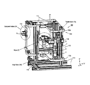
[00152] Preparation system 1000 includes, as seen in FIGS. 40A-40G, at
least the
following sub-systems and/or stations, namely, a rotation station (RS), a
weigh station (WS), a
transfer station (TS), component holders (CH), at least one manipulator (M),
at least one gripper
(G), and at least one barcode scanner (BS). Preparation system 1000 may be
considered a
Closed System Transfer Device (CSTD).
[00153] The Closed System Transfer Device (CSTD) of the present
disclosure, has been
produced for the safe transfer of potentially hazardous drugs used in the
compounding of cancer
treatments. The CSTD provides a means to make drug transfers between vials,
syringes and IV
bags without exposing the health care provider to the drug.
[00154] Early concepts for the CSTD included the possibility of
applying the CSTD
technology to an automated/robotic application. In this application, the CSTD,
vials, syringes,
etc. would be introduced to a standard pharmaceutical hood, then an automatic
or semi-automatic
28
CA 2920199 2017-07-05
CA 02920199 2016-02-02
WO 2015/017858 PCT/US2014/049609
preparation system would provide the motion, mixing, etc. required to develop
a suitable drug
for administration to a patient. The primary objective of such an approach
would be the
reliability, accuracy and repeatability afforded by an automated or semi-
automated method.
Further, the preparation system could be applied to multi-hood environments,
improving
throughput, and reducing the need for additional personnel, in particular
physicians and
pharmacologists to scrub and suit up.
Preparation system design:
[001551 The preparation system 1000 includes a number of components
that make up
subsystems which integrate into the top level preparation system. This
approach was conceived
for two reasons; it allows the preparation system 1000 to be discretized for
easier development,
and in the production case it will allow for 'plug & play' operation for
maintenance, repair and
upgrade.
1001561 The subsystems of the preparation system 1000 include, as
mentioned above, at
least a motion controller and drives; a manipulator (M); component holders (CI-
I); a
carousel/frame (1100); a gripper (0); a rotation station (RS); a transfer
station (TS); and a weigh
station (WS).
[001571 The
motion controller and drives is the overarching electronic controls system
that ties each subsystem into the control system. In this case the motion
controller is a Galil
DMC4050. There are five servo axes that are centrally controlled and can
operate independently
of each other, each driven by a 500W onboard amplifier. Additionally, the
controller provides
for additional digital and analog I/O for the control of solenoid valves,
input signals, analog
weight measurements, etc.
[00158] The
manipulator (M) includes a three mutually orthogonal axis system based on
integrated linear guide/ballscrew slides or rails 1110, 1112, 1114, in this
case Accutech USA
KM slides. Each of the slides is driven by a servo motor, with closed-loop
encoder position
feedback. Commutation of each motor is afforded by hall sensors.
1001591 A
carousel 1120 of the preparation system 1000 is responsible for the
translation
of the various compounding components, i.e. vials "V", syringes "1", vial
adapters 13, and
syringe adapters 11, from a loading position to a gripping position. The
carousel 1120 is based
29
CA 02920199 2016-02-02
WO 2015/017858 PCT/US2014/049609
on two horizontal axis gearbelts, one upper and one lower gearbelt 1122a,
1122b, respectively,
that operate in concert. Each gearbelt 1122a, 1122b is movably supported on a
series of
sprockets and the like. At least one of the gearbelts I 122a, 1122b may be
driven by a motor to
move the gearbelts 1122a, 1122b, in the manner of a conveyor belt, around
carousel 1120. In an
embodiment, as seen in FIG. 40B, a motor may be used to drive a drive shaft
1126, which drive
shaft 1126 drives a pair of driving belts, i.e., a first lower driving belt
1126a, and a second upper
driving belt I126b, wherein the driving belts 1126a, 1126b arc operatively
connected to
respective gearbelts 1122a, 1122b via respective sprockets and the like.
[00160] Component holders 1124 are affixed to each gearbelt 1122a,
1122b and are
positioned between them, and translate between them. In this manner, as the
gearbelts 1122a,
1122b are moved around carousel 1120, component holders 1124 are moved around
carousel
1120 to the various stations of preparation system 1000 (e.g,, rotation
station (RS), transfer
station (TS) and/or weigh station (WS)).
[00161] The component holders 1124, positioned at intervals along the
carousel belts or
gearbelts 1122a, 1122b, provide locations for the user to introduce
compounding components for
subsequent pull and use by a gripper (G). Each of the component holders 1124
features a pair of
jaws that are operated on by a meshed gearset and a torsion spring, so the two
jaws will operate
in coordination about the center plane, and provide sufficient engagement
pressure to maintain a
hold on the component until extraction by the gripper (G).
[00162] The gripper (G) is responsible for grasping a compounding component
in the
component holder 1124, removing it from one station or the component holder
1124, and then
placing it in another station. The gripper (G) is also responsible for
returning a component from
a station to the component holder 1124. The gripper (G) is further responsible
for effecting the
assembly and disassembly of the sub-assemblies, specifically the vial (V) and
vial adapter 13
sub-assembly, and the syringe "1" and syringe adapter 11 sub-assembly. With
reference to FIG.
400, the gripper (G) includes two jaws that can adapt to four components, by
way of
hermaphroditic jaws 1130 (including a first pair of fixed, opposed jaws 1132,
and a second pair
of translatable, opposed jaws 1134, wherein one jaw of the first pair of jaws
1132 is interposed
between the second pair of jaws 1134, and wherein a second jaw of the second
pair of jaws 1134
is interposed between the first pair of jaws 1132) thereof. The gripper (G)
features jaws 1130
CA 02920199 2016-02-02
WO 2015/017858 PCT/US2014/049609
that are coordinated by way of two gear racks and a pinion so that the jaws
always open and
close on a fixed center plane.
1001631 As illustrated in FIG. 40G, the gripper (G) functions by
translating the first pair of
jaws 1132 and the second pair of jaws 1134 relative to one another to grip a
component (e.g., a
syringe "I", a vial "V", a syringe adapter 11, a vial adapter 13, etc.) in one
of a first gripping
position (G1), a second gripping position (G2) and a third gripping position
(03). The first
gripping position (01) may be located between the first jaw of the first pair
ofjaws 1132 and the
first jaw of the second pair of jaws 1134; the second gripping position (02)
may be located
between the second jaw of the first pair of jaws 1132 and the first jaw of the
second pair of jaws
1134; and the third gripping position (G3) may be located between the second
jaw of the first
pair of jaws 1132 and the second jaw of the second pair of jaws 1134.
[001641 By way of example only, the first gripping position (01) of
gripper (G) may be
used to grip a syringe "I", the second gripping position (G2) of gripper (0)
may he used to grip a
vial adapter 13, and the third gripping position (G3) of gripper (G) may be
used to grip a syringe
adapter 11.
[001651 A rotation station (RS) of the preparation system 1000
provides, as seen in FIG.
40E, a clamping feature 1140 for the assembly of sub-assemblies and, if
desired or required, for
the rotation and/or oscillation of the various sub-assemblies. A pneumatic
actuator located
beneath the station provides 360 degrees of rotation to one of the rollers
1142 of the clamping
feature 1140. The rotation is used in the assembly of the syringe ".I" and
syringe adapter 11 by
way of a Luer lock tlu-ead. Further the rotation station (RS) is used to
rotate the vial "V" past a
scanner (optical or otherwise) for identification of the vial "V" and its
contents. The rotation
station (RS) is also equipped with two gear racks and a pinion to maintain
centrality to the center
plane.
1001661 Fluid
transfer is realized at a transfer station (TS) of the preparation system
1000.
The transfer station (TS), as seen in FIG. 40F, is configured as a linear
slide, similar to those in
the manipulator (M), and is likewise driven by a closed loop servo motor
controlled by the
motion controller. Adapters are configured to provide axial displacement of a
syringe plunger
"IP" relative to a syringe body "IB" (FIG. 40F). In operation, prior to
assembling a new or
empty syringe "I" to the syringe adapter 11, the transfer station (TS) draws a
prefill of air. Once
31
CA 02920199 2016-02-02
WO 2015/017858 PCT/US2014/049609
the compounding assembly (syringe, syringe adapter, vial adapter and vial) is
placed in the
transfer station (TS) the prefilled air is pushed into the vial adapter 13,
the entire transfer station
(TS) is inverted, and the drug (contained in the vial "V") is drawn into the
syringe "I" from the
vial "V".
Operation:
100167] The operation of the preparation system 1000 relies upon a number
of
independent sub-routines. The preparation system 1000 is capable of executing
a transfer of
fluid "L" (i.e., drug, etc.) from a vial "V" to a syringe "I", utilizing one
each of a syringe "I", a
syringe adapter 11, a vial "V" and a vial adapter 13. The program flow follows
in Table 1
below.
Table 1: basic program and function
SEQUENCE
NO. SUBROUTINE FUNCTION
VARIABL SETS POSITIONS, SPEEDS, ETC.
2 INITIAL ZEROES B,C,D,E
3 VPICK GRABS VIAL
4 VPLACE PLACES VIAL INTO ROTATION STATION
5 VAPICK GRABS VIAL ADAPTER
MAKES VIAL AND VIAL ADAPTER SUB-
6 VSUB , ASSEMBLY (VIAL SUB) BY FLUIDLY
CONNECTING (BY ROTATION) VIAL ADAPTER
AND 'VIAL
7 WEIGH MOVES VIAL SUB TO WEIGH STATION,
WEIGHS VIAL SUB, MESSAGES MASS
PLACES VIAL SUB BACK INTO VIAL ADAPTER
8 VSUBPLC
___________________________ HOLDER
9 SAP1CK GRABS SYRINGE ADAPTER
10 SAPLACE PLACES SYRINGE ADAPTER INTO ROTATION
STATION
11 SPICK GRABS SYRINGE
12 TRAN MOVES SYRINGE TO TRANSFER STATION ¨
13 PREFILL RETRACTS PLUNGER OF SYRINGE BY A
PRESET VOLUME
GRABS SYRINGE FROM TRANSFER STATION,
MOVES TO ROTATION STATION AND MAKES
14 SPEACE SYRINGE AND SYRINGE ADAPTER SUB-
ASSEMBLY (SYRINGE SUB) BY FLUIDLY
CONNECTING SYRINGE (BY ROTATION) TO
32
CA 02920199 2016-02-02
WO 2015/017858 PCT/US2014/049609
SYRINGE ADAPTER
15 SSTORE PLACES SYRINGE SUB INTO SYRINGE HOLDER
16 VAPICK GRABS VIAL SUB
17 VSUBROT RETURNS VIAL SUB TO ROTATION STATION
18 SPICK GRABS SYRINGE SUB
19 ASSEMBL ASSEMBLES SYRINGE SUB TO VIAL SUB
20 TRAN MOVES SYRINGE SUB TO TRANSFER STATION
PUSIIES AIR INTO VIAL ADAPTER,
21 FI,IJIDX 1NVERTS,RETRACTS FLUID INTO SYRINGE,
REVERTS
DISASSEMBLES VIAL SUB AND SYRINGE SUB
22 DISASS
ASSEMBLIES
23 SSTORE PLACES SYRINGE SUB INTO SYRINGE HOLDER
[001681 This sequence entails initializing the preparation system 1000,
assembling the vial
"V" and vial adapter 13, prefilling the syringe "I", assembling the syringe
adapter 11 to the
syringe "I", assembling the syringe sub to the vial sub, injecting the prefill
air into the vial
adapter 13 and drawing the prescribed volume of fluid from the vial "V".
[00169] In addition to the demonstration of feasibility of manipulating
compounding
components, the preparation system 1000 is capable of a relatively high degree
of accuracy and
repeatability in the transfer of fluids to and from the syringe "I" and vial
"V". A test comprised
of the steps described in Table 2 (below) was performed to evaluate the
accuracy and
repeatability of the preparation system.
Table 2: testing protocol for volumetric transfer accuracy and repeatability
Step Action
1 make up vial sub
2 weigh vial sub
3 record weight
introduce syringe to transfer
4 station
5 prefill air
6 remove from transfer station
7 make up syringe sub
make up assembly (syringe sub
8 to vial sub)
9 introduce to transfer station
10 push pretill air into vial sub
33
CA 02920199 2016-02-02
WO 2015/017858 PCT/US2014/049609
11 invert
12 draw fluid from vial
13 revert
14 remove from transfer station
15 break down assembly
16 weigh vial sub
17 record weight
empty syringe into graduated
18 cylinder _______
19 verify volume __
[001701 This test sequence was performed 6 times for 3 different volumes;
10, 15 and 20
ml. The results are summarized in Table 3.
Table 3: Volumetric transfer test results
__
Commanded volume: 10 1 I Commanded volume: 15 i Commanded volume: 20
Actual Actual Actual
Specimen volume Specimen volume Specimen volume
1 .
9.3 I 9.2 1 18.9
2 9.4 2 9.3 2 19.1
3 9.4 3 9.3 3 19.1
4 9.3 4 9.2 4 18.9
5 9.4 5 9.3 5 19.2
________ 6 9.7 6 ,'
, 9.6 6 18.8
______________________________________________ ,
average 9.32 average 9.29 average 19.02
_ std dev 0.05 std dev 0.05 std dev 0.15
accuracy 0.93 accuracy 0.62 accuracy 0.95
repeatability 0.99 repeatability 1.00 repeatability
0.99
[00171] For the purposes of this study, the accuracy is represented the
difference of
average reading to commanded volume, and the repeatability is represented by
the standard
deviation divided by the commanded volume.
1001721 The preparation system 1000 demonstrates the basic feasibility of
manipulating a
set of CSTD's as well as vials "V" and syringes "I". The preparation system
1000 performed
these tasks autonomously and can be operated semi-autonomously.
[001731 The preparation system 1000 further demonstrates very good
accuracy and
repeatability for the first pass assembly. Note especially the repeatability
figures, which are in
34
CA 02920199 2016-02-02
WO 2015/017858 PCT/US2014/049609
the 99% range. The repeatability is the more important of the two
measurements, as calibration
can bring the average (accuracy) into range, while maintaining the high level
of precision.
[00174] With reference to FIGS. 38A-38H, a process of operating
automated preparation
system 1000, in accordance with the principles of the present disclosure, is
provided. As seen in
FIG. 38A, at step 800, the process is initiated, At Step 802, an order is read
by preparation
system 1000, and at Step 804, an order is printed. At Step 806, it is
determined if the order
requires a medicament to be reconstituted or if the order is to be used in an
IV bag "B".
Reconstitution may he achieved by rocking, agitating and the like of the vial
"V" following
addition of diluents thereto.
[00175] If the order does not require reconstitution, then, as seen in FIG.
38B, at Step 808
a vial-syringe adapter is pulled. At Step 810a, a vial "V" containing the
medicament is pulled
and a vial cap assembly is pulled. At Step 810b, the vial cap assembly is
affixed to the vial "V".
At Step 810c, the vial-syringe adapter in connected to the vial cap assembly.
At Step 812a, a
first and a second syringe are pulled and a first syringe adapter is pulled.
At Step 812b, the order
printed at Step 804 is affixed to the first syringe, and the first syringe
adapter is attached to the
first syringe. At Step 814a, the first syringe is staged in the carousel 1100
of preparation system
1000 and at Step 814b, the first syringe is weighed (for example, in weigh
station (WS)). At
Step 816a, a plunger of the second syringe is pulled out, and at Step 816b,
the second syringe is
connected to vial-syringe adapter that was pulled at Step 808. At Step 818a,
the second syringe
is staged in the carousel 1100 of preparation system 1000, and at Step 818b,
the vial "V" is
spiked by the vial-syringe adapter. At Step 820, the first syringe, the second
syringe and the vial
are inverted.
[00176] As seen in FIG. 38C, at Step 822 a negative pressure or vacuum
is applied to the
vial "V" to extract contents from the vial "V" (e.g., medicament). At Step
824, the first syringe,
the second syringe and the vial "V" are reverted. At Step 826, the vial "V" is
unspikcd. At Step
828a, the vial "V" is weighed. If the weight of the vial "V" is not correct or
not equal to an
expected weight, at Step 828b, the vial "V" is unstaged from the carousel 1100
of preparation
system 1000, and at Step 828c, the vial "V" is set aside for disposition. If
the weight of the vial
"V" is correct or is equal to an expected weight, than at Step 830, the vial
"V" is scanned.
CA 02920199 2016-02-02
WO 2015/(117858 PCT/1152014/049609
1001771 As seen in FIG. 38D, at Step 832a, the first syringe is
scanned. If the information
from the scan does not equal the information of the order and if there is no
remaining drug, then
at Step 832b, the first syringe is unstaged from the carousel 1100 of the
preparation system 1000
and discarded. If the information from the scan does not equal the information
of the order and
if there is drug remaining, then at Step 832c, the second syringe and the vial-
syringe adapter are
unstaged from the carousel 1100 of the preparation system 1000. Then, at Step
832d, the vial-
syringe adapter is separated from the cap, at Step 832e, the vial-syringe
adapter is discarded and,
at Step 832f, the vial "V" is returned to storage. If the information from the
scan does equal the
information of the order and if there is drug remaining, then Steps 832c-832f
are once again
performed. If the information from the scan does equal the information of the
order and if there
is no drug remaining, then at Step 832g, the second syringe and the vial-
syringe adapter are
unstaged and discarded.
[00178] Simultaneously with the performance of some or all of Steps
832b-832g, as seen
in FIG. 3811, following the scanning of the first syringe at Step 832a, then
at Step 834a, if the
first syringe is not to be used in an IV bag "B" (see FIG. 37), then the first
syringe is ready.
Alternatively, at Step 834b, if the first syringe is to be used in an IV bag
"B", then an IV bag
adapter 17 is attached to the first syringe at Step 834c. Then, at Step 834d,
the IV bag "B" and
the IV bag adapter 17 are staged in the machine, at Step 834e, the IV bag
adapter 17 is spiked, at
Step 834f, the contents of the first syringe are injected into the IV bag "B",
and at Step 834g, IV
bag "B" is unspikcd. Then at Step 834h, the IV bag "B" is unstage as the IV
bag "B" is ready,
and at Step 834i, the first syringe is unstaged and discarded.
[001791
Referring back to FIG. 38A and with reference to FIG. 38E, if the order does
require reconstitution, then, at Step 836, a diluent is pulled. Then, at Step
838a, a first and a
second syringe are pulled and a first syringe adapter is pulled. At Step
8381), the order printed at
Step 804, is affixed to the first syringe, and the first syringe adapter is
attached to the first
syringe. At Step 838c, the first syringe is filled with the diluent, at Step
838d, the first syringe is
staged in the carousel 1100 of the preparation system 1000, and at Step 838e,
the first syringe is
weighed.
[00180]
Substantially simultaneously therewith, at Step 840a, a vial "V" containing
the
medicament, a vial cap and a vial-syringe adapter 13 is pulled. At Step 840b
the vial cap is
36
CA 02920199 2016-02-02
WO 2015/017858 PCT/US2014/049609
connected to the medicament vial "V" and, at Step 840b, the vial-syringe
adapter 13 is connected
to the vial cap. At Step 840c the vial-syringe adapter 13 is connected to the
vial cap. At Step
842a, the second syringe is connected to the vial-syringe adapter 13, and at
Step 842b, the
second syringe is connected to vial-syringe adapter 13 that was pulled at Step
838a. At Step
844a, the second syringe is staged in the carousel 1100 of the preparation
system 1000, and at
Step 844b the medicament vial "V" is spiked by the vial-syringe adapter 13. At
Step 846, a
negative pressure or vacuum is applied to the medicament vial while the
diluent is injected into
the medicament vial "V".
[00181] As
seen in FIG. 38F, if there needs to be a dwell time or a swirling of the vial
"V", at Step 848a, the vial "V" is removed from the carousel 1100 of the
preparation system
1000, at Step 848b, the vial "V" is taken to a dwell/swirl location, at Step
848e, the vial "V" is
then allowed to dwell or is swirled as needed, and at Step 848d, the vial "V"
is then re-staged in
the carousel 1100 of the preparation system 1000.
1001821 With
continued reference to FIG. 3814, following dwelling/swirling of the vial "V"
at steps 848a-848c, or if no dwelling/swirling is required, at Step 850, the
first syringe, the
second syringe and the vial "V" are inverted. At Step 852, a negative pressure
or vacuum is
applied to the vial "V" to extract contents from the vial "V" (e.g., the
reconstituted medicament).
At Step 854, the first syringe, the second syringe and the vial "V" are
reverted. At Step 856, the
vial "V" is unspiked. At Step 858a, the vial "V" is weighed. If the weight of
the vial "V" is not
correct or not equal to an expected weight, at Step 858b, the vial "V" is
unstaged from the
machine, and at Step 858c, the vial "V" is set aside for disposition. If the
weight of the vial "V"
is correct or is equal to an expected weight, then at Step 860, the vial "V"
is scanned.
[00183,1 As seen
in FIG. 38G, at Step 862a, the first syringe is scanned. If the information
from the scan does not equal the information of the order and if there is no
remaining drug, then
at Step 862b, the first syringe is unstaged from the carousel 1100 of the
preparation system 1000
and discarded. If the information from the scan does not equal the information
of the order and
if there is drug remaining, then at Step 862c, the second syringe and the vial-
syringe adapter are
unstaged from the carousel 1100 of the preparation system 1000. Then, at Step
862d, the vial-
syringe adapter is separated from the cap of the vial "V", at Step 862e, the
vial-syringe adapter is
discarded and, at Step 862f, the vial "V" is returned to storage. If the
information from the scan
37
CA 02920199 2016-02-02
WO 2015/017858 PCT/US2014/049609
does equal the information of the order and if there is drug remaining, then
Steps 862c-862f, are
once again performed. If the information from the scan does equal the
information of the order
and if there is no drug remaining, then at Step 862g, the second syringe and
the vial-syringe
adapter are unstaged and discarded.
[00184] Following the scanning of the first syringe at Step 862a, and
simultaneously with
the performance of some or all of Steps 862b-862g, as seen in FIG. 3814,
following the scanning
of the first syringe at Step 862a, then Steps 834a-834h may be performed, as
described above.
[00185]
Alternatively, referring back to FIG. 38A, if the order is to require the use
of an
IV bag "B", then at Step 870, an IV bag "B" is pulled, and at step 872, the
order is affixed to the
IV bag "B". Following the fixation of the order to the IV bag "B", then Steps
834a-834h may be
performed, as described above.
[00186] With
reference to FIGS. 39A-39C, a further process of operating automated
preparation system 1000, in accordance with the principles of the present
disclosure, is provided.
As seen in FIG. 39A, at step 900, the process is initiated by preparing and
loading preparation
system 1000. At Step 902, the patient regime order is reviewed, and at Step
904, the appropriate
vial is swabbed with an alcohol pad or the like.
[00187] If the
medicament in the vial "V" requires reconstitution, then at Step 906a, a
reconstitution vial adapter assembly 1 3 is attached to the lyopholized
medicament vial "V". At
Step 906b the lyopholized medicament vial "V" is loaded into a shaker device,
at Step 906c, a
diluent is injected into the lyopholized medicament vial "V", and at Step
906d, the shaker device
is activated to dissolve the powdered medicament with the diluent. At Step
906e, the vial "V" is
removed from the shaker, at Step 906f, the reconstitution vial "V" adapter
assembly 13 is
removed, and at Step 906g, the reconstitution vial adapter assembly 13 is
discarded.
[00188]
"Iltereafter or if the medicament in the vial "V" does not require
reconstitution, at
Step 908a a vial adapter assembly 13 is attached to the vial "V", and at Step
908b, the vials "V"
that are capped with the vial adapter assemblies 13 may be loaded into baskets
or trays (not
shown). The vials "V" may be locked into place by means of a twist lock
arrangement or the
like. At Step 908c, the proper loading of the vials "V" is verified.
38
CA 02920199 2016-02-02
WO 201S/017858 PCT/US2014/049609
[00189] At Step 910a, syringes are prepared by loading the syringes into
the housing of
preparation system 1000. Either 10ml or 60m1 syringes (in a compressed state)
are loaded. At
Step 910b, a cartridge having a plurality of syringe adapters is loaded into
the housing of
preparation system 1000.
[00190] As seen in FIG. 39B, at Step 912, preparation system 1000 is
configured. At Step
912a, the extraction volumes are imputed into system 1000, at Step 912b,
preparation system
1000 verifies that all the components are connected correctly, at Step 912c, a
system start is
initiated (optionally via wireless controller), at Step 912d, preparation
system 1000 registers
sequence commands, and at Step 912e, an extraction process begins.
[00191] At Step 914, the extraction process is performed. At Step 914a, as
seen in FIG
39B, a component holder (CH) selects an appropriate syringe. At Step 914b,
component holder
(CH) engages the selected syringe and secures the selected syringe into place
via clamping
mechanism or fingers. At Step 914e, component holder (CH) is slid back along
track or rails to a
syringe adapter assembly connection site (i.e., transfer station (TS)). At
Step 914d, a syringe
adapter 11 is connected to the syringe "I". At Step 914e, the syringe "I"
having the syringe
adapter 11 connected thereto is moved by component holder (CH) to an
extraction site (i.e.,
transfer station (TS)) corresponding to a loaded vial "V".
[00192] With component
holder (CH) engaging a plunger of the syringe "I", at Step 9! 5a,
component holder (CH) moves the syringe to a vial engagement access site. At
Step 915b, the
syringe "I" engages the capped vial "V", wherein a seal of the syringe adapter
11 makes
connection with a seal of the vial adapter 11. At Step 915c, component holder
(CH) continues to
advance the syringe "I" toward the vial "V" until a seal or stopper of the
vial "V" is engaged by a
seal of the vial adapter 11 and until a sealed connection is established
between the vial "V" and
the syringe "I". At Step 916, the extraction process begins.
[00193] As seen in
FIG. 39C, at Step 916a, component holder (CH) withdraws the plunger
relative to the syringe barrel of the syringe "1" to begin withdrawing fluid
from the vial "V" and
facilitate aspiration of fluid into the vial "V." At Step 916b, component
holder (CH) advances
the plunger relative to the barrel of the syringe "I" to inject fluid back
into the vial "V". At step
916c, component holder (CH) once again withdraws the plunger relative to the
barrel of the
syringe "I" to again withdraw fluid from the vial "V" to complete the transfer
of drug from the
39
CA 02920199 2016-02-02
WO 2015/017858 PCT/US2014/049609
vial "V" to the syringe "I". At Step 916d, the syringe "1" filed with the
medicament is
disengaged from the vial adapter 13. At Step 916e, component holder (CH) moves
away from
the vial "V" such that the seal of the vial adapter 13 is disengaged from the
seal of the vial "V"
and the seal of the syringe adapter 11 is disengaged from the seal of the vial
adapter 13.
[00194] At Step 918, as seen in FIG. 39C, component holder (CH), holding
the filled
syringe, is moved horizontally away from a tray (not shown) of vials "V". At
Step 920, loading
arm 714 may disengage and release the filled syringe. It is contemplated that
at least one tray
(not shown) may be provided within the carousel 1100 of the preparation system
1000. Each
tray may be configured to store vials "V" prior to and following manipulation
by component
holders (CH) to the various stations about carousel 1100.
[00195] Alternatively, at Step 922a, as seen in FIG. 39C, component
holder (CH) reorients
the filled syringe "I" to align a nose of the syringe with an access terminal
of an IV bag "B". At
Step 922b, component holder (CH) moves the nose of the syringe into the access
terminal of the
IV bag "R". With the nose of the syringe connected to the access terminal of
the IV bag "B", at
Step 922c, component holder (CH) actuates the plunger of the syringe to inject
the fluid of the
syringe into the IV bag "B". At Step 922d, component holder (CH) disengages
the syringe from
the access terminal of the IV bag "B".
[00196] At Step 924, component holder (CH) disengages the used and
empty syringe and
drops the used and empty syringe to a disposal tray. The entire process may be
repeated as many
times as necessary.
[00197] With reference to FIGS. 41A-42C, the process of operating
automated preparation
system 1000 of FIGS. 38A-39C has been annotated to illustrate which sub-
systems and/or
stations, namely, a rotation station (RS), a weigh station (WS), a transfer
station (TS),
component holders (CH), at least one manipulator (M), at least one gripper
((.1), and at least one
bareode scanner (BS), are used to achieve or accomplish the various steps and
the like thereof
[00198] Preparation system 1000 may include an error trapping protocol,
wherein
preparation system 1000 preforms and cheek and confirmation, at various stages
of the process,
to ensure that the correct components are being manipulated and that the
correct materials (i.e.,
drugs) are being reconstituted and/or reformulated.
CA 02920199 2016-02-02
WO 2015/017858 PCT/US2014/049609
[00199] For
example, as described above, the gripper (G) includes jaws that are opened
closed to grasp and release vials "V", vial adapters 13, syringes "I" and
syringe adapters 11. The
jaws of gripper (G) are coordinated by way of a single pinion engaged in two
racks, one per jaw
set. The relative distance between the jaw sets can be measured directly with
a linear measuring
device, such as a linear potentiometer or a Linear Variable Displacement
Transformer (LVDT),
or by an angular measuring device, such as a rotary potentiometer or encoder.
The pinion can be
driven by an electric motor which can have either an encoder or resolver
integrated into the main
shaft.
[00200] In
use, the distance measurements made by the gripper (G) can be used to qualify
and quantify the component (e.g., syringe adapter 11, vial adapter 13) being
grasped. The error
trapping protocol can, for instance determine that the size of the component
being grasped is not
consistent with the programmed component size. The error trapping protocol can
flag the
operator to clear the anomaly, or it can shut down the compounding operation.
Likewise, if a
component has become rnis-oriented within the equipment, the size will likely
be reported as
inconsistent with the expected size. The measurement can be used to quantify
the diameter of a
syringe, as well. With the diameter of the syringe known, the error trapping
protocol can
anticipate the required stroke required in the transfer station (TS) to
accommodate the required
volume.
[00201] A same or similar error trapping protocol can be implemented for
the rotation
station (RS) and for each of the component holders (CFI).
[00202] It will be understood that various modifications may be made to
the embodiments
disclosed herein. Therefore, the above description should not be construed as
limiting, but
merely as exemplifications of preferred embodiments. Those skilled in the art
will envision
other modifications within the scope and spirit of the claims appended
thereto.
41