Note: Descriptions are shown in the official language in which they were submitted.
87904-26
METHOD AND APPARATUS FOR TREATING DERMAL MELASMA
[0001]
FIELD OF THE DISCLOSURE
[0002] Exemplary embodiments of the present disclosure relates to
treating pigmented
tissue, and more particularly to methods and apparatus for treating dermal
melasma.
BACKGROUND INFORMATION
[0003] Melasma is a skin disorder of unknown etiology that causes a
blotchy
hyperpigmentation, often in the facial area. This condition is more common in
women than in
men. Although the specific cause(s) of melasma may not be well-understood, the
pigmented
appearance of melasma can be aggravated by certain conditions such as
pregnancy, sun
exposure, certain medications, such as, e.g., oral contraceptives, hormonal
levels, genetics, etc.
[0004] Exemplary symptoms of melasma include dark, irregularly-shaped
patches or
macules, which are commonly found on the upper cheek, nose, upper lip, and
forehead. These
patches often develop gradually over time. Melasma does not appear to cause
any other
symptoms, nor have other detrimental effects, beyond the cosmetic
discoloration.
[0005] Unlike many pigmented structures that are typically present in
the epidermal
region of skin (i.e., at or near the skin surface), dermal (or deep) melasma
is often characterized
by widespread presence of melanin and melanophages (including, e.g.,
excessively-pigmented
cells) in portions or regions of the underlying dermis. Accordingly, treatment
of dermal
melasma (e.g., lightening of the appearance of darkened pigmented regions) can
be particularly
challenging because of the presence of the greater difficulty in accessing and
affecting such
pigmented cells and structures located deeper within the skin. Accordingly,
conventional skin
rejuvenation treatments such as facial peels (laser or
1
Date Recue/Date Received 2020-12-04
CA 02920858 2016-02-09
WO 2015/021462 PCT/US2014/050518
chemical), dermabrasion, topical agents, and the like, which primarily affect
the overlying
epidermis, may not be effective in treating dermal melasma.
100061 It has been observed that application of light or optical
energy of certain
wavelengths can be strongly absorbed by pigmented cells, thereby damaging
them,
__________________________ However, an effective treatment of (let mai
melasma using optical energy introduces several
obstacles. For example, pigmented cells in the dermis must be targeted with
sufficient
optical energy of appropriate wavelength(s) to disrupt or damage them, which
may release
or destroy some of the pigmentation and reduce the pigmented appearance.
However, such
energy can be absorbed by pigment (e.g., chromophores) in the overlying skin
tissue, such
as the epidermis and upper dermis. This near-surface absorption can lead to
excessive
damage of the outer portion of the skin, and insufficient delivery of energy
to the deeper
dermis to affect the pigmented cells therein.
[0007] Fractional approaches have been developed that involve
application of
optical energy to small, discrete locations on the skin that are separated by
healthy tissue to
facilitate healing. However, such fractional approaches may "miss" many of the
pigmented
cells in the dermis, and effective targeting of such deeper cells may again
result in excessive
damage to the nearby healthy tissue.
100081 Therefore, it may be desirable to provide method and apparatus
that can
effectively target pigmented cells in the dermis and reduce the appearance of
miasma,
without generating excessive damage to healthy skin tissue or producing other
undesirable
side effects.
SUMMARY OF EXEMPLARY EMBODIMENTS OF THE DISCLOSURE
[0009] Exemplary embodiments of methods and apparatus can be provided
for a
treatment of dermal melasma and other pigmented defects within the dermis,
e.g., to lighten
the dark pigmented appearance of dermal melasma. The exemplary embodiments of
the
methods and apparatus can facilitate selective energy absorption by, and
thermal damage to,
pigmented structures within the dermis by focusing highly-convergent
electromagnetic
radiation (EMR), e.g., optical energy, having appropriate wavelengths onto the
pigmented
regions within the dermis. This exemplary procedure can result in heating
and/or thermal
damage to the pigmented regions, thereby disrupting the pigment and lightening
the
appearance of the skin, while avoiding unwanted thermal damage to surrounding
unpigmented tissue and the overlying tissue.
2
CA 02920858 2016-02-09
WO 2015/021462 PCT/US2014/050518
100101 According to exemplary embodiments of the present disclosure,
an apparatus
can be provided that can include a radiation emitter arrangement configured to
emit EMR,
and an optical arrangement configured to direct the EMR onto the skin being
treated and
focus it to a focal region within the dermis. A plate that is substantially
optically
transparent to the EMR can be provided on a portion of the apparatus that is
configured to
contact the surface of the skin being treated. Such plate can stabilize the
pliable skin tissue
and facilitate better control of the depth of the focal region below the skin
surface. A lower
surface of the plate can be substantially planar, or it may optionally be
slightly convex or
concave. The apparatus can further include a housing or handpiece that can
contain these
components and facilitate manipulation of the apparatus during its use.
100111 The EMR emitter can include, e.g., a waveguide or optical fiber
configured
to direct EMR from an external source, an EMR source such as one or more diode
lasers, a
fiber laser, or the like. If the emitter arrangement includes a source of EMR,
it can
optionally include a cooling arrangement configured to cool the EMR source(s)
and prevent
.. overheating of the source(s). A control arrangement can be provided to
control the
operation of the emitter arrangement including, e.g., turning the EMR source
on and off,
controlling or varying the power output of the EMR source, etc.
[0012] The EMR can have a wavelengths that is preferably greater than
about 600
nm, e.g., between about 625 nm and about 850 nm, or between about 650 nm and
750 nm.
Smaller wavelengths (e.g., less than about 600 nm) can be scattered
significantly within the
skin tissue, thereby having insufficient penetration depth to reach portions
of the dermal
layer with sufficient fluence and focus. Such smaller wavelengths can also
have a very high
melanin absorbance, which can generate increased EMR absorption by melanin in
the
overlying epidermal region and unwanted thermal damage to the surface region.
Such
smaller wavelengths can also have a higher absorbance by hemoglobin, a
competing
chromophore, which may be present in blood vessels. Significant EMR absorption
by
hemoglobin can cause unwanted thermal damage to such vessels. Absorbance of
EMR by
melanin generally decreases with increasing wavelength, so wavelengths longer
than about
850 nm may not be sufficiently absorbed by the dermal melanin to cause local
heating and
disruption of the pigmented structures.
[0013] The exemplary apparatus can include an optical arrangement
configured to
focus the EMR in a highly convergent beam. For example, the optical
arrangement can
include a focusing or converging lens arrangement having a numerical aperture
(NA) of
3
CA 02920858 2016-02-09
WO 2015/021462 PCT/US2014/050518
about 0.5 or greater, e.g., between about 0.5 and 0.9. The correspondingly
large
convergence angle of the EMR can provide a high fluence and intensity in the
focal region
of the lens (which can be located within the dermis) with a lower fluence in
the overlying
tissue above the focal region. Such focal geometry can help reduce unwanted
heating and
thermal damage in the overlying tissue above the pigmented dermal regions. The
exemplary optical arrangement can further include a collimating lens
arrangement
configured to direct EMR from the emitting arrangement onto the focusing lens
arrangement.
[0014] The exemplary optical arrangement can be configured to focus
the EMR to a
focal region having a width or spot size that is less than about 200 gm
(microns), for
example, less than 100 gm, or even less than about 50 pm, e.g., as small as 10
gm. Such
spot size can be selected as a balance between being small enough to provide a
high fluence
or intensity of EMR in the focal region (to effectively irradiate pigmented
structures in the
dermis), and being large enough to facilitate irradiation of large
regions/volumes of the skin
tissue in a reasonable treatment time.
[0015] The exemplary optical arrangement can also be configured to
direct the focal
region of the EMR onto a location within the dermal tissue that is at a depth
below the skin
surface of between about 120 ium and 400 gm, e.g., between about 150 gm and
300 gm.
Such exemplary depth range can correspond to typical observed depths of
pigmented
regions in skin that exhibits dermal melasma. This focal depth can correspond
to a distance
from a lower surface of the apparatus configured to contact the skin surface
and the location
of the focal region.
[0016] In further exemplary embodiments of the present disclosure, the
positions
and/or orientations of the EMR emitter arrangement and/or components of the
optical
arrangement can be controllable or adjustable relative to one another, such
that the path of
the EMR can be varied. Such variation in the path of the EMR can provide
corresponding
variations in the depth, width, and/or location of the focal region within the
dermis, and can
facilitate treatment of larger volumes of the skin tissue when the apparatus
is translated with
respect to the skin. Such relative movement of these components can also
facilitate
movement of the focal region within the skin tissue when the apparatus is held
stationary
relative to the skin, e.g., to treat larger regions of the skin without moving
the overall
apparatus.
4
CA 02920858 2016-02-09
WO 2015/021462 PCT/US2014/050518
[0017] In still further exemplary embodiments of the present
disclosure, the
exemplary focusing lens arrangement can include a plurality of micro-lenses,
e.g., convex
lenses, piano-convex lenses, or the like. Each of the micro-lenses can have a
large NA (e.g.,
between about 0.5 and 0.9). The micro-lenses can be provided in an array,
e.g., a square or
hexagonal array, to produce a plurality of focal regions in the dermal tissue
in a similar
pattern. A width of the micro-lenses can be small, e.g., between about lmm and
3 mm
wide. Micro-lenses 300 that are slightly wider or narrower than this can also
be provided in
certain embodiments. In yet further exemplary embodiments of the present
disclosure, the
micro-lenses can include cylindrical lenses, for example, convex cylindrical
lenses or piano-
convex cylindrical lenses. A width of such cylindrical micro-lenses can be
small, e.g.,
between about 1mm and 3 mm wide. A length of the cylindrical micro-lenses can
be
between, e.g., about 5 turn and 5 cm.
[0018] The exemplary radiation emitter arrangement and/or the
exemplary optical
arrangement can be configured to direct a single wide beam of EMR over the
entire array of
such micro-lenses or a portion thereof to simultaneously generate a plurality
of focal
regions in the dermis. In further exemplary embodiments, radiation emitter
arrangement
and/or the optical arrangement can be configured to direct a plurality of
smaller beams of
EMR onto individual ones of the micro-lenses. Such multiple beams can be
provided, e.g.,
by using a plurality of EMR sources (such as laser diodes), a beam splitter,
or a plurality of
waveguides, or by scantling a single beam over the individual micro-lenses. If
cylindrical
micro-lenses are provided, one or more beams of EMR can be scanned over such
cylindrical
lenses, e.g., in a direction parallel to the longitudinal axis of such
cylindrical lenses.
[0019] In yet another exemplary embodiment of the present disclosure,
the
exemplary cylindrical or spherical micro-lenses can different NA values,
different sizes or
radii, and/or different effective focal lengths than one another. Such
variations in the
geometry and optical properties of the micro-lenses can facilitate irradiation
of larger
volumes of the dermis.
[0020] The plate configured to contact the skin surface can optionally
be provided
as part of the focusing lens arrangement, e.g., it can be formed as the lower
surface of a
plano-convex lens or a plurality of such micro-lenses. The plate can
optionally be cooled,
e.g., by pre-cooling it prior to use or with an active cooling arrangement
(e.g. a Peltier
device, a conductive cold conduit, or the like). Such cooling can help protect
the epidermis
and upper portions of the dermis from unwanted thermal damage. An optical gel
or the like
5
CA 02920858 2016-02-09
WO 2015/021462 PCT/US2014/050518
(e.g. glycerol or a similar substance) can optionally be provided between the
plate and the
skin surface to reduce an optical index mismatch between the plate and the
skin, thereby
improving transmission of the EMR into the skin.
[0021] In further exemplary embodiments of the present disclosure, the
exemplary
apparatus can include one or more sensors configured to detect contact of the
apparatus with
the skin and/or speed of the apparatus over the skin surface during use. Such
exemplary
sensors can be coupled to a control arrangement of the EMR emitter or source,
and adapted
to generate signals capable of varying properties of the EMR, e.g., by varying
the power
emitted by the emitter arrangement based on the translational speed of the
apparatus, by
turning off the source(s) of EMR when the apparatus is stationary relative to
the skin
surface or moved away from the skin, etc. Such sensors and control
arrangements can
improve safety of the apparatus by preventing excessive irradiation and
unwanted thermal
damage to the skin.
[0022] It can be preferable to limit irradiation time (dwell time) of
a particular
location in the dermis to a short period of time, e.g., about 1-2 milliseconds
or less. Such
short dwell times can be achieved, e.g., by configuring the radiation emitter
arrangement to
provide discrete pulses of EMR. The exemplary interval between such pulses of
EMR can
be, e.g., on the order of about 50 milliseconds or more to provide spatial
separation between
regions of the dermis irradiated by successive pulses when the apparatus is
translated over
the skin. Short dwell times can also be achieved by translating the apparatus
over the skin
during use, e.g., at speeds of about 1 cm/s or greater, such that the focal
region does not
remain on a particular location in the dermis for longer than a few
milliseconds. In further
embodiments, optional sensors can also be used to control the EMR emitted by
the
apparatus to avoid longer local dwell times.
[0023] The power output of the exemplary emitter arrangement can be
selected to
provide a local fluence within each focal region that is between about 10-1000
J/cm2 for
EMR having a wavelength of about 650 mn, e.g., between about 50-500 J/cm2. The
estimated fluence within the focal region can be related to the spot size,
local dwell time,
and total beam power using conventional equations. Larger or smaller local
fluence values
can also be used when using faster or slower scan speeds and/or with shorter
or longer dwell
times, respectively. The fluence can be somewhat lower for shorter wavelengths
(which is
6
87904-26
more readily absorbed by melanin) or larger for longer wavelengths, for which
EMR absorption
by melanin is weaker.
[0024] In further embodiments of the disclosure, a method can be
provided for treating
dermal melasma that includes focusing at least one beam of EMR onto at least
one focal region
within the dermis, to generate selective absorption by pigmented cells or
structures within the
dermis while avoiding unwanted heating and damage to unpigmented tissue and
overlying tissue.
The EMR wavelength used, focal properties (e.g., NA value, focal depth, spot
size), scanning
speeds and/or pulsed EMR properties, EMR beam power, fluence within the focal
region(s), etc.,
can be provided in accordance with the various embodiments described herein.
[0024A] In accordance with another aspect, an apparatus is provided for
selectively
affecting pigmented regions in a layer of a skin tissue comprising:
a radiation arrangement configured to emit at least one electromagnetic
radiation (EMR)
beam having a wavelength between 600 and 850 nm;
an optical arrangement configured to direct and focus the EMR beam as a
convergent
beam into at least one focal region having a width less than 100 i.tm within a
dermis layer of a skin
tissue containing pigmented and unpigmented regions; and
a controller configured to control:
¨ a power output of the radiation arrangement to provide a
fluence of the EMR beam
to be between 10 and 1000 J/cm2 in the at least one focal region; and
¨ at least one of the radiation arrangement or the optical arrangement to scan
the at
least one EMR beam over an entirety of a treatment area of the skin tissue
within
the dermis layer, wherein a dwell time of the at least one EMR beam at the at
least
one focal region is 2 ms or less, and wherein a scan speed of the at least one
EMR
beam is between 5 mm/s to 5 cm/s; and
wherein the EMR beam is configured to provide selective energy absorption by
the
pigmented regions within the treatment area, and damages the pigmented regions
while
preventing damage to the unpigmented regions of the skin tissue within the
treatment area of the
skin tissue and overlaying the at least one focal region.
[0024B] In accordance with another aspect, a method is provided for
improving the
appearance of dermal melasma comprising:
7
Date Recue/Date Received 2023-06-21
87904-26
generating, by a radiation arrangement, at least one electromagnetic radiation
(EMR) beam
having a wavelength between 600 nm and 850 nm; and
focusing, by an optical arrangement, the at least one EMR beam into a
plurality of focal
regions within a dermis layer of a skin tissue containing pigmented and
unpigmented regions,
wherein a width of at least one of the plurality of focal regions is less than
100 microns;
scanning the plurality of focal regions of the at least one EMR beam over an
entirety of a
treatment area of the skin tissue within the dermis layer, wherein a dwell
time of the least one
EMR beam at the plurality of focal regions is 2 ms or less; and
controlling, using a controller:
¨ a power output of the radiation arrangement to provide a fluence of the EMR
beam
within each focal region of the plurality of focal regions between 10 and 1000
J/cm2 and
¨ at least one of the radiation arrangement or the optical
arrangement to scan the
EMR beam at a scan speed between 5 mm/s and 5 cm/s.
wherein the at least one EMR beam provides selective energy absorption by the
pigmented
regions of the skin tissue within the treatment area and damages the pigmented
regions while
preventing damage to the unpigmented regions of the skin tissue within the
plurality of focal
regions and an epidermal layer of the skin tissue overlying the plurality of
focal regions
[0024C] In accordance with another aspect, a cosmetic method is
provided for selectively
damaging pigmented cells within a dermis of a skin tissue. The method
comprises focussing at
least one electromagnetic radiation into a particular volume of the skin
tissue containing the
pigmented cells to irradiate them. A width of a focal region of the at least
one electromagnetic
radiation is less than 200 microns. A duration of irradiation is less than
about 2 milliseconds.
Unpigmented cells of the skin tissue that are at least one of overlying,
underlying, or adjacent to
the focal region are not damaged, and pigmented cells within the dermis of the
skin tissue are at
least one of thermally damaged or disrupted. The method also comprises
scanning the at least one
electromagnetic radiation with respect to the skin tissue at a scan speed
between 5 mm/s and 5
cm/s.
7a
Date Recue/Date Received 2023-06-21
87904-26
[0024D1 In accordance with another aspect, a method is provided,
comprising
generating, by a radiation arrangement, at least one electromagnetic radiation
(EMR) beam
having a wavelength between 600 nm and 850 nm;
focusing, by an optical arrangement including at least one lens having a
numerical aperture
between 0.5 and 0.9, the at least one EMR beam into a plurality of focal
regions within a dermis
layer of a skin tissue containing pigmented and unpigmented regions, wherein
the at least one
EMR beam provides selective energy absorption by the pigmented regions of the
skin tissue
within a treatment area, and wherein a width of at least one of the plurality
of focal regions is less
than 100 gm;
scanning the plurality of focal regions of the at least one EMR beam over the
treatment
area of the skin tissue within the dermis layer, wherein a dwell time of the
least one EMR beam at
the plurality of focal regions is 2 ms or less; and
controlling, using a controller:
¨ the radiation arrangement to emit the at least one EMR beam at a power
output
between 0.004 to 79 W; and
¨ at least one of the radiation arrangement or the optical arrangement to
scan the at
least one EMR beam at a scan speed of 5 mm/s to 5 cm/sec,
wherein the at least one EMR beam damages the pigmented regions of the skin
tissue
within the plurality of focal regions while preventing damage to the
unpigmented regions of the
skin tissue within the plurality of focal regions and an epidermal layer of
the skin tissue overlying
the plurality of focal regions.
[0024E] In accordance with another aspect, a method is provided,
comprising:
continuously emitting, using a radiation arrangement including at least one
continuous
wave laser, at least one continuous electromagnetic radiation (EMR) beam
having a wavelength
.. between 600 nm to 850 nm;
contacting at least one portion of an apparatus to a surface of a skin tissue;
directing and focusing, using an optical arrangement including a lens having a
numerical
aperture between 0.5 and 0.9, the at least one continuous EMR beam as a
converging beam into a
plurality of focal regions at one or more depths within a dermis layer of the
skin tissue, wherein a
width of at least one of the plurality of focal regions is less than 100 gm;
7b
Date Recue/Date Received 2023-06-21
87904-26
scanning the plurality of focal regions of the at least one continuous EMR
beam over a
treatment area of the skin tissue within the dermis layer which includes
pigmented and
unpigmented regions; and
controlling, using a controller:
¨ the radiation arrangement to emit the at least one continuous EMR beam at
a power
output between 0.004 to 79 W; and
¨ at least one of the radiation arrangement or the optical
arrangement to scan the at
least one continuous EMR beam at a scan speed of 5 mm/s to 5 cm/sec,
wherein a dwell time of the least one continuous EMR beam at the plurality of
focal
regions is 2 ms or less.
wherein the at least one continuous EMR beam provides (i) a selective energy
absorption
by the pigmented regions of the skin tissue within the treatment area, and
(ii) a thermal damage of
the pigmented region while preventing a thermal damage to the unpigmented
regions of the skin
tissue within the plurality of focal regions and overlying the plurality of
focal regions.
[0024F] In accordance with another aspect, an apparatus is provided for
selectively
affecting a pigmented region in a layer of a skin tissue, comprising
a radiation arrangement including at least one laser configured to emit at
least one
electromagnetic radiation (EMR) beam;
an optical arrangement including at least one lens configured to direct and
focus the at
least one EMR beam as a convergent beam into at least one focal region having
a width less than
100 gm within a dermis layer of the skin tissue containing pigmented and
unpigmented regions;
a sensor configured to detect a speed of a translation of the apparatus over a
surface of the
skin tissue, and provide signals which are based on the detected speed to
affect at least one
property of the at least one EMR beam; and
a controller configured to receive the signals from the sensor and to control:
¨ a power output of the radiation arrangement to provide a
fluence of the at least one
EMR beam between 10 and 1000 J/cm2 in the at least one focal region when the
signals indicate that the apparatus is in motion over the surface of the skin
tissue;
and
7c
Date Recue/Date Received 2023-06-21
87904-26
¨ at least one of the radiation arrangement or the optical
arrangement so as to scan
the at least one EMR beam over an entirety of a treatment area of the skin
tissue
within the dermis layer, wherein a dwell time of the at least one EMR beam at
the
at least one focal region is 2 ms or less.
wherein in operation, the at least one EMR beam (i) provides selective energy
absorption
by the pigmented regions of the skin tissue within the treatment area, and
(ii) damages the
pigmented regions while preventing damage to the unpigmented regions of the
skin tissue within
the treatment area and within an epidermal layer of the skin tissue overlying
the at least one focal
region.
[0025] These and other objects, features and advantages of the present
disclosure will
become apparent upon reading the following detailed description of exemplary
embodiments of
the present disclosure, when taken in conjunction with the appended drawings.
BRIEF DESCRIPTION OF THE DRAWINGS
[0026] Further objects, features and advantages of the present
disclosure will become
apparent from the following detailed description taken in conjunction with the
accompanying
figures showing illustrative embodiments, results and/or features of the
exemplary embodiments
of the present disclosure, in which:
[0027] FIG. 1 A is a side view of an illustration of one or more
radiations being focused
into pigmented dermal tissue;
[0028] FIG. TB is an exemplary absorbance spectrum graph for melanin;
[0029] FIG. 1C is an exemplary absorbance spectrum graph for
oxygenated and
deoxygenated hemoglobin;
[0030] FIG. 2 is a cross-sectional side view of a diagram of an
exemplary apparatus in
accordance with exemplary embodiments of the present disclosure;
[0031] FIG. 3 A is a schematic side view of an arrangement of micro-lenses
that can be
used with certain exemplary embodiments of the present disclosure;
[0032] FIG. 3B is a schematic top view of a first exemplary
arrangement of the micro-
lenses shown in FIG. 3A;
7d
Date Recue/Date Received 2023-06-21
CA 02920858 2016-02-09
WO 2015/021462 PCT/US2014/050518
[0033] FIG. 3C is a schematic top view of a second exemplary
arrangement of the
micro-lenses shown in FIG. 3A;
[0034] FIG. 3D is a schematic top view of an exemplary arrangement of
cylindrical
micro-lenses that can be used with certain exemplary embodiments of the
present
disclosure;
[0035] FIG. 3E is a schematic angled view of the exemplary arrangement
of
cylindrical micro-lenses shown in FIG. 3D;
[0036] FIG. 3F is a schematic side view of a further exemplary
arrangement of the
micro-lenses that can be used with further exemplary embodiments of the
present
disclosure;
[0037] FIG. 4 is a schematic cross-sectional side view of a further
exemplary
apparatus in accordance with still further exemplary embodiments of the
present disclosure;
[0038] FIG. 5 is an exemplary biopsy image of pig skin tattooed with a
melanin
solution to simulate the effects of dermal melasma;
[0039] FIG. 6A is an exemplary surface image of a region of pig skin
tattooed with
a melanin solution to simulate the effects of dermal melasma; and
100401 FIG. 6B is an exemplary surface image of the tattooed region of
pig skin
shown in FIG. 6A after it has been irradiated with focused electromagnetic
radiation in
accordance with exemplary embodiments of the present disclosure.
[0041] Throughout the drawings, the same reference numerals and characters,
unless otherwise stated, are used to denote like features, elements,
components, or portions
of the illustrated embodiments. Similar features may thus be described by the
same
reference numerals, which indicate to the skilled reader that exchanges of
features between
different embodiments can be done unless otherwise explicitly stated.
Moreover, while the
present disclosure will now be described in detail with reference to the
figures, it is done so
in connection with the illustrative embodiments and is not limited by the
particular
embodiments illustrated in the figures. It is intended that changes and
modifications can be
made to the described embodiments without departing from the true scope and
spirit of the
present disclosure as defined by the appended claims.
8
CA 02920858 2016-02-09
WO 2015/021462 PCT/US2014/050518
DETAILED DESCRIPTION OF EXEMPLARY EMBODIMENTS
100421 According to certain exemplary embodiments of the present
disclosure,
devices and methods can be provided for treating dermal (or deep) melasma. For
example,
electromagnetic radiation (EMR) such as, e.g., optical energy) at one or more
particular
wavelengths can be focused into the dermis, where the EMR can optionally be
pulsed
and/or scanned, such that the radiation is selectively absorbed by the
pigmented cells in the
dermis. Such absorption of the energy, together with the focusing geometry and
scanning
parameters, can selectively damage or destroy many of the pigmented cells
while reducing
or avoiding damage to surrounding unpigmented cells and to the overlying
epidermis.
[0043] An exemplary schematic side view of a section of skin tissue is
shown in
FIG. 1. The skin tissue includes a skin surface 100 and an upper epidermal
layer 110, or
epidermis, which can be, e.g., about 60-120 1.tm thick in the facial region.
The dermis can
be slightly thicker in other parts of the body. The underlying dermal layer
120, or dermis,
extends from below the epidermis 110 to the deeper subcutaneous fat layer (not
shown).
Skin exhibiting deep or dermal melasma can include a population of pigmented
cells or
regions 130 that contain excessive amounts of melanin.
[0044] In exemplary embodiments of the present disclosure, an
electromagnetic
radiation (FMR) 150 (e_g_, optical energy) can be focused into one or more
focal regions
160 that can be located within the dermis 120. The EMR 150 can be provided at
one or
more appropriate wavelengths that can be absorbed by melanin. The EMR
wavelength(s)
can be selected to enhance selective absorption by the pigmented regions 130
in the dermis
120.
100451 For example, a graph of an exemplary absorption spectrum for
melanin is
shown in the graph of FIG. 1B. The absorption of EMR by melanin is observed to
reach a
peak value at a wavelength of about 350 nm, and then decreases with increasing
wavelength. Although absorption of the EMR by the melanin facilitates heating
and/or
disruption of the melanin-containing regions 130, a very high melanin
absorbance can result
in high absorption by pigment in the epidermis 110 and reduced penetration of
the EMR
into the dermis 120. As illustrated in FIG. 1B, melanin absorption at EMR
wavelengths that
are less than about 500 nm are relatively high, such that wavelengths less
than about 500
rim may not be suitable for penetrating sufficiently into the dermis 120 to
heat and damage
or disrupt pigmented regions 130 therein. Such enhanced absorption at smaller
wavelengths
9
CA 02920858 2016-02-09
WO 2015/021462 PCT/US2014/050518
can result in unwanted damage to the epidermis 110 and upper (superficial)
portion of the
dermis 120, with relatively little unabsorbed EMR passing through the tissue
into the deeper
portions of the dermis 120.
[0046] Another significant chromophore observed in skin tissue is
hemoglobin,
which is present in blood vessels. Hemoglobin can be oxygenated (Hb02) or
deoxygenated
(Hb), where each form of Hemoglobin may exhibit slightly different EMR
absorption
properties. For example, exemplary absorption spectra for both Hb and Hb02 are
shown in
the graph of FIG. 1C. These spectra indicate a high absorption coefficient for
both Hb and
Hb02 at EMR wavelengths less than about 600 nm, with the absorbance decreasing
significantly at higher wavelengths. Strong absorption of EMR directed into
skin tissue by
hemoglobin (Hb and/or Hb02) can result in heating of the hemoglobin-containing
blood
vessels, resulting in unwanted damage to these vascular structures and less
EMR available
to be absorbed by the melanin.
[0047] Accordingly, it can be preferable to use EMR having wavelengths
greater
than 600 nm in certain exemplary embodiments of the present disclosure, e.g.,
about 625
nm or greater. Such wavelengths can increase selectivity of EMR absorption in
the dermis,
e.g., by reducing competing absorption by hemoglobin, and by also avoiding
excessive
absorption of the EMR by epidermal melanin (as described above) such that the
EMR can
penetrate into the dermis 120 and target pigmented regions 130 therein.
100481 For example, longer wavelengths of EMR tend to be scattered more
easily by
the non-homogeneous structure of skin tissue. Such scattering can reduce the
effective
penetration depth of EMR directed onto the tissue, and also inhibit focusing
of the EMR
beam 150 into a small focal region 160 as described herein. Further, the
absorbance of
melanin continues to decrease with increasing wavelength, as indicated in the
graph of FIG.
1B. Thus, EMR having wavelengths less than about 750 nm or 850 nm be well-
focused in
tissue to generate sufficient local intensity within the dermis 120, as well
as sufficiently
absorbed by dermal melanin to disrupt and/or damage pigmented regions 130.
[0049] Accordingly, exemplary embodiments of the present disclosure,
it is possible
to provide or use EMR having one or more wavelengths between about 600 min and
about
850 nm, e.g., between about 625 nm and about 800 nm, which is mostly in the
visible range
of light. In certain embodiments, the wavelength can be between about 650 nm
and 750
nm. In further exemplary embodiments of the present disclosure, wavelengths
less than
CA 02920858 2016-02-09
WO 2015/021462 PCT/US2014/050518
about 600 nm or greater than about 850 nm may be used, although EMR having
such
wavelengths may be provided with sufficient focusing and/or appropriate power
and
fluence, as described herein, to achieve sufficient quantity and selectivity
of absorption by
melanin in the dermis.
[0050] In further exemplary embodiments of the present disclosure, an
apparatus
200, schematically illustrated in a diagram of Fig. 2, can be provided to
treat dermal
melasma in skin using EMR 150, e.g., optical energy. For example, the
apparatus 200 can
include a radiation emitter arrangement 210, and an optical arrangement that
can be
provided between the radiation emitter arrangement 210 and the target tissue
to be treated.
For example, the optical arrangement can include a first lens arrangement 220
and a second
lens arrangement 230. These exemplary components can optionally be provided in
a
handpiece 250 or other housing or enclosure. The apparatus 200 can further
include a plate
240 having a lower surface configured to contact the surface 100 of the skin
tissue being
treated. An actuator arrangement 260 can be provided to control the operation
of the
apparatus 200, e.g., to activate and/or turn off the emitter arrangement 210,
control or adjust
certain operational parameters of the apparatus 200, etc. A power source (not
shown) for
the radiation emitter arrangement 210 can be provided. For example, the power
source can
include a battery provided within the handpiece 250, an electrical cord or
other conductive
connection provided between the emitter arrangement 210 and an external power
source
(e.g. an electrical outlet or the like), etc.
[0051] The radiation emitter arrangement 210 can include, e.g., one or
more laser
diodes, optical fibers, waveguides, or other components configured to generate
and/or emit
EMR 150 and direct it toward or onto the optical arrangement 220, e.g., onto
the first lens
arrangement 220. In certain exemplary embodiments of the present disclosure,
the radiation
emitter arrangement 210 can include one or more laser diodes that emit optical
radiation
150 having one or more wavelengths between about 600 nm and 850 nm, e.g.,
between
about 650 nm and 750 nm.
100521 In further exemplary embodiments of the present disclosure, the
radiation
emitter arrangement 210 can include distal ends of one or more waveguides
(e.g., optical
fibers) (not shown), where the waveguides can be configured or adapted to
direct EMR 150
from an external source (not shown) toward or onto the first lens arrangement
220. Such
exemplary external EMR source can be configured to piovide or direct EMR 150
to the
11
CA 02920858 2016-02-09
WO 2015/021462 PCT/US2014/050518
radiation emitter arrangement 210 having one or more wavelengths between about
600 nm
and 850 nm, e.g., between about 650 run and 750 nm.
100531 In further exemplary embodiments of the present disclosure, the
electromagnetic radiation (EMR) 150 (e.g., optical energy) can be focused into
one or more
.. focal regions 160 that can be located within the dermis 120, as shown
schematically in
FIGS. IA and 2. The exemplary optical arrangement can be configured to provide
one or
more highly-convergent beams of EMR 150, where each such beam can be emitted
from a
lower portion of the apparatus 200 and converge to a narrower focal region 160
located at a
particular distance below the lower surface of the apparatus 200, e.g., below
the lower
surface of the plate 240. Such convergence of the EMR 150 can produce a high
local
fluence and intensity within the focal region 160, while irradiating the
overlying tissue (e.g.
epidermis 110 and upper portion of the dermis 120) at a lower fluence.
In one additional exemplary embodiment of the present disclosure, the first
lens
arrangement 220 can be adapted and/or configured to direct EMR 150 from the
emitter
arrangement 210 towards or onto the second lens arrangement 230. The first
lens
arrangement 220 can include, e.g., one or more lenses, reflectors, partially-
or fully-silvered
mirrors, prisms, and/or beam splitters. For example, the first lens
arrangement 220 can be
configured to collimate or align the EMR 150 emitted from the emitter
arrangement 210
onto the second lens arrangement 230, as shown in FIG. 2. The first lens
arrangement 220
can include, e.g., an objective lens or the like.
[0054] The second lens arrangement 230 can be configured and/or
adapted to
receive EMR 150 from the first lens arrangement 220, and direct it into one or
more focal
zones 160 within the dermis 120, as shown in FIG. 1. For example, the first
lens
arrangement 220 can be a collimating lens, and the second lens arrangement 230
can serve
as a focusing lens that includes, e.g., a single objective lens as shown in
FIG. 2, one or more
piano-convex lenses or cylindrical lenses, or the like. Various exemplary
embodiments of
the optical arrangement that can be configured to produce one or more focal
regions 160 are
described in more detail herein below.
[0055] For example, as shown in the exemplary illustration in FIG. 2,
the highly-
.. convergent beam of EMR 150 is relatively "spread out" as it is passes
through the plate 240
(e.g., as it enters the surface 100 of the skin tissue when the apparatus 200
is placed on the
skin to irradiate it). Geometrical, temporal, and power characteristics of the
EMR 150 can
12
CA 02920858 2016-02-09
WO 2015/021462 PCT/US2014/050518
be selected as described herein, such that the fluence and intensity of the
EMR 150 at and
near the skin surface 100 are sufficiently low to avoid unwanted heating and
damage to the
surface tissue. The EMR 150 can then be focused to a sufficient intensity and
fluence
within the focal zone 160 to facilitate significant absorption of the EMR 150
by pigmented
regions 130 within or proximal to the focal region 160. In this manner,
exemplary
embodiments of the present invention can target pigmented regions 130 within
the dermis
120 to selectively heat and disrupt or damage them, without generating
unwanted damage in
the overlying tissue and surrounding unpigmented tissue.
[0056] Exemplary beam convergent angles of about 70-80 degrees are
illustrated in
FIGS. IA and 2, although this approximate value is merely an exemplary one. In
general,
the convergent angle can be about 40 degrees or greater, e.g., even about 90
degrees or
larger. Such non-narrow convergence angles can generate a large local
intensity and
fluence of EMR 150 at the focal region 160 while the corresponding fluence in
the
overlying (and underlying) tissue may be lower due to the beam
convergence/divergence. It
should be understood that other convergence angles are possible, and are
within the scope
of the present disclosure.
[0057] Accordingly, the effective numerical aperture (NA) of the
second lens
arrangement 230 is preferably large, e.g., greater than about 0.5, such as
between about 0.5
and 0.9. The numerical aperture NA is generally defined in optics as NA = n
sin 0, where n
is the refractive index of the medium in which the lens is working, and 0 is
one-half of the
convergence or divergence angle of the beam. The EMR 150 enters the lens
through
surrounding air, which has an index of refraction of about 1. Thus, an
exemplary
convergent half-angle 0 of the beam of EMR towards the focal region 160,
corresponding to
a NA value between about 0.5 and 0.9, can be between about 30 and 65 degrees.
Thus, the
exemplary range of the total convergence angle can be between about 60 and 130
degrees.
[0058] Larger values of the effective NA can provide a larger
convergence angle,
and a corresponding greater difference in the local beam intensity and fluence
between the
tissue surface 100 and the focal region 160. Accordingly, a larger NA value
can provide a
greater -safety margin" by providing less intense irradiation levels to the
overlying tissue
than to the pigmented regions 130, thereby reducing the likelihood of
generating thermal
damage in the overlying tissue. However, a larger NA value can decrease the
size of the
focal region 160 relative to the area of the incoming EMR beam, which can
thereby
irradiate a relatively smaller treatment volume of pigmented tissue within the
dermis 120.
13
CA 02920858 2016-02-09
WO 2015/021462 PCT/US2014/050518
Such smaller treatment volumes can reduce the efficiency of treating large
areas of skin in a
reasonable time. Exemplary NA values between about 0.5 and 0.9 can thus
provide a
reasonable compromise between safety factor and treatment efficiency, although
slightly
larger or smaller values of the NA may be used in certain embodiments (e.g.,
by adjusting
other system parameters appropriately, such as beam power, scanning speed,
etc.).
[0059] A width of the focal region 160 (e.g., a "spot size") can be
small, e.g., less
than about 200 gm, for example, less than 100 gm. In general, the focal region
can be
defined as the volumetric region in which the EMR 150 is present at a highest
intensity. For
example, the focal region 160 may not be present as an idealized spot because
of such
factors as scattering of the EMR 150 within the tissue, aberrations or
nonidealities in the
optical components (e.g. lenses and/or reflectors), variations in the path of
the incident rays
of EMR 150, etc. Further, the focal region 160 can be spread over a small
range of depths
within the tissue, as shown schematically in FIGS. lA and 2. In general, the
size and
location of the focal region relative to the apparatus 200 can be determined
or selected
based on properties and configuration of the optical arrangement (e.g., the
first and second
lens arrangements 220, 230), the characteristics of the EMR 150 provided by
the emitting
arrangement 210, and optical properties of the skin tissue being treated.
[00601 In certain exemplary embodiments, the width of the focal region
160 can be
less than 50 gm, e.g., as small as 10 gm. For example, a theoretical lower for
the spot size
can be approximated as 1.211/NA, where A is the wavelength of the
electromagnetic
radiation and NA is the numerical aperture of a lens. For a wavelength of
about 650 nm and
a NA of 0.5, the theoretical minimum spot size is about 1.6 microns. The
actual spot size
(or width of the focal region 160) can be selected as a balance between being
small enough
to provide a high fluence or intensity of EMR 150 in the focal zone 160 (to
damage
pigmented cells 130), and being large enough to irradiate a sufficiently large
volume of the
skin tissue in a short time. Also, a larger focal spot size can reduce the
difference in fluenee
between the focal region and the overlying tissue for a given NA value,
thereby increasing
the possibility of unwanted heating and/or damage to overlying tissue.
[0061] For a particular exemplary NA value of the focusing lens
arrangement 230,
the beam radius at the surface can be estimated as the focal depth multiplied
by the tangent
of the half-angle of convergence provided by the focusing lens. As an example,
an NA
value of 0.5 corresponds to a convergence half-angle of about 30 degrees, for
which the
tangent is 0.577. For an exemplary focal depth of 200 microns, the radius of
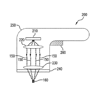
the converging
14
CA 02920858 2016-02-09
WO 2015/021462
PCT/US2014/050518
EMR beam at the skin surface 100 is about 115 microns (0.577 x 200), such that
the total
beam width at the surface is about 230 microns. The local fluence is inversely
proportional
to the local cross-sectional area of the beam for a particular beam energy.
Accordingly, for
a spot size (focal region width) of 20 microns, the ratio of fluence at the
focal region to that
at the skin surface is about (230/20)2, or about 130:1. The actual fluence
ratio may be
somewhat less due to absorption of some of the EMR energy between the skin
surface and
the focal region. Nevertheless, this exemplary calculation indicates the
relatively low
fluence in the surface regions of the skin (as compared to the fluence in the
focal region)
that can be generated when using a focusing lens having a high NA.
[0062] In further exemplary embodiments of the present disclosure, a
plurality of
such focal regions 160 can be generated simultaneously by the exemplary
apparatus and/or
the focal region(s) 160 may be scanned or traversed through the portions of
dermis 120
containing pigmented cells 130 to irradiate larger volumes of the dermis 120
in a reasonable
time, as described in more detail herein.
[0063] In certain exemplary embodiments, the depth of the focal region 160
below
the skin surface 100 can be between about 120 lam and 400 gm, e.g., between
about 150 lam
and 300 p.m. This exemplary depth range can generally correspond to the
observed depths
of pigmented regions 130 in skin that exhibits dermal melasma. The focal depth
can
correspond to a distance from a lower contact surface of the apparatus 200
(e.g., the lower
surface of the plate 240) and the focal region 160 of the EMR 150, because the
plate 240
may flatten out the underlying tissue when placed on the skin surface 100.
Accordingly, the
depth of the focal region 160 within the skin may be selected or controlled
based on a
configuration of the optical arrangement within the housing 250.
[0064] In
various exemplary embodiments of the present disclosure, the EMR 150
can be collimated (e.g., rays within the EMR beam are substantially parallel
to one another),
convergent, or divergent between the first lens arrangement 220 and second
lens
arrangement 230. In still further exemplary embodiments, the radiation emitter
arrangement 210 and/or components of the optical arrangement (e.g., the first
lens
arrangement 220 and/or the second lens arrangement 230) can be controllable or
adjustable
such that the path of the EMR 150 can be varied. Such exemplary variation in
the path of
the EMR 150 can provide corresponding variations in the depth, width, and/or
location of
the focal region 160 within the dermis 120 when the apparatus is held
stationary with
respect to the skin.
CA 02920858 2016-02-09
WO 2015/021462
PCT/US2014/050518
[0065] For example, the position and/or angle of the EMR 150 can be
shifted
relative to the optical axis of a lens in the second lens arrangement 230.
Alternatively or
additionally, the convergence or divergence of the EMR 150 entering or within
the optical
arrangement can be varied. Such variations in the EMR geometry and/or path can
provide
variations in the depth and/or lateral position of the focal region(s) 160. In
this manner,
larger volumes of the dermis 120 can be irradiated while the apparatus 200 is
held
stationary over the area of skin being treated. Such exemplary variation of
the focus region
characteristics can facilitate treatment of a plurality of depth ranges and/or
locations within
the dermis 120 containing pigmented cells or defects 130.
[0066] Exemplary adjustment and/or alteration of the geometry and/or path
of the
EMR 150 can be achieved, e.g., using one or more translators, movable mirrors,
beam
splitters and/or prisms, or the like, which may be coupled to the radiation
emitter
arrangement 210, the first lens arrangement 220, and/or the second lens
arrangement 230.
Further, these exemplary variations in locations of the focal region 160 can
also be
combined with a translation of the apparatus 200 over the area of skin being
treated to
irradiate larger volumes of the dermis 120, thereby targeting a greater number
of pigmented
cells 130 that can be present.
[0067] In further exemplary embodiments of the present disclosure, the
second lens
arrangement 230 can include a plurality of micro-lenses 300, e.g., as provided
in a
schematic side view of the exemplary configuration illustrated in FIG. 3A. For
example,
the micro-lenses 300 can include any conventional type of convergent lenses,
e.g., convex
lenses, or plano-convex lenses such as those shown in FIG. 3A. The micro-
lenses 300 can
be configured to focus EMR 150 into a plurality of focal regions 160 within
the underlying
dermis 120, as illustrated in FIG. 3A.
[0068] Each of the micro-lenses can have a large NA (e.g., between about
0.5 and
0.9), such that the EMR 150 converges from a relatively wide area at or near
the skin
surface 100 (with a relatively low intensity or local fluence) to a small
width (with higher
intensity or local fluence) in the focal region 160 within the dermis 120.
Such optical
properties can provide a sufficient intensity of EMR 150 within the focal
region 160 to
damage pigmented cells that absorb the radiation 150, while avoiding areas or
volumes of
high fluence or intensity away from the volume of dermis 120 containing
pigmented cells
130, thereby reducing likelihood of damaging overlying, underlying, and/or
adjacent
volumes of unpigmented skin tissue.
16
CA 02920858 2016-02-09
WO 2015/021462 PCT/US2014/050518
[0069] The micro-lenses 300 can be provided in a substantially square
or
rectangular array, such as that shown in the top view of such exemplary
configuration in
FIG. 3B. According to further exemplary embodiments of the present disclosure,
the micro-
lenses 300 can be provided in a hexagonal array, as shown in FIG. 3C. Other
exemplary
patterns and/or shapes of the micro-lenses 300 can be provided in still
further exemplary
embodiments. A width of the micro-lenses 300 can be small, e.g., between about
lmm and
3 mm wide. The exemplary micro-lenses 300 that are slightly wider or narrower
than this
can also be provided in certain exemplary embodiments.
[0070] In additional exemplary embodiments of the present disclosure,
the radiation
emitter arrangement 210 and/or the first lens arrangement 220 can be
configured to direct a
single wide beam of EMR 150 (such as, e.g., that shown in FIG. 2) over the
entire array of
micro-lenses 300 or a substantial portion thereof. Such exemplary
configuration can
generate a plurality of focal regions 160 in the dermis 120 simultaneously. In
further
exemplary embodiments, the radiation emitter arrangement 210 and/or the first
lens
arrangement 220 can be configured to direct a plurality of smaller beams of
EMR 150 onto
individual ones of the micro-lenses 300. According to still further exemplary
embodiments,
the radiation emitter arrangement 210 and/or the first lens arrangement 220
can be
configured to direct one or more smaller beams of EMR 150 onto a portion of
the array of
micro-lenses 300, e.g. onto a single micro-lens or a plurality of the micro-
lenses 300, and
the smaller beam(s) can be scanned over the array of the micro-lenses 300,
such that a
plurality of the focal regions 160 can be generated sequentially or non-
simultaneously in the
derrnis 120.
100711 In yet further exemplary embodiments of the present disclosure,
the micro-
lenses 300 can include cylindrical lenses, for example, convex cylindrical
lenses or piano-
convex cylindrical lenses, e.g., as shown in an exemplary top view in FIG. 3D
and
exemplary angled view in FIG. 3E. In the context used herein, 'cylindrical'
does not
necessarily require the rounded surface of the lens to be circular; it may
have an elliptical or
other smooth but non-circular profile in certain embodiments. Such cylindrical
lenses can
have a uniform profile in any cross-section that is perpendicular to the
longitudinal axis of
the lens.
100721 A width of the cylindrical micro-lenses 300 can be small, e.g.,
between about
lmm and 3 mm wide. The length of the cylindrical micro-lenses 300 can be
between about
5 mm and 5 cm, e.g., between about 5 mm and about 2 cm. This width and length
can be
17
CA 02920858 2016-02-09
WO 2015/021462 PCT/US2014/050518
selected based on such factors as the total power emitted by the radiation
emitter
arrangement 210, the overall size of the array of micro-lenses 300, etc. In
certain
exemplary embodiments, cylindrical micro-lenses 300 that are slightly shorter
or longer
and/or slightly narrower or wider can be provided.
[0073] In certain exemplary embodiments of the present disclosure, any of
the
exemplary arrays of the micro-lenses 300 can be provided on (or formed as part
of) the plate
240, as illustrated in FIG. 3E. Such configuration can facilitate placement of
the micro-
lenses 300 close to the skin surface 100, and also facilitate a more precise
depth of the focal
regions 160 within the dermis 120, e.g., when the plate 240 contacts the skin
surface 100
during use.
100741 In further exemplary embodiments of the present disclosure, the
radiation
emitter arrangement 210 and/or the first lens arrangement 220 can be
configured to direct a
single wide beam of EMR 150 (such as that shown in FIG. 2) over the entire
array of
cylindrical micro-lenses 300 or a substantial portion thereof. Such exemplary
configuration
can generate and/or produce a plurality of the focal regions 160 in the dermis
120
simultaneously that are elongated in one direction (e.g. along the
longitudinal axis of the
cylindrical micro-lenses 300) and narrow (e.g., less than about 200 gm wide,
less than about
100 IIM wide, less than about 50 gm wide, or as small as about 10 gm wide) in
a direction
orthogonal to the longitudinal axis of the cylindrical micro-lenses 300. Such
"line-focused"
EMR 150 can be used to more efficiently irradiate larger volumes of the dermis
120, e.g.,
when the exemplary apparatus 200 is scanned over the area of skin being
treated, for
example, in a direction substantially orthogonal to (or optionally at some
other angle to) the
longitudinal axis of the cylindrical micro-lenses 300,
[0075] According to yet additional exemplary embodiments of the
present
disclosure, the radiation emitter arrangement 210 and/or the first lens
arrangement 220 can
be configured to direct one or more smaller beams of EMR 150 onto one or more
of the
cylindrical micro-lenses 300. For example, the EMR 150 can be directed onto
one or more
cylindrical micro-lenses 300, e.g., over an elongated area 320 such as that
shown in FIG.
3D. The radiation emitter arrangement 210 and/or the first lens arrangement
220 can be
further configured to scan or traverse the irradiated area 320 over the
cylindrical micro-
lenses 300 (for example, using one or more movable mirrors, prisms,
waveguides, or the
like in the optical arrangement), e.g., along the longitudinal directions
indicated by the
arrows shown in FIGS. 3D and 3E (or back and forth along such direction), such
that a
18
CA 02920858 2016-02-09
WO 2015/021462 PCT/US2014/050518
plurality of the elongated focal regions 160 are progressively generated in
the dermis 120
during the scan. Such scanning of the EMR 150 can produce an irradiated focal
region 160
having a shape of an extended line within the dermis 120. The apparatus 200
can also be
traversed laterally over the region of skin being treated, e.g., in a
direction not parallel to the
longitudinal axes of the cylindrical micro-lenses 300, during the irradiation
such that the
elongated focal regions 160 can travel through the dermis 120 and irradiate a
larger volume
of tissue. For example, as described herein such lateral traversal can be
between about 5
mm/sec and 5 cm/sec. The scanning speed of the EMR beam along the axes of the
cylindrical can be larger, e.g., greater than about 10 cm/sec, to provide a
more uniform
.. irradiation of such larger volumes of tissue. The scan rate of the EMR 150
along the
cylindrical lens axes, traversal speed of the apparatus 200 over the skin,
power of the EMR
emitter arrangement 210, and width of the focal region 160 can be selected to
provide a
local fluence generated within portions of the the dermis 120 by the elongated
focal region
160 that is within the exemplary fluence ranges described herein.
[00761 In yet further exemplary embodiment of the present disclosure, some
of the
cylindrical or spherical micro-lenses 300 can have different NA values,
different sizes or
radii, and/or different effective focal lengths, e.g., as shown in the
exemplary schematic
diagram in FIG. 3F. The different focal depths of the micro-lenses 300 below
the skin
surface 100 can be, e.g., between about 120 gm and 400 gm, for example,
between about
150 ILM and 300 ftM. Such exemplary variations in the focal lengths can
produce focal
regions 160 at different depths, which can result in irradiation of larger
volumes of the
dermis 120 when the exemplary apparatus 200 is translated over the area of
skin being
treated, thereby targeting a greater number of pigmented cells 130 that may be
present (e.g.,
irradiating both shallower and deeper pigmented cells 130 in the derrnis 120).
[0077] The window or plate 240, if present, can be configured and/or
structured to
contact the surface 100 of the area of skin being treated. The lower surface
of the window
240 can be substantially planar, or it may be convex or concave in further
embodiments.
The window 240 can provide certain benefits during operation of the apparatus
200. For
example, the window 240 can facilitate precise positioning of the first and
second optical
arrangements 220, 230 relative to the skin surface 100, which can facilitate
accurate control,
selection and/or variation of the depth(s) of the focal region(s) 160 within
the skin.
[0078] The window 240 can further stabilize the soft skin tissue while
it is being
irradiated by the apparatus 200, which can facilitate control and uniformity
of the irradiation
19
CA 02920858 2016-02-09
WO 2015/021462
PCT/US2014/050518
profile. Pressure provided by the window 240 on the skin surface 100 can also
blanche (or
remove some blood from) the volume of skin tissue being irradiated, thereby
reducing the
amount of pigmented structures present locally (e.g. blood-filled vessels
containing
hemoglobin). Such blanching can facilitate increased selectivity of absorption
of the EMR
150 by pigmented cells 130 while reducing a risk of unwanted damage to blood
vessels.
[0079] In exemplary embodiments of the disclosure, the window 240 can
be cooled,
e.g., by pre-cooling it prior to using the apparatus 200 or by active cooling
using a
conventional cooling arrangement (e.g. a Peltier device, a conductive cold
conduit, or the
like). Such cooling can facilitate protection of the epidermis 110 and/or
upper portions of
the dermis 120 from unwanted damage while the pigmented cells 130 are being
irradiated
and/or damaged.
[0080] According to certain exemplary embodiments of the present
disclosure, the
window 240 can be provided as part of the second lens arrangement 230. For
example, the
second lens arrangement 230 can include a single piano-convex lens or a
plurality of piano-
convex lenses, such as those shown in FIG. 3A and 3D. Such lenses can be
affixed to or
formed as part of the window 240. The lower (planar) surface of such lenses
can provide
the benefits of the window 240 as described herein, e.g., precise positioning
of the second
lens arrangement 230 relative to the skin surface 100 to control depth of the
focal regions
160.
[0081] The actuator arrangement 260 can be configured to activate and/or
control
the radiation emitter arrangement 210 and/or an external EMR source that
provides
radiation to the radiation emitter arrangement 210, such that the irradiation
of an area of
skin by the EMR 150 can be controlled. The radiation emitter arrangement 210
and/or the
exemplary apparatus 200 can further include a conventional control arrangement
(not
shown) that can be configured to control and/or adjust the properties of the
EMR 150
directed onto the skin being treated.
[0082] For example, the apparatus 200 can include one or more sensors
(not shown)
configured to detect contact of the apparatus 200 with the skin surface 100
and/or speed or
displacement of the apparatus 200 over the skin surface 100 during use. Such
exemplary
sensors can generate signals capable of varying properties of the EMR 150,
e.g., by varying
the power emitted by the radiation emitter arrangement 210 based on the
translational speed
of the apparatus 200, by turning off the source(s) of EMR 150 when the
apparatus 150 is
CA 02920858 2016-02-09
WO 2015/021462
PCT/US2014/050518
stationary relative to the skin surface 100, etc. Such sensors and control
arrangements can
be provided as a safety feature, e.g. to prevent excessive irradiation and
unwanted damage
to the skin being treated, and are generally known in the art. Further
variations of such
conventional sensing and/or control arrangements can be used in embodiments of
the
present disclosure.
[0083] In
general, it can be preferable to expose a particular location in the dermis
to the focal region 160 for only a short period of time, e.g., to prevent
local build-up of heat
through absorption of the optical energy by melanin or other pigment. Long
local
irradiation times (or "dwell times") can generate heat faster and to a greater
extent than it
.. can safely diffuse into the surrounding tissue, which may lead to unwanted
damage to
unpigmented tissue. Thus, short-duration, intense irradiation of small areas
of pigmented
features 130 within the dermis 120 can disrupt the pigment and improve the
appearance of
melasma while avoiding excessive heat generation and unwanted thermal damage
to
surrounding unpigmented tissue. For example, typical sizes of pigmented cells
or structures
can be on the order of about 10 microns, and local thermal relaxation times
can be on the
order of about 0.1 to about 1-2 milliseconds. Longer local dwell times at
irradiation
intensities sufficient to heat and damage the pigmented structures 130 can
build up heat
locally faster than it can safely dissipate away.
[0084] Limiting
irradiation times (dwell times) at a particular focal region location
can be achieved in various ways. In one exemplary embodiment, the radiation
emitter
arrangement 210 can be configured to provide discrete pulses of EMR 150 into
the focal
regions 160. The interval between such pulses of EMR can be, e.g., on the
order of about
50 milliseconds or more even if the location of the focal region is moving
through the skin
tissue at a relatively slow speed of a few mm/s. These exemplary parameters
can result in a
distance between focal regions 160 irradiated by successive pulses of, e.g.,
about 50-100
microns, which can be greater than a width of the focal region 160 itself.
Accordingly, such
general parameters can facilitate spatial and temporal separation of the
successive irradiated
focal regions 160, such that local thermal relaxation can occur and buildup of
excess heat
can be avoided. The spot size, pulse duration, and/or total pulse energy can
be selected
based on the principles and guidelines described herein, using simple
calculations, to
provide a sufficient fluence within the focal region 160 to affect the
pigmented structures
130 while maintaining a sufficiently small dwell time (e.g. less than about 1-
2 ms).
21
CA 02920858 2016-02-09
WO 2015/021462
PCT/US2014/050518
[0085] In further exemplary embodiments of the present disclosure, the
focused
radiation 150 can be scanned over a region of skin affected by dermal melasma,
such that
the focal region(s) 160 may irradiate and damage a large number of the
pigmented cells
130. Such scanning can be performed with any of the embodiments described
herein. The
scanning can be done manually, e.g., using a conventional method of
translating a
handpiece over the area of skin to be treated. Alternatively, the apparatus
200 can
optionally be coupled to a translating arrangement that can be configured to
automatically
move the apparatus (or certain components thereof) over an area of skin to be
treated. Such
automatic translation can be provided as a pre-set pattern or as a random or
semi-random
path over the skin. In still further embodiments, one or more of the optical
components
(e.g. the first and/or second lens arrangement 220, 230) and/or the radiation
emitter
arrangement can be translated within the housing 250, such that the focal
region(s) 160 can
translate within the tissue while the housing 250 is held in a single position
relative to the
skin.
[0086] Average scan speeds (or ranges of such speeds) can be determined
based on
the general exemplary guidelines described herein. For example, for a
particular spot size
(which can be determined primarily by the properties of the optical
arrangement), the local
dwell (irradiation) time can be estimated as the spot size/width divided by
the translational
speed. As noted herein, such dwell time is preferably less than about 1-2
milliseconds to
avoid local heat buildup and unwanted thermal damage of unpigmented tissue.
Accordingly, a minimum scan speed can be estimated as the width of the focal
region 160
divided by 1 millisecond. For example, a spot size of 10 microns (0.01 mm)
would
correspond to a minimum scan speed of 0.01 mm/0.001 seconds, or about 10
min/sec (1
cm/sec). Scan rates for line-focused beams (e.g., produced by directing an EMR
beam onto
a cylindrical lens) can be estimated in a similar manner, e.g., where the
width of the focal
line corresponds to the width of the focal region and the scan speed is in a
direction
perpendicular to the focal line, or for other scanning configurations.
[0087] A power output of the radiation emitter arrangement 210 can be
selected
based on several factors including, e.g., the EMR wavelength, the number,
size, and/or
.. depth of the focal region(s) 160, optical characteristics and geometry of
the first and second
lens arrangements 220, 230, etc. The power output can be selected such that
the fluence in
the focal region 160 is sufficiently high to damage pigmented cells 130 that
absorb the
22
CA 02920858 2016-02-09
WO 2015/021462 PCT/US2014/050518
EMR 150 for short exposure times, while fluence at other depths (e.g., in the
epidermis 110)
is sufficiently low to minimize or avoid unwanted damage there.
100881 Based on some experimental observations, a local fluence within
the focal
region 160 that may be sufficient to affect melanin-containing structures
(e.g., pigmented
cells) can be between about 10-1000 J/cm2, for example, between about 50-500
J/cm2, for
EMR 150 having a wavelength of about 650 nm. This range of effective local
fluences can
increase slightly with increasing wavelength of the EMR 150 (and decrease with
decreasing
wavelength), based on the decreasing absorption factor for melanin at larger
wavelengths.
Larger or smaller local flue-nce values may also be provided when using faster
or slower
scan speeds, in further exemplary embodiments. Larger or smaller local fluence
values can
also be provided when using shorter or longer dwell times, respectively. The
local dwell
time can preferably remain less than about 1-2 milliseconds in such
embodiments.
100891 The exemplary fluence values and dwell times described herein
can be
understood to correspond to a single pulsed exposure onto, or a single
traversal of a scanned
focal region through, a particular location within the dermis_ For example, a
particular
location within the dermis 120 may be irradiated by scanning more than one
focal region
160 through it at different times, thereby providing a higher fluence at that
location.
However, local heat build-up can be avoided by providing a time interval
between
successive irradiations of the same location that is greater than a few
milliseconds.
[0090] The total power output of the radiation emitter arrangement 210
directed
onto a single focal spot 160 can thus be estimated and/or deteimined based on
the focal spot
size and scan speed. The fluence F (e.g., in j/cm2) can be calculated as the
EMR power
output P multiplied by the dwell time rand divided by the focal spot area A
(i.e., F=P IA).,
where the dwell time r can be estimated as the focal spot width D divided by
the scan speed
v (i.e., r=D/v). As an exemplary calculation, for EMR 150 having a wavelength
of about
650 nm, a focal spot width of about 20 microns, and a scan speed of about 1
cmis, the
power output P of a single EMR source (e.g., a laser diode) to achieve a level
of local
fluence in the focal region between about 10-1000 Pcm2 is between about 15 mW
and 1500
mW.
[0091] Typical scan speeds for a handpiece that is manually translated over
an area
of skin to be treated can be, e.g., on the order of about 5 mm/sec to about 5
cm/sec. Such
speeds correspond to traversing a distance of 5 cm (about 2 inches) in about 1-
10 seconds.
23
CA 02920858 2016-02-09
WO 2015/021462 PCT/US2014/050518
Accordingly, for a handpiece that is translated manually over the skin to
irradiate portions
of the dermis as described herein, the power output and focal geometry of the
apparatus 200
can be selected to provide a fluence at the irradiated locations within the
dermis that is
within the general range described herein.
[0092] Such exemplary power calculations can be based on the entire output
of the
laser diode being focused into one focal region. If the output from a single
source of EMR
is focused onto a plurality of focal regions (e.g., when using an optical
splitter or a wide
beam directed onto a plurality of micro-lenses), then the power output of the
EMR source
can be multiplied by the number of focal spots 160 to achieve the same local
fluence within
each focal region 160. EMR 150 can be provided as a continuous wave (CW) or
optionally
as a plurality of pulses. Alternatively, a plurality of EMR sources (e.g.
laser diodes or the
like) can be provided to generate a plurality of irradiated focal regions 160
simultaneously,
with the appropriate power level for each EMR source being estimated as
described above.
In certain embodiments, if one or more EMR beams are scanned over the focusing
lens
arrangement 230, the power of the EMR source can be selected based on the lens
properties,
scan speed, etc. to provide fluences and dwell times at locations of the
dermis irradiated by
the focal regions 160 that are within the general ranges described herein.
[0093] In certain exemplary embodiments of the present disclosure, the
radiation
emitter arrangement 210 can include a plurality of EMR emitters (e.g., laser
diodes or
waveguide ends). Such emitters can be provided in a linear array, such that
they lie
substantially along one or more straight lines. In further exemplary
embodiments, the
emitters can be arranged in a two-dimensional pattern, which can provide
further patterns of
EMR 150 directed onto the first lens arrangement 220. As described above, the
power
output of each emitter can be selected using a routine calculation based on
the focal spot
size and scan speed to generate a local fluence within each focal zone 160
that is within the
preferred range described herein.
[0094] A schematic diagram of a further exemplary apparatus 400 in
accordance
with certain exemplary embodiments of the present disclosure is shown in FIG.
4. The
exemplary apparatus 400 can be generally similar to the apparatus 200 shown in
FIG. 2, and
illustrates a few further features which may also be provided in the apparatus
200 such as,
e.g., a cooling arrangement for the EMR source or a lens cage. Exemplary
features of the
exemplary apparatus 200 can also be used with the exemplary apparatus 400,
including but
not limited to an array of micro-lenses 300, a housing 250, etc.
24
CA 02920858 2016-02-09
WO 2015/021462 PCT/US2014/050518
[0095] The apparatus 400 includes a lens cage 410 that can be provided
as an
enclosure or housing that encloses optical lenses 420, 430. A window 240 can
be provided
at one end of the lens cage 410. An aspheric focusing lens 420 can be used in
certain
embodiments to provide a larger front surface working distance than, e.g., a
microscope
objective lens. The distance between the front of the focusing lens 420 and
the target tissue
may be less than about 1 cm for large NA values as described herein, such that
the window
240 can also protect the lens 4from contacting the tissue directly, The NA of
the aspheric
focusing lens 420 can optionally be selectable, e.g. to vary the focal depth
beyond the
window 240.
[0096] The exemplary apparatus 400 further includes a laser diode (LD)
mounting
arrangement 440 coupled to the lens cage 410, which can accept one or more
laser diodes
450 that can be selected to emit energy in the visible and/or NIR ranges. A
driver 460 for
the laser diode(s) 450 can be provided, and the laser diodes 450 can be held
slightly above
threshold during operation with an applied DC bias current, which can
facilitate a rapid rise-
time in the pulse activation of the diode(s) 450. The pulse properties can be
controlled by a
pulse generator arrangement 470, e.g. a programmable function generator that
can be
configured to control the laser diode(s) 450 to produce single pulses or
sequences of pulses,
with selectable pulse widths (e.g. 30 ns and greater) and intervals between
pulses.
[0097] The LD mounting arrangement 440 can also include a
thermoelectric cooler
(TEC) arrangement coupled or connected to the laser diode mounting arrangement
440,
which can be controlled (e.g. with a fEC controller 480) to prevent the laser
diode(s) 450
from overheating during use. The apparatus 400 (as well as the apparatus 200
shown in
FIG. 2) can be used in various orientations, e.g., vertically, horizontally,
etc,, with the
window 240 pressed against a tissue provided at any angle to precisely
position the optics
relative to the tissue surface and thereby facilitate control of the focal
depth of the beam
within the tissue.
[0098] The exemplary apparatus 200 shown in FIG. 2 and the exemplary
apparatus
400 shown in FIG. 4 are illustrations of exemplary configurations, and other
embodiments
using various combinations and/or configurations of similar components can
also be used.
For example, different numbers and/or types of optical arrangements 220, 230
and/or
emitter arrangements 210 can be used to provide irradiation characteristics
and focal regions
160 within the dermis 120 as described herein. For example, in certain
embodiments, the
apparatus 200 can be provided in a shape factor similar to that of a handheld
razor, with the
CA 02920858 2016-02-09
WO 2015/021462 PCT/US2014/050518
radiation emitter arrangement 210 provided as one or more laser diodes,
optical
arrangements 220, 230 provided in the "head" of the razor, and a power source
(e.g. one or
more conventional alkaline cells or the like) provided in the handle. Other
form factors can
also be used in further embodiments of the disclosure. Similar features,
combinations
and/or variations can be provided for the apparatus 400.
[0099] Exemplary properties of the radiation emitter arrangement 210,
such as, e.g.,
wavelength(s) of EMR 150, power or intensity of the EMR 150, size and
numerical
apertures of the optical arrangements 220, 230, scanning speed or rate of the
first optical
arrangements 220 (if present), and/or target scan speed (or range thereof) of
the apparatus
.. 200 over the area of skin being treated, can be selected to provide
appropriate fluence,
intensity and/or dwell time of the EMR 150 on the pigmented cells during
operation of the
apparatus 200. Exemplary values and/or ranges for such parameters, as well as
certain basic
approaches that can be used to estimate their values as needed, are described
in more detail
herein. For example, such exemplary parameters can be selected to provide
sufficient local
fluence at the pigmented cells 130 to damage them and reduce the pigmented
appearance of
the skin, while avoiding unwanted damage to the epidermis 110 and unpigmented
volumes
of the dennis 120.
[00100] The exemplary effective dwell time can be estimated using
conventional
techniques based on an approximate width of pigmented cells 130 of about 10 gm
and the
local width (e.g., focal diameter or width) and speed of the focal region 160.
The speed of
the focal region 160 can be estimated based on a scan speed of EMR 150
provided by the
first lens arrangement 220 and/or radiation emitter arrangement 210 (if
present), optical
geometry of the optical arrangements 220, 230, and scan speed of the apparatus
200 over
the area of skin being treated.
100101] One or more exemplary parameters of the apparatus 200, 400 can be
selected
and/or adjusted once the other ones are known to provide a safe but effective
irradiation of
the pigmented cells 130 as described herein. For example, the exemplary
apparatus 200,
400 having known geometry (e.g. spot size or focal line width, and NA) of the
lens
arrangements 220, 230 or lenses 420, 430 (and internal scanning speed of EMR
beams, if
present), and a particular wavelength of EMR 150 can be provided. The power of
the EMR
source(s) can then be selected based on a target range of scanning speeds of
the apparatus
200 over the area to be treated. For example, the exemplary apparatus 200, 400
can be
traversed over an arc a of skin at a speed between about 1-5 cm/s, which
corresponds
26
CA 02920858 2016-02-09
WO 2015/021462 PCT/US2014/050518
approximately to the speed at which a conventional razor is traversed over
skin during
shaving. Using these exemplary parameters and the number of passes to be made
over the
treatment area, the local speed and dwell time of the focal region(s) 160 can
be estimated,
and a power output of the radiation emitter arrangement 210 can be selected or
adjusted to
provide an effective local fluence within the focal region 160 as described
herein. Such
calculations are routine and can be done by a person of ordinary skill in the
art.
[00102] In further exemplary embodiments of the present disclosure, a
method for
reducing the pigmented appearance of dermal melasma can be provided. The
exemplary
method can include directing and focusing electromagnetic radiation 150 as
described
herein onto a plurality of focal regions 160 within the dermis 120 using an
optical
arrangement, such that the EMR 150 is selectively absorbed by pigmented
regions 130 to
thermally damage or disrupt them, while avoiding unwanted thermal damage to
unpigmented regions and overlying tissue (e.g., the epidermis 110).
[00103] The EMR 150 can have a wavelength greater than about 600 nm,
e.g.,
between about 600 and 850 nm, or between 625 and 800 nm, or between about 650
and 750
nm. A width of the focal region within the dermis can be less than about 200
microns, e.g.,
less than about 100 microns, or less than about 50 microns, The spot size can
be greater
than the theoretical lower limit of a few microns.
100104] The EMR 150 can be focused using the optical arrangement, which
can
include one or more lens arrangements 220, 230. The focusing lens arrangement
230 having
a high NA, e.g., between about 0.5 and 0.9, can be used to focus the EMR 150
onto a focal
region 160. Such NA values can facilitate generation of high fluence in the
focal regions
160 within the dermis 120 while avoiding large fluences that may generate
unwanted
damage in the overlying tissue. Such focusing can be achieved using, e.g., the
single
focusing lens 230 (such as a convex objective lens or a piano-convex lens), a
plurality of
such lenses provided as an array of micro-lenses 300, one or more convex or
plano-convex
cylindrical lenses, or the like. The EMR 150 can be directed onto the focusing
lens
arrangements 230, and optionally scanned or pulsed over the one or more
focusing lens
arrangements 230, to irradiate a plurality of focal regions 160 in the dermis
120, either
simultaneously or sequentially.
[00105] In further exemplary embodiments of the present disclosure, an
optical gel or
the like (e.g. glycerol or a similar substance) can be provided between the
window 240 and
27
CA 02920858 2016-02-09
WO 2015/021462 PCT/US2014/050518
the skin surface 100 as a topical application to the skin surface 100. Such a
gel can reduce
an optical index mismatch between the window 240 and the skin, and it may
improve
transmission of the EMR 150 from the apparatus 200 into the dermis 120. The
gel can also
reduce friction between the exemplary apparatus 200 and skin surface 100,
thereby
facilitating a smoother translation of the apparatus 200 over the area of skin
being treated.
[00106] A particular location within the dermis 120 can be irradiated
by the focal
region with an irradiation (dwell) time that is less than about 2
milliseconds, e.g., to
facilitate local thermal relaxation of tissue that absorbs the EMR 150 and
avoid local
buildup of excess heat. Such short dwell times can be provided, e.g., by
scanning an
apparatus that provides the focused EMR 150 over the area of skin being
treated, by pulsing
the EMR source, and/or by moving components of the EMR source or emitter 210
and/or
optical arrangement, such that the location of the focal region(s) 160 within
the dermis 120
varies with time.
[00107] The local fluence within the focal region 160 can be, e.g.,
between about 10-
1000 J/cm2, e.g., between about 50-500 J/cm2, for EMR 150 having a wavelength
of about
650 mn. This range of effective local fluences can increase slightly with
increasing
wavelength of the EMR 150 (and decrease with decreasing wavelength), based on
the
decreasing absorption factor for melanin at larger wavelengths. Such fluence
can be related
to the focal properties of the optical arrangement (e.g., the focal spot
size), the translational
speed of the focal region 160 within the dermis 120, pulse duration of the
applied EMR 150,
etc. The surface of the skin 100 can optionally be cooled to further prevent
unwanted
thermal damage in the epidermis and/or upper dermis.
[00108] The exemplary method and apparatus and the associated
parameters
described herein can be generally based on a single pass of a focal region 160
over a
pigmented cell 130. The fluence needed to achieve the same thermal damage
effect based
on a plurality of passes varies approximately as the fourth root of the number
of passes n.
For example, a single pass of a focal region 160 over a pigmented cell 130 at
a particular
fluence would have a similar effect as 16 passes made with a focal region 160
having half
the particular fluence. Although a single pass may be more efficient than a
plurality of
passes, the exemplary apparatus 200, 400 can be configured to provide an
effective fluence
after a particular number of passes have been made. A plurality of passes can
provide a
greater safety margin to avoid unwanted damage to the epidermis while damaging
the
28
CA 02920858 2016-02-09
WO 2015/021462 PCT/US2014/050518
pigmented cells 130, e.g., it can accommodate a greater range of effective
translation speeds
of the apparatus 200 over the tieated area for multiple passes as compared to
if just a single
pass is made. The number of passes of the focal region(s) 160 through a
particular location
in the dermis 120 can depend, e.g., on the internal scan rate of EMR 150 over
the second
lens arrangement 230, if present, the number of focal regions 160 that may
pass throui a
given location during one pass of the entire apparatus 200 (e.g., a function
of the number,
size, and arrangement of micro-lenses 300, if present), as well as the number
of times the
apparatus 200 is translated over the area to be treated.
[00109] Other exemplary features and/or functions of the exemplary
apparatus 200,
400 described herein can also be used in conjunction with the exemplary
disclosed methods
for treating dermal melasma.
Example
[00110] An animal study using an exemplary spot-focused laser device
and model
system were used to test the efficacy of treating deep melasma using optical
radiation. The
study was performed on a female Yorkshire pig, as described below.
[00111] First, a deep-melasma condition was simulated by tattooing the
dermis using
a melanin-based ink. The ink was prepared by mixing synthetic melanin at a
concentration
of 20mg/mL in a 50:50 saline/glycerol solution. The resulting suspension was
then agitated
prior to being injected into 1 cm by 1 cm test sites on the animal subject
using a standard
tattoo gun. The tattooed sites were then allowed to settle over a period of a
week to allow
melanophages to phagocytoze the melanin granules in the dermis. The melanin
left in the
epidermis was substantially eliminated over this time period through natural
bodily
processes.
[00112] An exemplary biopsy image from a tattooed site that was allowed
to settle as
described herein, is shown in FIG, 5. The tissue sample was stained with
Fontana-Masson
stain to better image any melanin present. The dark spots evident in the
dermal layer in
FIG. 5 appear to be generally similar to those observed in patients having
deep/dermal
melasma. No such dark spots were seen in biopsy samples taken from untattooed
sites that
were similarly stained. Accordingly, the tattoo process described herein
appears to provide
a useful in vivo model of dermal melasma.
100113] An exemplary melasma treatment system was constructed based on
exemplary embodiments of the present disclosure described herein, which
includes a 200
29
87904-26
mW continuous wave (CW) diode laser configured to emit optical energy having a
wavelength
of about 658 nm, mounted on an x-y scanning platform. The scanner was capable
of scanning
speeds up to 15 mm/s. The laser beam was collimated and focused using two
lenses having a
numerical aperture (NA) of 0.62 to a depth of about 200 gm.
[00114] Test sites that were tattooed with melanin ink as described above,
and control
sites that have only tattooed borders to outline them, were both treated by
scanning the focused
laser beam across the sites in 10 parallel lines at different speeds. The
control sites were scanned
to assess any potential damage that may occur in Impigmented skin under the
different scanning
conditions performed.
[00115] An exemplary tattooed test site is shown in FIG. 6A. This image
shows the test
site after the tattoo has been allowed to settle for a week, just prior to
scanning with the laser
apparatus. The test site was scanned with the laser at a speed of 1-3 mm/sec,
using a 200 mW
CW output. The same test site is shown in FIG. 6B two weeks after it was
scanned with the
laser. There is a noticeable lightening of the appearance with no scarring or
scabbing evident,
even though only a portion of the tattooed area was irradiated with focused
optical energy.
These results indicate the general efficacy of the exemplary methods and
devices described
herein for reducing the hyperpigmented appearance of deep/dermal melasma.
[00116] The foregoing merely illustrates the principles of the present
disclosure. Various
modifications and alterations to the described embodiments will be apparent to
those skilled in
the art in view of the teachings herein. It will thus be appreciated that
those skilled in the art will
be able to devise numerous techniques which, although not explicitly described
herein, embody
the principles of the present disclosure and are thus within the spirit and
scope of the present
disclosure.
Date Recue/Date Received 2020-12-04