Note: Descriptions are shown in the official language in which they were submitted.
CA 02920868 2016-02-12
NAVIGATION OF AN ANGIOPLASTY GUIDEWIRE
CROSS-REFERENCE TO RELATED APPLICATION
This application is related to U.S. Patent Application
titled Angioplasty Guidewire, filed on even date with the
present application, and which is incorporated herein by
reference.
FIELD OF THE INVENTION
The present invention relates generally to invasive
medical procedures, and specifically to navigation of a
guidewire used in such procedures.
BACKGROUND OF THE INVENTION
In inserting a guidewire into a patient during a
medical procedure such as angioplasty, it is important that
the guidewire follows a correct path. One method for
tracking a guidewire is to use X-rays. However, X-rays are
ionizing radiation and it is preferable to reduce a
patient's exposure to X-rays as much as possible.
U. S. Patent Application 2011/0230758 to Eichler,
whose disclosure is incorporated herein by reference,
describes a system for determining the position of the
tip of a medical catheter within the body of a
patient. The method includes inserting a mapping position
system catheter into a tubular organ, and inserting a
medical catheter into the organ.
U. S. Patent 8,632,468 to Glossop et al., whose
disclosure is incorporated herein by reference, describes a
system for assisting/performing image-guided transjugular
intrahepatic portosystemic shunt (TIPS) procedures in a
portion of an anatomy of a patient. The system includes a
guide needle portion having a hollow tube with a bend
toward its distal tip, and a puncture needle portion that
1
CA 02920868 2016-02-12
includes at least one position indicating element at its
tip.
U. S. Patent 8,043,351 to Yon et al., whose disclosure
is incorporated herein by reference, describes a method for
performing a therapy for angioplasty. The disclosure refers
to a balloon tipped catheter, and describes how mapping
electrodes may be employed at the distal end of a warm
balloon.
U. S. Patent 7,317,819 to Janes, whose disclosure is
incorporated herein by reference, describes apparatus for
three-dimensional imaging. The disclosure refers to viewing
a specific portion of a three-dimensional image exposure,
e.g., for viewing an angioplasty device moving through a
vein or artery.
U. S. Patent Application 2006/0173298 to Tucker, whose
disclosure is incorporated herein by reference, describes
methods for using a catheter to generate a geographic map
of a venous structure of the heart and to generate an
electrical map of the electrical conduction patterns of the
heart for locating an aberrant electrical conduction
pattern.
U. S. Patent Application 2014/0081204 to Cohen et al.,
whose disclosure is incorporated herein by reference,
describes a remotely controlled catheter insertion system
which may include a mapping procedure and an angioplasty
procedure.
Documents incorporated by reference in the present
patent application are to be considered an integral part of
the application except that, to the extent that any terms
are defined in these incorporated documents in a manner
that conflicts with definitions made explicitly or
implicitly in the present specification, only the
2
CA 02920868 2016-02-12
definitions in the present specification should be
considered.
SUMMARY OF THE INVENTION
An embodiment of the present invention provides a
method, including:
advancing a first guidewire having a first diameter
through a lumen in the body of a patient, the first
guidewire being configured to be tracked by an
electromagnetic tracking system and an impedance tracking
system;
while advancing the first guidewire, recording first
signals of the electromagnetic tracking system and second
signals of the impedance tracking system, the first and the
second signals being generated in response to differing
positions of the first guidewire in the lumen;
recording respective correspondences between the first
and second signals at each of the differing positions;
withdrawing the first guidewire from the lumen;
after withdrawing the first guidewire, advancing
through the lumen a second guidewire, having a second
diameter smaller than the first diameter, and being
configured to be tracked by the impedance tracking system;
while advancing the second guidewire, receiving a
third signal of the impedance tracking system generated in
response to advancement of the second guidewire in the
lumen; and
applying the respective correspondences to the third
signal in order to determine a position of the second
guidewire in the lumen.
In an embodiment the first guidewire includes a coil
configured generate the first signals in response to a
magnetic field generated by the electromagnetic tracking
3
CA 02920868 2016-02-12
. .
system, and an electrode configured to inject a current
into the patient so as to generate the second signals as a
set of currents received by respective electrodes on skin
of the patient.
In a disclosed embodiment the second guidewire is not
trackable by the electromagnetic tracking system.
In a further disclosed embodiment the method includes,
while withdrawing the first guidewire from the lumen,
recording further respective correspondences between the
first and second signals at each of the differing positions
of the first guidewire in the lumen, and applying the
further respective correspondences to the third signal in
order to determine the position of the second guidewire.
There is also provided, according to an embodiment of
the present invention embodiment, apparatus, consisting of:
a first guidewire having a first diameter and
configured to be tracked by an electromagnetic tracking
system and an impedance tracking system;
a second guidewire, having a second diameter smaller
than the first diameter, and configured to be tracked by
the impedance tracking system; and
a processor, configured:
while the first guidewire is advanced through a lumen
in the body of a patient, to record first signals of the
electromagnetic tracking system and second signals of the
impedance tracking system, the first and the second signals
being generated in response to differing positions of the
first guidewire in the lumen;
to record respective correspondences between the first
and second signals at each of the differing positions;
after withdrawal of the first guidewire from the
lumen, to receive a third signal of the impedance tracking
4
CA 02920868 2016-02-12
system generated in response to advancement of the second
guidewire in the lumen; and
to apply the respective correspondences to the third
signal in order to determine a position of the second
guidewire in the lumen.
The present disclosure will be more fully understood
from the following detailed description of the embodiments
thereof, taken together with the drawings, in which:
BRIEF DESCRIPTION OF THE DRAWINGS
Fig. 1 is a schematic illustration of a guidewire
tracking system, according to an embodiment of the present
invention;
Fig. 2 is a schematic perspective diagram of a distal
portion of a mapping guidewire, according to an embodiment
of the present invention;
Figs. 3A and 3B are schematic cross-sections of the
mapping guidewire, according to an embodiment of the
present invention;
Fig. 4 is a schematic cross-section of a distal end of
a delivery guidewire, according to an embodiment of the
present invention; and
Fig. 5 is a flowchart of steps performed in
implementing the system of Fig. 1, according to an
embodiment of the present invention.
DETAILED DESCRIPTION OF EMBODIMENTS
OVERVIEW
In many medical procedures where a guidewire is
required to be inserted into a patient, it is difficult to
accurately track the guidewire as it is inserted. The
guidewire is typically used to deliver an element, such as
5
CA 02920868 2016-02-12
a balloon catheter, to a desired location. Larger
guidewires may be more easily tracked, since they may
incorporate elements such as sensors assisting in the
tracking. However, larger guidewires, together with the
elements they deliver, may be more likely to cause trauma
in the patient.
Embodiments of the present invention address this
problem by using two guidewires, a first, mapping
guidewire, and a second delivery guidewire. The delivery
guidewire has a smaller diameter than the mapping
guidewire. The mapping guidewire is configured to be
tracked by an electromagnetic tracking system and an
impedance tracking system, and while it is advanced into a
lumen of a patient signals for the two systems are
recorded. Correspondences between the two signals are
recorded for differing positions of the mapping guidewire
as it advances.
The mapping guidewire is then withdrawn and the
delivery guidewire is inserted into the lumen. The delivery
guidewire is configured to be tracked only by the impedance
tracking system, which enables the delivery guidewire to
have a smaller diameter than the mapping guidewire. The
impedance tracking system, used as a stand-alone system, is
typically less accurate than the electromagnetic tracking
system. However, embodiments of the present invention
overcome this inaccuracy by applying the correspondences
recorded for the mapping guidewire to the impedance
tracking system signals of the delivery guidewire, enabling
the position of the delivery guidewire to be accurately
determined.
6
CA 02920868 2016-02-12
SYSTEM DESCRIPTION
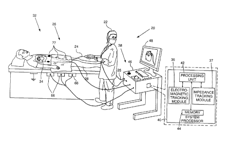
Reference is now made to Fig. 1, which is a schematic
illustration of a guidewire navigation system 20, according
to an embodiment of the present invention. For simplicity
and clarity, the following description, except where
otherwise stated, assumes an angioplasty procedure wherein
an operator 22 of system 20, herein assumed to be a medical
practitioner, inserts a guidewire 24 into a lumen 26 of a
patient 28. The angioplasty procedure may be indicated, for
example, for a case of chronic total occlusion. Typically
in the procedure, the guidewire is initially inserted into
the patient until a distal portion 32 of the guidewire
reaches a desired location in, or in proximity to a heart
34 of the patient.
Guidewire 24 is used to map the path followed by the
guidewire to the desired location in the region of heart
34, so that during the guidewire insertion, the path
followed by the distal portion is tracked. The guidewire is
then withdrawn, and a second guidewire 25, smaller in
diameter than guidewire 24, is inserted along the tracked
path to the heart desired location. Second guidewire 25 is
typically used to deliver a catheter, such as a balloon
catheter, to the desired location at the heart. Guidewire
24 is also referred to herein as mapping guidewire 24, and
guidewire 25 is also referred to herein as delivery
guidewire 25. The mapping guidewire is described in more
detail with reference to Figs. 2, 3A, and 3B, and the
delivery guidewire is described in more detail with
reference to Fig. 4.
System 20 may be controlled by a system processor 40,
comprising a processing unit (PU) 42 communicating with an
electromagnetic tracking module 36 and an impedance
tracking module 37. The functions of both modules are
7
CA 02920868 2016-02-12
described in more detail below. PU 42 also communicates
with a memory 44. Processor 40 is typically mounted in a
console 46, which comprises operating controls 38,
typically including a pointing device such as a mouse or
trackball, that operator 22 uses to interact with the
processor. The processor uses software stored in memory 44
to operate system 20. Results of the operations performed
by processor 40 are presented to the operator on a display
48, which typically presents a visual representation of the
paths taken by guidewire 24 and guidewire 25 in patient 28.
The software may be downloaded to processor 40 in
electronic form, over a network, for example, or it may,
alternatively or additionally, be provided and/or stored on
non-transitory tangible media, such as magnetic, optical,
or electronic memory.
For tracking the path of guidewire 24, embodiments of
the present invention use two tracking systems. A first
tracking system comprises an electromagnetic tracking
system, similar to that described in US Patent 6,690,963 to
Ben-Haim et al., whose disclosure is incorporated herein by
reference, and to that used in the CartoTM system produced
by Biosense-Webster of Diamond Bar, CA. The electromagnetic
tracking system is under control of electromagnetic
tracking module 36. The electromagnetic tracking system
comprises a plurality of magnetic field generators, herein
assumed to comprise three sets of generators 66, each set
comprising three orthogonal coils, so that the plurality of
generators comprises a total of nine coils. Generators 66
are placed in known locations beneath patient 28, the known
locations defining a frame of reference of the generators.
Module 36 controls, inter alia, the amplitude and frequency
of the alternating magnetic fields produced by the
generators.
8
CA 02920868 2016-02-12
The alternating magnetic fields interact with a coil,
described in more detail below, located within guidewire 24
and at the distal portion of the guidewire, so as to
generate alternating electropotentials in the coil, and the
electropotentials are received as a signal by tracking
module 36. The module, together with processing unit 42,
analyzes the received signal, and from the analysis is able
to determine a position, i.e., a location and an
orientation, of the guidewire coil in the defined frame of
reference.
In the electromagnetic tracking system processor 40
uses the location and orientation of the guidewire coil to
track the distal portion of the guidewire.
A second tracking system comprises an impedance
measuring tracking system, similar to that described in US
Patent 8,456,182 to Bar-Tal et al., whose disclosure is
incorporated herein by reference. The CartoTM system
produced by Biosense-Webster of Diamond Bar, CA also uses
an impedance measuring tracking system. The impedance
measuring tracking system is under control of impedance
tracking module 37.
The impedance measuring tracking system measures
currents between an electrode, described in more detail
below, located at the distal portion of guidewire 24, and a
plurality of generally similar patch electrodes 77, also
herein termed patches, which are positioned on the skin of
patient 28 in the vicinity of the region in which the
guidewire is operating. The currents between the guidewire
electrode and the patches vary according to the location of
the electrode, because of the different distances of the
distal portion from the patches, which cause different
impedances between the distal portion electrode and the
9
CA 02920868 2016-02-12
different patches. Module 37 may be configured to generate
an indication of the location from the different currents.
Typically the tracking by either or both of the
systems may be presented visually on display 48, for
example by incorporating an icon representing the guidewire
distal portion into an image of patient 28, as well as a
path taken by the icon.
Fig. 2 is a schematic perspective diagram of distal
portion 32 of mapping guidewire 24, and Figs. 3A and 3B are
schematic cross-sections of the guidewire, according to an
embodiment of the present invention. In Fig. 2, a terminal
portion of the distal portion has been cut-away to
illustrate the internal structure of the guidewire.
Guidewire 24 is formed from a hollow elastic metal tube 70,
which has an internal longitudinal lumen 86 and an axis of
symmetry 72. In a disclosed embodiment the material of the
tube is a nitinol alloy, and the tube has an outer diameter
of approximately 0.8 mm. and an inner diameter of
approximately 0.5 mm. Tube 70 is formed into a helix, by
having a laser cut a spiral channel 74 into the tube.
Typically, when the tube is formed by laser cutting of the
channel, the pitch of the channel is varied so that there
are two or more different pitches. The different pitches
give the guidewire the property that it has different
flexibilities in different sections of the guidewire.
A small conductive coil 76, made of insulated wire, is
inserted into lumen 86 so that it is located at a distal
end 78 of the tube. Fig. 3B is a cross-section of guidewire
24 taken at the location of the coil. Prior to insertion
the coil is wound on an elongate flexible core 80, herein
assumed to comprise a wire, and also referred to herein as
wire 80. After the coil has been formed the wire and coil
are inserted into lumen 86, so that the wire lies
CA 02920868 2016-02-12
. .
approximately along axis 72, and so that the coil has a
common axis of symmetry with axis 72. In the disclosed
embodiment referred to above coil 76 has an external
diameter of approximately 0.3 mm. Wires 82 connect the two
ends of coil 76 to the proximal end of the guidewire, and
proximal ends of the wires are connected to tracking module
36 so that the module receives an alternating
electropotential signal generated in the coil. Module 36 is
able to analyze the signal so as to determine the position
of the coil, and thus the position of distal end 78.
The outer surface of tube 70 is covered by a thin
layer 84 of biocompatible insulating polymer, the layer
acting as a sleeve for the tube. Layer 84 prevents fluids
from patient 28 contacting the outer surface of tube 70,
and/or penetrating into lumen 86. The layer also acts to
mechanically strengthen the guidewire. An electrode 90,
typically in the form of a ring, is attached to and
overlays layer 84. Fig. 3A is a cross-section of guidewire
24 taken at the location of the electrode. In some
embodiments there may be more than one such electrode. An
insulated wire 94 feeds from lumen 86, through spiral
channel 74 and an aperture 96 formed in layer 84, and
connects to the electrode. The wire conveys current between
the electrode, via the proximal end of the guidewire, and
module 37 and processing unit 42.
Fig. 4 is a schematic cross-section of a distal end
120 of delivery guidewire 25, according to an embodiment of
the present invention. Guidewire 25 has a circular cross-
section. In contrast to mapping guidewire 24, delivery
guidewire 25 does not have a conductive coil, and thus is
not trackable by the electromagnetic tracking module. The
absence of a conductive coil enables the diameter of the
delivery guidewire to be significantly smaller than the
11
CA 02920868 2016-02-12
diameter of the mapping guidewire. Delivery guidewire 25 is
formed as a conductive elongate flexible core 122,
typically a wire, and also referred to herein as wire 122.
Wire 122 is covered by a thin layer 124 of biocompatible
insulation polymer. A biocompatible conductive electrode
126 is formed at, or in close proximity to, the distal tip
of the delivery guidewire, and is galvanically connected to
wire 122. In some embodiments electrode 126 is in the form
of a ring which is crimped onto wire 122.
As is described with reference to the flowchart of
Fig. 5 below, current is injected from electrode 126 into
patient 28, and currents received by patches 77 in response
to the injected current are used to track the delivery
guidewire.
Fig. 5 is a flowchart of steps performed in
implementing system 20, according to an embodiment of the
present invention. In an initial step 200, mapping
guidewire 24 is inserted into patient 28, and is advanced
through a lumen of the patient, typically a vein, until
distal portion 32 reaches a desired location, herein
assumed to be in proximity to heart 34. While the guidewire
is being advanced, its location within the patient is
determined by electromagnetic tracking module 36 from
signals generated by coil 76. Also, while the guidewire is
being advanced, impedance tracking module 37 injects an
electric current from electrode 90 into patient 28, and
records respective signals, in the form of sets of
currents, received by patches 77 in response to the
injected current.
In a relationship step 202, as the guidewire is being
advanced, for each location determined by module 36
processor 40 registers the set of currents received by
patches 77, and records a correspondence between the
12
CA 02920868 2016-02-12
location and the set of currents. Thus, the processor
records a multiplicity of correspondences between the
locations of the guidewire and the registered current sets.
In a tracking step 204, also as the mapping guidewire
is being advanced, processor 40 presents an image of the
track of the distal portion of the guidewire on display 48.
The track image is generated from the locations of the
distal portion measured by the electromagnetic tracking
system, i.e., by electromagnetic tracking module 36. The
track image is typically superimposed on an image of the
patient. The presentation enables operator 22 to follow the
progress of the guidewire as it is inserted into the
patient, and if necessary to correct the track followed by
the guidewire. Typically, the track image is maintained on
display 48 for the remaining steps of the flowchart.
In a withdrawal step 206, once guidewire 24 has
reached the desired location (in proximity to heart 34),
the guidewire is withdrawn from the patient. Typically,
during the withdrawal, both modules 36 and 37 operate, so
that locations of the guidewire, and sets of currents
received by patches 77, are again registered by processor
40. In this case, the processor may incorporate the
locations and sets of currents acquired during the
withdrawal into the multiplicity of correspondences
recorded in step 202. Typically, such incorporation
enhances the accuracy of the correspondences. The locations
acquired during the withdrawal may also be used to enhance
the accuracy of, and/or to correct, the track image on
display 48.
In a second guidewire step 208, once mapping guidewire
24 has been withdrawn from the patient lumen, delivery
guidewire 25 is inserted into, and advanced through, the
lumen. While the delivery guidewire is being advanced
13
CA 02920868 2016-02-12
impedance tracking module 37 injects a current from
electrode 126 into patient 28, and the module records sets
of currents received by patches 77 in response to the
injected current.
In a final step 210, processor 40 treats each set of
patch currents acquired in step 208 as a signal, and
applies the correspondences recorded in the previous steps
to determine a location of the delivery guidewire. The
determined location may be imaged onto display 48,
typically by overlaying the track of the delivery guidewire
on the track of the mapping guidewire. The overlay enables
operator 22 to see any deviation from the expected path, as
well as to see the progress of the delivery guidewire.
It will be appreciated that the embodiments described
above are cited by way of example, and that the present
invention is not limited to what has been particularly
shown and described hereinabove. Rather, the scope of the
present invention includes both combinations and
subcombinations of the various features described
hereinabove, as well as variations and modifications
thereof which would occur to persons skilled in the art
upon reading the foregoing description and which are not
disclosed in the prior art.
14