Note: Descriptions are shown in the official language in which they were submitted.
CA 02921120 2016-02-11
WO 2015/022515
PCT/GB2014/052459
TITLE:
Intraocular lens system
FIELD OF THE INVENTION
The present invention relates to an intraocular lens system.
Throughout this application, the terms 'lens' and 'optic' are used
interchangeably. It should
be understood that optic refers to a refractive component of the intraocular
lens.
1
CA 02921120 2016-02-11
WO 2015/022515 PCT/GB2014/052459
BACKGROUND
The most common condition affecting the macula is age-related macular
degeneration
(AMD) - this is also the most common cause of significant visual loss in the
developed world.
AMD results in loss of the light-sensitive cells (photoreceptors), and
supporting tissue at the
back of the eye, in a specialised part of the retina known as the macula. The
condition most
often involves the very central part of the macula (the fovea), an area which
enables reading
and the recognition of faces. In the majority of patients with age-related
macular
degeneration loss of vision occurs over a number of years and the pattern of
visual loss
allows for the maintenance of small islands of functioning photoreceptors in
the macula.
These remaining islands of tissue may permit sufferers to read but, because
the density of
light-sensitive cells reduces with increasing eccentricity from the fovea,
visual resolution may
be impaired such that at 3 degrees nasal to the central fovea, the visual
acuity is reduced to
0.4 (compared with a visual acuity of 1.0 at 0 degrees), and at 5 degrees the
visual acuity is
further reduced to 0.34. Depending on the severity of the disease, patients
may benefit from
visual aids such as magnifying glasses, or the use of spectacle-mounted or
hand-held
telescopic devices that facilitate reading. Use of such devices is often
restrictive because
magnifying glasses are not easily portable (and require good lighting), and
telescopic
devices can severely reduce a patient's field of view. Despite the problems
associated with
reduced visual resolution, patients with age-related macular degeneration and
similar
conditions affecting the central visual field may still make effective use of
residual macular
tissue outside the damaged fovea (sometimes referred to as the 'preferred
retinal locus' or
PRL) although this may require the patient to learn to fixate eccentrically -
something that is
not always easily accomplished. One potential method of improving patients'
fixation is to
undertake surgery to introduce a device to modify the path of light in the eye
such that
images are focused on the PRL with or without a magnifying effect. However,
the precise
location of the PRL varies from patient-to-patient and accurate targeting of
the PRL using
such an approach is essential if a patient's vision is not to be made worse.
Furthermore, as
2
CA 02921120 2016-02-11
WO 2015/022515 PCT/GB2014/052459
the disease progresses and remaining islands of functioning retina shrink in
size, the
location of the PRL can shift and it may become necessary to alter the path of
light in the
eye to take account of this.
Current surgical approaches to the management of poor vision in age-related
macular
degeneration include the implantation of telescopic lenses, in some cases not
dissimilar to
those employed for use in cataract surgery. Such lenses have the advantage of
superior
optics without the disadvantages associated with the use of external
telescopic devices.
Furthermore, telescopic devices may be configured in such a way as to provide
a magnified
image that is focused on an area of healthy macula eccentric to the fovea.
Most existing
designs for these intraocular devices adopt variations on a Galilean telescope
system such
that a diverging intraocular lens (I0L) is sited in the eye behind a
converging 10L.
A basic paraxial approach to an intraocular Galilean telescope is as follows:
¨ D _______________________________________________
D =
M = E
citj M ¨
D = distance between lenses (assumed thin lenses)
M = magnification
fobj = focal length of the objective lens
foc = focal length of the ocular lens
Galilean intraocular telescopes that employ a light-diverging IOL located in
the posterior
chamber of the eye and a light-converging lens in the anterior chamber of the
eye are
disclosed by Orzalesi et al. (Ophthalmology Volume 114 Issue 5; 2007) and in
U.S. Pat. No.
3
CA 02921120 2016-02-11
WO 2015/022515 PCT/GB2014/052459
20120136438 A1. These systems provide for magnification of an image and the
deviation of
light to target healthier parts of the macula. The latter is achieved by
displacement of the
diverging lens in a direction perpendicular to the optic axis of the
converging lens by means
of an asymmetrical haptic design (the haptic is the supporting arm of an 10L,
most often
seen in pairs with each one attached at opposite sides of the implant to
ensure the position
of the IOL in the eye remains stable). By shortening one haptic and
lengthening the other it
is possible to shift the diverging IOL such that a prismatic effect is
achieved and light
focused eccentric to fixation. The arrangement has a number of disadvantages.
Firstly, the
prismatic effect is conferred by the diverging 10L, which lies behind the
converging IOL in
both instances, thereby making access difficult for the purpose of rotating
the diverging IOL
to target the PRL should its location change at a future date. Secondly, the
siting of an IOL in
the anterior chamber is known to be associated with secondary pathology such
as glaucoma
and damage to the cornea of the eye. Thirdly, the optics of such a
configuration are highly
dependent on the 10Ls remaining a fixed distance apart, for the purposes of
magnification,
and at a fixed displacement perpendicular to the optical axis (in the case of
the diverging
lens) for the purposes of targeting the PRL so that without consideration of
the optimal
configuration of the 10Ls in relation to one another the system has the
potential to make a
patient's vision even worse.
lntraocular telescopes that take advantage of 10Ls placed in fixed alignment
are disclosed in
U.S. Pat. Nos. 7186266; 6596026; 5391202; 7918886; 20040082995 and
20110153014.
The principal disadvantages of fixing the diverging lens to the converging
lens in these
systems are that: 1) The arrangement may not permit the displacement of one
lens in
relation to the other to create the prismatic effect necessary to target the
PRL (as is the case
with most cylindrical one-piece designs); 2) in some instances, the prismatic
effect, if
achieved, may not be modifiable without replacing the implant; 3) in the case
of systems
where the device (or part of the device) is implanted in the capsular bag,
fibrosis of the
capsular bag over the implant may prevent its easy replacement or rotation
should the need
4
CA 02921120 2016-02-11
WO 2015/022515 PCT/GB2014/052459
arise for adjustment in response to a change in the PRL; 4) the size of the
implant is
increased such that a larger incision in the eye is required to site it (this
is associated with
longer wound-healing time and increased astigmatism that may adversely affect
the quality
of vision). In addition the high dioptric power of the lenses employed
requires careful
consideration of the lens surfaces so as to optimise visual potential and
avoid poor
performance of the implant.
Consequently there exists the need for an intraocular lens systemthat reliably
focuses an
image on the PRL, whilst also being flexible enough to allow for changes in
the location of
the patient's PRL without the need for further surgery.
CA 02921120 2016-02-11
WO 2015/022515 PCT/GB2014/052459
SUMMARY OF INVENTION:
According to a first aspect of the present invention there is provided an
intraocular lens
system comprising at least one intraocular lens having an anterior surface and
a posterior
surface, wherein at least one surface of the lens (and preferably both
surfaces) is configured
to include asphericity to provide for a continuum of retinal images to be
focused at the retina
in an area between two retinal eccentricities.
Some embodiments of the invention include two (or more) lenses, for example an
anterior
light-converging intraocular lens for positioning within the eye, the anterior
lens having an
anterior surface and a posterior surface; and a posterior light-diverging
intraocular lens for
positioning within the eye posterior to the anterior lens, the posterior lens
having an anterior
surface and a posterior surface; wherein at least one of the surfaces of the
anterior lens and
surfaces of the posterior lens are modified surfaces which include asphericity
to provide for a
continuum of retinal images to be presented at the retina in an area between
two retinal
eccentricities.
To achieve this, an optimization process is used to determine the precise
values of the
radius and asphericity unique to each lens surface (given for instance as the
conic value of
each surface). There are multiple combinations of these values (radii and
conic values) that
may be employed to produce a similar optical performance for different angles
of retinal
eccentricity.By using multiple lenses, it is possible to magnify the images
presented on the
retina as well as to provide the desired continuum of images in an area
between two retinal
eccentricities.
The area between two retinal eccentricities may be an area that extends at
least 2 degrees,
preferably at least 3 degrees and more preferably at least 4 degrees from the
visual axis. In
some embodiments the area on which retinal images are focussed extends at
least 5
degrees from the visual axis. The area may extend to whole fovea.
6
CA 02921120 2016-02-11
WO 2015/022515 PCT/GB2014/052459
To provide the desired image characteristics, it will normally be preferred
that at least two of
the surfaces include asphericity. In some embodiments, at least three of the
lens surfaces
include asphericity. In other embodiments all lens surfaces (i.e. all four
surfaces where there
are two lenses) include asphericity.
Where two lenses are used, the anterior and posterior lenses may be separate
from one
another. Alternatively, they may be connected to one another by a physical
structure, for
example to hold them at a set distance apart. In some embodiments, the two
lenses can
have an optical transmission element between them, which may serve to connect
them.
In some embodiments, the system comprises at least two intraocular lenses
(10Ls) arranged
in the manner of a Galilean telescope to provide magnified images. Both lenses
are centred
on the visual axis. The use of asphericity provides for magnified images to be
presented to
the retina in an area extending 5 degrees or more from fixation. This
dispenses with the
need to induce a prismatic effect to target specific retinal loci, so there is
no need to offset
one lens in relation to the other in a direction perpendicular to the visual
axis. In this way
images may be focused in a continuum across the fovea (but not necessarily
limited to the
fovea) in individuals with poor central vision.
Optionally, a higher degree of asphericity may be conferred on any of the
modified surfaces
or a combination of surfaces. The tolerance of the system is advantageously
increased as a
result of further increasing asphericity in one of the modified surfaces. This
means that the
relative positioning of the two lenses is less critical and the system
therefore less sensitive to
variations in the separation between the two lenses that may arise, for
example, due to the
anatomy of the eye of the patient or differences in surgical technique. For
example, the
asphericity in said one of the modified surfaces may be between 2 and 4 times
as great as
the asphericity in one or more of the other surfaces including asphericity.
Preferably, the
posterior surface of the posterior lens has a higher amount of asphericity
than the remaining
surfaces in the system. The aberration may be any high order aberration
(particularly 4th to
7
CA 02921120 2016-02-11
WO 2015/022515 PCT/GB2014/052459
6th order); a spherical aberration or otherwise such that the tolerance of the
IOL system is
improved. The IOL system can therefore act to improve the patient's vision
over a range of
lens separation distances rather than at a specific separation distance. The
IOL system
avoids problems associated with other IOL systems where placement of the
system in a
patient's eye can actually result in a reduction in quality of the patient's
vision due to optical
effects associated with the relative locations of the two lenses.
The system advantageously permits relatively simple explantation of the lenses
should the
patient not tolerate the device or should it require replacement.
The use of two intraocular lenses (10Ls) in concert provides a way to maximize
the visual
potential of patients with age-related macular degeneration and other
progressive and non-
progressive conditions that affect the macula and central field of vision.
Optionally, the modified surfaces are rotationally symmetric polynomial conic
surfaces,
although other non-spherical surfaces may be used in other embodiments.
Optionally, the surface sag (z coordinate) of the modified surface is given
by:
z=
A possible, but not unique, combination of radii (r) and conic
constants (k) is the following:
r1= 6.6 mm; k1= -9;
r2= -5.7 mm; k2= -0.6;
T3= -13.3 mm; k3= -100;
T4= 4 mm; k4= -7.
8
CA 02921120 2016-02-11
WO 2015/022515 PCT/GB2014/052459
It should be noted that many other combinations of radii and conic constants
may
render similar values.
Preferably, the intraocular lens system comprises modification of all four
lens surfaces.
Preferably, all four surfaces in the intraocular lens system are rendered
aspherical.
Preferably, the intraocular lens system further includes: an anterior lens
positioning means;
and a posterior lens positioning means; wherein the anterior positioning means
is configured
such that when the anterior lens is positioned within the eye, the anterior
positioning means
locates the anterior lens so that it is aligned with the optical axis of the
eye; and wherein the
posterior lens positioning means is configured such that when the posterior
lens is
positioned within the eye, the posterior positioning means locates the
posterior lens in so
that it is aligned with the optical axis of the eye. In this way, the haptics
configured in a
symmetrical haptic design when in use such that the image focused onto the
retina by the
IOL system is focused at the fovea in an area extending between two retinal
eccentricities.
Optionally, the anterior lens and the anterior positioning means is a single-
piece; and/or the
posterior lens and the posterior positioning means is a single-piece. For
example, each lens
may be moulded to include its haptics.
Optionally, the modified surfaces include a second aberration, the second
aberration being a
Zernike polynomial for any one of: tilt, defocus, astigmatism, or coma. This
aberration may
be at least a 4th order aberration. Optionally, it may be no more than a 6th
order aberration.
In this way, the IOL system is further optimised to correct for additional
optical aberrations of
specific patients.
Preferably, one or both of the intraocular lenses is tinted yellow and thereby
configured such
that the lens material absorbs light having wavelengths below 390nm. In this
way, the
transverse chromatic aberration is reduced thereby optimizing the optics of
the system.
9
CA 02921120 2016-02-11
WO 2015/022515
PCT/GB2014/052459
Optionally, optics of the lenses are modified such that magnified images may
be focused on
the retina at a wide angle from the fovea! centre (beyond 5 degrees from the
fovea! centre).
Optionally a third intraocular lens with a surface incorporating a diffractive
property is
locatable between the first and second lens to increase the depth of focus of
the system.
Optionally a third intraocular lens with a surface incorporating asphericity
is locatable
between the first and second lens to increase the range of eccentricities
across which an
image is presented at the retina.
In some embodiments, the anterior lens is suitable to be positioned in the
anterior chamber
of the eye and the posterior lens is suitable to be located in the ciliary
sulcus of the eye. The
anterior lens may have a diameter which is no more than 5mm. Optionally, the
anterior lens
may have a diameter of no less than 4mm and no more than 5mm. In other
embodiments,
one or both lenses may have a diameter of no more than 6mm. In still further
embodiments,
lenses of diameters greater than 6mm may be used.
In some embodiments, the anterior lens is suitable to be positioned in the
ciliary sulcus of
the eye and the posterior lens is suitable to be positioned in the capsular
bag of the eye.
Again, the anterior lens may have a diameter which is no more than 5mm.
Optionally, the
anterior lens may have a diameter of no less than 4mm and no more than 5mm.
In some embodiments, both the anterior lens and the posterior lens are
suitable to be
positioned in the capsular bag of the eye.
In some embodiments the anterior lens is suitable for positioning in the
anterior chamber of
the eye and the posterior lens is suitable for positioning in the capsular bag
of the eye.
In some embodiments the anterior lens and the posterior lens are both suitable
for
positioning in the ciliary sulcus of the eye.
Preferably at least part of a lens is made from a biocompatible material.
CA 02921120 2016-02-11
WO 2015/022515
PCT/GB2014/052459
Preferably both lenses are made either partially or entirely from a
biocompatible material.
The biocompatible material may be silicone or an acrylic. The material may be
a rigid
material such as polymethylmethacrylate but may be a softer acrylic which may
have
hydrophobic or hydrophilic properties.
Optionally, the intraocular lens system includes haptics for the anterior lens
and/or posterior
lens(es) which are angled to enable said lens(es) to be tilted in a variety of
directions relative
to the optical axis of the eye.
Optionally, the anterior lens includes an opaque annulus. This is particularly
useful where
the natural pupils of the patient are large as it prevents blurring of the
retinal image that may
otherwise occur as a result of light which travels around the outside of the
lens.
Where the anterior lens is equipped with an opaque annulus, the annulus may
have an inner
diameter of between 5mm and 7mm. The annulus may be a separate feature which
is
connectable to the lens.
11
CA 02921120 2016-02-11
WO 2015/022515 PCT/GB2014/052459
Description of Figures
Embodiments of the current invention will be illustrated with reference to the
accompanying
drawings of which:
Figure 1 is a diagrammatic cross-sectional view of an eye;
Figure 2 is a top view and side view of the anterior IOL featured as part of
the systems
illustrated in FIGS. 2 and 6;
Figure 3 a top view and side view of the posterior IOL featured as part of the
systems
illustrated in FIGS. 2 and 6;
Figure 4 is a diagrammatic cross-sectional view of an eye featuring an
embodiment of the
current invention as set out in the present disclosure;
Figure 5 is a diagrammatic cross-sectional view of an embodiment of the
present invention in
an eye and associated light ray traces;
Figure 6 shows the off-axis image quality delivered by the present invention
compared with
that provided by a standard monofocal optic;
Figure 7 demonstrates the consistent image quality provided by the present
invention in the
range of 0 to 5 degrees of eccentricity from fixation at the retina and with
the distance
between the two optics of the system varying from 1.4mm to 1.7mm;
Figure 8 is a top view and side view of an embodiment of the anterior IOL; and
Figure 9 is a top view and side view of an embodiment of the posterior 10L.
12
CA 02921120 2016-02-11
WO 2015/022515 PCT/GB2014/052459
Description of Embodiments
One embodiment of the present invention comprises two separate 10Ls. The first
is a light-
converging lens shaped and sized for siting anteriorly to the second optic in
the ciliary sulcus
of the eye. The second is a posterior light-diverging lens shaped and sized
for siting in the
capsular bag. This embodiment is best employed with the 10Ls sited in these
positions but
other embodiments allow for siting of the light-converging lens in the
anterior chamber of the
eye and the light-diverging IOL in the ciliary sulcus or both 10Ls in the
ciliary sulcus or both
10Ls in the capsular bag. The 10Ls are stabilized in their relative positions
by means of
haptics attached to or continuous with the optic of each lens and the
configuration provides a
magnified image in the manner of a Galilean telescope. However, in order to
focus retinal
images across a range of retinal eccentricities from the foveal centre the
surfaces of the
intraocular lenses are rendered aspherical. This sacrifices optimum image
quality at a
specific retinal locus in exchange for the ability to focus a continuum of
images in an area
between two retinal eccentricities - thereby dispensing with the need to
induce a prismatic
effect in the lens train. Furthermore, embodiments of the present invention
can permit the
removal of the 10Ls during subsequent procedures and their replacement with
10Ls based
on the same design but with different dioptric powers such that more, or less,
magnification
of the retinal image may be provided.
An exemplary system comprises 4 rotationally symmetrical conic lens surfaces
which are
modified to render a continuum of images of consistent quality in an area
extending up to at
least 5 degrees from the fovea! centre (or an area of total diameter of 10
degrees centred
around the fovea! centre). Preferably all 4 lens surfaces in the system are
rendered
aspherical with the highest amount of asphericity conferred on the posterior
surface of the
posterior lens. This combination optimizes the quality of the images presented
to the retina
of the eye across a range of retinal eccentricities and increases the
tolerance of the system
to errors in IOL positioning.
13
CA 02921120 2016-02-11
WO 2015/022515 PCT/GB2014/052459
The flexible nature of embodiments of the present invention is made possible
by optimization
of the lens surfaces to correct for a range of optical aberrations.
Optimisation of the IOL
surfaces is required in the first instance because of the high dioptric powers
of the optics,
since these deviate from the thin lens paraxial formula described earlier.
Each surface of the
10Ls in the present invention has an aspherical surface. This affords
magnified images to be
presented to the fovea across a range of retinal eccentricities without the
need for
displacement of the two lenses relative to one another in a direction
perpendicular to the
visual axis. Flexibility is also afforded by the fact that there is an absence
of any coupling
between the two lenses of the exemplified embodiments of the present invention
- thereby
facilitating implantation of the 10Ls without the need for a large incision in
the eye that would
increase astigmatism and increase recovery time. Similarly, this feature
permits easier
explantation of the lenses (if so desired). However, because the distance
between the two
lenses along the optic axis is also a critical factor in determining the
quality of the retinal
image, a small shift in the position of the lenses relative to one another
along the optic axis
results in the generation of significant refractive error and degrades the
quality of the image
presented to the macula. Some embodiments of the current invention overcome
this problem
by inducing a higher degree of asphericity in one of the four lens surfaces in
the system
(preferably the posterior surface of the posterior lens). This increases the
depth of focus and
assures both a high quality of retinal image and a significant range of
positioning tolerance.
Other optional modifications to either or both 10Ls are included in the
disclosure for the
present invention; these variously include refinements to the optics, such as
to reduce
vignetting with larger pupils, and changes that permit a wider application of
the device. It is
contemplated that the kit will include a range of 10Ls of varying refractive
powers and
surfaces to confer a range of image magnifications and use of the invention in
a wide variety
of patients including those with conditions other than AMD and those with high
refractive
errors and astigmatism.
14
CA 02921120 2016-02-11
WO 2015/022515 PCT/GB2014/052459
It is a key feature of the present invention that the surfaces of each IOL
optic are
modelled/configured to induce spherical aberration, minimize optical
aberration and increase
the tolerance of IOL positioning. The surface characteristics of the
intraocular lenses used in
the present invention may be described using Zernike polynomials, these are a
complete set
of orthogonal polynomials defined on a unit circle which can be used to fit a
wavefront or
surface sag over a circular domain. They efficiently represent common errors
such as coma
and spherical aberration and are described according to the equation:
2.1.p,
Where p and e represent the normalized radius and the azimuth angle
respectively and a, is
the weighting coefficient for this term.
Table 1 shows the first 15 Zernike terms and the aberrations each term
signifies.
CA 02921120 2016-02-11
WO 2015/022515 PCT/GB2014/052459
2. 1 Piston
2 2pcosO Tilt x
2psin0 Tilt
4 (2P2 - I) Defocus
--
116 (2.p' sin2,19) Astigmatism Vt order (45)
6 (2p2 oos20) Astigmatism Ist order (0')
(3p3- Coma y
2p.icos0 Coma x.
9 vrd (p'sin0) Trifol130'
co0t) Trlfoii
11 (6p4- 61)24 .1) Spherical aberration
12 10 opl- 3p2)cos20 Astigmatism 2ordcr (0)
13 Nrid (4p4 3p-4)sin20 Astigmatism 2m ordcr (45).
14 ad (4A4cos40) Tetrafoii 0'
V 10 CO4 Sin 40 ) Tetraktii 22,5'
Table I
For the purposes of promoting a full understanding of the principles of the
present
disclosure, reference will now be made to the Figures. No limitation of the
scope of the
disclosure is intended. Any alterations and further modifications to the
described devices,
instruments, methods, and any further application of the principles of the
present disclosure
are fully contemplated as would normally occur to one skilled in the art to
which the
disclosure relates. In particular, it is fully contemplated that the features,
components, and/or
steps described with respect to one embodiment may be combined with the
features,
components, and/or steps described with respect to other embodiments of the
present
disclosure.
VVith reference to Figure 1, a representation of the human eye in cross-
section. The eye is
bounded by a tough fibrous coat, the sclera 1 which is absent anteriorly where
it meets the
16
CA 02921120 2016-02-11
WO 2015/022515 PCT/GB2014/052459
cornea 2. The cornea 2 is a transparent structure that provides the eye with
most of its
focusing power and forms the anterior boundary of the anterior chamber 3. The
posterior
chamber 4 is separated from the anterior chamber 3 by the iris 5. At the
anterior periphery of
the posterior chamber lies a depression known as the ciliary sulcus 6. The
iris 5 contains a
round, central hole known as the pupil 7 that allows the passage of light to
the natural
crystalline lens 8. The natural crystalline lens 8 is contained within a thin,
continuous
membrane known as the capsular bag 9 and attached to the capsular bag 9 are
attached
numerous fine ligaments known as the zonules 10. At their peripheral extent
the zonules 10
are attached to the ciliary muscle 11. Changes in the shape of the natural
crystalline lens 8
are made possible by the action of the ciliary muscle 11 and forces
transmitted via the
zonules 10 to the capsular bag 9 (an effect known as accommodation). The
natural
crystalline lens 8 acts to focus light rays on the fovea 12, a highly
specialised part of the
macula 13 which in itself is a specialised part of the retina 14 (the light
sensitive tissue at the
back of the eye). The retina 14 consists of multiple layers that include a
light-sensitive layer
of cells known as photoreceptors. The photoreceptors that facilitate colour
vision and high-
resolution vision (known as cones) are most highly concentrated at the macula
13 and, most
particularly, at the fovea 12 - an area that is essential for reading and
recognition of faces. It
may be seen that damage to the fovea 12 and macula 13 may prevent light that
has been
focused at these sites from being detected with a consequent failure of any
image being
processed in the brain. Finally, the optical axis 15 is an imaginary line that
defines the path
along which light propagates through an optical system. For a system such as
the eye the
optical axis 15 passes through the centre of curvature of the cornea 2 and
natural crystalline
lens 8 and coincides with the axis of rotational symmetry.
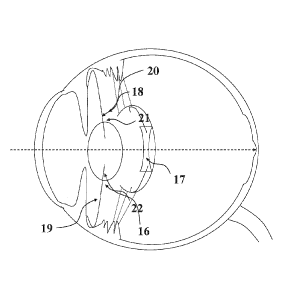
Referring now to both Figures 1 and 2. One embodiment of the present invention
comprises
an anterior light-converging IOL 16 located in the ciliary sulcus 6 and a
posterior light-
diverging IOL 17 located in the capsular bag 18. It should be noted that in
this embodiment
17
CA 02921120 2016-02-11
WO 2015/022515 PCT/GB2014/052459
the capsular bag 18 contains a circular defect anteriorly to facilitate
removal of the natural
crystalline lens or cataract in a manner consistent with current micro-
incisional techniques
employed during cataract surgery. The optical component of the anterior IOL 16
is
maintained in position by haptics in a symmetrical configuration such that the
first haptic 19
is the same length as the second haptic 20 ¨ the optical axis of the anterior
lens therefore
runs in line with that of the eye. The optic of the posterior IOL is
maintained in position in the
capsular bag by means of haptics attached such that the first haptic 21 is the
same length as
the second 22 ¨ the optical axis of the posterior lens therefore runs in line
with that of the
eye and that of the anterior lens. In this embodiment both optics 16, 17 are
made of a
hydrophobic material, such as soft acrylic polymer (refractive index 1.54;
Abbe number 40;
visible range transmission >92%; ultraviolet light transmission <0.5%), but
generally the
optics may be made from any transparent, biocompatible material used in
intraocular lens
construction, with calculations for optimisation of the optic surfaces (as set
out below)
revised accordingly. Similarly the haptics 19, 20, 21, 22 may be may be formed
of any
suitable polymeric material including polymethymethacrylate and/or
polypropylene. The 10Ls
are designed to be foldable to facilitate implantation via a wound in the eye
less than 5mm in
length.
Referring to Figures 1, 2, 3, 4, 5, 6 and 7, aspects of the 10Ls and their
arrangement are
discussed in more detail.
Figure 3 shows the anterior light-converging IOL in cross-section 23 and from
the top 24.
The anterior converging IOL consists of a light converging optic of a
thickness 25, a diameter
26 and a dioptric power such that in conjunction with the posterior 10L, light
may be focused
to provide a retinal image of a specific magnification across a range of
retinal eccentricities
at the macula. To achieve a retinal image of sufficient quality to benefit an
individual with
poor central vision, the optical design of the first lens is optimized such
that it consists of an
aspheric anterior surface 27 and an aspheric posterior surface 28. Each
surface of the first
18
CA 02921120 2016-02-11
WO 2015/022515 PCT/GB2014/052459
lens is a rotationally symmetric polynomial aspheric conic surface for which
the surface sag
(z co-ordinate) as a function of the radial coordinate r can be given by:
2=
1+ 1¨(1+k)c-2r2
Wherein,
i) c is the inverse of the radius of curvature R: c=1/R
ii) k is the conical constant (with a value ranging between -1 and 0
iii) a is an aspheric polynomial coefficient, additional to the conical
constant
The first lens is centred in line with the optical axis of the eye by means of
two haptics that
are attached to or continuous with the anterior optic such that the first
haptic 29 is the same
length as the second haptic 30. The optic is therefore sited equidistant 31,
32 from the point
at which each haptic is designed to make contact with the eye 6. It should be
noted that in
this embodiment both haptics are angled anteriorly from the point at which
they emerge from
the optic in such a way that the optic is sited in a plane that lies posterior
to that of the ciliary
sulcus ¨ in this way the anterior surface of the anterior IOL remains clear of
the iris 5.
However, the haptics may be designed for positioning of the optic in the
anterior chamber 3,
the ciliary sulcus 6 or the capsular bag 9 of the eye.
VVith reference to Figure 4, the posterior light-diverging IOL is shown in
cross-section 33 and
from the top 34. The posterior light-diverging IOL consists of a light-
diverging optic of a
central thickness 35, diameter 36 and dioptric power such that in conjunction
with the
anterior 10L, light may be focused on a region of the macula to provide
retinal images of a
specific magnification. Again, to achieve retinal images of sufficient quality
with this
configuration the optical design of the posterior optic is optimised such that
it consists of a
19
CA 02921120 2016-02-11
WO 2015/022515 PCT/GB2014/052459
rotationally symmetric polynomial aspheric anterior surface 37 and a
rotationally symmetric
polynomial aspheric conic posterior surface 38. For each surface the surface
sag z as a
function of the radial coordinate r can be given by expression:
cy
1+ V 1 ¨ + k)c2 r 2
as with the anterior optic.
By way of example only, the conical constants (k) in one embodiment of the
invention may
be (starting with the anterior surface of the anterior optic):
First surface: -9
Second surface: -0.6
Third surface: -110
Fourth surface: -7
Attached to or continuous with the posterior optic are two haptics 39, 40 of
equal length 41,
42. The haptics may be designed for positioning of the optic in the anterior
chamber 3, the
posterior chamber 4 or the capsular bag 9 of the eye. It should be noted that
in order to
achieve a maximal distance from the anterior IOL it may be necessary to angle
the haptics
39, 40 attached to the posterior IOL such that the optic lies in a plane
posterior to the site
where the haptics make contact with the periphery of the capsular bag 18.
VVith reference to
Figures 2 and 5, that show cross-sections of the arrangement of the anterior
IOL in relation
to the posterior IOL: The 10Ls are arranged in the eye in line with the
optical axis of the eye
such that the anterior light-converging IOL 46 is sited at an optimal distance
from the
posterior light-diverging IOL 47 resulting in a magnification of the retinal
image of 1.2 to 1.4.
CA 02921120 2016-02-11
WO 2015/022515 PCT/GB2014/052459
VVith reference to Figure 1, Figure 5, a diagrammatic representation of the
present invention
in a cross-section of the eye and lines representing the path of light 43
taken in the eye on
entering the cornea and passing through the optics of the present invention
44, 45 and
Figure 6. The optic of the first lens 44 is sited anteriorly to that of the
optic of the second lens
45 in the manner of a Galilean telescope and both lenses are centred with
their optical axes
in line with that of the eye 15. The surfaces of each optic are rendered
aspheric such that a
magnified image is simultaneously presented across a range of eccentricities
at the retina
46, 47, 48. The present invention is optimised to render an image of similar
optical quality in
an area 10 degrees off-axis (an area with a radius of 5 degrees from the
fovea! centre).
Figure 6 demonstrates the off-axis optimization of image quality achieved by
the present
invention at eccentricities of 0, 2.5 and 5 degrees from fixation when
compared with that
obtained with a standard 21 dioptre monofocal optic. The effect is such that a
magnified
image may be presented at a patient's preferred retinal locus without the need
to target this
area of the retina specifically and without requiring the patient to learn to
fixate eccentrically.
Furthermore, if the preferred retinal locus of the patient changes over time
they may
gradually learn to make use of an image presented at a different retinal
eccentricity from that
used initially.
Since even a small deviation from the intended axial positioning of the two
implants relative
to one another could produce a significant refractive error and degradation of
the image
presented at the retina, the current invention increases the tolerance of the
system for sub-
optimal implant axial positioning by rendering one of the surfaces in the
system,
preferentially the posterior surface of the second lens 38, more aspherical
than the other
optical surfaces in the system. This adds aberration and increases the depth
of focus of the
present invention. The precise amount of added aberration is determined to
assure both a
good enough quality of retinal image and a significant range of positioning
tolerance. This
feature of the present invention ensures that it is capable of delivering a
high quality of
21
CA 02921120 2016-02-11
WO 2015/022515 PCT/GB2014/052459
retinal image whilst accommodating variations in the practice of individual
surgeons and
alterations in the anatomy of the eye during the early and late post-operative
periods. The
benefits of added aberration, in increasing the tolerance of IOL positioning
in the present
invention and the quality of the image presented at the retina across a range
of eccentricities
by the present invention, are both shown in Figure 7. It may be seen that a
similar image
quality is delivered at angles of eccentricity ranging from 0 to 5 degrees and
that the quality
is maintained when the distance between the two lenses varies from 1.4mm to
1.7mm.
The optics of the system are further optimised to take account of transverse
chromatic
aberration induced by the vertical displacement of the implants relative to
one another 51,
this is achieved by adding a yellow tint to the implants during the
manufacturing process.
The addition of a yellow tint to the 10Ls also confers the added benefit of
macular protection
from ultraviolet radiation.
VVith reference to Table 1, it can be seen that the surfaces of the optics of
the 10Ls of the
present invention may be further optimised by the addition of values for
Zernike polynomials,
besides those for spherical aberration. The surfaces may be expressed as a
linear
combination of Zernike polynomials including those for tilt, defocus,
astigmatism, and coma,
such that optical aberrations for individual patients are minimised.
Consequent remodelling
of the lenses means that at least one lens design parameter is changed ¨ this
may include
the anterior surface shape and central radius and the posterior surface shape
and central
radius ¨ and 10Ls may be selected from a kit of lenses to achieve the desired
effect.
The materials, biomechanical properties, lengths and shapes of the haptics and
the
materials, surfaces, sizes and biomechanical properties of the anterior and
posterior optics
may be modified to achieve the desired retinal image (the haptics may form
part of a single
piece anterior or posterior IOL for example and may be permit siting of either
lens or both
lenses in the anterior chamber 3, posterior chamber 4 or capsular bag of the
eye 9). It is
further contemplated that a range of anterior and corresponding posterior
implants,
22
CA 02921120 2016-02-11
WO 2015/022515 PCT/GB2014/052459
consisting of a range of dioptric powers, optical surfaces, optic tints and
haptic configurations
may be included in the kit to facilitate targeting of the PRL in individual
patients with a wide
range of refractive errors (this includes toric optics to correct for high
astigmatism). Referring
now to Figure 8 which shows a version of the anterior light-converging IOL in
cross-section
49 and from the top 50. It is contemplated that with the current invention
there is risk of
visually significant vignetting occurring with larger pupil sizes,
particularly where levels of
vertical decentration between the anterior and posterior 10Ls are high. A
version of the
anterior light-converging IOL designed to prevent such vignetting is shown 49,
50. The
diameter of the optic is increased in this embodiment 51 with an added rim 52
rendered
opaque by the application of a biocompatible and stable opaque paint to its
surface 53.
Alternatively an opaque, rim may be located on the surface of the optic, for
example bonded
to the optic as originally conceived to create the same effect. The rim is of
sufficient width to
prevent vignetting with larger pupils. The refractive part of the optic
remains unaffected and
the haptics 54, 55, which are of equal lengths, insert into the optic as
previously described.
VVith reference to Figure 9, which shows a version of the posterior light-
diverging IOL in
cross-section 56 and from the top 57; the same, or a similar, effect may be
achieved by
increasing the diameter of the posterior optic 58 to include a rim 59 that may
be opaque and
bonded to the optic or, as shown in the illustration, rendered opaque by means
of a
biocompatible and stable opaque paint applied to its surface 60 (the
configuration of the
haptics remaining unchanged 61, 62).
In a further embodiment (not shown) the opaque rim may be located within part
of the optic
body.
Although the invention is described in the preferred embodiments illustrated
in the Figures
attached, no restriction is intended by this. The design and configuration of
the optical
surfaces, including application of a tint to refine optical properties, are
considered integral to
the present invention and may be applied in a variety of circumstances. For
example it is
23
CA 02921120 2016-02-11
WO 2015/022515 PCT/GB2014/052459
contemplated that an arrangement of the 10Ls may include positioning of the
anterior light-
converging IOL in the anterior chamber 3 and the posterior light-diverging IOL
in the
posterior chamber 4 or both 10Ls in the posterior chamber or both 10Ls in the
capsular bag
9 with revision of the optical surfaces, IOL dioptric powers and haptic
designs accordingly.
Further embodiments (not shown) include the application of diffractive
surfaces to one optic
or both optics to permit a range of focal points in the eye (and consequently
uncorrected
distance and near vision); and targeting of the PRL ¨ or the introduction of a
third optic with
one of the aforementioned characteristics, to either the anterior chamber, the
posterior
chamber or the capsular bag.
Again, whilst reference to use of the present invention in subjects with AMD
is made, no
restriction in terms of its use is intended. It is contemplated that the
present invention will be
used in a wide variety of clinical scenarios to achieve targeting of areas of
the macula
eccentric to fixation and with a range of magnification and refractive
capabilities. The present
invention is designed for insertion into the eye via a small (5mm) incision
with or without use
of a cartridge injector, an approach consistent with its use in the context of
surgical
techniques employed during natural crystalline lens or cataract extraction. As
such it is
expected that the present invention may be used in combination with natural
crystalline lens
extraction or at the time of cataract surgery or, if necessary, subsequent to
cataract
surgery/lens extraction (with its application - together with any necessary
modifications to the
optic surfaces, haptic design, optic materials and optic dioptric power - in
addition to or
instead of pre-existing implants in the eye).
In keeping with this approach, a range of monofocal 10Ls may be provided that
is designed
for use in cases where the present invention is not indicated at initial
surgery, but where the
natural crystalline lens is removed and the patient wishes to retain the
potential to use the
present invention at a later date. Under these circumstances, the optics of
the monofocal
24
CA 02921120 2016-02-11
WO 2015/022515 PCT/GB2014/052459
IOL implanted at the first operation will be optimised for use in conjunction
with the present
invention should this be required in the event that the patient develops a
macular disease.
A wide range of modification and substitution is contemplated with regards to
the present
disclosure, and the illustrations provided are not intended to restrict the
design of the present
invention or limit the applications of its use. Furthermore it is intended
that a variety of
permutations of the present invention may be created by incorporating the
various properties
as laid out in the Claims attached.