Note: Descriptions are shown in the official language in which they were submitted.
CA 02921234 2016-02-11
WO 2015/047910 PCT/US2014/056602
BIOMARKERS FOR DETECTION OF COLORECTAL CANCER
BACKGROUND OF THE INVENTION
Field of the Invention
[0001] The invention disclosed herein relates generally to the fields of
clinical testing
in oncology. Particularly, the invention provides a biomarker comprising
abnormally
methylated DNA fragments in a sample for detection of early stages (I and II)
of
colorectal cancer. More particularly, the invention provides methods for
detection of
early stages (I and II) of colorectal cancer using a marker comprising
abnormally
methylated DNA fragments in a sample.
Description of Related Art
[0002] Colorectal cancer (CRC), is the third most common form of cancer and
the
second leading cause of death among cancers worldwide, with approximately 1,
000,
000 new cases of CRC and 50, 000 deaths related to CRC each year (Bandres E,
Zarate R, Ramirez N, Abajo A, Bitarte N, Garcia-Foncillas J: Pharmacogenomics
in
colorectal cancer: the first step for individualized-therapy, World J
Gastroenterol 2007,
13(44):5888-5901; Kim H-J, Yu M-H, Kim H, Byun J, Lee CH: Non-invasive
molecular
biomarkers for the detection of colorectal cancer, BMB Rep 2008, 41(10):685-
692).
[0003] Most colorectal cancers develop slowly, beginning as small benign
colorectal
adenomas which progress over several decades to larger and more dysplastic
lesions
which eventually become malignant. This gradual progression provides multiple
opportunities for prevention and intervention.
[0004] The currently used methods for the early detection of CRC are the
Faecal
Occult Blood Test (FOBT) and the endoscopy. FOBT is simple, inexpensive and
the
least invasive method of screening available. Also, it has been shown through
prospective randomized trials that FOBT reduces CRC mortality, and
consequently the
evidence for its use is robust.
[0005] However, FOBT presents relatively high false negative and false
positive
rates, and it has particularly poor sensitivity for the detection of early-
stage lesions (see
e.g., Burch JA, Soares-Weiser K, St John DJ, Duffy S, Smith S, Kleijnen J,
Westwood M:
1
CA 02921234 2016-02-11
WO 2015/047910 PCT/US2014/056602
Diagnostic accuracy of faecal occult blood tests used in screening for
colorectal cancer:
a systematic review, J Med Screen 2007, 14(3):132-137; Allison JE, Tekawa IS,
Ransom LJ, Adrain AL: A comparison of fecal occult blood tests for colorectal-
cancer
screening, N Engl J Med 1996, 334(3):155-159; Greenberg PD, Bertario L, Gnauck
R,
Kronborg 0, Hardcastle JD, Epstein MS, Sadowski D, Sudduth R, Zuckerman GR,
Rockey DC: A prospective multicenter evaluation of new fecal occult blood
tests in
patients undergoing colonoscopy, Am J Gastroenterol 2000, 95(5):1331-1338).
[0006] In an attempt to improve on the false positive rates of FOBT, a new
Faecal
lmmunochemical testing (FIT) has been developed. It has slightly superior
performance
characteristics but at a greatly increased financial cost, and its
implementation has not
been effective as yet (see Newton KF, Newman W, Hill J: Review of biomarkers
in
colorectal cancer, Colorectal Disease 2012, 14(1):3-17).
[0007] On the other hand, colonoscopy offers significant improvements in
detection
rates for CRC but it also has important disadvantages associated, such as
inconvenience, high economic burden and potential major complications
(bleeding,
perforation) (see e.g., Winawer S, Fletcher R, Rex D, Bond J, Burt RW,
Ferrucci J,
Ganiats T, Levin T, Woolf S, Johnson D, et al.: Colorectal cancer screening
and
surveillance: clinical guidelines and rationale-Update based on new evidence,
Gastroenterol 2003, 124:544-560; Greenen JE, Schmitt MG, Wu WC, Hogan WJ:
Major
complications of colonoscopy: bleeding and perforation, Am J Dig Dis 1975,
20:231-
235).
[0008] Since none of the currently available methods are optimal, there is
an urgent
necessity of new diagnostic approaches in order to improve the outcome of CRC
screening programs.
[0009] DNA methylation biomarkers for noninvasive diagnosis of CRC and
precursor
lesions have been extensively studied. Different panels have been reported
attempting
to improve current protocols in clinical practice and several biomarkers (for
example
SEPT9 test) have been established to date (see e.g., Lofton-Day C. et al., DNA
methylation biomarkers for blood-based colorectal cancer screening, Clin Chem.
2008
Feb, 54(2):414-23; Grutzmann R. et al., Sensitive detection of colorectal
cancer in
peripheral blood by septin 9 DNA methylation assay, PLoS One. 2008,
3(11):e3759.;
deVos T. et al., Circulating methylated SEPT9 DNA in plasma is a biomarker for
colorectal cancer, Clin Chem. 2009 Jul, 55(7):1337-46; Ahlquist D. A. et al.,
The stool
z
CA 02921234 2016-02-11
WO 2015/047910 PCT/US2014/056602
DNA test is more accurate than the plasma septin 9 test in detecting
colorectal
neoplasia, Clin Gastroenterol Hepatol. 2012 Mar, 10(3):272-7.e1; Ladabaum U.
et al.,
Colorectal Cancer Screening with Blood-Based Biomarkers: Cost-Effectiveness of
Methylated Septin 9 DNA versus Current Strategies, Cancer Epidemiol Biomarkers
Prey.
2013 Sep, 22(9):1567-76). However, these tests suffer from low sensitivity in
detecting
early stages (I and II) of colorectal cancer and no definite biomarkers have
been
identified to date that can be reliably used for detecting CRC in blood
samples. Thus,
there is a clinical need for identifying specific biomarkers for early
detection of CRC that
can be tested in a noninvasive manner.
SUMMARY OF THE INVENTION
[0010] It is
against the above background that the present invention provides certain
advantages and advancements over the prior art.
[0011]
Although this invention is not limited to specific advantages or
functionality, it
is noted that the invention disclosed herein provides methods for detecting
cancer,
comprising:
(a)
analyzing a biological sample from a subject to detect a presence of a
biomarker in the sample, comprising
i. obtaining a DNA sample from a subject;
ii. digesting the DNA sample with a methylation-sensitive restriction
enzyme in the presence of a glycol compound;
iii. amplifying the digested sample of step (ii);
iv. quantifying amplification results from step (iii) using a real-time
quantitative PCR; and
v. analyzing DNA methylation status within a recognition site of the
methylation-sensitive restriction enzyme to detect a presence of the biomarker
in
the sample;
(b)
determining a methylation status of the biomarker detected in step (v);
and
3
CA 02921234 2016-02-11
WO 2015/047910 PCT/US2014/056602
(c) comparing the methylation status of the biomarker detected in the
sample
to cancer-positive and/or cancer-negative reference methylation status of the
biomarker
to detect whether the subject has cancer.
[0012] In preferred embodiments, the step of comparing further comprises:
(a) determining probabilities (P) of the biomarker being methylated or
unmethylated;
(b) determining cumulative probabilities of error (p) associated with
probabilities
(P) of the biomarker being methylated or unmethylated;
(c) determining cumulative probabilities of error for the biomarker in a
healthy
and a diseased state; and
(d) detecting that the subject has cancer if the cumulative probabilities of
error for
the biomarker in the healthy state is more than the cumulative probabilities
of error for
the biomarker in the diseased state.
[0013] In preferred embodiment, the subject is a mammal, wherein the mammal
is a
human.
[0014] In one aspect, the sample is a biological sample comprising blood,
blood
plasma, urine or saliva.
[0015] In further aspect, the biomarker comprises one or more DNA fragments
of
SEQ ID Nos. 1-12.
[0016] In further aspect, the cancer is colorectal cancer.
[0017] In further aspect, the colorectal cancer comprises early stage 1 and
11
colorectal cancer or late stage colorectal cancer.
[0018] In further aspect, the DNA comprises a genomic DNA.
[0019] In further aspect, the DNA sample is between about 1 pg and about 1
ng.
[0020] In further aspect, the DNA sample is about 300 pg.
[0021] In further aspect, the methylation-sensitive restriction enzyme
comprises
Hin61.
[0022] In further aspect, amplifying comprises amplifying using phi29 DNA
polymerase.
4
CA 02921234 2016-02-11
WO 2015/047910 PCT/US2014/056602
[0023] In further aspect, the amplifying further comprises amplifying using
a single
stranded DNA binding protein of E. co/i.
[0024] In further aspect, the real-time quantitative PCR comprises TaqMan
qPCR.
[0025] In further aspect, determining the DNA methylation status and/or
probability
of a methylation status comprises determining threshold cycle (CT) values.
[0026] The invention disclosed herein further provides a biomarker for
detecting
cancer, wherein the biomarker comprises one or more DNA fragments of SEQ ID
Nos.
1-12.
[0027] In one aspect, the cancer is colorectal cancer.
[0028] In further aspect, the colorectal cancer comprises early stage I and
ll
colorectal cancer or late stage colorectal cancer.
[0029] These and other features and advantages of the present invention
will be
more fully understood from the following detailed description of the invention
taken
together with the accompanying claims. It is noted that the scope of the
claims is
defined by the recitations therein and not by the specific discussion of
features and
advantages set forth in the present description.
BRIEF DESCRIPTION OF THE DRAWINGS
[0030] The following detailed description of the embodiments of the present
invention can be best understood when read in conjunction with the following
drawings,
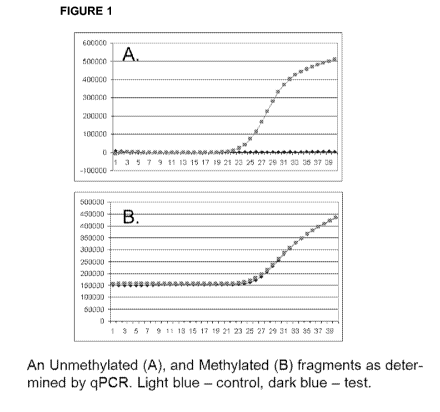
where like structure is indicated with like reference numerals and in which:
[0031] Figure 1 shows qPCR profile of methylated and unmethylated
fragments.
[0032] Skilled artisans will appreciate that elements in the figures are
illustrated for
simplicity and clarity and have not necessarily been drawn to scale. For
example, the
dimensions of some of the elements in the figures can be exaggerated relative
to other
elements to help improve understanding of the embodiment(s) of the present
invention.
DETAILED DESCRIPTION OF THE INVENTION
[0033] All publications, patents and patent applications cited herein are
hereby
expressly incorporated by reference for all purposes.
[0034] Methods well known to those skilled in the art can be used to
construct
genetic expression constructs and recombinant cells according to this
invention. These
CA 02921234 2016-02-11
WO 2015/047910 PCT/US2014/056602
methods include in vitro recombinant DNA techniques, synthetic techniques, in
vivo
recombination techniques, and PCR techniques. See, for example, techniques as
described in Maniatis et al., 1989, MOLECULAR CLONING: A LABORATORY MANUAL,
Cold
Spring Harbor Laboratory, New York; Ausubel et al., 1989, CURRENT PROTOCOLS IN
MOLECULAR BIOLOGY, Greene Publishing Associates and Wiley lnterscience, New
York,
and PCR Protocols: A Guide to Methods and Applications (Innis et al., 1990,
Academic
Press, San Diego, CA).
Definitions
[0035] Before describing the present invention in detail, a number of terms
will be
defined. As used herein, the singular forms "a", "an", and "the" include
plural referents
unless the context clearly dictates otherwise. For example, reference to a
"nucleic acid"
means one or more nucleic acids.
[0036] It is noted that terms like "preferably", "commonly", and
"typically" are not
utilized herein to limit the scope of the claimed invention or to imply that
certain features
are critical, essential, or even important to the structure or function of the
claimed
invention. Rather, these terms are merely intended to highlight alternative or
additional
features that can or cannot be utilized in a particular embodiment of the
present
invention.
[0037] For the purposes of describing and defining the present invention it
is noted
that the term "substantially" is utilized herein to represent the inherent
degree of
uncertainty that can be attributed to any quantitative comparison, value,
measurement,
or other representation. The term "substantially" is also utilized herein to
represent the
degree by which a quantitative representation can vary from a stated reference
without
resulting in a change in the basic function of the subject matter at issue.
[0038] As used herein, the terms "polynucleotide", "nucleotide",
"oligonucleotide",
and "nucleic acid" can be used interchangeably to refer to nucleic acid
comprising DNA,
RNA, derivatives thereof, or combinations thereof.
[0039] As used herein, the term "nucleic acid" refers to polynucleotides
such as
deoxyribonucleic acid (DNA), and, where appropriate, ribonucleic acid (RNA).
The term
should also be understood to include, as equivalents, analogs of either RNA or
DNA
made from nucleotide analogs, and, as applicable to the embodiment being
described,
single-stranded (such as sense or antisense) and double-stranded
polynucleotides.
6
CA 02921234 2016-02-11
WO 2015/047910 PCT/US2014/056602
[0040] The terms "compound", "test compound," "agent", and "molecule" are
used
herein interchangeably and are meant to include, but are not limited to,
peptides, nucleic
acids, carbohydrates, small organic molecules, natural product extract
libraries, and any
other molecules (including, but not limited to, chemicals, metals, and
organometallic
compounds).
[0041] The term "detection" is used herein to refer to any process of
observing a
marker, or a change in a marker (such as for example the change in the
methylation
state of the marker), in a biological sample, whether or not the marker or the
change in
the marker is actually detected. In other words, the act of probing a sample
for a marker
or a change in the marker, is a "detection" even if the marker is determined
to be not
present or below the level of sensitivity. Detection may be a quantitative,
semi-
quantitative or non-quantitative observation.
[0042] The term "including" is used herein to mean, and is used
interchangeably
with, the phrase "including but not limited to."
[0043] The term "isolated" as used herein with respect to nucleic acids,
such as DNA
or RNA, refers to molecules in a form which does not occur in nature.
Moreover, an
"isolated nucleic acid" is meant to include nucleic acid fragments which are
not naturally
occurring as fragments and would not be found in the natural state.
[0044] The term "or" is used herein to mean, and is used interchangeably
with, the
term "and/or", unless context clearly indicates otherwise.
[0045] The terms "phenotype" or "phenotypic status" are used herein
interchangeably and are meant to describe whether a subject has or does not
have a
particular disease.
[0046] The term "healthy state" means that a subject does not have a
particular
disease.
[0047] The term "diseased state" means that a subject has a particular
disease.
[0048] "Sample" or "biological sample" means biological material isolated
from a
subject. The biological sample may contain any biological material suitable
for detecting
the desired biomarkers, and may comprise cellular and/or non-cellular material
from the
subject. The sample may be isolated from any suitable biological tissue or
fluid such as,
for example but not limited to, blood, blood plasma, urine or saliva. A
"sample" includes
7
CA 02921234 2016-02-11
WO 2015/047910 PCT/US2014/056602
any material that is obtained or prepared for detection of a molecular marker
or a change
in a molecular marker such as for example the methylation state, or any
material that is
contacted with a detection reagent or detection device for the purpose of
detecting a
molecular marker or a change in the molecular marker.
[0049] A "subject" is any organism of interest, generally a mammalian
subject, such
as, for example, a human, monkey, mouse, or rabbit, and preferably a human
subject.
[0050] The term "biomarker" means an organic biomolecule(s) a marker and/or
a
panel of DNA fragments, such as for example but not limited to a panel of
methylated or
unmethylated DNA fragments, which are differentially present (i.e., present
with an
incorrect methylation status) in a biological sample taken from a subject or a
group of
subjects having a first phenotype (e.g., having a disease) as compared to a
biological
sample from a subject or group of subjects having a second phenotype (e.g.,
not having
the disease).
[0051] A biomarker may be differentially present at any level, but is
generally
present at a level that is increased by at least 5%, by at least 10%, by at
least 15%, by at
least 20%, by at least 25%, by at least 30%, by at least 35%, by at least 40%,
by at least
45%, by at least 50%, by at least 55%, by at least 60%, by at least 65%, by at
least 70%,
by at least 75%, by at least 80%, by at least 85%, by at least 90%, by at
least 95%, by at
least 100%, by at least 110%, by at least 120%, by at least 130%, by at least
140%, by
at least 150%, or more; or is generally present at a level that is decreased
by at least
5%, by at least 10%, by at least 15%, by at least 20%, by at least 25%, by at
least 30%,
by at least 35%, by at least 40%, by at least 45%, by at least 50%, by at
least 55%, by at
least 60%, by at least 65%, by at least 70%, by at least 75%, by at least 80%,
by at least
85%, by at least 90%, by at least 95%, or by 100% (i.e., absent).
[0052] A biomarker is preferably differentially present between different
phenotypic
statuses at a level that is statistically significant (i.e., a p-value less
than 0.05 and/or a q-
value of less than 0.10 as determined using the following, among others,
tests: Welch's
T-test, Wilcoxon's rank-sum Test, ANOVA, Kruskal-Wallis, Mann-Whitney, and
odds
ratio):
[0053] A biomarker, as described above, may provide a measure of relative
risk that
a subject belongs to one phenotypic status or another. Therefore, the
biomarker may be
useful for disease diagnostics.
8
CA 02921234 2016-02-11
WO 2015/047910 PCT/US2014/056602
[0054] A
"reference methylation status" of a biomarker means a methylation status
of the biomarker that is indicative of a particular disease state, phenotype,
or lack
thereof, as well as combinations of disease states, phenotypes, or lack
thereof. A
"positive" reference methylation status of the biomarker means a methylation
status that
is indicative of a particular disease state or phenotype. A "negative"
reference
methylation status of the biomarker means a methylation status that is
indicative of a
lack of a particular disease state or phenotype. Specifically, a reference
methylation
status of the biomarker in healthy subjects may be used to determine a
"negative
reference methylation status."
[0055] For
example, a "colorectal cancer-positive reference methylation status" of a
biomarker means a methylation status the biomarker that is indicative of a
positive
diagnosis of colorectal cancer in a subject, and a "colorectal cancer-negative
reference
methylation status" of a biomarker means a methylation status of the biomarker
that is
indicative of a negative diagnosis of colorectal cancer in a subject.
[0056] A
"reference methylation status" of a biomarker may be a combination of
relative methylation statuses of one and/or several DNA fragments, and such
reference
methylation status may be tailored to specific populations of subjects (e.g.,
a reference
methylation status may be age-matched so that comparisons may be made between
methylation statuses of DNA fragments in samples from subjects of a certain
age and for
a particular disease state, phenotype, or lack thereof in a certain age
group). Such
reference methylation status may also be tailored to specific techniques that
are used to
measure methylation status in biological samples (e.g., DNA methylation,
etc.), where
the methylation status may differ based on the specific technique that is
used.
[0057]
"Abnormally methylated" as used herein with respect to DNA fragments from
a sample, refers to fragments that are methylated in the sample when such
fragments
are supposed to be unmethylated or fragments that are unmethylated in the
sample
when such fragments are supposed to be methylated with respect to a "reference
methylation status" discussed above.
Overview
[0058] In
certain aspects, the invention relates to a biomarker, wherein the
biomarker comprises one or more abnormally methylated DNA fragments (see Table
1)
from a sample from a subject for detection of early stages (I and II) of
colorectal cancer
9
CA 02921234 2016-02-11
WO 2015/047910 PCT/US2014/056602
and methods for the in vitro detection of early stages (I and II) of
colorectal cancer by
determining the presence of said biomarker in the sample from the subject.
[0059] Generally, DNA methylation in fragments of Table 1 may be determined
for
biological samples from subjects diagnosed with colorectal cancer as well as
from one or
more other groups of human subjects (e.g., healthy control subjects not
diagnosed with
colorectal cancer), as well as from human subjects diagnosed with early stage
I and ll
colorectal cancer and human subjects diagnosed with late stages colorectal
cancer.
Biomarkers
[0060] Methylation status of the one or more DNA fragments comprising the
biomarker in biological samples from a subject having colorectal cancer was
compared
to the methylation status of the one or more DNA fragments comprising the
biomarker in
biological samples from the one or more other groups of subjects. A biomarker
comprising one or more abnormally methylated DNA fragments, including those
abnormally methylated at a level that is statistically significant, in the
methylation profile
of samples from subjects with colorectal cancer as compared to another group
(e.g.,
healthy control subjects not diagnosed with colorectal cancer) was used to
distinguish
those groups. In addition, abnormally methylated DNA fragments, including
those
abnormally methylated at a level that is statistically significant, in the
methylation profile
of samples from subjects with early stage I and ll colorectal cancer as
compared to late
stage colorectal cancer were also identified as biomarkers to distinguish
those groups.
[0061] The biomarker is discussed in more detail herein. The biomarker
comprising
one or more DNA fragments (see Table 1) was used for distinguishing subjects
having
colorectal cancer (early stage I and ll and/or late stage) vs. control
subjects not
diagnosed with colorectal cancer. The sequence information for DNA fragments
comprising the biomarker (see Table 1) is shown in Table 7. DNA methylation
sites are
shown in bold and are underlined.
[0062] Table 1. DNA fragments comprising the biomarker.
CA 02921234 2016-02-11
WO 2015/047910 PCT/US2014/056602
Status of
methylation
# Healthy CRC GENE Location Known function
Ca-activated K-chanel,
regulates calcium
1 Unmeth Meth KCN N4 19q13.2 influx
alkaline ceramidase 3,
positively regulates
2 Unmeth Meth ACER3 11q13.5 cell proliferation
GLI family zinc finger 4;
glioma-assoc.
3 Unmeth Meth GLI4 8q24.3 oncogene family
4 Unmeth Meth ZN F629 16p11.2
Mucin 2; loss of
expression -
Unmeth Meth MUC2 11p15.5 recurrence
Histone deacetylase 4
promotes CRC via
6 Unmeth Meth HDAC4 2q37.3 repression of p21
Perilipin3 binds
directly to the GTPase
7 Meth Unmeth PLIN3 19p13.3 RAB9 (RAB9A)
8 Meth Unmeth ZNF30 19q13.11
cadherin, [GE LAG
seven-pass G-type
9 Meth Unmeth CELSR1 22q13.3 receptor 1
Meth Unmeth unkown chr8:1094666-1094715
11 Meth Unmeth unkown chr2:583162-583222
12 Meth Unmeth NIPAL3 1p36.12-p35.1
[0063] Although the identities of some of the biomarkers are not known at
this time,
such identities are not necessary for the identification of the biomarkers in
biological
samples from subjects, as the "unnamed" biomarkers have been sufficiently
characterized by analytical techniques to allow such identification. The
methodology for
analytical characterization of all such "unnamed" biomarkers is described in
Example 1.
Detection of Colorectal Cancer
[0064] After the methylation status of the one or more DNA fragments
comprising
the biomarker are determined in the sample, the methylation status of the one
or more
DNA fragment comprising the biomarker compared to colorectal cancer-positive
and/or
11
CA 02921234 2016-02-11
WO 2015/047910 PCT/US2014/056602
colorectal cancer-negative reference methylation status to detect or aid in
detecting
whether the subject has colorectal cancer. Methylation status of the one or
more DNA
fragment comprising the biomarker in a sample, including those abnormally
methylated
or unmethylated a level that is statistically significant, matching the
colorectal cancer-
positive reference methylation status (e.g., methylation status that is the
same as the
reference methylation status, substantially the same as the reference
methylation status,
above and/or below the minimum and/or maximum of the reference methylation
status,
and/or within the range of the reference methylation status) are indicative of
a detecting
of colorectal cancer in the subject. Methylation status of the one or more DNA
fragment
comprising the biomarker in a sample, including those abnormally methylated or
unmethylated a level that is statistically significant, matching the
colorectal cancer-
negative reference methylation status (e.g., methylation status that is the
same as the
reference methylation status, substantially the same as the reference
methylation status,
above and/or below the minimum and/or maximum of the reference methylation
status,
and/or within the range of the reference methylation status) are indicative of
a detection
of no colorectal cancer in the subject.
[0065] The methylation status of the one or more DNA fragments comprising
the
biomarker may be compared to colorectal cancer-positive and/or colorectal
cancer-
negative reference methylation status using various techniques, including but
not limited
to a simple comparison (e.g., a manual comparison) of the methylation statuses
in the
biological sample to colorectal cancer-positive and/or colorectal cancer-
negative
reference levels. The methylation status of the one or more DNA fragments
comprising
the biomarker in the biological sample may also be compared to colorectal
cancer-
positive and/or colorectal cancer-negative reference methylation status using
one or
more statistical analyses (e.g., t-test, Welch's T-test, Wilcoxon's rank sum
test, random
forest).
[0066] The identification of a biomarker comprising one or more DNA
fragments for
colorectal cancer allows for the detection of (or for aiding in the detection
of) colorectal
cancer in asymptomatic subjects and/or subjects presenting with one or more
symptoms
of colorectal cancer. A method of detecting (or aiding in detecting) whether a
subject
has colorectal cancer may comprise (1) analyzing a biological sample from a
subject to
determine the presence of a biomarker comprising one or more DNA fragments for
colorectal cancer in the sample and (2) comparing the methylation status of
the one or
12
CA 02921234 2016-02-11
WO 2015/047910 PCT/US2014/056602
more DNA fragments comprising the biomarker in the sample to colorectal cancer-
positive and/or colorectal cancer-negative reference methylation status of the
one or
more DNA fragments comprising the biomarker. When such a method is used to aid
in
the detection of colorectal cancer, the results of the method may be used
along with
other methods (or the results thereof) useful in the clinical determination of
whether a
subject has colorectal cancer.
[0067] The methods of detecting (or aiding in detecting) whether a subject
has
colorectal cancer may also be conducted specifically to detect (or aid in
detecting)
whether a subject has an early stage I and ll colorectal cancer and/or late
stage
colorectal cancer. Such methods may comprise (1) analyzing a biological sample
from a
subject to determine the presence of a biomarker comprising one or more DNA
fragments in the sample of early stage I and ll colorectal cancer (and/or late
stage
colorectal cancer) in the sample, (2) determining the methylation status of
the one or
more DNA fragment comprising the biomarker, and (3) comparing the methylation
status
of the one or more DNA fragment comprising the biomarker in the sample to an
early
stage I and ll colorectal cancer-positive and/or an early stage I and ll
colorectal cancer-
negative reference methylation status (or late stage colorectal cancer-
positive and/or
late stage colorectal cancer-negative reference methylation status) in order
to detect (or
aid in the detection of) whether the subject has an early stage I and ll
colorectal cancer
(or late stage colorectal cancer).
[0068] Detection of (or aiding in the detection of) colorectal cancer using
above
described biomarker is based on detecting abnormal methylation status of DNA
fragments to detect early stages (I and II) of colorectal cancer. While each
fragment is
not sufficient to identify cancer with sufficient accuracy, the combination of
relative
probabilities of several fragments identifies the disease with very high
accuracy.
Abnormal methylation of the fragments (i.e., methylation that does not
correspond to
methylation status for the same fragments in healthy subjects) is detected
using the
technology for analysis of DNA methylation in ultra-small samples as described
below.
[0069] Thus, detection of (or aiding in the detection of) colorectal cancer
using
above described biomarker is based, in part, on determining the probabilities
(P) of
consensus reading (in regards to methylated status) for DNA fragments
comprising the
biomarker as shown in Table 2. The probabilities are recorded together with
the errors
(p) p=1-P for each of the DNA fragments comprising the biomarker.
13
CA 02921234 2016-02-11
WO 2015/047910
PCT/US2014/056602
[0070] Table 2. Methylation status of DNA fragments comprising the
biomarker.
1 2 3 4 5 6 7 8 9 10 11 12
Healthy (consensus) M MMM M
MMMMMMM
Probability of consensus reading M P 0.2 0.3 0.3
0.1 0.3 0.2 0.7 0.8 0.7 0.8 0.8 0.8
Error p =1-P 0.8 0.7 0.7 0.9 0.7 0.8 0.3
0.2 0.3 0.2 0.2 0.2
Cancer (consensus) M MMM M
MMMMMMM
Probability of consensus reading M P 0.7 0.7 0.8 0.7 0.7 0.8 0.2
0.3 0.3 0.3 0.2 0.2
Error p =1-P 0.3 0.3 0.2 0.3 0.3 0.2 0.8
0.7 0.7 0.7 0.8 0.8
[0071] The
first six DNA fragments are selected to produce unmethylated readout in
healthy control subjects not diagnosed with colorectal cancer and methylated
status in
subjects diagnosed with colorectal cancer. Accordingly, the probabilities (P)
of these
DNA fragments being methylated are small and the errors (p) are large in
healthy
subjects, while the probabilities (P) of these DNA fragments being methylated
are large
and the errors (p) are small in subjects diagnosed with colorectal cancer.
[0072] The
last six DNA fragments are selected to produce methylated readout in
healthy control subjects not diagnosed with colorectal cancer and unmethylated
status in
subjects diagnosed with colorectal cancer. Accordingly, the probabilities (P)
of these
DNA fragments being methylated are large and the errors (p) are small in
healthy
subjects, while the probabilities (P) of these DNA fragments being
unmethylated are
small and the errors (p) are large in subjects diagnosed with colorectal
cancer.
[0073] The
error rates associated with probabilities for each DNA fragment being
either methylated or unmethylated in healthy subjects and subjects diagnosed
with
colorectal cancer are summarized in Table 3, which are used for determining
whether a
subject has colorectal cancer as discussed in Example 2.
[0074] Table
3. Probabilities of error for each fragment being unmethylated or
methylated in healthy subjects and subjects diagnosed with colorectal cancer.
14
CA 02921234 2016-02-11
WO 2015/047910 PCT/US2014/056602
Healthy sample Cancer sample
p =1-P p =1-P
Status Status
# M UM ... # M UM
1 0.8 0.2 1 0.3 0.7
2 0.7 .... 0.3 2 0.3 0.7
3 0.7 0.3 3 0.2 0.8
4 0.9 0.1 4 0.3 0.7
5 0.7 0.3 5 0.3 0.7
6 0.8 0.2 6 0.2 0.8
7 0.3 0.7 7 0.8 0.2
8 0.2 0.8 8 0.7 0.3
9 0.3 0.7 9 0.7 0.3
10 0.2 0.8 10 0.7 0.3
11 0.2 ... 0.8 11 0.8 0.2
12 0.2 ... 0.8 12 0.8 0.2
[0075] The methylation status of one or more DNA fragment comprising the
biomarker may be determined in the methods of detecting and methods of aiding
in
detecting whether a subject has colorectal cancer. For example, the
methylation status
of one DNA fragment, two or more DNA fragments, three or more DNA fragments,
four
or more DNA fragments, five or more DNA fragments, six or more DNA fragments,
seven or more DNA fragments, eight or more DNA fragments, nine or more DNA
fragments, ten or more DNA fragments, etc., including a combination of all of
the DNA
fragments in Table 1, may be determined and used in such methods.
[0076] Determining methylation status of combinations of the DNA fragments
may
allow greater sensitivity and specificity in detecting colorectal cancer and
aiding in the
detection of colorectal cancer, and may allow better differentiation of
colorectal cancer
from other colorectal disorders (e.g., appendicitis, benign adenoma,
ulcerative colitis,
Crohn's disease, diverticular disease, Irritable Bowel Syndrome, etc.) or
other cancers
that may have similar or overlapping biomarkers to colorectal cancer (as
compared to a
subject not having colorectal cancer).
Discovery of Colorectal Biomarkers
[0077] The colorectal cancer biomarkers described herein were discovered
using
analysis of DNA methylation in selected fragments using ultra-small samples
(300 pg or
less) of genomic DNA extractable from clinical samples.
[0078] Briefly, DNA samples obtained from a subject was divided into two
parts; one
part was treated with the methylation-sensitive restriction enzyme and/or
methylation-
dependent restriction enzyme in defined conditions, while the other part was
incubated
without the enzyme and serves as the control. Genomic DNA in both parts was
CA 02921234 2016-02-11
WO 2015/047910
PCT/US2014/056602
amplified using genome-wide amplification with phi29 enzyme, and selected
fragments
were analyzed using TaqMan quantitative PCR. The ACt of the restriction enzyme-
treated and control parts of the sample were compared to determine the
methylation
status and/or probability of a methylation status of the recognition sites for
the restriction
enzyme within the selected fragments.
Digestion of ultra-small samples (300 pg or less) of genomic DNA with a
methylation-
sensitive restriction enzyme
[0079] In one embodiment, methylation-sensitive restriction enzyme Hin6I
(ThermoScientific) was used for restriction digestion (see Example 1). This
enzyme
recognizes the site GCGC, and does not cut DNA if the second nucleotide
(cytosine) is
methylated. Importantly, the reaction conditions generally used for
restriction digest with
Hin6I (see e.g., http://vvvvvv.thermoscientificbio.com/search/?term=Hin61) are
not suitable
for effective and efficient digestion of genomic DNA at ultra-low levels (300
pg or less).
[0080] Alternatively, a restriction digestion may be carried for example
with the
following, but not limiting, methylation-sensitive and methylation-dependent
restriction
enzymes and their isoschizomers as shown below in Tables 4 and 5.
[0081] Table 4. Methylation-sensitive restriction enzymes.
Restriction Enzyme Recognition Sequence Catalog Number
Aat I I GACGT1C Clontech: 1112A/B
Acc 11 CG1CG Clontech: 1002A/B
Aor13H 1 T1CCGGA Clontech: 1224A/B
Aor51H 1 AGC1GCT Clontech: 1118A/B
BspT1041 TT1CGAA Clontech: 1225A/B
BssH 11 G1CGCGC Clontech: 1119A/B
Cfr101 R1CCGGY Clontech: 1120A/B
Cla 1 ATI CGAT
Clontech: 1034A/B/AH/BH
Cpol CG1GWCCG Clontech: 1035A/B
Eco521 C1GGCCG Clontech: 1039A/B
Hae 11 RGCGClY Clontech: 1052A/B
Hap 11 C1CGG
Clontech: 1053A/B/AH/BH
Hha 1 GCG1C Clontech: 1056A/B
WI A1CGCGT
Clontech: 1071A/B/AH/BH
Nae 1 GCC1GGC Clontech: 1155A/B
Notl GC1GGCCGC Clontech: 1166A/B/BH
Nru I TCG1CGA Clontech: 1168A/B
Nsb 1 TGC1GCA Clontech: 1226A/B
PmaC 1 CAC1GTG Clontech: 1177A/B
16
CA 02921234 2016-02-11
WO 2015/047910
PCT/US2014/056602
Psp1406 I AA1CGTT Clontech: 1108A/B
Pvu I CGAT1CG Clontech: 1242A/B
Sac II CCGC1GG Clontech: 1079A/B
Sal I G1TCGAC
Clontech: 1080A/B/AH/BH
Sma I CCC1GGG
Clontech: 1085A/B/AH/BH
SnaB I TAC1GTA Clontech: 1245A/B
Dpnll 1GATC NEB: R0543S
Hpall C1CGG NEB: R0171S
Mspl C1CGG NEB: R0106S
Sall-HF G1TCGAC NEB: R31385
ScrFI CC1NGG NEB: R0110S
Wherein N = A or C or G or T; D = A or G or T; B = C or G or T; V = A or C or
G; R=A
orG;S=CorG;W=AorT;Y=CorT.
[0082] See e.g.,
http://vvwvv.clontech.com/takara/US/Products/Epigenetics/DNA_Preparation/MSRE_O
ver
view) and https://vvww.neb.com/products/epigenetics/methylation-sensitive-
restriction-
enzymes.
[0083] Table 5. Methylation-dependent restriction enzymes.
Restriction Enzyme Recognition Sequence Catalog
Number
Bisl GmCNGC
Glal GmCGmC
FspEl CmC(N)121 NEB: R06625
LpnPI CmCDG(N)101 NEB: R06635
McrBC PumC(N40-3000)FumC,I, NEB: M02725
MspJI mCNNR(N)121 NEB: R06615
Sgel m5C N N G (N)91
ThermoScientific: ER2211
MspJI mCNNR(N)91 NEB: R06615
FspEl CmC(N)121 NEB: R06625
AspBHI YSCNS(N)81(N)12SNGSR
Wherein N = A or C or G or T; D = A or G or T; B = C or G or T; V = A or C or
G; R=A
orG;S=CorG;W=AorT;Y=CorT.
[0084] See e.g., https://vvvvw.neb.com/products/epigenetics/methylation-
dependent-
restriction-enzymes; Karni et al., PNAS (2011); Murray, Microbiology (2002);
Sitaraman
et al., Gene (2011).
17
CA 02921234 2016-02-11
WO 2015/047910 PCT/US2014/056602
[0085] In
general, the cleavage by Hin6I or any other methylation-sensitive or
methylation-dependent enzyme can be detected, for example, but not limiting
to,
quantitative PCR (qPCR) and next generation sequencing (NGS).
[0086] In
order to accelerate digestion of a genomic DNA present at an ultra-low
concentration (300 pg or less), the inventors have advantageously determined
the
"optimal conditions" for digestion with a methylation-sensitive restriction
enzyme Hin6I
(see Example 1).
[0087] The
efficiency with which a restriction enzyme cuts its recognition sequence
at different locations in a piece of DNA can vary 10 to 50-fold. This is may
be due to
influences of sequences bordering the recognition site, which perhaps can
either
enhance or inhibit enzyme binding or activity (see,
e.g.,
http://vvvvvv.vivo.colostate.edu/hbooks/genetics/biotech/enzymes/cuteffects.htm
l).
[0088]
"Optimal conditions" of the digestion reaction are defined as "complete
digestion" of all unmethylated sites GCGC by the methylation-sensitive
restriction
enzyme Hin6I within an acceptable (<5 hr) timeframe. "Complete digestion" is
defined
as the absence of a specific PCR product from a target within the genome when
the
target contains an unmethylated site GCGC, and 40 cycles of PCR are performed.
Additionally, the "complete" digestion is defined as the absence of a specific
PCR
product following 40 cycles of qPCR, when the undigested part of the sample
demonstrates PCR product with CT range of between 17 and 27.
[0089]
Surprisingly and unexpectedly, the inventors have discovered that DNAzol
Direct, a glycol compound previously used for storing and/or processing of
biological
samples for direct use in PCR, may be advantageously added to the restriction
digestion
of a genomic DNA with a methylation-sensitive restriction enzyme Hin61. For
example,
as described in U.S. Patent No. 7,727,718 (incorporated herein by reference in
its
entirety) DNAzol Direct, a glycol compound, may comprise ethylene glycol,
polyethylene
glycols, polyglycol, propylene glycol, polypropylene glycol and glycol
derivatives
including polyoxyethylene lauryl ether, octylphenol-polyethylene glycol ether,
and
polyoxyethylene cetyl ether.
[0090] The
glycol compounds of this invention may further comprise 1,2-
propanediol, 1,3-butanediol, 1,4-butanediol, 1,4-
cyclohexanedimethanol-, 1,6-
hexanediol, butylene glycol, diethylene glycol, dipropylene glycol, ethylene
and
18
CA 02921234 2016-02-11
WO 2015/047910 PCT/US2014/056602
propylene glycol (including ethylene and propylene glycol monomers and
polymers, e.g.,
low molecular weight (less than 600) polyethylene glycols and low molecular
weight
(less than 600) polypropylene glycols), glycerol, long chain PEG 8000 (about
180
ethylene monomers), methyl propanediol, methyl propylene glycol, neopentyl
glycol,
octylphenol-polyethylene glycol ether, PEG-4 through PEG-100 and PPG-9 through
PPG-34, pentylene glycol, polyethylene glycol 200 (PEG 200 about 4 ethylene
monomers), polyethylene glycols, polyglycol, polyoxyethylene cethyl ether, and
octyl-
polyethylene glycol ether, polyoxyethylene cetyl ether, polyoxyethylene lauryl
ether,
polypropylene glycols, tetraethylene glycol, triethylene glycol,
trimethylpropanediol,
tripropylene glycol.
[0091] The glycol compounds of this invention may yet further comprise
ethylene
glycol, propylene glycol, polyethylene glycols, polypropylene glycols,
polyglycol and
glycol derivatives including polyoxyethylene lauryl ether, octylphenol-
polyethylene glycol
ether, and polyoxyethylene cetyl ether. The preferred organic solvents of this
invention
are polyethylene glycols and glycols derivatives. The most preferred solvents
are
polyethylene glycols.
[0092] Polyalkylene glycols comprise polyethylene glycol (PEG) and
polypropylene
glycol. PEGs are generally commercially available diols having a molecular
weight of
from 200 to 10,000 daltons, more preferably about 200-300 daltons. Suitable
PEGs can
be obtained from Spectrum Laboratory Products, Inc, (Gardena, Calif.,
Molecular weight
200, Cat. # PO 107). The molecular weight of the polyethylene glycol (PEG) can
range
from about 200 to about 10,000. Generally, the polyalkylene concentration will
depend
on the polyalkylene used. Depending on the weight range of polyethylene glycol
used,
the concentration can be adjusted. The PEG at a concentration from about 0.1%
to
about 100% and PPG, when added to a PCR mix, have been shown to inhibit the
effect
of impurities on PCR.
[0093] Surprisingly and unexpectedly, the inventors have discovered that
addition of
DNAzol Direct (Molecular Research Center, Inc.; Cat. # DN 131) to the reaction
mix (see
Example 1) resulted in an accelerated and complete digestion of a genomic DNA
present at an ultra-low concentration (300 pg or less). In a typical reaction
described
herein, acceleration of a complete restriction digestion with a methylation-
sensitive
restriction enzyme Hin6I was achieved with a genomic DNA sample at a
concentration of
2.33 ng/ml.
19
CA 02921234 2016-02-11
WO 2015/047910 PCT/US2014/056602
Efficient amplification of genomic DNA using phi29 Pol in the presence of the
E.coli ssb
protein
[0094]
Several useful methods have been developed that permit amplification of
nucleic acids. Most were designed around the amplification of selected DNA
targets
and/or probes, including the polymerase chain reaction (PCR), ligase chain
reaction
(LCR), self-sustained sequence replication (35R), nucleic acid sequence based
amplification (NASBA), strand displacement amplification (SDA), and
amplification with
Q13 replicase (Birkenmeyer and Mushahwar, J. Virological Methods, 35:117-126
(1991);
Landegren, Trends Genetics, 9:199-202 (1993)).
[0095] An
exemplary method is known as primer extension preamplification (PEP).
This technique uses random primers in combination with a thermostable DNA
polymerase to replicate copies throughout the genome. Exemplary conditions
that can
be used for PEP-PCR are described in Zhang et al., Proc. Natl. Acad. Sci. USA,
89:5847-51 (1992); Casas et al., Biotechniques 20:219-25 (1996); Snabes et
al., Proc.
Natl. Acad. Sci. USA, 91:6181-85 (1994,); or Barrett et al., Nucleic Acids
Res., 23:3488-
92 (1995).
[0096]
Further amplification methods may include, but not limited to, isothermal
strand displacement nucleic acid amplification as described in U.S. Pat. NOs.
6,214,587
or 5,043,272.
[0097] Other
non-PCR-based methods that can be used in the invention include, for
example, strand displacement amplification (SDA) which is described in Walker
et al.,
Molecular Methods for Virus Detection, Academic Press, Inc., 1995; U.S. Pat.
Nos.
5,455,166, and 5,130,238, and Walker et al., Nucl. Acids Res. 20:1691-96
(1992) or
hyperbranched strand displacement amplification which is described in Lage et
al.,
Genome Research 13:294-307 (2003).
[0098] Other
methods may include, but not limited to, are Nicking Enzyme
Amplification Reaction (NEAR) as described in
http://vvwvv.envirologix.com/artman/publish/article_314.shtml; nucleic acid
sequence-
based amplification (NASBA) as described in
http://vvwvv.premierbiosoft.com/tech_notes/NASBA.html; and Cross Priming
Amplification
as described in http://wvvw.readcube.com/articles/10.1038/srep00246?locale=en.
CA 02921234 2016-02-11
WO 2015/047910 PCT/US2014/056602
[0099] The
use of the following polymerases has been previously described: Bst and
Klenow fragment
(https://vvww.neb.com/applications/d na-am plification-and-
per/isothermal-amplification;
http://wvvw.neb-online.de/isothermal_amp.pdf); RPA
(http://alere-tech nologies.com/en/prod ucts/lab-solutions/isothermal-
amplification . html);
thermophilic Helicase-Dependent Amplification (tH
DA)
(http://vvwvv.biohelix.com/products/isoampiii_enzyme_mix.asp;
en .wikiped ia.org/wiki/Hel icase-dependent_amplification).
[00100] Phi29 DNA polymerase, for example, has proved useful in several
amplification methods, such as for example, but not limited to Multiple
Displacement
Amplification (MDA). MDA can be used to amplify linear DNA, especially genomic
DNA.
[00101] It has
been previously shown that inclusion of E. coli SSB in reaction mixtures
comprising linear DNA molecules leads to a much increased yield of amplified
DNA
products (see e.g., U.S. Publication No.: 20110065151, PCT/EP2009/056235,
published
as W02009141430 Al, Joneja et al., 2011; incorporated herein by reference in
its
entirety). Other E. coli SSB may include, but not limited to, ET SSB (NEW
England
Biolabs; Cat. No.: M02495), RecA (New England Biolabs; Cat. No.: M02495), T4
gene
32 protein (NEW England Biolabs; Cat. No.: M0300S), and Tth RecA (New England
Biolabs; Cat. No.: M24025).
[00102]
"Efficient amplification" as described herein is defined as the ability to
amplify
0.35 ng of DNA (one half of the 0.7 ng is used for the digestion with Hin61,
and one half ¨
as control) to no less than 10 g of product. Importantly, the DNA is
generally severely
fragmented, which makes the amplification reaction using phi29 polymerase very
inefficient.
Quantification of PCR fragments using TaqMan quantitative PCR
[00103]
Quantification of methylated or unmethylated CpG sites within amplified PCR
fragments was carried out using TaqMan probe-based real-time PCR method as
previously described (see e.g., 2011 MethyLight PCR Handbook, Qiagen;
Zeschnigk et
al., 32(16) Nucleic Acids Research (2004)).
[00104] In
general, TaqMan probe-based real-time PCR method allows the direct
quantification of the degree of methylation in a sample by using the threshold
cycle
values (CT) determined by qPCR. In general, the PCR reaction exploits the 5'
nuclease
activity of a DNA polymerase to cleave a TaqMan probe during PCR. The TaqMan
21
CA 02921234 2016-02-11
WO 2015/047910 PCT/US2014/056602
probe contains a reporter dye at the 5' end of the probe and a quencher dye at
the 3'
end of the probe. During the reaction, cleavage of the probe separates the
reporter dye
and the quencher dye, which results in increased fluorescence of the reporter.
Accumulation of PCR products is detected directly by monitoring the increase
in
fluorescence of the reporter dye (see e.g., TaqMan Universal PCR Master Mix
Protocol,
Applied Biosystems).
Determination of the DNA methylation status and/or probability of a
methylation status
[00105] Determination of the DNA methylation status and/or probability of a
methylation status within the recognition site of the methylation-sensitive or
methylation-
dependent restriction enzyme was based on the comparison of CT points in the
amplification plots of restriction enzyme-treated and control parts of the
same sample
using ACt method (including the scoring protocol) (see e.g., 2012 EpiTect
Methyl ll PCR
Array Handbook, Qiagen).
EXAMPLES
[00106] The Examples that follow are illustrative of specific embodiments
of the
invention, and various uses thereof. They are set forth for explanatory
purposes only,
and are not to be taken as limiting the invention.
Example 1: Discovery of Biomarkers for Colorectal Cancer
Samples
Cancer-positive samples were obtained from patients with established
colorectal cancer
as determined by a pathologist after resection of the tumour. Cancer-negative,
or
control samples, were obtained from individuals undergoing screening
colonoscopy that
did not detect any abnormalities. All samples were obtained from Caucasian
subjects,
matched by age and sex.
Genomic DNA preparation
[00107] High-quality genomic DNA is a prerequisite for a successful
digestion
reaction. Therefore, sample handling and genomic DNA isolation procedures are
crucial
to the success of the experiment. Residual traces of proteins, salts, or other
contaminants will either degrade the DNA or decrease the restriction enzyme
activities
necessary for optimal DNA digestion. Genomic DNA was isolated using DNeasy
Blood
22
CA 02921234 2016-02-11
WO 2015/047910
PCT/US2014/056602
and Tissue Kit (Qiagen). Genomic DNA samples were diluted or resuspended in
DNase-free water, or alternatively, in DNase-free 10 mM Tris buffer pH 8.0
without
EDTA. The measurement of concentration of genomic DNA and calculation of the
genomic DNA amount isolated was done with a PicoGreen reagent as described by
Life
Technologies, Invitrogen; (cat. # P7581).
Measurement of DNA concentration
[00108] The measurement of concentration of genomic DNA and calculation of the
genomic DNA amount isolated was done with a PicoGreen reagent as described by
Life
Technologies, Invitrogen; (cat. # P7581).
Restriction Digestion Protocol
[00109] The complete restriction digest was carried out according to the
following
protocol.
Reaction Mix:
H20 50%
DNAzol Direct (Molecular Research Center, Inc.; Cat. # DN 131) 35%
10x Buffer Tango (Fermentas; Cat. # BY5) 10%
Hin6I (Thermo Scientific; Cat. # ER0481 5%
[00110] The reaction mix was pipetted up and down to gently, but thoroughly
mix the
components and the tubes containing the reaction mix were briefly centrifuged
in a
microcentrifuge. Incubation of the complete restriction digest was carried out
for 210 min
at 42 C in a thermal cycler. The digested sample was used in the subsequent
amplification reaction.
Amplification of genomic DNA using phi29 Polymerase in the presence of the
E.coli SSB
protein
[00111] Mix the digested samples thoroughly by vortexing before use.
Centrifuge the
samples briefly in a microcentrifuge and proceed to step 1 of the
amplification reaction.
[00112] Amplification was done as described below.
Component 1 reaction
10x Buffer (NEBiolab, cat#: B02695) 5 I
1.3M Trehalose 13 I
23
CA 02921234 2016-02-11
WO 2015/047910 PCT/US2014/056602
0.5 mM Random Primer (1.1 g/u1) 5 I
25 mM dNTPs 10 I
10mg/m1 BSA 1 I
1 M DTT 0.25 I
H20 16W
[00113] 85 .1 of the Master Mix was added to a PCR tube, followed by
addition of 10
,1 of digested genomic DNA. The sample was slowly vortexed and spun down
before
incubation. The sample was subsequently incubated for 2 min at 95 C in a
thermal
cycler. After incubation, the sample was kept on ice for 30 seconds.
[00114] 4 ,1 phi29 DNA Polymerase (New England Biolabs; cat. # M0269L) and 1
.1
E. coli SSB-protein (10-20 ng/ml, Epicenter Technologies, an Illumina company;
cat. #
SSB02200) were added to the sample. The sample was then briefly vortexed, kept
on
ice 5 min, and spun down in a microcentrifuge. The sample was subsequently
incubated
for 16h at 30 C in a thermal cycler.
Quantification of PCR fragments using TaqMan quantitative PCR
[00115] Quantification of methylated or unmethylated CpG sites within
amplified PCR
fragments was carried out using TaqMan probe-based real-time PCR method as
previously described (see e.g., 2011 MethyLight PCR Handbook, Qiagen; TaqMan
Universal PCR Master Mix Protocol, Applied Biosystems; Zeschnigk et al.,
32(16)
Nucleic Acids Research (2004)).
Determination of the DNA methylation status and/or probability of a
methylation status
[00116] After the cycling program was completed, the CT values were determined
according to the following protocol. CT was calculated separately for control
and test
parts of the sample, and then the difference was calculated (ACt). ACt >8 was
considered significant and indicates unmethylated fragment (value 0). 2<ACt <8
was
considered undefined and the fragment is not scored. 0<ACt<2 was considered
significant, and the fragment was scored as methylated (value 1).
[00117] Each potentially informative fragment determined in the Discovery
phase by
microarray analysis is tested via qPCR in >=30 samples for each group and the
frequencies of unmethylated score and methylated score were recorded.
Fragments
24
CA 02921234 2016-02-11
WO 2015/047910 PCT/US2014/056602
with differences >0.75 were combined into the composite biomarker. The
fragments with
higher differences were preferentially selected in order to determine the
minimal number
of fragments and to bring the probability of error to less than 0.001%. For
example, if
each fragment has probability of error <0.25, so
that
0.25*0.25*0.25*0.25*0.25*0.25=0.000244 or 0.02%, so six fragments are
insufficient and
three additional are added: 0.000244*0.25*0.25*0.25=0.000003815 or 0.00015%.
Considering that some of the fragments may fail in the reaction, the actual
number of
components in the composite biomarker was no less than 12 with the cumulative
error
less than 0.00000006 or less than 0.000006%.
Selection of Biomarkers
[00118] Selection of informative fragments for TaqMan probe-based real-time
PCR
method was done based on (a) the highest difference in R=Cy5/Cy3 ratio between
test
and control samples, which has been determined previously in microarray-based
discovery experiments; (b) consistent difference between test and control
samples; and
confirmed by (c) Fisher's Exact test (see e.g., Handbook on Biological
Statistics found at
http://udel.edu/¨mcdonald/statfishers.html). At the end of the Discovery phase
up to 48
fragments for confirmation by qPCR are selected.
[00119] The techniques described in the preceding paragraphs allowed the
identification of the biomarkers of Table 1.
Example 2. Detection of Colorectal Cancer
[00120] As discussed above, detection of colorectal cancer is based on the
detection
of a biomarker comprising one or more DNA fragments (see Table 1) and
determining
the methylation status of the one or more DNA fragments. Specifically, the
detection of
colorectal cancer is based on the probabilities (P) of these DNA fragments
being
methylated or unmethylated in healthy subjects and the error rates (p)
associated with
probabilities (or probability of errors) for each DNA fragment being either
methylated or
unmethylated in healthy subjects and subjects diagnosed with colorectal cancer
(see
Table 6).
[00121] First,
the methylation status for twelve DNA fragments comprising the
biomarker (see Table 1) in the sample from a subject was determined. Second,
probabilities (P) of these DNA fragments being methylated or unmethylated and
cumulative probabilities of error were determined. The cumulative
probabilities of error
CA 02921234 2016-02-11
WO 2015/047910 PCT/US2014/056602
for each DNA fragment were multiplied as discussed above to yield cumulative
probabilities of error for healthy and diseased state (see Table 6).
[00122] The cumulative probabilities of error for healthy and diseased
state for twelve
DNA fragments comprising the biomarker were compared to each other. For sample
1,
the cumulative probabilities of error for healthy state were less than
cumulative
probabilities of error for a diseased state. Thus, sample 1 came from a
subject that did
not have early stages (I and II) of colorectal cancer. For sample 2, the
cumulative
probabilities of error for healthy state were greater than cumulative
probabilities of error
for a diseased state. Thus, sample 2 came from a subject that had early stages
(I and II)
of colorectal cancer.
[00123] Table 6. Detection of Colorectal Cancer.
Fragments
1 2 3 4 5 6 7 8 9 10 11 12
Probability of
Example 1 Status M UM M UM UM UM M UM UM M M M error
Conclusion
Errors Health 0.8 0.3 0.7 0.1 0.3 0.2 0.3 0.8 0.7 0.2
0.2 0.2 0.000001355 Healthy
Errors Cancer 0.3 0.7 0.2 0.7 0.7 0.8 0.8 0.3 0.3 0.7 0.8
0.8 0.000531063
Example 2 Status M UM M M M UM UM M UM UM UM M
Errors Health 0.8 0.3 0.7 0.9 0.7 0.2 0.7 0.2 0.7 0.8
0.8 0.2 0.000265531
Errors Cancer 0.3 0.7 0.2 0.3 0.3 0.8 0.2 0.7 0.3 0.3 0.2
0.8 0.000006096 Cancer
[00124] References
U.S. Publication No.: 2012/038930
U.S. Patent No.: 7,727,718
U.S. Patent No.: 5,945,515
U.S. Patent No.: 5,001,050
U.S. Patent No.: 4,683,202
U.S. Publication No.: 2006/0134650
U.S. Patent No.: 6,214,587
U.S. Patent No.: 5,043,272
U.S. Patent No.: 5,455,166
U.S. Patent No.: 5,130,238
Walker et al., Molecular Methods for Virus Detection, Academic Press, Inc.,
1995.
26
CA 02921234 2016-02-11
WO 2015/047910 PCT/US2014/056602
[00125] Table 7. Gene information and Sequences.
KCNN4; chromosome location 19q13.2
(Ca-activated K-channel, regulates calcium influx)
SEQ ID NO: 01
GCGGCATCGGGTTACACAGTATCTAGCTGGCAACCAGGATCTAGTTCCAATTCCCTGCTTGGA
ATTATTTTCCAGAGCAGTTCCAAATCATCCCCTTCCTAGGATCACAAAAAGCACCTACCTACA
GTGCATTCCGTGCTAATTGGGAAAATATGTCTCCTTCCTCCAAGGCAGAGGCAACCCTTTAGG
CAGGTCCCAGAGATAGGTTCGGAGACCGAACAGATGGCCTGTAAACCTGAGGCAGAGGTCAGG
CAGCCGGAAGGGAGGGGCTTTCTAGGGTCTGTGTGTGCGTTTGGGGAGACTGAAGGCTGCAGG
TGGAGGATTGGCTGGGGGCTTGTCTGTTGGTTCCTCTCACCCCAGTTGATGGGAGTGTGGGCA
AATTTCAGCCAGCAAGAGGAGAAGGGGTCAAAGTGTGAACTTTCTCCACTGCTTGGTCCTAGG
GGGCCTCAACCTGCACCGCGGCACAGGACGGCCGCCGTGGCTGTCCGGGGTTCCCCCCTGCGC
_
ATTTATGCCTCCATCACCCTCACCTCTCGGCCACGGACAGCACCCAGGCGGTGGTCAGCCAGA
GGCCAAGCGTGAGGCCGAGCAGCAGGCGGCCAGGGTGCGTGTTCATGTAAAGCTTGGCCACGA
ACCAGTGGCGGAAGCGGACTTGATTGAGAG
ACER3; chromosome location 11q13.5
(alkaline ceramidase 3, positively regulates cell proliferation)
SEQ ID NO: 02
GCCTGGGCGGCGGCGGCGGCGGCGTGATGGCTCCGGCCGCGGACCGAGAGGGCTACTGGGGCC
CCACGACCTCCACGCTGGACTGGTGCGAGGAGAACTACTCCGTGACCTGGTACATCGCCGAGT
TCTGTGAGTGTGGCCTGAGGAGGGGAGTGGGGGCGAGAGGGCACCGGGCTGAGGAGACGCCGT
GTGAGGAAGGCAAAGAGCGAACCTGGCCGCGAAGGGAGGTGCCAGGCCTGGCCCCGGGAGCTG
GAATGCGGCGCCCTGGGCCAGCGGGAGGCTGAGAGGAGCGGGCCGGGAGTCCAGTGTGTAGAG
_
GGAGGAGTACCGGGGTCTGGGAGGGAGGAAGGGGGCCTGAGGATTGGGGGGGCAGAAGAGCAG
TGGGAAGTGGGGAGCCCCTGCTGGACCTAAGGGGGAAAGCCTGAAGAGCCGGGTTGGGAATGG
GAATTCCTGCCCGAGAGCGGAGTGGGGCCAGGCTGGGAGAGTGGAGGACCCTGCCCCTTGGAA
TGAGGGCCCAGGACACCTGCTCTGCTGTTGCCACCACCAGAAGGGTACAGTTCCTAGCTTCGT
CTTTCCCCCAATCCGTGAGAATTCTACCGTCTTTCCCTTCCCTTTTCACTGGAATATTAGACC
TCCTTGCTCACCTCCAGGGAACAGTTTCACTAGTCTGAGATCTGAACCATCCCACCCCTATCC
CCCAGGATGTCTTCAAGTACCAGAGGTCATCTGCTCTCTGAGTATGATTATTCAACTGTCATC
TTGCACCAGGAGTCGAAGGCATCTTGCACCTAGCCTGTACCTTCTGCCCCTGCCAGGCTCCCA
27
CA 02921234 2016-02-11
WO 2015/047910 PCT/US2014/056602
AGAGCACAGAGGACCAAGTCCCTGCTCCATTCTGTCCTATCCAACTATCTAGGAGTTAGGGGT
CATCTGAGGACACTACTTCCACCGACTGCACCTTCTGAGGATTTAAGCATTCTTCTTTAGCGG
CTGCTCTGTCAGGCACTGCTGGTCAGGTTGGGCTTGTTCTGTGTGCCTATGTGGGTGTCTGTC
GLI4; chromosome location 8q24.3
(GLI family zinc finger 4; glioma-assoc. oncogene family)
SEQ ID NO: 03
GCCTGCGGCAAGGCCTTCGGCCAGAGCTCCCAGCTCATCCAGCACCAGCGGGTGCACTACCGC
GAGTAGCCGGGCGGGGGCTCGGGGCTCGGCCTCCTCCCTGCCCCCAACCCACCCTCCACCCCG
TCCCCCACGGTGGGCACTGCCCAGCACCGCATGCCACGTGTCCGGAATAAATTCTTTTTGATT
GTTGGAAGTGGGAGCCGGCACCTGCCTGGGTGAGACCTTGGGGCAGCTTCCTATCCCCGAGGA
CCCGCTGCGGGATGGGGGTGATGGGGCTGCTCCACCAAGACCTGCCATACAGGGCCACGGGGT
CCCTGGGGTCTGGCGGGCGGCCCGAGTGTCGTAGGGGAGGATCTGAGGCCTGGAGGTGTCCTG
ACTTGCCCAAAGCTGATACCCCACCATCAACACGGGAGGCGGGGGGGGGCGCGCCCAGAGCAG
_
GGGTCGAGGACGGGGCCAGTCTAGAAGTGCTCACAGGCCTGGCCAGGCTGCCTGTCTGCCACC
TGGGTGAGGGGTCTCTGGCAACTCGGTTCCCTTATGTATTTGGGAGGCCTCTGCTTCTGTAAA
TGCAGCAGGCTTCCCCACGTGCCCTGTCAGCTCTGCTGCCTCCATTCAGTGGGGGGCCTGCTG
GGCAGCAGTGGCCCGGGCTTCCTCTGCACCAGCCCCTTGCCCTGGGGTGTGGGGGCCCAGGGT
GTTCAGGTCTTGACAGGTGTGGGCTGGTACGGCTGGGCCTGCCGGGCCCTCTTCAGAGCTGCC
GGGACACTGCTTCTGGGCAGGGGAGTCTGGGCCACGAAGCTCTGGGAGAGCTCAGCTGGGGGT
GGCTCCAAGTGCTGAGTGCCAGTGATTCTGCCAGTGCCTTCTCCCTGCCCTGCCTGTGCCCTC
CGGGACAGC
ZNF629; chromosome location 16p11.2
SEQ ID NO: 04
GCCTTCCCCTAGGCCAATTCTATAATCAGGAAAGAGAAAGGGCTTTTCGTTGCCGTGGGTGAG
CTGATGCTGGAGGAGCACAGAGCGATCCAGGAAGCTCTCTCCGCAGTGGGAGCAGATGTAGGT
CTTGGAGGACAGCAGCCCCCGCCTCTGGCTGAAGCCCTCCTGACCCTCCGGCGGCTTAAGGGG
CTGTCCGGGGGCCTCCGCTCTGCCCTCCGCAGCCCCGGGGTAGGAATTCCCTCTGAAAGGGAG
CCTTGGGGATCGTAACTGAGGAGGTTTGGGGGCTGCGTGTGCGATGAGGCCGTCTGCATTTTT
GTAGGGGTTTTCTCCGATGTGGATCCTCTGATGTTGCATGAAGATGCCCTCGTCGTTGAAGCC
28
CA 02921234 2016-02-11
WO 2015/047910 PCT/US2014/056602
CTTTCCGCACACGAGACACTTGTGCGGCTTGGCTCCCGGCGGCGGGGTCAGCAGGGAGGGGTC
CCCGAGCCCCAGCAGGCTGTCGCCCTGGGCCCTACGCGCTGGGGTCTTCCCCCTCTCATGGAT
_
CACCCGGTGCTGCTCCAGCTCGTGGGCTTCCAGGAAGGCCTTCCCGCAGTCGGAGCACACGAA
CAGGTTCTCGTCCATGTGCGTGCGGACGTGGGTAATAAGGTTGGAGCTCTGGCTGAAGCTCTT
GCCGCACTCGGGGCACTTGTAGGGCTTCTCGCCGGTGTGCGTGCGGCGGTGCTGGATAAGGTG
GGAGCTGCGGATGAAGCTCTTGCCGCAGTCGGAACACTTGTAGGGCTTCTCACCGGTGTGGAT
GC
MUC2; chromosome location 11p15.5
(Mucin 2; loss of expression - recurrence)
SEQ ID NO: 05
GCCTGCACCGCCAAGGGCGTCATGCTGTGGGGCTGGCGGGAGCATGTCTGCAGTGAGTGCCGT
CCCCGTGGGCTGCATCCTGGGGATGGGGTCCGGGCTTTGAGCTCCTGGGACGGGGCTGGGGGC
CCTGAGCACGGGTGGTCCAGGGAGAGGGGTCGGCCCCCTGCAGCCACGGACCAGGCTCCAGCT
TCGTCAGCCGGTGGTAGCAGGAAACCAGCAACTCCTATAGCAAGGGGCGGCCACGTAGCAGGG
GCAGAACCTGGGGTGGGCCTGGAGCTGTGGCGGCCGAGTGTGGGAGTGGGTCCCAGAGTGTGC
ACTCCCTGGCCCCCTGGCCACCCTGGGGATGGGAGCTGGGCGTCTGGCTCTTCCCGTCCCTCA
CACCACCCCGTGGTCCTCTGCAGACAAGGATGTGGGCTCCTGCCCCAACTCGCAGGTCTTCCT
GTACAACCTGACCACCTGCCAGCAGACCTGCCGCTCCCTCTCCGAGGCCGACAGCCACTGTCT
CGAGGGCTTTGCGCCTGTGGACGGCTGCGGCTGCCCTGACCACACCTTCCTGGACGAGAAGGG
_
CCGCTGCGTACCCCTGGCCAAGTGCTCCTGTTACCACCGCGGTCTCTACCTGGAGGCGGGGGA
TGTGGTCGTCAGGCAGGAAGAACGATGGTGGGTACCTGCTCGGGGGTCAGGTGTGGCGTGGGG
GCGGGGGAGCTCCTTCTGAACCTGCCCCAAGCGGAGACCTGGGAGTCTCTACCTGGGGAAGCT
GAGACACCCAAGGCTGAGGGGTGCCTGGGGTGGGGGGCGCTGAGAGGCATCAGGCTCACATCT
_
GCGGGGAAGCTGCGGGCTGTCTGTGGCCGTCCTGCATGGGCCCCGCTCATCCCTGGCCTTTTC
CACAGTGTGTGCCGGGATGGGCGGCTGCACTGTAGGCAGATCCGGCTGATCGGCCAGAGTAAG
TGGCACTGCCCCGGCCACCCCTCCCCAGCCACCCCTCCCTGCCTGCCCTGGCCACCCTCCCCG
GCCACCCCTCCCGGGCCTGCCTGAGACCCCCAGCTTCAGCTGGAGCTGAGGTGGCCCCTCCGT
CCCACAGGCTGCACGGCCCCAAAGATCCACATGGACTGCAGCAACCTGACTGCACTGGCCACC
TCGAAGCCCCGAGCCCTCAGCTGCCAGACGCTGGCCGCCGGCTATGTGCGTGTTGGGGGC
29
CA 02921234 2016-02-11
WO 2015/047910 PCT/US2014/056602
HDAC4; chromosome location 2q37.3
(Histone deacetylase 4 promotes CRC via repression of p21)
SEQ ID NO: 06
GCAATCATAGCTCACTGTAAGCTTGAGCTCCTGGGCTCAAGTGATCCTCCTACCTCAGACTCC
CAAATAGATGGCAGTTAATTAAAAAAACAAAATTGTAGAGAAGGGGTCTTGCTATGTTGCCCA
GGCTGGTCTCGAACTCCTGGGCTCAAGCCATTCTCCCACCTCAGCCTCCTGAGTAGCTGGGAC
TACAGGTGCACACCACTGCACCCAGATACGTTTTCTTCTTTTTTGATGAAACAAGATCTTGCT
CTGTTGCTGGGGCTGGTCTCAAACCCCTGGGCTCACGTGATCCTCCCGCCTTAGCTTCCTAAA
GCTCTGGGATTACGAGCGTGAGCTGCCTCACCCGGCCACTGGTGGGTTGCTTTTTGTTGGTCT
TGCTCCCCTTATGGAGGAAGAGGGGACGGTGAGAGGGTACGGGATAAGCAGGCATCCTGGCAA
CCAGAGTGGCCCGAGGAACTTTCTGTGGAGGAAATTTAGTGAATCAGGGGCTCCGGGCTGGCT
CCAGAGTGGGGCTTCCACCAGCTGGTGATTCTTCCTGGAGGATGAGGCTCAGGCCAGGGAAAG
GATGAGCAAAGCATAGAGTGGGGTGTGTGTGCGAGGCAGCCACCGGATGCCCGAGGCATAGAG
TGGGGAGTGCGTGCGAGGCAGCCACCAGACGCCCGAGGCATAGAGTGGGGTGTGTGTGCGAGG
CAGCCACCGGACGCCTGAGGCATAGAGTGGGGTGTGCGTGCGAGGCAGCCACCGGACGCCCGA
GGCATAGAGTGGGGAGTGCGTGCGAGGCAGCCACCGGACGCCTGAGGCATAGAGTGGGGTGTG
CGTGCGAGGCAGCCACTGGATGCCTGTGCTCCATGAGTGGCTGCGCTGGCACAGCAGGACTGG
CGCCCATGGGATGCCACCCACGTCACACTGTCGTCCCTGTGTATTCTTCAATCCCTCTACGAC
_
AGGGGTCCCCACCTCCGGCCGTGGACGGGTAGCAGCAGTCCCTGGCCTGTTAGGAAATGGGCC
ACAAAGCAGGAGGTAAGTGGCAGGCTAGGGAGCATTCCCGCCAGAGCTCAAGCTCCTGCCGGA
TCAGTGGTGGCATTAGATTCTCACAGGAGTGTGATCCTGTTGTGAACTGTGCGTGTGGGGGAT
TTAGGATGCATGGTCCTTATGAGAATCTAACGCCTGATGATCTGAGGTGGTGGAAGTTTCATC
CCGAAACCATTCTCCTGCGTCCCCCACCCCTGTCCACAGAAAAAACCATCTTCCACGAAACCG
GTCCCTGGTGCCAAAAAGGTCGGGACCGATTGCCGCTCTACAACAAATGC
PLIN3; chromosome location 19p13.3
(Perilipin3 binds directly to the GTPase RAB9 (RAB9A))
SEQ ID NO: 07
GCCCTCTGGTGGCTGCTGTGGGGAGGAGACTGTGGTGGATGAGGGCGGGAGCTGGTGAGCAGG
CA 02921234 2016-02-11
WO 2015/047910 PCT/US2014/056602
ACAGAGGGGACTGCGTTAGTGATGAGATTCCAAGATGCCCGGGAGAAGTGGCAGGGACGAGGC
GGCAGTGAGTGTCGGCACAGACCCCAGGAGGCCGACAGCGGCTTCCGGTCAGGGGGCCTGGGG
AGGGGTCCCAGAGCAGCCCGCTGGCCACACTTACCCAGTTCGGCATCCGTAAGGGGCAGGTGG
TTGTCCGCCCACTCCTCCGACTTCCCCAGCACCGTGTCGACCCCACTCAACACCATCTGGCCC
AAGCGGGAGCCCATGACCGATTGGACGCCGCCGGTCACTACGGACTTTGTCTTGTCCACGCCG
CTCTGCACAGCACCGCGGGTCGCGTCCACCGCCTCCGACAATTGGGTGGCCACCGTGTCCTTG
GCGCTAGACACCATCTCTTGGGCCCCCGACACCTTAGACGACACAAGCTCCTTGGTGTCCGCC
_
AGGACCTAGGAGATGCAACAGCATCAGCATCTCTGCCTTCCCTCCATATCTGGGCACCCCTCC
CCTGCACCCCAACTTCCAGGGAGACCGAGGCGGGGAGC
ZNF30; chromosome location 19q13.11
SEQ ID NO: 08
GCCGGGCATGCTCGGCGGTGTGACGGCTCAGGACTGCATTTCCCAGAGGCTGCAGCTATCCGG
CCAATGTAGCCTGAAACTACATTTCTCAGCGGCCACTGGAACGACCTCAATCTCTGCCTCCTC
GCCAGTTCATTGTGGTCGTTGACCCGGCAGCGAGCTTTGGAGTTCATCGAGGGAGAAGTCAGC
_
GCCCAGCTCCGAGGTTGGAGCAGCCCCGCCGGGCAACTTGAATTTCTGCAAACGAACACAGCA
_
CCGGGAGCTCTGCAGACCTGTGTCGGCGCGGAACCCGGACTGAGACATGCGTGAGCGTTGGGT
_
GGACCGGGCGAGGATCCCGGGCCGGCGAGTGCGGGAGCGGCAGGGCAGGGAGGGTGCGTCGGC
CGGGGCCGGTGTGCATCCGCGAAGACTGGGTGCATGGCCTCCATGCGAACCTGAGCTATTAAT
ATTTGTTACTATTTTGGATAAAATCACTGTAATTGATTTATGTAAAGGAGCAAAAGACTCTTC
AACTCTCAGTTTAAAAAGGAAACGATAGTTATGATACCTTTTGCATGCAGCGGGAAGAAATGG
GATTGCCAGGAAGCCTCTTCTTGTTTGGAAAAAACTGTATAAAGTATTTACACCTTTTAAAGA
TGAGAGCAATGTCATCTGAAAATTATCAGTGCAGGGAAAAGGACTTCAAAGGATCTGTTGTGC
AGATTACTTAACTAATGACAAAATTATGTAAGAAAGGAGAGCAAGATGACAGCTGTAAACATT
TCCATCAATCTCCATATTGCACAGAAATAGGACCCAGCTTTTTCTTAAGGTTCTTCAATTTTG
CATTATCCCACAGCAGTAGCTCTCTCTCTCTAGCTGCTAGGGGACAGAGGAAATTGAAATGTC
AGAGAATCTTTTCTGTTGGTTTTTTATTTGTTTGTTTTTAAAGAAGAGTTGCTCTTAATTTTT
TAGTTAGAATTAAAAGAAAGCATGCCAGAGAAACTTACGTTTTAAGTAAAAAGTGGAAACAGG
TCGGGTGCCATGGCTCATGCCTGTAATCCCAGCACTTTGGGAAGCTGAGGCGGGTGGATTGCC
TGAGCTCAGGAGTTCGAGACCACCAACATGGCAACATGGTGAAACCCCGTCTCTACTAAAAAT
ATAAAAATTAGCCAGGCATGGTGGCGTGC
31
CA 02921234 2016-02-11
WO 2015/047910 PCT/US2014/056602
CELSR1; chromosome location 22q13.3
(cadherin, EGF LAG seven-pass G-type receptor 1)
SEQ ID NO: 09
GCTGGGTGCGAATCACACCGGACGTGGGCTCGATGTAGAAGTCCCCATCGCCGTCGTCCCCAC
CCTGGAAGGTGTACAGCAGACGCCCATTGGGACCTGAGTCCCGGTCCGTGGCAGAGACCTGGA
GGATGCTGGTCGAGGGTGGAGCATCCTCAAAGATGGAACCCTGGTAGAAATCCCACAGGAACT
GGGGTGCATTGTCATTGGCATCGAGGATGAGGATCTCTAGGGTGGTGGTGTCTGATTTCTGCG
GGATGCCGTTGTCCTGGGCCATGATGGTCAGCGTGTAGGCGACCTGGTTCTCATAGTCCAGCT
CCATCATGGTGTACATGGTGCCACTGTCGGGGTCAATGCGGAACTGCGGCACGGGGTCCTGAA
TCACGTAGGTGATGCGGGCATTCTCTCCTGTGTCCTCATCGTTGGCACTGAGGGTAGCAATGG
AGGTGCCCACAGGCCTGTCCTCACTGACACTCACTGTGTAATGGGAGCTCTGAAAGACAGGCC
TGTGGGTGTTGGCATCAGTGACGTTGATTAGGACATGCGCAGTGTGCGACCGTGTGCCGTCGG
_
ATGCTGTCACCGCCAGCACGTACTGCTGCTCCTGCTTGTAGTCCAGAGGTAGCGCCAGGGTGA
_
TGAGGCCGCCCCCTCTCTGGCTGCTGAGTGCAAAGCGGTTCCGGGTGTTGCCGCCTGTGAGCT
GGTAGGTAATCACACTGTTGGCGTCACGGTCGCGGGCCTGCAGGGTCAGCACGCTGCTCCCCA
CGGCCGCATCCTCATTCAGACGAAGCTCGTAGGTGGGCTGCGTGAACACCGGGTCGTTGTCAT
TCACGTCCAGCACCGTGATGGACACGCTGGTGGAGGAGCTCATGGGGGGCCAGCCGTGGTCCA
CCGCCTCCACCCCGAAGCTGTAGTGCTCCACCTCCTCGCGGTCCAGCTCGGCACACACTGTGA
TCCAACCGGAGCTGTTGTGGATCTGGAAGGGGAAGTCAGGGGTGGGGGCAGGATTCTTAGGCC
CAGC
unknown; chromosome location chr8:1094666-1094715
SEQ ID NO: 10
GCGTTTTCACCGCCCTGTGCTGGAAAGGCACTTAGGAAGATAATGAATATAAACTCACACTAT
CTGGACACAGATGGAGAAGGCGGTGGAGCATTCGAGTGGATGATTAAAGAGAAAAACAAATCA
GGAGGTAAAATTACTGTTTATGGGCCAGGGAGGCCACGTCCTAAAGTTTAGTGGAATTGTGCT
TTAGAAAGAATGCTGTAAGAAATCCAGAAGCTGTGAAGACGGTAAAGACAATGATGACAGTGA
GCTTTCTTGTTTCTTTGAGGCTTCCGAATGCTCCTCCCCAGTCTGCGTCCTGCTTTGACTGGA
CGTTGCAAACAAAAGATTCTTGCTTTGTCTGTCTCCATCCTTTCGACCACCTCCAGAAGCTAC
AGGAAATAAACGCTCTTTCCATCCTGGTCCCTTTGCCACCCACAAATACAGAGAAGTTGCGTC
32
CA 02921234 2016-02-11
WO 2015/047910 PCT/US2014/056602
TAGGTAAATATTAATCTCTGCTTCTGCTTTTCCTTCCTGTGTGCTGTGAATACAGGCCCTGTC
TGCAGTTTTACTTTTGGCTGAAGTAGCCCATGCTCTAGGGTCCATCCAGGAAACACACAGCGC
_
ACAGTCAAACCGCAGACGGCCTGTACCCACAGTCAAACCACAGACGGCCTGTATGCACAACCA
AACCGCAGACGGCCTGTACCCACAGTCAGACCGCAGACGGCCTGTATGAACAGTCAAACCACA
GACGGCCTGTACCCACAGTCAAACCGCAGACGGCCTGTACCCACAGTCAAACCGCAGACGGCC
TGTACCCACAGTCAGACCGCAGATGGCCTGTATGAACAGACAAACCACAAATGGCCTGTATTC
ACAATGCAAAGGAAGGAAAAGCAAAAGCAAAAGTTAATATTCACCTAGATGCAACTTCTCTGT
ATTAGTAGGTGGCCAAGGGACCAGGATGAAAAGAGCATTTATTTCCTGTAGCTTCTGGAGGTG
GCCAGGAGGATGGAGACAGACAAAGCAACCAGAAACACAACTTTCCCCCCAGCCACCATCCAG
AAACACAGCTTCCCTCCAGCCACTGTACAGAAACACAGCTTGCCCCACCCAGCCACCATCCGG
AAACACAACTCCCACCCACCCACCATCCCTCCAGGAAGCCGCTGTTTTTAATCCCCTCCCATG
AGTTATGAATTGTGTCTGGTGTGGTGGACCCTGGAGCATGGGCTTGTTGGCTGCGGTTCCACT
CGCCCAGCGTGGGGCCTGGGAGACCTGGCTGAGCTGGTGTGTGGTGTCCTCTGTACATGACTC
CACTGTGGTCTCCCGTCCTGTGGGTGTGCATGCTTCATCCATCCATTGCAACGTCAACAGACC
CCTCTCCTCCTTCCACTTCTCTCCTCCTGTTTTCTAGTTTGAAACTCTTACCAATAATGCTGC
TGTAAACATCTTCTGCATATTTTTGGTGAATCTATGGATGTATTCTTTTTTTTATTATACTTT
AAGTTTTAGGGTACATGTGCACAATGTGCAGGTTAGTTACATATGTATACATGTGCCATGCTG
GTGTGCTGCACCCATTA
unknown; chromosome location chr2: 583162-583222
SEQ ID NO: 11
TAATAGTTAATGCTAGCAAACAGTGAAATGTAATTAGGGCAGAGAGACGCTGAGGCTCATTAG
AAAGAACAACAACGCTGAGCTGTGAGCCGGAGGAGGCAGCCGGGTTCTGATGGAAGCTGCCTC
GACCACCAGAACAACACCGCAAGCGTCCAGCAGCAGTAAGGGGCACAAGCTGCCTCGACCACC
AGGCCAATGCCACCAGCGTCCAGCAGCAGCGAGGGGCACAAGCTGCCTCGACCACCAGAACAA
TGCCGCCAGCGTCCAGCAGCAGTGAGGGGCACAAGCTGCCTCGACCACCAGAACAACGCCGCC
AGCGTCCAGCAGCAGTGAGGGGCACAAGCTGCCTCGACCACCAGAACAATGCCGCCAGCGTCC
AGCAGCAACGAGGGGCACAAGCTGCCTCGACCACCAGAACAACACCGCAAGCGTCCAGCAGCA
GCGAGGGGCACGGAGAGCAGGCAGTGCAGAAGTCAAACCCCTAACAGCCACAGGAAACTCAGG
GCAAACGGAAGCGTTTCCATTCTCCAGCCCTTCTTTCAATATTCTTAACATGAGCAATCCATG
AGCCCTCATTTTGCAGCCCACAGAACCTCAGCCAGCGTGTGAGGAAGAAGCTCCAGGCGGCGG
33
CA 02921234 2016-02-11
WO 2015/047910 PCT/US2014/056602
CAGCCAGCGTGTGAGGAAGAAGCTCCACGCGGCGGCCAGTGTGTGAGGAAGAAGCTCCACGCG
GCGGCGGCCAGTGTGTAAGAAAGAAGCTCCACGCAGCGGCCAGCGTGTGAGGAAGAAGCTCCA
CGCAGCGGCCAGCGTGTGAGGAAGAAGCTCCAGGCGGCGGCGGCCAGCGTGTGAGGAAGAAGC
TCCACGCGGCGGCCAGTGTGTGAGGAAGAAGCTCCACGCGGCGGCCAGTGTGTGAGAAAAGCT
CCACGCAGCGGCCAGCGTGTGAGGAAGAAGCTCCAGGCGGCGGCCGCCAGCGTGTGAGGAAGA
AGCTCCACGCGGCGCTTGCTCAGGGAATCCTGCTCCAGGGCGTGCTCACTTGCTGTTATTGTG
_
TTTTATTTTTCCTGAGACTGTAAATGGAGCGGATAGAAGTTCAGAACCATCGGTCCCTCTTCT
TCCTGGGTCATCCTGAGCTCGGCAGTGAGAGCACCTACGACTAGGGAGCGGCCGAGCAGAGGG
AACAAGGCCGTGCCCGCTAAGGTTCTCCCGGGACGGTGGCGAGCCCACGCTTGCCAGGCATGA
CGCCTCGACCTCCAGCGTCCAGAGCGTCCCTTCATTGGTTCACAGGAACTTTTCACATGTGTC
CGTCCACTTTTCTTAGGAATATTTATTTAGGTGAGGTTATTCATTCTGACACTGGAAGAAAAG
TGCAAAACCTCGTGTGGACTTCGTAGGTGGAGCATTTGAGTTATCATCGGAAAACTAGAGCCC
GGACTGTATGAGGAAGGTAATTCATGTTTACAACTGATTATTGCTTTGGGTGATTTTCTCTAA
TGCAATAATAAAAATAGTAGAAAGAAACTTTTCAACTGTGAAACCCAAACTTAATATTACTAT
ATCATTATTATCAGTCTTTAAACACCTATTTCAGACAAGTTTTTTAAAATATAAAGACAAGAC
CTAATAAGAGGTGTGAGTTTTACAAATATACCAGAAAAGTGTGTGCCTGAATAAGTGTTGACC
CCTCAGAGTGACCCCTGCTGGTCGCAGGGAACCTGTTCCCATCACGTCCCCACTCACCCACAA
GGCAGC
NIPAL3; chromosome location 1p36.12-p35.1
SEQ ID NO: 12
CGAAATGCCTGCCAACTTCTGACTGGCAGGCAGTCTGGCAAATCAAATCGCGACCTTTGAAAG
CAAAACACTGCAGCATCTTGGCAGCTCTGAATTGGGAAGGGATGAAGGAGGCTGTGCCTCCGG
GTTGCACGAAGAGTCCGAGTCATTTCTCAGAAGGTTTTGATAGGTGGGCCTTAGAGGAGACGC
CGCCGGTGAGTAGTGATTAACACCGGGAGGAAGGGGAATTGAATTTAACCTTCGTTTTTTTCT
GGAAAAAGCGAAGTCACCTAACGTCCCCTAGTGTACATACCCTTCCTTCTTACTGTCACCAGC
CTCGCCAACCTGGGTCCCGTTGCCTTGGAATGTTCTTTCCAGTTTTGCATCGAGGCCAAGAGG
AGCGGGGGCATGGGCTACCTTACTAAAGGTGATGCCAGGCTCTACCAAACCAGGAAGTGACAT
GGAGTTAACTTTGCCAGAATTTCTCCTCTTCGTGCCGAGCGGCTCGGGCTTCCTGGCGGCAGC
AGATGGTGGAGTTAGCAGGTGGGATGAGGGGAGGCGTTCTTGGTCTAAGCCCGCTTCTGGAAC
AGAGGTGCTGTCTCCTCGAGTTGTAAGTTTCCAGCTCAGTGGGACGGGACGGAAGAATGTAAC
34
CA 02921234 2016-02-11
WO 2015/047910 PCT/US2014/056602
CTTCTCGTGAGCCAAAGCCGAGGAACGGGAAGCTTGGCAGGGAACTGGCGCTCACCTCCAGAA
_
GCCAGATCGTCGGGTGGTGGGAAAGAGCGTGTTTTATTGATTTGTTCAGAAAGAGGCAAATTC
GAATACAGACGCTATGAGCCACGGCTGTCTCATTTGTAAAACGTGCTCTGTGGGATTGGTGAA
ATCCGTCATCGAGATAAACGGGGTGGGAATGGAAGCAGAGCACCTAGTGAATTCTCATTCCTT
CCTTGGGTCAGTGACCACGTGCTCTAATTGTGGGGTGGTTGACAACGCAGAGGTGACTGCTTG
CCTCTCGGGCATATGTAGGTCCTGAAGAAATGCTTCGAAATCAGGAAAAGAGAGTCACCAGGT
GAAAAGTATGTGTCTTATAAGGGAGTACAGCTTGCAAAGGGTTCCTCCAGGCTTTCAGTCCGG
ACTCCCACCCAGCTGAGGGAGAGCCTTCAATCTTTGCAGGCGATGCTTCAGAGGCCTGCGAGT
TCCTGAGGCAGAGAGGGAAGCTGCTTTTTAAAAGAAAAATTAAACCACAGCAATGCCAACCAC
CACAAACAAAAGCAAAACCAGAAAAGCACTTGGGCAAACTACTCTGAAGGATGTTAGGAGGCC
TGGGTTCCCACCACTCCCTTGATTGACATGTCGCTAAGGTCTGTTGGCTTTCTCTGACCTCCT
GTGGGTGGGGCTGAGTATATCTGCTTGTTGAAGCTCTGGAGTTGTGGTTGATTAGGCCTTAGA
AAGGCATTCTTGACTGCGAAGGGGCCACAGTGCACCCAGTGCTTAACCAGCCTGCATTTAGTC
AGTCGGTTGGTATGTATACAAGTCACTGTCAGGCTCTGGGCTAGATCTCATTTAGTCAGTTGG
TTGGTATGTATAGAAGTCACTGTCAGGCTCTGGGCTAGATCTCAGCTGGGAGCAACTGAACAG
GGTATTCCGCAAACACCTCACTGGAGTTTGC
[00126] Having described the invention in detail and by reference to
specific
embodiments thereof, it will be apparent that modifications and variations are
possible
without departing from the scope of the invention defined in the appended
claims. More
specifically, although some aspects of the present invention are identified
herein as
particularly advantageous, it is contemplated that the present invention is
not necessarily
limited to these particular aspects of the invention.