Note: Descriptions are shown in the official language in which they were submitted.
- 1 -
NEUROTHERAPEUTIC NANOPARTICLE COMPOSITIONS AND DEVICES
[0001]
BACKGROUND OF THE INVENTION
[0002] The invention is in the field of compositions for
neuroprotection,
particularly compositions that promote and protect neural cells in the central
nervous system of a mammal such as a human. Also described are methods for
repairing tissues of the central nervous system of a mammal such as a human.
Neurodegenerative diseases represent the largest area of unmet clinical need
in the
Western world. They are characterised by a progressive loss of the structure
or
function of neurons in the nervous system (neurodegeneration) and include
Alzheimer's Disease (AD), Parkinson's Disease (PD) and a host of other rarer
conditions such as Huntington's Disease (I ID), Frontotemporal dementia (FTD)
and Amyotrophic Lateral Sclerosis (ALS). The process of neurodegeneration is
not well understood and so the diseases that stem from it have no effective
cures,
nor is it possible to slow down their progression, as yet.
[0003] Chronic neuroclegenerative disorders (NDD) of the central
nervous
system, which target the aging brain, are set to increase as the population
ages and
finding ways to better understand and treat these conditions is a major
challenge
given the personal and economic costs of these conditions. These disorders are
defined by the loss of specific populations of neurons with a characteristic
pathological pattern of protein aggregation- for example in the case of PD the
loss,
of the nigrostriatal dopaminergic pathway and the presence of alpha synuclein-
containing Lewy bodies.
CA 2921491 2019-12-19
CA 02921491 2016-02-16
WO 2014/031883 PCT/US2013/056246
- 2 -
[0004] While this is a useful starting point by which to define these
diseases, it
is though important to realise that these chronic neurodegenerative disorders:
(i) have a much greater extent of pathological burden than was once
recognised and as such these diseases target a whole range of different
neuronal populations, rather than just one neuronal network;
(ii) have pathology that is not confined to the neurons but involves glial
cells
and an inflammatory element;
(iii) often display mixed profiles of pathology typically with a
significant
vascular disease burden in the brain of some of those conditions that
affect the more elderly;
(iv) are heterogeneous with a complex aetiology.
[0005] Taking Parkinson's Disease (PD) as an example, this is a
degenerative
disorder of the central nervous system (CNS) that currently affects
approximately
1% of people over 65 years of age and is likely to become more common as the
population ages and lives longer. It is characterised clinically by the
development
of bradykinesia, rigidity and a resting tremor, which has been attributed in
part to
the progressive degeneration of the dopaminergic input from the substantia
nigra
to the striatum of the brain. It is increasingly being understood that PD is a
disorder which has widespread pathology from its onset and that, therefore,
the
nigral pathology is only part of a much more diffuse pathological process.
However, the core loss of the dopaminergic nigrostriatal pathway is not
disputed.
[0006] The progressive loss of dopamine can be treated with a range of
symptomatic dopaminergic drug therapies, particularly in the early stages of
the
disease. However, as symptoms progress with time and coupled to the long-term
use of dopaminergic drug therapies, a range of problems arise including the
development of drug-induced motor complications such as "on-off' fluctuations
and levodopa-induced dyskinesias (LID). At this stage of the disease, drug
therapies become increasingly disappointing in terms of a reliable therapeutic
benefit. Therefore, other therapeutic approaches are used including more
invasive
ways of delivering more continuous dopaminergic therapy, such as apomorphine
pumps and DuoDopa (constant delivery of L-Dopa into the small bowel), as well
CA 02921491 2016-02-16
WO 2014/031883 PCT/US2013/056246
- 3 -
as neurosurgical interventions such as deep brain stimulation, especially of
the
subthalamic nucleus.
[0007] These latter therapies can be effective, but only ever treat the
symptoms
without any attempt to repair the underlying and progressive disease. Thus,
these
treatments also start to fail, in part because of this progressive nature of
the non-
nigral, non-motor aspects of Parkinson's Disease and in part because of the
continued loss of nigral dopaminergic neurones. Therefore, whilst a better
understanding of disease pathogenesis may enable better treatment of all
aspects of
PD, more restorative approaches to repairing the dopaminergic nigrostriatal
tract,
including cell replacement, neurotrophic support and pharmacological and gene
therapies, may also prove very, useful.
[0008] Thus, NDD are characterised by a slow insidious progression with
increasing misery for the patient and their family, and increasing burden on
healthcare systems worldwide. Alzheimer's Disease (AD) afflicts some 8 million
in the Western World; PD around 120,000 in the UK; 1 million in the USA; and 4
million worldwide. Huntington's Disease cases number some 6,000 in the UK, and
30,000 in the USA. Development of strategies to improve treatment of NDD is a
pressing priority. Currently patients with NDD are managed in general
neurology/medical or specialist clinics, and offered some symptomatic drugs
which, whilst helpful in some of these conditions, are often only useful in
the early
stages of disease. Early management is more in the community, but over time
there are increasing co-morbidities that in turn greatly escalate costs in
their
management.
[0009] Stopping or slowing down the disease process at the early stages of
NDD conditions would represent a very major therapeutic advance with far
reaching benefits to those afflicted, and within the health care organisations
worldwide.
[0010] At the cellular level attempts to slow down or reverse the
neurodegenerative disease process have produced variable results. One of the
most
effective reparative therapies in patients to date has been with
allotransplants of
dopamine neuroblasts obtained from foetal ventral mesencephalic (VM) tissue.
Some grafted patients have responded well and come off anti-PD medication for
CA 02921491 2016-02-16
WO 2014/031883 PCT/US2013/056246
- 4 -
years, whilst others have shown no or only modest clinical improvements.
Moreover, a subset of patients also developed severe, off-state graft-induced
dyskinesias, which in a few cases have required additional neurosurgical
intervention. The reasons behind this heterogeneity of outcomes and the
emergence of graft-induced dyskinesias, in particular, are unknown. There is,
therefore, an urgent need for an optimised and more standardised procedure
that
will translate into more consistently efficacious transplants with minimal
side-
effects. Current cell harvest procedures typically incur an 80% cell death
rate of an
already scare cell resource; therefore, there is a need to reduce the cell
death rate
and reduce the amount of tissue required for allo- or autografting by
optimising
procedures for cellular therapy. Thus, in cell therapy for PD, problems arise
from
the scarcity and ex vivo fragility of fetal dopaminergic cells.
[0011] The newly developed capacity to re-programme adult somatic cells
from patients with neurodegenerative diseases has opened up new possibilities
in
this area. The technology of inducible stem cells has been used to better
understand these diseases and in addition provide a potential future resource
for
cell transplantation.
[0012] .. Other experimental treatments aim to repair the core pathology, for
example by delivering soluble growth factors to rescue the diseased cells from
dying, or by immunising against the protein that lies at the core of the
pathology
(e.g. amyloid in Alzheimer's disease). However, such approachs have so-far
failed
to deliver substantial clinical benefits. One exception exists where L-DOPA-
synthesising enzymes were delivered via lentivirus to the substantia nigra.
Whilst
this exception proves that repair at the level of neuro-biochemistiy is
possible,
viral-mediated delivery involves risk of unwanted side-effects due to viral
components in addition to generating an immune response within the patient
against the therapeutic protein itself. Use of soluble growth factors alone is
not
simple, and may incur substantial off-target side-effects including the risk
of
carcinogenesis. Even targeted delivery of growth factor using gene therapy
into the
CNS, including leukaemia inhibitory factor (LIF) gene therapy, revealed the
issue
of increased endogenous inflammatory gene expression profiles and severe
cachexia due to long term high level of LIF exposure (Prima et al, 2004,
CA 02921491 2016-02-16
WO 2014/031883 PCT/US2013/056246
- 5 -
Endocrinology). Thus there is an outstanding need for a means of controlled,
transient, paracrine-type delivery of growth factor to the CNS at
physiological
doses where the aim is to stimulate endogenous repair within the CNS. This
need
is combined with the need to protect the therapeutic growth factor from
degradation by circulating proteases in the blood, plus the need to avoid
troughs
and peaks of exposure to the growth factor that are associated with bolus
delivery.
[0013] In addition to chronic neurodegenerative disorders, damage to cells
within the CNS may arise following traumatic injury, hypoxic injury as may
occur
in newborns, and axonal damage occuring as a result of demeylinating
disorders.
[0014] In summary, the need to improve the treatment of NDDs, injuries of
the
CNS, hypoxic injury in newborns and trauma arising from demyelinating
disorders
by repairing or replacing damaged CNS neural tissue requires an approach that
is
simple, transient, non-invasive and non-inflammatory, with the aim of
harnessing
endogenous repair and slowing down, stopping or even reversing disease
progression.
[0015] Nanomedicine is now recognised worldwide as representing new
opportunities in clinical medicine. Currently untreatable illnesses including
NDD
present key future targets for nano-therapeutic intervention. Within the CNS
endogenous neural stem and precursor cells (NSC and NPC) constitute up to 10%
of the brain, providing a potential resource of healthy cells can be exploited
to
replace diseased neural tissue by stimulation with neural growth factors.
[0016] Accordingly, the present invention seeks to overcome or at least
reduce
the problems that exist in the treatment of tissue damage within the CNS
including
that caused by neurodegenerative diseases by providing a nanotherapeutic
composition for targeted delivery of factors to expand, and/or to protect
and/or to
differentiate neural stem cells, and/or neural progenitor cells and/or induced
pluripotent stem cells. This includes recruiting the endogenous stem cells
that exist
in the adult brain and -which are able to replace damaged cells and so
maintain
good brain function. The invention enables (i) expansion and protection of
healthy
brain cells; (ii) improved cell therapy for NDD; and (iii) development of
neuronal
models of clinical disease to identify new therapies including
nanotherapeutics to
abrogate the clinical disease process. By delivering critical neural growth
factors
CA 02921491 2016-02-16
WO 2014/031883 PCT/US2013/056246
- 6 -
direct to neural progenitor cells either ex vivo, or direct to endogenous
cells within
the brain, the invention will stop or even reverse disease progression.
SUMMARY OF THE INVENTION
[0017] A first aspect of the invention provides a composition for the
treatment
of neurodegenerative disease comprising:
a) a pharmaceutically acceptable carrier solution; and
b) a plurality of biodegradable nanoparticles, wherein the nanoparticles
comprise a targeting moiety that is able to bind selectively to the surface of
a neural
stem cell and wherein the nanoparticles further comprise leukaemia inhibitory
factor
(LIF).
[0018] .. In a specific embodiment of the invention the targeting moiety is
further able to bind selectively to the surface of one or more of the group
consisting of: a pluripotent stem cell; a totipotent stem cell; an embryonic
stem
cell (ESC); an induced pluripotent stem cell (iPSC); a T lymphocyte; an
ectodermal cell; a precursor cell having commitment to a neurectodermal
lineage;
a neural cell; a neuroglial cell, and a neuronal cell.
[0019] In an embodiment of the invention the nanoparticles comprise a
biodegradable polymer layer that encapsulates the LIF. Optionally, the polymer
comprises poly(lactic)-co-glycolic acid (PLGA) and/or PLA. In an alternative
embodiment of the invention the nanoparticles comprise a lipid layer that
encapsulates the LIF so as to form a liposome nanoparticle, optionally the
lipid
layer may comprise a phospholipid bilayer.
[0020] According to a specific embodiment of the invention the targeting
moiety is selected from a monoclonal antibody; a polyclonal antibody; an
antigen-
binding antibody fragment; a ligand; an aptamer and a small molecule. In one
embodiment of the invention the targeting moiety binds specifically to a Thy-1
antigen present on the surface of the neural stem cell and/or the neural
progenitor
cell and/or the induced pluripotent stem cell.
[0021] In a particular embodiment of the invention the nanoparticles
further
comprise one or more of the following therapeutic (compounds) biologics: brain-
CA 02921491 2016-02-16
WO 2014/031883 PCT/US2013/056246
- 7 -
derived neurotrophic factor (BDNF) or an agonist thereof; epidermal growth
factor
(EGF) or an agonist thereof; glial cell-derived neurotrophic factor (GDNF) or
an
agonist thereof; retinoic acid and derivatives thereof; ciliary neurotrophic
factor
(CTNF) or an agonist thereof; Wnt5A.
[0022] According to an embodiment of the invention the nanoparticles
suitably
have a diameter of at least about 50nm and at most about 300nm; optionally at
least about 100nm and at most about 200nm. Suitably the nanoparticles are
capable of degrading of a period of time in order to effect timed release of
the
encapsulated LIF. Optionally the period of time may be selected from the group
consisting of: 1, 2, 3, 4, 5, 6, 7, 8, 9, 10, 12 days; 1, 2, 3, 4, 5 or 6
weeks; and up to
six months.
[0023] A second aspect of the invention provides a method for expanding a
population of stem cells having the capacity to act as a neural precursor cell
comprising exposing the cells to a plurality of biodegradable nanoparticles,
wherein the nanoparticles comprise a targeting moiety that is able to bind
selectively to the surface of the stem cells and wherein the nanoparticles
further
comprise leukaemia inhibitory factor (LIF).
[0024] In an embodiment of the invention, the stem cells having the
capacity
to act as a neural precursor cell are selected from one or more of the group
consisting of: neural stem cells; neural progenitor cells; pluripotent stem
cells;
totipotent stem cells; embryonic stem cells (ESCs); induced pluripotent stem
cell
(iPSCs); induced neural cells (iN); induced dopaminergic cells (iDA); induced
oligodendrocytes (iOD); ectodermal cells; precursor cells having commitment to
a
neurcetodermal lineage; neural cells; and neuronal cells.
[0025] In an embodiment of the invention the nanoparticles comprise a
biodegradable polymer layer that encapsulates the LIF. Suitably the polymer
comprises poly(lactic)-co-glycolic acid (PLGA) and/or PLA or a suitable
biocompatible equivalent. In an alternative embodiment of the invention the
nanoparticles comprise a lipid layer that encapsulates the LIF so as to form a
liposome nanoparticle, suitably a phospholipid bilayer.
[0026] In a particular embodiment of the invention the nanoparticles
comprise
a targeting moiety that is selected from a monoclonal antibody; a polyelonal
CA 02921491 2016-02-16
WO 2014/031883
PCT/US2013/056246
- 8 -
antibody; an antigen-binding antibody fragment; a ligand; and a small
molecule.
Suitably the targeting moiety may bind specifically to a Thy-1 antigen present
on
the surface of the stem cell.
[0027] According to a specific embodiment of the invention the
nanoparticles
further comprise one or more of the compounds selected from: brain-derived
neurotrophic factor (BDNF) or an agonist thereof; epidermal growth factor
(EGF)
or an agonist thereof; glial cell-derived neurotrophic factor (GDNF) or an
agonist
thereof; retinoic acid and derivatives thereof; ciliary neurotrophic factor
(CTNF)
or an agonist thereof; Wnt5A.
100281 In embodiments of the invention the method is carried out in
vitro, ex
vivo or in vivo.
[0029] A third aspect of the invention provides a method for treating a
subject
suffering from a neurodegenerative disease (NDD) or CNS damage comprising
administering to the subject a pharmaceutical composition comprising a
plurality
of biodegradable nanoparticles, wherein the nanoparticles comprise a targeting
moiety that is able to bind selectively to the surface of a neural precursor
cell and
wherein the nanoparticles further comprise leukaemia inhibitory factor (LIF).
Suitably, the neural precursor cell comprises a neural stem cell and/or a
neural
progenitor cell. In an embodiment of the invention the targeting moiety is
further
able to bind selectively to the surface of one or more of the group consisting
of: a
pluripotent stem cell; a totipotent stem cell; an embryonic stem cell (ESC);
an
induced pluripotent stem cell (iPSC); induced neural cells (iN); induced
dopaminergic cells (iDA); induced oligodendrocytes (i0D); a T lymphocyte; an
ectodermal cell, a precursor cell having commitment to a neurectodermal
lineage;
a neural cell; and a neuronal cell.
[0030] According to a specific embodiment of the invention the subject
is an
animal, suitably a mammal, optionally selected from the group consisting of:
sheep; cattle; rodents; rabbits; pigs; cats; dogs; and primates. Where the
mammal
is a primate the primate may be a human.
[0031] A fourth aspect of the invention provides for a nanoparticle
device
comprising:
CA 02921491 2016-02-16
WO 2014/031883 PCT/US2013/056246
- 9 -
a biodegradable carrier material, a therapeutic compound, and a targeting
moiety;
wherein the carrier material is configured so as to encapsulate the
therapeutic
compound and wherein the carrier material further defines a surface, upon and
within which surface is located the targeting moiety,
the nanoparticle device further characterised in that the therapeutic
compound comprises LIF and the surface located targeting moiety comprises an
antibody, or an antigen binding fragment of an antibody, that specifically
hinds to an
antigen present on the cell surface of a stem cell having the capacity to act
as a
neural precursor cell.
[0032] Tn a particular embodiment of the invention the biodegradable
carrier
material degrades at a rate that allows for controlled release of the LIF over
a pre-
determined period of time. Suitably, the targeting moiety binds specifically
to a
Thy-1 antigen present on the surface of the stem cell. In a further embodiment
the
moiety binds specifically to a NCAM antigen present on the surface of the
cell. In
yet a further embodiment the moiety binds specifically to a GDNF receptor al
(GDNFR- al) located on the surface of the cell.
[0033] In an embodiment of the invention the nanoparticle device further
comprises one or more of the following therapeutic compounds: brain-derived
neurotrophic factor (BDNF) or an agonist thereof; epidermal growth factor
(EGF)
or an agonist thereof; glial cell-derived neurotrophic factor (GDNF) or an
agonist
thereof; retinoic acid and derivatives thereof; ciliary neurotrophic factor
(CTNF)
or an agonist thereof; Wnt5A.
[0034] In a particular embodiment of the invention the nanoparticle device
has
a diameter of at least about 50nm and at most about 300nm; optionally at least
about 100mn and at most about 200nm.
[0035] A fifth aspect of the invention provides for a compositions or
nanoparticle devices as described above for use in the treatment of NDD and
CNS
damage. According to a specific embodiment of the invention the compositions
or
nanoparticle devices are suitable for use in the treatment of one or more
diseases
selected from the group consisting of: Alzheimer's Disease (AD), Parkinson's
- I 0 -
Disease (PD); Huntington's Disease (HD); Frontotemporal dementia (FTD); and
Amyotrophic Lateral Sclerosis (ALS).
[0036] A sixth aspect of the invention provides a composition for
the treatment
of NDD and CNS repair comprising:
a) a pharmaceutically acceptable carrier solution; and
b) a plurality of biodegradable nanoparticles, wherein the nanoparticles
comprise a targeting moiety that is able to bind selectively to the surface of
a neural
stem cell and/or a neural progenitor cell and wherein the nanoparticles
further
comprise XAV939.
10037] A seventh aspect of the invention provides for a
combinatorial
composition for the treatment of NDD comprising:
a) a pharmaceutically acceptable carrier solution;
b) a first population of biodegradable nanoparticles, wherein the first
nanoparticles comprise a targeting moiety that is able to bind selectively to
the
surface of a neural stem cell and/or a neural progenitor cell and wherein the
first
nanoparticles further comprise leukaemia inhibitory factor (LIF); and
c) a second population of biodegradable nanoparticles, wherein the second
population of nanoparticles comprise a targeting moiety that is able to bind
selectively to the surface of a neural stem cell and/or a neural progenitor
cell and
wherein the second nanoparticles further comprise one or more of the compounds
selected from: brain-derived neurotrophic factor (BDNF) or an agonist thereof;
epidermal growth factor (EGF) or an agonist thereof; glial cell-derived
neurotrophic
factor (GDNF) or an agonist thereof; retinoic acid and derivatives thereof;
ciliary
neurotrophic factor (CTNF) or an agonist thereof; Wnt5A; and XAV939.
BRIEF DESCRIPTION OF THE DRAWINGS
[0038]
[0039] The foregoing will be apparent from the following more
particular
description of example embodiments of the invention, as illustrated in the
accompanying drawings in which like reference characters refer to the same
parts
CA 2921491 2019-12-19
CA 02921491 2016-02-16
WO 2014/031883 PCT/US2013/056246
- 11 -
throughout the different views. The drawings are not necessarily to scale,
emphasis instead being placed upon illustrating embodiments of the present
invention.
[0040] Figure 1 shows A) a diagram of the LIF receptor consisting of two
proteins: gp130 and gp190; and B) Immunocytochemistry of 5 day old E14 VM
cultures with antibodies against tyrosine hydroxylase and gpl 30 or gpl 90
demonstrating that dopaminergic neurons express gp130 and gp190.
[0041] Figure 2 shows a graph indicating that dissociation of F14 VM tissue
in
LW' supplemented medium increases the number of dopaminergic neurons in
subsequent monolayer culture. Isolated ventral midbrain tissue from E14 rat
foetuses was dissociated in growth medium alone or medium supplemented with
0.1ng/m1 LIF.
[0042] Figure 3 shows that supplementing growth medium with 0ing/m1 LIF
increases the dopaminergic cell count at 3 and 5 days in vitro. A) shows a
graph of
results demonstrating that supplementing the medium with 0.1ng/m1 LIF
significantly increased the number of dopaminergic neurons at 3 and 5 days in
vitro. B) Examplary immunocytochemistry images of El4 VM cultures after 5
days in vitro demonstrates the increased number of tyrosine hydroxylase
positive
neurons (highlighted) in cultures grown with 0.1ng/m1 LIF. The scale bar
represents 10011m.
[0043] Figure 4 shows micrographs indicating that dopaminergic neurons in
E14 VM cultures express the GDNF receptor al. The scale bar represents
2511111.
[0044] Figure 5 shows a graph (A) and immunocytochemistry (B) indicating
that treatment of E14 VM cultures with nanoparticles targeted to the GDNF
receptor al increases the dopaminergic cell count at 3 days in vitro.
[0045] Figure 6 shows micrographs indicating that for monolayer cultures
derived from El 4 VM cells previously expanded as neuro spheres
immunocytochemieal analysis revealed presence of immature neurons (flIII
tubulin) and astrocytes (GFAP). The scale bars represent 501Am.
[0046] .. Figure 7 shows graphs that indicate that expansion of E 14 VM neural
progenitor cells with 0.1 ng/ml LIF has no impact on subsequent
differentiation.
Expansion of E14 VM as neurospheres in medium supplemented with 0.1ng/m1
CA 02921491 2016-02-16
WO 2014/031883 PCT/US2013/056246
- 12 -
LIF had no significant effect on subsequent levels of neural or astroglial
differentiation in monolayer cultures produced from dissociated neurospheres.
[0047] Figure 8 shows micrographs indicating that a proportion of
dopaminergic neurons in El4 VM cultures undergo apoptosis. El 4 VM cultures
were fixed after 2 days in vitro and analysed via immtmocytochemis try.
[0048] Figure 9 shows graphs of the results of treatment of E14 VM
cultures
with 0.1ng/m1LIF or targeted LIF nanoparticles after 2, 3 and 5 days,
demonstrating a significant reduction in the level of dopaminergic apoptosis
at 2
days in vitro.
[0049] Figure 10 shows micrographs indicating that serotonin neurons
express
GDNF receptor al (GDNFR-al). Analysis of the stained culture demonstrated that
serotonin neurons from El 4 VM express GDNFR-al both on their soma and
neurites. The scale bar represents 501im.
[0050] Figure 11(A-H) shows graphs of results indicating that rat El 4
VM
cultures respond to Thy-1 targeted nanoparticles. The nanoparticles were
directed
to Thy-1 using biotinylated anti-Thy-1 in the NP surface: they carried a cargo
of
7,8 dihydroxyflavone (7,8 DHF), a BDNF agonist that binds TrkB, the BDNF
receptor.
[0051] Figure 12 shows the experimental protocol for transplanting
primary
isolates of rat VM cells into the striatum of isogenic Lewis rats.
[0052] Figure 13 shows graphs indicating the response of lesioned
recipient
Lewis rats following transplantation of isogenic foetal VM cells treated with
either
empty nanoparticles, LIF nanoparticles or BDNF nanoparticles, or untreated
cells.
[0053] Figure 14 shows micrographs demonstrating the expansion of
primary
human foetal ventral mesencephalon culture cell numbers to provide sufficient
cells for testing LIF therapeutic nanoparticles. Primary = primary cultures;
Passage
0 = first subculture; Passage 1 = second subculture.
[0054] Figure 15 shows micrographs with the amplified cells of Figure
14
used to test the effect of LIF nanoparticles at increasing concentrations
(dose) on
dopaminergic cell maturation and overall cell survival.
[0055] Figure 16 shows a graph providing quantification of the results
of
Figure 15.
- 13 -
[0056] Figure 17 shows the protocol for testing the effect of LIF-
nanoparticle
treatment targeted to Thy-I on human foetal VM cell grafts in vitro.
[0057] Figure 18 shows a schematic of a protocol to measure the
effect of
nanotherapy in vitro by incubating hfVM cells for 24h at 37 C together with
Thy-
1 targeted particles loaded with various cargos prior to transplantation into
the
striatum of a nude rat. f3
[0058] Figure 19 shows photographs and micrographs of sections of
striatum
of a nude rat brain that comprises LIF-nano treated hfVM cells. A: low power
section showing graft stained for HuNu and TH positive cells, enlarged in B.
Further enlargement in C shows large numbers of HuNu staining cells plus some
TH+ cells both within the graft site and spreading out from this site.
[0059] Figure 20 shows photographs and micrographs of sections of
striatum
of a nude rat brain following transplantation of XAV939-Nano treated hfVM
cells.
A: low power section showing striatum with grafted hfVM cells (nuclei) on left
"grafted striatum" ¨ shown in higher power in B. 13 also shows human
dopaminergic cell within the graft site (DA cell). Ungrafted striatal tissue
(C) acts
as endogenous control for specificity of HuNu staining of hfVM: no stained
nuclei
are present.
[0060] Figure 21 shows photographs and micrographs of sections of
striatum
of a nude rat brain following transplantation of Retinoic Acid (RA)-Nano
treated
hfVM cells. A: low power section showing striatum with grafted hfVM cells
(black nuclei) where the injection needle tract (solid arrow) is marked by the
presence of the HuNu stained nuclei. B shows a different section of the same
recipient as in A, at higher power, showing surviving cells plus some cell
debris
(solid arrow): the dashed arrow indicates human dopaminergic TH+ cells. C
shows
a further higher power of the grafted cells in situ plus cell debris.
[0061] Figure 22 shows photographs and micrographs of sections of
striatum
of a nude rat brain following transplantation of control Empty-Nano (i.e.
nanoparticles targeted to Thy-1 but without any cargo) treated hfVM cells. A:
low
power section showing striatum with grafted hfVM cells (nuclei) where the
injection needle tract (solid arrow) is marked by the presence of the
=
CA 2921491 2019-12-19
- 14 -
I luNu stained nuclei. B and C show higher magnifications of the grafted
cells,
where cell debris (pale clumps) is also visible.
100621 Figure 23 is graph showing survival benefit of
nanotherapeutics for TH
positive dopaminergic cells according to the protocol of Figure17. EM-NP
represents empty nanoparticle control.
100631 Figure 24 is graph showing total cell numbers survival
benefit of
nanotherapeutics counting all DAPI positive cells according to the protocol of
Figure 17.
100641 Figure 25 is graph showing preferential survival benefit on
dopaminergic cells expressed as percentage for each treatment according to the
protocol of Figure 17.
100651
100661
100671
100681
CA 2921491 2019-12-19
- 15 -
[0069]
[0070]
[0071]
[0072]
[0073]
[0074]
[0075]
[00761
[0077]
CA 2921491 2019-12-19
-16-
[0078]
[0079]
10080]
[0081]
[0082]
[0083]
CA 2921491 2019-12-19
- 1 7 -
DETAILED DESCRIPTION OF THE INVENTION
[0084] A description of example embodiments of the invention
follows.
A number of definitions are provided that will assist in the understanding of
the
invention. Unless otherwise defined, all technical and scientific terms used
herein
have the same meaning as commonly understood by one of ordinary skill in the
art
to which this invention belongs.
[0085] As used herein, the term "comprising" means any of the
recited
elements are necessarily included and other elements may optionally be
included
as well. "Consisting essentially of' means any recited elements are
necessarily
included, elements that would materially affect the basic and novel
characteristics
of the listed elements are excluded, and other elements may optionally be
included. "Consisting of' means that all elements other than those listed are
excluded. Embodiments defined by each of these terms are within the scope of
this
invention.
[0086] The term "antibody" as used herein denotes a protein that
is produced
in response to an antigen that is able to combine with and bind to the
antigen,
preferably at a specific site on the antigen, known as an epitope. The term as
used
herein includes antibodies of polyclonal and monoclonal origin, unless stated
otherwise. Also included within the term are antigen binding fragments of
naturally or non-naturally occurring antibodies, for example, the "Fab
fragment",
'Fab' fragment" (a Fab with a heavy chain hinge region) and "F(ab')2 fragment"
(a
dimer of Fab' fragments joined by a heavy chain hinge region).
[0087] The term "growth factor" as used herein denotes a naturally
occurring
substance capable of stimulating cellular growth, proliferation and
differentiation.
Growth factors are important for regulating a variety of cellular processes
and
typically act as signaling molecules between cells. Certain combinations of
growth
factors create gradients able to guide cell differentiation in a temporal and
spatial
manner.
[0088] The term "induced pluripotent stem cells" (IFS cells) as
used herein
denotes a type of pluripotent stem cell artificially derived from a non-
pluripotent
CA 2921491 2019-12-19
CA 02921491 2016-02-16
WO 2014/031883 PCT/US2013/056246
- 18 -
cell by inducing the forced expression of specific genes. Typically, the non-
pluripotent cell is an adult somatic cell. IPS cells can be used to generate
immuno-
compatible cell types for cell based therapy, thereby avoiding the use of
immune
suppressive treatment.
[0089] The compositions and methods of the invention can be utilised with
any stem cells that exhibit the capacity to act as a neural precursor cell or
to
differentiate into a neural stem cell. Such stem cells may be selected from
one or
more of the group consisting of: neural stem cells; neural progenitor cells;
pluripotent stem cells; totipotent stem cells; embryonic stein cells (ESCs);
induced
pluripotent stem cell (iPSCs); eetodermal cells; precursor cells having
commitment to a neurectodermal lineage; neural cells; and neuronal cells. In
certain embodiments of the invention where the stem cells are ESCs, the ESCs
may be derived from sources other than a human embryo.
[0090] The term "neural stem cells" (NSCs) as used herein denotes self-
renewing multipotent cells that are capable of generating the main phenotypes
of
the nervous system, including neurons, astrocytes and oligodendrocytes.
[0091] The term "neural progenitor cells" (NPCs) as used herein denotes
oligopotent cells that are at a further stage of differentiation compared to
NSCs
and are destined to differentiate into specific target cells.
[0092] The terms "induced neuron" (iN) and "induced dopaminergic cell"
(iDA) and "induced oligodendrocyte" (i0D) are used to denote cells derived by
transdifferentiation from differentiated somatic cell types usually
fibroblastic in
origin.
[0093] The invention provides nanoparticle-mediated delivery of compounds,
such as growth factors, signalling proteins, cytokines and small molecules in
novel
combinations, as a novel means to repair damaged tissue in the CNS of an
animal,
such as a human. The clinical benefit is considerable for patients with
neurodegenerative diseases or other tissue damage within the CNS including
demyelinating injury. Compounds may be delivered individually or in
combinatorial compositions, thereby allowing for synergistic therapeutic
activity
to be localised to the point of need in the recipient.
- 19 -
[0094] LIF is a member of the IL-6 family of cytokines, which are
growth
factors, LIF is a secreted signalling factor that binds to and signals via
heterodimers of the LIF-specific receptor subunit, "gp190" and the signal-
transducing receptor subunit "gp130". Downstream, intracellular signal
propagation following LIF/LIF-R engagement occurs via both (i) the JAK/STAT
pathway especially via the transcription factor STAT-3, and (ii) the MAPKinase
pathway. Within the immune system there is an exquisite ability to
discriminate
between "self" and "non-self" that is orchestrated by antigen-specific T
lymphocytes. Genomic plasticity enables differentiation of naive CD4+ T
lymphocytes into either regulatory cells (Treg) that express the transcription
factor
Foxp3 and actively prevent auto-immune self-destruction, or effector cells
(Tett)
that attack and destroy their cognate target. Importantly, LIF supports Treg
maturation in contrast to IL-6 which drives development of the pathogenic Th17
effector phenotype (Gao et al 2009 Cell Cycle). The inventors have previously
demonstrated that nanopartiole-mediated targeted delivery of LIF can be used
to
guide tolerogenesis in a patient (see International (PCT) Publication No. WO
2009/053718).
[0095] Working in the CNS, the inventors made the unexpected
discovery that
nanopartiele-mediated targeted delivery of LIF to neural precursor has a
profound
protective effect that is markedly superior to that of soluble LIF. The cells
were of
the CNS where there is commitment to a neural cell fate, such as for neural
stem
cells, neural, neuronal oligodendrocyte and glial progenitor cells. This
enables
these nano-LIF-treated cell populations to be used therapeutically with
unexpectedly high efficacy, such as in the treatment of NDD and other CNS
conditions,
[0096] In the CNS, LIF is thought to act predominantly as an
injury factor,
optimising the pool of neural precursors available for repopulation during
repair
(Pitman et al 2004, Mol Cell Neuroscience). LIF promotes neural stem cell self-
renewal in the adult brain, regulating the emergence of more differentiated
cell
types, which ultimately leads to an expansion of the neural stem cell pool
(Bauer,
S. et al., 2006), LIF also stimulates the proliferation of parenchymal glial
progenitors, in particular oligodendrocyte progenitor cells, through the
activation
CA 2921491 2019-12-19
CA 02921491 2016-02-16
WO 2014/031883 PCT/US2013/056246
- 20 -
of gp130 receptor signaling within these cells. This effect of LIF can be used
to
enhance the generation of oligodendroeytes and suggests that LIF has both
reparative and protective activities that makes it a suitable candidate for
the
treatment of CNS demyelinating disorders and injuries (Deverman, B.E. et at.,
2012). Furthermore, LIF has been shown to directly prevent ofigodendrocyte
death
in animal models of multiple sclerosis, which is a disabling inflammatory
demyelinating disease of the CNS, and this effect complements endogenous LIF
receptor signalling, which already serves to limit oligodendrocyte loss during
immune attack (Butzkueven, H. et al., 2002). LIF has also been shown to up-
regulate the re-expression of NPCs in the brain of a Parkinson's Disease mouse
model (Liu, J. et al., 2009).
[0097] However, when considering LIF as a potential therapeutic, it is
important to note that LIF is tightly regulated in vivo under physiological
conditions and that recombinant LIF (rLIF) administered systemically in high
bolus doses is toxic. Low doses of rLIF are ineffective due to rapid
degradation by
serum proteases ¨ part of the physiological control imposed on endogenous LIF
in
vivo.
[0098] In order to harness the immense therapeutic potential of LIF as
a
therapeutic within the CNS, the inventors have created a device that permits
(i)
specific targeting to sites of need within the CNS and (ii) provides low dose
paracrine-type delivery of cargo sustained over several days or weeks,
followed by
complete degradation and elimination of the degradation products device
including
via CSF transit flow. Unexpectedly, by bringing the LIF-loaded nanoparticles
into
direct contact with cells surface receptors via the targeting moiety, the
continuous
low dose paraerine-type delivery of LIF achieves profound efficacy in
promoting
and protecting the CNS-derived cells as is shown in the Examples described
herein.
[0099] In an embodiment of the invention, LIF-containing nanoparticles
are
provided that are capable of being targeted at neural stem cells and/or neural
progenitor cells, in particular at specific markers located on the surface of
these
cells. The nanoparticles can be targeted to stem cells committed to or capable
of
following a neural lineage, including neural stem cells and neural progenitor
cells
- 21 -
in vitro (for example to test the nanoparticle efficacy and cytokine release
rate,
etc.), ex vivo (for later transplantation of LIF expanded neural cell
populations into
a patient) and/or in vivo (i.e. direct administration of nanoparticles into a
patient
requiring treatment for a neurodegenerative disorder).
[00100] The nanoparticle ¨ also referred to as the nanoparticle device ¨
suitably
comprises a biodegradable non-toxic polymer that encapsulates LIF polypeptide
(multiple cytokine polypeptides are typically encapsulated) either alone or in
combination with one or more other factors. In this way the LIF represents a
cargo
load that is delivered by the nanoparticle. Suitably, the polymer comprises
the
copolymer poly(lactic)-co-glycolic acid (PLGA), which is an FDA approved
biodegradable and biocompatible copolymer that allows for the slow release of
LIF into the micro-environment of the target cell(s). PLGA undergoes
hydrolysis
(biodegrades) in the body to produce the original monomers, lactic acid and
glycolic acid. It is possible to adjust the polymer degradation time by
altering the
ratio of these monomers in the PLGA copolyrner. Hydrolysis of the polymeric
matrix releases entrapped bioactive LIF in a sustained manner. Nanoparticulate
devices and compositions are described in US-2010/0151436.
[00101] Alternatively, the nanoparticle polymer may comprise a combination of
PLGA and poly(lactic acid) (otherwise known as polylactide - PLA). PLA is
biodegradable thermoplastic aliphatic polyester derived from renewable
resources.
The ratios of PLGA and PLA can be varied to provide optimal delivery of LIF to
neural stem cells and/or neural progenitor cells. The ratios can also be
varied
depending on whether the nanoparticles are to be delivered in vitro, ex vivo
or in
vivo.
100102] The above-described polymers have several features that make them
ideal materials for use in the nanoparticles of the present invention: 1)
control over
the size range of the nanoparticles, an important feature for ensuring that
the
nanoparticles can pass through biological barriers (such as the blood brain
barrier)
when used in active therapy (i.e. in vivo delivery of nanoparticles to CNS and
brain tissue); 2) reproducible biodegradability without the necessary addition
of
enzymes or cofactors; 3) capability for temporal and special control of
sustained
Date Recue/Date Received 2021-02-08
CA 02921491 2016-02-16
WO 2014/031883 PCT/US2013/056246
- 22 -
release of encapsulated, protected neurally active factors (such as LIF) that
may be
tuned in the range of days to months by varying factors such as the PLGA to
PLA
copolymer ratios; 4) well-understood fabrication methodologies that offer
flexibility over the range of parameters that can be used for fabrication,
including
choices over the polymer material, solvent, stabiliser, and scale of
production; 5)
control over surface properties facilitating the introduction of modular
functionalitics into the surface; and 6) the polymers are impermeable to serum
proteases.
[00103] The nanoparticles of the invention are typically sized at least 50 nut
(nanometres), suitably at least approximately 100 nm and typically at most 200
nm, although suitably up to 300 nm in diameter. In one embodiment of the
invention the nanoparticle device has a diameter of approximately 100 nm. This
is
so that they are below the threshold for reticuloendothelial system
(mononuclear
phagocyte system) clearance, i.e. the particle is small enough not to be
destroyed
by phagocytic cells as part of the body's defence mechanism.
[00104] The nanoparticle device of the invention may suitably deliver the
encapsulated cargo over a period of time that may be controlled by the
particular
choice or formulation of the encapsulating biodegradable non-toxic polymer or
biocompatible material. One exemplary temporal release profile comprises a
pulse
of LIF release - characterized by release of up to 50% by weight of the amount
of
the cargo - associated with the nanoparticulate device in 1-5 days following
the
introduction into a subject. Following the pulse, the residual amount is
slowly
released over an extended period of time (e.g., 1, 2, 3, 4, 5, 6, 7, 8, 9, 10,
12 days
or 2, 3, 4, 5 or more weeks) following the pulse period. In another embodiment
of
the invention the initial pulse may be reduced to less than 50% of the amount
of
the cargo, less than 30% or even less than 10% by weight of the total cargo.
Likewise, the device may be configured so as to only deliver the cargo over a
sustained period of time of 1, 2, 3, 4, 5, 6, 7, 8, 9, 10, 12 days, 2, 3, 4, 5
or more
weeks, or up to six months. It will be appreciated that the release profile
will be
best optimised to suit the clinical needs of the patient and the particular
NDD that
is being treated.
CA 02921491 2016-02-16
WO 2014/031883 PCT/US2013/056246
- 23 -
[001051 Targeting of the nanoparticles to a specified cell surface marker on
the
cell of choice, for example a neural stem cell and/or a neural precursor cell,
is
typically achieved by locating a targeting moiety, such as an antibody, on the
surface of the nanoparticle. The targeting moiety is able to bind selectively
to the
cell of choice via affinity-targeted ligand interactions, Cell-specific
targeting is
achieved by the choice of surface-bound antibody. Thus, the nanoparticles of
the
invention further comprise a surface exposed antibody that specifically binds
to
the cell of choice. Suitable targeting moieties include monoclonal antibodies,
polyclonal antibodies, antigen binding antibody fragments, ligands, and small
molecules. Suitable antibody fragments or derivatives from a variety of
sources
may include: F,b, scFv, Bis-scFv , VH, VL, V-NAR, VhH or any other antigen-
binding single domain antibody fragment. The specific binding activity may
also
be localised within another antibody-like affinity binding protein including
lactoferrins, cathelicidins, ficolins, collagenous lectins and defensins.
[00106] The nanoparticle polymer can suitably be decorated with functional
avidin or streptavidin groups on the nanoparticle surface to enable
modification of
the surface through the robust attachment of biotinylated ligands such as
specific
cell-targeting antibodies.
[00107] The Thy-1 antigen (Reif and Allen, 1964) has been identified as one
suitable target to localise nanoparticles of the invention to the surface of
neural
stem cells and neural progenitor cells. It may be beneficial to target the
nanoparticles to the Thy-1 antigen rather than a cell surface receptor so as
to avoid
any potential interference of receptor function of the target cell. Thy-1
(also
known as CD90 - Cluster of Differentiation 90) is a 25-37 kDa heavily N-
glycosylated, glycophosphatidylinositol anchored conserved cell surface
protein
with a single V-like immunoglobulin domain. It can be used as a marker for a
variety of stem cells, including neural stem cells, and for the axonal
processes of
mature neurons. T lymphocytes also express Thy-1 on their cell surface. The co-
targeting of the nanoparticles of the invention to neural committed stem
cells,
neural progenitor cells and additionally T lymphocytes is of great benefit
when
using the nanoparticles to expand and protect a population of neural stem
cells
and/or neural progenitor cells ex vivo for transplantation into a subject. T
CA 02921491 2016-02-16
WO 2014/031883 PCT/US2013/056246
- 24 -
lymphocytes mature towards Tõg under the influence of LIF so that, when the
time
comes for cell transplantation, a population of the transplanted cells treated
with
nanoparticles of the invention will be surrounded by an artificial stroma
comprising, for eXample, LIF-containing nanoparticles that promote both cell
survival and repress adverse immune reactions to enhance engraftment of
transplanted cells in the CNS. Thus, in one embodiment of the invention,
I,IF's
neurogenic and tolerogenic dual characteristics make it an ideal choice of
factor
for endogenous support of brain repair and suppression of inappropriate immune
activity and a profound synergistic effect is provided by the LIF encapsulated
nanoparticles.
[00108] The link between IL6, a potent inducer of pathogenic inflammatory
TH17 lymphocytes and neurodegenerative disease progression is of further
relevance, since the inventors have found that LIF directly suppresses both
IL6
activity and TH17 cell development and instead promotes tolerogenic Treg cells
(Gao et al 2009; Park et al 2011). This correlates with the recent finding
that Treg
opposes TH17-driven dopaminergic neurodegeneration in a mouse model of
Parkinson's Disease (Reynolds et al 2010); and that LIF opposes pathogenic
TH17
cells in an experimental allergic encephalitis (EAE) model of multiple
sclerosis, a
demyelinating disease of the CNS (Cao et al 2011).
[00109] It will be appreciated by the skilled person that other alternative
cell
surface markers may be used for targeting nanoparticle devices of the
invention to
neural stem cells and neural progenitor cells, or other pluripotent cells
having the
capacity to differentiate into neural cells. By way of non-limiting example,
one
alternative target is the glial cell line derived neutotrophic factor receptor
al
(GDNF-R al). Hence, in specific embodiments of the invention if Thy-1 is the
target cell surface marker the nanoparticle may comprise an anti-Thy-1
antibody in
its surface. Likewise, if GDNF-R al is the target cell surface marker the
nanoparticle may comprise an anti- GDNF-R al antibody on its surface.
[00110] The nanoparticles of the invention enable the sustained delivery of
factors, such as multiple LIF molecules, to ensure a relatively high
concentration
of factor precisely within the mieroenvironment of the targeted cells to
expand and
CA 02921491 2016-02-16
WO 2014/031883 PCT/US2013/056246
- 25 -
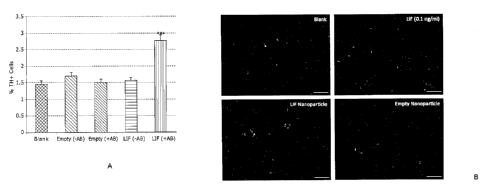
protect the cells, whilst avoiding toxic systemic exposure of the recipient
subject
to the therapeutic cytokine.
[001111 In an embodiment of the invention, the nanoparticles arc suspended in
a
biocompatible solution to faint a composition that can be targeted to a
location on
a cell, within a tissue or within the body of a patient or animal (e.g. the
composition can be used in vitro, ex vivo or in vivo). Suitably, the
biocompatible
solution may be phosphate buffered saline or any other pharmaceutically
acceptable carrier solution. One or more additional pharmaceutically
acceptable
carriers (such as diluents, adjuvants, excipients or vehicles) may be combined
with
the nanoparticles of the invention in a pharmaceutical composition. Suitable
pharmaceutical carriers are described in "Remington's Pharmaceutical Sciences"
by E. W. Martin. Pharmaceutical formulations and compositions of the invention
are formulated to conform to regulatory standards and can be administered
orally,
intravenously, topically, or via other standard routes. Administration can be
systemic or local or intranasal or intrathecal.
[00112] In further embodiments of the invention, other growth factors,
signalling proteins and small molecules may be encapsulated within the
nanoparticles either in addition to or instead of LIF to expand, protect
and/or
differentiate neural stem cells, neural progenitor cells or other pluripotent
cells
having the capacity to differentiate into neural cells. The provision of other
factors
and/or molecules in addition to LIE may augment the efficacy of LIF or the
tolerogenic effect of the composition when used in vivo.
[00113] Other potential neurogenic and/or neuroprotective agents for
encapsulation in nanoparticles include growth factors such as brain-derived
neurotrophic factor (BDNF), the BDNF-agonist 7,8 dihydroxy flavone (7,8-DHF)
epidermal growth factor (EGF), glial cell-derived neurotrophic factor (GDNF),
ciliary neurotrophic factor (CTNF), amongst others, retinoic acid (RA) and
derivatives thereof, and the signalling protein Wnt5A. Derivatives of retinoic
acid
may include, but are not limited to, 9-cis RA, 13-cis RA, N-(4-hydroxyphenyl)
retinamide (4-HPR), and all-trans retinoic acid (ATRA). Agonists of neural
growth
factors can also be encapsulated in the nanoparticles. By way of example, the
=BDNF agonist 7,8 dihydroxyflavone (7,8,DHF) is shown in the present Examples
CA 02921491 2016-02-16
WO 2014/031883 PCT/1JS2013/056246
- 26 -
to increase the yield of TH+ neuronal cells in primary rat E 14 VM tissue
treated
with nanoparticles that encapsulate the agonist. Optional additional factors,
such as
anti-oxidants, or transforming growth factor beta (TGF-f3) that promotes
responsiveness to GDNF, or retinoic acid that plays an important role in
multipotency, may also be included in the nanoparticles. Single or multiple
agents
may be combined with LlE in the same nanoparticle, or may be used individually
in one nanoparticle, for nanoparticle delivery to target cells.
[00114] Taking EGF as an example, this growth factor has a unique role as a
mediator of dopamine-induced precursor cell proliferation in the sub-
ventricular
zone of the brain. EGF receptors are reduced in Parkinson's Disease, therefore
targeted paracrine delivery of nanoparticles containing EGF can increase
dopamine-induced precursor cell proliferation due to the increase in EGF
potency.
[00115] Wnt5a (Wingless-type MMTV integration site family member 5A) is a
signaling protein that in humans is encoded by the WNT5A gene. Members of the
Wnt5a class of proteins activate non-canonical Wnt pathways, which involve
different kinases such as protein kinase C, calmodulin-dependent protein
kinase II
and c-Jun N-terminal kinase, as well as phosphatases and GTPases. Non-
canonical
Wnt pathways inhibit the canonical Wnt¨h-catenin pathway. Human frizzled-5
(hFz5) is a receptor for the human Wnt5A protein. Wnt5A has been implicated as
a tumour suppressor gene. Importantly, Wnt5A has been identified for use in
the
treatment of primary midbrain precursor cells to induce their differentiation
into
dopaminergic (DA) neurons. Therefore, sustained nanoparticle delivery of Wnt5a
(either with or without LIF) to dopaminergic precursor cell populations will
support DA cell differentiation in addition to increasing dopaminergic
precursor
cell recovery ex vivo and also their survival following subsequent
transplantation
into patients suffering from Parkinson's Disease.
[00116] In an embodiment of the invention the nanoparticles may also comprise
as the cargo - in addition to or instead of LIF - the small molecule XAV939
(structure shown below).
CA 02921491 2016-02-16
WO 2014/031883 PCT/US2013/056246
-27 -
H 0
N
cF3
¨N
XAV939 is a known inhibitor of the Wnt/f3-catenin signalling pathway that
mediates
13-catenin degradation by inhibiting the poly-ADP-ribosylating enzymes
tankyrase 1
and tankyrase 2, which in turn stabilises axin. Both tankyrase isoforms
interact with
a highly conserved domain of axin and stimulate its degradation through the
ubiquitin-proteasome pathway (Huang et al., 2009). Importantly, XAV939
promotes
remyelination of demyelinated nerve axons by stabilising Axin2. Axin2 itself
is
regulatory and provides a therapeutic target in new born brain injury and for
remyelination. Axin2 is expressed in immature oligodendrocyte precursor cells
(OPC), including those residing in active MS lesions. Axin2 plays a role in
feedback
regulation of the wnt signalling pathway: since wnt signalling can act to
inhibit OPC
differentiation in both adult remyelination models and developmental
myelination,
manipulation of Axin2 levels in OPC can repress wnt signalling and promote
accelerated differentiation of OPC to oligodendrocytes (OD) capable of
remyelinating nerve axons within the CNS. By inhibiting tankyrase, involved in
Axin2 degradation, XAV939 promotes remyelination (Fancy et al. 2011). Direct
injection of XAV939 direct into spinal cord lesions promotes markedly
accelerated
OD differentiation after demyelinating injury. Hence, the nano-XAV939 device
of
the present invention targeted to the surface of, for example, demyelinated
axons
provides a non-invasive focussed means of simarly promoting remyclination.
[00117] The nanoparticles and compositions of the invention can be delivered
to target cells in vitro, for example to test their efficacy, and also ex vivo
for the
transplantation of L1F expanded and/or protected target cells into the adult
brain of
patients suffering from neurodegenerative disease. Cell therapy promotes brain
repair by maintaining or replacing populations of vulnerable neurons and/or
expanding the endogenous neural stem cells and progenitor cells that populate
the
brain, providing an enriched source of healthy precursor cells with the
potential to
mediate repair. Cell therapy can provide precursor cells as autografts (for
example,
CA 02921491 2016-02-16
WO 2014/031883 PCT/US2013/056246
- 28 -
derived from patient skin fibroblasts by trans-differentiation to a required
phenotypic precursor cell ¨ IPS cell) or allografts (for example, from foetal
precursor cells). In an embodiment of the invention the transplanted cells may
be
dopaminergic cells.
[00118] The nanoparticles and compositions of the invention can also be
delivered to target cells in vivo. In vivo use requires that the nanoparticles
of the
invention are able to cross the blood brain barrier so that they can access
the target
cells within the brain of the patient. Self-administered intra-nasal delivery
of the
nanoparticles and compositions of the invention is one way in which the
nanoparticles can reach the target cells to promote endogenous repair and
replacement of damaged brain tissues, and to protect healthy brain structure
from
toxic damage associated with disease states.
[00119] The nanoparticles and compositions of the invention can be used in the
treatment of various neurodegenerative diseases, including Alzheimer's
Disease,
Parkinson's Disease, Amyotrophic lateral sclerosis and Huntington's Disease,
amongst others, and will provide huge socio-economic benefit to patients
suffering
from neurodegenerative diseases and their families. By way of example,
dopaminergic cell replacement therapy is the focus for the treatment of
Parkinson's Disease.
[00120] IPS cells are an alternative source of cells for therapy and the
nanoparticles and compositions of the invention can be targeted to IPS cells
to
expand, protect and/or differentiate these cells for use in cellular therapy
in the
treatment of NDD and CNS trauma. Likewise the nanoparticle devices of the
invention may be used to expand or admix with stem cell preparations ex-vivo
prior to introduction into a subject. In such an embodiment of the invention
the
stem cells may be adult derived, foetal-derived, derived from IPS cells, or
from
any other allogenic
[00121] The invention further provides for combinatorial compositions that
comprise mixtures of populations of nanoparticles that comprise more than one
therapeutic agent per nanoparticle, or different nanparticles each comprising
a
different therapeutic agent, for the treatment of neurodegenerative disease.
Such
combinatorial compositions may suitably comprise a pharmaceutically acceptable
CA 02921491 2016-02-16
WO 2014/031883 PCT/US2013/056246
- 29 -
carrier solution; at least a first population of biodegradable nanoparticles,
wherein
the first nanoparticles comprise a targeting moiety that is able to bind
selectively
to the surface of a neural stem cell and/or a neural progenitor cell and
wherein the
first nanoparticles further comprise leukaemia inhibitory factor (LIF); and at
least
second population of biodegradable nanoparticles, wherein the second
population
of nanoparticles comprise a targeting moiety that is able to bind selectively
to the
surface of a neural stem cell and/or a neural progenitor cell and wherein the
second
nanoparticles further comprise one or more other than HT. Suitably, the second
nanoparticles may comprise compounds selected from: brain-derived neurotrophic
factor (BDNF); epideintal growth factor (EGF); glial cell-derived neurotrophic
factor (GDNF); ciliary neurotrophic factor (CTNF); retinoic acid, and
derivatives
thereof; Wnt5A; and XAV939.
[00122] The invention is further exemplified in the following non-limiting
examples.
Example 1
1.1 Dopaminergic neurons derived from E14 ventral mesencephalon (VM) from
rat foetuses express the components of the LIF receptor complex
[00123] The expression of gp130 and gp190, the two components of the LIF
receptor complex (Figure la), on dopaminergic neurons of embryonic day 14
CE14') VM was analysed via immunocytochemistry of E14 VM cultures after 3
days in vitro ('./NV') (Figure lb). Figure lb shows that both components of
the
LIF receptor complex are expressed by dopaminergic neurons in E14 ventral
meseneephalon (VM) cultures. A) The LIF receptor is a heterodimer consisting
of
two proteins: gp130 and gp190. B) Immunocytochemistry of 5 day old E14 VM
cultures with antibodies against tyrosine hydroxylase and gp130 or gp190
demonstrated that dopaminergic neurons express gp130 and gp190. Dopaminergic
neurons were demonstrated to express both gp130 and gp190, suggesting a
potential for responsiveness to LIF treatment.
1.2 LIF treatment during tissue dissociation increases the subsequent number
of
dopaminergic neurons
CA 02921491 2016-02-16
WO 2014/031883 PCT/US2013/056246
- 30 -
[00124] The VM of E14 rat foetuses was dissected and dissociated in medium
with or without 0.1ng/m1 soluble LIF. The tissue was then plated in monolayer
culture and grown for 2, 3 or 5 days prior to fixing. Dissociated cells were
seeded
in monolayer cultures and fixed after 2, 3 or 5 days in vitro (DIV). Culture
derived
from cells dissociated in LIF supplemented medium were found via
immunocytochemical analysis to contain significantly more tyrosine hydroxylase
positive neurons after 2 days in vitro but not later time points. Subsequent
immunocytochemistry of fixed culture demonstrated that cultures derived from
tissue dissociated in the presence of 0.1ng/m1LIF had significantly more TH+
neurons after 2 days in vitro; this effect was lost at 3 and 5 days in vitro
(Figure 2).
1.3 Dopaminergic cell count in E14 VM cultures can be increased by
supplementing growth medium with 0.1 ng/ml soluble LIF
[00125] The VM of E14 rat foetuses was dissected and dissociated in standard
conditions. Primary E14 VM tissue was dissociated and grown as monolayer
cultures. After plating cells were chronically treated with soluble LIF in
their
growth medium ranging from 0.1ng/m1 to 10Ong/ml. Subsequent
immunocytochemistry demonstrated that supplementation of growth medium with
0.1ng/m1 LIF was able to significantly increase the number of tyrosine
hydroxylase positive neurons after 3 and 5 days in vitro (Figure 3). Treatment
of
E14 VM cultures with all LIF dosages above 0.1ng/m1 had no significant effect
on
the number of TH positive neurons.
1.4 Dopaminergic neurons express the glial cell line derived neurotrophic
factor
receptor al
[00126] Before the effect of nanoparticle treatment on E14 VM cultures could
be investigated it was necessary to identify a cell surface protein that could
be used
as a target for antibodies on the nanoparticle surface. Given the known
neurotrophic effect of glial cell line derived neurotrophic factor (`GDNF') on
dopaminergic neurons the expression of the GDNF receptor al CGDNF-R al in
E14 VM cultures was analysed via immunocytochemistry with the aim of
potentially using this protein as a nanoparticle target. The monolayer culture
was
fixed after 5 days in vitro and analysed for expression of GDNFR-al through
immunocytochemistry. Dual staining with tyrosine hydroxylase demonstrated that
CA 02921491 2016-02-16
WO 2014/031883 PCT/US2013/056246
- 31 -
individual neurons express both TH and GDNFR-al. Hence, as expected,
dopaminerQic neurons were found to express this protein (Figure 4).
1.5 LIF nanoparticles targeted via antibodies against GDNF-R al increase the
tyrosine hydroxylase positive cell count in E14 VM cultures
1001271 To investigate the effect of LIF nanoparticle treatment on tyrosine
hydroxylase positive cell counts, primary E14 VM was mixed with LIF
nanoparticles (targeted or non-targeted) or empty nanoparticles (targeted or
non-
targeted) immediately prior to plating in monolayer culture. El 4 VM tissue
was
mixed with 100111 of a 1mg/m1 nanoparticle solution immediately prior to
plating.
Nanopartieles were either empty nanoparticles (with or without surface bound
anti-GDNFR-al antibodies) or LIF nanoparticles (with or without anti-GDNFR-al
antibodies). Irmnunocytochemical analysis of these cultures after 3 days in
vitro
revealed a significant increase in the number of tyrosine hydroxylase positive
neurons in the cultures treated with targeted LIF nanoparticles. Cultures were
fixed
after 3 days in vitro and analysed via immunocytochemistry for tyrosine
hydroxylase. Plating cells with targeted LIF nanoparticles significantly
increased
the TH positive cell count at 3 days in vitro Non-targeted LIF nanoparticles
and
empty nanoparticles had no effect on the TH+ cell count (Figure 5 (A) and
(B)).
1.6 Treatment of E14 VM derived neurospheres with 0.1 ng/ml soluble LIF has no
effect on subsequent differentiation in monolayer culture
1001281 To investigate the effect of Lit' treatment on the differentiation of
El 4
VM, tissue was grown as neurospheres in expansion medium with or without
0.1ng/m1 soluble LIF. Primary ventral midbrain tissue was expanded in medium
containing the mitogens EGF and FGF-2 for 5 days. These neurospheres were then
dissociated into single cells and plated in monolayer culture in the absence
of LIF.
After 5 days of growth these cultures were analysed via immunocytochemistry
for
neural and astroglial differentiation (Figure 6, showing morphology + or -
LIF).
The presence of LIF during the expansion of E14 VM had no effect on subsequent
differentiation (Figure 7 showing results after 5 and 10 days).
1.7 Treatment of E14 VM monolayer cultures with soluble LIF or targeted LIF
nanoparticles reduces levels of dopaminergic apoptosis
CA 02921491 2016-02-16
WO 2014/031883 PCT/US2013/056246
- 32 -
[00129] A subset of tyrosine hydroxylase neurons co-localised with cleaved
caspase-3 and a condensed nucleus, both markers of apoptotic cells. This
indicates
that a proportion of dopaminergic neurons in E14 VM cultures undergo apoptosis
during culture (Figure 8), contributing to the decrease in the number of these
neurons as culture time progresses. Immunocytochemical analysis was performed
to determine whether LIF treatment (soluble or targeted nanoparticles)
decreased
the number of apoptotic dopaminergic neurons in these cultures.
[00130] El 4 VM rnonolayer cultures, treated with soluble LIF or LIF/empty
nanoparticles were fixed after 2, 3 or 5 days. Immunocytochemical analysis for
cells positive for tyrosine hydroxylase, cleaved caspase-3 (CC-3) and a
condensed
nucleus demonstrated a significant reduction in dopaminergic apoptosis. It was
found that LIF treatment resulted in reduced numbers of apoptotic dopaminergic
neurons after 2 days in vitro (Figure 9). A trend towards reduced apoptosis in
the
presence of LIF remained after 3 days in vitro but did not reach statistical
significance. Together with the finding that LIF does not bias El 4 VM towards
neural differentiation, this result suggests the increase in TH+ cells seen
with
chronic LIF treatment is an effect of increased dopaminergic cell survival.
1.8 Serotonin neurons in E14 VM cultures express GDNFR-al
[00131] Contaminating scrotonin neurons in foetal grafts have been linked to
the development of graft-induced dyskinesias (` Gins') in Parkinson's Disease
patients. It was therefore of interest to determine whether LIF treatment had
any
effect on the number of serotonin neurons in E14 VM cultures. An E14 VM
culture was fixed after 5 days in vitro and stained with antibodies against
GDNFR-
al and serotonin. As a first step, immunocytochemistry was performed to reveal
whether serotonin neurons in these cultures express GDNFR-al, the protein
being
used to target LIF nanoparticles. Dual staining for serotonin and GDNFR-al
demonstrated that serotonin neurons express GDNFR-a1 (Figure 10).
1.9 Anti-Thy-1 directed nanotherapy: either nanoparticle-delivered BDNF, or
nanoparticle-delivered 7,8 dihydroxy-flavone (7, 8-DHF) improves yield of T11+
cells and this is comparable to treatment with soluble BDNF, or soluble 7, 8-
DHF
[00132] To compare the effect of brain-derived neurotrophic factor (BDNF), or
the BDNF agonist 7,8-dihydroxy flavone (7,8-DHF), when in a nano-particulate
CA 02921491 2016-02-16
WO 2014/031883 PCT/US2013/056246
- 33 -
formulation targeted to Thy-1, versus free, primary rat El 4 VM tissue was
mixed
with 1001.11 of nanoparticle solution (0.05mg; 0.1mg; 1.0mg nanoparticles/m1),
or
with free growth factor (10nM; 100nM; 1 M; 10uM) immediately prior to plating.
After first confirming presence of Thy-1 antigen on the surface of TH+ neurons
(data not shown), anti-Thy-1 decorated nanoparticles were prepared as either
empty; or BDNF-nanoparticles; or 7,8 DHF-nanoparticles. Cultures were fixed
after 7 days in vitro and analysed via immunocytochemistry for tyrosine
hydroxylase positive cells. Plating cells with targeted BDNF-, or 7,8 DHF-
nanopartieles significantly increased the TM positive cell count to levels
comparable with the effect of free BDNF or 7,8-DHF. Analysis of cells
demonstrated a response to BDNF, and to the BDNF-agonist 7,8-dihydroxy
flavone (7,8-DRF), delivered in nano-formulation targeted to Thy-1. This is
shown
for 7,8 DIIF-nanoparticles in Figure 11 (panels A and B) where the dose-
response
curve is similar to that reported by Jang et al (Tang et al, Proc. Natl. Acad.
Sei.
USA, 2010), with the exception of the high dose (10 M) decline observed here.
[00133] The experiment also tested for the effect of BDNF and 7,8-DHF on
serotonergie cells versus dopaminergic cells where a constant ratio was found
(Figure 11, panels C and D). Measurement of both longest neurite length and
number of primary neurites revealed a significant increase for both parameters
following treatment with BDNF or BDNF-nano (data not shown): unexpectedly,
neither soluble 7,8-DHF nor 7,8-DHF-nano altered neurite length or number
(Figure 11, panels E ¨ H).
1.10 Rat fetal VM grafts treated ex vivo with LIF or BDNF nanoparticles prior
to
grafting into the striatum of lesioned syngeneic recipients show no evidence
of
adverse effects though do not significantly alter the response to amphetamine.
[00134] Following transplantation surgery rats in all groups continued to gain
weight. Post-transplantation weight gain was not affected by nanoparticle
supplementation of grafted tissue. Two way repeated measures ANOVA:
significant effect of time F174,4175 = 99.30, p <0.001, no effect of group
F3,24= 1.3,
p = 0.311, no time x group interaction F9,24 = 0.74, p > 0.05. Figure 13
(upper).
[00135] In the amphetamine-induced rotation assay, there was a significant
reduction in net ipsilateral rotation across all groups. There was no
significant
CA 02921491 2016-02-16
WO 2014/031883 PCT/US2013/056246
- 34 -
effect of nanop article supplementation on recovery rate in the reduction of
amphetamine induced rotation post-transplant. Two way repeated measures
ANOVA: significant effect of time F 17,411= 18.41, p <0.001, no effect of
group
F3,24 = 1.89, p = 0.158, no time x group interaction F9,24 = 1.21, p > 0.05.
Figure 13
(lower).
Example 2
2.1 Human: Monolayer and neurosphere cultures ¨ expansion of cell numbers to
provide sufficient cells for testing therapeutic LIF nanoparticles
[00136] 6 ¨ 8 week old human foetal midbrain was dissected and cultured as
neurospheres in proliferation medium before being sectioned and stained. Upon
passage, parallel cultures as monolayer were grown in differentiation medium.
Dopaminergic cells are positive for tyrosine hydroxylase (TH). Total cell
numbers
are stained with nuclear antigen (IIoechst). See Figure 12, where: Primary =
primary cultures; Passage 0 = first subculture; Passage 1 = second subculture.
[00137] Results show (i) maturation of TH+ neurons within the neurosphere
microenvironment; (ii) differentiation of TH+ neurons grown in monolayer.
Passage 2 also contains dopaminergic (DA) cells. These amplified cells were
used
to test LIF-nano effects on DA cell maturation and overall cell survival.
2.2 Human foetal ventral mesencephalon LIF-nanoparticle therapy enhances
human dopaminergic neuron derivation
[00138] After establishing culture conditions for expansion of primary human
foetal VM cells, these cells were used to test therapeutic efficacy of the LIF-
nano
device (see Figure 14). Cultures were stained for tyrosine hydroxylase (1H)
after 5
days in culture in the presence or absence of LIF nanoparticles targeted to
Thy-1.
Three sets of experiments were completed. A dose of 1/100 LIF-nanoparticles
targeted to Thy-1 resulted in both (i) 3-fold increase in overall cell numbers
and
(ii) a percentage fold increase of 2.5% TH+ cells within this overall cell
population. Thus, there was a greater than 5-fold increase of dopaminergic
cells as
a result of LIF-nano therapy (see Figures 15 and 16).
CA 02921491 2016-02-16
WO 2014/031883 PCT/US2013/056246
- 35 -
Example 3
3,1 Treatment of human foetal ventral rnesencephalon (WM) with LIF-
nanoparticle therapy, or XAV939-nanoparticle therapy, enhances human
dopaminergic neuron derivation and increases hfVM cell survival both in vitro
and in vivo.
[00139] To measure the effect of nanotherapy in vivo: hfVM cells were
prepared as for the in vitro experiments as outlined in Figure 17, primary
human
fetal mesencephalon tissue was stored at 4 C for upto 4 days in Hibernate E
storage medium. The cells were then seeded on coverslips and cultured 4d in
differentiation medium after which cells were stained by DAPI to enumerate
nuclei and for tyrosine hydroxylase to identify and enumerate differentiated
dopaminergic cells. Pooled tissue was then prepared for cell transplantation
following the clinical TransEuro Protocol. The protocol summarised in Figure
18
follows that of the TransEuro clinical trial assessing hfVM cell grafts as
cell
therapy in patients with Parkinson's disease: http://www.transeuro.org.uk. The
harvested cells were divided into four aliquots in proliferation medium and
treated
with nanoparticles targeted to Thy-1 and carrying a cargo of (i) no cargo;
(ii) LIF;
(iii) XAV939; or (iv) retinoic acid for upto 24h. The cells were then
transplanted
in to the striatum of nude rats aged between 12 - 16 weeks following the
protocol
in Figure 22 using standard techniques. At 3 months the rats were perfused
with
BrdU according to standard protocols and then culled when the brain was
harvested and sectioned for immune-cytochemical analysis.
[00140] Human nuclear antigen specific antibody (HuNu) stained transplanted
human cells: tyrosine hydroxylase staining revealed human dopaminergic (DA)
cells within the grafts (Figures 19, 20, 21 and 22). Beta III tubulin stained
neurons,
and BrdU identified any dividing cells post infusion and pre-cull. Numbers and
localisation of cells were identified following image capture (Irnagescope
Aperio).
The results show highly significant increased survival and distribution of
transplant-derived neurons and DA cells in the striatum of rats receiving
grafts
pretreated with LIT-nano, or with XAV939-nano, when compared to the empty-
nano controls. In particular the results quite clearly show the surprising and
beneficial effects of nanoparticle devices of the invention (see Figures 19(C)
and
CA 02921491 2016-02-16
WO 2014/031883 PCT/US2013/056246
- 36 -
20(B)) on cell survival and differentiation in the brain compared to control
(see
Figure 22(C)).
[00141] In vitro experiments paralleled the above in vivo study, but instead
of
transplantation, the cells were seeded onto coverslips in differentiation
medium
and grown for 4 days, fixed, and stained for DA cells and total neurons. Here
six
groups of nanoparticles were tested namely empty-nano; LIF-nano; XAV939-
nano; LIF+XAV939-nano; BDNF-nano; and GDNF-nano; and the results are
shown in Figures 23, 24 and 25. All cargos promoted cell survival with
increased
numbers of TH+ cells compared to the empty nanoparticle control group (Em-NP).
Example 5
5.1 Preparation of surface targeted nanoparticles containing hLIF inLIF or
XAV939
[00142] Human IAF (Santa Cruz cat. SC-4377), mouse LIF (Santa Cruz cat.
SC-4378), or XAV939 (Sigma Aldrich cat. X3004) was encapsulated in avidin-
coated PLGA nanoparticles using a modified water/oil/water double emulsion
technique.
[00143] Briefly, 501..tg of cytokine was dissolved in 200 Itt PBS or 1 mg of
XAV939 dissolved in DMSO at a concentration of 10 mg/ml (100 ul) was added
dropwise with vortexing to 100 mg PGLA (50/50 monomer ratio, Durect Corp.
cat. B0610-2) in 2 ml dichloromethane. The resulting emulsion was added to 4
ml
of aqueous surfactant solution containing 2.5 mg/ml polyvinyl alcohol (PVA)
(Sigma-Aldrich cat. 363138) and 2.5 mg/ml avidin-palmitate bioconjugatc (see
5.2
below), and sonicated to create an emulsion containing nano-sized droplets of
polymer/solvent, encapsulated cytokine and surfactant. Solvent was removed by
magnetic stirring at room temperature; hardened nanoparticles were then washed
3x in DI water and lyophilized for long-term storage.
[00144] Targeted nanoparticles were formed by reacting the avidin-coated NPs
in PBS with 4 id biotin-antibody (0.5 mg/ml) per mg NP for 15 minutes and used
itnmediately. Nanoparticle size and morphology are analyzed via scanning
electron microscopy and dynamic light scattering in lx PBS (Brookhaven
Instruments, ZetaPALS). Drug or cytokine release was measured by incubating
CA 02921491 2016-02-16
WO 2014/031883 PCT/US2013/056246
- 37 -
particles in PBS at 37 C and measuring cytokine or drug concentrations in
supernatant fractions by ELISA or UV Spectroscopy. Total encapsulation was
approximated as the amount of LIF or XAV939 released over a seven day period
and percent encapsulation efficiency calculated as total encapsulation divided
by
maximum theoretical encapsulation. Capture of biotinylated ligands was
quantified using biotin-R-phycoelythrin as a model protein. NPs were suspended
at 1.0 mg/ml in lx PBS, and 200 ul added to eppendorfs containing varying
concentrations of biotin-R-PE. NPs were reacted for 15-30 minutes at room
temperature, centrifuged for 10 minutes at 12k RPM, and the remaining biotin-R-
PE in the supernatant quantified by fluorescence at excitation/emission
533/575nm.
5.2 Preparation of the avidin-pahnitate bioconjugate for use in surface
modification of biodegradable nanoparticles
[00145] Stable avidin-lipid conjugates were formed using a zero-length
crosslinking agent to create a covalent bond between the lipid carboxyl end
groups
and free amines on the avidin protein. Fatty acid (Palmitic acid, Sigma) was
first
reacted in 0.1x PBS with 1-ethyl-343-dimethylaminopropyl] carbodiimide (EDC)
and N-hydroxylsulfosuccinimide (sulfo-NHS) (Invitrogen) to convert the
terminal
carboxyl group to an amine-reactive sulfo-NHS ester. Avidin (Sigma) at 5 mg/ml
was then reacted with 10-fold molar excess of the NHS-functionalized fatty
acid in
0.1x PBS and the solution was gently mixed at 37 C for 2 hours. Reactants were
then dialyzed against 1.0x PBS at 37oC for 24 hours to remove excess reactants
and/or hydrolyzed esters.
[00146] The above protocol may be adapted for encapsulation of the other
compounds described herein.
[00147] Although particular embodiments of the invention have been disclosed
herein in detail, this has been done by way of example and for the purposes of
illustration only. The aforementioned embodiments are not intended to be
limiting
with respect to the scope of the appended claims, which follow. It is
contemplated
by the inventors that various substitutions, alterations, and modifications
may be
made to the invention without departing from the spirit and scope of the
invention
as defined by the claims.
- 38 -
REFERENCES
Aklyama et al (1997) In vivo effect of recombinant human LIF in primates. Jpn
J
Cancer Res 88: 578 ¨ 583
Barker RA. (2012) Stem Cells and Neurodegenerative disease - Where is it all
going? Regen.Med 2012 Nov; 7(6 Suppl): 26-31.
Bauer S and Patterson PH (2006) Leukemia inhibitory factor promotes neural
stem
cell self-renewal in the adult brain. J Neurosci 26:12089-99.
Butzkueven H, Zhang JG, Soilu-Hanninen et al (2002) LIF receptor signaling
limits
immune-mediated demyelination by enhancing oligodendrocyte survival. Nat Med
8(6):613-9.
Devennan BE and Patterson PH (2012) Exogenous leukemia inhibitory factor
stimulates oligodendrocyte progenitor cell proliferation and enhances
hippocampal
remyelination. J Neurosci 32:2100-2109.
Fancy SP, Harrington EP, Yuen TJ, Silbereis JC, Zhao C, Baranzini SE, Bruce
CC,
Otero JJ, Huang EJ, Nusse R, Franklin RJ, Rowitch DH (2011) Axin2 as
regulatory
and therapeutic target in newborn brain injury and remyelination Nat Neurosci.
2011
Jun 26;14(8):1009-16
Gao, Thompson L, Zhou Q, Putheti P, Fahmy TM, Strom TB, and Metcalfe S (2009)
Treg versus Th17 lymphocyte lineages are cross-regulated by LIF versus IL-6.
Cell
Cycle 8:9, 1444-1450,
Gillespie LN, Clark GM, Bartlett PF and Marzella PL (2001) LIF is more potent
than BDNF in promoting neuritc outgrowth of mammalian auditory neurons in
vitro.
Neuroreport, 12 2: 275-279.
CA 2921491 2019-12-19
CA 02921491 2016-02-16
WO 2014/031883 PCT/US2013/056246
- 39 -
Liu J, Zang D. (2009) Response of neural precursor cells in the brain of
Parkinson's
disease mouse model after LIF administration. Neurol Res. (7):681-6.
Jang et al (2010). A selective TrkB agonist with potent neurotrophie
activities by
7,8-dihydroxyflavone. PNAS 107; 2687-2692
Huang SM, Mishina YM, Liu S, Cheung A, Stegmeier F, Michaud GA, Charlat 0,
Wiellette E, Zhang Y, Wiessner S, Hild M, Shi X, Wilson CJ, Mickanin C, Myer
V.
Fazal A, Tomlinson R, Serluca F, Shao W, Cheng H, Shultz M, Rau C, Schirle M,
Schlegl J, Ghidelli S. Fawell S. Lu C, Curtis D, Kirschner MW, Lengauer C,
Finan
PM, Tallarieo JA, Bouwmeester T, Porter JA, Bauer A, Cong F (2009) Tankyrase
inhibition stabilizes axin and antagonizes Wnt signaling Nature. 2009 Oct
1;461(7264):614-20
Niwa et al (2009) A parallel circuit of LIF signalling pathways maintains
pluripotency of mouse ES cells. Nature Jul 2;460(7251):118-22
Park J, Gao W, Whiston R, Strom TB, Metcalfe S and Fahmy TM (2011)
Modulation of CD4+ T lymphocyte lineage outcomes with targeted nanoparticle-
mediated cytokine delivery. Mol Phatin. 8(1):143-52.
Pitman et al (2004) LIF receptor signalling modulates neural stem cell renewal
Mol
Cell Neurol 27 : 255-266.
V. Prima, M. Tennant, 0. S. Gorbatyuk, N. Muzyczka, P. J. Scarpace and S.
Zolotukhin (2004) Differential Modulation of Energy Balance by Leptin, Ciliary
Neurotrophic Factor, and Leukemia Inhibitory Factor Gene Delivery: Microarray
Deoxyribonucleic Acid-Chip Analysis of Gene Expression Endocrinology 145 (4):
2035
- 40 -
Reif AE, Allen JM (1964) The AKR thymic antigen and its distribution in
leukaemias and nervous tissues J Exp Med. 1964 Sep 1;120:413-33
Reynolds AD, Stone DK, Hutter JAL, Benner EJ, Mosley RL and Gendelman HE
(2010) Regulatory T cells attenuate Th17 cell-mediated nigrostriatal
dopaminergic
neurodegeneration in a model of Parkinson's Disease. J Immunology. (184):2261-
2271.
[00148]
[00149] While this invention has been particularly shown and
described with
references to example embodiments thereof, it will be understood by those
skilled
in the art that various changes in form and details may be made therein
without
departing from the scope of the invention encompassed by the appended claims.
CA 2921491 2019-12-19