Note: Descriptions are shown in the official language in which they were submitted.
CA 02921502 2016-02-17
WO 2015/02-1089 PCT/BR201-
1/000301
1
PROCESS FOR PRODUCING MULTIPOTENT STEM CELLS AND
PROGENITORS
The present invention generally relates to a
non-enzymatic process for producing multipotent stem cells and
progenitors from the growth of stem cell niches.
Background of the invention
It is known that stem cell populations are at particular
anatomical locations of the human body - i.e., natural niches
- which guarantee their maintenance and the cell interactions
necessary to allow division of these cells and participation
in appropriate homeostasis and tissue repair. Thus, the stem
cell niche is the functional unit of any tissue containing
several types of cells residing in harmonic interaction with
its extracellular matrix, in a microenvironment that provides
short-range intercellular signals for maintenance of the
non-differentiated state of cells.
In this invention, in a particular and non-exclusive
manner, suitable stem cell niches comprise those typically
included in extraembryonic tissues expelled by a woman body at
childbirth, particularly the placenta, amniotic membrane and
the umbilical cord, including its constituents (for example,
veins, arteries, epithelium and connective tissue) , preferably
after blood removal.
In the text that follows, just for ease of expression and
as representative of natural stem cell niches (NSCN) adequate
to the invention, specific references will be made to the intact
umbilical cord tissue (UCT), including mesenchymal connective
tissue (Wharton's jelly), arteries, vein and outer epithelium,
after blood withdrawal, said mention not limiting, in any way,
the invention with regard to the use of other NSCN such as, for
example, nerve, muscle, fat, bone, skin and organ-derived
(e.g., liver, lungs, heart, spleen, liver, pancreas, testes,
ovaries or even biopsy-derived) tissues, but without being
CA 02921502 2016-02-17
WO 2015/024089
PCT/BR2014/000301
2
limited by these included, as well as other postnatal and adult
tissues, or even cancer tissue with NSCN.
Various isolation methods of stem cell from human tissues
or substrates are known. However, until now, no method can
guarantee the obtainment of cells in large-scale in response
to the growing demand of stem cells and their derivatives for
therapies, and for large-scale production of bioactive
molecules (peptides, growth factors, hormones, etc.) that have
trophic properties of pericytes and which could replace SC
(stem cell) in many therapies.
There are countless mentions in the state of the art to
procedures in which various substrates in the human body are
submitted to enzymatic processes to obtain and/or retrieve stem
cells. However, even with the use of enzymatic processes, there
is no disclosure of methods that produce stem cells in large
amounts.
It is known that substantial amounts of stem cells are
required, among others, for the production of tissues, organ .
transplants, tissue and/or complete organ printing, healing of
an injury directly in the patient and immunosuppression in many
autoimmune diseases. There are references in bibliography
that these treatments require an amount of 2 x 10⁶ cells per
kilogram of patient weight, these stem cells being standardized
in relation to ST properties and the number of cell passages.
As will be seen below, the present invention provides a
simple process with high yield for obtaining large amounts of
stem cells and progenitors, in an essentially non-enzymatic
manner.
Description of the Figure
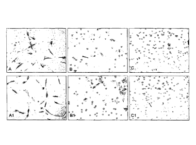
Figure 1 - Cell populations obtained by the inventive process
and by 2 alternative enzymatic methods, after 72 hours in
cultivation in A, B, C and after 5 days in Al, 81 and Cl.
Description of the Invention
CA 02921502 2016-02-17
WO 2015/024089
PC1713R2014/000301
=
3
The present invention relates to an innovative process for
producing adult stem cells, particularly multipotent
mesenchymal/stromal stem ceLls and perivascular precursors,
such as pericytes.
Differently from the prior art processes, the process of
the invention is essentially non-enzymatic and is based on the
propagation and isolation of population of stem cells that
actively proliferate in their niches, in a biologically
synergistic manner (that is, in equilibrium with their
environment). The way of verifying if a particular tissue is
a stem cell niche is not part of the invention, which is known
by the person skilled in the art, for example, by
immunofluorescence and immunohistochemistry.
The present invention provides an advantageous and robust
process for producing cells with 100% of success and
effectiveness with respect to the processed tissues, allowing
the obtainment of large amounts of cells and minimum time delays
related to the isolation process.
The mesenchymal stem cells (MSC) isolated in large amounts
from the process of the invention exhibit all the
characteristics of mesenchymal stem cells and of pericyte type,
either for production of medicinal molecules used directly in
therapy or in the pharmaceutical industry for drug production.
The SC obtained by the process of the invention presents a large
capacity of differentiation (induced or spontaneous) in vitro
and in vivo; with large capacity of tissue regeneration using
culture media and agents and/or substrates ("scaffold") in
liquid, rigid or gelatinous support phase, or extracellular
matrix (complex of macromolecules: fibrous components,
proteins and polysaccharides) of diverse nature, natural or
synthesized.
A characteristic of stem cells/progenitors produced by
the process of the invention is having the same pattern of
CA 02921502 2016-02-17
WO 2015/024089
PCT/BR2014/000301
4
expression of markers related to the non-differentiated state
of in vivo cells present within cultivated fragments.
The process of the invention comprises, in an innovative
manner, the in vitro cultivation of Sc niches (NSCN). Without
being a justification of the invention, it is known that the
in vitro cultivation of stem cells outside their usual niche
may induce changes in karyotype or phenotype stability, in the
molecular signature and genomic stability. Additionally, due
to the unequal division of stem cells (which produce a stem cell
and a progenitor), they lose multipotentiality with cell
passages (ability to produce abroad spectrum of differentiated
cell types). This mixed population of mesenchymal stem cells
derived from the natural niche can provide greater biological
synergy of cells isolated according to the process of the
invention, which dispenses prior removal of blood vessels or
any other tissue, only withdrawing the blood present.
A feature of the process of the invention is the floating
culture (or 3D) of tissue fragments of NSCN, promoting
substantial amounts of stem cells and progenitors
substantially free from genetic and biological changes.
According to the invention, the tissue fragments keep
floating in basal culture medium, producing stem
cells/progenitors stem cells. The release of SC occurs through
natural migration of cells from NSCN fragments to the plastic
of culture bottle, ensuring that these cells will maintain the
homogeneity of molecular profile of the population and survival
unchanged after this natural insulation.
It is observed that the culture of floating tissue
fragments, according to the invention, typically exhibits the
following dynamics in SC production: with the culture of NSCN
fragments in suspension, efficient diffusion of gases and
nutrients throughout the fragment occurs, the SC leave the
state of quiescence and have their proliferation in the niches
CA 02921502 2016-02-17
WO 2015/024089
PCT/BR2014/000301
stimulated (i.e. have their number increased by symmetric
division), and their migration also occurs from inside the NSCN
fragments to the surface thereof in response to tissue
fragmentation. Hence, the fragments become denser and
temporarily touch/adhere to the culture bottle substrate,
releasing the Sc to migrate out of the debris and adhere to the
substrate. After adhesion of debris to the substrate, an
increase of migration to the detriment of proliferation occurs,
since the diffusion of nutrients/gases apparently becomes less
intense. The fragment then tends to become lighter due to the
extensive migration of SC to the substrate and, autonomously
or by simple stirring of the culture bottle, the fragment
detaches from the substrate and floats. Once floating again,
the diffusion of gases and nutrients throughout the tissue is
reestablished and a new increase in proliferation of SC in the
niches occurs, and the fragment becomes denser again,
establishing a new cycle of adhesion and release of SC and
subsequent flotation. This
flotation-proliferation-sedimentation-adhesion-migration-re
lease system can be repeated several times during cultivation
of tissue fragments from SC natural niches, producing colonies
of stem cells. As time goes by, multiple colonies consisting
of inventive cells can be observed at the bottom of the culture
bottle.
According to a preferred embodiment, the cell culture at
the bottom of the culture bottle - a relatively homogeneous
culture of stem cells with stem cell/progenitor
characteristics - is kept semi-confluent, particularly with
70% to 90% of confluence, in order to prevent spontaneous cell
differentiation. The term "semi-confluent" means a culture
that is not so dense in a manner that would allow substantial
contact of cells with each other, as is verified in high density
confluent cultures.
CA 02921502 2016-02-17
WO 2015/024089
PCT/BR2014/000301
6
A particular embodiment of the invention additionally
includes modifications of the basal culture medium, by the
addition of growth factors and/or other bioactive molecules
that may alter the characteristics of cells arising from the
process.
In a particular embodiment of the invention, the NSCN
tissue to be cultivated may be submitted to an initial mild
pretreatment with dissociation enzymes (for example, trypsin,
collagenase, TrypLE's, etc.) before the immersion of these
tissues in culture medium. Such pretreatment does not aim at
an extensive digestion of the tissue, but only at a small
relaxation of tissue coherence to facilitate the movement of
SC when immersed in the basal culture medium.
After the end of the invention process, further enzymatic
treatment of adhered SC and their respective cell passages to
other vessels for in vitro or ex vivo growth or expansion of
the resulting stem cells/progenitors favors isolation of
pericytes.
A peculiarity of the process of the invention is the
removal and transfer of fragments from NSCN, for example, by
any adequate mechanic manner, for example, with the aid of
devices such as a pipette, clamp, needle, or appliances, or
simply by pouring the contents from a container to another.
After transferring these NSCN tissue pieces, (floating
fragments) to a new container, they can continue the production
of SC with passage zero (without passage), since no enzymatic
treatment was performed.
The process of the invention uses NSCN fragments, which
comprise stem cells that proliferate within the tissue and keep
expressing markers of various types of stem cells even after
several mechanical tissue transfers mentioned.
The process of the invention is minimally invasive on the
use of niches comprised in extraembryonic tissues expelled by
CA 02921502 2016-02-17
WO 2015/024089
PCT/BR2014/000301
7
a woman body at childbirth, particularly the umbilical cord,
amniotic membrane and placenta. Among other advantages of stein
cells contained, their youth should be mentioned. Mammalian
aging is associated with a reduction in tissue regeneration,
increase of occurrence of degenerative diseases and cancer.
Since stem cells regenerate many adult tissues and when these,
by accumulation of mutations, can contribute to cancer
development, age-related modifications in stem cells probably
contribute to age-related morbidity. Consistent with this, the
role of stem cells in various tissues decreases with age,
possibly resulting in the loss of expression of tumor
suppressors, DNA damage, changes in cell physiology and changes
in the tissue environment. It remains unknown whether declines
in stem cell function during aging influence the organism
longevity; however, the mechanisms that influence longevity
also modulate age-related morbidity, in part, through effects
on stem cells. Therefore, the stem cells of extraembryonic
tissues related to childbirth have extreme importance to cell
therapy, since they are young cells - highlighting the fact that
cells obtained by the process of the invention are not embryonic
cells.
Although it is not essential, an initial wash of NSCN
tissue is appropriate to the process of the invention,
particularly for blood removal before the fragmentation of
tissue that will grow.
Particularly, NSCN wash is made externally and
internally. It is externally made, for example, with distilled
water or sterile buffered saline solution (PBS or physiological
solution and antibiotics (such as penicillin and streptomycin
2%) ) . It is internally made, for example, by washing the inside
of the blood vessels (arteries and veins) with distilled or
sterile buffered saline solution (PBS or physiological
solution and antibiotics (such as penicillin and streptomycin
CA 02921502 2016-02-17
WO 2015/024089
PCTBR2014/000301
8
The fragmentation of NSCN to be cultured according to the
process of the invention can be made from any suitable way, for
example, with sharp blades, by manual or mechanized cutting,
under sterile conditions. Preferably, but not essentially, the
cut must be made with little pressure on the tissue, without
heating, adequately with a water jet cutter under pressure.
In a particular embodiment, the present invention also
relates to repetitive cryopreservation/thawing of tissue
fragments of NSCN(s), thus allowing the continued production
of new stem cells/progenitors in high-scale, particularly when
involving NSCN tissue from a single patient and in low cell
passages (typically less than or equal to 5). However, this
aspect does not limit the process of the invention, which can
use NSCN from a single patient, or from two or more distinct
patients, or can concomitantly use different NSCNs from one or
more patients, or with more NSCN passages.
The cells obtained in accordance with the process of the
present invention express, in the non-differentiated state, a
group of MSC markers (C1J29, CD73, CD90, CD105) concurrently
with pericyte markers (CD140b and CD166) and neither express
hematopoietic lineage markers (CD34 and 0D45) nor
histocompatibility markers (HLA-DR). The cells obtained from
the process of the invention are multipotent, capable of
self-renewal with continuous proliferation and in vitro
differentiation, with the use of known conditions, for example,
addition of inducing agents (for example, retinoic acid,
dimethyl sulfoxide, etc.),growth factors and cytokines. Under
these conditions, the stem cells/progenitors of the invention
differentiate into distinct cell types, such as bone, cartilage
and fat. Since stem cells/progenitors obtained according to the
invention also express CD31 marker, which is characteristic of
endothelial progenitor cells, they can additionally
CA 02921502 2016-02-17
WO 2015/024089
PCT/BR2014/000301
9
differentiate into muscle, endothelial and nerve cells.
Cells and/or fragments obtained by the process of the
invention are suitable in many possibilities of use, such as
in therapeutic, non-therapeutic, biotechnological and
pharmaceutical uses.
Thus, the present invention aims at a process for
obtaining stem cells, particularly multipotent stem
cells/progenitors from the culture of NSCN tissue,
characterized by comprising the following steps:
A - obtainment of one or more NSCN;
B - pre-preparation of one or more NSCN;
C - preparation of tissue fragments of one or more NSCN;
D - promotion of SC propagation by culturing NSCN fragments in
a basal culture medium;
E - separation of SC from NSCN fragments;
F - optionally, separated NSCN fragments in E return to step
D.
The mention of SC in process steps includes stem cells and
progenitors.
Step A to the process of the invention is particularly
performed with the obtainment of a particular NSCN, such as
umbilical cord. Accordingly, without excluding any other, the
NSCN(s) used in the process of the invention are tissues from
woman childbirth, particularly umbilical cord, amniotic
membrane and placenta, even more particularly the intact
umbilical cord, with arteries and vein, Wharton's jelly and
epithelium. Nervous,
muscle, fat, bone, skin and
organ-derived tissues, such as of liver, lungs, heart, spleen,
liver, pancreas, testes, ovaries or even biopsy-derived
tissues, are adequate to the invention, but are not limited by
these included, as well as other postnatal and adult tissues,
and even cancerous tissue having NSCN.
According to item (B) above, NSCN is submitted to a
CA 02921502 2016-02-17
W02015/024089
PCT/BR2014/000301
pre-preparation, which may consist of cleaning, washing,
tissue pre-cuts, grinding, compression, mild pretreatment with
dissociation enzymes (collagenase, dispase, trypsin, TrypLEm%
among others), etc.
5 Particularly,
the NSCN tissue is submitted to
cleaning/washing, aiming at blood removal and other substrates
that may adversely affect Sc propagation during cultivation,
for example, by induction of differentiation, intoxication,
contamination, etc. In the particular case of umbilical cord,
10 proper cleaning
is carried out both internally and externally,
for example, with one or more of distilled water, saline
solution, physiological solution and antibiotics (for example,
penicillin and streptomycin). Internal washing is carried out,
for example, by injecting the substrate into the inner tissue
vessels, and removing the material thus dragged.
In a particular embodiment, the washing and cleaning of
umbilical cord is carried out, for example, in pre-cut parts
of 5 to 10 cm from the umbilical cord vein, avoiding the
inclusion of blood clots.
Still in accordance with step B, the pre-preparation may
involve a mild initial pretreatment of the tissue with
dissociation enzymes (for example, trypsin, collagenase,
TrypLE, etc.) . Such pretreatment does not aim at the extensive
digestion of the tissue, but only at a small relaxation of
tissue coherence to facilitate the movement of SC when immersed
in the basal culture medium.
In relation to step C of the process of the invention, a
manner to fragment the NSCN tissue, as already mentioned, is
in any way suitable for its purpose. In the case of umbilical
cord, fragments obtained from transversal and/or longitudinal
cuttings until reach, for example, cubes with dimensions from
0.5 to 1 cm, are adequate. Any other size or fragment format
is included in the scope of the invention, even particles
CA 02921502 2016-02-17
WO 2015/024089
PCT/BR2014/000301
11
obtained by tissue grinding.
Still in relation to step C, in a particular embodiment
of the invention, the NSCN fragmentation aims at using only
specific parts of intact tissues for growth in the following
step D. For example, in the case of umbilical cord, fragments
exclusively of the veins, arteries, epithelium or Wharton's
jelly may be used, each one generating particular stem
cells/progenitors. They are fragments from NSCN constituents
(vein, artery, epithelium, etc.) that can be used separately,
according to the desired purpose, for producing cells with
molecular signature, differentiation potential and production
of specific active molecules, or can be used together.
In relation to step D of the process of the invention, the
basal medium for culturing fragments of umbilical cord and/or
NSCN cells is anyone that allows the propagation/expansion and
isolation of stem cells and progenitors with the same
phenotypic and molecular characteristics. For example, an
adequate medium is DMEM/F12 ("Dulbecco's modified Eagle's
medium"/Ham's F12, 1:1, of Invitrogen, USA) or any equivalent
substrate known by a person skilled in the art, typically
containing amino acids, proteins, serum and antibiotics.
Said basal culture medium adequately comprises, for
example, 15 wt% serum. Particularly, said serum is bovine
derived such as, for example, bovine fetal serum, and human sera
are also adequate (for example, platelet-rich or -poor plasma)
and other animals, including mixtures thereof, as well as other
natural or synthetic reagents which may allow SC isolation.
The culture medium of step D adequately contains
antibiotic and/or amino acids. The antibiotics used are
comprised in the technical knowledge of the skilled technician,
for example, a combination of penicillin and streptomycin or
gentamicin. Amino acids useful for carrying out the invention
are glutamine, non-essential amino acids or mixtures thereof,
CA 2921502 2017-05-29
12
among others.
The frequent exchange/renewal of culture medium is
particularly carried out in step D, since the fragments of
NSCN tissues spend medium more quickly than cells. The medium
exchange as pH changes from basic to acid is appropriate,
said change being assessed, for example, by change of the
growth medium color, typically from pink to yellow, or in
any other appropriate manner.
Optionally, after step D of the process of the
invention, a mixture can be prepared containing NSCN
fragments and stem cells obtained (for example, by removing
part or all the basal culture medium), such mixture being
preserved for use in a future moment. The preservation of
such mixture is, for example, via cryopreservation, in ways
known by a person skilled in the art. The use of this mixture,
in a future moment is properly performed with thawing, new
insertion in basal culture medium and subsequent separation
of cells/progenitors from fragments, according to item E
above.
In a particular embodiment, the mixture of NSCN and/or
stem cells obtained and/or conditioned by culture can also
be freeze-dried, with additional uses beyond reuse in the
process of the invention (step D).
According to step E of the process of the invention,
after one culture cycle, fragments of NSCN tissue are
mechanically isolated and can - according to step F- be
resubmitted to cultivation of step (D), either immediately
subsequent or after storage (cryopreservation). The reuse of
fragments in new cultures can be made until exhaustion of
the ability to release SC/progenitors. Such fragments from
step E can be mixed with new fragments, originated after
steps A, B and C, which have not been previously used, for
cultivation in step D.
In a particular embodiment, NSCN fragments may also be
decellularized, digested and/or freeze-dried after step E
CA 02921502 2016-02-17
WO 2015/024089
PCT/B112014/000301
13
aiming at additional uses besides of reuse, according to step
F of the process of the invention.
The treatment that can be given to adhering SC/progenitors
after separation of fragments, according to item E above, is
known itself by a person skilled in the art. Cells are typically
washed, for example, with sterile buffered saline solution,
with or without antibiotics, to then carry out their
dissociation (since cells are half-confluent) , whether by
mechanical or enzymatic means, to perform their harvest.
.. Preferably, this dissociation is performed by enzymatic means,
particularly by using Tryplem (marketed by Invitrogen, a US
company) , which is an exceptionally pure recombinant enzyme
free of animal components and mild to cells. A solution of about
0.25-0.05% of trypsin/ethylenediaminetetraacetic acid (EDTA)
can be also used, which is marketed, for example, by
Sigma-Aldrich. Thereafter, the harvested cells are typically
submitted to cell passage by enzymatic means, or cryopreserved
for later use, according to processes known by a person skilled
in the art.
In a particular embodiment, the SC/progenitors obtained
by the process of the invention, isolated according to step E
of the process of the invention, may be also submitted to
freeze-drying for specific uses. The culture
medium
conditioned by cells and/or fragments can be also freeze-dried
for producing substrates of cultures, bioactive molecules,
nutritional supplements and for aesthetic use.
The cryopreservation of tissue fragments of NSCN or
SC/progenitors, or mixtures thereof, possible after step E of
the process of the invention, is adequately conducted in a
freezer, at temperatures around -80 C and subsequently in
liquid nitrogen, at temperatures around -196 C, according to
the knowledge of a person skilled in the art.
After item E of the process of the invention, isolated
CA 02921502 2016-02-17
WO 2015/024089
PCT/BR2014/000301
14
SC/progenitors are typically submitted to enzymatic cell
passage, according to process known by a person skilled in the
art, in number adequate to the intended purpose. Great
stability of SC markers of the present invention is noted after
a large number of passages, for example, 25 or more.
The culturing of NSCN tissue fragments and multiplication
by cell passage of SC/progenitors resulting from the process
of the present invention can be carried out on microcarriers
or in known bioreactors intended for producing cells in
high-scale, as is known in the technical field.
EXAMPLES
Exemplary embodiments of the instant invention are given
below in order to illustrate their realization, without
imparting any limitations beyond those expressed in the
attached claims.
Example 1
Processing of umbilical cord and cell culture
The description of this example represents the mean number
of countless embodiments performed in the same manner.
From an intact umbilical cord a 5-cm fragment was
prepared, which was washed twice inside and out (in this case
with a needle on a syringe) with sterile buffered saline
solution [0.01 M PBS, pH 7.4] containing antibiotics [100
units/mL penicillin and 100 pg/mL streptomycin] to eliminate,
to the maximum possible extent, contamination with blood. In
the first internal washings the, washing solution was still
contaminated with blood, and had reddish color. Additionally,
sterile water injected in the umbilical cord vessels was used,
and the washing solution remained colorless. Then, using a
scalpel, the cord was cut longitudinally and transversally to
obtain about 35 pieces of 0.5 x 0.5 cm approximately, and then
they were transferred to a 75 cm2 flask (Corning, NY) containing
DMEM/F12 medium (Dulbecco's modified Eagle medium/Ham's F12,
CA 02921502 2016-02-17
WO 2015/024089
PCT/BR2014/000301
1:1, marketed by Invitrogen, a US Company) supplemented with
15% fetal bovine serum (FBS, marketed by Hyclone, a US Company),
100 pg/mL penicillin, 100 pg/mL streptomycin, 2 mM L-glutamine
and 2 mM of non-essential amino acids. The bottle was kept in
5 a CO2 greenhouse under wet atmosphere and at 37 C. The
fragments began to release the cells from days 2-3 or 5-7, an
individual variation observed in other embodiments of the
process of the invention. Cell culture growth of fragments of
umbilical cord was kept in these conditions for two weeks. The
10 medium color remained rose (alkaline) and not yellow (acidic)
by daily replacement, and it was verified that the tissue
consumes the medium very quickly. The fragments kept floating
or wafting in the culture medium. Once the bottle bottom was
covered by cells, forming individual half-confluent colonies,
15 in 5-7 or 9-11 days, according to variation observed with
different fragments, the fragments were transferred, by simply
pouring to another bottle of the same size.
Cells adhered to the bottles had a morphology similar to
fibroblasts, high rate of proliferation and nearly 4 x 10⁶ cells
were generated from the 35 fragments in 2 weeks, Umbilical cord
cells exhibited high capacity of forming individual colonies
and, in passage 1, the frequency of formation of cell colonies
was around 100 colonies/100 plated cells in a 90 cm2plate. The
growth kinetics of a single colony derived from umbilical cord
cells was measured at passage 1. During 16 days, cells were
collected and counted daily and changes in the growth rate were
not observed. Changes in the morphology or growth pattern of
these stem cells/progenitors were not verified after 25 tickets
either.
These cells showed normal karyotype in all lines obtained
and no changes could be observed after 10 passages.
Example 2 - Comparison of the method of the invention with
enzymatic method of state of the art
CA 02921502 2016-02-17
WO 2015/024089
PCT/BR2014/000301
16
An umbilical cord was isolated and split into three equal
parts (nearly 5 cm). The first part was processed according to
Example 1 and was transferred directly to the growth medium.
The second and third parts were washed and processed
fragmented, as described in example 1, to then be treated with
collagenase (0.1% collagenase for two hours) and TrypLET" (for
30 minutes), respectively. It can be observed, in figure 1, the
difference between cell populations obtained by these three
methods, after 72 hours in cultivation in A, B, C and after 5
days in Al, Bl and Cl. The difference in cell morphology can
be observed, which is more defined and fusiform in A and Al,
as well as on the amount of adhered cells that begin to form
colonies. The proliferation of these cells was evaluated by
plating equal number of cells (103 on 25 cm2), it being observed
that while the inventive cells reached 90% of confluence with
an amount of 106 cells in 5 days and were frozen, the cells
obtained by enzymatic methods reached only 70% of confluence
(see table 1 below). The number of passages was not counted
during the transfer of fragments, hence, each transfer will
produce cells in passage 0. Thus, taking into account the
multiple transfers of fragments, the number of cells in a low
passage (up to P5 or passage 5) counted as the limit passage
on their therapeutic use is practically unlimited.
Table 1 - comparison of aspects of the invention in relation
to alternative enzymatic methods of figure 1.
Factors for cell Floating Collagenase TrypLem
production non-enzymatic According to the
According to the state of the art
invention
Cell proliferation
higher high high
Tissue freezing possible possible possible
Cell freezing possible possible possible
CA 02921502 2016-02-17
WO 2015/924089
PCT/BR2014/000301
17
Number of passages
for therapy unlimited limited limited
Number of cells unlimited limited limited
Production on an
industrial scale unlimited limited limited
for
biotechnological
and therapeutic
application
Example 3 - Characterization by flow cytometry of cells
obtained according to the process of invention, cultured in
vitro.
For the analysis by flow cytometry, antibodies against
cell surface molecules and their respective control isotypes
were used, such as: human anti-CD45 monoclonal, (Sigma company,
USA), CD90 (of BD-Pharmigen company, USA) and CD105, CD73 (of
Serotec company, United Kingdom). One million of cells are
incubated with antibodies during 30 minutes on ice, washed with
PBS containing 2% fetal bovine serum and 1 pM sodium azide,
followed by addition of FITC (fluorescein isotiocianate) or PE
(phycoerythrin). The analysis by flow cytometry is performed
on a FACS (Fluorescence-Activated Cell Sorter, from Becton,
Dickson and Company, USA) using CELLQuest software (from
Becton, Dickson and Company, USA).
The characterization by flow cytometry in the passage
discloses that the cells obtained according to the process of
the invention are positive for mesenchymal stem cell markers.
In vitro differentiation
For neuronal differentiation, inventive cells are kept
confluent for a week in the 25 cm2 culture bottle, containing
DMEM medium supplemented with 20% Knockout serum (from
Invitrogen company, USA), 100 units/mL penicillin, 100 pg/mL
CA 02921502 2016-02-17
WO 2015/024089
PCT/BR2014/000301
18
streptomycin, and 2 mM L-glutamine. Thus, they are collected
by using a 0.05% trypsin/EDTA solution and plated at high
density in 35 cm2petri dishes containing the same culture type.
The neuronal differentiation is also induced by addition of
all-trans retinoic acid (RA) (Sigma Company, USA). The
suspension of inventive cells obtained by trypsinization is
transferred to a 35 cm2petri dish, pretreated with 0.1% agarose
solution (Sigma Company, USA) containing neurobasal culture
medium (Invitrogen Company, USA) supplemented with B27. After
24 hours, the cells form spherical structures and neuronal
differentiation is induced by addition of RA (retinoic acid)
and DMSO (dimethyl sulfoxide) at a final concentration of 10-7
M and 0.05%, respectively, said medium being daily exchanged.
After four days of culture under non-adherent conditions, SLS
adhere on plates treated with 0.1% gelatin containing a
suitable culture medium.
For adipogenic differentiation, cells were cultured in
DMEM medium with 10% fetal bovine serum, 0,25M isobutyl methyl
xanthine, 10 pM insulin and 1% penicillin. The exchange of
inductor medium was carried out every 3 days and maintained
during 20 days. After this period, cells were fixed for 60
minutes at room temperature with 4% paraformaldehyde and washed
a few times with 70% ethanol. In the following step, they were
incubated at room temperature for five minutes with Oil Red 0,
the excess of dye was removed with a few washes with distilled
water. The cells show positive staining for Von Kossa.
For chondrogenic differentiation, cells were cultured in
DMEM medium with 1% bovine fetal serum, 6.25 pm insulin, 10
ng/mLTGF-pl and 1% penicillin. The exchange of inductor medium
was carried out every 3 days and maintained during 21 days.
After this period, cells were fixed with 4% paraformaldehyde
at room temperature and stained with Alcian Blue. The
CA 02921502 2016-02-17
WO 2015/024089
PCT/BR2014/000301
19
chondrogenic differentiation can be confirmed by specific
staining for safranin and toluidine.
It is known that, from the information provided herein,
with the aid of the given examples, the person skilled in the
art can achieve embodiments not explicitly mentioned in this
document, which perform similar functions to attain results of
the same nature that are thus included in the scope of
protection realized by the attached claims.