Note: Descriptions are shown in the official language in which they were submitted.
DRUG ELUTION FOR IN VIVO PROTECTION
OF BIO-SENSING ANALYTES
[0001] The present application claims the benefit of priority to U.S.
Provisional Application
Serial No. 61/868,179, filed on August 21, 2013.
BACKGROUND
[0002] Field of Invention
[0003] The present invention relates generally to measuring an analyte in a
medium of a living
animal using a system including a sensor implanted or inserted into the living
animal. Specifically,
the present invention relates to a sensor that utilizes one or more
therapeutic agents, which may be
incorporated within a drug eluting polymer matrix, an analyte indicator,
and/or a membrane
covering at least a portion of the analyte indicator.
[0004] Discussion of the Background
[0005] A sensor may be implanted within a living animal (e.g., a human) and
used to
measure an analyte (e.g., glucose, oxygen, cardiac markers, low-density
lipoprotein (LDL), high-
density lipoprotein (HDL), or triglycerides) in a medium (e.g., interstitial
fluid (ISF), blood, or
intraperitoneal fluid) within the living animal. The sensor may include a
light source (e.g., a
light-emitting diode (LED) or other light emitting element), indicator
molecules, and a
photodetector (e.g., a photodiode, phototransistor, photoresistor or other
photosensitive element).
Examples of implantable sensors employing indicator molecules to measure an
analyte are
1
Date Recue/Date Received 2021-01-18
described in U.S. Pat. Nos. 5,517,313 and 5,512,246.
[0006] A sensor may include an analyte indicator, which may be in the form
of indicator
molecules embedded in a graft (i.e., layer or matrix). For example, in an
implantable fluorescence-
based glucose sensor, fluorescent indicator molecules may reversibly bind
glucose and, when
irradiated with excitation light (e.g., light having a wavelength of
approximately 378 nm), emit an
amount of light (e.g., light in the range of 400 to 500 nm) that depends on
whether glucose is bound
to the indicator molecule.
[0007] If a sensor is implanted in the body of a living animal, the
animal's immune system may
begin to attack the sensor. For instance, if a sensor is implanted in a human,
white blood cells may
attack the sensor as a foreign body, and, in the initial immune system
onslaught, neutrophils may be
the primary white blood cells attacking the sensor. The defense mechanism of
neutrophils includes
the release of highly caustic substances known as reactive oxygen species. The
reactive oxygen
species include, for example, hydrogen peroxide.
[0008] Hydrogen peroxide and other reactive oxygen species may degrade the
indicator
molecules of an analyte indicator. For instance, in indicator molecules having
a boronate group,
hydrogen peroxide may degrade the indicator molecules by oxidizing the
boronate group, thus
disabling the ability of the indicator molecule to bind glucose.
[0009] Glucocorticoids are used with cardiac pace makers and eye surgery to
reduce
inflammation. For instance, the following European patent application
publications describe
pace-maker leads and controlled release of steroids: EP2416783 Al (-Improved
glucocorticoid
therapy"), EP1477187 Bl (-Formulation for controlled release of drugs by
combining
hydrophilic and hydrophobic agents"), EP1637164 A2 (Improved formulation for
controlled
2
Date Recue/Date Received 2021-01-18
CA 02922095 2016-02-22
WO 2015/027018 PCT/US2014/052010
release of drugs by combining hydrophilic and hydrophobic agents"), and
EP2303227 A2
("Controlled release corticosteroid compositions and methods for the treatment
of optic
disorders"). However, these devices do not have analyte indicators, and the
glucocorticoid is not
used to reduce degradation of an analyte indicator. Instead, the
glucocorticoid is used to stop
scar tissue from building up.
[0010] There is presently a need in the art for improvements in reducing
analyte indicator
degradation.
SUMMARY
[0011] The present invention overcomes the disadvantages of prior systems
by providing,
among other advantages, reduced analyte indicator degradation.
[0012] One aspect of the present invention provides a sensor that may be
for implantation or
insertion within a living animal and measurement of an analyte in a medium
within the living
animal. The sensor may include a sensor housing, an analyte indicator covering
at least a portion
of the sensor housing, and one or more therapeutic agents that reduce
deterioration of the analyte
indicator.
[0013] In some embodiments, the sensor may include at least one drug
eluting polymer
matrix, and the one or more therapeutic agents may be dispersed within the
drug eluting polymer
matrix. In some embodiments, the drug eluting polymer matrix may cover at
least a portion of
the sensor housing. In some embodiments, the drug eluting polymer matrix may
be within the
sensor housing. The sensor housing may be perforated to allow elution of the
one or more
therapeutic agents from a drug eluting polymer matrix within the sensor
housing.
[0014] In some embodiments, the one or more therapeutic agents may be
incorporated in the
analyte indicator. In some embodiments, the sensor may include a membrane
covering at least a
3
CA 02922095 2016-02-22
WO 2015/027018 PCT/US2014/052010
portion of the analyte indicator, and the one or more therapeutic agents are
incorporated within
the membrane.
[0015] Further variations encompassed within the systems and methods are
described in the
detailed description of the invention below.
BRIEF DESCRIPTION OF THE DRAWINGS
[0016] The accompanying drawings, which are incorporated herein and form
part of the
specification, illustrate various, non-limiting embodiments of the present
invention. In the
drawings, like reference numbers indicate identical or functionally similar
elements.
[0017] FIG. 1 is a schematic view illustrating a sensor system embodying
aspects of the
present invention.
[0018] FIG. 2 illustrates a perspective view of a sensor embodying aspects
of the present
invention.
[0019] FIG. 3 illustrates an exploded view of a sensor embodying aspects of
the present
invention.
[0020] FIGS. 4 and 5 illustrate perspective views of sensor components
within the sensor
body/shell/capsule of a sensor embodying aspects of the present invention.
[0021] FIG. 6 illustrates a side view of a sensor embodying aspects of the
present invention.
[0022] FIG. 7 illustrates a cross-sectional end view of a sensor embodying
aspects of the
present invention.
[0023] FIG. 8 illustrates a sensor having a dip coated drug-eluting polymer
matrix
embodying aspects of the present invention.
[0024] FIGS. 9A-9G illustrate examples of sensors having a preformed drug-
eluting polymer
matrix embodying aspects of the present invention.
4
CA 02922095 2016-02-22
WO 2015/027018 PCT/US2014/052010
[0025] FIG. 10 illustrates the release profile of dexamethasone acetate
from a drug-eluting
polymer matrix according to one embodiment.
[0026] FIG. 11 is a graph showing experimental results comparing analyte
modulation in
sensors having no drug eluting polymer matrix with an embodiment of a sensor
having a ring-
shaped drug-eluting polymer matrix after 30 days in an animal model.
DETAILED DESCRIPTION OF PREFERRED EMBODIMENTS
[0027] FIG. 1 is a schematic view of a sensor system embodying aspects of
the present
invention. In one non-limiting embodiment, the system includes a sensor 100
and an external
transceiver 101. In the embodiment shown in FIG. 1, the sensor 100 may be
implanted in a
living animal (e.g., a living human). The sensor 100 may be implanted, for
example, in a living
animal's arm, wrist, leg, abdomen, peritoneum, or other region of the living
animal suitable for
sensor implantation. For example, in one non-limiting embodiment, the sensor
100 may be
implanted beneath the skin (i.e., in the subcutaneous or peritoneal tissues).
In some
embodiments, the sensor 100 may be an optical sensor. In some embodiments, the
sensor 100
may be a chemical or biochemical sensor.
[0028] A transceiver 101 may be an electronic device that communicates with
the sensor 100
to power the sensor 100 and/or receive measurement information (e.g.,
photodetector and/or
temperature sensor readings) from the sensor 100. The measurement information
may include
one or more readings from one or more photodetectors of the sensor and/or one
or more readings
from one or more temperature sensors of the sensors. In some embodiments, the
transceiver 101
may calculate analyte (e.g., glucose) concentrations from the measurement
information received
from the sensor 100.
CA 02922095 2016-02-22
WO 2015/027018 PCT/US2014/052010
[0029] In some non-limiting embodiments, the transceiver 101 may be a
handheld device or
an on-body/wearable device. For example, in some embodiments where the
transceiver 101 is an
on-body/wearable device, the transceiver 101 may be held in place by a band
(e.g., an armband
or wristband) and/or adhesive, and the transceiver 101 may convey (e.g.,
periodically, such as
every two minutes, and/or upon user initiation) measurement commands (i.e.,
requests for
measurement information) to the sensor 100. In some embodiments where the
transceiver 101 is
a handheld device, positioning (i.e., hovering or swiping/waving/passing) the
transceiver 101
within range over the sensor implant site (i.e., within proximity of the
sensor 100) may cause the
transceiver 101 to automatically convey a measurement command to the sensor
100 and receive
a reading from the sensor 100.
[0030] In some embodiments, the transceiver 101 may include an inductive
element 103,
such as, for example, a coil. The transceiver 101 may generate an
electromagnetic wave or
electrodynamic field (e.g., by using a coil) to induce a current in an
inductive element 114 of the
sensor 100, which powers the sensor 100. The transceiver 101 may also convey
data (e.g.,
commands) to the sensor 100. For example, in a non-limiting embodiment, the
transceiver 101
may convey data by modulating the electromagnetic wave used to power the
sensor 100 (e.g., by
modulating the current flowing through a coil 103 of the transceiver 101). The
modulation in the
electromagnetic wave generated by the transceiver 101 may be
detected/extracted by the sensor
100. Moreover, the transceiver 101 may receive data (e.g., measurement
information) from the
sensor 100. For example, in a non-limiting embodiment, the transceiver 101 may
receive data by
detecting modulations in the electromagnetic wave generated by the sensor 100,
e.g., by
detecting modulations in the current flowing through the coil 103 of the
transceiver 101.
6
CA 02922095 2016-02-22
WO 2015/027018 PCT/US2014/052010
[0031] The inductive element 103 of the transceiver 101 and the inductive
element 114 of the
sensor 100 may be in any configuration that permits adequate field strength to
be achieved when
the two inductive elements are brought within adequate physical proximity.
[0032] In some embodiments, the sensor 100 includes a sensor housing 102
(i.e., body, shell,
capsule, or encasement), which may be rigid and biocompatible. In exemplary
embodiments,
sensor housing 102 may be formed from a suitable, optically transmissive
polymer material, such
as, for example, acrylic polymers (e.g., polymethylmethacrylate (PMMA)).
[0033] In some embodiments, sensor 100 may include an analyte indicator. In
some non-
limiting embodiments, the analyte indicator may be a polymer graft 106 coated,
diffused,
adhered, or embedded on at least a portion of the exterior surface of the
sensor housing 102. The
polymer graft 106 may cover the entire surface of sensor housing 102 or only
one or more
portions of the surface of housing 102. As an alternative to coating the graft
106 on the outer
surface of sensor housing 102, the graft 106 may be disposed on the outer
surface of the sensor
housing 102 in other ways, such as by deposition or adhesion. In some
embodiments, the
polymer graft 106 may be a fluorescent glucose indicating polymer. In one non-
limiting
embodiment, the polymer is biocompatible and stable, grafted onto the surface
of sensor housing
102, designed to allow for the direct measurement of glucose in interstitial
fluid (ISF), blood, or
intraperitoneal fluid after implantation of the sensor 100.
[0034] In some embodiments, the analyte indicator (e.g., polymer graft 106)
of the sensor
100 may include indicator molecules 104. The indicator molecules 104 may be
distributed
throughout the entire graft 106 or only throughout one or more portions of the
graft 106. The
indicator molecules 104 may be fluorescent indicator molecules (e.g., TFM
having the chemical
name 9-[N-[6-(4,4,5,5,-tetramethy1-1,3,2-dioxaborolano)-3-
(trifluoromethyl)benzyll-N-[3-
7
CA 02922095 2016-02-22
WO 2015/027018 PCT/US2014/052010
(methacrylamido)propylamino]methy1]-10-EN-[6-(4,4,5,5,-tetramethyl-1,3,2-
dioxaborolano)-3-
(trifluoromethyObenzyl]-N-[2-(carboxyethyl)amino]methyl]anthracene sodium
salt) or light
absorbing, non-fluorescent indicator molecules. In some embodiments, the
indicator molecules
104 may reversibly bind an analyte (e.g., glucose, oxygen, cardiac markers,
low-density
lipoprotein (LDL), high-density lipoprotein (HDL), or triglycerides). When an
indicator
molecule 104 has bound an analyte, the indicator molecule may become
fluorescent, in which
case the indicator molecule 104 is capable of absorbing (or being excited by)
excitation light 329
and emitting light 331. In one non-limiting embodiment, the excitation light
329 may have a
wavelength of approximately 378 nm, and the emission light 331 may have a
wavelength in the
range of 400 to 500 nm. When no analyte is bound, the indicator molecule 104
may be only
weakly fluorescent.
[0035] In some embodiments, the sensor 100 may include a light source 108,
which may be,
for example, a light emitting diode (LED) or other light source that emits
radiation, including
radiation over a range of wavelengths that interact with the indicator
molecules 104. In other
words, the light source 108 may emit the excitation light 329 that is absorbed
by the indicator
molecules in the matrix layer/polymer 104. As noted above, in one non-limiting
embodiment,
the light source 108 may emit excitation light 329 at a wavelength of
approximately 378 nm.
[0036] In some embodiments, the sensor 100 may also include one or more
photodetectors
(e.g., photodiodes, phototransistors, photoresistors or other photosensitive
elements). For
example, in the embodiment illustrated in FIG. 1, sensor 100 has a first
photodetector 224 and a
second photodetector 226. However, this is not required, and, in some
alternative embodiments,
the sensor 100 may only include the first photodetector 224. In the case of a
fluorescence-based
sensor, the one or more photodetectors may be sensitive to fluorescent light
emitted by the
8
CA 02922095 2016-02-22
WO 2015/027018 PCT/US2014/052010
indicator molecules 104 such that a signal is generated by a photodetector
(e.g., photodetector
224) in response thereto that is indicative of the level of fluorescence of
the indicator molecules
and, thus, the amount of analyte of interest (e.g., glucose).
[0037] Some part of the excitation light 329 emitted by the light source
108 may be reflected
from the polymer graft 106 back into the sensor 100 as reflection light 333,
and some part of the
absorbed excitation light may be emitted as emitted (fluoresced) light 331. In
one non-limiting
embodiment, the emitted light 331 may have a different wavelength than the
wavelength of the
excitation light 329. The reflected light 333 and emitted (fluoresced) light
331 may be absorbed
by the one or more photodetectors (e.g., first and second photodetectors 224
and 226) within the
body of the sensor 100.
[0038] Each of the one or more photodetectors may be covered by a filter
112 (see FIG. 3)
that allows only a certain subset of wavelengths of light to pass through. In
some embodiments,
the one or more filters 112 may be thin glass filters. In some embodiments,
the one or more
filters 112 may be thin film (e.g., dichroic) filters deposited on the glass
and may pass only a
narrow band of wavelengths and otherwise reflect most of the received light.
In some
embodiments, the filters may be thin film (dichroic) filters deposited
directly onto the photo
detectors and may pass only a narrow band of wavelengths and otherwise reflect
most of the
light received thereby. The filters 112 may be identical (e.g., both filters
112 may allow signals
to pass) or different (e.g., one filter 112 may be a reference filter and
another filter 112 may be a
signal filter).
[0039] In one non-limiting embodiment, the second (reference) photodetector
226 may be
covered by a reference photodiode filter that passes light at the same
wavelength as is emitted
from the light source 108 (e.g., 378 nm). The first (signal) photodetector 224
may detect the
9
amount of fluoresced light 331 that is emitted from the molecules 104 in the
graft 106. In one non-
limiting embodiment, the peak emission of the indicator molecules 104 may
occur around 435 nm,
and the first photodetector 224 may be covered by a signal filter that passes
light in the range of
about 400 nm to 500 nm. In some embodiments, higher glucose
levels/concentrations correspond
to a greater amount of fluorescence of the molecules 104 in the graft 106,
and, therefore, a greater
number of photons striking the first photodetector 224.
[0040] In some embodiments, sensor 100 may include a substrate 116. In some
embodiments,
the substrate 116 may be a circuit board (e.g., a printed circuit board (PCB)
or flexible PCB) on
which circuit components (e.g., analog and/or digital circuit components) may
be mounted or
otherwise attached. However, in some alternative embodiments, the substrate
116 may be a
semiconductor substrate having circuitry fabricated therein. The circuitry may
include analog
and/or digital circuitry. Also, in some semiconductor substrate embodiments,
in addition to the
circuitry fabricated in the semiconductor substrate, circuitry may be mounted
or otherwise attached
to the semiconductor substrate 116. In other words, in some semiconductor
substrate embodiments,
a portion or all of the circuitry, which may include discrete circuit
elements, an integrated circuit
(e.g., an application specific integrated circuit (ASIC)) and/or other
electronic components, may be
fabricated in the semiconductor substrate 116 with the remainder of the
circuitry is secured to the
semiconductor substrate 116, which may provide communication paths between the
various
secured components. In some embodiments, circuitry of the sensor 100 may
incorporate some or
all of the structure described in U.S. Patent Application No. 13/650,016 with
particular reference to
FIG. 11D.
[0041] In some embodiments, the one or more photodetectors (e.g.,
photodetectors 224 and
226) may be mounted on the semiconductor substrate 116, but, in some preferred
embodiments,
Date Recue/Date Received 2021-01-18
CA 02922095 2016-02-22
WO 2015/027018 PCT/US2014/052010
the one or more photodetectors may be fabricated in the semiconductor
substrate 116. In some
embodiments, the light source 108 may be mounted on the semiconductor
substrate 116. For
example, in a non-limiting embodiment, the light source 108 may be flip-chip
mounted on the
semiconductor substrate 116. However, in some embodiments, the light source
108 may be
fabricated in the semiconductor substrate 116.
[0042] FIGS. 2-7 illustrate a non-limiting embodiment of a sensor 100
embodying aspects of
the present invention that may be used in the sensor system illustrated in
FIG. 1. FIGS. 2 and 3
illustrate perspective and exploded views, respectively, of the non-limiting
embodiment of the
sensor 100.
[0043] In some embodiments, as illustrated in FIG. 3, the sensor housing
102 may include an
end cap 113. In some embodiments, the sensor 100 may include one or more
capacitors 118.
The one or more capacitors 118 may be, for example, one or more tuning
capacitors and/or one
or more regulation capacitors. The one or more capacitors 118 may be too large
for fabrication
in the semiconductor substrate 116 to be practical. Further, the one or more
capacitors 118 may
be in addition to one or more capacitors fabricated in the semiconductor
substrate 116.
[0044] In some embodiments, as illustrated in FIG. 3, the sensor 100 may
include a reflector
119 (i.e., mirror). Reflector 119 may be attached to the semiconductor
substrate 116 at an end
thereof In a non-limiting embodiment, reflector 119 may be attached to the
semiconductor
substrate 116 so that a face portion 121 of reflector 119 is generally
perpendicular to a top side of
the semiconductor substrate 116 (L e., the side of semiconductor substrate 116
on or in which the
light source 108 and one or more photo detectors 110 are mounted or
fabricated) and faces the
light source 108. The face 121 of the reflector 119 may reflect radiation
emitted by light source
11
CA 02922095 2016-02-22
WO 2015/027018 PCT/US2014/052010
108. In other words, the reflector 119 may block radiation emitted by light
source 108 from
exiting the axial end of the sensor 100.
[0045] According to one aspect of the invention, an application for which
the sensor 100 was
developed (although by no means the only application for which it is suitable)
is measuring
various biological analytes in the living body of an animal (including a
human). For example,
sensor 100 may be used to measure glucose, oxygen, toxins, pharmaceuticals or
other drugs,
hormones, and other metabolic analytes in, for example, the human body.
[0046] The specific composition of the polymer graft 106 and the indicator
molecules 104
may vary depending on the particular analyte the sensor is to be used to
detect and/or where the
sensor is to be used to detect the analyte (e.g., in the in subcutaneous
tissues, blood, or
peritoneum). Preferably, however, graft 106 should facilitate exposure of the
indicator
molecules to the analyte. Also, it is preferred that the optical
characteristics of the indicator
molecules (e.g., the level of fluorescence of fluorescent indicator molecules)
be a function of the
concentration of the specific analyte to which the indicator molecules are
exposed.
[0047] FIGS. 4 and 5 illustrate perspective views of the sensor 100. In
FIGS. 4 and 5, the
sensor housing 102, filters 112, and the reflector 119, which may be included
in some
embodiments of the sensor 100, are not illustrated. As shown in the
illustrated embodiment, the
inductive element 114 may comprise a coil 220. In one embodiment, coil 220 may
be a copper
coil, but other conductive materials, such as, for example, screen printed
gold, may alternatively
be used. In some embodiments, the coil 220 is formed around a ferrite core
222. Although core
222 is ferrite in some embodiments, in other embodiments, other core materials
may
alternatively be used. In some embodiments, coil 220 is not formed around a
core. Although
12
coil 220 is illustrated as a cylindrical coil in FIGS. 4 and 5, in other
embodiments, coil 220 may be
a different type of coil, such as, for example, a flat coil.
[0048] In some embodiments, coil 220 is formed on ferrite core 222 by
printing the coil 220
around the ferrite core 222 such that the major axis of the coil 220
(magnetically) is parallel to the
longitudinal axis of the ferrite core 222. A non-limiting example of a coil
printed on a ferrite core
is described in U.S. Patent No. 7,800,078. In an alternative embodiment, coil
220 may be a wire-
wound coil. However, embodiments in which coil 220 is a printed coil as
opposed to a wire-wound
coil are preferred because each wire-wound coil is slightly different in
characteristics due to
manufacturing tolerances, and it may be necessary to individually tune each
sensor that uses a wire-
wound coil to properly match the frequency of operation with the associated
antenna. Printed coils,
by contrast, may be manufactured using automated techniques that provide a
high degree of
reproducibility and homogeneity in physical characteristics, as well as
reliability, which is
important for implant applications, and increases cost-effectiveness in
manufacturing.
[0049] In some embodiments, a dielectric layer may be printed on top of the
coil 220. The
dielectric layer may be, in a non-limiting embodiment, a glass based insulator
that is screen printed
and fired onto the coil 220. In an exemplary embodiment, the one or more
capacitors 118 and the
semiconductor substrate 116 may be mounted on vias through the dielectric.
[0050] In the illustrated embodiment, the one or more photodetectors 110
include a first
photodetector 224 and a second photodetector 226. First and second
photodetectors 224 and 226
may be mounted on or fabricated in the semiconductor substrate 116.
[0051] FIGS. 6 and 7 illustrate side and cross-sectional views,
respectively, of the sensor 100
according to one embodiment. As illustrated in FIGS. 6 and 7, the light source
108 may be
13
Date Recue/Date Received 2021-01-18
CA 02922095 2016-02-22
WO 2015/027018
PCT/US2014/052010
positioned to emit light that travels within the sensor housing 102 and
reaches the indicator
molecules 104 of the polymer graft 106, and the photodetectors 110, which may
be located
beneath filters 112, may be positioned to receive light from the indicator
molecules 104 of the
polymer graft 106.
[0052] The implantation or insertion of a medical device, such as a bio-
sensor, into a
user/patient's body can cause the body to exhibit adverse physiological
reactions that are
detrimental to the functioning of the device. The reactions may range from
infections due to
implantation surgery to the immunological response of a foreign object
implanted in the body.
That is, the performance of the implantable bio-sensor can be hindered or
permanently damaged
in vivo via the immunological response to an infection or the device itself.
In particular, the
perforniance of the analyte indicator may be deteriorated by the immunological
response of the
body into which the sensor 100 is implanted. For example, as explained above,
white blood
cells, including neutrophils, may attack an implanted sensor 100. The
neutrophils release, inter
alio, hydrogen peroxide, which may degrade indicator molecules 104 (e.g., by
oxidizing a
boronate group of an indicator molecule and disabling the ability of the
indicator molecule to
bind glucose).
[0053] The sensor 100 may include one or more drug-eluting polymer
matrices. In one
embodiment, the drug eluting polymer matrix may cover at least a portion of
the sensor housing
102. One or more therapeutic agents may be dispersed within the drug eluting
polymer matrix
(e.g., an inert polymer matrix). In some embodiments, the one or more
therapeutic agents may
reduce or stop the migration of neutrophils from entering the wound space and,
thus, reduce or
stop the production of hydrogen peroxide and fibrotic encapsulation.
Accordingly, in some
14
CA 02922095 2016-02-22
WO 2015/027018 PCT/US2014/052010
embodiments, the one or more therapeutic agents may reduce deterioration of
the analyte
indicator (e.g., polymer graft 106).
[0054] In some non-limiting embodiments, the drug-eluting polymer matrix
may be applied
to the sensor housing 102 via dip coating. FIG. 8 illustrates a sensor 100
having a dip coated
drug-eluting polymer matrix 828. In the embodiment illustrated in FIG. 8, the
dip coated drug-
eluting polymer matrix 828 covers a portion of the sensor housing 102.
However, this is not
required, and, in alternative embodiments, the dip coated drug-eluting polymer
matrix 828 may
cover a different portion of the sensor housing 102 or the entire sensor
housing 102. In some
non-limiting embodiments, as an alternative to dip coating, the drug-eluting
polymer matrix may
be applied to the sensor housing 102 via spray coating.
[0055] In some non-limiting embodiments, as an alternative to a dip or
spray coated drug-
eluting polymer matrix, the drug-eluting polymer matrix may have a pre-formed
shape such as,
for example, a ring or sleeve. Other pre-formed shapes are possible, such as,
for example and
without limitation, a shell (e.g., conformal shell), cylinder, or any suitable
monolith (e.g.
rectangular). FIG. 9A illustrates an example of a preformed, ring-shaped drug-
eluting polymer
matrix 930 that covers a portion of sensor housing 102. As illustrated in FIG.
9B, the ring-
shaped drug-eluting polymer matrix 930 may wrap around a portion of the sensor
housing 102.
In some alternative embodiments, the ring-shaped drug-eluting polymer matrix
930 may be
wider or narrower than the ring-shaped drug-eluting polymer matrix 930
illustrated in FIG. 9B.
For instance, in one non-limiting embodiment, the preformed, ring-shaped drug-
eluting polymer
matrix 930 may have a width equal to the width of the sensor 100 (except for
the portion
constituting the polymer graft 106) and wrap around the entire width of the
sensor 100. In
another non-limiting embodiment, as illustrated in FIG. 9C, the ring-shaped
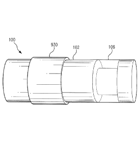
drug-eluting
CA 02922095 2016-02-22
WO 2015/027018 PCT/US2014/052010
polymer matrix 930 may be located adjacent the polymer matrix 106. Although
the ring-shaped
drug-eluting polymer matrix 930 is located on one side of the polymer matrix
106 in
embodiment illustrated in FIG. 9C, the ring-shaped drug-eluting polymer matrix
930 could be
located to the other side of the polymer matrix 106 or on both sides of the
polymer matrix 930.
[0056] In some non-limiting embodiments, as illustrated in FIG. 9D, the
sensor housing 102
may include a groove 932, and the ring-shaped drug-eluting polymer matrix 930
may be
positioned in the groove 932. The edges of the groove 932 may assist in
holding the ring-shaped
drug-eluting polymer matrix 930 in place on the sensor housing 102.
[0057] In some non-limiting embodiments, the analyte indicator (e. g. ,
polymer graft 106)
may a have thin layer (e. g. , 10 nm) on the outside of the graft 106. The
thin layer may protect
against indicator molecule degradation. The thin layer may be platinum, and
the platinum may
be sputtered onto the outside surface of the graft 106, which may include the
indicator molecules
104. Platinum rapidly catalyzes the conversion of hydrogen peroxide into water
and oxygen,
which are harmless to the sensor. The rate of this reaction is much faster
than the boronate
oxidation; thus, the platinum would provide protection against oxidation by
reactive oxygen
species. Although platinum is the catalyst of the conversion of hydrogen
peroxide into water and
oxygen in some embodiments, in alternative embodiments, other catalysts of
this reaction, such
as, for example, palladium or catalase, may be used for the thin layer instead
of or in addition to
platinum.
[0058] In some non-limiting embodiments, as illustrated in FIG. 9E, the
sensor 100 may
include a membrane 934 covering at least a portion of the analyte indicator.
In one non-limiting
embodiment, the membrane 934 may be an analyte permeable membrane. The
membrane 934
may be positioned over the polymer graft 106 (and over any thin layer on the
outside of the graft
16
CA 02922095 2016-02-22
WO 2015/027018 PCT/US2014/052010
106). The membrane 934 may be opaque and, therefore, perform a light-blocking
function. In
other words, the opaque nature of the membrane 934 may serve the function of
effectively
blocking the extraneous light from over stimulating the indicator molecules
104 of the graft 106.
In some non-limiting embodiments, the opaque membrane 934 may be physically
attached over
the graft 106 after boring an additional, smaller well into the
capsule/housing 102.
[0059] In some embodiments, the membrane 934 may be porous. In other words,
the
membrane 934 may be structured so that it channels one or more analytes (e.g.,
glucose) to the
graft 106. For example, in one non-limiting embodiment, the membrane 934 may
have small
pores (e.g., pores having a pore size of microns or less) that block white
blood cells (e.g.,
neutrophils), which are between 6 and 12 microns in diameter, from reaching
the underlying
graft 106 to attack it. The small pores, however, would at the same time be
large enough to
allow the analyte to reach the graft 106. In this way, a porous membrane 934
having small pores
would increase sensor longevity while not affecting the ability of the sensor
100 to measure the
analyte.
[0060] In some embodiments, the opaque membrane 934 may be made from a
material that
does not react adversely to the body's defenses. In non-limiting embodiments,
the material from
which the opaque membrane 934 is made may additionally be both porous (e.g.,
to allow and
analyte, such as glucose, to flow through it) and opaque (e.g., to prevent
light from traveling
through it). For example, in some embodiments, the membrane (e.g., mesh)
material may be a
material such as nylon, cellulose acetate, polypropylene (PP), polyvinyl
alcohol (PVA),
polybutylene terephthalate (PBT), polyether ether ketone (PEEK),
polyanhydride, polyamide,
polyvinylchloride (PVC), polyethersulfone (PES), polyethylene terephthalate
(PET),
polyvinylidene difluoride (PVDF), polytetrafluoroethylene (PTFE), and/or
polycarbonate.
17
[0061] In some embodiments, the membrane 934 may be a porous, opaque
diffusion membrane
that is configured to: substantially prevent white blood cells from passing
through the membrane,
permit an analyte of interest to pass through the membrane to the graft, and
substantially prevent
transmission of light of at least a specified wavelength or range of
wavelengths through the
membrane.
[0062] In some embodiments, to enhance biocompatibility and/or
hydrophilicity, the membrane
934 may comprise an additional thin layer, and/or the membrane 934 may
comprise multiple mesh
layers. In some embodiments, the membrane 934 and the one or more therapeutic
agents may have
an additive effect in reducing oxidation of the analyte indicator.
[0063] In some embodiments, the sensor 100 may have one or more of the
features described in
U.S. Patent Application Nos. 14/142,000 and 14/142,017, filed on December 27,
2013.
[0064] In some embodiments, the one or more therapeutic agents, which may
be dispersed
within the drug eluting polymer matrix, may include one or more anti-
inflammatory drugs, such as,
for example, non-steroidal anti-inflammatory drug (e.g., acetylsalicylic acid
(aspirin) and/or
isobutylphenylpropanoic acid (ibuprofen)). In some non-limiting embodiments,
the one or more
therapeutic agents dispersed within the drug-eluting polymer matrix may
include one or more
glucocorticoids. In some non-limiting embodiments, the one or more therapeutic
agents may
include one or more of dexamethasone, triamcinolone, betamethasone,
methylprednisolone,
beclometasone, fludrocortisone, derivatives thereof, and analogs thereof. In
some embodiments,
the one or more therapeutic agents may reduce the production of hydrogen
peroxide by neutrophils
and macrophages. In some embodiments, the one or more therapeutic agents may
reduce
deterioration of the analyte indicator (e.g., polymer graft 106).
18
Date Recue/Date Received 2021-01-18
CA 02922095 2016-02-22
WO 2015/027018 PCT/US2014/052010
[0065] In some non-limiting embodiments, the drug eluting polymer matrix
may release the
one or more therapeutic agents distributed throughout the polymer matrix in a
controlled manner.
For instance, in various embodiments, the drug eluting polymer matrix may
release the one or
more therapeutic agents in a controlled manner over a period of hours, days,
weeks, or months.
FIG. 10 illustrates the release profile of dexamethasone acetate from a drug-
eluting polymer
matrix according to one non-limiting embodiment.
[0066] As described above, in some embodiments, the sensor 100 may include
one or more
drug eluting polymer matrices (e.g., drug-eluting polymer matrix 930) located
outside the sensor
housing 102 and covering at least a portion of the sensor housing 102.
However, this is not
required, and, in some embodiments, the sensor 100 may additionally or
alternatively include
one or more drug eluting polymer matrices located within the sensor housing.
For example, as
shown in FIGS. 9F and 9G, the sensor 100 may include a drug eluting polymer
matrix 936 and
939, respectively, within the sensor housing 102. In some non-limiting
embodiments, the sensor
housing 102 may include one or more perforations (e.g., holes, openings,
cavities, grooves, or
channels), which may allow elution of the one or more therapeutic agents. For
example, in the
non-limiting embodiment illustrated in FIG. 9F, the drug eluting polymer
matrix 936 may be in a
form of a solid (e.g. cylinder) positioned in a hole or cavity 938 foimed in
an end portion of
sensor housing 102 (e.g. end portion 113). In the embodiment of FIG. 9G, the
hole or cavity 938
is formed in the direction substantially parallel to the longitudinal axis A
of sensor 100. In other
non-limiting embodiments, as illustrated in FIG. 9G, a drug eluting polymer
matrix 939 may be
in a form of a solid (e.g. cylinder) positioned in a channel or cavity 940
formed in an end portion
sensor housing 102 (e.g. end portion 113). In the embodiment of FIG. 9G, the
channel or cavity
940 is formed in the direction substantially orthogonal to the longitudinal
axis A of sensor 100.
19
CA 02922095 2016-02-22
WO 2015/027018 PCT/US2014/052010
Although the embodiment of FIG. 9G shows the drug eluting polymer matrix 940
extending
from one end of the sensor to the opposite end, this is not necessary. In
other embodiments, the
channel or cavity 940 and/or the a drug eluting polymer matrix 939 may extend
only a portion of
the way into either one or both of either end of end portions of sensor
housing 102. In some
embodiments, by having the drug eluting polymer matrix 936 and/or 939 within
the sensor
housing 102, the sensor housing 102 may protect the drug eluting polymer
matrix 936 and/or
939.
[0067] As described above, in some embodiments, the sensor 100 may include
one or more
therapeutic agents dispersed within one or more drug eluting polymer matrices
(e.g., drug-eluting
polymer matrix 930). However, this is not required, and, in some embodiments,
one or more
therapeutic agents may alternatively or additionally be incorporated within an
analyte indicator
(e.g., polymer graft 106) and/or a membrane (e.g., membrane 934) covering at
least a portion of
the analyte indicator. For example, in one non-limiting embodiment, the sensor
100 may not
have a drug eluting polymer matrix and may instead have one or more
therapeutic agents
incorporated within a membrane that covers at least a portion of the analyte
indicator. In another
non-limiting embodiment, the sensor 100 may not have a drug eluting polymer
matrix and may
instead have one or more therapeutic agents incorporated within an analyte
indicator (e.g.,
polymer graft 106), which may or may not be covered by a membrane. In yet
another non-
limiting embodiment, the sensor 100 may include a drug eluting polymer matrix
(e.g., drug-
eluting polymer matrix 930) and may also include one or more therapeutic
agents incorporated
within one or more of an analyte indicator and a membrane, which may cover at
least a portion
of the analyte indicator.
CA 02922095 2016-02-22
WO 2015/027018 PCT/US2014/052010
[0068] In some embodiments, the one or more therapeutic agents may be
chemically
incorporated within the drug eluting polymer matrix, membrane, or hydrogel
and/or polymer
containing the analyte indicator. In some non-limiting embodiments, one or
more therapeutic
agents may be incorporated within the drug eluting polymer matrix, membrane,
or hydrogel
and/or polymer containing the analyte indicator via covalent bonds. The drug
eluting polymer
matrix, membrane, or hydrogel and/or polymer containing the analyte indicator
may release the
one or more therapeutic agents when one or more of the covalent bonds are
broken. For
example, in one non-limiting embodiment, the covalent bonds may break in the
presence of
water (e.g., in the presence of water in the interstitial fluid, blood, or
intraperitoneal fluid).
However, this is not required, and, in some alternative embodiments, the
covalent bonds may
additionally or alternatively break through exposure to ultraviolet or visible
light. In some non-
limiting embodiments, the covalent bonds may break through exposure to light
emitted by the
light source 108. For example, in one embodiment, exposure to the excitation
light 329 (e.g.,
having a wavelength of approximately 378 nm) emitted by the light source 108
may cause the
covalent bonds to break. Moreover, the light source 108 may be controlled to
emit light in a
manner (e.g., blinking at specific intervals and/or intensities) that alters
(e.g., increases the rate at
which one or more therapeutic agents are released) the elution profile of the
one or more
therapeutic agents (e.g., to maximize effectiveness in preventing oxidation of
the indicator
species). In some embodiments, a wavelength in a specific range (e.g., 150nm-
1000nm or 300-
600nm) may be necessary to photocleave (i.e., break the covalent bonds and
release), and the
wavelength of the light emitted by the light source 108 of the sensor 100 may
be within in the
specific range.
21
CA 02922095 2016-02-22
WO 2015/027018 PCT/US2014/052010
[0069] In some embodiments, the sensor 100 may include multiple drug
eluting components.
In some non-limiting embodiments, the sensor 100 may include any combination
of one or more
external drug eluting polymer matrices (i.e., drug-eluting polymer matrices
located outside the
sensor housing 102 and covering at least a portion of the sensor housing 102),
one or more
internal drug eluting polymer matrices (i.e., drug-eluting polymer matrices
located within the
sensor housing 102), one or more analyte indicators having one or more
therapeutic agents
incorporated therein, and/or one or more membranes covering at least a portion
of an analyte
indicator and having one or more therapeutic agents incorporated therein. For
example, in one
non-limiting embodiment, the sensor 100 may have two external drug eluting
polymer matrices.
In another non-limiting embodiment, the sensor 100 may have one external drug
eluting polymer
matrix, one internal drug eluting polymer matrix, and a membrane covering at
least a portion of
an analyte indicator and having one or more therapeutic agents incorporated
therein.
[0070] In some embodiments, the multiple drug eluting components may have
different
elution/release rates. For example, in one non-limiting embodiment, the sensor
100 may include
a first drug eluting polymer matrix and a second drug eluting polymer matrix,
the first drug
eluting polymer matrix may release one or more therapeutic agents dispersed
within the first
drug eluting polymer matrix at a first rate, and the second drug eluting
polymer matrix may
release one or more therapeutic agents dispersed within the second drug
eluting polymer matrix
at a second rate that is different from the first rate. For example, in one
non-limiting
embodiment, a faster release rate may be used on the initial immune response
(e.g. 0-21 days),
and a slower release rate may be used as a maintenance release to moderate any
chronic immune
response (e.g. 14-365+ days). In some embodiments, additional drug eluting
components may
have various rates of releasing one or more therapeutic agents.
22
CA 02922095 2016-02-22
WO 2015/027018 PCT/US2014/052010
[0071] An implanted sensor including a drug-eluting polymer matrix,
membrane, or analyte
indicator may have improved performance over a sensor that does not include a
drug-eluting
polymer matrix. For instance, the controlled release of one or more
therapeutic agents (e.g., by a
drug-eluting polymer matrix) may have improved longevity and functionality.
FIG. 11 is a graph
showing the experimental results comparing analyte modulation in sensors
having no drug
eluting polymer matrix and no membrane 934 with a non-limiting embodiment of
sensor 100
having a ring-shaped drug-eluting polymer matrix 930 after 30 days in an
animal model (rat).
FIG. 11 shows that, after 30 days in the animal model, the analyte indicators
of the sensors
having no drug eluting polymer matrix and no membrane 934 had an average
modulation of
approximately 15% relative to the modulation in in vitro conditions (IVC),
where there is no
immunological response. See results labeled "Sensor". In contrast, in the
sensor having a drug-
eluting polymer matrix, the analyte indicator had a modulation of greater than
90% relative to the
modulation in IVC after 30 days in the animal model. See results labeled
"Sensor + Dex". Thus,
the non-limiting experimental results of the analyte indicator modulation of
30 days in the animal
model show that a drug-eluting polymer matrix significantly increases sensor
longevity and
functionality.
[0072] Embodiments of the present invention have been fully described above
with reference
to the drawing figures. Although the invention has been described based upon
these preferred
embodiments, it would be apparent to those of skill in the art that certain
modifications,
variations, and alternative constructions could be made to the described
embodiments within the
spirit and scope of the invention. For example, although in some embodiments,
the analyte
sensor 100 may be an optical sensor, this is not required, and, in one or more
alternative
embodiments, the analyte sensor may be a different type of analyte sensor,
such as, for example,
23
CA 02922095 2016-02-22
WO 2015/027018 PCT/US2014/052010
an electrochemical sensor, a diffusion sensor, or a pressure sensor. Also,
although in some
embodiments, the analyte sensor 100 may be an implantable sensor, this is not
required, and, in
some alternative embodiments, the analyte sensor may be a transcutaneous
sensor having a wired
connection to an external transceiver. For example, in some alternative
embodiments, the
analyte sensor 100 may be located in or on a transcutaneous needle (e.g., at
the tip thereof). In
these embodiments, instead of wirelessly communication using an antenna (e.g.,
inductive
element 114), the analyte sensor may communicate with the external transceiver
using one or
more wires connected between the external transceiver and a transceiver
transcutaneous needle
including the analyte sensor. For another example, in some alternative
embodiments, the analyte
sensor may be located in a catheter (e.g., for intravenous blood glucose
monitoring) and may
communicate (wirelessly or using wires) with an external transceiver.
24