Note: Descriptions are shown in the official language in which they were submitted.
1
TITLE: A TRANSCRIPTOMIC INDEX FOR CHARACTERIZING THE
CELLULAR REPAIR RESPONSE AFTER SOFT TISSUE INJURY IN
DIARTHRODIAL JOINTS
RELATED APPLICATIONS
[001] Blank.
STATEMENT AS TO RIGHTS TO INVENTIONS MADE UNDER
FEDERALLY SPONSORED RESEARCH AND DEVELOPMENT
[002] This invention was made with Government support of Grant No.
R01-AR057066 awarded by the National Institutes of Health. The Federal
Government has certain rights in this invention.
TECHNICAL FIELD
[003] The present invention generally relates to methods for determining
the quality of recovery from joint injury and to methods of treating joint
injury
based on such determinations.
BACKGROUND
[004] Increasing degrees of force applied to joints result in joint injury.
Such joint injury is frequently seen as a result of trauma, for example
chondral
lesions are often seen in athletes, and are typically associated with acute
inflammation. The treatment of joint injuries (such as ligamentous rupture or
meniscal tearing) and rehabilitation of the patient after such injuries
involves a
number of components. Immediate care after the injury typically includes rest,
cold application, compression and elevation. The aim of this treatment is to
minimize inflammation, hemorrhage, pain and cellular metabolism during the
acute post-injury phase and to optimize the potential for subsequent recovery.
Date Recue/Date Received 2020-12-18
CA 02922143 2016-02-22
WO 2015/038474 PCT/US2014/054550
2
[005] such initial treatment is often foiloN,Aesd by protection of the
injured
tissues by immobilization for 1-3 weeks after the injury. Immobilization aims
to
allow healing to begin and to proceed undisturbed and it also prevents re-
injury
of the joint which often results in longer recovery times and can have long
term
effects. After tissue healing has begun, typically beyond 3 weeks post injury,
controlled mobilization is introduced. At 4-8 weeks post injury, more vigorous
rehabilitation to recover muscle mass and joint function can begin.
[006] Orthopedic repair of severe injuries is often performed as soon as
acute swelIng and hemorrhage of the injury subsides. However, physicians
currently do not have a system or method available to differentiate between
acute injury requiring invasive treatment and injuries that will heal
sufficiently
without such treatment.
101371 It is particularly important to identify injuries that can result in
joint
deterioration before such deterioration begins Currently, this kind of
information can be obtained only by MRI imaging of the structural components
of the joint to determine whether critical structural measures, such as
cartilage
volume, are being maintained or undergoing progressive degeneration.
Typically, imaging is required at intervals of about 1-2 years after injury.
Assay
of marker proteins in joint fluid can also be used to detect and monitor joint
deterioration. However, because such marker assays are generally based on a
very limited number of gene products, and the abundance of the products in the
fluid can be highly variable, their predictive capacity tends to be very
limited.
SUMMARY OF THE PREFERRED EMBODIMENTS
[0081 In one aspect, the present invention provides a method for
characterizing the quality of the repair response after injury to a joint of a
human or veterinary subject. The method includes determining mRNA
expression levels of a plurality of genes expressed in a tissue sample taken
from an intra-articular region of the joint. In one embodiment, the plurality
of
genes includes at least the genes listed in Table 1, Table 2 and Table 3. A
CA 02922143 2016-02-22
WO 2015/038474 PCT/US2014/054550
3
reparative index score indicative of the quality of the repair response is
calculated based on the mRNA expression levels of these genes.
[009] In one embodiment, the mRNA expression levels are detennined
using a Reverse Transcriptase -Real Time PCR assay. The tissue sample can
include cartilage, synovium, meniscai tissue, joint capsule lining, ligaments
or
combinations of at least two of these materials.
[010] In certain embodiments, calculating the reparative index score
includes comparing the mRNA expression levels from the patient tissue with
first standard expression levels of the genes and second standard expression
ievels of the genes, where the first standard expression levels are indicative
of
a reparative profile and where the second standard expression levels are
indicative of a non-reparative profile. The reparative index score is based on
relative values of the mRNA expression levels from the patient tissue, the
first
standard expression levels and the second standard expression levels.
[011] The first standard expression levels can be post-injury expression
levels from a wild-type mouse and the second standard expression levels can
be post-injury expression levels from a mouse lacking hyaluronan synthase-1.
The method can also include applying a correction factor to adjust the
relative
abundance of the murine levels with respect to the genes expressed in the
patent tissue sample.
[0/2] In other embodiments, the plurality of genes includes at least the
genes listed in Table 4, Table 5 and Table or at least the genes listed in
Table?, Table 8 and Table 9. The plurality of genes can also include the
genes listed in Table 13, Table 14, and/or Table 15.
10131 Another aspect of the present invention provides a method for
treating an injury to a joint of a human or veterinary subject including
administrating a therapy depending on the reparative index score as discussed
above. In certain embodiments, the therapy is surgical reconstruction,
physical
therapy, viscosupplementation HA therapy, diet recommendations, life-style
CA 02922143 2016-02-22
WO 2015/038474 PCT/US2014/054550
4
change recommendations, anti-inflammatory creams, anti-inflammatory gels or
sprays, heat and freeze treatments, non-steroidal anti-inflammatory drugs
(NSAIDs), acupuncture, complementary and alternative medicines, steroid
injections or steroid tablets.
BRIEF DESCRIPTION OF THE DRAWINGS
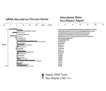
[014] Figure 1 includes bar charts illustrating the relative mRNA
abundance for the 22 fibrosis genes included in Table 7,
[91 53 Figure 2 includes bar charts illustrating the relative mRNA
abundance for the 22 wound repair genes included in Table 8.
[016] Figure 3 includes bar charts illustrating the relative mRNA
abundance for the 22 inflammation genes included in Table 9.
DETAILED DESCRIPTION OF THE PREFERRED EMBODIMENT'S
Definitions
[017] Unless otherwise defined, all technical and scientific: terms used
herein have the same meaning as commonly understood by one of ordinary
skill in the art to which this Invention pertains. In case of conflict, the
present
document, including definitions, will control. Preferred methods and materials
are described below, although methods and materials similar or equivalent to
those described herein can be used in the practice or testing of the present
invention.
[018] The uses of the terms "a" and "an" and 'the" and similar
references in the context of describing the invention (especially in the
context of
the following claims) are to be construed to cover both the singular and the
plural: unless otherwise indicated herein or clearly contradicted by context.
Recitation of ranges of values herein are merely intended to serve as a
shorthand method of referring individually to each separate value falling
within
the range, unless otherwise indicated herein, and each separate value is
CA 02922143 2016-02-22
WO 2015/038474 PCT/US2014/054550
incorporated into the specification as if it were individually recited herein.
All
methods described herein can be performed in any suitable order unless
otherwise indicated herein or otherwise deafly contradicted by context The
use of any and all examples, or exemplary ianguage (e.g., such as, for
example') provided herein, is fltended merely to better illuminate the
invention
and does not pose a limitation on the scope of the invention unless othetwise
claimed. No language in the specification should be construed as indicating
any non-claimed element as essential to the practice of the invention.
[019] As used herein the terms "comprise(s)," "include(s)," "having,"
"has," "can," and variants thereof, are intended to be open-ended transitional
phrases, terms, or words that do not preclude the possibility of additional
acts.
The present invention also contemplates other embodiments "comprising,"
"consisting or and 'consisting essentially of," the embodiments or elements
presented herein, whether explicitly set forth or not.
Method of Determining the Quality of Repair Response After Joint Injury
0201 For the purpose of promoting an understanding ot the principles of
the invention, reference will now be made to embodiments, some of which are
illustrated in the drawings, and specific language will be used to describe
the
same, ft will nevertheless be understood that no limitation of the scope of
the
invention is thereby intended. Any alterations and further modifications in
the
described embodiments, and any further applications of the principles of the
invention as described herein are contemplated as would normally occur to one
skilled in the art to which the invention relates, In the discussions that
follow, a
number of potential features or selections of assay methods, methods of
analysis, or other aspects, are disclosed, it is to be understood that each
such
disclosed feature or features can be combined with the generalized features
discussed, to form a disclosed embodiment of the present invention
[021] One aspect of the present invention provides a Method of generating
a reparative index that is indcative of the quality of the repair response
after a
CA 02922143 2016-02-22
WO 2015/038474 PCT/US2014/054550
6
itsint injury. The joint can be any joint in the body of a human or veterinary
subject including, but not limited to, the knee, .shoulder, hp, elbow or ankle
joints, or a joint of the hand, foot or spine. The method includes a gene
expression analysis of a tissue sample taken from the intra-articular space of
the joint.
(0221 In one embodiment, the method determines changes in the
transcriptome of tissue containing stromal and multipotent progenitor cells
proximate to the injury site. Specifically, the transcript abundance of a set
of
"fibroproliferative' and "profibrotic' genes in such joint tissues is
predictive of
the quality of the long term repair process. Such genes include genes in the
fibrosis, oytokine and/or NFkB pathways that are involved in fibrosis, wound
healing and inflammation. In a murine model the determination of expression
levels of such genes as early as 4 weeks post-injury is predictive of the
quality
of joint repair which can be expected.
[023) In one embodiment, the tissue sample is obtained from the intra-
articular region. As used herein the term "intra-articular refers to the space
inside of a joint between two bones, specifically to the portion of the joint
contained by the joint capsule. The sample can include cartilage, synovium,
compressed meniscal tissue, joint capsuie lining, ligaments or combinations of
at least two of these materials.
[024] In certain embodiments, the subject is a human subject and the
tissue sample is obtained at between 4 weeks and 20 weeks, 4 weeks and 16
weeks, 4 weeks and 12 weeks or 6 weeks and 10 weeks after the injury. In
other embodiments multiple tissue samples are obtained, each at a different
time after the injury. Such a procedure allows the quality of the repair
response
to be monitored as time progresses. For example, the quality of natural
healing
of the joint or the response of the injury to a particular treatment can be
determined by observing the changes in gene expression over time. For
example, the tissue sample can be obtained during the first arthroscopic (or
open joint) evaluation of the injury. in certain cases, depending on the
CA 02922143 2016-02-22
WO 2015/038474 PCT/US2014/054550
surgeons preferences, a second sample could be obtained at a later time (such
as at surgery or at a second arthroscopic evaluation)
[0251 The gene expression analysis can be performed by any method
known in the art. Methods suitable for such an analysis include, but are not
limited to. Northern blot, Nuclease Protection Assay, In Situ Hybridization
and
Real Time PCR. In a preferred embodiment, gene expression is measured by
quantification of mRNA using Reverse Transcriptase Real Time Polymer Chain
Reaction (RT2-PCR.) For example, levels of mRNA can be measured using
commercially available products such as the RT2 Profiler PCR Array, available
from Olagen, Inc. (Valencia, Calif.)
10261 In various embodiments, expression levels of genes associated
with fibrosis, wound healing and inflammation are measured. In one such
ernlx)diment, at least the genes listed in Table 1 (fibrosis genes), Table 2
(wound healing genes) and Table 3 (inflammation genes) are determined in the
gene expression analysis. In another embodiment, at least the genes listed in
Table 4, Table 5 and Table 6 are determined. In yet another embodiment, at
least the genes listed in Table 7. Table 8 and Table 9 are determined. In
other
embodiments, expression levels of at least some of the genes listed in Tables
13, 14, and 15 are also determined. For example, 10, 20, 40, 60, 60 or 65 of
the genes included in Table 13 and/or 5, 10 or 15 of the genes listed in Table
14 can also be determined in the gene expression analysis.
[027] In certain embodiments, the expression levels of the genes
discussed above are used to calculate a reparative index score that is
indicative of the quality of the repair process after joint injury in a human
or
veterinary subject. For example, the reparative index can be indicative as to
whether the injury displays a wound healing response resulting in repair of
the
injury (reparative profile) of an aberrant response leading to joint
degeneration,
fibrosis and/or chronic inflammation (non-reparative profile.)
CA 02922143 2016-02-22
WO 2015/038474 PCT/US2014/054550
8
[028] In one embodiment, the expression levels of genes from a patient
tissue sample taken from the region of a joint injury are compared with
standard
expression levels, for example non-injured expression levels, for the
corresponding genes from the same tissue location in the same or a different
species. For example, the standard expression levels can be gene expression
levels known to be indicative of a reparative profile. In another embodiment,
the expression levels from Me patient sample are compared with standard
expression levels that are indicative of a non-reparative profile. In yet
another
embodiment, the expression levels from the patient are compared with
standard expression levels indicative of both reparative and non-reparative
profiles. Where further quantification of the reparative index score is
required,
additional standards can be included for comparison with the expression levels
from the patient.
[029] An exemplary protocol for calculating a reparative index score is
described belovir for the purposes of illustration only. It is not intended
that the
disclosed methods be limited to this illustrative, embodiment,
[03O] Expression levels of the required genes selected from those listed
in Tables 1-19 and 13-15 are determined in wild type mice (WI) and mice
lacking nyaluronan synthase-1 (Has1-1,-. MSI:106590) at approximately four
weeks post cartilage injury in the knee joint. In this injury model, the WT
mice
display classic wound healing responses in soft tissues, resulting in a
reparative response. In comparison, Haslet- mice show aberrant wound
healing responses, leading to fibrosis, chronic inflammation and OA pathology
(for example, cartilage degeneration and osteophyte formation.) Pathway-
specfic gene array analyses in combination with gross morphology,
immunochemistry confirms healing response in WT mice but chronic fibrosis
and inflammatory status in the joints of Hasl-/- mice. At 4 weeks post-injury
in
murine model, the transcript abundance of these genes in such joint tissues is
predictive of the quality of the long term repair process.
CA 02922143 2016-02-22
WO 2015/038474 PCT/US2014/054550
9
[031] Expression levels for WT mice (repair) and Has1-1- mice (no
repair) are shown in Tables 1-9 for 66 genes involved in fibrosis, wound
hewing
or inflammation. These tables also slow the ratio of the expression levels of
these genes in WI mice and Hast-/- mice. All of the genes in Tables 1-9 are
shown to be up-regulated by at least 7.8 fold in the Has1-/- mice as compared
to WT mice. The additional genes are listed in The 13 are up-regulated by
approximately 3-8 fold in Hasl-/- mice as compared to WT mice. The genes
listed in Table 14 are down-regulated by approximately 3-8 fold in HAs1-1-
mice
as compared to WT mice.
[0321 Figure 1 illustrates the relative mRNA abundance for the 22
fibrosis genes included in Table 7. Here, expression in Has 1-1- mice is shown
to be upgraded up to just over 100 fold. Figure 2 illustrates the relative
mRNA
abundance for the 22 wound repair genes included in Table 8. For these
genes, expression in Has 1-i- mice is shown to be upgraded up to 200 told.
Figure 3 illustrates the relative mRNA abundance for the 22 inflammation genes
included in Table 9. For these cones, expression in Has 1-/- mice is shown to
be upgraded up to just under 400 fold.
[033] in one embodiment, the expression levels of a number of fibrosis,
wound healing and inflammation related cones having similar expression levels
in WI and Hasl-f- mice are measured. For example, such "non-responsive"
murine genes can be considered to be genes that are up- or down regulated by
less than 3 fold in Hasl-/- mice as compared to their levels in WT mice. Table
lists the expression levels of 8 such genes.
10343 The expression levels of the non-responsive murine genes, for
example the WT mice non-responder genes, are then compared with the
expression levels of the corresponding genes in the patient tissue sample. An
average gene ratio is calculated. For example, such a calculation for the
fibrosis pathway is illustrated in Table 11. This ratio can be used as a
murinefhuman correction factor to normalize expression level differences
between the human and murine genes. The WI and Hasl-/- expression levels
CA 02922143 2016-02-22
WO 2015/038474 PCT/US2014/054550
of the murine genes (for example, those genes listed in Tables 1-9, 13-16) are
multiplied by the correction factor to generate an equivalent human range for
each gene.
A reparative index, score can be calculated by, for example,
assigning a score of zero to the bottom value of the human range for each gene
and a score of '10 to the top value of the human range for each gene, with
each
intermediate value being scored as appropriate on this scale. A score is
assigned for each of the genes measured in the patient sample. for example for
each of the 48 genes listed in Tables 4, 5 and S. and the scores added to give
the reparative index score. This total score will range from zero to 160 for
each
pathway of 16 genes and from zero to 480 for the three combined pathways. A
score of zero will be indicative of very good repair and a score of 180 (or
480)
as very poor repair. Table '12 illustrates the calculation of the reparative
index
for a human patient based on the expression levels of the list of six genes in
Table 1.
Methods of Treatment
[036) Another aspect of the present invention provides methods for treating
an injury to a joint of a human or veterinary subject including administrating
a
therapy depending on the reparative index score as discussed above. In
certain embodiments, the therapy is surgical reconstruction, physical therapy,
viscosupplementation HA therapy, diet recommendations or life-style change
recommendations. In other embodiments, the treatment includes
administration of anti-inflammatory creams, gels or sprays, heat and freeze
treatments, non-steroidal anteinflammatory drugs (NSAIDs), acupuncture,
complementary and alternative medicines, steroid injections or steroid
tablets.
Examples
Example 1 ¨ Protocol Used to Obtain Tissue Samples for Analysis
CA 02922143 2016-02-22
WO 2015/038474
PCT/US2014/054550
11
1037j Samples for use in the de,termination of a reparative index
for a
joint injured patent are typically obtaine,d from one or more of the
following:
cartilage, synovium, meniscal tissues. joint capsule lining and lioaments
(including the ACL, PCL and perimeniscal ligaments.) Samples are usually
obtained during the first post-injury arthroscopy. In some cases: where the
extent of soft tissue involvement has been determined by MRI or other imaging
modalities, the biopsies are taken during surgical repair. Tissue (typically
50
milligrams wet weight) proximate to the injury site, such as perimeniscal
synovium in the case of rneniscal tears, are immediately placed in RNALater
(Qiagen Inc.) and stored at 4 C for RNA stabilization. RNA (at least 2
micrograms) is analyzed on Qiagen RT2- Profiler PCR arrays for components of
the Fibrosis, Wound Healing and Inflammation pathways. Equivalent arrays
from other suppliers can aisiD be used. If multiple biopsies are obtained, an
index can be generated from each to evaluate the efficacy of the therapies in
use.
Example 2 Gene Assay Analysis of a Cartilage Injury Model in Mice
[038] Cartilage injury 'in the knee joint of wild type mice (C5713L6) and
mice lacking hyaluronan synthase-1 MGI:106590)
is analyzed as
described below. The knee joint space is opened and a non-bleeding, partial
thickness scalpel wound created in the atticular surface of the femoral
patellar
groove. After iavage of the wound, the joint is closed surgically and the mice
are allowed free cage activity with food and water ad libidum.
[039] Knees are dissected (skin and muscle-free) from mice at 0, 3, 7,
14 and 28 days after surgery. The repair/healing status is evaluated by gross
morphology histology and immunohistochemistry with a panel of antibodies
against matrix repair and innate inflammatory responses. in addition, a
quantitative real time pciymerase chain reaction assay is used to quantify the
abundance for mRNAs on the Fibrosis, Common cytokine and NEkB array
plates (Qiagen Inc Product Nos. PAMM-225Z, PAMM-021Z and PAMM-120Z.)
CA 02922143 2016-02-22
WO 2015/038474 PCT/US2014/054550
12
[040] Data obtained from gross morphology: histology and
immunohistochemistry of the wild-type and Hast-i- mice at each time point are
evaluated. Wild-type mice exhibit a transient inflammatory phase and
fibrogenic phase that is most evident in the stromal/progenitor cell rich-
tissues
adjacent to the cartilage injury site. At later times this early response is
arrested and recovery from the initial injury is substantial. In contrast, for
Has1-
I- mice the post-injury soft tissue histopathology is not arrested Instead it
is
transformed into further fibro-proliferative responses and classic
osteparthritic
changes that include cartilage loss from initially uninjured sites, such as
the
tibial and patellar surfaces, and osteophyte development on lateral and medial
aspects of the femoral groove,
[041] In summary, as assessed by this panel of methods, the reparative
response is highly effective in wild-type mice but essentially absent in Has1-
1-
mice, RT2 PCR array analyses for components of the fibrosis, wound healing
and inflammation pathways reveals that wild-type and Hest -/- mice exhibit a
markedly different expression of genes in all 3 pathways. 50 genes from each
pathway which are found to best discriminate between the 2 genotypes are
chosen for use in generation of a reparative index.
[042] In parallel mRNA analyses of murine and human joint tissues: the
murine and human models routinely show similar values for the abundance of
transcripts for genes of interest. The abundance values for both munne and
human samples are controlled internally against the house-keeping gene:
glyceraldehyde-3-phosphate dehydrogenase (GAPDH).) This allows
standardization of the analysis of human gene expression against murine
values obtained under the same assay conditions,
(043]
............. =
= No
nt Reoeir
Inhbe j 0.1 6,6
=
2 amp 9.0 894.9 69.2
3 MA9 0,2 17.1 -- 66.4
4 Pbts 0.0 781 84.6
CA 02922143 2016-02-22
.
WO 2015/038474 PCT/US2014/054550
13
r
- J 1111 1 0.09 1 864
0.0 I 22;3
gm] Table 1: Transcript Abundance - Fibrosis Pathway: Gene Set 1
(0451
i I ts.12
...................... I .................. lam. Bea& RInir
I i in 0.1 172 j 207.4
1 .................. 2 c.s12 0.0 7.1 j 206.7
i=
3 Wimple 0.1 1 17.2 190.8
1
[ 4 1 Pia 0,1 i 8.9 142.4
: 6 1 Fel0 0.2 18.5 82.7
1
................... 6 ii..8 0.4 : 18
10461 Table 2; Transcript Abundance - Wound Healing Pathway: Gene Set 1
[047]
mg
I ........................ 201 autit
I 4 Sottp25 0.00 .... 16.23 2441 i
-
2 i l MO 0.06. 12.21 196.2 _
3 i C.ycl3 0.08 16.01 180.3
1
4 1 11.21 0.00 9.31 149.6
5 OS 0 06 11.0,6 177.8
=
[ 6 , Mail 1 t.-06 i not I 363.4 ,
[048] Table 3 : Transcript Abundance - Inflammation Pathway: Gene Set 1
10491 .........................
Ng. I
_am Reptilt, Reavkr 1
1 lohbe 0.1 6.5 ' 105.0
t
2 1 ay.' T 9.0 894.9 99.2
.'
3 i trni 02 171 86.4
4 i Fbrs 0.9 78.6 84.6
. 4
i 1 .................. tni i 0.09 6.54 96.7 .
6 i Earl': 0.6 j 22.3 39.0
i
7 Cen 0.9 25.9 30.0
8 . Cal3a 1435 27331 19.0
9 #13ra2 1.3 21.0 16.1
:
L 10 1 lifinja3 __ 14.7 218.8 14.9
11 1 Cala 2.0 28.3 110
i
12 ....................................... Pas' " O.? = 10.1 14.8
. , 3
CA 02922143 2016-02-22
WO 2015/038474 PCT/US2014/054550
14
,
13 ..I. roti __ 1.2 1 171
4- 14.0
14 CaVI :265 389.3 14.7
15 C0112. 2.0 28.3 14.0
16 ccnt : i 3 _____ 14.7 . 11.8
[0501 Table 4: Transcript Abundance Fibrosis Pathway Gene Set 2
(0511 ....
lb
....................... 29Dst .. .Repoir, 1
1 j 112 01 , - 17.2 201 4
4
2 Can _ 0.0 7.1 _1 206.7
3 mmpra 0.1 190.8
-1--.17.2
4 Pig o.1 8.9 1424
5 atm) 0.2 10.5 82.7
= a 11..6 04 18.3 61.3 )
7 11/0 0.8 30.0 , no )
8 Nif 54.7 19933 34.61
9 Minp2 162.2 ______ 3183.6 20.9
10 .................. EGFI 0.7 13.8 19.6
11 nits es 10.3 17.5
t-
12 Ptifsl 12 4õ.., 212 17.6
13 PRO 1,8 27.4
14 Kg" 2.1 28.6 13.6
/5
18 Csil 0.2 .. 2.0 12.8 i
10521 Table 5: Transcript Abundance Wound Healing Pathway Gene Set 2
r" a..., No Rekait ; Ira I
i I Snap25 0.08 . 15.23 244.8 .1 2,,J
1120 0.08 12.21 1962 i
3 -I-
Cx 03 0.08 15.01 160.3
.1-
4 1121 0.08j, 0.31 140.6
6 .. 113 0.06 t 11.06 ...... 177,8
6 ifilbl 0.06 1 22.61 363.4
7 C0122 0.1 8.8 83.3 4
8 1112b 0,2 13.6 71.7j
9 C4a 0.2 3.9- 225
10 Creil 0,3 11.2 34.9
_
0,4 16,1 37.9
12 Lit 4..1 0,5 17.9 30.6
13 Fast. I 1 7 28.8 17.2
1
14 1118 I 10.6 110.9 I 11.2 ,
=
CA 02922143 2016-02-22
WO 2015/038474 PCT/US2014/054550
................................... , ......
15 ' nie
I 3.7 1 50.2 i 13.$
16 L...1e I 8,1 I no I 10.6 ,
10541 Table 6: Transcript Abundance Inflammation Pathway Gene Set 2
r TP551 .......... tia
..:-. Gene Repair_ Busk. NR/R.
I rnhbe- c.i.1 6.5 105.0
4.--
1 2 Bon* 5 0 894,9 99.2
i 3
Ifn T 0.2 17.1 86.4
1 4 Film Li ?8.4 84,6
,
5. sto .............. 0.09 6.54 98.7
8 Edni 0.6 22.3 39.0 i
7 oc./3 0.9 ____ 2.5.9 30.0 1
?-----
I 8 Co130 1435 27331 19.0
i.
.9 1113ra2 1.3 21,0 16.1 ,
i i
t 10 litni.p3 .... 14.7 216.8- 14.91
1 11 Co112 f___ 2.0_5555 28,3 14.0
i 12 1An, 1 1 10,1 14.8
r 1.3 TO/ 1.2 17.1 1 14.01
t14 1 ciso 26.5 . aftp.3-.... .....
15 Cc112 2.0 28.3 14.0
l=
t 16 Calf 1.3 14.7 11.8
i /7 Snail 1.5 ie.7 11.4
s 18 tgfb2 ,..,,... 4.0 -44,7 r '
11.2_1
I 19 CV, 5t35 .0 -6138.4 10.5
sea o.a 9.0 __ 10.7
21 õteb3 ._ 6.2 64.7 105
1 22 -1
Tiroff3 1 36.0- 380.2 : 10.8.)
[069 Table 7: Transcript Abundance Fibrosis Pathway Gene Set 3
MU
1
........... , gsim, 520.1:1 !tell _ ISA
1 112 0.1 t 17.2 207,4
......... 2 CsI2 1 0.0 ' 7,1 2067 i
..,... 3 , ithwja 0.1 1 17.2 190.8 I
L.L.......V ..... Pig. 0.1 83 142.4
5 Faf10 0.2 18.5 82 7 ' ...1
t....
6 .11.11 0,4 1 1a3 513
f=-=
7 00 1 0.8 30.8 39.9
8 Me i 94.7 ,,. 1893.3 i 34.6
CA 02922143 2016-02-22
WO 2015/038474 PCT/US2014/054550
16
................................... T ...
91 ain3p2 j.,1512 3183.6 I 209
.10 EGF 0,7 13.8 19.5
1
11 IVO -o.e i 7e.3 = 1 .5
i.
.12 Ptgs2 .õ, 1 2 21.2 1 176
I
13 Plat 1.8 27.4 z 15,5
14 1110 21 28.6 4.._ 11.8 i
15 .... iirge .... OA 1.3 I 13.4 1
16 04/3 0.2 2.6 1 12.6 i
======
17 rspci2 65.8 876.4 12.1 1
18 Np3 0,7 8.0 12.0k
i
19 1405 19.7 210,7 10.7
20 Wan Vi"
= = -,
"I' 1
21 J. 140P 8.4
-.<
-I
22 iVfA 6.0 47.0 1 7.8 I
10681 Table 8: Transcript Abundance Wound Healing Pathway Gene Set 3
10591 .. = NO. I
, gi411V. ........... RODair RopoW lea_
1 _ Sitv2 .. 0.06 _ 1523 244.8
2 1120 0.06 12.21 190.2
3 Cxel3 .
0.08 1501 180.3
4 112i 0.06 9.31 . 149.6
03 0.06 11.08 177.8
6- Ifsbl Ø06 1 22.61 363A
-k
7 C0122 0.1 8.6 j 86,3
8 1112b 0.2 13,6 J 71.7
i g C4s 0.2 3.9 22.6
:
i 10 Cxel 0.3 11,2 34.9
1
! 11 1/24 0,4 18.1 37.9
i., 12 -, 1.11 0.5 17,9 36.6
L /3 Fasts 1,7 28.6 17.2
i-
1 14 ... HI 8 10.5 116.0 11.2
' 15 .. 1/18 3.7 50.2 13.5
16 ESta 8.1 85.0 10.5
17 Elef2a la 6.2 54.8 10A
_
18 41.t2 8.1 860 10.5
- 19 &Usti a 5.2 54.8 10.5
20 timpt 0,1 17.7 ......... 9.6
21 Ma 7,2 69.0 9.6
22 Adns .. 0.5 42 9.0 - ,.. _ .
CA 02922143 2016-02-22
WO 2015/038474 PCT/US2014/054550
'17
[060] Table 9 : Transdnpt Abundance Inflammation Pathway Gene Set 3
(061] ___
oen - , Re )air
PRI 1'119*
Atttl MN: 113,1 la
i-
Csi2rb 321 07.0
lel 76,1 163.1 Ira
____.....
. qb :40-3 111MIIIII
Steil 100.2 1111=1111=
51813 lirrillirrall 2.6
. .........
rni 7,8 111121111011111
rietseth I 23.5 illeilltill
O62] Table 10: Normalizing Gene Set 1
10631 ..................
1 Data from Patient A
s Patient A for IAItirine for
non- non-
responder responder
Qom's COORS
4...
Mal 31 0.45
esf2th 25 0.70
.......
37 0.47
1..tb 1 10 01
...
Stati -45 a.R.L._.4
slats, 5.0 0.28
Tat 2.1 0,26
irstrsfl b 18 an
, A versue ratio 0.0
[064] Table 11; Calculation of Fibrosis Pathway Index; Ratio of non-
responder genes
to651 _________
1 Transcript Transcnpt Transcript Transcript Patient
A ,. Patient A - '
iAbundanc,3 Abundance Abundant)* Abundance 'Transcript Index Score
iFibrosis Fibrosis Fibrosis Fibrosis Abundance
1 Pathway Pathway Pathway Patinsiny .. Fibrosis
(nit.:(ine) {mutt* (human) ; (human) Pathway
: : . e - ' r N! !Waist._ .Rsaa LN.9 Remit*
1 initbe 0.1 ... 6.5 0.053 i 34 4.6 10
CA 02922143 2016-02-22
WO 2015/038474 PCT/US2014/054550
18
2 aTel ' 9.0 f 894.9 4'7 464 261 6 .... .-
1 0.12 9.1 7.9 8
3 ling
.-
4 PbM 0 9 789 "7 42 _____ 23 5
., ................................................................... ¨
di 1 0.09 8.54 *= ...... b 4,3 9
r
6 Edw./ 0.6 Z 22.3 f3= 34 13.2 3.6 .. 3
............4
Total 41
Scom 1
[066j Table 12: Calculation of Fibrosis Pathway index for a Human Patient
O671
i
I Gone r Gene
1 I z Adel 32 SeTinel ....
2 WO. 33 I Sotto/obi
1 = =
3 ; BAT3 34 Solad2
i .. = _,
4 1 Ceir 36 ; Sofas:33
6 ,.õ j CciS 36 .. i Soma
6 I Condi 37 1 Stund8
7 1 Ccil 38 i Smad7
1.µ $ I COO 39 i Statf
I-
9
40 .LAfeitp2#6 = F3 41 1 Atm
11 t F8 42 NM)/
12 Gadd45b 43 lkkol
=# 4 Ind 44 Roth
....... 14 .t, Rim 45 I Sole
16 112r 46 .. 1 5od2
16 MIS 47 1 SitutSb
17 : Ins 48 i InfatiO
Is 1 III/9 49 i Agt
1
19 1 924 -50 i Cca
20 1 1127 61 I Cebpk,
21 i tnhba 62 Cxeol _________
...
....... 22 .1 Lofty, 63 .,.1L.......
23 .......... 4 Ltb 54 E Jun
24 4, Paull 3 , .. ,...._ 5 .....õ1 Ltbp%, ........._
....... ...... ........... ..........
25 1 Agt N i Mtprei
26 Ccr2 .57 Roo
27 L.Cobpb 58 I Setpinel
25 ! Cxcr4 % i Sespintil
----t
29 En 60 ' Smad2 ..
3-
30 ' Play 61 I $melt _1
t i
31 1. Sorptnefo 6.2 __ L Sam* __
1 I 63 i Stnad7
64 41 Sian
1 65 1 Timp4 ,
O68] Table 13 : Additional Genes Included in the Fibrosis, Wound Healing
or Inflammation Pathways ¨ Set 1
CA 02922143 2016-02-22
WO 2015/038474 PCT/US2014/054550
=
[069)
Gene
Cs12
2 114
.3- ins2
............... 4 finm,81)
6
6 Gdel5
7 11,142
8 1t13
..................... inn
.................... ; 425
...... .... .. t!ct
1 ..
Thfsf
.========4
13 Tntisf4 .....
_______________ 14 .. 1 /113
114
on] Table 14 Additional Genes Included in the Fibrosis, Wound Healing
or inflammation Pathways ¨ Set 2
10711 .............................
I Gem .............................
=+-
1 /lean
2 I Arrthp
3 Cotlat
4 Cot102
Cq/241.1
6 Co13a1
=
7 Heal
a thts2 ..
e Thu3
10 ttihl
11 hih2
12 Stab2
1:3 l'nfaipe
............... 14 110317
10721 Table 16 : Extraaellular Matrix and Hyaluronan Network-Associated
Genes Related to the Fibrosis, Wound tlealing or Inflammation Pathways
(0731 Although the invention has been described and illustrated with
reference to specific illustrative embodiments thereof, it is not intended
that the
invention be limited to those iliustrative embodiments: Those skilled in the
art
20
will recognize that variations and modifications can be made without departing
from the true scope and spirit of the invention as defined by the claims that
follow. It is therefore intended to include within the invention all such
variations
and modifications as fall within the scope of the appended claims and
equivalents thereof.
In some aspects, embodiments of the present invention as
described herein include the following items:
1. A method for classifying the quality of a repair response after
injury to a
joint of a human or veterinary subject, comprising:
determining mRNA expression levels of a plurality of genes expressed in a
tissue sample taken from an intra-articular region of the joint, the plurality
of genes comprising the genes lnhibin beta E (Inhbe), Bone morphogenetic
protein 1 (Bmp1), Interferon gamma (Ifng), Fibrosin (Fbrs), Interleukin 11
(1111),
Endothelin 1 (Edn1), Interleukin 2 (112), Colony stimulating factor 2
(granulocyte-
macrophage) (Csf2), Matrix metallopeptidase 1a (interstitial collagenase)
(Mmp1a), Plasminogen (Pig), Fibroblast growth factor 10 (Fgf10), Interleukin 6
(116), Synaptosomal-associated protein 25 (Snap25), Interleukin 20 (1120),
Chemokine (C-X-C motif) ligand 3 (Cxcl3), Interleukin 21 (1121), Interleukin 3
(113) and Interferon beta 1 fibroblast (Ifnb1),
and calculating a reparative index score based on the mRNA expression
levels of
the plurality of genes,
wherein calculating the reparative index score comprises:
comparing the mRNA expression levels with first standard expression levels
of the genes and second standard expression levels of the genes, wherein the
first
standard expression levels are indicative of a reparative profile and wherein
the
second standard expression levels are indicative of a non-reparative profile;
Date Recue/Date Received 2020-12-18
21
assigning a sub-score for each of the genes, wherein the sub-score is a
measure of the mRNA expression level of each gene relative to the first
standard
expression level and the second standard expression level for each gene, and
summing the sub-scores to obtain the reparative index score; and applying a
correction factor to adjust the relative abundance of the first and second
expression levels with respect to the plurality of genes expressed in the
tissue
sample;
wherein the reparative index score is indicative of the quality of the repair
process, and wherein the reparative index score is indicative of a non-
reparative
profile if the reparative index score is indicative of an elevated mRNA
expression level of the genes.
2. The method of item 1, wherein the first standard expression levels are
post-injury expression levels from a wild-type mouse and wherein the second
standard expression levels are post-injury expression levels from a mouse
lacking hyaluronan synthase-1.
3. The method of item 1 or 2, wherein the first standard expression levels
and
the
second standard expression levels are expression levels measured at between
2 weeks and 4 weeks post-injury.
4. The method of any one of items 1 to 3, wherein determining mRNA
expression levels comprises performing a Reverse Transcriptase -Real Time-PCR
assay.
5. The method of any one of items 1 to 4, wherein the plurality of genes
comprises
the genes lnhibin beta E (Inhbe), Bone morpho genetic protein 1 (Bmp1),
Interferon
gamma (Ifng), Fibrosin (Fbrs), Interleukin 11 (1111), Endothelin 1 (Edn1),
Chemokine
(C-C motif) ligand 3 (CcI3), Collagen, type III, alpha 1 (Col3a1), Interleukin
13
Date Recue/Date Received 2020-12-18
22
receptor, alpha 2 (1113ra2), Matrix metallopeptidase 3 (Mmp3), Chemokine (C-C
motif) ligand 12 (Cd12), Fas ligand (TNF superfamily, member 6) (Fast), TGFB-
induced factor homeobox 1 (Tgifl ), Caveolin 1, caveolae protein (Cavl),
Chemokine
(C-C motif) ligand 11 (CcI11), Interleukin 2 (112), Colony stimulating factor
2
(granulocyte-macrophage) (Csf2), Matrix metallopeptidase la (interstitial
collagenase) (Mmpla), Plasminogen (Pig), Fibroblast growth factor 10 (Fgf10),
Interleukin 6 (116), Interleukin 10 (1110), Macrophage migration inhibitory
factor (Mu),
Matrix metallopeptidase 2 (Mmp2), Epidermal growth factor (Egf), lntegrin beta
8
(1tgb8), Prostaglandin-endoperoxide synthase 2 (Ptgs2), Selectin, platelet
(Plat),
Inte grin beta 6 (1tgb6), Tissue inhibitor of metalloproteinase 1 (Timpl),
Colony
stimulating factor 3 (granulocyte) (Csf3), Synaptosomal-associated protein 25
(Snap25), Interleukin 20 (1120), Chemokine (C-X-C motif) ligand 3 (Cxcl3),
Interleukin
21 (1121), Interleukin 3 (113), Interferon beta 1, fibroblast (lfnbl),
Chemokine (C-C
motif) ligand 22 (CcI22), Interleukin 12B (1112b), Complement component 4A
(Rodgers blood group) (C4a), Chemokine (C-X-C motif) ligand 1 (Cxcll ),
Interleukin
24 (1124), Leukemia inhibitory factor (Lit), Fos ligand (TNF superfamily,
member 6)
(Fast), Interleukin 18 (1118). Interleukin 16 (1116) and Early growth response
2 (Egr2).
6. The method of any one of items 1 to 4, wherein the plurality of
genes
comprises
the genes lnhibin beta E (lnhbe), Bone morphogenetic protein 1
(Bmpl),Interferon
gamma (lfng), Fibrosin (Fbrs),Interleukin 11 (1111), Endothelin 1 (Ednl),
Chemokine
(C-C motif) ligand 3 (CcI3), Collagen, type III, alpha 1 (Col3a1),Interleukin
13
receptor, alpha 2 (1113ra2), Matrix metallopeptidase 3 (Mmp3), Chemokine (C-C
motif) ligand 12 (Cd12), Fas ligand (TNF superfamily, member 6) (Fast), TGFB-
induced factor homeobox 1 (Tgif1), Caveolin 1, caveolae protein (Cavl),
Chemokine
(C-C motif) ligand 12 (CcI12), Chemokine (C-C motif) ligand 11 (CcI11), Snail
homolog 1 (Drosophila) (Snail), Transforming growth factor, beta 2 (Tgfb2),
Connective tissue growth factor (Ctgt), Bone morphogenetic protein 7 (Bmp7),
Transforming growth factor, beta 3 (Tgfb3), Tissue inhibitor of
metalloproteinase 3
(Timp3), Interleukin 2 (112), Colony stimulating factor 2 (granulocyte-
macrophage)
(Csf2), Matrix metallopeptidase la (interstitial collagenase) (Mmpla),
Plasminogen
Date Recue/Date Received 2020-12-18
23
(Pig), Fibroblast growth factor 10 (Fgf10), Interleukin 6 (116), Interleukin
10 (1110),
Macrophage migration inhibitory factor (Mit), Matrix metallopeptidase 2
(Mmp2),
Epidermal growth factor (Egt), lntegrin beta 8 (Itgb8),Prostaglandin-
endoperoxide
synthase 2 (Ptgs2), Selectin, platelet (Plat), lntegrin beta 6 (Itgb6),Tissue
inhibitor of
metalloproteinase 1 (Timpl), Colony stimulating factor 3 (granulocyte) (Csf3),
Thrombospondin 2 (Thbs2), lntegrin alpha 3 (Itga3), lntegrin beta 5 (Itgb5),
Versican
V1 iso form (Vcan V1), Hepatocyte growth factor (Hgt), Vascular endothelial
growth
factor A (Vegfa), Synaptosomal-associated protein 25 (Snap25), Interleukin 20
(1120,
Chemokine (C-X-C motif) ligand 3 (Cxcl3),Interleukin 21 (1121), Interleukin 3
(113),
Interferon beta 1, fibroblast (Ifnbl), Chemokine (C-C motif) ligand 22
(CcI22),
Interleukin 128 (1112b), Complement component 4A (Rodgers blood group) (C4a),
Chemokine (C-X-C motif) ligand 1 (Cxcll), Interleukin 24 (1124), Leukemia
inhibitory
factor (Lit), Fas ligand (TNF superfamily, member 6) (FasL), Interleukin 18
(1118),
Interleukin 16 (1116), Early growth response 2 (Egr2), 8-cell
leukemia/lymphoma 2
related protein Ala (8c12a1a), Interferon alpha 4 (Ifna4), Interleukin 1 alpha
(111 a) and
Adrenomedullin (Adm).
7.
The method of item 5 or 6, wherein the plurality of genes further comprises
the genes Adrenomedullin (Adm), Baculoviral IAP repeat-containing 2 (Birc2),
Baculoviral IAP repeat-containing 3 (Birc3), Chemokine (C-C motif) ligand 12
(Cd12),
Chemokine (C-C motif) ligand 5 (CcI5), Cyclin D1 (Ccndl), Chemokine (C-C
motif)
receptor 5 (Ccr5), CD80 antigen (Cd80), Epidermal growth factor receptor
(Egfr),
Coagulation factor III (F3), Coagulation factor VIII (F8), Growth arrest and
DNA-
damage-inducible 45 beta (Gadd45b), Interleukin 1 receptor, type II (II1r2),
Interleukin 1 receptor antagonist (111 rn), Interleukin 2 receptor, alpha
chain (II2ra),
Interleukin 15 (1115), Interleukin 18 (1118), Interleukin 1 family, member 9
(111f9),
Interleukin 24 (1124), Interleukin 27 (1127), lnhibin beta-A (Inhba), Left
right
determination factor 1 (Leftyl), Lymphotoxin B (Ltb), Tumor necrosis factor
(ligand)
superfamily, member 13 (Tnfsf13), Angiotensinogen (serpin peptidase inhibitor,
clade
A, member 8) (Agt), Chemokine (C-C motif) receptor 2 (Ccr2), CCAAT/enhancer
binding protein (C/E8P), beta (Cebpb), Chemokine (C-X-C motif) receptor 4
(Cxcr4),
Endoglin (Eng), Plasminogen activator, urokinase (Plau), Serine (or cysteine)
Date Recue/Date Received 2020-12-18
24
peptidase inhibitor, clade A, member 1a (Serpina1a), Serine (or cysteine),
peptidase
inhibitor, clade E, member 1 (Serpine1), Serine (or cysteine) peptidase
inhibitor,
clade H, member 1 (Serpinh1), MAD homolog 2 (Drosophila) (Smad2), MAD
homolog 3 (Drosophila) (Smad3), MAD homolog 4 (Drosophila) (Smad4), MAD
homolog 6 (Drosophila) (Smad6), MAD homolog 7 (Drosophila) (Smad7), Signal
transducer and activator of transcription 1 (Stat1), Mitogen-activated protein
kinase
kinase 6 (Map2k6) Myelocytomatosis oncogene (Myc), Nuclear factor of kappa
light
polypeptide gene enhancer in B-cells 1, p105 (Nfkb1), NAD(P)H dehydrogenase,
quinone 1 (Nqo1), Avian reticuloendotheliosis viral (v-re!) oncogene related B
(Relb),
Selectin, endothelial cell (Sele), Superoxide dismutase 2, mitochondrial
(50d2),
Signal transducer and activator of transcription 58 (5tat5b), Tumor necrosis
factor
(ligand) superfamily, member 10 (Tnfsf10), Integrin beta 3 (Itgb3), Jun
oncogene
(Jun), Latent transforming growth factor beta binding protein 1 (Ltbp1),
Matrix
metallopeptidase 9 (Mmp9) and Tissue inhibitor of metalloproteinase 4 (Timp4).
8. The method of any one of items 5 to 7, wherein the plurality of genes
further
comprises the genes Colony stimulating factor 2 (granulocyte-macrophage)
(Csf2),
Interleukin 4 (114), Insulin II (Ins2), Bone morphogenetic protein 8b (Bmp8b),
Cardiotrophin 2 (Ctf2), Growth differentiation factor 15 (Gdf15), Interferon
alpha 2
(Ifna2), Interleukin 13 (1113), Interleukin 17C (1117c), Interleukin 25
(1125), Interleukin 9
(119), Tumor necrosis factor (ligand) superfamily, member 18 (Tnfsf18), Tumor
necrosis factor (ligand) superfamily, member 4 (Tnfsf4), Interleukin 13 (1113)
and
Interleukin 4 (114).
9. The method of any one of items 5 to 8, wherein the plurality of genes
further
comprises the genes Aggrecan (Acan), Alpha-1-Microglobulin/Bikunin Precursor
(Ambp), Collagen, type I, alpha 1 (Col1a1), Collagen, type I, alpha 2
(Col1a2),
Collagen, type II, alpha 1 (Col2a1), Collagen, type III, alpha 1 (Col3a1),
Hyaluronan
synthase 1 (Has1), Hyaluronan synthase 2 (Has2), Hyaluronan synthase 3 (Has3),
Inter-Alpha-Trypsin Inhibitor Heavy Chain 1 (Itih1), Inter-Alpha-Trypsin
Inhibitor
Heavy Chain 2 (Itih2), Stabilin 2 (5tab2), Tumor Necrosis Factor, Alpha-
Induced
Date Recue/Date Received 2020-12-18
25
Protein 6 (Tnfaip6) and Versican (Vcan).
10. The method of any one of items 1 to 10, wherein the tissue sample
comprises material selected from the group consisting of cartilage, synovium,
compressed
meniscal tissue, joint capsule lining, ligaments, and combinations of at least
two of these materials.
11. Use of anti-inflammatory creams, anti-inflammatory gels or sprays, non-
steroidal anti-inflammatory drugs (NSAIDs), injectable steroid or steroid
tablet for
treating an injury to a joint of a human or veterinary subject, wherein the
subject
has a reparative index score which is determined by the method of item 1.
12. The use according to item 11, wherein the tissue sample comprises
material
selected from the group consisting of cartilage, synovium, meniscal tissue,
joint capsule lining, ligaments, and combinations of at least two of these
materials.
13. The use according to item 11 or 12, wherein the subject is a human
subject
and
wherein the mRNA expression levels are determined at between 4 weeks and
12 weeks post-injury.
14. The use according to any one of items 11 to 13, wherein the plurality
of
genes comprises the genes lnhibin beta E (Inhbe), Bone morphogenetic protein 1
(Bmpl), Interferon gamma (Ifng), Fibrosin (Fbrs), Interleukin 11 (1111),
Endothelin 1
(Ednl), Chemokine (C-C motif) ligand 3 (CcI3), Collagen, type III, alpha 1
(Col3a1),
Interleukin 13 receptor, alpha 2 (I113ra2), Matrix metallopeptidase 3 (Mmp3),
Chemokine (C-C motif) ligand 12 (CcI12), Fas ligand (TNF superfamily, member
6)
(Fasl), TGFB-induced factor homeobox 1 (Tgifl), Caveolin 1, caveolae protein
(Ca vi), Chemokine (C-C motif) ligand 11 (CcI11), Interleukin 2 (112), Colony
stimulating factor 2 (granulocyte-macrophage) (Csf2), Matrix metallopeptidase
la
(interstitial collagenase) (Mmpl a), Plasminogen (Pig), Fibroblast growth
factor 10
Date Recue/Date Received 2020-12-18
26
(Fgf10), Interleukin 6 (116), Interleukin 10 (1110), Macrophage migration
inhibitory
factor (Mit), Matrix metallopeptidase 2 (Mmp2), Epidermal growth factor (Egt),
lntegrin beta 8 (Itgb8), Prostaglandin-endoperoxide synthase 2 (Ptgs2),
Selectin,
platelet (Plat), lntegrin beta 6 (Itgb6), Tissue inhibitor of
metalloproteinase 1 (Timpl),
Colony stimulating factor 3 (granulocyte) (Csf3), Synaptosomal-associated
protein 25
(Snap25), Interleukin 20 (1120), Chemokine (C-X-C motif) ligand 3 (Cxcl3),
Interleukin
21 (1121), Interleukin 3 (113), Interferon beta 1, fibroblast (Ifnbl),
Chemokine (C-C
motif) ligand 22 (CcI22), Interleukin 128 (I112b), Complement component 4A
(Rodgers blood group) (C4a), Chemokine (C-X-C motif) ligand 1 (Cxcll ),
Interleukin
24 (1124), Leukemia inhibitory factor (Lit), Fos ligand (TNF superfamily,
member 6)
(Fos , Interleukin 18 (1118). Interleukin 16 (1116) and Early growth response
2 (Egr2).
15. The use according to any one of items 11 to 14, wherein the
plurality of
genes comprises the genes lnhibin beta E (Inhbe), Bone morphogenetic protein 1
(Bmpl),Interferon gamma (Ifng), Fibrosin (Fbrs),Interleukin 11 (1111),
Endothelin 1
(Ednl), Chemokine (C-C motif) ligand 3 (CcI3), Collagen, type III, alpha 1
(Col3a1),Interleukin 13 receptor, alpha 2 (I113ra2), Matrix metallopeptidase 3
(Mmp3), Chemokine (C-C motif) ligand 12 (CcI12), Fas ligand (TNF superfamily,
member 6) (Fos , TGFB-induced factor homeobox 1 (Tgifl ), Caveolin 1, caveolae
protein (Cavl), Chemokine (C-C motif) ligand 12 (Cd12), Chemokine (C-C motif)
ligand 11 (CcI11), Snail homolog 1 (Drosophila) (Snail), Transforming growth
factor,
beta 2 (Tgfb2), Connective tissue growth factor (Ctgt), Bone morpho genetic
protein 7
(Bmp7), Transforming growth factor, beta 3 (Tgfb3), Tissue inhibitor of
metalloproteinase 3 (Tim p3), Interleukin 2 (112), Colony stimulating factor 2
(granulocyte-macrophage) (Csf2), Matrix metallopeptidase la (interstitial
collagenase) (Mmpl a), Plasminogen (Pig), Fibroblast growth factor 10 (Fgf10),
Interleukin 6 (116), Interleukin 10 (1110), Macrophage migration inhibitory
factor (Mu),
Matrix metallopeptidase 2 (Mmp2), Epidermal growth factor (Egf), lntegrin beta
8
(Itgb8),Prostaglandin-endoperoxide synthase 2 (Ptgs2), Selectin, platelet
(Plat),
lntegrin beta 6 (Itgb6),Tissue inhibitor of metalloproteinase 1 (Timpl),
Colony
stimulating factor 3 (granulocyte) (Csf3), Thrombospondin 2 (Thbs2), lntegrin
alpha 3
(Itga3), lntegrin beta 5 (Itgb5), Versican V1 iso form (Vcan V1), Hepatocyte
growth
Date Recue/Date Received 2020-12-18
27
factor (Hgt), Vascular endothelial growth factor A (Vegfa), Synaptosomal-
associated
protein 25 (Snap25), Interleukin 20 (1120, Chemokine (C-X-C motif) ligand 3
(Cxcl3),Interleukin 21 (1121), Interleukin 3 (113), Interferon beta 1,
fibroblast (Ifnbl),
Chemokine (C-C motif) ligand 22 (CcI22), Interleukin 12B (I112b), Complement
component 4A (Rodgers blood group) (C4a), Chemokine (C-X-C motif) ligand 1
(Cxcll ), Interleukin 24 (1124), Leukemia inhibitory factor (Lit), Fas ligand
(TNF
superfamily, member 6) (FasL), Interleukin 18 (1118), Interleukin 16 (1116),
Early
growth response 2 (Egr2), B-cell leukemia/lymphoma 2 related protein Ala
(Bc12a1a), Interferon alpha 4 (/fna4), Interleukin 1 alpha (111 a) and
Adrenomedullin
(Adm).
16. The use according to any one of items 11 to 15, wherein the
plurality of
genes further comprises the genes Adrenomedullin (Adm), Baculoviral IAP repeat-
containing 2 (Birc2), Baculo viral IAP repeat-containing 3 (Birc3), Chemokine
(C-C
motif) ligand 12 (Cd12), Chemokine (C-C motif) ligand 5 (CcI5), Cyclin D1
(Ccnd1),
Chemokine (C-C motif) receptor 5 (Ccr5), CD80 antigen (Cd80), Epidermal growth
factor receptor (Egfr), Coagulation factor III (F3), Coagulation factor VIII
(F8), Growth
arrest and DNA-damage-inducible 45 beta (Gadd45b), Interleukin 1 receptor,
type II
(II1r2), Interleukin 1 receptor antagonist (Om), Interleukin 2 receptor, alpha
chain
(II2ra), Interleukin 15 (1115), Interleukin 18 (1118), Interleukin 1 family,
member 9 (II1f9),
Interleukin 24 (1124), Interleukin 27 (1127), lnhibin beta-A (lnhba), Left
right
determination factor 1 (Leftyl), Lymphotoxin B (Ltb), Tumor necrosis factor
(ligand)
superfamily, member 13 (Tnfsf13), Angiotensinogen (serpin peptidase inhibitor,
clade
A, member 8) (Agt), Chemokine (C-C motif) receptor 2 (Ccr2), CCAAT/enhancer
binding protein (C/EBP), beta (Cebpb), Chemokine (C-X-C motif) receptor 4
(Cxcr4),
Endoglin (Eng), Plasminogen activator, urokinase (Plau), Serine (or cysteine)
peptidase inhibitor, clade A, member la (Serpinala), Serine (or cysteine),
peptidase
inhibitor, clade E, member 1 (Serpinel), Serine (or cysteine) peptidase
inhibitor,
clade H, member 1 (Serpinhl ), MAD homolog 2 (Drosophila) (Smad2), MAD
homolog 3 (Drosophila) (Smad3), MAD homolog 4 (Drosophila) (Smad4), MAD
homolog 6 (Drosophila) (Smad6), MAD homolog 7 (Drosophila) (Smad7), Signal
transducer and activator of transcription 1 (Statl), Mitogen-activated protein
kinase
Date Recue/Date Received 2020-12-18
28
kinase 6 (Map2k6) Myelocytomatosis oncogene (Myc), Nuclear factor of kappa
light
polypeptide gene enhancer in B-cells 1, p105 (Nfkb1), NAD(P)H dehydrogenase,
quinone 1 (Nqo1), Avian reticuloendotheliosis viral (v-re!) oncogene related B
(Relb),
Selectin, endothelial cell (Sele), Superoxide dismutase 2, mitochondrial
(Sod2),
Signal transducer and activator of transcription 58 (5tat5b), Tumor necrosis
factor
(ligand) superfamily, member 10 (Tnfsf10), lntegrin beta 3 (Itgb3), Jun
oncogene
(Jun), Latent transforming growth factor beta binding protein 1 (Ltbp1),
Matrix
metallopeptidase 9 (Mmp9) and Tissue inhibitor of metalloproteinase 4 (Timp4).
17. The use according to any one of items 11 to 16, wherein the plurality
of
genes further comprises the genes Colony stimulating factor 2 (granulocyte-
macrophage) (Csf2), Interleukin 4 (114), Insulin II (In52), Bone morpho
genetic protein
8b (Bmp8b), Cardiotrophin 2 (Cff2), Growth differentiation factor 15 (Gdf15),
Interferon alpha 2 (Ifna2), Interleukin 13 (1113), Interleukin 17C (1117c),
Interleukin 25
(1125), Interleukin 9 (119), Tumor necrosis factor (ligand) superfamily,
member 18
(Tnfsf18), Tumor necrosis factor (ligand) superfamily, member 4 (Tnfsf4),
Interleukin
13 (1113) and Interleukin 4 (114).
18. The use according to any one of items 11 to 17, wherein the plurality
of
genes further comprises the genes Aggrecan (Acan), Alpha-1-
Microglobulin/Bikunin
Precursor (Ambp), Collagen, type I, alpha 1 (Col1a1), Collagen, type I, alpha
2
(Col1a2), Collagen, type II, alpha 1 (Col2a1), Collagen, type III, alpha 1
(Col3a1),
Hyaluronan synthase 1 (Has1), Hyaluronan synthase 2 (Has2), Hyaluronan
synthase
3 (Has3), Inter-Alpha-Trypsin Inhibitor Heavy Chain 1 (Itih1), Inter-Alpha-
Trypsin
Inhibitor Heavy Chain 2 (Itih2), Stabilin 2 (5tab2), Tumor Necrosis Factor,
Alpha-
Induced Protein 6 (Tnfaip6) and Versican (Vcan).
Date Recue/Date Received 2020-12-18