Note: Descriptions are shown in the official language in which they were submitted.
=
- 1 -
DELIVERY OF MEDICAL DEVICES
[0001]
BACKGROUND
[0002] Walls of the vasculature, particularly arterial walls, may
develop areas of
pathological dilatation called aneurysms. As is well known, aneurysms have
thin, weak walls
that are prone to rupturing. Aneurysms can be the result of the vessel wall
being weakened by
disease, injury, or a congenital abnormality. Aneurysms could be found in
different parts of the
body, and the most common are abdominal aortic aneurysms and brain or cerebral
aneurysms in
the neurovasculature. When the weakened wall of an aneurysm ruptures, it can
result in death,
especially if it is a cerebral aneurysm that ruptures.
[0003] Aneurysms are generally treated by excluding the weakened part
of the vessel
from the arterial circulation. For treating a cerebral aneurysm, such
reinforcement is done in
many ways including: (i) surgical clipping, where a metal clip is secured
around the base of the
aneurysm; (ii) packing the aneurysm with small, flexible wire coils (micro-
coils); (iii) using
embolic materials to "fill" an aneurysm; (iv) using detachable balloons or
coils to occlude the
parent vessel that supplies the aneurysm; and (v) intravascular stenting.
[0004] Intravascular stents arc well known in the medical arts for
the treatment of
vascular stenoses or aneurysms. Stents are prostheses that expand radially or
otherwise within a
vessel or lumen to provide support against the collapse of the vessel. Methods
for delivering
these intravascular stents are also well known.
[0005] In conventional methods of introducing a compressed stent into
a vessel and
positioning it within in an area of stenosis or an aneurysm, a guiding
catheter having a distal tip
is percutaneously introduced into the vascular system of a patient. The
guiding catheter is
advanced within the vessel until its distal tip is proximate the stenosis or
aneurysm. A guidewire
positioned within an inner lumen of a second, inner catheter and the inner
catheter are advanced
through the distal end of the guiding catheter. The guidewire is then advanced
out of the distal
end of the guiding catheter into the vessel until the distal portion of the
guidewire carrying the
CA 2922305 2017-07-28
CA 02922305 2016-02-24
WO 2015/031025 PCT/US2014/050270
- 2 -
compressed stent is positioned at the point of the lesion within the vessel.
Once the compressed
stent is located at the lesion, the stent may be released and expanded so that
it supports the
vessel.
SUMMARY
[0006] At least one aspect of the disclosure provides methods and
apparatuses for
delivering an occluding device or devices (e.g., stent or stents) in the body.
The occluding
device can easily conform to the shape of the tortuous vessels of the
vasculature. The occluding
device can be used in a variety of applications. For example, in some
embodiments, the
occluding device can direct the blood flow within a vessel away from an
aneurysm.
Additionally, such an occluding device can allow adequate blood flow to be
provided to adjacent
structures such that those structures, whether they are branch vessels or
oxygen demanding
tissues, are not deprived of the necessary blood flow.
[0007] The delivery of an intravascular stent to a treatment site within
the vessel of a
patient requires substantial precision. Generally, during the implantation
process, a stent is
passed through a vessel to a treatment location. The stent can be expanded at
the treatment
location, often by allowing a first end of the stent to expand and thereafter
slowly expanding the
remainder of the stent until the entire stent has been expanded. The process
of initially
contacting the vessel wall as the first end of the stent expands can be
referred to as "landing" the
stent. The final position of the stent within the vessel is generally
determined by its initial
placement or landing within the vessel. In some situations, the stent may
initially be "landed" in
a suboptimal location within the vessel. Using traditional methods and
apparatuses, it may be
very difficult for a clinician to reposition the stent within the vessel. For
example, a clinician
may be unable to recapture, collapse, withdraw, or resheath the stent back
into the catheter after
the stent has been partially expanded within the vessel. As such, the initial
landing is critical to
successful placement of the stent.
[0008] The subject technology is illustrated, for example, according to
various
aspects described below. Various examples of aspects of the subject technology
are described as
numbered embodiments (1, 2, 3, etc.) for convenience. These are provided as
examples and do
not limit the subject technology. It is noted that any of the dependent
embodiments may be
combined in any combination with each other or one or more other independent
embodiments, to
CA 02922305 2016-02-24
WO 2015/031025 PCT/US2014/050270
- 3 -
form an independent embodiment. The other embodiments can be presented in a
similar manner.
The following is a non-limiting summary of some embodiments presented herein:
Embodiment 1. A stent delivery system, comprising:
a core member having an intermediate portion and an elongate, spiral-cut tube
extending proximally of the intermediate portion, the tube having first and
second flex
zones, the second flex zone being proximal of the first flex zone, and a
transition zone
between the first and second flex zones;
the first flex zone having a bending stiffness of less than 12 N*mmA2 so as to
be
navigable through the internal carotid artery bifurcation, the spiral cut of
the tube in the
first flex zone having a first pitch;
the second flex zone having a bending stiffness of greater than 60 N*mm^2, the
spiral cut of the tube in the second flex zone having a second pitch different
from the first
pitch;
wherein the spiral cut of the tube in the transition zone changes from the
first
pitch to the second pitch in a series of pitch transitions, the spiral cut
pitch in the
transition zone increasing by an overall percent increase from the first pitch
to the second
pitch, such that the average overall percent increase achieved per transition
is 15% or
less; and
a stent carried by the intermediate portion.
Embodiment 2. The system of Embodiment 1, wherein the pitch
transitions of the
spiral cut of the tube have a density along the transition zone greater than 1
transition per
centimeter.
Embodiment 3. The system of Embodiment 1, wherein the pitch of the
spiral cut of
the tube increases by over 150% from the first pitch to the second pitch in a
proximal direction in
the transition zone.
Embodiment 4. The system of Embodiment 1, wherein the first flex zone
length is
greater than 60 mm.
Embodiment 5. The system of Embodiment 1, wherein the second flex zone
length
is greater than 30 mm.
Embodiment 6. The system of Embodiment 1, wherein the second flex zone
bending stiffness is 60-100 N*mm^2.
CA 02922305 2016-02-24
WO 2015/031025 PCT/US2014/050270
- 4 -
Embodiment 7. The system of Embodiment 1, wherein the transition zone
comprises about 25 pitch transitions.
Embodiment 8. The system of Embodiment 1, wherein the first flex zone
is
navigable to the M1 bifurcation.
Embodiment 9. The system of Embodiment 8, wherein the second flex zone
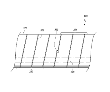
is
navigable to the common carotid artery.
Embodiment 10. The system of Embodiment 1, further comprising a second
transition zone distal of the first flex zone, the spiral cut of the tube in
the second transition zone
decreasing from the second pitch in a second series of pitch transitions, the
second series of pitch
transitions having a density along the second transition zone greater than
five transitions per
centimeter.
Embodiment 11. The system of Embodiment 1, wherein a distal end of the
first flex
zone is spaced 8-12 mm from a proximal end of the stent.
Embodiment 12. The system of Embodiment 11, wherein a distal end of the
second
flex zone is spaced 225-275 mm from a proximal end of the stent.
Embodiment 13. The system of Embodiment 1, wherein the spiral cut of
the tube
prevails along a cut length of the tube, the cut length being greater than 50
cm.
Embodiment 14. The system of Embodiment 13, wherein the spiral cut is
contiguous along the cut length.
Embodiment 15. The system of Embodiment 13, further comprising a
polymeric
outer layer disposed over the outer surface of the tube along at least a
portion of the cut length,
wherein the spiral cut is not cut into the polymeric outer layer.
Embodiment 16. The system of Embodiment 15, wherein the polymeric outer
layer
covers the entire cut length of the tube.
Embodiment 17. A stent delivery system, comprising:
a core member having an intermediate portion and an elongate, spiral-cut tube
extending proximally of the intermediate portion, the tube having an uncut-
tube bending
stifffiess and a first flex zone located near a distal end of the tube, and a
transition zone
extending proximally from the first flex zone;
CA 02922305 2016-02-24
WO 2015/031025 PCT/US2014/050270
- 5 -
the first flex zone having a bending stiffness of less than 5% of the uncut-
tube
bending stiffness so as to be navigable through the carotid siphon, the spiral
cut of the
tube in the first flex zone having a first pitch;
wherein the spiral cut of the tube in the transition zone increases from the
first
pitch in a proximal direction in a series of pitch transitions, the spiral cut
pitch in the
transition zone increasing by an overall percent increase from the first
pitch, such that the
average overall percent increase achieved per transition is 15% or less; and
a stent carried by the intermediate portion.
Embodiment 18. The system of Embodiment 17, wherein the pitch
transitions of the
spiral cut of the tube have a density along the transition zone greater than 1
transition per
centimeter.
Embodiment 19. The system of Embodiment 17, wherein the pitch of the
spiral cut
of the tube increases by over 150% from the first pitch in a proximal
direction in the transition
zone.
Embodiment 20. The system of Embodiment 17, wherein the first flex zone
length is
greater than 60 mm.
Embodiment 21. The system of Embodiment 17, wherein the transition zone
comprises about 25 pitch transitions.
Embodiment 22. The system of Embodiment 17, wherein the first flex zone
is
navigable to the M1 bifurcation.
Embodiment 23. The system of Embodiment 17, further comprising a second
transition zone distal of the first flex zone, the spiral cut of the tube in
the second transition zone
decreasing from the second pitch in a second series of pitch transitions, the
second series of pitch
transitions having a density along the second transition zone greater than
five transitions per
centimeter.
Embodiment 24. The system of Embodiment 17, wherein a distal end of the
first
flex zone is spaced 8-12 mm from a proximal end of the stent.
Embodiment 25. The system of Embodiment 17, wherein the spiral cut of
the tube
prevails along a cut length of the tube, the cut length being greater than 50
cm.
Embodiment 26. The system of Embodiment 25, wherein the spiral cut is
contiguous along the cut length.
CA 02922305 2016-02-24
WO 2015/031025 PCT/US2014/050270
- 6 -
Embodiment 27. The system of Embodiment 25, further comprising a
polymeric
outer layer disposed over the outer surface of the tube along at least a
portion of the cut length,
wherein the spiral cut is not cut into the polymeric outer layer.
Embodiment 28. The system of Embodiment 27, wherein the polymeric outer
layer
covers the entire cut length of the tube.
Embodiment 29. The system of Embodiment 17, wherein the tube has an
outer
diameter of 0.040" or less, and a wall thickness of 0.010" or less.
Embodiment 30. A stent delivery system, comprising:
a core member having an intermediate portion and an elongate, spiral-cut tube
extending proximally of the intermediate portion, the tube having first and
second flex
zones, the second flex zone being proximal of the first flex zone, and a
transition zone
between the first and second flex zones;
the first flex zone having a bending stiffness of less than 220 N*mmA2 so as
to be
navigable through the aortic arch, the spiral cut of the tube in the first
flex zone having a
first pitch,
the second flex zone having a bending stiffness of greater than 250 N*mm^2.,
the
spiral cut of the tube in the second flex zone having a second pitch different
from the first
pitch,
wherein the spiral cut of the tube in the transition zone changes from the
first
pitch to the second pitch in a series of pitch transitions, the spiral cut
pitch in the
transition zone increasing by an overall percent increase from the first pitch
to the second
pitch, such that the average overall percent increase achieved per transition
is 10% or
less; and
a stent carried by the intermediate portion.
Embodiment 31. The system of Embodiment 30, wherein the pitch
transitions of the
spiral cut of the tube have a density along the transition zone greater than 1
transition per
centimeter.
Embodiment 32. The system of Embodiment 30, wherein the pitch of the
spiral cut
of the tube increases by over 35% from the first pitch to the second pitch in
a proximal direction
in the transition zone.
CA 02922305 2016-02-24
WO 2015/031025 PCT/US2014/050270
- 7 -
Embodiment 33. The system of Embodiment 30, wherein the first flex zone
length is
greater than 200 mm.
Embodiment 34. The system of Embodiment 30, wherein the second flex
zone
length is greater than 30 mm.
Embodiment 35. The system of Embodiment 30, wherein the second flex
zone
bending stiffness is 250-310 N*mm^2.
Embodiment 36. The system of Embodiment 30, wherein the transition zone
comprises about 8 pitch transitions.
Embodiment 37. The system of Embodiment 30, wherein a distal end of the
first
flex zone is spaced 480-540 mm from a proximal end of the stent.
Embodiment 38. The system of Embodiment 37, wherein a distal end of the
second
flex zone is spaced 780-820 mm from a proximal end of the stent.
Embodiment 39. The system of Embodiment 30, wherein the spiral cut of
the tube
prevails along a cut length of the tube, the cut length being greater than 50
cm.
Embodiment 40. The system of Embodiment 39, wherein the spiral cut is
contiguous along the cut length.
Embodiment 41. The system of Embodiment 39, further comprising a
polymeric
outer layer disposed over the outer surface of the tube along at least a
portion of the cut length,
wherein the spiral cut is not cut into the polymeric outer layer.
Embodiment 42. The system of Embodiment 41, wherein the polymeric outer
layer
covers the entire cut length of the tube.
Embodiment 43. A stent delivery system, comprising:
a core member having an intermediate portion and an elongate, spiral-cut tube
extending proximally of the intermediate portion, the tube having first and
second flex
zones, the second flex zone being proximal of the first flex zone, and a
transition zone
between the first and second flex zones;
the first flex zone having a bending stiffness of less than 120 N*mmA2 so as
to be
navigable to the common carotid artery, the spiral cut of the tube in the
first flex zone
having a first pitch,
CA 02922305 2016-02-24
WO 2015/031025 PCT/US2014/050270
- 8 -
the second flex zone having a bending stiffness of greater than 180 N*mm^2,
the
spiral cut of the tube in the second flex zone having a second pitch different
from the first
pitch
wherein the spiral cut of the tube in the transition zone changes from the
first
pitch to the second pitch in a series of pitch transitions, the spiral cut
pitch in the
transition zone increasing by an overall percent increase from the first pitch
to the second
pitch, such that the average overall percent increase achieved per transition
is 10% or
less; and
a stent carried by the intermediate portion.
Embodiment 44. The system of Embodiment 43, wherein the pitch
transitions of the
spiral cut of the tube have a density along the transition zone greater than
0.5 transitions per
centimeter.
Embodiment 45. The system of Embodiment 43, wherein the pitch of the
spiral cut
of the tube increases by over 80% from the first pitch to the second pitch in
a proximal direction
in the transition zone.
Embodiment 46. The system of Embodiment 43, wherein the first flex zone
length is
greater than 50 mm.
Embodiment 47. The system of Embodiment 43, wherein the second flex
zone
length is greater than 200 mm.
Embodiment 48. The system of Embodiment 43, wherein the second flex
zone
bending stiffness is 190-210 N*mm^2.
Embodiment 49. The system of Embodiment 43, wherein the transition zone
comprises about 10 pitch transitions.
Embodiment 50. The system of Embodiment 43, wherein a distal end of the
first
flex zone is spaced 300-340 mm from a proximal end of the stent.
Embodiment 51. The system of Embodiment 50, wherein a distal end of the
second
flex zone is spaced 480-540 mm from a proximal end of the stent.
Embodiment 52. The system of Embodiment 43, wherein the spiral cut of
the tube
prevails along a cut length of the tube, the cut length being greater than 50
cm.
Embodiment 53. The system of Embodiment 52, wherein the spiral cut is
contiguous along the cut length.
CA 02922305 2016-02-24
WO 2015/031025 PCT/US2014/050270
- 9 -
Embodiment 54. The system of Embodiment 52, further comprising a
polymeric
outer layer disposed over the outer surface of the tube along at least a
portion of the cut length,
wherein the spiral cut is not cut into the polymeric outer layer.
Embodiment 55. The system of Embodiment 54, wherein the polymeric outer
layer
covers the entire cut length of the tube.
Embodiment 56. A stent delivery system, comprising:
a core member having an intermediate portion and an elongate, spiral-cut tube
extending proximally of the intermediate portion, the tube having first,
second, and third
flex zones and first and second transition zones, the first transition zone
between the first
and second flex zones, the second transition zone between the second and third
flex
zones,
the core member being configured such that (i) a bending stiffness of the
first flex
zone is greater than a bending stiffness of the second flex zone and a bending
stiffness of
the third flex zone and (ii) the bending stiffness of the second flex zone is
greater than the
bending stiffness of the third flex zone, for providing distal pushability of
portions of the
core member distal to the first flex zone,
the spiral cut of the tube has (i) a first pitch in the first flex zone, (ii)
a second
pitch in the second flex zone, (iii) a third pitch in the third flex zone, and
(iv) changing in
the first transition zone from the first pitch to the second pitch in a series
of pitch
transitions and (v) in the second transition zone from the second pitch to the
third pitch in
a series of pitch transitions for preventing buckling of the tube in the first
and second
transition zones when the tube is pushed; and
a stent carried by the intermediate portion.
Embodiment 57. The system of Embodiment 56, wherein the spiral cut of
the tube
prevails along a cut length of the tube, the cut length being greater than 50
cm.
Embodiment 58. The system of Embodiment 57, wherein the spiral cut is
contiguous along the cut length.
Embodiment 59. The system of Embodiment 58, further comprising a
polymeric
outer layer disposed over the outer surface of the tube along at least a
portion of the cut length,
wherein the spiral cut is not cut into the polymeric outer layer.
CA 02922305 2016-02-24
WO 2015/031025 PCT/US2014/050270
- 10 -
Embodiment 60. The system of Embodiment 59, wherein the polymeric outer
layer
covers the entire cut length of the tube.
Embodiment 61. The system of Embodiment 56, wherein the tube comprises
an
uncut segment at a distal portion of the tube.
Embodiment 62. A method of operating a stent delivery system, the
method
comprising:
inserting a core member comprising a varying-stiffness elongate tube into a
tortuous catheter,
advancing the tube through the tortuous catheter by bending the tube in a
transition zone of the tube, thereby forming a curving, non-kinking bend in
the transition
zone.
Embodiment 63. The method of Embodiment 62, wherein the transition zone
is
located between two flex zones of the tube.
Embodiment 64. The method of Embodiment 63, wherein one or both flex
zones has
a substantially constant bending stiffness.
Embodiment 65. The method of Embodiment 62, wherein the tube is spiral-
cut
along a cut length of the tube, and the cut length is greater than 50 cm.
Embodiment 66. The method of Embodiment 65, wherein the spiral cut of
the tube
is contiguous along the cut length.
Embodiment 67. The method of Embodiment 62, wherein advancing the tube
comprises navigating the tube through the aortic arch.
Embodiment 68. The method of Embodiment 62, wherein advancing the tube
comprises navigating the tube through the carotid siphon.
Embodiment 69. The method of Embodiment 62, performed with the core
member
of any of s 1-Embodiment 61.
Embodiment 70. The method of Embodiment 62, wherein the catheter
extends into
the internal carotid artery, and advancing the tube comprises navigating a
portion of the core
member through the internal carotid artery without buckling the tube.
Embodiment 71. A stent delivery system, comprising:
an elongate core member sized for insertion into a blood vessel, the core
member
configured for advancing a stent toward a treatment location in the blood
vessel, the core
CA 02922305 2016-02-24
WO 2015/031025 PCT/US2014/050270
- 11 -
member comprising a longitudinally extending tube having a helical cut
extending along
the tube, the helical cut having an axial length of at least 50 cm and being
continuous
along the axial length.
Embodiment 72. The system of Embodiment 71, wherein the helical cut
comprises a
void in the shape of a helix that extends along the axial length of the tube,
wherein the void is
continuous along the axial length.
Embodiment 73. The system of Embodiment 72, wherein the void comprises
multiple helical slots.
Embodiment 74. The system of Embodiment 73, wherein the helical slots
are
arranged in a contiguous, end-to-end manner.
Embodiment 75. The system of Embodiment 74, wherein the void further
comprises
at least one connection aperture that joins adjacent helical slots.
Embodiment 76. The system of Embodiment 75, wherein the helical slots
and the at
least one connection aperture together form the continuous void.
Embodiment 77. The system of Embodiment 74, wherein the at least one
connection
aperture is a circle.
Embodiment 78. The system of Embodiment 77, wherein the at least one
connection
aperture has a diameter of about 100 microns.
Embodiment 79. The system of Embodiment 77, wherein the at least one
connection
aperture has a diameter of greater than 50 microns.
Embodiment 80. The system of Embodiment 77, wherein the at least one
connection
aperture has a diameter at least twice a width of a helical slot.
Embodiment 81. The system of Embodiment 73, wherein each of the helical
slots
has a slot width of about 25 microns.
Embodiment 82. The system of Embodiment 73, wherein at least one of the
helical
slots has a slot width of about 70 microns or less.
Embodiment 83. The system of Embodiment 71, wherein the helical cut
forms a cut
pattern.
Embodiment 84. The system of Embodiment 71, wherein the tube has a
diameter of
2.3 mm or less.
CA 02922305 2016-02-24
WO 2015/031025 PCT/US2014/050270
- 12 -
Embodiment 85. The system of Embodiment 71, wherein the tube has a wall
thickness of 0.010" or less.
Embodiment 86. A stent delivery system comprising a hypotube having an
elongate
tubular body having a first section and a continuous helical cut extending
about the first section,
the cut having an axial length of at least 50 cm.
Embodiment 87. The system of Embodiment 86, wherein the cut comprises a
plurality of individual helical slots interconnected in an end-to-end manner.
Embodiment 88. The system of Embodiment 87, wherein each individual
helical
slot has an axial length of less than or equal to about 15 cm.
Embodiment 89. The system of Embodiment 87, wherein adjacent individual
helical
slots interconnect via an aperture extending through the hypotube, the
adjacent individual helical
slots extending from the aperture.
Embodiment 90. The system of Embodiment 86, further comprising a second
section, proximal to the first section, wherein a proximal end of the cut
terminates proximal to
the second section.
Embodiment 91. The system of Embodiment 86, wherein the tube further
comprises
an uncut region distal to the cut.
Embodiment 92. The system of Embodiment 86, wherein a pitch of the
helical cut
varies over the length of the cut.
Embodiment 93. The system of Embodiment 92, the pitch of the helical
cut changes
from a first pitch to a second pitch within a longitudinal segment length of
about 5 mm or less.
Embodiment 94. The system of Embodiment 92, the pitch of the helical
cut changes
from a first pitch to a second pitch within a longitudinal segment length of
about 3 mm or less.
Embodiment 95. The system of Embodiment 92, the pitch of the helical
cut changes
from a first pitch to a second pitch within a longitudinal segment length of
about 2 mm or less.
Embodiment 96. The system of Embodiment 92, the pitch of the helical
cut changes
from a first pitch to a second pitch within a longitudinal segment length of
about 1.0 mm.
Embodiment 97. The system of Embodiment 92, wherein the pitch of the
helical cut
changes within a longitudinal distance of about 10 cm or more from an endpoint
of the cut.
Embodiment 98. The system of Embodiment 92, wherein the pitch of the
helical cut
changes within a longitudinal distance of about 20 cm or more from an endpoint
of the cut.
CA 02922305 2016-02-24
WO 2015/031025 PCT/US2014/050270
- 13 -
Embodiment 99. The system of Embodiment 92, wherein the pitch of the
helical cut
changes within a longitudinal distance of about 30 cm or more from an endpoint
of the cut.
Embodiment 100. The system of Embodiment 92, wherein the pitch of the
helical cut
changes in magnitude from a first segment to a second segment by 0.2
mm/rotation or less.
Embodiment 101. The system of Embodiment 92, wherein the pitch of the
helical cut
changes in magnitude from a first segment to a second segment by 0.1
mm/rotation or less.
Embodiment 102. The system of Embodiment 92, wherein the pitch of the
helical cut
changes in magnitude from a first segment to a second segment by 0.01
mm/rotation or less.
Embodiment 103. The system of Embodiment 92, wherein the pitch of the
helical cut
changes in magnitude from a first segment to a second segment by 0.005
mm/rotation or less.
Embodiment 104. A method of manufacturing a stent delivery system, the
method
comprising:
mounting a hypotube in a cutting device having a cutting head;
aligning the hypotube with the cutting head; and
while rotating and axially moving the hypotube relative to the cutting head,
cutting the hypotube to form a helically extending cut having an axial length
of at least 50
cm.
Embodiment 105. The method of Embodiment 104, wherein the cutting
comprises
cutting multiple helical slots to form the helically extending cut.
Embodiment 106. The method of Embodiment 105, wherein the cutting
comprises
cutting the helical slots in a contiguous, end-to-end manner.
Embodiment 107. The method of Embodiment 106, wherein the cutting
comprises
cutting at least one connection aperture at an end of a helical slot.
Embodiment 108. The method of Embodiment 107, wherein the aligning the
cutting
head with the at least one connection aperture to begin cutting a subsequent
helical slot from the
at least one connection aperture.
Embodiment 109. The method of Embodiment 107, wherein the cutting at
least one
connection aperture comprises cutting a circle at an end of a helical slot.
Embodiment 110. The method of Embodiment 104, further comprising
releasing the
hypotube and repositioning and remounting the hypotube in the cutting device
after completing a
cut.
CA 02922305 2016-02-24
WO 2015/031025 PCT/US2014/050270
- 14 -
Embodiment 111. The method of Embodiment 110, wherein the repositioning
and
remounting comprises aligning the cutting head with an end of the cut.
Embodiment 112. The method of Embodiment 110, wherein the cutting the
hypotube
comprises making three or more contiguous, end-to-end cuts to create the
helically extending
cut.
Embodiment 113. A method of operating a stent delivery system, the
method
comprising:
inserting a core member into a catheter in a tortuous configuration, the core
member comprising a longitudinally extending tube having a helical cut
extending along
the tube, the helical cut having an axial length of at least 50 cm and being
continuous
along the axial length; and
pushing the core member through the tortuous catheter; and
by pushing the core member, causing the tube to flex along the helical cut,
thereby facilitating advancement of the core member through the tortuous
catheter.
Embodiment 114. The method of Embodiment 113, wherein the core member
comprises a plurality of flex zones, and the pushing comprises advancing at
least one flex zone
across a tortuosity of the catheter such that the tube forms a curving, non-
kinking bend across the
tortuosity.
Embodiment 115. The method of Embodiment 113, wherein a pitch of the
helical cut
varies over the length of the cut to provide a variable flexibility to the
tube during advancement
through the tortuous catheter.
Embodiment 116. The method of Embodiment 113, wherein inserting the core
member into the catheter comprises doing so without buckling the tube.
Embodiment 117. The method of Embodiment 113, wherein the tube has an
outside
diameter of 2.3 mm or less.
Embodiment 118. The method of Embodiment 113, wherein the tube has a
wall
thickness of .010" or less.
Embodiment 119. The method of Embodiment 113, wherein pushing the core
member through the catheter comprises moving a stent through the catheter with
the core
member.
CA 02922305 2016-02-24
WO 2015/031025 PCT/US2014/050270
- 15 -
Embodiment 120. The method of Embodiment 119, further comprising
releasing the
stent from the core member.
Embodiment 121. The method of Embodiment 113, wherein pushing the core
member through the tortuous catheter comprises pushing the tube through the
tortuous catheter.
Embodiment 122. A method of operating a stent delivery system, the
method
comprising:
inserting a core member into a blood vessel of a patient, the core member
comprising a longitudinally extending tube having a helical cut extending
along the tube
and an axial length of at least 50 cm, the helical cut being continuous along
the axial
length;
advancing the core member to the internal carotid artery; and
by advancing the core member, causing the tube to flex along the helical cut,
thereby facilitating advancement of the core member to the internal carotid
artery.
Embodiment 123. The method of Embodiment 122, further comprising
distally
advancing the core member through the internal carotid artery to the middle
cerebral artery of the
patient.
Embodiment 124. The method of Embodiment 122, wherein the core member
comprises a plurality of flex zones, and the method further comprises
advancing at least one flex
zone across the aortic arch such that the tube forms a curving, non-kinking
bend across the aortic
arch.
Embodiment 125. The method of Embodiment 122, further comprising
distally
advancing the core member through the carotid siphon.
Embodiment 126. The method of Embodiment 122, wherein the cut length is
greater
than 60 cm.
Embodiment 127. The method of Embodiment 122, wherein a pitch of the
helical cut
varies over the length of the cut to provide a variable flexibility to the
tube during advancement
through the blood vessel.
Embodiment 128. The method of Embodiment 122, wherein advancing the core
member to the internal carotid artery comprises doing so without buckling the
tube.
Embodiment 129. The method of Embodiment 122, wherein the tube has an
outside
diameter of 2.3 mm or less.
CA 02922305 2016-02-24
WO 2015/031025 PCT/US2014/050270
- 16 -
Embodiment 130. The method of Embodiment 122, wherein the tube has a
wall
thickness of .010" or less.
Embodiment 131. The method of Embodiment 122, wherein advancing the core
member comprises moving a stent with the core member.
Embodiment 132. The method of Embodiment 131, further comprising
releasing the
stent from the core member.
Embodiment 133. The method of Embodiment 122, wherein advancing the core
member to the internal carotid artery comprises positioning the tube so that
it extends from the
aortic arch to the internal carotid artery.
Embodiment 134. The method of Embodiment 122, wherein advancing the core
member to the internal carotid artery comprises advancing the tube to the
internal carotid artery.
Embodiment 135. A stent delivery system, comprising:
a core member having a distal segment;
a stent engagement member positioned along the core member distal segment and
coupled to the core member, the engagement member comprising an outer surface;
and
a stent extending along the core member distal segment such that the outer
surface
of the engagement member engages an inner surface of the stent along at least
a portion
of only a distal half of the stent for transmitting an axial force from the
core member to
only the stent distal half.
Embodiment 136. The system of Embodiment 135, wherein an axial force on
the core
member is transmitted to the stent only through the engagement member.
Embodiment 137. The system of Embodiment 135, wherein a proximal end of
the
engagement member is positioned distal to a midpoint of the stent such that
transmission of a
distal axial force allows the engagement member to pull the stent.
Embodiment 138. The system of Embodiment 135, wherein the engagement
member
is rotatably coupled to the core member.
Embodiment 139. The system of Embodiment 135, wherein the engagement
member
is positioned in an axial gap between restraints, coupled to the core member,
for permitting
rotational movement of the engagement member relative to the core member.
CA 02922305 2016-02-24
WO 2015/031025 PCT/US2014/050270
- 17 -
Embodiment 140. The system of Embodiment 139, wherein the positioning of
the
engagement member in the axial gap permits translation movement of the
engagement member
relative to the core member.
Embodiment 141. The system of Embodiment 135, wherein the engagement
member
is a first engagement member, and the system further comprises a second stent
engagement
member coupled to the core member and positioned proximal to the first stent
engagement
member.
Embodiment 142. The system of Embodiment 141, wherein a distal end of
the second
stent engagement member is positioned proximal to a midpoint of the stent such
that
transmission of a distal axial force allows the second stent engagement member
to push the stent.
Embodiment 143. The system of Embodiment 141, wherein the second stent
engagement member is rotatably coupled to the core member.
Embodiment 144. The system of Embodiment 141, wherein the second stent
engagement member is positioned in an axial gap between restraints, coupled to
the core
member, for permitting rotational movement of the second stent engagement
member relative to
the core member.
Embodiment 145. The system of Embodiment 144, wherein the positioning of
the
second stent engagement member in the axial gap permits translation movement
of the second
stent engagement member relative to the core member.
Embodiment 146. The system of Embodiment 135, wherein the engagement
member
comprises a generally tubular body.
Embodiment 147. The system of Embodiment 135, further comprising a
radially
expandable member coupled to the core member proximal to the engagement
member, the
radially expandable member having a collapsed position and an expanded
position, wherein in
the expanded position, the radially expandable member is configured to engage
a proximal
portion of the stent.
Embodiment 148. The system of Embodiment 147, wherein the radially
expandable
member comprises a balloon coupled to the core member proximal to the
engagement member,
the balloon being inflatable to engage a proximal portion of the stent.
Embodiment 149. The system of Embodiment 148, wherein the core member
comprises an inflation lumen extending axially to the balloon.
CA 02922305 2016-02-24
WO 2015/031025 PCT/US2014/050270
- 18 -
Embodiment 150. The system of Embodiment 147, wherein the radially
expandable
member comprises a wedge component having an outer portion configured to
expand radially
when the core member is proximally retracted such that the wedge component
engages with the
stent to transmit a proximal force to the stent.
Embodiment 151. The system of Embodiment 135, further comprising a stent
cover
component having a first end coupled to the core member distal segment and a
second end
extending from the first end, the second end configured to at least partially
surround at least a
distal portion of a stent carried by the stent delivery system.
Embodiment 152. The system of Embodiment 151, wherein the cover
component
first end is positioned in an axial gap between first and second restraints
such that the first end is
rotatably coupled to the core member distal segment.
Embodiment 153. The system of Embodiment 151, wherein a distal end of
the
engagement member is spaced less than 1 mm proximal to the second end of the
cover
component.
Embodiment 154. The system of Embodiment 151, wherein a distal end of
the
engagement member is spaced distal to the second end of the cover component
such that the
second end is configured to at least partially surround a portion of the
engagement member.
Embodiment 155. The system of Embodiment 151, wherein a proximal end of
the
engagement member is positioned adjacent to the second end of the cover
component such that
the cover component extends longitudinally along an entire length of the
engagement member.
Embodiment 156. The system of Embodiment 135, further comprising a
catheter
having a lumen configured to receive the core member, engagement member, and
stent, wherein
the stent is radially compressed between an inner surface of the catheter and
the outer surface.
Embodiment 157. The system of Embodiment 135, wherein the stent is a
self-
expanding stent.
Embodiment 158. The system of Embodiment 135, further comprising a
retraction-
only interface positioned along the core member distal segment proximal of the
stent
engagement member.
Embodiment 159. The system of Embodiment 158, wherein the retraction-
only
interface comprises a balloon.
CA 02922305 2016-02-24
WO 2015/031025 PCT/US2014/050270
- 19 -
Embodiment 160. The system of Embodiment 158, wherein the retraction-
only
interface comprises an expandable pad.
Embodiment 161. A stent delivery system, comprising:
a catheter having a lumen and an inner surface extending along the lumen;
a core member, extending within the catheter lumen, having a distal segment
and
a device interface; and
a stent extending along the core member distal segment, at least a portion of
only
a distal half of the stent being radially compressed between the interface and
the catheter
inner surface such that a distal axial force exerted on the core member is
transmitted
through the interface to pull the stent in a distal direction.
Embodiment 162. The system of Embodiment 161, wherein a proximal end of
the
interface is positioned distal to a midpoint of the stent.
Embodiment 163. The system of Embodiment 161, wherein the interface
comprises a
stent engagement member coupled to the distal segment of the core member, the
engagement
member comprising an outer surface configured to engage an inner surface of
the stent.
Embodiment 164. The system of Embodiment 161, wherein the device
interface is a
first device interface, and the system further comprises a second device
interface, proximal to the
first device interface, configured to engage the stent along a proximal half
thereof.
Embodiment 165. The system of Embodiment 164, wherein the second device
interface comprises a second stent engagement member coupled to the distal
segment of the core
member, the second stent engagement member comprising an outer surface
configured to engage
an inner surface of the stent.
Embodiment 166. The system of Embodiment 164, wherein the second device
interface comprises an expandable member coupled to the core member proximal
to the first
stent engagement member, the radially expandable member having a collapsed
position and an
expanded position, wherein in the expanded position, the radially expandable
member is
configured to engage a proximal portion of the stent.
Embodiment 167. The system of Embodiment 166, wherein the radially
expandable
member comprises a balloon coupled to the core member proximal to the first
stent engagement
member, the balloon being inflatable to engage a proximal portion of the
stent.
CA 02922305 2016-02-24
WO 2015/031025 PCT/US2014/050270
- 20 -
Embodiment 168. The system of Embodiment 164, wherein the second device
interface comprises a retraction-only interface.
Embodiment 169. The system of Embodiment 168, wherein the retraction-
only
interface comprises a balloon.
Embodiment 170. The system of Embodiment 168, wherein the retraction-
only
interface comprises an expandable pad.
Embodiment 171. The system of Embodiment 161, further comprising a stent
cover
component having a first end coupled to the core member distal segment and a
second end
extending from the first end, the second end configured to at least partially
surround at least a
distal portion of a stent carried by the stent delivery system.
Embodiment 172. The system of Embodiment 171, wherein a distal end of
the
interface is spaced less than 1 mm proximal to the second end of the cover
component.
Embodiment 173. The system of Embodiment 171, wherein a distal end of
the
interface is spaced distal to the second end of the cover component such that
the second end is
configured to at least partially surround a portion of the interface.
Embodiment 174. The system of Embodiment 171, wherein a proximal end of
the
interface is positioned adjacent to the second end of the cover component such
that the cover
component extends longitudinally along an entire length of the interface.
Embodiment 175. A method of advancing a stent delivery assembly through
a
tortuous catheter, the method comprising:
moving a core assembly distally within a lumen of the catheter, the core
assembly
comprising a stent engagement member that is engaged with at least a portion
of a stent
along only a distal half of the stent;
by moving the core assembly, pulling the stent distally within the catheter
lumen,
the engagement member configured such that friction between the engagement
member
and the core member is less than friction between the engagement member and
the stent.
Embodiment 176. The method of Embodiment 175, wherein the moving
comprises
causing the stent to rotate with respect to a core member of the core
assembly.
Embodiment 177. The method of Embodiment 176, further comprising
rotating the
core member to steer the core assembly to avoid damaging vasculature adjacent
to a treatment
site within a blood vessel.
CA 02922305 2016-02-24
WO 2015/031025 PCT/US2014/050270
-21 -
Embodiment 178. The method of Embodiment 175, further comprising
applying a
proximally oriented retracting force on the core assembly to retract the stent
into the catheter
after a distal portion of the stent has been expanded outside of the catheter.
Embodiment 179. The method of Embodiment 178, wherein the applying
comprises
inflating a balloon, coupled to a core member of the core assembly, to engage
a proximal portion
of the stent prior to applying the proximally oriented force.
Embodiment 180. The method of Embodiment 175, further comprising
advancing the
core assembly distally until at least a distal portion of the stent extends
distally beyond the
catheter such that the stent distal portion expands from a collapsed
configuration.
Embodiment 181. The method of Embodiment 180, wherein the advancing
comprises
inflating a balloon, coupled to a core member of the core assembly, to engage
a proximal portion
of the stent prior to advancing the stent distal portion distally beyond the
catheter.
Embodiment 182. The method of Embodiment 181, wherein the advancing
comprises, after the stent distal end extends distally beyond the catheter,
advancing the stent by
transferring a distal pushing force to the stent via the balloon until a
proximal portion of the stent
is distally beyond the catheter.
Embodiment 183. The method of Embodiment 175, further comprising
partially
expanding the stent distally of the catheter, and retracting the stent into
the catheter with a
retraction-only interface.
Embodiment 184. The method of Embodiment 183, wherein pulling the stent
distally
comprises doing so without applying any substantial distal pulling force to
the stent by the
retraction-only interface.
Embodiment 185. A stent delivery system, comprising:
a core member having a first section and a second section distal to the first
section, the second section having a bending stiffness per unit length that is
less than a
bending stiffness per unit length of the first section;
an introducer sheath having a lumen configured to receive the core member
therethrough, the introducer sheath having a length of at least about 80 cm;
and
a microcatheter having a lumen and a proximal end configured to interface with
a
distal end of the introducer sheath for delivering the core member into the
microcatheter
lumen.
CA 02922305 2016-02-24
WO 2015/031025 PCT/US2014/050270
- 22 -
Embodiment 186. The system of Embodiment 185, wherein the sheath length
is equal
to or greater than a length of the core member second section.
Embodiment 187. The system of Embodiment 185, wherein the first section
has a
substantially constant bending stiffness per unit length.
Embodiment 188. The system of Embodiment 185, wherein the sheath length
is
between about 80 cm and about 150 cm.
Embodiment 189. The system of Embodiment 188, wherein the sheath length
is about
106 cm.
Embodiment 190. The system of Embodiment 185, wherein the core member
comprises a marker visible through the introducer sheath.
Embodiment 191. The system of Embodiment 190, wherein the marker is
disposed
along the core member in the first section thereof.
Embodiment 192. The system of Embodiment 190, wherein the introducer
sheath
comprises titanium dioxide.
Embodiment 193. The system of Embodiment 185, wherein the core member
comprises a solid wire in the first section.
Embodiment 194. The system of Embodiment 185, wherein the core member
comprises a hollow tubular member in the second section.
Embodiment 195. The system of Embodiment 194, wherein at least a portion
of the
hollow tubular member comprises a spiral cut.
Embodiment 196. The system of Embodiment 195, wherein the spiral cut
extends
along about 60 cm to about 100 cm of a length of the second section.
Embodiment 197. The system of Embodiment 196, wherein the spiral cut
extends
along about 86 cm of the length of the second section.
Embodiment 198. A stent delivery system, comprising:
a core member having (i) a stiff section having a first bending stifffiess and
(ii) a
soft section having a second bending stiffness that is less than the first
bending stiffness,
the second bending stiffness varying spatially along the soft section;
an introducer sheath covering any portion of the core member having a bending
stiffness that is less than the first bending stiffness, the introducer sheath
having a length
of at least about 80 cm; and
CA 02922305 2016-02-24
WO 2015/031025 PCT/US2014/050270
- 23 -
a microcatheter having a lumen and a proximal end configured to interface with
a
distal end of the introducer sheath for delivering the core member into the
microcatheter
lumen.
Embodiment 199. The system of Embodiment 198, wherein the bending
stiffness of
the stiff section is substantially constant.
Embodiment 200. The system of Embodiment 198, wherein the stiff section
is
proximal to the soft section.
Embodiment 201. The system of Embodiment 198, wherein the sheath length
is
between about 80 cm and about 150 cm.
Embodiment 202. The system of Embodiment 201, wherein the sheath length
is about
106 cm.
Embodiment 203. A method of manufacturing a stent delivery system, the
method
comprising:
providing a core member and an introducer sheath configured to extend over the
core member, the core member comprising a stiff proximal section configured to
allow a
clinician to grasp the core member for advancing the core member relative to
the sheath;
and
inserting the core member into the sheath such that the sheath covers any
portion
of the core member having a bending stiffness less than a bending stiffness of
the
proximal section and such that only the proximal section is exposed for
gripping.
Embodiment 204. The method of Embodiment 203, wherein the inserting
comprises
advancing the core member into the sheath until a proximal end of the sheath
is positioned
axially over a distal end of the stiff proximal section.
Embodiment 205. The method of Embodiment 203, wherein the inserting
comprises
aligning a marker on the core member with a proximal end of the sheath.
Embodiment 206. A method of advancing a stent delivery system, the
method
comprising:
positioning a distal end of the stent delivery assembly adjacent to a proximal
end
of a guide catheter for moving the core member into a lumen of the catheter,
the core
member comprising a proximal first section and a distal second section that is
more
CA 02922305 2016-02-24
WO 2015/031025 PCT/US2014/050270
- 24 -
flexible than the first section, the stent delivery assembly comprising an
introducer sheath
extending over the entire distal second section; and
while grasping a proximal end of the introducer sheath, grasping only the core
member first section to apply a distal axial force to advance the core member
into the
catheter lumen.
Embodiment 207. The method of Embodiment 206, wherein the core member
comprises a marker visible through the introducer sheath, the method further
comprising
advancing the core member into the catheter lumen until the marker reaches a
first position
visible within the introducer sheath, the first position of the marker
corresponding to a position
of a stent carried on the core member within the catheter.
Embodiment 208. The method of Embodiment 206, further comprising
proximally
withdrawing the introducer sheath from over the core member when the marker
reaches the first
position.
Embodiment 209. A stent delivery system, comprising:
a core member having a distal segment;
a stent engagement member having a generally tubular body positioned about the
core member distal segment and rotatably coupled to the core member, the
engagement
member comprising an inner layer having a first durometer and an outer layer
having a
second durometer less than the first durometer; and
a stent extending along the core member distal segment such that an inner
surface
of the stent is engaged by the engagement member outer layer for facilitating
rotation of
the stent relative to the core member.
Embodiment 210. The system of Embodiment 209, wherein the inner layer
comprises
a substantially cylindrical inner surface surrounding the core member.
Embodiment 211. The system of Embodiment 209, wherein the inner layer
comprises
a
Embodiment 212. The system of Embodiment 209, wherein the outer layer
comprises
a durometer of between about 10A to about 50A.
Embodiment 213. The system of Embodiment 212, wherein the outer layer
comprises
a durometer of between about 15A to about 40A.
CA 02922305 2016-02-24
WO 2015/031025 PCT/US2014/050270
- 25 -
Embodiment 214. The system of Embodiment 213, wherein the outer layer
comprises
a durometer of about 20A.
Embodiment 215. The system of Embodiment 209, wherein the inner layer
comprises
polyimide and the outer layer comprises silicone.
Embodiment 216. The system of Embodiment 209, wherein the outer layer
comprises
a substantially cylindrical outer surface for contacting the stent.
Embodiment 217. The system of Embodiment 209, wherein the outer layer
comprises
a plurality of protrusions for contacting the stent.
Embodiment 218. The system of Embodiment 209, wherein the outer layer is
adhered
to the inner layer.
Embodiment 219. The system of Embodiment 209, wherein the stent is
moveable
within a tubular component by virtue of engagement with the engagement member.
Embodiment 220. The system of Embodiment 209, further comprising a
sheath
having a lumen configured to receive the core member, engagement member, and
stent, wherein
the stent is radially compressed between an inner surface of the sheath and
the engagement
member outer layer.
Embodiment 221. The system of Embodiment 220, wherein friction between
the
engagement member and the stent is greater than friction between the sheath
inner surface and
the stent.
Embodiment 222. The system of Embodiment 209, wherein the engagement
member
comprises a pad.
Embodiment 223. The system of Embodiment 209, wherein the stent is a
self-
expanding stent.
Embodiment 224. A stent delivery system, comprising:
a core member having a distal segment;
a stent engagement member positioned about the core member distal segment and
rotatably coupled to the core member, the engagement member comprising an
inner layer
and an outer layer having a durometer of less than 50A; and
a stent extending along the core member distal segment such that an inner
surface
of the stent is engaged by the engagement member outer layer for facilitating
rotation of
the stent relative to the core member.
CA 02922305 2016-02-24
WO 2015/031025 PCT/US2014/050270
- 26 -
Embodiment 225. The system of Embodiment 224, wherein the inner layer
comprises
a substantially cylindrical inner surface surrounding the core member.
Embodiment 226. The system of Embodiment 224, wherein the inner layer
comprises
a coil.
Embodiment 227. The system of Embodiment 224, wherein the outer layer
comprises
a durometer of between about 10A to about 50A.
Embodiment 228. The system of Embodiment 227, wherein the outer layer
comprises
a durometer of between about 15A to about 40A.
Embodiment 229. The system of Embodiment 228, wherein the outer layer
comprises
a durometer of about 20A.
Embodiment 230. The system of Embodiment 224, wherein the inner layer
comprises
a durometer of between about 70A to about 100A.
Embodiment 231. The system of Embodiment 224, wherein the inner layer
comprises
polyimide and the outer layer comprises silicone.
Embodiment 232. A method of manufacturing a stent delivery system, the
method
comprising:
forming a tubular body of a first material having a first durometer; and
dipping the tubular body in a second material to form an outer layer of the
second
material on the body, wherein the second material, when in solid form, has a
second
durometer less than the first durometer.
Embodiment 233. The method of Embodiment 232, wherein the forming
comprises
dipping a wire in the first material to form the tubular body
Embodiment 234. The method of Embodiment 233, wherein the dipping the
wire
comprises dipping the wire in polyimide to form the tubular body.
Embodiment 235. The method of Embodiment 233, wherein the dipping the
wire
comprises repeatedly dipping the wire such that the tubular body has an outer
diameter of from
about 0.343 mm to about 0.380 mm.
Embodiment 236. The method of Embodiment 233, wherein the forming
comprises
selecting a wire having an outer diameter of less than or equal to 0.25 mm.
CA 02922305 2016-02-24
WO 2015/031025 PCT/US2014/050270
- 27 -
Embodiment 237. The method of Embodiment 232, wherein the dipping
comprises
repeatedly dipping the tubular body in the second material such that the outer
layer has an outer
diameter of about 0.579 mm to about 0.635 mm.
Embodiment 238. The method of Embodiment 232, wherein the dipping
comprises
dipping the tubular body in silicone, ChronoPrene, Pebax , or polyurethane.
Embodiment 239. The method of Embodiment 232, further comprising cutting
the
tubular body to form an engagement member.
Embodiment 240. The method of Embodiment 239, wherein the cutting
comprises
cutting the tubular body to a length of from about 2.1 mm to about 2.5 mm.
Embodiment 241. The method of Embodiment 239, further comprising
positioning
the engagement member over a core member of the stent delivery system.
Embodiment 242. A method of advancing a stent delivery assembly through
a
tortuous catheter, the method comprising:
moving a core assembly distally within a lumen of the catheter;
by moving the core assembly, moving a stent distally within the catheter
lumen;
by moving the core assembly, causing the stent, together with and supported on
a
stent engagement member of the core assembly, to rotate with respect to a core
member
of the core assembly, the engagement member being configured such that
friction
between the engagement member and the core member is less than friction
between the
engagement member and the stent.
Embodiment 243. The method of Embodiment 242, wherein the moving
comprises
contacting an inner layer of the engagement member with the core member and an
outer layer of
the engagement member with the stent, wherein the causing the stent to rotate
about the core
member comprises causing the inner layer to rotate or slide with respect to
the core member
while the outer layer is substantially stationary with respect to the stent.
Embodiment 244. The method of Embodiment 242, further comprising
rotating the
core member to steer the core assembly to avoid damaging vasculature adjacent
to a treatment
site within a blood vessel.
Embodiment 245. The method of Embodiment 242, wherein the moving
comprises
distally advancing the core assembly through the aortic arch of a patient.
Embodiment 246. A stent delivery system, comprising:
CA 02922305 2016-02-24
WO 2015/031025 PCT/US2014/050270
- 28 -
a microcatheter having a lumen with an internal diameter;
a core member having a proximal segment and a distal segment, the proximal
segment comprising a hollow, tubular portion having an external diameter such
that the
tubular portion fills a majority of space in the microcatheter lumen; and
a stent carried on the core member distal segment such that distal advancement
or
proximal withdrawal of the core member results in distal advancement or
proximal
withdrawal, respectively, of the stent within the microcatheter;
wherein the core member tubular portion provides core member pushability by
providing (i) column strength to the core member during distal advancement
within the
microcatheter and (ii) radial support of the tubular portion against a wall of
the
microcatheter lumen to reduce buckling tendency of the core member.
Embodiment 247. The system of Embodiment 246, wherein the tubular
portion
external diameter is between about 60% and about 98% of the microcatheter
internal diameter.
Embodiment 248. The system of Embodiment 246, wherein the tubular
portion
external diameter is between about 75% and about 95% of the microcatheter
internal diameter.
Embodiment 249. The system of Embodiment 248, wherein the tubular
portion
external diameter is between about 90% and about 93% of the microcatheter
internal diameter.
Embodiment 250. The system of Embodiment 246, wherein the tubular
portion
external diameter is between about 0.35 mm to about 0.70 mm.
Embodiment 251. The system of Embodiment 250, wherein the tubular
portion
external diameter is between about 0.45 mm to about 0.65 mm.
Embodiment 252. The system of Embodiment 251, wherein the tubular
portion
external diameter is about 0.51 mm.
Embodiment 253. The system of Embodiment 246, wherein the proximal
segment
comprises a solid core wire coupled to a proximal end of the tubular portion.
Embodiment 254. The system of Embodiment 253, wherein the proximal
segment
comprises a sheath extending from a proximal end of the distal segment to the
proximal end of
the tubular portion.
Embodiment 255. The system of Embodiment 254, wherein the proximal
segment
comprises a solid core wire coupled to a proximal end of the tubular portion
and the sheath is
bonded to the solid core wire and to a distal end of the tubular portion.
CA 02922305 2016-02-24
WO 2015/031025 PCT/US2014/050270
- 29 -
Embodiment 256. The system of Embodiment 254, wherein an assembly of the
sheath and the proximal segment has an outer diameter of about 0.61 mm.
Embodiment 257. The system of Embodiment 246, wherein the distal segment
comprises a core wire and the proximal segment comprises a tubular member
coupled to the core
wire.
Embodiment 258. The system of Embodiment 246, wherein the tubular
portion
comprises a helical cut extending along an axial length of at least 50 cm.
Embodiment 259. A method of advancing a stent delivery system through a
torturous
microcatheter, the method comprising:
moving a core assembly distally within a lumen of the microcatheter, the lumen
having an internal diameter;
by moving the core assembly, moving a core member distally within the
microcatheter lumen, the core member having a proximal segment and a distal
segment,
the proximal segment comprising a hollow, tubular portion having an external
diameter
such that the tubular portion fills a majority of space in the microcatheter
lumen;
by moving the core assembly, forcing the tubular portion into radial contact
with a
wall of the microcatheter lumen such that the tubular portion is operative to
(i) provide
column strength to the core member during distal advancement within the
microcatheter
and (ii) reduce buckling tendency of the core member.
Embodiment 260. The method of Embodiment 259, further comprising
distally
advancing the core assembly such that a stent carried by the core assembly is
permitted to extend
out of the microcatheter and expand.
Embodiment 261. The method of Embodiment 260, further comprising
proximally
retracting the core member prior to releasing the stent such that the stent is
recaptured to within
the microcatheter.
Embodiment 262. A stent delivery system, comprising:
a core member having a distal segment;
first and second restraints coupled to the core member distal segment and
axially
spaced apart from each other to provide an axial gap, the first and second
restraints each
having an outer profile that tapers radially inwardly, in a direction away
from the gap
CA 02922305 2016-02-24
WO 2015/031025 PCT/US2014/050270
- 30 -
such that the first restraint tapers in a distal direction and the second
restraint tapers in a
proximal direction; and
a stent cover component having a first end positioned in the axial gap between
the
first and second restraints such that the first end is rotatably coupled to
the core member
distal segment.
Embodiment 263. The system of Embodiment 262, wherein the stent cover
component has at least one second end extending from the first end, the at
least one second end
being configured to at least partially surround at least a distal portion of a
stent carried by the
stent delivery system.
Embodiment 264. The system of Embodiment 262, wherein the first end of
the stent
cover component is formed separately from the core member such that the first
end is rotatable
about and slidable along the core member between the first and second
restraints.
Embodiment 265. The system of Embodiment 262, wherein the first
restraint is
positioned distally of the second restraint, the first restraint having an
outer profile that is less
than an outer profile of the second restraint.
Embodiment 266. The system of Embodiment 265, wherein the first
restraint has a
maximum outer diameter less than a maximum outer diameter of the second
restraint.
Embodiment 267. The system of Embodiment 262, wherein the first
restraint has a
maximum outer diameter less than a maximum cross-sectional profile of the
stent cover
component.
Embodiment 268. The system of Embodiment 262, further comprising (i)
third and
fourth restraints rotatably coupled to the core member distal segment and
axially spaced apart
from each other to provide a second axial gap and (ii) a stent engagement
member rotatably
coupled to the core member distal segment in the second axial gap between the
first and second
restraints.
Embodiment 269. The system of Embodiment 268, wherein the stent
engagement
member is formed separately from the core member such that it can rotate about
and slide along
the core member between the third and fourth restraints.
Embodiment 270. The system of Embodiment 269, further comprising a stent
positioned over and engaged by the stent engagement member such that the stent
is freely
rotatable about the core member.
CA 02922305 2016-02-24
WO 2015/031025 PCT/US2014/050270
-31 -
Embodiment 271. The system of Embodiment 270, wherein the stent has an
inner
diameter, the inner diameter of the stent being greater than maximum cross-
sectional profiles of
the third and fourth restraints.
Embodiment 272. The system of Embodiment 269, further comprising a stent
positioned over and engaged by the stent engagement member, the stent having
an inner
diameter that is greater than maximum cross-sectional profiles of the third
and fourth restraints.
Embodiment 273. The system of Embodiment 268, wherein the engagement
member
has a maximum outer diameter, the maximum outer diameter of the engagement
member being
greater than maximum cross-sectional profiles of the third and fourth
restraints.
Embodiment 274. The system of Embodiment 268, wherein the second axial
gap has
an axial length that is between about 0.30 mm and about 0.50 mm greater than
an axial length of
the stent engagement member.
Embodiment 275. The system of Embodiment 274, wherein the axial length
of the
second axial gap is about 0.40 mm greater than the axial length of the stent
engagement member.
Embodiment 276. The system of Embodiment 262, wherein the axial gap has
an axial
length of between about 0.50 mm and about 0.70 mm.
Embodiment 277. The system of Embodiment 276, wherein the axial length
of the
axial gap is about 0.60 mm.
Embodiment 278. The system of Embodiment 262, further comprising an
introducer
sheath having a lumen configured to receive the core member, the first and
second restraints, and
the stent cover component.
Embodiment 279. A stent delivery system, comprising:
a core member having a distal segment;
first and second restraints coupled to the core member distal segment and
axially
spaced apart from each other to provide an axial gap, the first and second
restraints each
having an outer profile that tapers radially inwardly in directions away from
the gap; and
a stent engagement component at least partially disposed in the axial gap
between
the first and second restraints such that the component is slidably and
rotatably coupled to
the core member distal segment.
Embodiment 280. The system of Embodiment 279, wherein the stent
engagement
component comprises a stent cover component having (i) a first end positioned
in the axial gap
CA 02922305 2016-02-24
WO 2015/031025 PCT/US2014/050270
- 32 -
between the first and second restraints such that the first end is rotatably
coupled to the core
member distal segment and (ii) at least one second end extending from the
first end, the at least
one second end being configured to at least partially surround at least a
distal portion of a stent
carried by the stent delivery system.
Embodiment 281. The system of Embodiment 279, wherein the stent
engagement
component comprises a stent engagement member rotatably coupled to the core
member distal
segment in the gap between the first and second restraints.
Embodiment 282. The system of Embodiment 281, wherein the first and
second
restraints have maximum outer cross-sectional profiles that are less than a
maximum diameter of
the stent engagement member.
Embodiment 283. The system of Embodiment 282, wherein the first and
second
restraints have different maximum outer cross-sectional profiles.
Embodiment 284. The system of Embodiment 279, further comprising (i) a
third
restraint spaced apart from the first and second restraints and providing a
second axial gap and
(ii) a second stent engagement component rotatably coupled to the core member
distal segment
in the second axial gap.
Embodiment 285. The system of Embodiment 279, further comprising a stent
carried
by the core member, the stent having an inner diameter that is greater than
maximum cross-
sectional profiles of the first and second restraints.
Embodiment 286. The system of Embodiment 279, wherein the delivery
system
comprises a first radiopaque marker, the catheter comprises a second
radiopaque marker, the first
and second radiopaque markers being longitudinally movable relative to each
other and
longitudinally alignable with each other such that the system achieves a pre-
release position
beyond which additional distal advancement of the core member permits release
of a stent from
the delivery system.
Embodiment 287. The system of Embodiment 286, wherein the first
restraint
comprises the first radiopaque marker, and a distal portion of the catheter
comprises the second
radiopaque marker.
Embodiment 288. The system of Embodiment 287, wherein the second
radiopaque
marker is positioned at the catheter distal end.
CA 02922305 2016-02-24
WO 2015/031025 PCT/US2014/050270
- 33 -
Embodiment 289. The system of Embodiment 287, wherein the first
restraint is
positioned distally of the second restraint.
Embodiment 290. A method of delivering a stent delivery system, the
method
comprising:
inserting the delivery system into a curved path, the delivery system
comprising a
catheter, a core member disposed within the catheter, first and second
restraints coupled
to the core member, a stent engagement component coupled to the core member
between
the first and second restraints, and a stent having a first portion (i)
supported on the stent
engagement component and (ii) extending over at least one of the first and
second
restraints, the first and second restraints each having a longitudinally
tapered end;
causing the core member to bend in the curved path, more than the core member
could if the first and second restraints were not tapered, without causing the
first and
second restraints to compress the stent against an inner wall of the catheter.
Embodiment 291. The method of Embodiment 290, wherein the first
restraint is
positioned distally of the second restraint, the method further comprising
advancing the core
member until the first restraint is determined to be positioned adjacent to
the distal end of the
catheter.
Embodiment 292. The method of Embodiment 291, further comprising holding
the
axial position of the core member relative to the catheter, when the first
restraint is determined to
be positioned adjacent to the distal end of the catheter, until initial
placement of the stent is
determined to be correct.
Embodiment 293. The method of Embodiment 291, wherein the delivery
system
comprises a first radiopaque marker, the catheter comprises a second
radiopaque marker
longitudinally movable relative to the first radiopaque marker, and the
advancing comprises
longitudinally aligning the first and second radiopaque markers such that the
system achieves a
pre-release position beyond which additional distal advancement of the core
member permits
release of the stent from the delivery system.
Embodiment 294. The method of Embodiment 293, wherein the first
restraint
comprises the first radiopaque marker, a distal portion of the catheter
comprises the second
radiopaque marker, and the advancing comprises observing an image of the first
radiopaque
CA 02922305 2016-02-24
WO 2015/031025 PCT/US2014/050270
- 34 -
marker and the second radiopaque marker as the core member is advanced
relative to the
catheter.
Embodiment 295. The method of Embodiment 294, wherein the second
radiopaque
marker is positioned at the catheter distal end, and the advancing comprises
longitudinally
aligning the first restraint with the catheter distal end.
Embodiment 296. The method of Embodiment 291, further comprising
advancing the
first and second restraints distally of the catheter distal end such that the
stent first portion is
released and the stent is disengaged from the delivery system.
Embodiment 297. The method of Embodiment 290, wherein the stent first
portion
undergoes a bend of at least about 300
.
Embodiment 298. The method of Embodiment 290, wherein the causing
comprises
causing the stent first portion to undergo the bend without causing the first
and second restraints
to contact an inner surface of the stent.
Embodiment 299. The method of Embodiment 290, wherein the causing
comprises
causing the stent first portion to undergo a bend of at least about 45
without causing the first
and second restraints to compress the stent against the inner wall of the
catheter.
Embodiment 300. The method of Embodiment 299, wherein the causing
comprises
causing the stent first portion to undergo the bend without causing the first
and second restraints
to contact an inner surface of the stent.
Embodiment 301. The method of Embodiment 290, wherein the causing
comprises
causing the stent first portion to undergo a bend of at least about 60
without causing the first
and second restraints to compress the stent against the inner wall of the
catheter.
Embodiment 302. The method of Embodiment 290, wherein the causing
comprises
causing the stent first portion to undergo a bend of at least about 90
without causing the first
and second restraints to compress the stent against the inner wall of the
catheter.
Embodiment 303. The method of Embodiment 290, wherein the causing
comprises
causing the stent first portion to undergo a bend of at least about 1100
without causing the first
and second restraints to compress the stent against the inner wall of the
catheter.
Embodiment 304. The method of Embodiment 290, wherein the delivery
system
further comprises third and fourth restraints coupled to the core member
distally of the first and
second restraints, the third and fourth restraints being spaced apart to
provide a gap wherein a
- 35 -
first end of a stent cover component is coupled to the core member, the stent
first portion
extending over the first, second, and third restraints, wherein the causing
comprises causing the
stent first portion to undergo the bend without causing the first, second, and
third restraints to
compress the stent against the inner wall of the catheter.
Embodiment 305. The
method of Embodiment 304, wherein the causing comprises
causing the stent first portion to undergo the bend without causing the first,
second, and third
restraints to contact an inner surface of the stent.
Embodiment 306. The
method of Hmbodiment 290, wherein the causing comprises
advancing the core member and the stent through the aortic arch.
[0008a] According to an aspect, there is provided a stent delivery system
comprising a
hypotube having an elongate tubular body having a first section and a
continuous helical cut
extending about the first section, the cut having an axial length of at least
50 cm, wherein a pitch
of the helical cut varies over the length of the cut, the length of the cut
having a first segment, the
cut having a first pitch magnitude in the first segment, wherein the pitch of
the cut changes, at
both ends of the first segment, from the first magnitude by 0.2 mm/rotation or
less, and wherein
the cut comprises first and second longitudinally adjacent spirals that are
joined by a connection
aperture, and wherein the pitch of the cut varies in both the first and second
spirals.
[0008b]
According to another aspect, there is provided a stent delivery system,
comprising: an elongate core member sized for insertion into a blood vessel,
the core member
configured for advancing a stent toward a treatment location in the blood
vessel, the core
member comprising a longitudinally extending tube having a helical cut
extending along the
tube, the helical cut having an axial length of at least 50 cm and being
continuous along the axial
length, wherein a pitch of the helical cut varies over the length of the cut,
the length of the cut
having a first segment, the cut having a first pitch magnitude in the first
segment, wherein the
pitch of the cut changes, at both ends of the first segment, from the first
magnitude by 0.2
mm/rotation or less, wherein the cut comprises first and second longitudinally
adjacent spirals
that are joined by a connection aperture, and wherein the pitch of the cut
varies in both the first
and second spirals.
[0009]
Additional features and advantages of the subject technology will be set forth
in the description below, and in part will be apparent from the description,
or may be learned by
CA 2922305 2017-07-28
-3 5a-
practice of the subject technology. The advantages of the subject technology
will be realized and
attained by the structure particularly pointed out in the written description
and embodiments
hereof as well as the appended drawings.
[0010] It is to be understood that both the foregoing general
description and the
following detailed description are exemplary and explanatory and are intended
to provide further
explanation of the subject technology.
BRIEF DESCRIPTION OF THE DRAWINGS
[0011] The accompanying drawings, which are included to provide further
understanding of the subject technology and are incorporated in and constitute
a part of this
specification, illustrate aspects of the disclosure and together with the
description serve to
explain the principles of the subject technology.
[0012] Figure 1 is a side, cross-sectional view of a medical device
delivery system
disposed within a body lumen, according to some embodiments.
[0013] Figure 2 is a side, cross-sectional view of a core assembly of
the medical
device delivery system shown in Figure 1, according to some embodiments.
[0014] Figure 3 is an enlarged side, cross-sectional view of the
delivery system
shown in Figure 1.
[0015] Figure 4 is another enlarged side, cross-sectional view of the
delivery system
shown in Figure 1.
CA 2922305 2017-07-28
CA 02922305 2016-02-24
WO 2015/031025 PCT/US2014/050270
- 36 -
[0016] Figure 5 is a side, cross-sectional view of a medical device
delivery system in
a first position, adjacent to a target location, according to some
embodiments.
[0017] Figure 6 is a side, cross-sectional view of the delivery system
shown in Figure
5, wherein the system is in a second position in which a stent thereof is
partially expanded and a
distal cover is disengaged from the stent, according to some embodiments.
[0018] Figure 7 is a side, cross-sectional view of the delivery system
shown in Figure
5, wherein the distal cover is moved to an everted position, according to some
embodiments.
[0019] Figure 8 is a side, cross-sectional view of the delivery system
shown in Figure
5, wherein the stent has been retracted into a catheter of the system,
according to some
embodiments.
[0020] Figure 9 is a side, cross-sectional view of the stent expanded at
the target
location, according to some embodiments.
[0021] Figures 10 and 11 are partial perspective views of an engagement
member,
according to some embodiments.
[0022] Figure 12 is a side, cross-sectional view of a medical device
delivery system
being advanced through a torturous pathway, according to some embodiments.
100231 Figure 13 is another side, cross-sectional view of a core
assembly, according
to some embodiments.
[0024] Figure 14 is a schematic view of a laser cutting machine
performing a laser
cut in a catheter.
[0025] Figure 15 is a schematic view of a laser cutting machine
performing a laser
cut and a catheter, according to some embodiments.
[0026] Figure 16 is an enlarged side view illustrating drawbacks of
prior art methods
for creating a spiral cut in a tubular member.
[0027] Figure 17 is an enlarged side view of contiguous or continuous
spiral cut in a
tubular member, according to some embodiments.
[0028] Figure 18 is a flowchart illustrating representative steps of a
method of
performing a helical cut in a tubular member, according to some embodiments.
[0029] Figure 19 is a schematic view of human vasculature, separated
into
representative zones, according to some embodiments.
CA 02922305 2016-02-24
WO 2015/031025 PCT/US2014/050270
-37-
100301 Figure 20 is a schematic side view of human neurovasculature
representative
of some of the neurovasculature accessible with embodiments of the delivery
systems disclosed
herein.
[0031] Figure 21 is a graph illustrating the relationship between cut
pitch and
distance from a cut distal end of a helical cut in a tubular member, according
to some
embodiments.
[0032] Figure 22 is a perspective view of a medical device delivery
system,
according to some embodiments.
[0033] Figures 23-25 are side views of a medical device delivery system,
illustrating
relative positions of a catheter, a sheath, and a core member and a visible
guide system,
according to some embodiments.
[0034] Figure 26 is a side, cross-sectional view of another core
assembly, according
to some embodiments.
[0035] Figure 27 is a side, cross-sectional view of another core
assembly, according
to some embodiments.
[0036] Figures 28 and 29 are side, cross-sectional views of device
interfaces for
providing enhanced proximal re-sheathing capability, according to some
embodiments.
DETAILED DESCRIPTION
[0037] In the following detailed description, numerous specific details
are set forth to
provide a full understanding of the subject technology. It should be
understood that the subject
technology may be practiced without some of these specific details. In other
instances, well-
known structures and techniques have not been shown in detail so as not to
obscure the subject
technology.
[0038] Figures 1-8 depict embodiments of a medical device delivery
system 100
which may be used to deliver and/or deploy a medical device, such as but not
limited to a stent
200, into a hollow anatomical structure such as a blood vessel 102. The stent
200 can comprise a
proximal end 202 and a distal end 204. The stent 200 can comprise a braided
stent or other form
of stent such as a laser-cut stent, roll-up stent, etc. The stent 200 can
optionally be configured to
act as a "flow diverter" device for treatment of aneurysms, such as those
found in blood vessels
including arteries in the brain or within the cranium, or in other locations
in the body such as
- 38 -
peripheral arteries. The stent 200 can optionally be similar to any of the
versions or sizes of the
PIPELINETm Embolization Device marketed by Covidien of Mansfield,
Massachusetts USA.
The stent 200 can further alternatively comprise any suitable tubular medical
device and/or other
features, as described herein.
[0039] As shown in Figure 1, the depicted medical device delivery system
100 can
comprise an elongate tube or catheter 110 which slidably receives a core
assembly 140
configured to carry the stent 200 through the catheter 110. Figure 2
illustrates the core assembly
140 without depicting the catheter 110 for clarity. The depicted catheter 110
(see Figures 1, 3-8)
has a proximal end 112 and an opposing distal end 114 which can be positioned
at a treatment
site within a patient, an internal lumen 116 extending from the proximal end
112 to the distal end
114, and an inner surface 118 facing the lumen 116. At the distal end 114, the
catheter 110 has a
distal opening 120 through which the core assembly 140 may be advanced beyond
the distal end
114 in order to expand or deploy the stent 200 within the blood vessel 102.
The proximal end
112 may include a catheter hub 122. The catheter 110 can define a generally
longitudinal axis
A-A extending between the proximal end 112 and the distal end 114. When the
delivery system
100 is in use, the longitudinal axis need not be straight along some or any of
its length.
[0040] The catheter 110 can optionally comprise a microcatheter. For
example, the
catheter 110 can optionally comprise any of the various lengths of the
MARKSMAN I'M catheter
available from Covidien of Mansfield, Massachusetts USA. The catheter 110 can
optionally
comprise a microcatheter having an inner diameter of about 0.030 inches or
less, and/or an outer
diameter of 3 French or less near the distal end 114. Instead of or in
addition to these
specifications, the catheter 110 can comprise a microcatheter which is
configured to
percutaneously access the internal carotid artery, or a location within the
ncurovasculature distal
of the internal carotid artery, with its distal opening 120.
[0041] Information regarding additional embodiments of the catheter 110,
and
additional details and components that can optionally be used or implemented
in the
embodiments of the catheter described herein, can be found in U.S. Patent
Application
Publication No. US 2011/0238041 Al, published on September 29. 2011, titled
Variable
Flexibility Catheter.
CA 2922305 2017-07-28
CA 02922305 2016-02-24
WO 2015/031025 PCT/US2014/050270
- 39 -
[0042] The core assembly 140 can comprise a core member 160 configured
to extend
generally longitudinally through the lumen 116 of the catheter 110. The core
member 160 can
have a proximal end or section 162 and a terminal or distal end 164, which can
include a tip coil
165. The core member 160 can also comprise an intermediate portion 166 located
between the
proximal end 162 and the distal end 164, which intermediate portion is the
portion of the core
member 160 onto or over which the stent 200 is positioned or fitted or extends
when the core
assembly 140 is in the pre-deployment configuration as shown in Figures 1-5.
[0043] The core member 160 can generally comprise any member(s) with
sufficient
flexibility, column strength and thin-ness to move the stent 200 or other
medical device through
the catheter 110. The core member 160 can therefore comprise a wire, or a tube
such as a
hypotube, or a braid, coil, or other suitable member(s), or a combination of
wire(s), tube(s),
braid(s), coil(s), etc. The embodiment of the core member 160 depicted in
Figures 1-8 is of
multi-member construction, comprising a proximal wire 168, a tube 170 (e.g., a
hypotube)
connected at its proximal end to a distal end of the proximal wire 168, and a
distal wire 172
connected at its proximal end to a distal end of the tube 170. An outer layer
174, which can
comprise a layer of lubricious material such as PTFE (polytetrafluoroethylene
or TEFLONTm) or
other lubricious polymers, can cover some or all of the tube 170 and/or
proximal wire 168. The
proximal and/or distal wires 168, 172 may taper or vary in diameter along some
or all of their
lengths. The proximal wire 168 may include one or more fluorosafe markers 176,
and such
marker(s) can be located on a portion of the wire 168 that is not covered by
the outer layer 174,
e.g., proximal of the outer layer 174. This portion of the wire 168 marked by
the marker(s) 176,
and/or proximal of any outer layer 174, can comprise a bare metal outer
surface.
100441 The core assembly 140 can further comprise a proximal device
interface 180
and/or a distal device interface 190 that can interconnect the medical device
or stent 200 with the
core member 160. The proximal device interface 180 can comprise a proximal
engagement
member 182 that is configured to underlie the stent 200 and engage an inner
wall of the stent. In
this manner, the proximal engagement member 182 cooperates with the overlying
inner wall 118
of the catheter 110 to grip the stent 200 such that the proximal engagement
member 182 can
move the stent 200 along and within the catheter 110, e.g., as the user pushes
the core member
160 distally and/or pulls the core member proximally relative to the catheter
110, resulting in a
corresponding distal and/or proximal movement of the stent 200 within the
catheter lumen 116.
CA 02922305 2016-02-24
WO 2015/031025 PCT/US2014/050270
- 40 -
[0045] The proximal engagement member 182 can be fixed to the core
member 160
(e.g., to the distal wire 172 thereof in the depicted embodiment) so as to be
immovable relative to
the core member 160, either in a longitudinal/sliding manner or a
radial/rotational manner.
Alternatively, as depicted in Figures 1-8, the proximal engagement member 182
can be coupled
to (e.g., mounted on) the core member 160 so that the proximal engagement
member 182 can
rotate about the longitudinal axis A-A of the core member 160 (e.g., of the
distal wire 172),
and/or move or slide longitudinally along the core member. In such
embodiments, the proximal
engagement member 182 can have an inner lumen that receives the core member
160 therein
such that the proximal engagement member 182 can slide and/or rotate relative
to the core
member 160. Additionally in such embodiments, the proximal device interface
180 can further
comprise a proximal restraint 184 that is fixed to the core member 160 and
located proximal of
the proximal engagement member 182, and/or a distal restraint 186 that is
fixed to the core
member 160 and located distal of the proximal engagement member 182. The
proximal and
distal restraints 184, 186 can be spaced apart along the core member 160 by a
longitudinal
distance that is greater than the length of the proximal engagement member, so
as to leave one or
more longitudinal gaps 187 between the proximal engagement member 182 and one
or both of
the proximal and distal restraints 184, 186, depending on the position of the
proximal
engagement member between the restraints. When present, the longitudinal
gap(s) 187 allow the
proximal engagement member 182 to slide longitudinally along the core member
160 between
the restraints 184, 186. The longitudinal range of motion of the proximal
engagement member
182 between the restraints 184, 186 is approximately equal to the total length
of the longitudinal
gap(s) 187.
[0046] Instead of or in addition to the longitudinal gap(s) 187, the
proximal device
interface 180 can comprise a radial gap 188 (Figure 3) between the outer
surface of the core
member 160 and the inner surface of the proximal engagement member 182. Such a
radial gap
188 can be formed when the proximal engagement member 182 is constructed with
an inner
luminal diameter that is somewhat larger than the outer diameter of the
corresponding portion of
the core member 160. When present, the radial gap 188 allows the proximal
engagement member
182 to rotate about the longitudinal axis A-A of the core member 160 between
the restraints 184,
186. The presence of longitudinal gaps 187 of at least a minimal size on
either side of the
CA 02922305 2016-02-24
WO 2015/031025 PCT/US2014/050270
-41 -
proximal engagement member 182 can also facilitate the rotatability of the
proximal engagement
member.
[0047] One or both of the proximal and distal restraints 184, 186 can
have an outside
diameter or other radially outermost dimension that is smaller than the
outside diameter or other
radially outermost dimension of the proximal engagement member 182, so that
one or both of the
restraints 184, 186 will tend not to contact the inner surface of the stent
200 during operation of
the core assembly 140.
[0048] In the proximal device interface 180 shown in Figures 1-8, the
stent 200 can
be moved distally or proximally within the catheter 100 via the proximal
engagement member
182. During distal movement, the distal end of the proximal restraint 184
bears on the proximal
end of the engagement member 182, and the engagement member urges the stent
200 distally via
frictional engagement with the inner surface of the stent 200 (assisted by the
overlying catheter
110). During proximal movement, the proximal end of the distal restraint 186
bears on the distal
end of the engagement member 182, which in turn moves the stent 200 proximally
via such
frictional engagement. Proximal movement of the stent 200 relative to the
catheter 110 can be
employed when withdrawing or re-sheathing the stent 200 back into the distal
end 114 of the
catheter 110, as will be discussed in greater detail below. When the stent 200
has been partially
deployed and a portion of the stent remains disposed between the proximal
engagement member
182 and the inner wall of the catheter (see Figures 6, 7), the stent 200 can
be withdrawn back
into the distal opening 120 of the catheter by moving the core assembly 140
(including the
engagement member 182) proximally relative to the catheter 110 (and/or moving
the catheter
110 distally relative to the core assembly 140). Re-sheathing in this manner
remains possible
until the engagement member 182 and/or catheter 110 have been moved to a point
where the
engagement member 182 is beyond the distal opening 120 of the catheter 110 and
the stent 200
is released from between the member 182 and the catheter 110.
[0049] Optionally, the proximal edge of the proximal engagement member
182 can
be positioned just distal of the proximal edge of the stent 200 when in the
delivery configuration
shown in Figures 1-5. In some such embodiments, this enables the stent 200 to
be re-sheathed
when as little as about 3 mm of the stent remains in the catheter 110.
Therefore, with stents 200
of typical length, resheathability of 75% or more can be provided (i.e. the
stent 200 can be re-
sheathed when 75% or more of it has been deployed).
CA 02922305 2016-02-24
WO 2015/031025 PCT/US2014/050270
- 42 -
[0050] The distal device interface 190 can comprise a distal engagement
member 192
that can take the form of, for example, a distal device cover or distal stent
cover (generically, a
-distal cover"). The distal cover 192 can be configured to reduce friction
between the medical
device or stent 200 (e.g., the distal portion or distal end thereof) and the
inner surface 118 of the
catheter 110. For example, the distal cover 192 can be configured as a
lubricious, flexible
structure having a free first end or section 192a that can extend over at
least a portion of the stent
200 and/or intermediate portion 166 of the core assembly 160, and a fixed
second end or section
192b that can be coupled (directly or indirectly) to the core member 160.
[0051] The distal cover 192 can have a first or delivery position,
configuration, or
orientation (see, e.g., Figures 1-5) in which the distal cover can extend
proximally relative to the
distal tip 164, or proximally from the second section 192b or its (direct or
indirect) attachment to
the core member 160, and at least partially surround or cover a distal portion
of the stent 200.
The distal cover 192 can be movable from the first or delivery orientation to
a second or
resheathing position, configuration, or orientation (see, e.g., Figures 7-8)
in which the distal
cover can be everted such that the first end 192a of the distal cover is
positioned distally relative
to the second end 192b of the distal cover 192 to enable the resheathing of
the core assembly
140, either with the stent 200 carried thereby, or without the stent.
[0052] The distal cover 192, particularly the first end 192a thereof,
can comprise one
or more flexible, generally longitudinally extending strips, wings, or
elongate portions that are
coupled to or integrally formed with the second end 192b. The distal cover 192
can be
manufactured or otherwise cut from a tube of the material selected for the
distal cover or from
multiple radial portions of such a tube. In such embodiments the first section
192a may be
formed as multiple longitudinal strips cut from the tube, and the second
section 192b may be an
uncut (or similarly cut) length of the tube. Accordingly, the second section
192b and the
proximally extending strips of the first section 192a may form a single,
integral device or
structure. In some embodiments, the distal cover 192 comprises only one, or no
more than two
strips, wings, or elongate portions.
[0053] In some embodiments, the distal cover 192 may comprise a tube or
a
longitudinally slit tube, and the first section 192a can include two or more
semi-cylindrical or
partially cylindrical strips or tube portions separated by a corresponding
number of generally
parallel, longitudinally oriented cuts or separations formed or otherwise
positioned in the
CA 02922305 2016-02-24
WO 2015/031025 PCT/US2014/050270
- 43 -
sidewall of the tube. Therefore, when in the pre-expansion state, as shown in
Figures 1-5, the
first section 192a may generally have the shape of a longitudinally split or
longitudinally slotted
tube extending or interposed radially between the outer surface of the stent
or device 200 and the
inner surface 118 of the catheter 110.
[0054] In various embodiments, the strips, wings, or elongate portions
of the first
section 192a may collectively span substantially the entire circumference of
the outer surface of
the stent 200 (e.g., where the cuts between the strips are splits of
substantially zero width), or be
sized somewhat less than the entire circumference (e.g., where the cuts
between the strips are
slots having a nonzero width). In accordance with some embodiments, the width
of the strips,
wings, or elongate portions of the first section 192a can be between about 0.5
mm and about 4
mm. The width can be about 0.5 mm to about 1.5 mm. In accordance with some
embodiments,
the width can be about 1 mm.
[0055] The strips, wings, or elongate portions of the first section 192a
can also extend
longitudinally over at least a portion of the distal portion of the stent 200.
In various
embodiments, the first section 192a can extend between about 1 mm and about 3
mm, or
between about 1.5 mm and about 2.5 mm, or about 2 mm, over the distal portion
of the stent.
[0056] The first section 192a and the second section 192b can define a
total length of
the distal cover 192. In some embodiments, the total length can be between
about 4 mm and
about 10 mm. The total length can also be between about 5.5 mm and about 8.5
mm. In some
embodiments, the total length can be about 7 mm.
[0057] The strips of the first section 192a may be of substantially
uniform size. For
example, the first section 192a can comprise two strips spanning approximately
180 degrees
each, three strips spanning approximately 120 degrees each, four strips
spanning approximately
90 degrees each, or otherwise be divided to collectively cover all or part of
the circumference of
the stent, etc. Alternatively, the strips may differ in angular sizing and
coverage area without
departing from the scope of the disclosure. In one embodiment, only two strips
or tube portions
are employed in the first section 192a. The use of only two strips can
facilitate radial expansion,
distal movement and/or fold-over or everting of the first section 192a, as
discussed herein, while
minimizing the number of free or uncontained strips in the blood vessel lumen
and any potential
for injuring the vessel by virtue of contact between a strip and the vessel
wall.
CA 02922305 2016-02-24
WO 2015/031025 PCT/US2014/050270
- 44 -
[0058] The distal cover 192 can be manufactured using a lubricious
and/or
hydrophilic material such as PTFE or Teflon , but may be made from other
suitable lubricious
materials or lubricious polymers. The distal cover can also comprise a
radiopaque material
which can be blended into the main material (e.g., PTFE) to impart
radiopacity. The distal cover
192 can have a thickness of between about 0.0005" and about 0.003". In some
embodiments, the
distal cover can be one or more strips of PTFE having a thickness of about
0.001".
[0059] The distal cover 192 (e.g., the second end 192b thereof) can be
fixed to the
core member 160 (e.g., to the distal wire 172 or distal tip 164 thereof) so as
to be immovable
relative to the core member 160, either in a longitudinal/sliding manner or a
radial/rotational
manner. Alternatively, as depicted in Figures 1-3 and 5-8, the distal cover
192 (e.g., the second
end 192b thereof) can be coupled to (e.g., mounted on) the core member 160 so
that the distal
cover 192 can rotate about the longitudinal axis A-A of the core member 160
(e.g., of the distal
wire 172), and/or move or slide longitudinally along the core member. In such
embodiments, the
second end 192b can have an inner lumen that receives the core member 160
therein such that
the distal cover 192 can slide and/or rotate relative to the core member 160.
Additionally in such
embodiments, the distal device interface 190 can further comprise a proximal
restraint 194 that is
fixed to the core member 160 and located proximal of the (second end 192b of
the) distal cover
192, and/or a distal restraint 196 that is fixed to the core member 160 and
located distal of the
(second end 192b of the) distal cover 192. The proximal and distal restraints
194, 196 can be
spaced apart along the core member 160 by a longitudinal distance that is
greater than the length
of the second end 192b, so as to leave one or more longitudinal gaps 197
between the second end
192b and one or both of the proximal and distal restraints 194, 196, depending
on the position of
the second end 192b between the restraints. When present, the longitudinal
gap(s) 197 allow the
second end 192b and/or distal cover 192 to slide longitudinally along the core
member 160
between the restraints 194, 196. The longitudinal range of motion of the
second end 192b and/or
distal cover 192 between the restraints 194, 196 is approximately equal to the
total length of the
longitudinal gap(s) 197.
[0060] Instead of or in addition to the longitudinal gap(s) 197, the
distal device
interface 190 can comprise a radial gap 198 between the outer surface of the
core member 160
(e.g., of the distal wire 172) and the inner surface of the second end 192b.
Such a radial gap 198
can be formed when the second end 192b is constructed with an inner luminal
diameter that is
CA 02922305 2016-02-24
WO 2015/031025 PCT/US2014/050270
- 45 -
somewhat larger than the outer diameter of the corresponding portion of the
core member 160.
When present, the radial gap 198 allows the distal cover 192 and/or second end
192b to rotate
about the longitudinal axis A-A of the core member 160 between the restraints
194, 196. The
presence of longitudinal gaps 197 of at least a minimal size on either side of
the second end 192b
can also facilitate the rotatability of the distal cover.
[0061] One or both of the proximal and distal restraints 194, 196 can
have an outside
diameter or other radially outermost dimension that is smaller than the (e.g.,
pre-depl oym ent)
outside diameter or other radially outermost dimension of the distal cover
192, so that one or
both of the restraints 194, 196 will tend not to bear against or contact the
inner surface 118 of the
catheter 110 during operation of the core assembly 140.
[0062] In the embodiment depicted in Figures 1-3 and 5-8, the second end
192b of
the distal cover 192 includes an internal hoop 192c which can comprise a
(metallic or polymeric)
coil as depicted, or other generally rigid, tubular or cylindrical internal
member such as a short
segment of relatively stiff polymeric or metallic tubing. The internal hoop
192c can be contained
in an annular enclosure or loop(s) formed by the second end 192b, or otherwise
attached to or
integrated into the second end 192b in a manner that tends to maintain an
inside diameter of the
distal cover 192 in the second end 192b that is larger than the outside
diameter of the adjacent
portion of the core member 160 (or the wire 172 thereof). In other words, the
hoop 192c can
help maintain the presence of the radial gap 198 between the inside diameter
of the second end
192b and the outside diameter of the core member 160 or distal wire 172.
[0063] The annular enclosure or loop(s) of the second end 192b can be
formed by
wrapping a portion of a sheet or tube of the distal cover material (e.g.,
PTFE) around the sidewall
and through the lumen of the hoop 192c and adhering, gluing or heat bonding an
end of the
wrapped portion of the sheet or tube to the adjacent, proximally extending
portion of the sheet or
tube. Thus are formed two layers that are adhered together on the proximal
side of the hoop 192.
Where the distal cover material comprises PTFE, unsintered PTFE can be used to
enable
bonding the two portions of the material together with heat and pressure,
which is not typically
possible with "ordinary" or sintered PTFE.
[0064] In operation, the distal cover 192, and in particular the first
section 192a, can
generally cover and protect the distal end 204 of the stent 200 as the stent
200 is moved distally
within the catheter 110. The distal cover 192 may serve as a bearing or buffer
layer that, for
CA 02922305 2016-02-24
WO 2015/031025 PCT/US2014/050270
- 46 -
example, inhibits filament ends of the distal end 204 of the stent 200 (where
the stent 200
comprises a braided stent) from contacting the inner surface 118 of the
catheter 110, which could
damage the stent 200 and/or catheter 110, or otherwise compromise the
structural integrity of the
stent 200. Since the distal cover 192 may be made of a lubricious material,
the distal cover 192
may exhibit a low coefficient of friction that allows the distal end 204 of
the stent 200 to slide
axially within the catheter 110 with relative ease. The coefficient of
friction between the distal
cover and the inner surface of the catheter can be between about 0.02 and
about 0.4. For
example, in embodiments in which the distal cover and the catheter are formed
from PTFE, the
coefficient of friction can be about 0.04. Such embodiments can advantageously
improve the
ability of the core assembly to pass through the catheter, especially in
tortuous vasculature.
[0065] Further, as shown in Figures 1-5, at least a portion of the
distal cover 192 can
at least partially extend or be interposed radially between the distal portion
of the stent 200 and
the inner surface 118 of the catheter 110 in the first position,
configuration, or orientation. In the
first orientation, the first section 192a of the distal cover 192 can extend
from the second section
192b in a proximal direction to a point where the first section is interposed
between the distal
portion of the stent 200 and the inner surface 118 of the catheter 110. In
this orientation, the first
section of the distal cover can take on a "proximally oriented" position or
configuration.
[0066] The core assembly 140 shown in Figures 1-4 can operate as
illustrated in
Figures 5-9. The core assembly 140 can be distally advanced until the distal
portion of the stent
200 is positioned distally beyond the distal end 114 of the catheter 110 to
permit expansion of
the distal portion of the stent 200 into a lumen 104 of the blood vessel 102.
As the distal portion
of the stent 200 expands, it can cause the distal cover 192 to be opened or
moved from the first
orientation. Because (when braided) the stent 200 can often foreshorten as it
expands, the stent
200 can withdraw from engagement with the distal cover 192, as shown in Figure
6.
[0067] After the distal cover 192 has become disengaged from the stent
200 to reach
the state shown in Figure 6, the cover can proceed to the second orientation
as shown in Figure 7,
as oncoming blood flow and/or other forces urge the first section 192a
distally relative to the
core member 160. Alternatively, the distal cover 192 can remain substantially
in the disengaged,
proximally-extending configuration shown in Figure 6 until the core assembly
140 is withdrawn
proximally into the catheter 110, at which point the distal end 114 of the
catheter 110 can force
the approaching first section 192a of the cover 192 to evert or otherwise take
on the second
CA 02922305 2016-02-24
WO 2015/031025 PCT/US2014/050270
-47 -
configuration as shown in Figures 7-8. In each case, the distal cover 192 can
move toward an
everted position or configuration in which the first section 192a of the
distal cover 192 is flipped,
everted or rotated to extend in a distal direction or in a "distally oriented"
position or
configuration. In some embodiments of a distally-oriented second
configuration, all or at least a
portion of the first section 192a is located distal of all or at least a
portion of the second section
192b.
[0068] The stent 200 can be further unsheathed and subsequently released
into
position in the lumen 104 of the vessel 102, e.g., across and/or spanning a
neck 106 of an
aneurysm 108 formed in the wall of the vessel 102 (as shown in Figure 9), or
the stent 200 can
be retracted and withdrawn back into the catheter 110 (as shown in Figure 8),
if needed. In
either situation, when the distal portion of the core assembly 140 is
withdrawn into the lumen
116 of the catheter 110, the distal cover 192 can be retracted into the
catheter 110 in the second
position, configuration, or orientation, in which the distal cover 192 can be
at least partially
everted, as shown in Figures 7 and 8. This can facilitate complete resheathing
of the stent 200
and/or the core assembly 140 within the catheter 110.
[0069] In some embodiments, in the first orientation, the first section
192a of the
distal cover 192 is positioned outside of a radial space 210 located between
the core assembly
160 or axis A-A (in either case distal of the second section 192b or the
location where the distal
cover 192 is connected to the core member) and the inner wall of the catheter
110, as shown in
Figure 3. The distal cover 192 can extend proximally from the second section
192b and/or
connection location, and away from the radial space 210. Additionally, in some
such
embodiments, in the second orientation, some or all of the first section 192a
of the distal cover
192 can extend distally through the radial space 210 upon retraction of the
core assembly 140
into the catheter 110, as shown in Figure 8.
[0070] Further, in some embodiments, the first section 192a of the
distal cover 192
can radially overlap with the distal end 204 of the stent 200 at an overlap
point 212 along the
core member 160. As illustrated in Figure 3, the overlap point 212 can be
located along the core
member 160 at or near a distal end 214 of the intermediate portion 166 of the
core member 160,
or at any location along the core member 160 that underlies an overlap of the
(first section 192a
of the) distal cover 192 over the stent 200 when the core assembly 140 is in
its pre-deployment
configuration shown in Figures 1-3 and 5. Additionally, in some such
embodiments, in the
CA 02922305 2016-02-24
WO 2015/031025 PCT/US2014/050270
- 48 -
second orientation, the first section 192a of the distal cover 192 no longer
overlaps with the
(distal end 204 of) the stent 200 at the overlap point 212 (and the first
section 192a can be
located distally of such location), upon retraction of the core assembly 140
into the catheter 110,
as shown in Figure 8.
[0071] In the second orientation, as shown in Figures 7-8, there is no
longer radial
overlap of the stent 200 and the cover 192 at the overlap point 212 or at the
distal end 214 of the
intermediate section 166. Thus, after disengagement of the distal cover 192
from the stent 200,
the core assembly 140 can be proximally withdrawn into the catheter 110 and
the distal cover
192 can generally extend in a distal direction away from the overlap point
212. As also shown in
Figure 8, at such time that the stent 200 is resheathed or withdrawn into the
catheter 110 after
partial expansion or deployment, the stent 200 and the distal cover 192 will
not overlap at the
overlap point 212. Thus, the distal cover 192 will not overlap the stent 200
or the overlap point
212 after at least partial expansion of the stent 200 when the core assembly
140 is withdrawn
into the catheter 110. Further, once the distal cover 192 is disengaged, the
intermediate portion
166 of the core member 160 can be positioned radially adjacent to the distal
end 114 of the
catheter 110 with the distal cover 192 being positioned outside of the radial
space between the
intermediate portion 166 and the (inner wall 118 of the) catheter 110.
Accordingly, the
movement and configuration of the distal cover 192 can enable the core
assembly 140 to provide
radial clearance between the core member 160 or the intermediate portion 166
and the catheter
110 for facilitating resheathing of the core member 160 and stent 200, as
shown in Figures 7-8.
100721 Structures other than the herein-described embodiments of the
distal cover
192 may be used in the core assembly 140 and/or distal device interface 190 to
cover or
otherwise interface with the distal end 204 of the stent 200. For example, a
protective coil or
other sleeve having a longitudinally oriented, proximally open lumen may be
employed.
Suitable such protective coils include those disclosed in U.S. Patent
Application Publication No.
2009/0318947 Al, published on December 24, 2009, titled SYSTEM AND METHOD FOR
DELIVERING AND DEPLOYING AN OCCLUDING DEVICE WITHIN A VESSEL.
[0073] In embodiments of the core assembly 140 that employ both a
rotatable
proximal engagement member 182 and a rotatable distal cover 192, the stent 200
can be rotatable
with respect to the core member 160 about the longitudinal axis A-A thereof,
by virtue of the
rotatable (connections of the) proximal engagement member 182 and distal cover
192. In such
CA 02922305 2016-02-24
WO 2015/031025 PCT/US2014/050270
- 49 -
embodiments, the stent 200, proximal engagement member 182 and distal cover
192 can rotate
together in this manner about the core member. When the stent 200 can rotate
about the core
member 160, the core assembly 140 can be advanced more easily through tortuous
vessels as the
tendency of the vessels to twist the stent and/or core assembly is negated by
the rotation of the
stent, proximal engagement member and distal cover about the core member. In
addition, the
required push force or delivery force is reduced, as the user's input push
force is not diverted into
torsion of the stent and/or core member. The tendency of a twisted stent
and/or core member to
untwist suddenly or "whip" upon exiting tortuosity or deployment of the stent,
and the tendency
of a twisted stent to resist expansion upon deployment, are also reduced or
eliminated. Further,
in some such embodiments of the core assembly 140, the user can "steer" the
core assembly 140
via the tip coil 165, particularly if the coil 165 is bent at an angle in its
unstressed configuration.
Such a coil tip can be rotated about the axis A-A relative to the stent 200,
engagement member
182 and/or distal cover 192 by rotating the distal end 162 of the core member
160. Thus the user
can point the coil tip in the desired direction of travel of the core
assembly, and upon
advancement of the core assembly the tip will guide the core assembly in the
chosen direction.
[0074] As noted, embodiments of the distal cover can provide various
advantages.
For example, the use of the distal cover can allow the core assembly to be
easily urged toward
the treatment site within the catheter. This can advantageously reduce the
delivery force
required to move the core assembly through the catheter. Further, a flexible
distal cover such as
the depicted distal cover 192 can also allow the distal portion of the stent
to open or expand
radially immediately as the distal portion of the stent exits the catheter.
The distal cover can be
easily urged away from the first or encapsulating position or configuration
such that the
expansion of the stent is not hindered and expansion can be predictable to the
clinician. Where
employed, this can be a significant improvement over prior art devices that
used a relatively rigid
tube, such as a coil to distally restrain a distal end of the stent, which
could impede or make
unpredictable the proper expansion or deployment of the distal end of the
stent.
[0075] Further, where the first portion 192a is flexible, evertible,
and/or provides a
minimal cross-section, the intermediate portion of the core assembly can be
easily recaptured
within the catheter (with or without the stent coupled thereto (e.g., mounted
thereon)) to facilitate
resheathing. Thus, the catheter can remain in place in the vasculature and the
entire core
assembly can be withdrawn therefrom. This can enable the clinician to
"telescope" one or more
CA 02922305 2016-02-24
WO 2015/031025 PCT/US2014/050270
- 50 -
other stents (e.g., delivering more than one stent such that it overlaps with
another stent) without
having to remove the catheter, saving time and reducing trauma to the patient.
This also enables
the clinician to remove the core assembly and stent entirely from the catheter
in the event of a
failure to deploy or other evident defect in the stent, and insert another
core assembly and stent
through the same catheter, with the same time savings and reduction in trauma.
[0076] In other embodiments, the distal device interface 190 can omit
the distal cover
192, or the distal cover can be replaced with a component similar to the
proximal engagement
member 182. Where the distal cover 192 is employed, it can be connected to the
distal tip coil
165, e.g., by being wrapped around and enclosing some or all of the winds of
the coil 165, or
being adhered to or coupled to the outer surface of the coil by an adhesive or
a surrounding
shrink tube. In still other embodiments, the distal device interface 190 (or
the proximal device
interface 180) can be omitted altogether.
[0077] Additional details regarding the proximal engagement member will
now be
discussed, with reference especially to Figures 3, 10 and 11. Some embodiments
of the proximal
engagement member 182 can be of multi-layer construction, which can be useful
for facilitating
rotation of the engagement member 182 and/or stent 200 about the core member
160. For
example, the proximal engagement member 182 can comprise a generally tubular
or cylindrical
inner layer 230, and another generally tubular or cylindrical outer layer 232
that overlies the
inner layer 230. The outer layer 232 can be adhered to or otherwise securely
joined to the inner
layer 230 so that the two cannot rotate or move longitudinally relative to
each other during the
ordinary use of the core assembly 140 and delivery system 100.
[0078] The inner layer 230 and outer layer 232 can differ in mechanical
properties
such as hardness. For example, the outer layer 232 can comprise a relatively
soft material to
facilitate relatively high-friction or "high-grip" contact with the inner
surface of the stent 200.
The inner layer can be formed from a relatively hard or stiff material to
facilitate low-friction
engagement with the adjacent portion of the core member 160, and high hoop
strength to resist
inward deflection or collapse of the inner lumen 234 of the proximal
engagement member 182.
Such inward deflection or collapse can result in "pinching" the core member
160 with the inner
layer 230 and consequent degradation of the ability of the proximal engagement
member 182 to
rotate and/or move longitudinally with respect to the core member 160. When
contact does
occur between the inner surface of the inner layer 230 and the outer surface
of the core member
CA 02922305 2016-02-24
WO 2015/031025 PCT/US2014/050270
-51 -
160, the relatively hard/stiff material of the inner layer 230 minimizes the
friction resulting from
such contact.
[0079] In some embodiments of the multi-layer proximal engagement
member, the
outer layer 232 can be formed from a relatively soft polymer or elastomer such
as silicone,
rubber (e.g., ChronopreneTm), thermoplastic polyurethane (e.g., TecoflexTm) or
polyether block
amide (e.g., PebaxTm). Whether made of such materials, or of other materials,
the outer layer
232 can have a durometer of between 10A and 50A, or between 15A and 40A, or
about 20A, or
about 25A.
[0080] Instead of or in addition to the above-recited materials and/or
properties of the
outer layer 232, in some embodiments, the inner layer 230 can be formed from
polyimide, e.g., a
polyimide tube; alternatively a tubular metallic coil (e.g., a stainless steel
coil) could be
employed, or a metal tube, either with or without slots or a spiral cut formed
in the sidewall.
Whether made of such materials, or of other materials, the inner layer 230 can
have a higher
durometer than the outer layer 232, e.g., above 70D or between 70D and 100D.
[0081] In some embodiments, the inner and outer layers 230, 232 can be
integrally
formed. For example, both layers could be formed from a single cylinder of
soft material
wherein the harder/stiffer inner layer comprises the radially inner portions
of the cylinder which
have been treated or processed to become harder/stiffer. Or the reverse could
be done, wherein a
cylinder of hard material is processed to make its outer layer softer and/or
higher-friction.
[0082] Although, as disclosed above, the outer layer 232 can be made
from a variety
of materials, silicone is particularly preferred because it offers a high
coefficient of friction, high
heat resistance to facilitate sterilization, and high creep resistance to
resist being "imprinted"
with, or interlocked with, the filament or strut pattern of the adjacent
medical device or stent 200.
The high coefficient of friction of silicone also facilitates the use of a
relatively short proximal
engagement member, e.g., (for delivery of a neurovascular stent) less than 5
mm, less than 3 mm,
between 1 mm and 3 mm, or between 2 mm and 2.5 mm. It is also preferred to use
a silicone
outer layer 232 in combination with a thermoset material (such as polyimide)
for the inner layer
230, of a higher durometer than the outer layer 232, or generally to use
thermoset materials for
both the inner and outer layers 230, 232, with the outer layer of lower
durometer than the inner
layer.
CA 02922305 2016-02-24
WO 2015/031025 PCT/US2014/050270
- 52 -
[0083] Despite these advantages of silicone, it is difficult to process
in a manner
useful to form a multi-layer tubular component like the proximal engagement
member 182, e.g.,
via co-extrusion. Because of this difficulty, it was necessary for the
inventors to develop a
method of manufacturing the proximal engagement member 182 with a silicone
outer layer 232
and an inner layer of higher-durometer thermoset material such as polyimide.
[0084] In one embodiment, the proximal engagement member 182 can be
manufactured as follows. A length of polyimide tubing of approximately 100 mm
in length can
be placed over a metallic mandrel so that the mandrel passes through the lumen
of the tubing.
The mandrel is sized to fit closely within the tubing lumen so as to hold the
tubing in place on
the mandrel via frictional engagement with the inner wall of the tubing. In
addition, the close fit
of the mandrel helps to seal the tubing lumen from inflow of silicone material
during the
subsequent dip coating of the tubing. Once the tubing is on the mandrel, the
mandrel is mounted
on a dipping fixture.
[0085] A silicone reservoir is provided in the form of a vertical, open-
topped
cylinder, and the cylinder is prepared by wiping the inner surfaces of it with
70% isopropyl
alcohol and allowing it to dry for 5 minutes. The mounted polyimide tubing is
prepared in a
similar manner by wiping it twice with a lint-free cloth wetted with 70%
isopropyl alcohol and
allowing it to dry for 5 minutes. Once the tubing is dry, it is "painted" with
a primer (e.g., MED-
163 Primer from NuSil Technology of Carpinteria, California USA) by first
wetting the bristles
of an applicator brush with a pipette full of the primer, and then painting
the tubing (held along
with the mandrel in a vertical orientation from the dipping fixture) with the
wet brush with a
bottom-to-top motion in a first pass, and then in a second pass after rotating
the tubing and
mandrel 90 degrees about the vertical axis of the tubing and mandrel. Once the
primer has been
applied to the tubing in this manner, the tubing is allowed to dry while
exposed in a humidity
chamber at 50%-70% relative humidity and 23 -28 C temperature for 30-45
minutes.
[0086] Flowable silicone material is prepared using, for example, a 2-
part medical
silicone such as MED-4011 (Parts A and B) from NuSil Technology of
Carpinteria, California
USA. The silicone elastomer (Part A) and liquid crosslinker (Part B) are
combined in a mix of
parts elastomer with 1 part crosslinker, and mixed in a sealed container in a
centrifugal mixer
at 3000 rpm for 60 seconds. After mixing, the silicone is allowed to sit for
ten minutes before
the container is unsealed.
CA 02922305 2016-02-24
WO 2015/031025 PCT/US2014/050270
- 53 -
[0087] The flowable silicone is then poured into the reservoir cylinder,
and the
reservoir is positioned in a programmable dipping apparatus beneath a
vertically moveable
dipping actuator. The dipping fixture, mandrel and tubing are mounted on the
dipping actuator
with the mandrel and tubing in a vertical, downward-extending orientation, and
the vertical axis
of the mandrel and tubing aligned with the central vertical axis of the
reservoir cylinder. The
dipping apparatus is then operated to lower the dipping actuator, mandrel and
tubing to a position
in which the lower end of the tubing is just above the surface of the
silicone. The tubing and
mandrel are then lowered or dipped into the silicone substantially along a
straight line at a
velocity of 2.29 mm per minute, over a stroke distance of 110 mm. At the
bottom of the stroke,
the dipping actuator, tubing and mandrel are raised out of the silicone at a
velocity of 400
mm/minute.
[0088] The fixture, mandrel and coated tubing are then removed from the
dipping
apparatus and placed in an oven at 100 C temperature for 15 minutes. In the
oven, the tubing
and mandrel are oriented vertically but inverted relative to their orientation
employed during the
dipping process. After removal from the oven, the coated tubing is allowed to
cool for 5
minutes. After cooling, the tubing is sliced into individual proximal
engagement members 182
with a series of cuts made along the tubing orthogonal to the longitudinal
axis of the tubing.
[0089] In some embodiments, the proximal engagement member can have an
axial
length of 2.25 mm, overall outside diameter of 0.02275-0.02500", inside
diameter of 0.010",
inner layer 230 thickness (e.g., polyimide tubing wall thickness) of 0.0015",
outer layer 232
thickness greater than 0.003", and inner layer 230 outside diameter of 0.0135"
or less.
[0090] The use of a "high-grip" material such as silicone for the outer
layer 232
makes practical the use of a proximal engagement member 182 that is relatively
short in axial
length (i.e., the dimension measured along or parallel to the longitudinal
axis A-A). The
proximal engagement member can be less than 5.0 mm in axial length, or less
than 3.0 mm in
axial length, or between 1.3 mm and 5.0 mm in axial length, or between 1.3 mm
and 3.0 mm in
axial length. Generally, a shorter proximal engagement member 182 is
advantageous because
shortness tends to reduce the tendency of the engagement member 182 to stiffen
the core
assembly 140 and delivery system 100. Accordingly there is made possible in
some
embodiments an engagement member 182 that not only can rotate about the core
member 160
CA 02922305 2016-02-24
WO 2015/031025 PCT/US2014/050270
- 54 -
but can also effectively grip the inner surface of the stent 200 even at
lengths below 5 mm, or
below 3 mm.
[0091] As may be observed from Figures 10 and 11, the outer surface 236
of the
outer layer 232 can comprise a generally smooth surface as shown in Figure 10,
or a non-smooth
surface such as that shown in Figure 11, comprising, for example, a number of
outwardly
projecting and longitudinally extending ridges 238 that alternate with
longitudinally extending
recesses 240. Other patterns of projecting members and recesses, such as
combinations of spikes
and recessed portions, can also be employed.
[0092] With reference now to Figures 3, 4 and 12, it may be observed
that the distal
restraint 186 of the proximal device interface 180, and/or the proximal and/or
distal restraints
194, 196 of the distal device interface 190, can each optionally comprise a
tapered portion 250
and a cylindrical or non-tapered portion 252. In the proximal device interface
180, the distal
restraint 186 can form a tapered portion 250 that is located distal of its non-
tapered portion 252,
and tapers down in diameter or cross-sectional size as it extends distally,
away from the proximal
engagement member 182. In the distal device interface 190, the proximal
restraint 194 can form
a tapered portion 250 that is located proximal of its non-tapered portion 252,
and tapers down in
diameter or cross-sectional size as it extends proximally, away from the
distal engagement
member 192; the distal restraint 196 can form a tapered portion 250 that is
located distal of its
non-tapered portion 252, and tapers down in diameter or cross-sectional size
as it extends
distally, away from the distal engagement member 192. Accordingly, in the
depicted
embodiment each of the restraints 186, 194, 196 forms a tapered portion 250
that tapers radially
inwardly as it extends away from its respective engagement member 182/192
and/or its
respective longitudinal gap(s) 187/197.
[0093] By incorporating the tapered portion(s) 250, the restraint(s)
186, 194, 196 can
provide the benefit of relatively large diameter or cross-sectional size in
the non-tapered portion
252 (effective longitudinal restraint of the engagement member 182/192) and/or
relatively long
axial length (secure attachment to the core member 160) without suffering the
drawback of
increased stiffness or reduced bendability of the core assembly 140 and
delivery system 100.
This may be understood best with reference to Figure 12, which shows the
delivery system 100
including the core assembly 140 passing through a bend in the vessel 102. In
this drawing it can
be observed that the tapered portion 250 of the distal restraint 186 of the
proximal device
CA 02922305 2016-02-24
WO 2015/031025 PCT/US2014/050270
- 55 -
interface 180 provides ample clearance for the sharply bending adjacent
portion of the catheter
110 and stent 200, as compared to a non-tapered restraint of similar length
and cross-sectional
size or diameter. Accordingly the tapered restraint 186 allows the core
assembly 140 and core
member 160 to bend more sharply (and/or to bend without the restraint
contacting the inner
surface of the stent 200) in the vessel 102 than would be possible with a non-
tapered restraint of
similar axial length and cross-sectional size or diameter. In this manner the
risk of a distal corner
of the restraint 186 impinging on the inner surface of the stent 200 and
creating a pressure
concentration that can require a higher push force from the user, is reduced.
[0094] With further reference to Figure 3, in some embodiments the
distal restraint
196 of the distal device interface 190 may have a smaller (maximum) outside
diameter or cross-
sectional size than the proximal restraint 194 of the distal interface 190.
Such a smaller distal
restraint can help provide radial clearance for the everted first end 192a of
the distal cover 192
during retraction into the catheter 110.
[0095] As seen in Figure 13, in other embodiments, one, some or all of
the restraints
184, 186, 194, 196 can comprise a tapered coil. Such coil(s) can be formed
from wire stock with
a tapering diameter; when wound into a coil the resulting coil tapers to a
smaller diameter in the
smaller diameter region of the wire. Restraints in the form of coils can
provide a high degree of
flexibility and improve the bendability of the core assembly 140 and delivery
system 100.
[0096] One, some or all of the restraints 184, 186, 194, 196 can be
formed from a
radiopaque material (e.g., platinum, iridium, alloys thereof, etc.), so as to
facilitate visibility of
the respective portions of the core assembly 140 in a patient via fluoroscopy
or other imaging. In
one configuration, at least the distal restraint 186 of the proximal device
interface 180 is
radiopaque, and the catheter 110 is radiopaque at or near its distal tip, so
as to indicate to the user
that the proximal engagement member 182 is soon to exit the distal end of the
catheter 110, and
the delivery system 100 or core assembly 140 as a result will lose the
capability to withdraw the
stent 200 back into the catheter 110. Accordingly the user can observe via
fluoroscopy that the
distal restraint 186 is approaching the distal end 114 of the catheter 110 and
thereby recognize
that the delivery system 100 or core assembly 140 will soon lose the
capability to withdraw the
stent 200 back into the catheter 110.
[0097] As mentioned previously, the core member 160 can optionally be of
multi-
member construction, and can include the tube 170 which can comprise a
hypotube. The tube
CA 02922305 2016-02-24
WO 2015/031025 PCT/US2014/050270
- 56 -
170 can have a sidewall that is "uncut" or without openings or voids formed
therein.
Alternatively, the tube 170 can have openings, voids or cuts formed in the
sidcwall to enhance
the flexibility of the tube. This may be done by cutting a series of slots in
the sidewall along part
or all of the length of the tube, or cutting or drilling a pattern of other
openings in the sidewall, or
cutting a spiral-shaped void in the sidewall.
[0098] In some embodiments, for example where the delivery system is to
be used in
narrow and/tortuous vasculature, such as the neurovasculature, the tube 170
can be of relatively
small outside diameter (e.g., 0.040" or less, or 0.030" or less, or 0.027" or
less, or about 0.020");
have a relatively thin sidewall thickness (e.g., 0.0050" or less, or 0.0040"
or less, or about
0.0030", or between 0.0025" and 0.0035"); and/or be of relatively long overall
length (e.g., 50
cm or more, or 60 cm or more, or 70 cm or more, or 80 cm or more, or about 91
cm). Instead of
or in addition to any one or combination of such dimensions, the tube can have
a relatively long
cut length (the length of the portion of the tube in which opening(s),
void(s), cut(s), spiral(s)
is/are present) of 50 cm or more, or 60 cm or more, or 70 cm or more, or 80 cm
or more, or about
86 cm.
[0099] A relatively long, small-diameter and/or thin-walled spiral-cut
tube offers
certain advantages for use in the core member 160 in narrow and/tortuous
vasculature, such as
the neurovasculature. The tube can be made highly flexible (or inflexible as
the case may be)
where necessary by use of an appropriate spiral pitch, and the column strength
or "pushability"
of the tube can be maintained largely independent of its flexibility, as the
diameter of the tube
can remain constant along its length, in contrast with a long tapering wire
which must sacrifice
pushability for flexibility as it narrows. The combination of high flexibility
and pushability can
facilitate easier navigation into difficult, tortuous vascular locations.
[0100] Despite these advantages, difficulties can arise when attempting
to make a
relatively long, small-diameter and/or thin-walled spiral-cut tube. Figure 14
illustrates some of
these difficulties in the context of a laser cutting machine 300, in which the
tube 170 is supported
at one end in a moveable and rotatable chuck 302 and at the other end in a
stationary bushing
304. A laser 306, also stationary, is positioned between the chuck 302 and the
bushing 304 and
oriented to emit a cutting laser beam 308 at the sidewall of the tube 170 as
the tube passes by the
laser 308. The chuck 302 is programmable to rotate the tube 170 and move it
laterally relative to
the laser beam 308 at selected rates of rotation and lateral movement, to form
a spiral cut in the
CA 02922305 2016-02-24
WO 2015/031025 PCT/US2014/050270
- 57 -
sidewall of the tube at a desired pitch and location. The process begins with
the chuck 302
positioned at the maximum distance away from the laser 306 and bushing 304
(with a maximum
working length WL of tube 170 extending therebetween), and the chuck 302 and
tube 170
coupled thereto move laterally toward the laser 306 and bushing 304 while
rotating until the
chuck 302 reaches a minimum distance from the laser and bushing (with a
minimum working
length WL of tube 170 extending therebetween). However, when the working
length WL of the
tube 170 is long relative to its diameter and/or wall thickness, the tube 170
can sag as shown in
Figure 14, and such sag can interfere with accurate cutting of a desired
spiral pattern in the tube
170. Such a long working length WL can also lead to twisting of the tube 170
over the working
length, as rotational friction in the bushing 304 resists rotation of the tube
170 driven by the
chuck 302. The longer the working length WL, the more the tube tends to twist
as a result of
friction in the bushing 304. The resulting twisting of a long tube 170 leads
to torsional error in
the spiral pattern cut by the laser beam 308, which can be exacerbated as the
torsion repeatedly
builds up in the tube 170 and is released as the torsion periodically
overcomes the friction in the
bushing. In these circumstances, the tube near the bushing 304 tends to rotate
in "bursts" rather
than at a steady rate. Finally, at an overly long working length WL the tube
170 is susceptible to
buckling as it is pushed toward the bushing 304 by the chuck 302.
[0101] In contrast, Figure 15 shows the benefits of a relatively short
working length
WL: sag, torsional error and/or buckling can be reduced or eliminated
altogether. However, the
inventors discovered that at the desired tube diameter and/or wall thickness
the usable working
length WL was much smaller than the desired overall length or cut length
(e.g., 50 cm or more)
of the tube 170. As an initial solution, the inventors thought to form such a
longer spiral by
linking together a number of separate, longitudinally adjacent spirals that
are cut individually
over an acceptably short working length WL. For example, five separate
longitudinally adjacent
cuts could be made, each at a working length of about 12 cm, in a -linked-
together" fashion to
form a long spiral cut of about 60 cm in length.
[0102] Figure 16 illustrates a problem that arises when attempting to
link together
separate spirals. The depicted tube 170 includes a first spiral 320 formed in
the sidewall 322,
and a second spiral 324 formed in the tube 170 and longitudinally adjacent to
the first spiral 320.
Each spiral 320, 324 comprises a respective void 326, 328 in the sidewall 322
that advances
along the tube in a helical or spiraling form. The two spirals 320, 324 are
longitudinally adjacent
CA 02922305 2016-02-24
WO 2015/031025 PCT/US2014/050270
- 58 -
but not contiguous or continuous. Due to limitations in the laser cutting
machine 300, the
proximal end of the second spiral 324 cannot be positioned close enough to the
distal end of the
first spiral 320 to make the two spirals contiguous or continuous. Instead,
the two spirals 320,
324 are separated by a discontinuity 330 between the distal end of the first
spiral 320 and the
proximal end of the second spiral 324. Such a discontinuity can be a source of
cracks formed in
the sidewall 322 when the tube 170 is subject to bending, twisting or other
stresses encountered
in vascular use.
[0103] Figure 17 illustrates one embodiment of a solution to the
problems of
discontinuity and crack formation. In the embodiment of Figure 17, the two
spirals 320, 324 are
formed in the same manner as in Figure 16 but the spirals (and their
respective voids 326, 328)
are joined by a connection aperture 332. The connection aperture 332 can
comprise an
additional void that is formed (e.g., cut) in the sidewall 322 and is
contiguous or continuous with
the voids 326, 328 of the first and second spirals 320, 324. Accordingly, the
connection aperture
332 and the voids 326, 328 can be considered to form a single, contiguous or
continuous void
extending along the contiguous or continuous first and second spirals 320,
324. The connection
aperture 332 can comprise a circle, as shown in Figure 17, or any other
suitable shape such as an
ellipse or polygon. A circle is thought to be advantageous due to a tendency
to minimize the
possibility of crack formation near the juncture of the voids 326, 328.
[0104] In various embodiments of the tube 170, a relatively long
contiguous or
continuous helical or spiral cut can be provided in the sidewall of the tube.
For example, the
tube 170 can have such a helical or spiral cut over any of the various cut
lengths specified above
or elsewhere herein for the tube 170. A tube 170 having such a helical or
spiral cut have also
have any one or combination of the various outside diameters, sidewall
thicknesses and/or
overall lengths specified above or elsewhere herein for the tube 170.
[0105] The long contiguous or continuous helical or spiral cut can be
implemented as
discussed herein, e.g., as with respect to Figure 17. Two or more
longitudinally adjacent spirals,
cuts, slots or voids can be formed contiguously or continuously in the
sidewall of the tube 170
and joined at their adjacent ends by connection aperture(s) 332 to form a
spiral or helical cut, slot
or void that is contiguous or continuous along the overall length or along the
cut length of the
tube 170. In some embodiments, the individual spirals, cuts, slots or voids
can be about 15 cm in
length, or 15 cm or less in length. These need not be uniform in length along
the tube or cut
CA 02922305 2016-02-24
WO 2015/031025 PCT/US2014/050270
- 59 -
length; for example the first or last spiral, cut, slot or void can be made
somewhat shorter in
order to achieve a cut length that is not an even multiple of the length of
the individual spirals.
[0106] In some embodiments, one or more terminal apertures may be
employed in
the spiral or helical cut, slot or void. Such terminal aperture(s) can similar
to any of the
connecting apertures 332 disclosed herein, with the exception that they are
positioned at one or
both terminal ends of the spiral rather than at a juncture of two or more
individual spirals. In still
other embodiments of the tube 170, a spiral or helical cut, slot or void is
employed with terminal
aperture(s) at one or both terminal ends and no connecting apertures along the
cut length. One or
multiple such spirals may be formed in the sidewall 322 of a single tube 170.
Where employed,
the terminal aperture(s) can serve as a stress relief or measure against
sidewall crack formation at
the end(s) of the spiral. One example of a terminal aperture 334 can be seen
in Figures 1-2 and
5-8.
[0107] Instead of or in addition to a spiral that is contiguous or
continuous over a
relatively long overall length or cut length of the tube 170, the pitch of the
spiral can be
controlled precisely over a long overall length or cut length. For example,
the pitch of the spiral
can vary over the cut length such that a pitch of a specific magnitude can
prevail along a
relatively short segment of the cut length, for example 5 mm or less, or 3 mm
or less, or 2 mm or
less, or about 1.0 mm. In this manner, the spiral pitch can be finely adjusted
in small increments
of the cut length thereby facilitating superior control over the mechanical
properties of the tube
170 (e.g., bending stiffness, column strength) in various portions of the
tube. Therefore, the tube
170 can have a pitch that varies in magnitude (including a specific "first
pitch magnitude") along
the overall length or cut length of the tube, and the first pitch magnitude
can prevail along a first
segment of the cut length. The first segment can have a length (measured along
the axis A-A) of
mm or less, or 3 mm or less, or 2 mm or less, or about 1.0 mm. The magnitude
of the pitch can
change from the first magnitude at one or both ends of the first segment. The
first segment can
be located (e.g., in a contiguous or continuous void) anywhere along the cut
length, including
location(s) relatively far from the endpoints of the cut length, e.g., more
than 10 cm away, or
more than 20 cm away, or more than 30 cm away from an endpoint of the cut
length.
[0108] Instead of or in addition to achievement of a particular pitch
magnitude in one
or more short segments of the cut length (and/or a spiral that is contiguous
or continuous over a
relatively long overall length or cut length of the tube 170), the pitch
magnitude can be
CA 02922305 2016-02-24
WO 2015/031025 PCT/US2014/050270
- 60 -
controlled precisely so that it can vary in relatively small increments. (The
pitch can be
expressed in mm/rotation.) For example, the pitch can vary in magnitude by 0.2
mm/rotation or
less, or 0.1 mm/rotation or less, or 0.01 mm/rotation or less, or 0.005
mm/rotation or less. Thus
is provided another manner in which the spiral can be finely controlled to
facilitate desired
mechanical properties in various portions of the tube 170. Therefore, the tube
170 can have a
pitch that varies in magnitude (including a specific "first pitch magnitude")
along the overall
length or cut length of the tube, and the first pitch magnitude can prevail
along a first segment of
the cut length. The magnitude of the pitch can change from the first magnitude
by 0.2
mm/rotation or less, or 0.1 mm/rotation or less, or 0.01 mm/rotation or less,
or 0.005
mm/rotation or less, at one or both ends of the first segment. The first
segment can be located
(e.g., in a contiguous or continuous void) anywhere along the cut length,
including location(s)
relatively far from the endpoints of the cut length, e.g., more than 10 cm
away, or more than 20
cm away, or more than 30 cm away from an endpoint of the cut length.
[0109] In one embodiment, the tube 170 has an overall length of 91 cm,
cut length of
86 cm, outside diameter of 0.020", wall thickness of 0.003", spiral cut (slot)
width of 25 microns,
circular connection apertures with a diameter of 100 microns, and individual
spiral cut lengths of
about 15 cm.
[0110] Figure 18 depicts in flowchart form one embodiment of a method
350 of
forming a relatively long spiral cut in the sidewall 322 of the tube 170,
using equipment such as
the laser cutting machine 300 described herein with reference to Figures 14-
15. The method 350
begins at 352 by gripping the tube 170 with a rotating tool such as the chuck
302, followed at
354 by aligning or aiming the laser 306 with or at a portion of the tube 170,
such as one of the
proximal and distal ends thereof Next, at 356 rotation and axial (lateral)
advancement of the
tube 170 relative to the laser 306 is commenced, at rates selected to obtain
the desired spiral
pitch, with the rotating tool or chuck 302. In this manner the laser 306
begins to cut a helical or
spiral void in the sidewall of the tube 170. This is continued 358 until the
void has been formed
along the desired spiral segment length (e.g., 15 cm, or 15 cm or less). At
360, once the terminal
end of the spiral segment has been formed, the rotating tool or chuck 302
(and/or the laser 306)
is operated so as to form the connecting aperture 332 at the terminal end and
contiguous or
continuous with the just-formed spiral void. Then at 362 the tube 170 is
secured in place relative
to the laser 306 and bushing 304 via for example a selectively actuatable tube
grip that can be
CA 02922305 2016-02-24
WO 2015/031025 PCT/US2014/050270
- 61 -
incorporated into the bushing 304 or elsewhere in the machine 300, while the
chuck 302 releases
its grip on the tube 170 and retracts laterally away from the laser 306 and
bushing 304 to the
home position. Once in the home position, the chuck 302 grips the tube 170
once again and the
actuatable tube grip releases the tube. At 364, the chuck 302 and/or laser 306
is operated to aim
or align the laser at or with the aperture 332. Once the laser 306 is so aimed
or aligned, the
chuck or rotating tool can be operated again as in 356 to rotate and laterally
advance the tube 170
relative to the laser 306. Thus the laser 306 begins to cut another spiral
segment in the tube
sidewall. Because of the initial positioning of the laser beam 308 in the
aperture 332, the new
spiral segment begins at the perimeter of the aperture and the new segment is
contiguous or
continuous with the aperture 332 and the previous segment. As indicated in
368, acts 358-366
can now be repeated until the desired number of spiral segments and connecting
apertures 332
are formed, over a desired cut length of the tube 170.
[0111] Figures 19-20 show a vascular access route 400 that can be
employed in some
embodiments of methods of using the delivery system 100, particularly in such
methods of using
the delivery system 100 to deliver a medical device or the stent 200 to the
neurovasculature. The
route 400 can begin with percutaneous access into one of the femoral arteries
402 and then
proceed to the abdominal aorta 404 and to the aortic arch 406. From the aortic
arch 406 the route
400 can proceed up to and through the neck 408 through (A) the brachiocephalic
artery 410 and
(i) right common carotid artery 412 or (ii) right vertebral artery 414, or (B)
the left common
carotid artery 416, or (C) the left subclavian artery 418 and left vertebral
artery (not shown).
When the route 400 passes through the (right) common carotid artery 412 it can
then proceed
past the (right) carotid bifurcation 420 into the (right) internal carotid
artery (ICA) 422. (The
ICA commonly displays high tortuosity as shown at 424.) At the end of the ICA
the route 400
can continue into one of the ICA's terminal branches, the middle cerebral
artery (MCA) 426 or
the anterior cerebral artery (ACA) 428. In the MCA 426 the route 400 can
proceed through the
M1 segment, to or beyond the M1 bifurcation 430.
[0112] When the route 400 passes through the (right) vertebral artery
414, it
frequently encounters vertebral tortuosity such as shown at 432. From either
vertebral artery, the
route 400 can proceed through the basilar artery (not shown) to or past the
basilar tip, posterior
cerebral arteries (not shown), or posterior communicating arteries (not
shown).
- 62 -
[0113] Instead of beginning at access via the femoral artery 402, the
route 400 may
begin at access via the left 418 or right 434 subclavian artery and proceed
into the aortic arch
406, right common carotid artery 412 or right vertebral artery 414, and beyond
as described
above.
[0114] As seen in Figure 19, the various embodiments of the vascular
access route
400 may be divided into up to four zones: Zone 1, characterized by the
relatively straight, large-
diameter femoral artery 402 and abdominal aorta 404; Zone 2, including the
sharply turning
aortic arch 406 and its junctions with the arteries branching from the arch
406 toward the neck
408; Zone 3, with the common carotid and proximal vertebral arteries, and
proximal ICA; and
Zone 4, characterized by highly tortuous segments of the ICA 422 or vertebral
artery 414, and/or
smaller-diameter vessels that are frequently tortuous, such as the MCA 426 and
leading up to or
beyond the M1 bifurcation 430.
[0115] In some embodiments, the tube 170 can comprise a spiral-cut tube
and the
pitch of the spiral can vary along the overall length and/or cut length of the
tube. The pitch can
vary at a constant rate, or a non-constant rate. One or more segments of the
cut length can have
constant pitch, and these can be combined with one or more segments that have
varying pitch.
The tube 170 can incorporate spiral-cut and non-spiral-cut portions.
[0116] In some embodiments, the cut portion of the tube 170 can have two
or more
segments wherein the pitch is substantially constant (e.g., to impart
mechanical properties suited
to a desired one of the Zones indicated in Figure 19) and these constant-pitch
segments can be
joined by segments in which the pitch varies. For example, a proximal segment
may have a
relatively high substantially constant pitch (in mm/rotation) to make the tube
170 relatively stiff
in that segment, and a distal segment may have a relatively low substantially
constant pitch (in
mm/rotation) to make the tube 170 relatively flexible in that segment. These
two segments may
be joined by a varying-pitch segment in which the pitch is gradually reduced
from that of the
proximal segment to that of the distal segment. In this manner the tube 170
can incorporate a
stiff proximal section for pushability and column strength, and a flexible
distal section for
navigability in tortuous vessels. The tube 170 can accommodate a relatively
large change in
pitch and stiffness between the proximal segment and the distal segment by
making the change
in pitch sufficiently gradual along the length of the varying-pitch segment.
This can be done by
incorporating a sufficient number of pitch transitions along the length of the
varying-pitch
CA 2922305 2017-07-28
- 63 -
segment. The number of pitch transitions per unit length of the tube can be
considered a pitch
transition density or PTD.
[0117] If, in a varying-pitch segment positioned between two segments
that differ
significantly in pitch or stiffness, the PTD is too low, the change in
pitch/stiffness at any
individual pitch transition will be relatively high; as a result the tube 170
may have an unduly
high tendency to kink at such an individual pitch transition as the tube is
advanced through a
tortuous vessel and/or a high push force is exerted on the tube. In other
words, if the tube
incorporates an abrupt transition from a high-stiffness section to a low-
stiffness section, the tube
may be likely to kink at the transition point or segment when encountering a
sharp turn in a
vessel and/or application of a high push force.
[0118] Therefore, in order to accommodate in the tube 170 multiple
segments that
differ significantly in pitch/stiffness (and for example thereby tailor the
mechanical properties of
the tube segments to the various anatomical regions of the access route 400),
without unduly
increasing the tendency of the tube to kink, it can be useful to employ
varying-pitch segments or
transition zones that have a relatively high PTD or a relatively high overall
number N of
transitions. When the tube is forced to bend at or near a transition zone
characterized by
sufficiently high PTD and/or sufficiently high N, the bend becomes "spread"
among the
individual transitions in the transition zone, resulting in a gradual, arcing
bend rather than a
sudden, sharp kink.
[0119] Figure 21 illustrates a varying pitch that may be used in some
embodiments of
the tube 170. The tube 170 may incorporate one or more multiple segments or
flex zones of
substantially or relatively constant pitch or stiffness, such as one, some or
all of the zones Z1,
Z2, Z3 (which can include two smaller zones Z3A, Z3B), and/or Z4 (which can
include two
smaller zones Z4A, Z4B). The flex zones can decrease in pitch/stiffness as the
tube extends
distally, e.g., with Z1 > Z2 > Z3 > Z4 in pitch and/or stiffness. The zone Z1
can have a pitch
and/or stiffness that is sufficiently flexible for navigation in Zone 1 of the
access route 400
(Figure 19), through the femoral artery 402 and abdominal aorta 404, while
retaining pushability
and column strength to move the distal portions of the core assembly 140
through Zones 2, 3 and
4. The zone Z2 can have a pitch and/or stiffness that is sufficiently flexible
for navigation in
Zone 2 of the access route 400, particularly across the aortic arch and making
a turn from the
arch and extending into the one of the arteries leading to the neck
(brachiocephalic 410, left
CA 2922305 2017-07-28
- 64 -
common carotid 418 or left subclavian 418). The zone 73 can have a pitch
and/or stiffness that
is sufficiently flexible for navigation in Zone 3 of the access route 400,
particularly in the
common carotid artery 412, or proximal portions of the internal carotid artery
422 or vertebral
artery 414. The zone Z4 can have a pitch and/or stiffness that is sufficiently
flexible for
navigation in Zone 4 of the access route 400, particularly in the tortuous
distal portions of the
internal carotid artery 422 (such as the carotid siphon) and vertebral artery
414, and/or the
middle cerebral artery 426 to the M1 bifurcation 430.
(0120] The flex zones Z1, Z2, Z3, Z4 can vary significantly relative to
each other in
pitch and/or stiffness in order to accommodate their respective target
anatomies. For example,
the zone Z4 can have a bending stiffness less than 5%, or less than 3%, or
less than 2%, or less
than 1% of the bending stiffness of the tube 170 when uncut. The zone Z3 can
have a bending
stiffness (A) greater than 8%, or greater than 10%, or greater than 12% of the
bending stiffness
of the tube 170 when uncut; and/or (B) less than 22%, or less than 20%, or
less than 18%, or less
than 17% of the bending stiffness of the tube 170 when uncut. The zone Z2 can
have a bending
stiffness (A) greater than 27%, or greater than 29%, or greater than 30% of
the bending stiffness
of the tube 170 when uncut; and/or (B) less than 36%, or less than 34%, or
less than 33% of the
bending stiffness of the tube 170 when uncut. The zone Z1 can have a bending
stiffness (A)
greater than 38%, or greater than 40%, or greater than 42% of the bending
stiffness of the tube
170 when uncut; and/or (B) less than 50%, or less than 46%, or less than 44%
of the bending
stiffness of the tube 170 when uncut. The foregoing bending stifThess values
and ranges can be
implemented with reference to a tube 170 of any dimensions disclosed herein,
including but not
limited to a tube 170 having an outside diameter of 0.040" or less and/or a
wall thickness of
0.010" or less. Such a tube may be constructed from materials including
polymers, and metals
including nitinol and stainless steels such as 304 or 304L stainless steel.
One suitable tube 170 is
constructed from 304L stainless steel with an outside diameter of 0.020" and a
wall thickness of
0.003".
101211 Instead of or in addition to the bending stiffnesses specified
above, the zones
Z1, Z2, Z3 and/or Z4 can have one, some or all of the following bending
stiffnesses in Newtons
times millimeters squared (N*mmA2): Z4, less than 12, less than 10, less than
8, or about 5; Z3B,
60-100, or 70-90, or about 80; Z3A, 90-130, 100-120, or about 110; Z2, 180-
220, 190-210, or
about 205; and/or Z1, greater than 250, greater than 270, or about 280, or 250-
310, or 270-290.
CA 2922305 2017-07-28
- 65 -
The uncut tube 170 can have a stiffness of 600-700, 625-675, or about 650. The
foregoing
bending stiffness values and ranges can optionally be normalized (to account
for any differences
in measuring equipment) with reference to a value of 340 N*mm^2 for 0.017"
diameter solid
wire made from 304 stainless steel.
[0122] One, some or all of transition zones Ti, T2, T3A and/or T3B can
optionally
be provided to incorporate these differences in pitch/stiffness while
minimizing any resulting
tendency of the tube to kink between the flex zones. The transition zones Ti,
12, 13A and/or
T3B can have relatively high PTD or N, as discussed above. For example, the
transition zone Ti
can have a PTD greater than 1.0 transitions per centimeter (T/cm), or of 2.0
T/cm or greater, or
of about 2.0 T/cm; the transition zone T2 can have a PTD greater than 0.5
T/cm, or of 0.74 T/cm
or greater, or of about 0.74 T/cm; the transition zone T3A can have a PTD
greater than 1.5 T/cm,
or of 2.2 T/cm or greater, or of about 2.2 T/cm; the transition zone T3B can
have a PTD greater
than 1.0 T/cm, or of 1.8 T/cm or greater, or of about 1.8 T/cm; and the
transition zone T4 can
have a PTD greater than 6.0 T/cm, or of 8.9 T/cm or greater, or of about 8.9
T/cm.
[0123] The transition zone T3B can provide a transition in flexibility
from the
relatively soft zone Z4, which can have a bending stiffness (such as any of
those discussed above
for Z4) suitable for navigating the distal ICA and M1 segment of the MCA, up
to the stiffer zone
Z3. Along the transition zone T3B, the pitch can increase significantly from
the pitch employed
in the zone Z4, by over 150%, over 200%, over 250%, or about 254%, to the
pitch employed in
zone Z3. The transition zone T3B can comprise a number of individual pitch
transitions, such
that the average overall percent increase in pitch achieved per individual
transition is 15% or
less, or 12% or less, or 11% or less, or 10.5% or less, or about 10.1%. (Such
an average is
computed by dividing the total percent increase in pitch achieved in the
transition zone by the
total number of transitions in the zone.) Instead of or in addition to any of
these averages, the
transition zone T3B can achieve a reduction in stiffness of greater than 75%,
or greater than
85%, or greater than 90%, or about 94.5%, from the zone Z3 (particularly Z3B)
to the zone Z4.
[0124] The transition zone T2 can provide a transition in flexibility
from the zone Z3,
which can have a bending stiffness (such as any of those discussed above for
Z3) suitable for
navigating the common carotid artery, proximal internal carotid artery, and/or
proximal vertebral
artery, to the stiffer zone Z2 which can have a stiffness (such as any of
those discussed above for
Z2) suited to crossing the aortic arch and/or extending into one of the
arteries leading from the
CA 2922305 2017-07-28
CA 02922305 2016-02-24
WO 2015/031025 PCT/US2014/050270
- 66 -
arch toward the neck. Along the transition zone T2, the pitch can increase
significantly from the
pitch employed in the zone Z3, by over 80%, over 100%, over 120%, or about
125%, to the pitch
employed in zone Z2. The transition zone T2 can comprise a number of
individual pitch
transitions, such that the average overall percent increase in pitch achieved
per individual
transition is 20% or less, or 15% or less, or 13% or less, or about 12.5%.
(Such an average is
computed by dividing the total percent increase in pitch achieved in the
transition zone by the
total number of transitions in the zone.) Instead of or in addition to any of
these averages, the
transition zone T2 can achieve a reduction in stiffness of greater than 35%,
or greater than 40%,
or greater than 45%, or about 47%, from the zone Z2 to the zone Z3.
[0125] The transition zone Ti can provide a transition in flexibility
from the zone Z2,
to the stiffer zone Z1 which can have a stiffness (such as any of those
discussed above for Z1)
suited to passing through the femoral artery and abdominal aorta, and
providing pushability for
the more distal portions of the core assembly 140. Along the transition zone
Ti, the pitch can
increase significantly from the pitch employed in the zone Z2, by over 35%,
over 40%, or about
45%, to the pitch employed in zone Z1 . The transition zone Ti can comprise a
number of
individual pitch transitions, such that the average overall percent increase
in pitch achieved per
individual transition is 10% or less, or 8% or less, or 6% or less, or about
5.6%. (Such an
average is computed by dividing the total percent increase in pitch achieved
in the transition
zone by the total number of transitions in the zone.) Instead of or in
addition to any of these
averages, the transition zone Ti can achieve a reduction in stiffness of
greater than 15%, or
greater than 20%, or greater than 25%, or about 27%, from the zone Z1 to the
zone Z2.
[0126] Some, one or all flex zones Z1, Z2, Z3, Z4 can have a length
greater than 30
mm, or greater than 40 mm. For example, the zone Z4 can have a length of 60 mm
or more, or
80 mm or more, or 80-120 mm, or about 100 mm. The zone Z3B can have a length
of 40-60
mm, or about 50 mm and the zone Z3A can have a length of 50-70 mm, or about 60
mm. The
zone Z2 can have a length greater than 200 mm, or 200-300 mm, or 225-275 mm,
or about 250
mm. The zone Z1 can have a length of 50-70 mm, or about 60 mm.
[0127] Instead of or in addition to any one or combination of the
lengths specified
above, the zones can be situated along the tube 170 with their respective
distal ends located at the
following distances from the distal end of the tube, or from the proximal end
of the stent 200: Z4,
8-12 mm, or about 10 mm; Z3B, 225-275 mm, or 240-260 mm, or about 250 mm; Z3A,
300-340
CA 02922305 2016-02-24
WO 2015/031025 PCT/US2014/050270
- 67 -
mm, or 310-330 mm, or about 320 mm; Z2, 480-540 mm, 490-530 mm, or about 515
mm; and/or
Z1, 780-820 mm, or 790-810 mm, or about 800 mm. By employing these locations
along the
tube, the zones Z1, Z2, Z3 and/or Z4 can be configured to occupy the
anatomical regions
described herein as corresponding to such region(s) when the distal end of
zone Z4 or the
intermediate region 166 is located within the M1 segment of the MCA.
[0128] The tube 170 can optionally include a transition zone T4 at the
distal end of
the cut length, e.g., distal of and adjacent to the zone Z4. The transition
zone T4 can be
configured to serve a "steering" function to point the tube 170 in the
direction of travel of the
distal portions of the core member 160 (e.g., distal wire 172) as those distal
portions navigate
turns within the vasculature. Accordingly the zone T4 can have a relatively
high PTD (e.g., over
T/cm, over 7 T/cm, or about 9 T/cm), a relatively short length (e.g., less
than 15 mm, or less
than 12 mm, or 8-10 mm, or about 9 mm), and/or an average stiffness less than
the stiffness of
the zone Z4 (e.g., a stiffness that decreases from that of zone Z4 as zone T4
extends distally).
[0129] Numerous parameters for various aspects of a spiral cut of the
tube 170 are
specified above. The scope of the present disclosure includes any single one
or any combination
of any number of the specified parameters. No one parameter, and no one value
of any such
parameter, should be regarded as essential.
[0130] Referring now to Figures 22-25, in some embodiments, the core
assembly 140
(and optionally together with the stent 200 or medical device carried thereby)
can be packaged
in, or pre-loaded in an introducer sheath 450 to thereby form a pre-load
assembly 460. Such a
pre-load assembly 460 and introducer sheath 450 can facilitate rapid transfer
of the core
assembly 140 and stent 200 into the catheter 110 via the hub 122 and/or
proximal end 112. This
can enable, for example, the catheter 110 to be selected independently of the
core assembly 140
and stent 200. The core assembly 140 and stent 200 can be packaged in a pre-
loaded condition
in the introducer sheath 450 (e.g., with the resulting pre-load assembly in a
coiled configuration),
and the introducer sheath connected to the proximal end of the catheter 110 to
enable delivery of
the stent 200 via the catheter 110. The introducer sheath can have an inside
diameter that is
approximately equal to the inside diameter of the catheter 110, and a tapered
distal tip (not
shown) to facilitate connection with the proximal end of the catheter 110.
[0131] As seen in Figures 22-25, after connection of the distal end of
the introducer
sheath 450 to the proximal end 112 of the catheter 110, the pre-load assembly
460 and catheter
CA 02922305 2016-02-24
WO 2015/031025 PCT/US2014/050270
- 68 -
110 arc in the state shown in Figures 22-23, in which the core assembly 140
and stent are inside
the sheath 450, proximal of the catheter 110. From this state the core
assembly 140 and stent
200 can be advanced into the catheter 110 by gripping the exposed portion of
the core member
160 proximal of the sheath 450, and pushing the core assembly 140 distally,
thereby reaching the
state shown in Figure 24, with the stent 200 and much of the core assembly now
located in the
catheter 110. At this point the introducer sheath 450 can be disconnected from
the catheter 110
and retracted over the proximal portion of the core member 160, either to
expose a portion of the
core member proximal of the catheter 110, or retracted entirely from the core
member 160 and
discarded. Figure 25 shows the result of complete retraction of the sheath
450; a portion of the
core member 160 is exposed for gripping proximal of the proximal end of the
catheter 110. The
user can then grip the core member 160 there and push the core assembly 140
and stent 200
further distally into the catheter 110 to proceed with delivery and/or
deployment of the stent
according to any of the methods disclosed herein.
[0132] The introducer sheath 450 can be made relatively long, e.g., 80
cm or more, or
90 cm or more, or 100 cm or more, or about 106 cm. Alternatively, the
introducer sheath 450
can have a length equal to or longer than the length of the core assembly 140
from the distal tip
to the proximal end of the cut length of the tube 170. As still another
alternative, the length of
the introducer sheath 450 can be sufficient to cover the entire length of the
core assembly 140
from its distal tip extending proximally, except for a proximal grip region
462 of the core
member 160 that is at or near the full insertable diameter of the core member
160 and is at or
near full stiffness (e.g., lacks significant flexibility enhancements such as
a spiral cut or a pattern
of slots or other openings formed or cut in the sidewall of a tube, or lacks
significant tapering in
the case of a wire). In the case of the core assembly 140 shown in Figures 1-
8, the exposed
proximal grip region can comprise the proximal wire 168 and/or an uncut
portion of the tube
170.
[0133] An introducer sheath of such length advantageously prevents the
user from
gripping or pushing on any of the "soft" or highly flexible portions of the
core assembly 140 or
core member 160 when advancing the core assembly 140 and stent 200 into the
catheter 110,
thus protecting such soft/flexible portions from damage. In addition, the
introducer sheath 450
helps resist buckling or kinking of the core member 160 while the core
assembly 140 is being
CA 02922305 2016-02-24
WO 2015/031025 PCT/US2014/050270
- 69 -
pushed into the catheter 110 via the grip region 462, by constraining the
amount to which the
core member 160 can bend sideways under a compressive load.
101341 As may be observed in Figures 23-25, before advancement of the
core
assembly 140 and stent 200 distally from the sheath 450 into the catheter 110,
the sheath 450
covers the entire core assembly 140 and core member 160 except for the
proximal grip region
462. The user is therefore forced to grip the core member 160 in the proximal
grip region 462 to
advance it into the catheter 110 (and/or prevented from grasping the core
member 160 anywhere
else). After reaching the state shown in Figure 24, the proximal grip region
462 is still the only
exposed portion of the core assembly 140, although a smaller portion of the
region 462 is now
exposed. (Optionally, the sheath 450 and core member 160/proximal wire 168 can
be sized so
that the proximal end of the core member 160 is flush with the proximal end of
the sheath 450
upon reaching the state shown in Figure 24, or any similar state wherein the
stent 200 is proximal
of the distal end 114 of the catheter 110.) After partial or complete
retraction of the introducer
sheath 450 (Figure 25), the proximal grip region 462 is again the only portion
of the core
assembly 140 and core member 160 that is exposed proximal of the catheter 110.
Again the user
can grip the core member 160 only in the proximal grip region while pushing
the core assembly
140 distally into the catheter 110.
101351 Instead of or in addition to the length(s) specified above, the
introducer sheath
can have a sidewall which is translucent and/or contrast-enhancing. For
example, the sidewall
can be of a translucent white or translucent yellow color (as opposed to clear
or transparent).
Optionally, a translucent white sidewall can be made by including titanium
dioxide in the
material or polymer used for forming the sheath 450. With a translucent and/or
contrast-
enhancing sidewall, the fluorosafe marker(s) 176 can be made black in color,
such as via surface
oxidation of the proximal wire 168 with a laser or other heat treatment.
101361 The translucent and/or contrast-enhancing sheath 450 can enhance
visibility of
the fluorosafe marker 176, in a manner superior to a transparent sheath 450,
during advancement
of the core assembly 140 (particularly when the sheath lumen contains a liquid
such as saline) as
shown in Figures 23-25. Prior to advancement of the core assembly 140 (Figure
23), the
fluorosafe marker 176 can be located proximal of the sheath 450, or in a
proximal portion of the
sheath 450. As the core assembly 140 and core member 160 are advanced into the
catheter 110,
the fluorosafe marker 176 is visible through the sidewall of the sheath 450 so
that the user can
CA 02922305 2016-02-24
WO 2015/031025 PCT/US2014/050270
- 70 -
observe the movement of the fluorosafe marker 176 within the sheath 450 until
it reaches a
position near the proximal end of the catheter (Figures 24, 25), thereby
signaling to the user that
the distal end of the stent 200 is about to exit the distal end 114 of the
catheter 110. Recognizing
this, the user can stop advancement of the core assembly 140 until ready to
move further and
deploy the stent 200. If the proximal end of the core member 160 reaches the
proximal end of
the sheath 450 before the fluorosafe marker 176 and stent 200 reach their
positions shown in
Figure 24, the user can nonetheless note the position of the fluorosafe marker
176 through the
sidewall of the sheath 450, to enable the user to find the marker 176 upon
retraction and/or
removal of the sheath 450. After retraction/removal of the sheath 450, the
user can further
advance the core member 140 distally (if necessary) to reach the state shown
in Figure 25, in
which the fluorosafe marker 176 is just proximal of the proximal end 112 of
the catheter 110 and
the distal end of the stent is just proximal of the distal end 114 of the
catheter 110. By observing
the position of the fluorosafe marker 176, the user recognizes that the stent
is soon to emerge
from the distal end 114 of the catheter 110, and that it is now appropriate to
activate fluoroscopic
imaging to observe deployment of the stent into a blood vessel via such
imaging. Heretofore
during some or all of the advancement of the core assembly 140, the imaging
had been kept
deactivated to minimize patient exposure to radiation.
[0137] Figure 26 shows an additional embodiment of the core assembly 140
(with the
stent 200) which can be identical in structure, function and method(s) of use
to any of the other
embodiments of the core assembly 140 described herein, except as further
described as follows.
In this embodiment, the proximal device interface 180 (including for example
the proximal
engagement member 182 and/or its restraints 184, 186) can be located in a
distal portion of the
stent 200, e.g., in the distal half of the stent 200, overlapping with or just
proximal of the distal
cover 192, or partially or wholly overlapping with the distal cover 192.
Further, according to
some embodiments, that the proximal device interface 180 be located only in
the distal half of
the stent 200 does not mean that the proximal device interface 180 extends
along the entire distal
half, but instead can refer to embodiments in which the proximal device
interface extends along
less than the distal half.
[0138] For example, the proximal engagement member 182 can be located so
that its
distal end is less than 1 mm proximal of the proximal end of the cover 192, or
distal of such
location. With the proximal device interface 180 and proximal engagement
member 182 so
CA 02922305 2016-02-24
WO 2015/031025 PCT/US2014/050270
- 71 -
located, the member 182 can urge the stent 200 distally primarily by "pulling"
the stent from a
distal portion thereof, applying force to a point or region in a distal
portion, or near the distal
end, of the stent. When moving or pulling the stent in this fashion, the
amount of push force
necessary to be exerted through the core member 160 is reduced because the
tendency of the
stent to expand radially (as can occur when it is pushed distally and
longitudinally compressed
by a force applied to a point or region near the proximal end of the stent) is
reduced. Optionally,
in the embodiment of Figure 26 there may be no additional structures proximal
of the
engagement member 182 and/or interface 180 that transmit force from the core
member 160 or
wire 172 to the stent 200.
[0139] Figure 27 depicts an additional embodiment of the core assembly
140 which
can be identical to the embodiment of Figure 26, with the addition of a second
proximal device
interface 180' in a proximal portion of the stent 200, in addition to the
distally located interface
180 described with reference to Figure 26. The second interface 180' and/or
its engagement
member 182' can be located in a proximal portion of the stent 200, e.g., near
the proximal end or
in the proximal half of the stent 200. In such an arrangement, both the
interfaces 180, 180'
and/or members 182, 182' can urge the stent 200 distally in response to a
distal push force
exerted on the core member 160, thereby both "pulling" the stent from the
distal portion and
"pushing" it from the proximal portion. This can also reduce the amount of
push force necessary
to be exerted through the core member 160 to advance the stent into or through
the catheter 110.
In addition, the interface 180' and member 182' when located near the proximal
end of the stent
200 can facilitate re-sheathing the stent 200 even when most of the stent 200
(e.g., except for the
proximal-most portion thereof) has been deployed.
[0140] In the embodiments of Figures 26 and 27, any of the embodiments
of the
proximal device interface 180 and proximal engagement member 182 described
herein (rotating,
non-rotating, sliding, non-sliding, and any other varieties) can be employed.
[0141] Figures 28 and 29 depict additional embodiments of proximal
device
interfaces 500, 520 that may be incorporated into the core assembly 140 of
Figure 26 to provide
enhanced proximal re-sheathing capability. Accordingly, either of the
interfaces 500, 520 can be
incorporated in the core assembly 140 in a proximal portion of the stent 200,
e.g., near the
proximal end or in the proximal half of the stent 200. The device interfaces
500, 520 can be
CA 02922305 2016-02-24
WO 2015/031025 PCT/US2014/050270
- 72 -
considered retraction-only interfaces in that they function only (or provide
the option of
functioning only) in a retraction or resheathing mode.
[0142] The interface 500 of Figure 28 comprises a balloon 502 coupled to
(e.g.,
mounted on) the core member 160 in a proximal portion of the stent 200. The
balloon 502 can
be kept deflated or otherwise disengaged with the stent 200 throughout
operation of the core
assembly 140 until it is desired to re-sheath the stent 200 or otherwise
retract it proximally along
the catheter 110. The balloon 502 can be inflated via an inflation lumen 504
to engage the inner
surface of the stent 200, thereby gripping the stent 200 in cooperation with
the catheter 110 in a
manner similar to the engagement member 182. Upon so engaging or gripping the
stent 200, the
balloon 502 can be used to retract a partially-deployed stent 200 back into
the catheter 110 by
pulling the core member 160 proximally, in accordance with any of the re-
sheathing methods
described herein. The balloon 502 can be further employed to withdraw the
stent 200 entirely
from the catheter 110, or it can optionally be deflated and the stent 200 can
be re-deployed using
the proximal engagement member 182 (which has now re-engaged the retracted
stent 200 so that
the member 182 can urge the stent 200 distally from the catheter 110 in
response to a distal push
force applied to the core member 160). As yet another option, the balloon 502
can be kept
deflated during distal advancement of the stent 200 through the catheter 110
until the distal end
of the stent 200 is about to emerge from the distal end 114. At that point the
balloon 502 can be
inflated and both the balloon 502 and engagement member 182 can be used to
push the stent 200
distally and deploy it. The balloon 502 can be employed to deploy the proximal
portion of the
stent 200, e.g., before and/or after the member 182 has emerged from the
catheter 110 and
become disengaged with the stent 200, while remaining available to re-sheath
the stent 200 as
described above.
[0143] The inflation lumen 504 can be incorporated into the core member
160 via an
inflation tube 506 that passes through the lumen of the tube 170 and extends
to the proximal end
of the core member 160 (in which case the proximal wire 168 can be replaced
with a similar
length of hypotube). The distal portion of the inflation tube 506 can extend
past the distal end of
the tube 170 into the interior of the balloon 502. There, the tube 506 can be
connected to a
proximal end of the distal wire 172, which extends distally therefrom.
[0144] Figure 29 depicts another embodiment of a retraction-only
proximal device
interface that can be incorporated in the core assembly 140 in a proximal
portion of the stent 200,
CA 02922305 2016-02-24
WO 2015/031025 PCT/US2014/050270
-73 -
e.g., near the proximal end or in the proximal half of the stent 200. The
interface 520 of Figure
29 comprises a radially expanding member 522 that interacts with a wedge or
cone 524 to
expand radially, and engage an inner surface of the stent 200, only when the
core member 160 is
retracted. Accordingly, the interface 520 can remain in the radially
contracted, non-engaging
state shown in Figure 29 (and therefore not transmit push force from the core
member 160 to the
stent 200) during distal advancement of the core assembly 140 and stent 200.
When the stent
200 has been partially deployed, and it is desired to re-sheath the stent 200,
the core member 160
can be retracted, causing the expanding member 522 to expand radially and
engage the stent.
The expanding member 522 thus can grip the stent 200 in cooperation with the
catheter 110 in a
manner similar to the engagement member 182. Upon so engaging or gripping the
stent 200, the
expanding member 522 can be used to retract a partially-deployed stent 200
back into the
catheter 110 by pulling the core member 160 proximally, in accordance with any
of the re-
sheathing methods described herein. If desired, the expanding member 522 can
be further
employed to withdraw the stent 200 entirely from the catheter 110.
[0145] Figures 1, 5-9 and 12 depict some embodiments and methods of use
of the
medical device delivery system 100. First, the catheter 110 can be inserted
into the patient's
vasculature via a percutaneous access technique or other suitable method of
access. The distal
end 114 of the catheter 110 is then advanced to a treatment site or location
in the blood vessel
102, using for example any of the access routes 400. The blood vessel 102 may
comprise a vein
or artery, such as an artery in a brain or within a cranium of the patient. As
previously
mentioned, the catheter 110 can comprise a microcatheter. A guide catheter
(not shown) can be
used instead of or in addition to the catheter 110; for example, the guide
catheter can first be
placed in the vasculature so that it extends part or all of the way to the
treatment site and a
microcatheter or other catheter then inserted through the guide catheter to
the treatment site.
[0146] The treatment location may be near the aneurysm 108 formed in a
wall of the
blood vessel 102, and advancing the catheter 110 to the treatment location may
include
advancing the distal end 114 and/or distal opening 120 to a location that is
distal of the aneurysm
108 (e.g., Figure 5). Such advancement of the catheter 110 may include
advancing the distal end
114 and/or distal opening 120 distally across the ostium or neck 106 of the
aneurysm 108, to the
location in the vessel 102 distal of the aneurysm.
CA 02922305 2016-02-24
WO 2015/031025 PCT/US2014/050270
- 74 -
[0147] Once the catheter 110 has been inserted, it may extend proximally
from the
distal end 114 and/or distal opening 120 at the treatment location, through
the vascular access
site, to the proximal end 112 and/or hub 122 which are preferably situated
outside the patient's
body.
[0148] After the catheter 110 has been placed, the core assembly 140
(with the stent
200 carried thereby) can be inserted, distal end first, into the lumen 116 of
the catheter 110 via
the hub 122 and/or proximal end 112. Where the core assembly 140 is initially
at least partially
contained within the introducer sheath 450 (Figures 22-25), the distal end of
the introducer
sheath 450 can be inserted into the proximal end of the catheter 110 and the
core assembly 140 is
advanced distally through the introducer sheath until the distal core assembly
and stent 200 exit
the distal end of the introducer sheath and pass into the lumen 116 of the
catheter 110. Such
advancement of the core assembly 140 can comprise gripping the core member 160
in the
proximal grip region 462 as a result of its exposure proximal of the proximal
end of the sheath
450 (and/or of the sheath 450 preventing the gripping of any other portion of
the core assembly
140). When the core assembly 140 and stent have been sufficiently advanced,
the introducer
sheath 450 can be retracted from the proximal end of the catheter 110 and/or
discarded. Once
the sheath 450 has been so retracted/discarded, the proximal grip region 462
can be exposed for
gripping proximal of the catheter proximal end 112, and the region 462 can be
the only portion
of the core assembly available for gripping by the user. (Other method steps,
acts or functions
disclosed herein with reference to Figures 22-25 can also optionally be
performed in connection
with the presently discussed method(s).)
[0149] The core assembly 140 and stent 200 are at this point disposed in
the catheter
110 generally as depicted in Figure 1. In particular, the stent 200 and distal
portion of the core
assembly 140 can be positioned in the lumen 116 of the catheter 110, with the
stent 200
generally in contact with the inner surface 118 of the catheter 110 except
where the first section
192a of the distal cover 192 is extending or interposed radially between the
distal end 204 of the
stent 200 and the inner surface 118 of the catheter 110. Further, the core
member 160 can extend
proximally of the proximal end 112 and/or hub 122 of the catheter 110 to a
location outside of
the patient's body, so that the proximal portions (e.g., proximal wire 168
where employed,
and/or the proximal grip region 462) of the core member 160 can be easily
accessed.
CA 02922305 2016-02-24
WO 2015/031025 PCT/US2014/050270
- 75 -
[0150] Next, the core assembly 140 with the stent 200 can be axially
advanced
distally within the lumen 116 of the catheter 110, toward the distal end 114
of the catheter 110
and treatment location. Where the core assembly 140 includes a proximal
engagement member
182 and/or a distal cover 192 that can rotate about the core member 160,
advancing the core
assembly (in this method or in any method of advancing the core member 140
through a tortuous
catheter, such as when such catheter is disposed in a laboratory model of
vasculature) can further
comprise rotating the stent 200, engagement member 182 and/or distal cover 192
about the core
member 160. This can optionally be done without significant twisting of the
core member 160
and/or stent 200.
[0151] Where the core assembly 140 includes one or more restraints 184,
194 and/or
196 having a tapered portion 250 (see Figure 12), advancing the core assembly
140 (in this
method or in any method of advancing the core member 140 through a tortuous
catheter) can
further comprise bending the core assembly 140 and core member 160 more
sharply (and/or
without the restraint 184, 194 and/or 196 contacting the inner surface of the
stent 200) in the
vessel 102 than would be possible with a non-tapered restraint 184, 194 and/or
196 of similar
axial length and cross-sectional size or diameter.
[0152] Where the core member 160 includes a tube 170 with transition
zones T3B,
T3A, T2 and/or Ti, advancing the core assembly 140 (in this method or in any
method of
advancing the core member 140 through a tortuous catheter) can further
comprise forming a
rounded, arc-like and/or non-kinking bend in the tube 170 in one or more of
such transition
zones T3B, T3A, T2 and/or Ti, e.g., between the portions of the tube
longitudinally adjacent to
the transition zone(s) being so bent.
[0153] Where the core member 160 includes a tube 170 with flex zones Z4,
Z3, Z2
and/or Z1, advancing the core assembly 140 (in this method or in any method of
advancing the
core member 140 through a tortuous catheter) can further comprise any one or
combination of
the following: advancing zone Z4 into or through the cavernous ICA, the
carotid siphon, the M1
segment of the MCA, and/or the M2 segment of the MCA; advancing zone Z3 into
the proximal
portion of the ICA, proximal of the cavernous ICA, and/or into or through the
common carotid
artery; advancing zone Z2 into or through the aortic arch, and/or into any of
the arteries
originating at the arch and leading toward the neck; and/or advancing zone Z1
into the femoral
artery and/or the abdominal aorta. The respective flex zone(s) can occupy one,
some or all of the
CA 02922305 2016-02-24
WO 2015/031025 PCT/US2014/050270
- 76 -
foregoing anatomical regions while the stent 200 is carried by the core
assembly 140 and
positioned in the M1 or M2 regions of the MCA, or while the intermediate
portion 166 is in such
location.
[0154] Where the core assembly 140 comprises a proximal device interface
180
and/or engagement member 182 positioned in a distal portion or half of the
stent 200 (e.g.,
Figures 26-27), advancing the core assembly 140 (in this method or in any
method of advancing
the core member 140 through a tortuous catheter) can further comprise pulling
the stent 200, or
the proximal portions or proximal half thereof through the catheter 110 with
the interface 180
and/or engagement member 182. This can optionally further comprise exerting
less push force
on the core member 160 than would be required in a similar delivery system
that lacks a
proximal device interface 180 and/or engagement member 182 positioned in a
distal portion or
half of the stent 200. Furthermore, if such a core assembly comprises a
retraction-only interface
in a proximal portion or half of the stent 200, advancing the core assembly
can comprise doing
so with the retraction-only interface disengaged from the stent.
[0155] As the stent 200 and distal cover 192 are advanced toward the
distal end 114
and treatment location, the first section 192a of the distal cover 192 remains
extending or
interposed radially between the outer surface and/or distal end 204 of the
stent 200 and the inner
surface 118 of the catheter 110. Thus, the distal cover 192 may inhibit the
distal end 204 of the
advancing stent 200 (e.g., the filament ends thereof) from damaging, abrading,
or gouging the
catheter 110, and from thereby impeding progress of the stent 200 along the
catheter 110. This
may, in turn, avoid damage to the stent 200 such as by longitudinal
compression resulting from
high friction generated between the distal end 204 of the stent 200 and the
catheter 110 while
distally directed force is applied to the proximal portions of the stent 200.
[0156] Where the treatment location is near the aneurysm 108 and the
distal end 114
and/or distal opening 120 of the catheter 110 has been advanced to a location
that is distal of the
aneurysm, advancement of the core assembly 140 with the stent 200 toward the
distal end 114
and treatment location can include advancing the distal portion of the core
assembly 140 and the
distal end 204 of the stent 200 distally through the catheter 110 across the
ostium or neck 106 of
the aneurysm, to a location in the vessel 102 distal of the aneurysm.
[0157] As the stent 200 moves closer to the distal end of the catheter
110, the user
can observe the fluorosafe marker 176 (when present) approaching the proximal
end of the
CA 02922305 2016-02-24
WO 2015/031025 PCT/US2014/050270
- 77 -
catheter and thereby recognize that the stent is or will soon be close to
exiting the distal end of
the catheter. Having recognized this, the user can activate fluoroscopic
imaging to view the exit
of the stent from the distal catheter end via such imaging, and then proceed
to urge the core
assembly distally and thereby cause the stent to exit the distal end of the
catheter.
[0158] To begin expansion of the stent 200 (see Figures 5-9), the core
assembly 140
may be held stationary and the catheter 110 may be withdrawn proximally over
the stent 200 and
distal portion of the core assembly 140, as shown in Figures 6-7. (Optionally,
the core assembly
and stent can be advanced distally while performing this step, instead of or
in addition to
withdrawal of the catheter.) Where the core assembly 140 comprises a
selectively activatable
interface such as the balloon 502 (Figure 28) in a proximal portion or half of
the stent 200, the
interface can now be activated (e.g., the balloon now inflated and thereby
changed from a
deflated, disengaged condition to an inflated condition in which it engages
the inner wall of the
stent) to assist in urging the stent out of the catheter 110. In any event, as
a result, the stent 200
(except for any portion retained within the catheter 110) can be released and
permitted to expand
into engagement with the inner wall of the blood vessel 102, as shown in
Figures 6-7. Some
embodiments of the stent 200 (such as certain braided stents) can shorten
axially while
expanding radially. As a result of (i) any axial foreshortening of the stent
200, (ii) radial
expansion of the stent 200, and/or (iii) radial expansion of the distal cover
192 in response to
radial expansion of the stent 200, the strips or tube portions of the first
section 192a of the distal
cover 192 can disengage from contact with the distal end 204 of the stent 200,
while in some
embodiments separating and moving radially outward as well.
[0159] As the distal portion of the stent 200 expands, it can cause the
distal cover 192
to be opened or moved from the first orientation. When the stent 200 can
foreshorten as it
expands, the stent 200 can withdraw from engagement with the distal cover 192,
as shown in
Figure 6. After the distal cover 192 has become disengaged from the stent 200
to reach the state
shown in Figure 6, the cover can proceed to the second orientation as shown in
Figure 7, as
oncoming blood flow and/or other forces urge the first section 192a distally.
Alternatively, the
distal cover 192 can remain substantially in the disengaged, proximally-
extending configuration
shown in Figure 6 until the core assembly 140 is withdrawn proximally into the
catheter 110, at
which point the distal end 114 of the catheter 110 can force the approaching
first section 192a of
the cover 192 to evert or otherwise take on the second configuration as shown
in Figures 7-8.
CA 02922305 2016-02-24
WO 2015/031025 PCT/US2014/050270
- 78 -
[0160] In some embodiments, as the distal cover 192 disengages from the
stent, it no
longer covers the distal end 204 of the stent 200; instead, its first section
192a is now spaced
distally from the stent distal end 204 as shown in Figure 6. In this state,
the strips or tube
portions forming the first section 192a can be free or unconfined within the
lumen of the blood
vessel 102. As similarly noted above, the strips or tube portions can have
free first ends, as well
as second ends that are coupled to the core assembly 140. The free first ends
can cover at least a
portion of the stent distal portion during delivery of the stent. Further,
when the stent is
expanded and/or the core assembly 140 is proximally withdrawn into the
catheter, the strips or
tube portions can be everted, such that free first ends of the strips, wings,
or elongate portions are
drawn together distal to the second ends thereof
[0161] The pullback of the catheter 110 (and/or distal movement of the
core assembly
140) and expansion of the stent 200 may be done in multiple discrete steps.
For example, the
catheter 110 may initially be pulled back proximally only part of the way as
shown in Figures 6-
7, and only the distal portion 204 of the stent 200 expanded into engagement
with the vessel
wall. Such initial partial expansion facilitates anchoring the distal portion
of the stent in the
vessel 102, which in turn facilitates longitudinal stretching or compression
of the stent 200 as
desired by the clinician during or prior to expansion of the remaining
portions of the stent 200
into the vessel 102. Initial partial expansion can also facilitate
confirmation by the clinician that
the distal portion of the stent 200 has "landed" in the desired location in
the vessel 102 (e.g.,
distal of the neck or ostium of any aneurysm formed in the vessel wall) prior
to expansion of the
remaining portions of the stent 200. Generally, where an aneurysm is present
in the vessel 102,
proper placement of the stent 200 can include positioning a distal portion of
the stent 200 in the
vessel lumen distal of the aneurysm neck 106 and a proximal portion of the
stent in the vessel
lumen proximal of the aneurysm neck 106, such that the stent 200 extends
across the neck
(Figure 9). Where the expanded stent 200 is appropriately configured, it may
then perform a
therapeutic flow-diverting function with respect to the aneurysm 108.
[0162] While the delivery system 100 is in the configuration shown in
Figure 6 or 7,
with the proximal end 202 of the stent 200 retained within the catheter 110
between the proximal
engagement member 182 and the inner wall 118 of the catheter, the partially
expanded stent 200
can be resheathed or retracted proximally into the catheter 110 as shown in
Figure 8. The
engagement member 182 and catheter 110 can secure, grip, or engage the stent
200 to a
CA 02922305 2016-02-24
WO 2015/031025 PCT/US2014/050270
- 79 -
sufficient degree to permit the catheter 110 to be advanced distally over the
partially expanded
stent 200 (and/or the core member 160 withdrawn proximally relative to the
catheter 110) until
the stent 200 is again positioned in the lumen 116 of the catheter 110. Thus,
the engagement
member 182 can exert a proximal force on the stent 200 as the stent 200 is
withdrawn or
retracted into the catheter 110. Where the core assembly includes a retraction-
only interface in a
proximal half or portion of the stent (e.g., Figures 28-29), the retraction-
only interface can be
activated and employed to retract the stent proximally into the catheter 110.
Thus, the retraction-
only interface can exert a proximal force on the stent 200 as the stent 200 is
withdrawn or
retracted into the catheter 110.
[0163] Figures 6-7 also show a first aspect of a process of resheathing
the stent 200,
during or prior to the stent 204 being drawn into the lumen 116 of the
catheter 110. Because the
previously stent-engaging portion (e.g., the first section 192a) of the distal
cover 192 has moved
radially outward from the core member 160 (e.g., Figure 6) and/or distally
relative to the core
member 160 (e.g., Figure 7), it does not impede the entrance of the distal
portion and distal end
204 of the stent 200 into the distal opening 120 of the catheter 110 (e.g., to
get to the state shown
in Figure 8) during resheathing. Accordingly, the resheathing process can
comprise moving the
stent 200 (including the distal end 204) into the catheter 110 through the
distal opening 120
while the previously stent-engaging portion (e.g., the first section 192a) of
the distal cover 192 is
in a second, everted, or resheathing configuration in which the stent-engaging
portion is disposed
radially outward from the core member 160 and/or the first section 192a of the
distal cover 192
is disposed distally relative to the core member 160, the second section 192b,
and/or the distal tip
165, in comparison to a first, encapsulating, or delivery configuration (e.g.,
Figure 1, 3) of the
stent-engaging portion (e.g., the first section 192a) of the distal cover 192.
[0164] Figure 8 shows a second aspect of the resheathing process
currently under
discussion. In this aspect of the process, the core assembly 140 can be moved
further proximally
into the catheter 110 (and/or the catheter 110 is moved further distally over
the core assembly
140) until the distal cover 192 enters the catheter 110 via the distal opening
120. As noted
above, the first section 192a of the distal cover 192 is preferably
sufficiently flexible to evert and
thereby attain the second, everted, or resheathing configuration shown in
Figures 7-8. In the
second, everted, or resheathing configuration, the first section 192a of the
distal cover 192 can
extend generally in a distal direction, away from the stent 200, and/or extend
distally of the
CA 02922305 2016-02-24
WO 2015/031025 PCT/US2014/050270
- 80 -
second section 192b of the distal cover 192. Further, in some embodiments, the
first section
192a of the distal cover 192 can also radially overlap the distal tip 165
and/or the distal restraint
196. Instead of or in addition to these aspects of the second, everted, or
resheathing
configuration, the distal cover 192 can be radially small enough to extend
into the lumen 116 of
the catheter 110, either partially or wholly as shown in Figure 8, and/or the
entire distal cover
192 can be spaced distally from the distal end 204 of the stent 200 in the
lumen 116 of the
catheter 110.
[0165] Accordingly, in accordance with some embodiments of methods
disclosed
herein, when operating the delivery system 100, a clinician can check the
initial partial expansion
of the stent 200 (e.g., as shown in Figures 6-7) and, if the initial placement
is unsatisfactory or if
the initial expansion of the stent 200 is unsatisfactory, the clinician can
recapture, collapse,
withdraw, or resheath the stent 200 into the catheter 110, as described above
with respect to
Figures 6-8. After resheathing, the clinician can attempt to deploy the stent
again, as described
herein, beginning for example with the state depicted in Figure 8, and
resulting for example, in
the state depicted in Figures 6-7 or 9. Resheathing can also be performed, and
the delivery
system 100 and stent 200 removed from the patient entirely, if for example,
the delivery and/or
expansion of the stent 200 damages or reveals a defect in, or improper sizing
of, the stent 200 or
delivery system 100. After an initial partial expansion of the stent 200, the
depicted core
assembly 140 can optionally be entirely removed with the stent 200 from the
catheter 110
without need to remove the catheter 110 from the blood vessel 102. In this
manner, access to the
treatment site in the blood vessel 102 can be maintained via the catheter 110
and, if desired,
additional attempts to deliver the stent 200 can be made through the catheter
110.
[0166] If the initial expansion of the stent 200 in the vessel 102 is
satisfactory, full
deployment and expansion can be completed to result in the state depicted in
Figure 9. The
proximal end 202 of the stent 200 may be released from the catheter 110 by
holding the core
member 160 stationary and withdrawing the catheter proximally relative to the
core member 160
and the stent 200 until the distal opening 120 is proximal of the proximal end
202 of the stent
200. No longer constrained by the catheter 110, the proximal end 202 of the
stent 200 can now
expand into contact with the wall of the vessel 102, as shown Figure 9. (Note
that until this
point, according to an aspect of some embodiments, the partially expanded
stent 200 had been
fully resheathable.) The fully deployed stent 200 extends across the neck 106
of the aneurysm
-81 -
108, and can optionally perform a therapeutic flow-diverting function with
respect to the
aneurysm.
[0167] Following full expansion of the stent 200, the core assembly 140
can be drawn
back into the catheter 110. Both the catheter 110 and core assembly 140 can be
withdrawn from
the patient, either simultaneously or sequentially. However, when the stent
has been successfully
released, the core assembly 140 can also be entirely removed from the catheter
110, with the
catheter 110 remaining in place, and a second core assembly can be inserted
into the catheter
lumen. The second core assembly can be configured to deliver a second stent to
the treatment
site in order to perform, e.g., a telescoping procedure.
[0168] In the present disclosure, numerous references are made to moving
the
catheter 110 axially over the core assembly 140, and moving the core assembly
140 axially
within the catheter 110. Except where specifically noted to the contrary, all
such references to
one form of this relative movement should be understood to include the other
as an alternative.
[0169] Information regarding additional embodiments of the medical
device delivery
system 100, and additional details, components and methods that can optionally
be used or
implemented in or with the embodiments of the delivery system 100 described
herein, can be
found in U.S. Patent Application No. 13/664547, filed on October 31, 2012,
titled METHODS
AND APPARATUS FOR LUMINAL STENTING. The delivery system 100 and methods
disclosed herein can optionally be similar to any of the delivery systems or
methods disclosed in
the above-incorporated application, except as further described herein.
[0170] The apparatus and methods discussed herein are not limited to the
deployment
and use of a medical device or stent within the vascular system but may
include any number of
further treatment applications. Other treatment sites may include areas or
regions of the body
including any hollow anatomical structures.
[0171] Although the detailed description contains many specifics, these
should not be
construed as limiting the scope of the subject technology but merely as
illustrating different
examples and aspects of the subject technology. It should be appreciated that
the scope of the
subject technology includes other embodiments not discussed in detail above.
Various other
modifications, changes and variations may be made in the arrangement,
operation and details of
the method and apparatus of the subject technology disclosed herein without
departing from the
CA 2922305 2017-07-28
CA 02922305 2016-02-24
WO 2015/031025 PCT/US2014/050270
- 82 -
scope of the present disclosure. Unless otherwise expressed, reference to an
element in the
singular is not intended to mean "one and only one" unless explicitly stated,
but rather is meant
to mean "one or more." In addition, it is not necessary for a device or method
to address every
problem that is solvable (or possess every advantage that is achievable) by
different
embodiments of the disclosure in order to be encompassed within the scope of
the disclosure.
The use herein of "can" and derivatives thereof shall be understood in the
sense of "possibly" or
"optionally" as opposed to an affirmative capability.