Note: Descriptions are shown in the official language in which they were submitted.
CA 02922338 2016-02-24
WO 2015/031525
PCT/US2014/052991
SHAPEABLE RE-ENTRY DEVICES AND
ASSOCIATED SYSTEMS AND METHODS
TECHNICAL FIELD
[0001] This
application claims the benefit of U.S. Provisional Patent Application
No. 61/870,554 filed August 27, 2013, which is incorporated herein by
reference in its
entirety.
TECHNICAL FIELD
[0002] The
present technology relates generally to systems, methods, and devices for
accessing and/or treating vascular abnormalities and/or complications. In
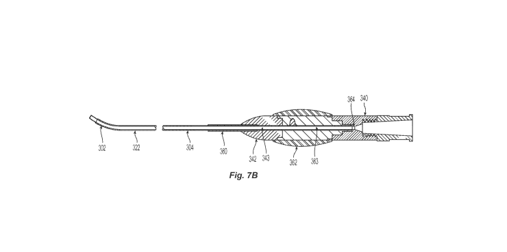
particular, several
embodiments are directed to intravascular devices having shapeable distal
portions for
addressing occlusions within a body vessel, including those related to
peripheral vascular
disease states, cardiovascular diseases, cerebrovascular diseases, and others.
BACKGROUND
[0003] Chronic
total occlusions ("CTO") are vascular lesions characterized by heavy
atherosclerotic plaque within the blood vessel, resulting in complete (or
nearly complete)
obstruction of blood flow across the lesion. Such occlusions can occur
anywhere in a
patient's vascular system. Since most lesions form gradually over a long
period of time, the
ischemic tissue downstream of the lesion has time to form collateral
circulation. For example,
in the case of coronary arteries, collateral vessels can form from the
proximal artery and
connect into the distal artery ("ipsilateral collaterals"), or collateral
vessels can form from the
other major arterial branches and connect into the distal artery
("contralateral collaterals").
When the lesion finally becomes a total occlusion, the collateral circulation
is typically
sufficient to keep the distal tissue alive, though ischemic. Accordingly, it
is desirable to
reestablish blood flow through or around the blockage in blood vessels by
crossing the CTO
and advancing therapeutic devices, such as a balloon angioplasty catheter, to
dilate and treat
the CTO. Likewise, in some cases it may be necessary to cross a CTO to gain
access to a
location along the vasculature distal to the CTO.
[0004] CTOs are
more difficult to cross than partially occluded lesions because, rather
than navigate a pre-existing lumen, a guidewire must either penetrate the
lesion or, when
CA 02922338 2016-02-24
WO 2015/031525
PCT/US2014/052991
penetrating the occlusion is impractically difficult and/or complicated, go
around the lesion
via a sub-intimal layer of a vessel wall. Figures 1A-1F, for example, are
schematic cross-
sectional side views of a conventional device used to treat such a CTO.
Referring first to
Figure 1A, a guidewire 12 and/or a catheter 10 is forced into a sub-intimal
layer SL adjacent
to an occlusion 0. Once the guidewire 12 and/or catheter 10 traverse the
occlusion 0, the
device(s) will require re-entry into a true lumen TL of the vessel V. For
example, to re-enter
the true lumen TL, many current techniques employ advancing the guidewire 12
such that it
bends and/or loops back onto itself at its distal end 16 as the guidewire 12
is advanced
distally through the sub-intimal layer SL. As best seen in Figures 1B and 1C,
for example,
such techniques can create a "sub-intimal tunnel." As best seen in Figures 1D-
1F, once a
looped end 14 of the guidewire 12 is distal to a distal end of the CTO, the
guidewire 12 can
be forced to re-enter the true lumen TL, thereby creating a large hole H
and/or flap in the
vessel wall V. Forcing re-entry utilizing a looped guidewire, however, may
cause
complications for the patient, such as unnecessarily extending the sub-intimal
tunnel,
perforation of the vessel V, and/or undesirable dissection of the vessel V
that requires
additional treatment.
[0005] Other
current methods of re-entering the true lumen TL during treatment involve
utilizing one of the currently available re-entry devices. Such re-entry
devices, however,
typically have larger diameters than the original tools utilized in the
procedure. In many
procedures, for example, the catheter in use is removed and the introducer
sheath is replaced
with a larger sheath (e.g., 7-8 Fr sheath). Such a transition, however, can
cause significant
disruptions to the procedure and dramatically increase procedure time and x-
ray exposure for
the patient.
BRIEF DESCRIPTION OF THE DRAWINGS
[0006] Many
aspects of the present disclosure can be better understood with reference
to the following drawings. The components in the drawings are not necessarily
to scale.
Instead, emphasis is placed on illustrating clearly the principles of the
present disclosure.
Furthermore, components can be shown as transparent in certain views for
clarity of
illustration only and not to indicate that the illustrated component is
necessarily transparent.
[0007] Figures
1A-1F are schematic cross-sectional side views of a conventional device
used to treat a CTO by traversing the CTO via a sub-intimal space.
2
CA 02922338 2016-02-24
WO 2015/031525
PCT/US2014/052991
[0008] Figure
2A is a side perspective view of an intravascular device in a low-profile
configuration configured in accordance with an embodiment of the present
technology.
[0009] Figure
2B is a side perspective view of the intravascular device of Figure 2A
with an angled distal portion configured in accordance with an embodiment of
the present
technology.
[0010] Figure 3
is an enlarged side view of the distal portion of the device of Figure 2B
without a guidewire and/or intravascular catheter placed therethrough.
[0011] Figure 4
is an enlarged side view of the distal portion of the device of Figure 2B
with an intravascular catheter and guidewire placed therethrough in accordance
with an
embodiment of the present technology.
[0012] Figure 5
is an enlarged cross-sectional view of a portion of the elongated
member of the device shown in Figure 7A configured in accordance with an
embodiment of
the present technology.
[0013] Figure
6A is an enlarged cross-sectional view of a portion of the elongated
member of the device shown in Figure 3 configured in accordance with an
embodiment of the
present technology.
[0014] Figure
6B is an enlarged cross-sectional view of another portion of the
elongated member of the device shown in Figure 3 configured in accordance with
an
embodiment of the present technology.
[0015] Figure
7A is an enlarged side view of the intravascular device of Figure 2B
configured in accordance with the present technology.
[0016] Figure
7B is an enlarged cross-sectional side view of the intravascular device of
Figure 2B mapped to Figure 7A in accordance with the present technology.
[0017] Figure
7C is a side view of a shaping mandrel configured in accordance with the
present technology.
[0018] Figure
8A is a partially-schematic side view of an intravascular device in a low-
profile configuration, configured in accordance with an embodiment of the
present
technology.
3
CA 02922338 2016-02-24
WO 2015/031525
PCT/US2014/052991
[0019] Figure
8B is a partially-schematic side view of the intravascular device of
Figure 8A having an angled distal portion configured in accordance with an
embodiment of
the present technology.
[0020] Figure 9
is an enlarged cross-sectional view of a portion of the elongated
member of the device shown in Figure 8B in accordance with an embodiment of
the present
technology.
[0021] Figure
10 is a side view of a distal portion of an intravascular device having a
bracing member configured in accordance with another embodiment of the present
technology.
[0022] Figure
11 is a partial cross-sectional side view of a distal portion of the
intravascular device of Figure 10 disposed within an outer sheath in
accordance with the
present technology.
[0023] Figure
12 is a side perspective view of a distal portion of an intravascular device
having two inflatable support rings configured in accordance with the present
technology.
[0024] Figure
13 is a side view of a distal portion of an intravascular device having a
one-sided expandable member configured in accordance with the present
technology.
[0025] Figure
14 is a side view of a distal portion of an intravascular device having a
bracing member and an expandable member configured in accord ance with the
present
technology.
[0026] Figures
15A-15G are anatomical cross-sectional side views illustrating a method
for using an intravascular device and/or one or more interventional devices
for crossing
and/or treating a CTO in accordance with an embodiment of the present
technology.
[0027] Figure
16A and 16B are side views of a proximal marker configured in
accordance with embodiments of the present technology.
[0028] Figures
17A and 17B are side views of a proximal marker configured in
accordance with another embodiment of the present technology.
DETAILED DESCRIPTION
[0029] The
present technology relates generally to systems, methods, and devices for
crossing and treating CTOs. Specific details of several embodiments of the
present
technology are described herein with reference to Figures 2-17B. Although many
of the
4
CA 02922338 2016-02-24
WO 2015/031525
PCT/US2014/052991
embodiments are described below with respect to devices and methods for
crossing and/or
treating CT0s, any vascular occlusion in addition to those described herein
may be crossed
and/or treated within the scope of the present technology (e.g., full
occlusions, partial
occlusions, occlusions resulting from a thrombus, occlusions resulting from an
embolism,
occlusions resulting from atherosclerosis, etc.). Additionally, other
embodiments of the
present technology can have different configurations, components, or
procedures than those
described herein. For example, other embodiments can include additional
elements and
features beyond those described herein, or other embodiments may not include
several of the
elements and features shown and described herein.
[0030] For ease
of reference, throughout this disclosure identical reference numbers are
used to identify similar or analogous components or features, but the use of
the same
reference number does not imply that the parts should be construed to be
identical. Indeed, in
many examples described herein, the identically-numbered parts are distinct in
structure
and/or function.
[0031]
Generally, unless the context indicates otherwise, the terms "distal" and
"proximal" within this disclosure reference a position relative to an operator
or an operator's
control device. For example, "proximal" can refer to a position closer to an
operator or an
operator's control device, and "distal" can refer to a position that is more
distant from an
operator or an operator's control device.
I. Selected Embodiments
[0032] Figure
2A is a side perspective view of an intravascular device 300 in a low-
profile or generally straight configuration in accordance with an embodiment
of the present
technology. The intravascular device 300 can include proximal portion 306
having a
handle 310, as well as a distal portion 302 and an elongated shaft 304
extending between the
handle 310 and the distal portion 302. The handle 310 can be configured to be
positioned at a
location external to a patient, and the elongated shaft 304 can be configured
to locate the
distal portion 302 intravascularly at or near a complete or partial occlusion
within a blood
vessel of the patient. The intravascular device 300 can have a lumen 326
(Figure 5)
extending proximally from an opening 330 at a distal end of the device 300 to
an outlet 331 at
the handle 310 of the device 300.
[0033] Figure
2B is a side view of the intravascular device 300 with the distal
portion 302 in an angled or treatment configuration (labeled in Figure 2B as
302'). In some
CA 02922338 2016-02-24
WO 2015/031525
PCT/US2014/052991
embodiments, the distal portion 302 may be "cold-worked" during manufacturing
into a
permanent, angled shape. In other embodiments, the distal portion 302 of the
intravascular
device 300 in Figure 2A may be composed of a malleable or shapeable material
so that a
clinician can manually transform the generally straight distal portion 302
into an angled distal
portion 302'. For example, the intravascular device 300 can come as part of a
kit that
includes one or more shaping mandrels (see, for example, the mandrel 370 shown
in
Figure 7C). The mandrels can come in a variety of configurations (e.g.,
different diameters,
shapes, angles, etc.) to address different needs presented by the particular
patient's
vasculature. The clinician can place the mandrel at least partially within or
over the distal
portion 302 and bend or manipulate the distal portion 302 into the angle or
shape of the
mandrel. In some embodiments, the clinician can manually bend the distal
portion 302 to a
desired angle based on specific requirements presented by a particular
procedure (e.g.,
crossing, steering, targeting for a particular area within the vasculature,
etc.). For example,
the clinician can manipulate the distal portion 302 without a mandrel, or in
some
embodiments, the mandrel can be flexible such that the mandrel bends with the
distal
portion 302 and primarily functions to keep the internal diameter of the
distal portion 302
from collapsing or kinking during the shaping process.
[0034] Often
times, the clinician may find it beneficial and/or necessary to utilize
multiple shapes and/or angles during a procedure. As such, the distal portion
302 of the
present technology is configured to be repeatedly angled, shaped, and/or
manipulated during
a procedure. For example, a clinician may initially utilize a first angle (for
example, a 45
angle) or shape but realize, after inserting the device 300, that a greater
angle (e.g., 70 , 60 ,
50 , etc.), lesser angle (e.g., 30 , 20 , 15 , 10 , etc.), or different shape
may be needed to
navigate the vasculature and/or re-enter the true lumen. The clinician can
remove the
device 300 from the patient and bend, angle, shape, and/or otherwise
manipulate the device to
a second angle or shape that is different from the first angle or shape. The
clinician can then
re-insert the device 300 with the second angle or shape. The clinician can
remove and re-
shape or manipulate the distal portion 302 as many times as desired during a
single procedure
(e.g., 2 times, 3 times, 4 times, etc.). In some embodiments, the distal
portion 302 can be
bent at multiple portions along its longitudinal axis.
[0035] Although
the shape of the distal portion 302 can be affected by the clinician
and/or mandrel, once a desired shape is set the distal portion 302 has
sufficient rigidity to
retain its desired shape when subjected to tortuous anatomy or when a
guidewire and/or
6
CA 02922338 2016-02-24
WO 2015/031525
PCT/US2014/052991
interventional device is placed therethrough. The shapeable distal portion 302
can be made
from shape memory plastic, Nitinol, stainless steel, titanium, tungsten,
tantalum, Elgiloy, and
other suitable materials. In some embodiments, the shapeable distal portion
can be between
about 0.25 inches to about 1.50 inches in length along its longitudinal axis.
In some
embodiments, the shapeable distal portion can be a different color than the
remainder of the
elongated shaft for identification purposes. In other embodiments, however,
the shapeable
distal portion may have a different arrangement and/or include different
features.
[0036] Figure 5
is an enlarged cross-sectional view of a portion of the elongated
shaft 304 proximal to the distal portion 302. Figures 7A and 7B show an
enlarged side view
and a partial cross-sectional side view, respectively, of the intravascular
device of Figure 2B.
Referring to Figures 5, 7A and 7B together, the elongated shaft 304 may
include one or more
layers configured to rotate along a central axis independently of one another.
For example,
the elongated shaft 304 can include an outer sheath 324 and a tubular rotating
member 322
within the outer sheath 324. As shown in Figure 7B, a proximal section of the
outer sheath
324 can be fixed to a first control knob 342 (e.g., at a first portion 343),
and a proximal
section of the rotating member 322 can be fixed to a second control knob 362
(e.g., at a
second portion 363). Accordingly, rotational motion applied to the first knob
342 (e.g., by a
clinician) causes rotation of the outer sheath 324, and rotation of the second
knob 362 causes
rotation of the rotating member 322. In some embodiments, a proximal region
360 of the
outer sheath 324 is reinforced to provide strain relief to the outer sheath
324 and/or the
rotating member 322 during rotation. The outer sheath 324 may be made of a
flexible
polymer (e.g., polyurethane, polyether block amide copolymer sold under the
trademark
PEBAX, etc.) or any suitable material, and may have an outer diameter of about
1.9 Fr to
about 5 Fr. In other embodiments, however, the outer sheath 324 may have a
different
arrangement and/or include different features. The intravascular catheter can
accommodate a
range of guidewire sizes (e.g., 0.010 inches, 0.014 inches, 0.018 inches,
0.035 inches and
0.038 inches).
[0037] The
rotating member 322 can be made of metal, plastic, and/or any suitable
material with sufficient rigidity to adequately transfer rotational forces
and/or provide
accurate responsiveness at its distal end when actuated by a clinician located
at a proximal
portion 306 of the device 300. The rotating member 322 can be a solid,
slotted, coiled,
and/or braided tube (e.g., a braid-reinforced polyimide), and in some
embodiments the
rotating member 322 can have any suitable shape and/or configuration. In
some
7
CA 02922338 2016-02-24
WO 2015/031525
PCT/US2014/052991
embodiments, the intravascular device 300 may include a lubricious coating
between the
rotating member 322 and the outer sheath 324 to allow for friction free or
relatively low
friction manipulation of the rotating member 322 within the sheath 324 in
various anatomical
tortuosity. Without being bound by theory, it is believed that such layering
renders the
tortuosity impact on torsional response insignificant relative to torque
transmission.
[0038] The
inner lumen 326 of the rotating member 322 can be coated with a lubricous
coating 320 (e.g., polytetrafluoroethylene (PTFE), fluorinated ethylene
propylene (FEP),
hydrophilic, etc.) applied directly to the inner walls of the rotating member
322. The inner
lumen 326 extends distally from an opening 331 at the handle 310 to an opening
330 at a
distal portion 302. The
inner lining 320 allows for fluid, guidewires, and/or other
intravascular devices (collectively referred to herein as "interventional
devices D") to be
slidably positioned within the lumen 326 of the rotating member 322. For
example, the
handle 310 can include an opening 331for insertion of an interventional device
such as the
crossing device disclosed in International Patent Application No.
PCT/US2010/047170, filed
August 30, 2010, entitled "SYSTEMS, METHODS AND DEVICES FOR ABLATION,
CROSSING, AND CUTTING OF OCCLUSIONS," which is incorporated herein by
reference in its entirety. In some embodiments, however, the handle 310 may
have a
different arrangement and/or include different features.
[0039] Figure 3
is a side view of the distal portion 302 of the intravascular device 300
of Figure 2B without an interventional device D placed therethrough, and
Figure 4 is a side
view of the distal portion 302 including an interventional device D. Referring
to Figures 3
and 4 together, the distal portion 302 can be a generally hollow tube having
an attachment
region 316 carried by, fixed to, and/or contiguous with a distal region 305 of
the elongated
shaft 304. The distal portion 302 can also include an angled region 318
extending distally at
an angle cto from the attachment region 316 and terminating at an atraumatic
distal tip 334.
[0040] In some
embodiments, the attachment region 316 of the distal portion 302 is
attached to the rotating member 322 such that rotation of the rotating member
322 causes
rotation of the distal portion 302. For example, as shown in Figure 6A, the
distal portion 302
can be defined by an angled tube 308, and at least a portion of the attachment
region 316 of
the angled tube can be placed over at least a portion of a distal region of
the rotating member
322. The overlapping portions can then be joined together (e.g., via adhesive,
crimping,
soldering, etc.). The outer sheath 324 may be advanced over the rotating
member 322 until a
distal end of the outer sheath 324 comes in contact with a proximal end of the
angled
8
CA 02922338 2016-02-24
WO 2015/031525
PCT/US2014/052991
tube 308. Accordingly, the transition between the outer sheath 324 and the
distal portion 320
may be seamless allowing for a low profile and maximum crossing ability.
[0041] In
embodiments where the distal portion is not shapeable by the clinician, the
distal portion 302 can be made from stainless steel, Nitinol, Elgiloy, other
metals and/or
similarly stiff materials that allow the distal portion 302 to retain its bent
and/or angled shape
while passing through the tortuous vasculature and/or when an interventional
device (such as
the interventional device D) is slidably positioned through the lumen 326 at
or distal to a
bend 350 in the distal portion 302. Many current devices have distal portions
that are heat-
treated to have an angled configuration. Such heat-treated devices, however,
do not retain
their shapes once an interventional device D is passed therethrough and/or the
device is
subject to a tortuous anatomy. In contrast with such current devices, the non-
shapeable distal
portion 302 of the present technology is not heat-treated, and rather
comprises a hollow, stiff
tube 308 that is "cold-worked" into a permanent, angled shape that is
unaffected by
anatomical tortuosity and/or placement of an interventional device D
therethrough.
[0042]
Regardless of whether the distal portion is shapeable or non-shapeable, the
angle
lac) remains the same even when an interventional device D is advanced through
the
lumen 326 of the distal portion 302 and extends through the opening 330 at the
distal tip 334.
Accordingly, the angled distal portion 302 allows for the predictable
angulation of a separate
intravascular device and/or guidewire, regardless of the shape of the separate
intravascular
device or the guidewire. Likewise, it is important to note that the angled
distal portion 302 is
not subject to any control wires and/or actuation device that can be
manipulated at a proximal
portion 306 of the device 300 to cause deflection at the distal portion 302.
In some
embodiments, all or a portion of the distal portion 302 may be coated with or
comprised at
least in part of radiopaque material to aid in positioning the device.
[0043] Even
with the angled distal portion 302, the overall profile of the intravascular
device 300 remains no greater than 7 Fr. In some embodiments, the overall
profile of the
intravascular device remains no great than 5 Fr. The angle cto of the distal
portion 302 can
vary (e.g., between about 1 to about 90 degrees). In some embodiments, for
example, the
angle cto is between about 10 and about 40 degrees. In other embodiments, the
angle cto is
between about 25 degrees and about 35 degrees (e.g., about 25 degrees, about
30 degrees,
about 35 degrees, etc.). In some embodiments, the distal portion 302 may be
configured to
have any suitable angle and/or length relative to the length of the
intravascular device 300.
9
CA 02922338 2016-02-24
WO 2015/031525
PCT/US2014/052991
[0044 The
distal tip 334 can be atraumatic and have a generally tapered shape. In
some embodiments, the distal tip 334 can also be configured to engage another
element of the
intravascular device 300. For example, the opening 330 at the distal end of
the distal tip 334
can define a passageway for receiving a guidewire (not shown) for delivery of
the treatment
device using over-the-wire ("OTW") or rapid exchange ("RX") techniques. In
other
embodiments, however, the distal tip 334 may have a different arrangement
and/or include
different features. The distal tip 334 may also be radiopaque to aid
positioning of the device.
[0045] Figure
8A is a side view of an intravascular device 800 in a low-profile or
generally straight configuration configured in accordance with an embodiment
of the present
technology. The intravascular device 800 can include a handle or luer 810 at a
proximal
portion 306, a distal portion 802, and an elongated shaft 804 extending
between the luer 810
and the distal portion 802. The luer 810 can be configured to be positioned at
a location
external to a patient, and the elongated shaft 804 can be configured to locate
the distal
portion 802 intravascularly at or near a complete or partial occlusion within
a blood vessel of
the patient. The intravascular device 800 can have a lumen 826 extending from
an
opening 330 at a distal end of the device 300 to an outlet 331 at the handle
310 of the
device 300.
[0046] Figure
8B is a side view of the intravascular device 800 with the distal
portion 802 in an angled or treatment configuration 802'. In some embodiments,
the distal
portion 802 is "cold-worked" during manufacturing into a permanent, angled
shape. In other
embodiments, the distal portion 802 of the intravascular device 800 in Figure
8A can be
made of a malleable or shapeable material so that a clinician can manually
transform the
generally straight distal portion 802 into an angled distal portion 802'. For
example, the
intravascular device 800 can come as part of a kit that includes a shaping
mandrel 370 (see
Figure 7C). The clinician may place the mandrel 370 at least partially within
or over the
distal portion 802 to keep the internal diameter of the distal portion 802
from collapsing or
kinking while it is manually bent to the desired shape. In some embodiments,
the clinician
may manually bend the distal portion 802 to a desired angle based on specific
requirements
presented by a particular procedure (e.g., crossing, steering, targeting for a
particular area
within the vasculature, etc.). Although the shape of the distal portion 802
can be manipulated
by the clinician, once a desired shape is set the distal portion 802' has
sufficient rigidity to
retain its desired shape when subjected to tortuous anatomy or with a
guidewire and/or
interventional device placed therethrough. The shapeable distal portion 802
can be made
CA 02922338 2016-02-24
WO 2015/031525
PCT/US2014/052991
from shape member plastic, Nitinol, stainless steel, titanium, tungsten,
Elgiloy, and others.
The shapeable distal portion can be between about 0.25 inches to about 0.30
inches in length
along its longitudinal axis.
[0047] Figure 9
shows an enlarged cross-sectional view of the intravascular device of
Figure 8B. The elongated shaft 804 can include an outer layer 824 and a braid-
reinforced
polyimide layer 822 within the outer layer 824. During manufacturing, a
polymer (e.g.,
polyether block amide copolymer sold under the trademark PEBAX) may be melted
over the
braid 822 to form the outer layer 824. In some embodiments, a proximal region
860 of the
outer layer 824 can be reinforced by additional material (e.g., additional
polyether block
amide copolymer sold under the trademark PEBAX is melted onto the braid) to
provide strain
relief to the outer layer 824 when rotated. Rotational motion applied to the
luer 810 (e.g., by
a clinician) causes rotation of the outer layer 824 along the entire
longitudinal axis of the
elongated shaft 804. The outer sheath may be made of a flexible polymer (e.g.,
polyether
block amide copolymer sold under the trademark PEBAX). The inner walls 826 of
the braid
may also include a lubricous coating 820 as described above.
[0048] The
braided layer 822 may extend partially or completely along the longitudinal
axis of the distal portion. The braided layer 822 is expected to provide a
high burst strength
(e.g., greater than or equal to 2000 psi) and additional kink resistance.
[0049] Figure
10 is a perspective view of a distal portion of an intravascular
device 1200 having a bracing member 1250 configured in accordance with another
embodiment of the present technology. In the
illustrated embodiment, the bracing
member 1250 can have a generally "humpback shape" configured to allow for
additional
deflection from the vessel wall. The bracing member 1250 can be made from
metal and/or
plastic materials. The bracing member 1250 can be delivered in a low-profile
or delivery
configuration, then expand upon proximal retraction of an outer sheath 1300
(see Figure 11)
to add additional support or stability to the distal portion 1202 of the
intravascular
device 1200. In some embodiments, the bracing member 1250 could automatically
and/or
manually extend and/or retract from a layer and/or lumen of the elongated
shaft 1204 (e.g.,
the outer sheath 1224) to add stability. In some embodiments, the sheath 1300
can be
partially retracted to selectively control the angle of the distal portion
1202 between about 0
and about 90 degrees.
11
CA 02922338 2016-02-24
WO 2015/031525
PCT/US2014/052991
[0050] Figure
12 illustrates an intravascular device 1400 configured in accordance with
another embodiment of the present technology. As shown in Figure 12, the
intravascular
device 1400 may have one or more inflatable or expandable rings 1402
configured to position
and support a distal portion 1404 of the device 1400 within the vessel. For
example, once the
distal portion 1404 is positioned at a target site, the rings 1402 can be
expanded to force at
least a portion of the distal portion 1404 away from an adjacent occlusion or
vessel wall. In
embodiments where the intravascular device includes a shapeable distal
portion, expansion of
the rings 1402 helps guide deflection of the distal portion. In embodiments
where the
intravascular device 1400 includes a pre-shaped distal portion, however,
expansion of the
rings 1402 does not generally affect angulation of the distal portion. Rather,
in such
embodiments, the rings 1402 may reinforce the pre-formed angle of the device
1400 and also
stabilize the device with respect to the surrounding anatomy. In some
embodiments, the rings
1402 can be independently expanded or deployed to provide additional
directionality and/or
stability to the device 1400.
[0051] Figure
13 is a side view of another embodiment of an intravascular device 1500
having a one-sided expandable member 1502. Once expanded, the expandable
member 1502
provides additional angulation to the distal portion 1504 and support to the
device 1500. For
example, in some embodiments, the expandable member 1502 aids in deflection of
the distal
portion 1504 off of the vessel wall and/or in redirecting the device 1500.
[0052] Figure
14 shows yet another embodiment of an intravascular device 1600 having
a combination of expandable members 1602 (e.g., balloons, inflatable rings,
etc.) and a brace
1604. Such a combination is expected to provide both directionality and
stability to the distal
portion 1606. The expandable members 1602 may be inflated or expanded
independent from
deployment of the brace 1604. In some embodiments, an expandable member 1602
may be
inflated or deflated, thereby allowing the pre-formed wire 1604 to bend and
provide an
additional angle to the device 1600. In some embodiments, the rings 1602
and/or pre-formed
wire 1604 can also be sheathed for delivery through the vasculature until the
distal portion of
the device is positioned at a desired location inside the vessel, as discussed
above with
reference to Figures 12 and 13.
II. Selected Delivery Systems and Methods
[0053] The
ability to percutaneously access the remote vasculature is well-known and
described in the patent and medical literature. Once percutaneous access is
achieved (for
12
CA 02922338 2016-02-24
WO 2015/031525
PCT/US2014/052991
example, through the femoral or iliac veins), the interventional tools and
supporting
catheter(s) may be advanced to the target vessel or CTO and positioned at or
proximate to the
CTO in a variety of manners, as described herein.
[0054] Figures
17A-17G illustrate one example for using an intravascular device 300
and/or one or more interventional devices to cross and/or treat a CTO.
Referring first to
Figure 17A, a guidewire 800 may be advanced along the vasculature until the
guidewire 800
is precluded from further distal movement by a proximal region of the CTO and
inadvertently
or purposely enters the sub-intimal layer SL (Figure 17B). At this point, as
best seen in
Figure 17C, the distal portion 302 of the device 300 can be advanced distally
over the
guidewire 800 and into the sub-intimal layer SL. As shown in the top view of
Figure 17D,
once in the sub-intimal layer, the angled distal portion 302 can be advanced
distally through
the sub-intimal layer SL until the distal portion 302 of the device 300 is
positioned at or distal
to a distal end of the CTO,. While the angled distal portion 302 is moved
through the sub-
intimal space, the angled region 318 is generally perpendicular to a true
lumen TL of the
vessel V. The distal portion 302 can be advanced through the sub-intimal layer
SL using
known imaging systems and techniques such as fluoroscopy, x-ray, MRI,
ultrasound or others.
Radiopaque material can be incorporated into the guidewire 800, distal portion
302, and/or
along any portion of the intravascular device 300 to provide additional
visibility under
imaging guidance. Such marker materials can be made from tungsten, tantalum,
platinum,
palladium, gold, iridium, or other suitable materials.
[0055] Once the
distal portion 302 reaches the distal end of the CTO, the clinician can
actuate the knob 362 (Figures 2A-2B and 7A-7B) or rotate the handle (Figures
8A and 8B) to
rotate the distal portion 302 (via the rotating member 322) so that the angled
region 318 is
directed towards the true lumen TL of the vessel V, as shown in the top and
side views of
Figures 17E - 17F, respectively.
[0056] As shown
in Figures 16A and 16B and Figures 17A and 17B, in particular
embodiments, the intravascular device may include a marking 352 on the luer or
handle that
aligns with the bend 350 such that a clinician can identify (from an
extracorporeal location)
the direction of the angle cto and/or projection of the angled region 318. For
example, the
knob 362 may have a marking 352 at a circumferential position corresponding
with the
bend 350.
13
CA 02922338 2016-02-24
WO 2015/031525
PCT/US2014/052991
[0057] A
piercing element (not shown) can be advanced through the distal portion 302
to penetrate the sub-intimal lining and facilitate re-entry into the true
lumen TL. As
discussed above, a guidewire 800 and/or an interventional device ID can be
advanced through
the opening 330 and into the true lumen TL of the vessel V in a direction
and/or angle
dictated by the angle cto of the distal portion 302 (as best seen in Figure
15G).
[0058] In some
embodiments the intravascular device 300 can include one or more
distal markers that could be utilized by the various imaging techniques
described above. For
example, in some embodiments, the distal portion 302 could have markings
(e.g., holes,
grooves, radiopaque markings, etc.) along its length so that when an
interventional device
reaches the distal portion 302, corresponding markings on the interventional
device will align
and confirm that the guidewire and/or interventional device have reached the
distal
portion 302 of the device 300. Additionally, such distal markings could be
utilized when the
device is used as a diagnostic catheter or angiographic catheter. Saline or
dye could be
flushed through the device and out the distal portion through the various
holes.
[0059] It
should be noted that the intravascular device described herein is not limited
to
a re-entry device. For example, various embodiments of the present technology
could also be
used when trying to reach areas of the vasculature with tortuosity and/or to
provide steering
while traversing such anatomy. Because the angled shape of the distal portion
stays true and
does not lose its configuration, multiple wires and other devices could be fed
through the
elongated shaft from an opening 331 at the proximal portion 306 and
selectively positioned
without the concern of having to compensate for any changes to the pre -set
dimensions prior
to entering the anatomy. In some embodiments, for example, the system may be
used to
reach a complex location having multiple bends, twists, and anatomical
variability.
III. Examples
[0060] The
following examples are illustrative of several embodiments of the present
technology:
1. An intravascular device, comprising:
a handle at a proximal portion;
an elongated member coupled to and extending between the handle and an angled
distal portion, wherein the elongated member includes¨
an outer sheath; and
14
CA 02922338 2016-02-24
WO 2015/031525
PCT/US2014/052991
a hollow rotational member with the sheath, wherein the rotational member is
coupled to the angled distal portion, and wherein rotation of the
rotational member directly causes rotation of the angled distal portion;
and
wherein the angled distal portion¨
defines a lumen therethrough,
has a first section and a second section that extends distally at an angle
from
the first section, and
the angle between the first section and the second section remains the same
when an interventional device is at least partially within the lumen of
the distal portion and spans at least a portion of the first section and at
least a portion of the second section.
2. An intravascular device, comprising:
a handle at a proximal portion;
an elongated member coupled to and extending between the handle and a distal
portion, wherein the elongated member includes¨
an outer sheath; and
a hollow rotational member with the sheath, wherein the rotational member is
coupled to the distal portion, and wherein rotation of the rotational
member directly causes rotation of the distal portion; and
wherein, in response to a force applied by a clinician while the distal
portion is
located external to the patient, the distal portion is moveable between:
a first configuration that is configured to be intravascularly delivered; and
a second configuration that is configured to be intravascularly delivered, and
wherein the second configuration is different from the first
configuration.
3. The device of example 1 wherein the angle between the first section and
the
second section remains the same when a guidewire is at least partially within
the lumen of the
distal portion.
4. The device of any of examples 1-3 wherein the distal portion is not heat
set.
CA 02922338 2016-02-24
WO 2015/031525
PCT/US2014/052991
5. The device of any of example 1 wherein the angle is about 30 degrees.
6. The device of any of examples 1-5 wherein the distal portion further
comprises an atraumatic distal tip.
7. The device of any of examples 1-6 wherein¨
the rotational member has a distal end and a proximal end;
the distal end of the rotational shaft is coupled to the distal portion; and
the proximal end of the rotational shaft is coupled to a knob located at the
handle.
8. The device of any of examples 1-7 wherein at least a portion of the
distal
portion is radiopaque.
9. The device of any one of examples 2, 4 and 6-8 wherein¨
the first configuration makes a first angle with respect to the longitudinal
axis of the
elongated member; and
the second configuration makes a second angle with respect to the longitudinal
axis of
the elongated member, wherein the second angle is different than the first
angle.
10. The device of any one of examples 2, 4 and 6-8 wherein¨
the first configuration is a rounded configuration having a first diameter;
and
the second configuration is a rounded configuration having a second diameter
that is
different than the first diameter.
11. The device of any one of examples 2, 4 and 6-8 wherein¨
the first configuration is a rounded configuration;
the second configuration is a bent configuration.
12. The device of any one of examples 2, 4 and 6-11 wherein the distal
portion is
further moveable to a third configuration that is different than at least one
of the first
configuration and the second configuration.
16
CA 02922338 2016-02-24
WO 2015/031525
PCT/US2014/052991
13. The device of any one of examples 2,4 and 6-11 wherein¨
the distal portion is further moveable to a third configuration that is
different than the
first configuration and the second configuration, and
when in the third configuration, the distal portion is configured to be
intravascularly
delivered.
14. A method of using a treatment device having a distal portion, the
method
comprising:
intravascularly delivering a distal portion in a first configuration;
removing the distal portion in the first configuration to an extracorporeal
location;
reconfiguring the distal portion into a second configuration that is different
than the
first configuration; and
intravascularly delivering the distal portion in a second configuration.
15. The method of example 14 wherein the first configuration is a rounded
configuration having a first diameter and the second configuration is a
rounded configuration
having a second diameter that is different than the first diameter.
16. The method of example 14 wherein the first configuration makes a first
angle
with respect to the longitudinal axis of the treatment device and the second
configuration
makes a second angle with respect to the longitudinal axis of the treatment
device, wherein
the second angle is different than the first angle.
17. The method of example 14 wherein reconfiguring the distal portion
includes
bending the distal portion.
18. The method of any one of examples 14-17 wherein intravascularly
delivering
the device includes delivering the distal portion to an intravascular location
proximate to a
chronic total occlusion.
19. The method of any one of examples 14-18, further comprising:
removing the distal portion in the second configuration to an extracorporeal
location;
17
CA 02922338 2016-02-24
WO 2015/031525
PCT/US2014/052991
reconfiguring the distal portion into a third configuration that is different
than at least
one of the first configuration and the second configuration; and
intravascularly delivering the distal portion in a third configuration.
IV. Conclusion
[0061] The
above detailed descriptions of embodiments of the technology are not
intended to be exhaustive or to limit the technology to the precise form
disclosed above.
Although specific embodiments of, and examples for, the technology are
described above for
illustrative purposes, various equivalent modifications are possible within
the scope of the
technology, as those skilled in the relevant art will recognize. For example,
while steps are
presented in a given order, alternative embodiments may perform steps in a
different order.
The various embodiments described herein may also be combined to provide
further
embodiments.
[0062] From the
foregoing, it will be appreciated that specific embodiments of the
technology have been described herein for purposes of illustration, but well-
known structures
and functions have not been shown or described in detail to avoid
unnecessarily obscuring the
description of the embodiments of the technology. Where the context permits,
singular or
plural terms may also include the plural or singular term, respectively.
[0063]
Moreover, unless the word "or" is expressly limited to mean only a single item
exclusive from the other items in reference to a list of two or more items,
then the use of "or"
in such a list is to be interpreted as including (a) any single item in the
list, (b) all of the items
in the list, or (c) any combination of the items in the list. Additionally,
the term "comprising"
is used throughout to mean including at least the recited feature(s) such that
any greater
number of the same feature and/or additional types of other features are not
precluded. It will
also be appreciated that specific embodiments have been described herein for
purposes of
illustration, but that various modifications may be made without deviating
from the
technology. Further, while advantages associated with certain embodiments of
the
technology have been described in the context of those embodiments, other
embodiments
may also exhibit such advantages, and not all embodiments need necessarily
exhibit such
advantages to fall within the scope of the technology. Accordingly, the
disclosure and
associated technology can encompass other embodiments not expressly shown or
described
herein.
18