Note: Descriptions are shown in the official language in which they were submitted.
81795160
RAPID TARGETING ANALYSIS IN CROPS FOR DETERMINING DONOR INSERTION
CROSS-REFERENCE TO RELATED APPLICATIONS
This application claims the benefit, under 35 U.S.C. 119(e), to U.S.
Provisional
Patent Application No. 61/873,719, filed September 4, 2013 and U.S.
Provisional Patent
Application No. 61/899,569, filed November 4, 2013.
REFERENCE TO SEQUENCE LISTING SUBMITTED ELECTRONICALLY
The official copy of the sequence listing is submitted electronically via EFS-
Web
as an ASCII formatted sequence listing with a file named "226007 5T25.txt",
created on
November 04, 2013, and having a size of 68.6 kilobytes and is filed
concurrently with the
specification.
FIELD OF THE INVENTION
The subject disclosure relates generally to the fields of molecular biology
and
biochemistry. The subject disclosure concerns a method for analyzing the
genomic site of
insertion of an integrated donor polynucleotide. The method is applicable for
high throughput
analysis of the integrated donor polynucleotide and can be used to minimize
the detection of false
positive results. Furthermore, the method uses cell based targeting and
analysis, without the need
for production of generating a stably targeted plant.
BACKGROUND OF THE INVENTION
Targeted genome modification of plants has been a long-standing and elusive
goal
of both applied and basic research. Targeting genes and gene stacks to
specific locations in the
plant genome will improve the quality of transgenic events, reduce costs
associated with
production of transgenic events and provide new methods for making transgenic
plant products
such as sequential gene stacking. Overall, targeting trangenes to specific
genomic sites is likely to
be commercially beneficial. Significant advances have been made in the last
few years towards
development of methods and compositions to target and cleave genomic DNA by
site specific
nucleases (e.g., Zinc Finger Nucleases (ZFNs), Meganucleases, Transcription
Activator-Like
Effector Nucelases (TALENS) and Clustered Regularly Interspaced Short
Palindromic
Repeats/CRISPR-associated nuclease (CRISPR/Cas) with an engineered crRNA/tracr
RNA), to
- 1 -
Date Recue/Date Received 2020-12-04
81795160
induce targeted mutagenesis, induce targeted deletions of cellular DNA
sequences, and facilitate
targeted recombination of an exogenous donor DNA polynucleotide within a
predetermined
genomic locus. See, for example, U.S. Patent Publication No. 20030232410;
20050208489;
20050026157; 20050064474; and 20060188987, and International Patent
Publication No. WO
2007/014275. U.S. Patent Publication No. 20080182332 describes use of non-
canonical zinc
finger nucleases (ZFNs) for targeted modification of plant genomes and U.S.
Patent Publication
No. 20090205083 describes ZFN-mediated targeted modification of a plant EPSPs
genomic locus.
Current methods for targeted insertion of exogenous DNA typically involve co-
transformation of
plant tissue with a donor DNA polynucleotide containing at least one transgene
and a site specific
nuclease (e.g., ZFN) which is designed to bind and cleave a specific genomic
locus. This causes
the donor DNA polynucleotide to stably insert within the cleaved genomic locus
resulting in
targeted gene addition at a specified genomic locus.
Unfortunately, reported and observed frequencies of targeted genomic
modification indicate that targeting a genomic loci within plants is
relatively inefficient. The
reported inefficiency necessitates the screening of a large number of plant
events to identify a
specific event containing the targeted genomic loci. The screening method
should also be
applicable as a high throughput method for the rapid identification of plant
events containing a
targeted genomic loci. In addition, as targeted gene insertion occurs in
conjunction with random
gene insertion, screening methods must be designed to specifically identify
targeting of genomic
loci within a background of random insertions and to discern the genomic
integration from
exogenous plasmid DNA which may produce false-positive results. Furthermore,
the assay
should be sensitive enough to detect an event occurring in a single cell,
wherein that cell contains
the only targeted event amongst thousands of other non-targeted cells. Most
reported plant event
analyses rely on a single analytical method for confirming targeting which may
lead to inaccurate
.. estimation of targeting frequencies and low confidence outcomes. A need
exists for development
of improved molecular assay methods, particularly for high-throughput
analysis, that can detect
site specific chromosomal integrations and discern these events from exogenous
plasmid DNA.
Finally, current methods for assessing targeted genomic modifications are
based on generation of
stable plants and are time and cost intensive. Accordingly, there is a need
for an analytical
method that allows rapid targeting assessment at a
- 2 -
Date Recue/Date Received 2020-12-04
CA 02922823 2016-02-29
WO 2015/034885 PCT/US2014/053832
large number of genomic loci and screening of a large number of site-specific
nucleases to
identify and confirm the insertion of a polynucleotide donor sequence within
the targeted
genomic loci.
The foregoing examples of the related art and limitations related therewith
are
intended to be illustrative and not exclusive. Other limitations of the
related art will become
apparent to those of skill in the art upon a reading of the specification.
BRIEF SUMMARY OF THE INVENTION
In an embodiment, the disclosure relates to an assay for detecting site
specific
integration of a polynucleotide donor sequence within a genomic target site,
wherein: a
genomic DNA is amplified with a first round of PCR to produce a first amplicon
using a first
Out-PCR primer designed to bind to the genomic DNA target site; a first In-PCR
primer
designed to bind the integrated polynucleotide donor sequence, and the first
amplicon is
amplified with a second round of PCR using primers specific to sequences
located within the
first amplicon to produce a second amplicon; and, the presence of the second
amplicon is
detected, wherein the production of the second amplicon indicates the presence
of the site
specific integration event.
In an aspect of the embodiment, the genomic target site comprises an
endogenous or an engineered genomic target site. In another aspect of the
embodiment, the first
In-PCR primer is provided at a lower concentration than the first Out-PCR
primer. In an
embodiment, the first round of PCR is conducted using a relative concentration
of first Out-
PCR primer to first In-PCR primer of about 4:1, 3:1 or 2:1. In another
embodiment, the first In-
PCR primer comprises a concentration of 0.05 ¨ 0.091.1.M, and the first Out-
PCR primer
comprises a concentration of at least 0.11.1M.
In a subsequent aspect of the embodiment, the second round of PCR comprises a
second Out-PCR primer designed to bind to the genomic DNA target site of the
first amplicon
and a second In-PCR primer designed to bind the integrated polynucleotide
donor sequence of
the first amplicon. In an embodiment, the second In-PCR primer is provided at
a lower
concentration than the second Out-PCR primer. In another embodiment, the
second round of
.. PCR is conducted using a relative concentration of second Out-PCR primer to
second In-PCR
primer of about 4:1, 3:1 or 2:1. In a further embodiment, the second In-PCR
primer comprises a
concentration of 0.05 ¨ 0.1 M, and the second Out-PCR primer comprises a
concentration of
0.2 ILIM.
- 3 -
81795160
In a further aspect of the embodiment, the genomic DNA comprising the site
specific integration of the polynucleotide donor sequence within the genomic
target site is a
plant genomic DNA. As an embodiment, the plant genomic DNA is isolated from a
monocotyledonous plant. As another embodiment, the plant genomic DNA is
isolated from a
dicotyledonous plant.
In another aspect of the embodiment, the cleavage of the genomic DNA target
site with a site specific nuclease results in the site specific integration of
the polynucleotide
donor sequence within the genomic target site. As an embodiment, the site
specific nuclease is
selected from the group consisting of a Zinc Finger nuclease, a CRISPR
nuclease, a TALEN
nuclease, or a meganuclease. In a subsequent embodiment, the site specific
integration of the
polynucleotide donor sequence within the genomic target site occurs via a Non
Homologous
End Joining mechanism.
In an aspect of the embodiment, the detecting step is an agarose gel of the
second amplicon or a sequencing reaction of the second amplicon.
In yet another aspect of the embodiment, the disclosure relates to a method
for
detecting site specific integration of a polynucleotide donor sequence within
a genomic target
site of transfected plant cells comprising: amplifying a genomic DNA with a
first round of
PCR to produce a first amplicon, wherein said PCR is conducted using a first
Out-PCR primer
designed to bind to the genomic target site and a first In-PCR primer designed
to bind the
polynucleotide donor sequence, further wherein said first In-PCR primer is
provided at a
lower concentration than the first Out-PCR primer; amplifying the first
amplicon with a
second round of PCR using primers specific to sequences located within the
first amplicon to
produce a second amplicon; and, detecting the presence of a second amplicon,
wherein the
production of a second amplicon indicates the presence of a site specific
integration event. In
other embodiments, the plant cell is a protoplast plant cell. In an
embodiment, the detection
of the site specific integration is performed on a mixed population of
targeted and non-
targeted plant cells, wherein the non-targeted plant cells do not contain a
polynucleotide donor
sequence within a genomic target site.
In addition to the exemplary aspects and embodiments described above, further
aspects and embodiments will become apparent by study of the following
descriptions.
- 4 -
Date Recue/Date Received 2020-12-04
81795160
In an embodiment, there is provided a method for detecting site specific
integration of a polynucleotide donor sequence within a genomic target site,
the method
comprising: a. amplifying a genomic DNA with a first round of PCR to produce a
first
amplicon using a first Out-PCR primer designed to bind to the genomic DNA
target site and a
first In-PCR primer designed to bind the integrated polynucleotide donor
sequence, wherein
the first In-PCR primer is provided at a lower concentration than the first
Out-PCR primer and
said first Out-PCR primer and said first In-PCR primer pair are selected to
amplify reverse
orientation inserted polynucleotide donor sequences, wherein the reverse
orientation is
relative to the orientation of the original transformation vector; b.
amplifying the first
amplicon with a second round of PCR using primers specific to sequences
located within the
first amplicon to produce a second amplicon; and, c. detecting the presence of
the second
amplicon, wherein the production of the second amplicon indicates the presence
of the site
specific integration event.
In an embodiment, there is provided a method for detecting site specific
integration of a polynucleotide donor sequence within a genomic target site of
transfected
plant cells, said method comprising a. amplifying a genomic DNA with a first
round of PCR
to produce a first amplicon, wherein said PCR is conducted using a first Out-
PCR primer
designed to bind to the genomic target site and a first In-PCR primer designed
to bind the
polynucleotide donor sequence, further wherein said first In-PCR primer is
provided at a
lower concentration than the first Out-PCR primer and said first Out-PCR
primer and said
first In-PCR primer pair are selected to amplify reverse orientation inserted
polynucleotide
donor sequences, wherein the reverse orientation is relative to the
orientation of the original
transformation vector; b. amplifying the first amplicon with a second round of
PCR using
primers specific to sequences located within the first amplicon to produce a
second amplicon;
and, c. detecting the presence of a second amplicon, wherein the production of
a second
amplicon indicates the presence of a site specific integration event.
BRIEF DESCRIPTION OF THE FIGURES
Figure 1 illustrates a plasmid map of pDAB111845.
- 4a -
Date Recue/Date Received 2020-12-04
CA 02922823 2016-02-29
WO 2015/034885 PCT/US2014/053832
Figure 2 illustrates a plasmid map of pDAB111846.
Figure 3 illustrates a plasmid map of pDAB117415.
Figure 4 illustrates a plasmid map of pDAB117416.
Figure 5 illustrates a plasmid map of pDAB117417.
Figure 6 illustrates a plasmid map of pDAB117419.
Figure 7 illustrates a plasmid map of pDAB117434.
Figure 8 illustrates a plasmid map of pDAB117418.
Figure 9 illustrates a plasmid map of pDAB117420.
Figure 10 illustrates a plasmid map of pDAB117421.
Figure 11 illustrates a representation of the universal donor polynucleotide
sequence for integration via NHEJ.
Figure 12 illustrates a representation of the universal donor polynucleotide
sequence for integration via HDR. The label -HA" indicates homology arms; and
the label
-ZFN BS" indicates ZFN binding site (for monomer).
Figure 13 illustrates the constructs used for targeting and validation of the
universal donor polynucleotide system integration within the Zea mays select
genomic loci
targeting validation. A) ZFN design space with location of the ZFN pairs. B)
Configuration of
the ZFN expression construct. The label "NLS" indicates Nuclear Localization
Signal, the
label "ZFP" indicates Zinc Finger Protein. C) universal donor polynucleotide
for NHEJ
mediated targeting of Zea mays select genomic loci. Z1-Z6 represent ZFN
binding sites
specific for a Zea mays select genomic loci target. The number of ZFN sites
can vary from 3-6.
Vertical arrows show unique restriction sites and horizontal arrows represent
potential PCR
primer sites. The universal donor polynucleotide system is a short (110 bp)
sequence that is
common to all donors used for integration within Zea mays select genomic loci.
Figure 14 illustrates a plasmid map of pDAB8393.
Figures 15A & 15B illustrate the ZFN cleavage activity at Zea mays selected
genomic loci targets. Cleavage activity is represented as number of sequences
with Indels at
the ZFN cleavage site per one million high quality reads. Figure 15A
represents the data in a
bar graph form. Figure 15B represents the data as a table.
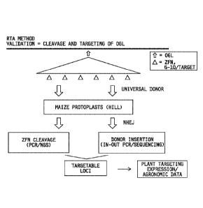
Figure 16 illustrates the validation of Zea mays selected genomic loci targets
using NHEJ based Rapid Targeting Analysis method.
- 5 -
CA 02922823 2016-02-29
WO 2015/034885 PCT/US2014/053832
Figure 17 illustrates plasmid constructs transformed into Zea mays via random
integration that comprise the events used for flanking sequence analysis and
trans gene
expression studies.
Figure 18 illustrates donor insertion via NHEJ at an ELP in protoplast Rapid
Targeting Analysis. Insertion can occur in a forward or a reverse orientation.
Figure 19 illustrates disruption of the ZFN cleavage sites in ELP1. Disruption
is
represented as a decrease in qPCR signal in terms of target to reference
ratio. On average 22%
and 15% reduction in signal is observed for ZFN1 and ZFN3 respectively.
Figure 20 illustrates the sequence of In-Out amplified PCR products. Four
clones from each In-Out PCR were sequenced and the results demonstrated intact
target donor
junctions and processed end junctions. The sequences listed correspond with
SEQ I NO: 248 as
predicted, SEQ ID NO:249 as A9-1, SEQ ID NO:250 as A9-2, SEQ ID NO:251 as A9-
5, SEQ
ID NO:252 as A9-6, SEQ ID NO:253 as G8-1, SEQ ID NO:254 as G8-2, SEQ ID NO:255
as
G8-5, SEQ ID NO:256 as G8-6, SEQ ID NO:257 as G9-1, SEQ ID NO:258 as G9-2, SEQ
ID
NO:259 as G9-6, SEQ ID NO:260 as H9-1, SEQ ID NO:261 as H9-2, SEQ ID NO:262 as
H9-
5, and SEQ ID NO:263 as H9-6.
Figure 21 illustrates donor insertion via NHEJ at E32 in protoplast Rapid
Targeting Analysis. Insertion can occur in a forward or a reverse orientation.
Figure 22 illustrates a schematic showing the relation of the primers designed
for
the donor polynucleotide and the zinc finger binding sequence.
Figure 23 illustrates a plasmid map of pDAB7221.
Figure 24 illustrates a schematic of probe/primers for the locus disruption
assay.
The F2 ZFN binding sites for the FAD2 2.3 and 2.6 genes and primers used for
the disruption
assay are indicated.
Figure 25 provides the sequence of In-Out PCR products resulting from NHEJ
targeting of a donor sequence using the F2, ZFN2 zinc finger nuclease in the
FAD2 2.3 locus.
The reference sequence (top of figure) represents the configuration of the
targeted insertion of
the donor vector in a reverse orientation. The single-stranded ends of the
DNAs resulting from
FokI digestion were filled in to create the reference sequence. Sanger
sequences are shown.
The F2, ZFN2 ZFN binding sequences are underlined. Plasmid clones with a
similar sequence
to the specified sequence are listed to the right.
- 6 -
81795160
DETAILED DESCRIPTION
I. Overview
Novel methods have now been disclosed for rapid screening, identification and
characterization of site specific nuclease targeted plant events. The methods
can be used to
analyze the integration a donor polynucleotide within a genomic target locus
via a first and
second amplification reaction. The first and second amplification reactions
are an "In-Out"
PCR amplification reaction for screening the 3' and/or the 5' junction
sequences of a donor
DNA polynucleotide targeted within a genomic locus. The presence of an
amplified product
which contains the 3' and/or 5' junction sequence indicates that the donor DNA
polynucleotide is present within the targeted genomic locus.
The disclosed screening assays describe high quality, high throughput
processes for identifying and obtaining targeted transgene insertion events.
Deployment of
the screening assay allows for large numbers of plant events to be analyzed
and screened to
select specific events which have a donor DNA polynucleotide inserted within a
targeted
genomic locus, and to discern these events from false-positive results.
Moreover, the
disclosed methods can be deployed as high throughput assays allowing for the
rapid and
efficient identification of a subset of samples that can then be further
analyzed by other
molecular confirmation methods. The presently disclosed subject matter
includes plants and
plant cells comprising nuclease targeted plant events selected utilizing the
novel screening
methods. Furthermore, the methodology is readily applicable for the analysis
of any plant
species.
II. Terms
Unless defined otherwise, all technical and scientific terms used herein have
the same meaning as commonly understood by one of ordinary skill in the art to
which this
disclosure relates. In case of conflict, the present application including the
definitions will
control. Unless otherwise required by context, singular terms shall include
pluralities and
plural terms shall include the singular.
In order to further clarify this disclosure, the following terms,
abbreviations
and definitions are provided.
- 7 -
Date Recue/Date Received 2020-12-04
CA 02922823 2016-02-29
WO 2015/034885
PCT/US2014/053832
As used herein, the terms "comprises", -comprising", -includes", -including",
"has", -having", -contains", or "containing", or any other variation thereof,
are intended to be
non-exclusive or open-ended. For example, a composition, a mixture, a process,
a method, an
article, or an apparatus that comprises a list of elements is not necessarily
limited to only those
elements but may include other elements not expressly listed or inherent to
such composition,
mixture, process, method, article, or apparatus. Further, unless expressly
stated to the contrary,
-or" refers to an inclusive or and not to an exclusive or. For example, a
condition A or B is
satisfied by any one of the following: A is true (or present) and B is false
(or not present), A is
false (or not present) and B is true (or present), and both A and B are true
(or present).
The term -invention" or -present invention" as used herein is a non-limiting
term and is not intended to refer to any single embodiment of the particular
invention but
encompasses all possible embodiments as disclosed in the application.
As used herein, the term -plant" includes a whole plant and any descendant,
cell,
tissue, or part of a plant. The term -plant parts" include any part(s) of a
plant, including, for
example and without limitation: seed (including mature seed, immature seed,
and immature
embryo without testa); a plant cutting; a plant cell; a plant cell culture; a
plant organ (e.g.,
pollen, embryos, flowers, fruits, shoots, leaves, roots, stems, and related
explants). A plant
tissue or plant organ may be a seed, callus, or any other group of plant cells
that is organized
into a structural or functional unit. A plant cell or tissue culture may be
capable of regenerating
a plant having the physiological and morphological characteristics of the
plant from which the
cell or tissue was obtained, and of regenerating a plant having substantially
the same genotype
as the plant. In contrast, some plant cells are not capable of being
regenerated to produce
plants. Regenerable cells in a plant cell or tissue culture may be embryos,
protoplasts,
meristematic cells, callus, pollen, leaves, anthers, roots, root tips, silk,
flowers, kernels, ears,
cobs, husks, or stalks.
Plant parts include harvestable parts and parts useful for propagation of
progeny
plants. Plant parts useful for propagation include, for example and without
limitation: seed;
fruit; a cutting; a seedling; a tuber; and a rootstock. A harvestable part of
a plant may be any
useful part of a plant, including, for example and without limitation: flower;
pollen; seedling;
tuber; leaf; stem; fruit; seed; and root.
A plant cell is the structural and physiological unit of the plant. Plant
cells, as
used herein, includes protoplasts and protoplasts with a partial cell wall. A
plant cell may be in
the form of an isolated single cell, or an aggregate of cells (e.g., a friable
callus and a cultured
- 8 -
CA 02922823 2016-02-29
WO 2015/034885 PCT/US2014/053832
cell), and may be part of a higher organized unit (e.g., a plant tissue, plant
organ, and plant).
Thus, a plant cell may be a protoplast, a gamete producing cell, or a cell or
collection of cells
that can regenerate into a whole plant. As such, a seed, which comprises
multiple plant cells
and is capable of regenerating into a whole plant, is considered a -plant
part" in embodiments
herein.
The term -protoplast", as used herein, refers to a plant cell that had its
cell wall
completely or partially removed, with the lipid bilayer membrane thereof
naked. Typically, a
protoplast is an isolated plant cell without cell walls which has the potency
for regeneration into
cell culture or a whole plant.
As used herein, -endogenous sequence" defines the native form of a
polynucleotide, gene or polypeptide in its natural location in the organism or
in the genome of
an organism.
The term -isolated" as used herein means having been removed from its natural
environment.
The term -purified", as used herein relates to the isolation of a molecule or
compound in a form that is substantially free of contaminants normally
associated with the
molecule or compound in a native or natural environment and means having been
increased in
purity as a result of being separated from other components of the original
composition. The
term "purified nucleic acid" is used herein to describe a nucleic acid
sequence which has been
separated from other compounds including, but not limited to polypeptides,
lipids and
carbohydrates.
As used herein, the terms -polynucleotide". "nucleic acid", and "nucleic acid
molecule" are used interchangeably, and may encompass a singular nucleic acid;
plural nucleic
acids; a nucleic acid fragment, variant, or derivative thereof; and nucleic
acid construct (e.g.,
messenger RNA (mRNA) and plasmid DNA (pDNA)). A polynucleotide or nucleic acid
may
contain the nucleotide sequence of a full-length cDNA sequence, or a fragment
thereof,
including untranslated 5' and/or 3' sequences and coding sequence(s). A
polynucleotide or
nucleic acid may be comprised of any polyribonucleotide or
polydeoxyribonucleotide, which
may include unmodified ribonucleotides or deoxyribonucleotides or modified
ribonucleotides
or deoxyribonucleotides. For example, a polynucleotide or nucleic acid may be
comprised of
single- and double-stranded DNA; DNA that is a mixture of single- and double-
stranded
regions; single- and double-stranded RNA; and RNA that is mixture of single-
and double-
stranded regions. Hybrid molecules comprising DNA and RNA may be single-
stranded,
- 9 -
CA 02922823 2016-02-29
WO 2015/034885 PCT/US2014/053832
double-stranded, or a mixture of single- and double-stranded regions. The
foregoing terms also
include chemically, enzymatically, and metabolically modified forms of a
polynucleotide or
nucleic acid.
It is understood that a specific DNA refers also to the complement thereof,
the
sequence of which is determined according to the rules of deoxyribonucleotide
base-pairing.
As used herein, the term -gene" refers to a nucleic acid that encodes a
functional
product (RNA or polypeptide/protein). A gene may include regulatory sequences
preceding
(5' non-coding sequences) and/or following (3' non-coding sequences) the
sequence encoding
the functional product.
As used herein, the term -coding sequence" refers to a nucleic acid sequence
that
encodes a specific amino acid sequence. A -regulatory sequence" refers to a
nucleotide
sequence located upstream (e.g., 5' non-coding sequences), within, or
downstream (e.g., 3' non-
coding sequences) of a coding sequence, which influence the transcription, RNA
processing or
stability, or translation of the associated coding sequence. Regulatory
sequences include, for
example and without limitation: promoters; translation leader sequences;
introns;
polyadenylation recognition sequences; RNA processing sites; effector binding
sites; and stem-
loop structures.
As used herein, the term "polypeptide" includes a singular polypeptide, plural
polypeptides, and fragments thereof. This term refers to a molecule comprised
of monomers
(amino acids) linearly linked by amide bonds (also known as peptide bonds).
The term
"polypeptide" refers to any chain or chains of two or more amino acids, and
does not refer to a
specific length or size of the product. Accordingly, peptides, dipeptides,
tripeptides,
oligopeptides, protein, amino acid chain, and any other term used to refer to
a chain or chains of
two or more amino acids, are included within the definition of "polypeptide",
and the foregoing
terms are used interchangeably with "polypeptide" herein. A polypeptide may be
isolated from
a natural biological source or produced by recombinant technology, but a
specific polypeptide
is not necessarily translated from a specific nucleic acid. A polypeptide may
be generated in
any appropriate manner, including for example and without limitation, by
chemical synthesis.
In contrast, the term "heterologous" refers to a polynucleotide, gene or
polypeptide that is not normally found at its location in the reference (host)
organism. For
example, a heterologous nucleic acid may be a nucleic acid that is normally
found in the
reference organism at a different genomic location. By way of further example,
a heterologous
nucleic acid may be a nucleic acid that is not normally found in the reference
organism. A host
-10-
CA 02922823 2016-02-29
WO 2015/034885 PCT/US2014/053832
organism comprising a hetereologous polynucleotide, gene or polypeptide may be
produced by
introducing the heterologous polynucleotide, gene or polypeptide into the host
organism. In
particular examples, a heterologous polynucleotide comprises a native coding
sequence, or
portion thereof, that is reintroduced into a source organism in a form that is
different from the
corresponding native polynucleotide. In particular examples, a heterologous
gene comprises a
native coding sequence, or portion thereof, that is reintroduced into a source
organism in a form
that is different from the corresponding native gene. For example, a
heterologous gene may
include a native coding sequence that is a portion of a chimeric gene
including non-native
regulatory regions that is reintroduced into the native host. In particular
examples, a
heterologous polypeptide is a native polypeptide that is reintroduced into a
source organism in a
form that is different from the corresponding native polypeptide.
A heterologous gene or polypeptide may be a gene or polypeptide that comprises
a functional polypeptide or nucleic acid sequence encoding a functional
polypeptide that is
fused to another gene or polypeptide to produce a chimeric or fusion
polypeptide, or a gene
encoding the same. Genes and proteins of particular embodiments include
specifically
exemplified full-length sequences and portions, segments, fragments (including
contiguous
fragments and internal and/or terminal deletions compared to the full-length
molecules),
variants, mutants, chimerics, and fusions of these sequences.
As used herein, the term -modification" can refer to a change in a
polynucleotide
disclosed herein that results in reduced, substantially eliminated or
eliminated activity of a
polypeptide encoded by the polynucleotide, as well as a change in a
polypeptide disclosed
herein that results in reduced, substantially eliminated or eliminated
activity of the polypeptide.
Alternatively, the term -modification" can refer to a change in a
polynucleotide disclosed
herein that results in increased or enhanced activity of a polypeptide encoded
by the
polynucleotide, as well as a change in a polypeptide disclosed herein that
results in increased or
enhanced activity of the polypeptide. Such changes can be made by methods well
known in the
art, including, but not limited to, deleting, mutating (e.g., spontaneous
mutagenesis, random
mutagenesis, mutagenesis caused by mutator genes, or transposon mutagenesis),
substituting,
inserting, down-regulating, altering the cellular location, altering the state
of the polynucleotide
or polypeptide (e.g., methylation, phosphorylation or ubiquitination),
removing a cofactor,
introduction of an antisense RNA/DNA, introduction of an interfering RNA/DNA,
chemical
modification, covalent modification, irradiation with UV or X-rays, homologous
-11-
CA 02922823 2016-02-29
WO 2015/034885 PCT/US2014/053832
recombination, mitotic recombination, promoter replacement methods, and/or
combinations
thereof.
The term "derivative", as used herein, refers to a modification of a sequence
set
forth in the present disclosure. Illustrative of such modifications would be
the substitution,
.. insertion, and/or deletion of one or more bases relating to a nucleic acid
sequence of a coding
sequence disclosed herein that preserve, slightly alter, or increase the
function of a coding
sequence disclosed herein in crop species. Such derivatives can be readily
determined by one
skilled in the art, for example, using computer modeling techniques for
predicting and
optimizing sequence structure. The term -derivative" thus also includes
nucleic acid sequences
having substantial sequence identity with the disclosed coding sequences
herein such that they
are able to have the disclosed functionalities for use in producing
embodiments of the present
disclosure.
The term -promoter" refers to a DNA sequence capable of controlling the
expression of a nucleic acid coding sequence or functional RNA. In examples,
the controlled
coding sequence is located 3' to a promoter sequence. A promoter may be
derived in its entirety
from a native gene, a promoter may be comprised of different elements derived
from different
promoters found in nature, or a promoter may even comprise rationally designed
DNA
segments. It is understood by those skilled in the art that different
promoters can direct the
expression of a gene in different tissues or cell types, or at different
stages of development, or
in response to different environmental or physiological conditions. Examples
of all of the
foregoing promoters are known and used in the art to control the expression of
heterologous
nucleic acids. Promoters that direct the expression of a gene in most cell
types at most times
are commonly referred to as "constitutive promoters." Furthermore, while those
in the art have
(in many cases unsuccessfully) attempted to delineate the exact boundaries of
regulatory
sequences, it has come to be understood that DNA fragments of different
lengths may have
identical promoter activity. The promoter activity of a particular nucleic
acid may be assayed
using techniques familiar to those in the art.
The term -operably linked" refers to an association of nucleic acid sequences
on
a single nucleic acid, wherein the function of one of the nucleic acid
sequences is affected by
another. For example, a promoter is operably linked with a coding sequence
when the promoter
is capable of effecting the expression of that coding sequence (e.g., the
coding sequence is
under the transcriptional control of the promoter). A coding sequence may be
operably linked
to a regulatory sequence in a sense or antisense orientation.
-12-
CA 02922823 2016-02-29
WO 2015/034885 PCT/US2014/053832
The term -expression", as used herein, may refer to the transcription and
stable
accumulation of sense (mRNA) or antisense RNA derived from a DNA. Expression
may also
refer to translation of mRNA into a polypeptide. As used herein, the term
"overexpression"
refers to expression that is higher than endogenous expression of the same
gene or a related
gene. Thus, a heterologous gene is "overexpressed" if its expression is higher
than that of a
comparable endogenous gene.
As used herein, the term -transformation" or -transforming" refers to the
transfer
and integration of a nucleic acid or fragment thereof into a host organism,
resulting in
genetically stable inheritance. Host organisms containing a transforming
nucleic acid are
referred to as -transgenic", -recombinant", or "transformed" organisms. Known
methods of
transformation include, for example: Agrobacterium tumefaciens- or A.
rhizogenes-mediated
transformation; calcium phosphate transformation; polybrene transformation;
protoplast fusion;
electroporation; ultrasonic methods (e.g., sonoporation); liposome
transformation;
microinjection; transformation with naked DNA; transformation with plasmid
vectors;
transformation with viral vectors; biolistic transformation (microparticle
bombardment); silicon
carbide WHISKERS-mediated transformation; aerosol beaming; and PEG-mediated
transformation.
As used herein, the term "introduced" (in the context of introducing a nucleic
acid into a cell) includes transformation of a cell, as well as crossing a
plant comprising the
nucleic acid with a second plant, such that the second plant contains the
nucleic acid, as may be
performed utilizing conventional plant breeding techniques. Such breeding
techniques are
known in the art. For a discussion of plant breeding techniques, see Poehlman
(1995) Breeding
Field Crops, 4th Edition, AVI Publication Co., Westport CT.
Backcrossing methods may be used to introduce a nucleic acid into a plant.
This
technique has been used for decades to introduce traits into plants. An
example of a description
of backcrossing (and other plant breeding methodologies) can be found in, for
example,
Neiman (1995), supra; and Jensen (1988) Plant Breeding Methodology, Wiley, New
York,
NY. In an exemplary backcross protocol, an original plant of interest (the -
recurrent parent") is
crossed to a second plant (the "non-recurrent parent") that carries the
nucleic acid be
introduced. The resulting progeny from this cross are then crossed again to
the recurrent
parent, and the process is repeated until a converted plant is obtained,
wherein essentially all of
the desired morphological and physiological characteristics of the recurrent
parent are
recovered in the converted plant, in addition to the nucleic acid from the non-
recurrent parent.
- 13-
CA 02922823 2016-02-29
WO 2015/034885 PCT/US2014/053832
-Binding" refers to a sequence-specific, non-covalent interaction between
macromolecules (e.g., between a protein and a nucleic acid). Not all
components of a binding
interaction need be sequence-specific (e.g., contacts with phosphate residues
in a DNA
backbone), as long as the interaction as a whole is sequence-specific. Such
interactions are
generally characterized by a dissociation constant (Kd) of 10-6 M-1 or lower. -
Affinity" refers to
the strength of binding: increased binding affinity being correlated with a
lower Kd.
A "binding protein" is a protein that is able to bind non-covalently to
another
molecule. A binding protein can bind to, for example, a DNA molecule (a DNA-
binding
protein), an RNA molecule (an RNA-binding protein) and/or a protein molecule
(a protein-
binding protein). In the case of a protein-binding protein, it can bind to
itself (to form
homodimers, homotrimers, etc.) and/or it can bind to one or more molecules of
a different
protein or proteins. A binding protein can have more than one type of binding
activity. For
example, zinc finger proteins have DNA-binding, RNA-binding and protein-
binding activity.
-Recombination" refers to a process of exchange of genetic information between
two polynucleotides, including but not limited to, donor capture by non-
homologous end
joining (NHE.1) and homologous recombination. For the purposes of this
disclosure,
"homologous recombination (HR)" refers to the specialized form of such
exchange that takes
place, for example, during repair of double-strand breaks in cells via
homology-directed repair
mechanisms. This process requires nucleotide sequence homology, uses a "donor"
molecule to
template repair of a -target" molecule (i.e., the one that experienced the
double-strand break),
and is variously known as -non-crossover gene conversion" or -short tract gene
conversion",
because it leads to the transfer of genetic information from the donor to the
target. Without
wishing to be bound by any particular theory, such transfer can involve
mismatch correction of
heteroduplex DNA that forms between the broken target and the donor, and/or -
synthesis-
dependent strand annealing", in which the donor is used to resynthesize
genetic information that
will become part of the target, and/or related processes. Such specialized HR
often results in an
alteration of the sequence of the target molecule such that part or all of the
sequence of the
donor polynucleotide is incorporated into the target polynucleotide. For HR-
directed
integration, the donor molecule contains at least one region of homology to
the genome
(-homology arms") of least 50-100 base pairs in length. See, e.g., U.S. Patent
Publication No.
20110281361.
In the methods of the disclosure, one or more targeted nucleases as described
herein create a double-stranded break in the target sequence (e.g., cellular
chromatin) at a
-14-
CA 02922823 2016-02-29
WO 2015/034885 PCT/US2014/053832
predetermined site, and a -donor" polynucleotide, having homology to the
nucleotide sequence
in the region of the break, can be introduced into the cell. The presence of
the double-stranded
break has been shown to facilitate integration of the donor sequence. The
donor sequence may
be physically integrated or, alternatively, the donor polynucleotide is used
as a template for
repair of the break via homologous recombination, resulting in the
introduction of all or part of
the nucleotide sequence as in the donor into the cellular chromatin. Thus, a
first sequence in
cellular chromatin can be altered and, in certain embodiments, can be
converted into a sequence
present in a donor polynucleotide. Thus, the use of the terms "replace" or -
replacement" can be
understood to represent replacement of one nucleotide sequence by another, (i.
e. , replacement
of a sequence in the informational sense), and does not necessarily require
physical or chemical
replacement of one polynucleotide by another.
"Cleavage" refers to the breakage of the covalent backbone of a DNA molecule.
Cleavage can be initiated by a variety of methods including, but not limited
to, enzymatic or
chemical hydrolysis of a phosphodiester bond. Both single-stranded cleavage
and double-
stranded cleavage are possible, and double-stranded cleavage can occur as a
result of two
distinct single-stranded cleavage events. DNA cleavage can result in the
production of either
blunt ends or staggered ends. In certain embodiments, fusion polypeptides are
used for targeted
double-stranded DNA cleavage.
The terms -plasmid" and -vector", as used herein, refer to an extra
chromosomal
element that may carry one or more gene(s) that are not part of the central
metabolism of the
cell. Plasmids and vectors typically are circular double-stranded DNA
molecules. However,
plasmids and vectors may be linear or circular nucleic acids, of a single- or
double-stranded
DNA or RNA, and may carry DNA derived from essentially any source, in which a
number of
nucleotide sequences have been joined or recombined into a unique construction
that is capable
of introducing a promoter fragment and a coding DNA sequence along with any
appropriate 3'
untranslated sequence into a cell. In examples, plasmids and vectors may
comprise
autonomously replicating sequences for propagating in bacterial hosts.
Polypeptide and -protein" are used interchangeably herein and include a
molecular chain of two or more amino acids linked through peptide bonds. The
terms do not
refer to a specific length of the product. Thus, "peptides", and
"oligopeptides", are included
within the definition of polypeptide. The terms include post-translational
modifications of the
polypeptide, for example, glycosylations, acetylations, phosphorylations and
the like. In
addition, protein fragments, analogs, mutated or variant proteins, fusion
proteins and the like
- 15-
81795160
are included within the meaning of polypeptide. The terms also include
molecules in which one or
more amino acid analogs or non-canonical or unnatural amino acids are included
as can be
synthesized, or expressed recombinantly using known protein engineering
techniques. In addition,
inventive fusion proteins can be derivatized as described herein by well-known
organic chemistry
.. techniques.
The term -fusion protein" indicates that the protein includes polypeptide
components
derived from more than one parental protein or polypeptide. Typically, a
fusion protein is expressed
from a fusion gene in which a nucleotide sequence encoding a polypeptide
sequence from one protein
is appended in frame with, and optionally separated by a linker from, a
nucleotide sequence encoding a
polypeptide sequence from a different protein. The fusion gene can then be
expressed by a
recombinant host cell as a single protein.
Embodiments of the Present Invention
In an embodiment, the disclosure relates to an assay for detecting site
specific
.. integration of a polynucleotide donor sequence within a genomic target
site.
In some embodiments a genomic DNA is assayed for detecting site specific
integration of a polynucleotide donor sequence within a genomic target site.
In aspects of the
embodiment, the genomic DNA comprises; a chromosomal genomic DNA, a
mitochondrial genomic
DNA, a transposable element genomic DNA, a genomic DNA derived from a viral
integration, an
.. artificial chromosome genomic DNA (see PCT/US2002/017451 and
PCT/US2008/056993, included
herein as non-limiting examples), and other sources of genomic DNA.
In some embodiments, the genomic DNA is amplified via the Polymerase Chain
Reaction (PCR). In aspects of the embodiment, PCR generally refers to the
method for increasing the
concentration of a segment of a target sequence in a mixture of genomic DNA
without cloning or
.. purification (U.S. Pat. Nos. 4,683,195; 4,683,202; and 4,965,188). This
process for amplifying the
target sequence comprises introducing an excess of two oligonucleotide primers
to the DNA mixture
containing the desired target sequence, followed by a precise sequence of
thermal cycling in the
presence of a DNA polymerase. The two primers are complementary to their
respective strands of the
double stranded target sequence. To effect amplification, the mixture is
denatured and the primers then
annealed to their complementary sequences within the target molecule.
Following annealing, the
primers are extended with a polymerase so as to form a new pair of
complementary strands.
- 16 -
Date Recue/Date Received 2020-12-04
CA 02922823 2016-02-29
WO 2015/034885 PCT/US2014/053832
The steps of denaturation, primer annealing and polymerase extension can be
repeated many
times (i.e., denaturation, annealing and extension constitute one -cycle";
there can be numerous
"cycles") to obtain a high concentration of an amplified segment of the
desired target sequence.
The length of the amplified segment of the desired target sequence is
determined by the relative
positions of the primers with respect to each other, and therefore, this
length is a controllable
parameter. By virtue of the repeating aspect of the process, the method is
referred to as the
-polymerase chain reaction" (hereinafter -PCR"). Because the desired amplified
segments of
the target sequence become the predominant sequences (in terms of
concentration) in the
mixture, they are said to be -PCR amplified."
In other embodiments, the PCR reaction produces an amplicon. As an aspect of
the embodiment, amplicon refers to the product of the amplification reaction
generated through
the extension of either or both of a pair of amplification primers. An
amplicon may contain
exponentially amplified nucleic acids if both primers utilized hybridize to a
target sequence.
Alternatively, amplicons may be generated by linear amplification if one of
the primers utilized
does not hybridize to the target sequence. Thus, this term is used generically
herein and does
not necessarily imply the presence of exponentially amplified nucleic acids.
Amplification of a selected, or target, nucleic acid sequence may be carried
out
by any suitable method. See generally, Kwoh et al., Am. Biotechnol. Lab. 8, 14-
25 (1990).
Examples of suitable amplification techniques include, but are not limited to,
polymerase chain
reaction, ligase chain reaction, strand displacement amplification (see
generally G. Walker et
al., Proc. Natl. Acad. Sci. USA 89, 392-396 (1992); G. Walker et al., Nucleic
Acids Res. 20.
1691-1696 (1992)), transcription-based amplification (see D. Kwoh et al.,
Proc. Natl. Acad Sci.
USA 86, 1173-1177 (1989)), self-sustained sequence replication (or "3SR") (see
J. Guatelli et
al., Proc. Natl. Acad. Sci. USA 87, 1874-1878 (1990)), the Q13 replicase
system (see P. Lizardi
et al., BioTechnoloay 6, 1197-1202 (1988)), nucleic acid sequence-based
amplification (or
"NASBA") (see R. Lewis, Genetic Engineering News 12 (9), 1 (1992)), the repair
chain
reaction (or "RCR") (see R. Lewis, supra), and boomerang DNA amplification (or
"BDA") (see
R. Lewis, supra). Polymerase chain reaction is generally preferred.
In another embodiment, the amplification of the genomic DNA is completed via
a PCR reaction using primers. In an aspect of the embodiment, the primers may
comprise a
first set of primers, a second set of primers, a third set of primers, and so
forth. As such, the
designation "first", -second", -third", etc. indicate the order by which the
primer sets are used
in a nested PCR reaction. For example, the "first" set of primers are used
initially in a first
- 17 -
CA 02922823 2016-02-29
WO 2015/034885 PCT/US2014/053832
PCR reaction to amplify a polynucleotide sequence. Next, the -second" set of
primers are used
in a second PCR reaction to amplify the product of the first PCR reaction.
Then the -third" set
of primers are used in a third PCR reaction to amplify the product of the
second PCR reaction
and so forth. In other aspects of the embodiment, the primers may be an -Out"
primer that is
designed to bind the genomic DNA target site, or an -In" primer that is
designed to bind a
polynucleotide donor sequence that is integrated within the genome of an
organism. In other
embodiments the first set of primers may be comprised of an In and an Out
primer, or may be
designed to comprise two distinct In primers, or two distinct Out primers. In
an embodiment,
the term primer refers to an oligonucleotide that is complementary to a DNA
template to be
amplified in an appropriate amplification buffer. In certain embodiment the
primers may be
from 10 Bp to 100 Bp, 10 Bp to 50 Bp or 10 Bp to 25 Bp in length.
In an embodiment of the subject disclosure the In primer is provided at a
lower
concentration than the Out primer. An aspect of the embodiment includes, a
relative
concentration of Out primer to In primer of about 10:1. 9:1, 8:1, 7:1, 6:1,
5:1, 4:1, 3:1 or 2:1. In
another aspect, the embodiment includes where the In primer comprises a
concentration of
0.001, 0.005, 0.01, 0.02, 0.03, 0.04, 0.05, 0.06, 0.07. 0.008, or 0.09 M, and
the Out primer
comprises a concentration of at least 0.111M. In a further aspect, the
embodiment includes
where the In primer comprises a concentration of 0.01, 0.02, 0.03, 0.04, 0.05,
0.06, 0.07. 0.08,
0.09, 0.1, 0.11 0.12, 0.13, 0.14, 0.15, 0.16, 0.17, 0.18 or 0.19 M, and the
Out primer comprises
a concentration of at least 0.2 M.
In some embodiments, the genomic integration site is a plant genomic DNA. In
an aspect plant cells which are transformed in accordance with the present
disclosure includes,
but is not limited to, any higher plants, including both dicotyledonous and
monocotyledonous
plants, and particularly consumable plants, including crop plants. Such plants
can include, but
are not limited to, for example: alfalfa, soybeans, cotton, rapeseed (also
described as canola),
linseed, corn, rice, brachiaria, wheat, safflowers, sorghum, sugarbeet,
sunflowers, tobacco and
turf grasses. Thus, any plant species or plant cell can be selected. In
embodiments, plant cells
used herein, and plants grown or derived therefrom, include, but are not
limited to, cells
obtainable from rapeseed (Bras sica napus); indian mustard (Bras sica juncea);
Ethiopian
.. mustard (Brassica carinata); turnip (Brassica rapa); cabbage (Brassica
oleracea); soybean
(Glycine max); linseed/flax (Linum usitatissimum); maize (also described as
corn) (Zea mays);
safflower (Carthamus tinctorius); sunflower (Helianthus annuus); tobacco
(Nicotiana tabacum);
Arabidopsis thaliana; Brazil nut (Betholettia excelsa); castor bean (Ricinus
communis); coconut
- 18 -
81795160
(Cocus nucifera); coriander (Coriandrum sativum); cotton (Gossypium spp.);
groundnut
(Arachis hypogaea); jojoba (Simmondsia chinensis); oil palm (Elaeis guineeis);
olive (Olea
eurpaea); rice (Oryza sativa); squash (Cucurbita maxima); barley (Hordeum
vulgare);
sugarcane (Saccharum officinarum); rice (Oryza sativa); wheat (Triticum spp.
including
Triticum durum and Triticum aestivum); and duckweed (Lemnaceae sp.). In some
embodiments, the genetic background within a plant species may vary.
With regard to the production of genetically modified plants, methods for the
genetic engineering of plants are well known in the art. For instance,
numerous methods for
plant transformation have been developed, including biological and physical
transformation
protocols for dicotyledenous plants as well as monocotyledenous plants (e.g.,
Goto-Fumiyuki
et al., Nature Biotech 17:282-286 (1999); Miki et al., Methods in Plant
Molecular Biology and
Biotechnology, Glick, B. R. and Thompson, J. E. Eds., CRC Press, Inc., Boca
Raton,
pp. 67-88 (1993)). In addition, vectors and in vitro culture methods for plant
cell or tissue
transformation and regeneration of plants are available, for example, in
Gruber et al., Methods
in Plant Molecular Biology and Biotechnology, Glick, B. R. and Thompson, J. E.
Eds., CRC
Press, Inc., Boca Raton, pp. 89-119 (1993).
A large number of techniques are available for inserting DNA into a plant host
cell. Those techniques include transformation with disarmed T-DNA using
Agrobacterium
tumefaciens or Agrobacterium rhizogenes as the transformation agent, calcium
phosphate
transfection, polybrene transformation, protoplast fusion, electroporation,
ultrasonic methods
(e.g., sonoporation), liposome transformation, microinjection, naked DNA,
plasmid vectors,
viral vectors, biolistics (microparticle bombardment), silicon carbide
WHISKERS mediated
transformation, aerosol beaming, or PEG as well as other possible methods.
For example, the DNA construct may be introduced directly into the genomic
DNA of the plant cell using techniques such as electroporation and
microinjection of plant
cell protoplasts, or the DNA constructs can be introduced directly to plant
tissue using
biolistic methods, such as DNA particle bombardment (see, e.g., Klein et al.
(1987) Nature
327:70-73). Additional methods for plant cell transformation include
microinjection via
silicon carbide WHISKERS mediated DNA uptake (Kaeppler et al. (1990) Plant
Cell Reporter
9:415-418). Alternatively, the DNA construct can be introduced into the plant
cell via
nanoparticle transformation (see, e.g., US Patent Application No. 12/245,685).
- 19 -
Date Recue/Date Received 2020-12-04
CA 02922823 2016-02-29
WO 2015/034885 PCT/US2014/053832
Another known method of plant transformation is microprojectile-mediated
transformation wherein DNA is carried on the surface of microprojectiles. In
this method, the
vector is introduced into plant tissues with a biolistic device that
accelerates the
microprojectiles to speeds sufficient to penetrate plant cell walls and
membranes. Sanford et al.,
Part. Sci. Technol. 5:27 (1987), Sanford, J. C., Trends Biotech. 6:299 (1988),
Sanford. J. C.,
Physiol. Plant 79:206 (1990), Klein et al., Biotechnology 10:268 (1992).
Alternatively, gene transfer and transformation methods include, but are not
limited to, protoplast transformation through calcium chloride precipitation,
polyethylene
glycol (PEG)- or electroporation-mediated uptake of naked DNA (see Paszkowski
et al. (1984)
EMBO J 3:2717-2722, Potrykus et al. (1985) Molec. Gen. Genet. 199:169-177;
Fromm et al.
(1985) Proc. Nat. Acad. Sci. USA 82:5824-5828; and Shimamoto (1989) Nature
338:274-276)
and electroporation of plant tissues (D'Halluin et al. (1992) Plant Cell
4:1495-1505).
A widely utilized method for introducing an expression vector into plants is
based on the natural transformation system of Agrobacterium. Horsch et al.,
Science 227:1229
(1985). A. tumefaciens and A. rhizogenes are plant pathogenic soil bacteria
known to be useful
to genetically transform plant cells. The Ti and Ri plasmids of A. tumefaciens
and A.
rhizogenes, respectively, carry genes responsible for genetic transformation
of the plant. Kado,
C. I., Crit. Rev. Plant. Sci. 10:1 (1991). Descriptions of Agrobacterium
vector systems and
methods for Agrobacterium-mediated gene transfer are also available, for
example, Gruber et
al., supra, Miki et al., supra, Moloney et al., Plant Cell Reports 8:238
(1989), and U.S. Patent
Nos. 4,940,838 and 5,464,763.
If Agrobacterium is used for the transformation, the DNA to be inserted should
be cloned into special plasmids, namely either into an intermediate vector or
into a binary
vector. Intermediate vectors cannot replicate themselves in Agrobacterium. The
intermediate
vector can be transferred into Agrobacterium tumefaciens by use of a helper
plasmid
(conjugation). The Japan Tobacco Superbinary system is an example of such a
system
(reviewed by Komari et al., (2006) In: Methods in Molecular Biology (K. Wang,
ed.) No. 343:
Agrobacterium Protocols (2nd Edition, Vol. 1) Humana Press Inc., Totowa, NJ,
pp.15-41; and
Komori et al., (2007) Plant Physiol. 145:1155-1160). Binary vectors can
replicate themselves
both in E. coli and in Agrobacterium. They comprise a selection marker gene
and a linker or
polylinker which are framed by the right and left T-DNA border regions. They
can be
transformed directly into Agrobacterium (Holsters, 1978). The Agrobacterium
used as host cell
is to comprise a plasmid carrying a vir region. The Ti or Ri plasmid also
comprises the vir
- 20 -
CA 02922823 2016-02-29
WO 2015/034885 PCT/US2014/053832
region necessary for the transfer of the T-DNA. The vir region is necessary
for the transfer of
the T-DNA into the plant cell. Additional T-DNA may be contained.
The virulence functions of the Agrobacterium tumefaciens host will direct the
insertion of a T- strand containing the construct and adjacent marker into the
plant cell DNA
when the cell is infected by the bacteria using a binary T DNA vector (Bevan
(1984) Nuc. Acid
Res. 12:8711-8721) or the non-binary T-DNA vector procedure (Horsch et al.
(1985) Science
227:1229-1231). Generally, the Agrobacterium transformation system is used to
engineer
dicotyledonous plants (Bevan et al. (1982) Ann. Rev. Genet 16:357-384; Rogers
et al. (1986)
Methods Enzymol. 118:627-641). The Agrobacterium transformation system may
also be used
to transform, as well as transfer, DNA to monocotyledonous plants and plant
cells. See U.S.
Patent No. 5, 591,616; Hernalsteen et al. (1984) EMBO J 3:3039-3041; Hooykass-
Van
Slogteren et al. (1984) Nature 311:763-764; Grimsley et al. (1987) Nature
325:1677-179;
Boulton et al. (1989) Plant Mol. Biol. 12:31-40; and Gould et al. (1991) Plant
Physiol.
95:426-434. Following the introduction of the genetic construct into
particular plant cells, plant
cells can be grown and upon emergence of differentiating tissue such as shoots
and roots,
mature plants can be generated. In some embodiments, a plurality of plants can
be generated.
Methodologies for regenerating plants are known to those of ordinary skill in
the art and can be
found, for example, in: Plant Cell and Tissue Culture, 1994, Vasil and Thorpe
Eds. Kluwer
Academic Publishers and in: Plant Cell Culture Protocols (Methods in Molecular
Biology 111,
1999 Hall Eds Humana Press). The genetically modified plant described herein
can be cultured
in a fermentation medium or grown in a suitable medium such as soil. In some
embodiments, a
suitable growth medium for higher plants can include any growth medium for
plants, including,
but not limited to, soil, sand, any other particulate media that support root
growth (e.g.,
vermiculite, perlite, etc.) or hydroponic culture, as well as suitable light,
water and nutritional
supplements which optimize the growth of the higher plant.
Transformed plant cells which are produced by any of the above transformation
techniques can be cultured to regenerate a whole plant which possesses the
transformed
genotype and thus the desired phenotype. Such regeneration techniques rely on
manipulation of
certain phytohormones in a tissue culture growth medium, typically relying on
a biocide and/or
herbicide marker which has been introduced together with the desired
nucleotide sequences.
Plant regeneration from cultured protoplasts is described in Evans, et al., -
Protoplasts Isolation
and Culture" in Handbook of Plant Cell Culture, pp. 124-176, Macmillian
Publishing Company,
New York, 1983; and Binding, Regeneration of Plants, Plant Protoplasts, pp. 21-
73, CRC Press,
-21 -
CA 02922823 2016-02-29
WO 2015/034885 PCT/US2014/053832
Boca Raton, 1985. Regeneration can also be obtained from plant callus,
explants, organs,
pollens, embryos or parts thereof. Such regeneration techniques are described
generally in Klee
et al. (1987) Ann. Rev. of Plant Phys. 38:467-486.
In other embodiments, the plant cells which are transformed are not capable of
regeneration to produce a plant. Such cells are said to be transiently
transformed. Transiently
transformed cells may be produced to assay the expression and/or functionality
of a specific
transgene. Transient transformation techniques are known in the art, and
comprise minor
modifications to the transformation techniques described above. Those with
skill in the art may
elect to utilize transient transformation to quickly assay the expression
and/or functionality of a
specific transgenes, as transient transformation are completed quickly and do
not require as
many resources (e.g., culturing of plants for development of whole plants,
self-fertilization or
crossing of plants for the fixation of a transgene within the genome, etc.) as
stable
transformation techniques.
In an embodiment the donor polynucleotide can be introduced into essentially
any plant. A wide variety of plants and plant cell systems may be engineered
for site specific
integration of the donor polynucleotide of the present disclosure and the
various transformation
methods mentioned above. In an embodiment, target plants and plant cells for
engineering
include, but are not limited to, those monocotyledonous and dicotyledonous
plants, such as
crops including grain crops (e.g., wheat, maize, rice, millet, barley), fruit
crops (e.g., tomato,
apple, pear, strawberry, orange), forage crops (e.2., alfalfa), root vegetable
crops (e.g., carrot,
potato, sugar beets, yam), leafy vegetable crops (e.g., lettuce, spinach);
flowering plants (e.g.,
petunia, rose, chrysanthemum), conifers and pine trees (e.g., pine fir,
spruce); plants used in
phytoremediation (e.g., heavy metal accumulating plants); oil crops (e.g.,
sunflower, rape seed)
and plants used for experimental purposes (e.g., Arabidopsis).
In other embodiments, the polynucleotide donor sequences are introduced into a
plant cell for site specific targeting within a genomic target site. In such
embodiments, the
plant cell may be a protoplast plant cell. The protoplasts can be produced
from various types of
plant cells. Accordingly, those having ordinary skill in the art may utilize
different techniques
or methodologies to produce the protoplast plant cell. For example, the
generation and
production of protoplasts are provided by: Green and Phillips, Crop Sc., 15
(1975) 417-421;
Harms et al. Z. Pflan ZeT1Z aechtg., 77 (1976) 347-351; European patent
applications EP-
0,160,390, Lowe and Smith (1985); EP-0,176,162, Cheng (1985); and EP-
0,177,738, Close
(1985); Cell Genetics in Higher Plants, Dudits et al., (eds), Akademiai Kiado,
Budapest (1976)
- 22 -
CA 02922823 2016-02-29
WO 2015/034885 PCT/US2014/053832
129-140, and references therein; Harms, ''Maize and Cereal Protoplasts-Facts
and
Perspectives," Maize for Biological Research, W. F. Sheridan, ed. (1982);
Dale, in: Protoplasts
(1983); Pottykus et al (eds.) Lecture Proceedings, Experientia Supplement= 46,
Potryk-us et
al., cds, Birkhauser, Basel (1983) 31-41, and references therein. Plant
regeneration from
cultured protoplasts is described in Evans et al. (1983) "Protopl.ast
Isolation and Culture,"
Handbook of Plant Cell Cultures 1, 124-176 (MacMillan Publishing Co., New
York; Davey
(1983) "Recent Developments in the Culture and Regeneration of Plant
Protoplasts,"
Protoplasts, pp. 12-29, (Birkhauser, Basel); Dale (1983) " Protoplast Culture
and Plant
Regeneration of Cereals and Other Recalcitrant Crops," Protoplasts pp. 31-41,
(Birkhauser,
Basel); Binding (1985) "Regeneration of Plants," Plant Protoplasts, pp, 21-73,
(CRC Press,
Boca Raton, FL).
Selection of target sites; ZFPs and methods for design and construction of
fusion
proteins (and polynucleotides encoding same) are known to those of skill in
the art and
described in detail in U.S. Patent Nos. 6,140,081; 5,789,538; 6,453,242;
6,534,261; 5,925,523;
6,007,988; 6,013,453; 6,200,759; WO 95/19431; WO 96/06166; WO 98/53057;
WO 98/54311; WO 00/27878; WO 01/60970 WO 01/88197; WO 02/099084; WO 98/53058;
WO 98/53059; WO 98/53060; WO 02/016536 and WO 03/016496.
In subsequent embodiments, the DNA binding domain comprising one or more
DNA binding sequences is bound by a zinc finger binding protein, a
meganuclease binding
protein, a CRIPSR, or a TALEN binding protein.
In certain embodiments, the composition and methods described herein employ a
meganuclease (homing endonuclease) binding protein or meganuclease DNA-binding
domain
for binding to the donor molecule and/or binding to the region of interest in
the genome of the
cell. Naturally-occurring meganucleases recognize 15-40 base-pair cleavage
sites and are
commonly grouped into four families: the LAGLIDADG family, the GIY-YIG family,
the His-
Cyst box family and the HNH family. Exemplary homing endonucleases include I-
SceI, I-
Ceul, PI-Pspl, P1-See, I-SceIV, I-CsmI, I-PanI, I-SceII, I-PpoI, I-ScellI, I-
CreI, I-Tevl, I-TevII
and I-TevIII. Their recognition sequences are known. See also U.S. Patent No.
5,420,032;
U.S. Patent No. 6,833,252; Belfort et al. (1997) Nucleic Acids Res. 25:3379-
3388; Dujon et
al. (1989) Gene 82:115-118; Perler et al. (1994) Nucleic Acids Res. 22, 1125-
1127; Jasin
(1996) Trends Genet. 12:224-228; Gimble et al. (1996) J. Mol. Biol. 263:163-
180; Argast et
al. (1998) J. Mol. Biol. 280:345-353 and the New England Biolabs catalogue.
- 23 -
81795160
In certain embodiments, the methods and compositions described herein make use
of a nuclease that comprises an engineered (non-naturally occurring) homing
endonuclease
(meganuclease). The recognition sequences of homing endonucleases and
meganucleases such as
I-SceI, I-CeuI, PI-PspI, PI-Sce, I-SceIV, I-CsmI, I-PanI, I-SceII, I-PpoI, I-
SceIII, I-CreI, I-TevI,
I-TevII and I-TevIII are known. See also U.S. Patent No. 5,420,032; U.S.
Patent No. 6,833,252;
Belfort et al. (1997) Nucleic Acids Res. 25:3379-3388; Dujon et al. (1989)
Gene 82:115-118;
Perler et al. (1994) Nucleic Acids Res. 22, 1125-1127; Jasin (1996) Trends
Genet. 12:224-228;
Gimble et al. (1996) J. Mol. Biol. 263:163-180; Argast et al. (1998) J. Mol.
Biol. 280:345-353
and the New England Biolabs catalogue. In addition, the DNA-binding
specificity of homing
endonucleases and meganucleases can be engineered to bind non-natural target
sites. See, for
example, Chevalier et al. (2002) Molec. Cell 10:895-905; Epinat et al. (2003)
Nucleic Acids Res.
31:2952-2962; Ashworth et al. (2006) Nature 441:656-659; Paques etal. (2007)
Current Gene
Therapy 7:49-66; U.S. Patent Publication No. 20070117128. The DNA-binding
domains of the
homing endonucleases and meganucleases may be altered in the context of the
nuclease as a
whole (i.e., such that the nuclease includes the cognate cleavage domain).
In other embodiments, the DNA-binding domain of one or more of the nucleases
used in the methods and compositions described herein comprises a naturally
occurring or
engineered (non-naturally occurring) TAL effector DNA binding domain. See,
e.g., U.S. Patent
Publication No. 20110301073. The plant pathogenic bacteria of the genus
Xanthomonas are
known to cause many diseases in important crop plants. Pathogenicity of
Xanthomonas depends
on a conserved type III secretion (T3 5) system which injects more than 25
different effector
proteins into the plant cell. Among these injected proteins are transcription
activator-like (TAL)
effectors which mimic plant transcriptional activators and manipulate the
plant transcriptome (see
Kay et al (2007) Science 318:648-651). These proteins contain a DNA binding
domain and a
transcriptional activation domain. One of the most well characterized TAL-
effectors is AvrBs3
from Xanthomonas campestgris pv. Vesicatoria (see Bonas et al (1989) Mol Gen
Genet
218: 127-136 and W02010079430). TAL-effectors contain a centralized domain of
tandem
repeats, each repeat containing approximately 34 amino acids, which are key to
the DNA binding
specificity of these proteins. In addition, they contain a nuclear
localization sequence and an
acidic transcriptional activation domain (for a review see Schornack S, et al
(2006) J Plant Physiol
163(3): 256-272). In addition, in the phytopathogenic bacteria Ralstonia
solanacearum two genes,
designated brgll and hpx17 have been found that are homologous to the AvrBs3
family of
- 24 -
Date Recue/Date Received 2020-12-04
81795160
Xanthomonas in the R. solanacearum biovar 1 strain GMI1000 and in the biovar 4
strain RS1000
(See Heuer et al (2007) App! and Envir Micro 73(13): 4379-4384). These genes
are 98.9%
identical in nucleotide sequence to each other but differ by a deletion of
1,575 bp in the repeat
domain of hpx17. However, both gene products have less than 40% sequence
identity with
AvrBs3 family proteins of Xanthomonas. See, e.g., U.S. Patent Publication Nos.
20110239315,
20110145940 and 20110301073.
Specificity of these TAL effectors depends on the sequences found in the
tandem
repeats. The repeated sequence comprises approximately 102 bp and the repeats
are typically
91-100% homologous with each other (Bonas et al, ibid). Polymorphism of the
repeats is usually
located at positions 12 and 13 and there appears to be a one-to-one
correspondence between the
identity of the hypervariable diresidues at positions 12 and 13 with the
identity of the contiguous
nucleotides in the TAL-effector's target sequence (see Moscou and Bogdanove,
(2009) Science
326:1501 and Boch et al (2009) Science 326:1509-1512). Experimentally, the
natural code for
DNA recognition of these TAL-effectors has been determined such that an HD
sequence at
positions 12 and 13 leads to a binding to cytosine (C), NG binds to T, NI to
A, C, G or T, NN
binds to A or G, and ING binds to T. These DNA binding repeats have been
assembled into
proteins with new combinations and numbers of repeats, to make artificial
transcription factors
that are able to interact with new sequences and activate the expression of a
non-endogenous
reporter gene in plant cells (Boch et al, ibid). Engineered TAL proteins have
been linked to a
FokI cleavage half domain to yield a TAL effector domain nuclease fusion
(TALEN) exhibiting
activity in a yeast reporter assay (plasmid based target). See, e.g., U.S.
Patent Publication
No. 20110301073; Christian et al ((2010)< Genetics epub
10.1534/genetics.110.120717).
In other embodiments, the nuclease is a system comprising the CRISPR
(Clustered
Regularly Interspaced Short Palindromic Repeats)/Cas (CRISPR Associated)
nuclease system.
The CRISPR/Cas is a recently engineered nuclease system based on a bacterial
system that can be
used for genome engineering. It is based on part of the adaptive immune
response of many
bacteria and archea. When a virus or plasmid invades a bacterium, segments of
the invader's
DNA are converted into CRISPR RNAs (crRNA) by the 'immune' response. This
crRNA then
associates, through a region of partial complementarity, with another type of
RNA called
tracrRNA to guide the Cas9 nuclease to a region homologous to the crRNA in the
target DNA
next to a photospacer adjacent motif (PAM) NGG. Cas9 cleaves the DNA to
generate blunt ends
at the DSB at sites specified by a 20-nucleotide guide sequence contained
within the crRNA
- 25 -
Date Recue/Date Received 2020-12-04
81795160
transcript. Cas9 requires both the crRNA and the tracrRNA for site specific
DNA recognition and
cleavage. This system has now been engineered such that the crRNA and tracrRNA
can be
combined into one molecule (the "single guide RNA"), and the crRNA equivalent
portion of the
single guide RNA can be engineered to guide the Cas9 nuclease to target any
desired sequence
adjacent to a PAM(see Jinek et al (2012) Science 337, p.816-821, Jinek et al,
(2013), eLife
2:e00471, and David Segal, (2013) eLife 2:e00563). Thus, the CRISPR/Cas system
can be
engineered to create a DSB at a desired target in a genome, and repair of the
DSB can be
influenced by the use of repair inhibitors to cause an increase in error prone
repair.
In certain embodiments, the DNA binding domain of one or more of the nucleases
used for in vivo cleavage and/or targeted cleavage of the genome of a cell
comprises a zinc finger
protein. Preferably, the zinc finger protein is non-naturally occurring in
that it is engineered to
bind to a target site of choice. See, for example, See, for example, Beerli et
al. (2002) Nature
Biotechnol. 20:135-141; Pabo et al. (2001) Ann. Rev. Biochem. 70:313-340;
Isalan et al. (2001)
Nature Biotechnol. 19:656-660; Segal et al. (2001) Curr. Opin. Biotechnol.
12:632-637; Choo et
al. (2000) Curr. Opin. Struct. Biol. 10:411-416; U.S. Patent Nos. 6,453,242;
6,534,261; 6,599,692;
6,503,717; 6,689,558; 7,030,215; 6,794,136; 7,067,317; 7,262,054; 7,070,934;
7,361,635;
7,253,273; and U.S. Patent Publication Nos. 2005/0064474; 2007/0218528;
2005/0267061.
An engineered zinc finger binding domain can have a novel binding specificity,
compared to a naturally-occurring zinc finger protein. Engineering methods
include, but are not
limited to, rational design and various types of selection. Rational design
includes, for example,
using databases comprising triplet (or quadruplet) nucleotide sequences and
individual zinc finger
amino acid sequences, in which each triplet or quadruplet nucleotide sequence
is associated with
one or more amino acid sequences of zinc fingers which bind the particular
triplet or quadruplet
sequence. See, for example, co-owned U.S. Patents 6,453,242 and 6,534,261.
Exemplary selection methods, including phage display and two-hybrid systems,
are disclosed in US Patents 5,789,538; 5,925,523; 6,007,988; 6,013,453;
6,410,248; 6,140,466;
6,200,759; and 6,242,568; as well as WO 98/37186; WO 98/53057; WO 00/27878; WO
01/88197
and GB 2,338,237. In addition, enhancement of binding specificity for zinc
finger binding
domains has been described, for example, in co-owned WO 02/077227.
- 26 -
Date Recue/Date Received 2020-12-04
CA 02922823 2016-02-29
WO 2015/034885 PCT/US2014/053832
In addition, as disclosed in these and other references, zinc finger domains
and/or multi-fingered zinc finger proteins may be linked together using any
suitable linker
sequences, including for example, linkers of five or more amino acids in
length. See, also, U.S.
Patent Nos. 6,479,626; 6,903,185; and 7,153,949 for exemplary linker sequences
6 or more
amino acids in length. The proteins described herein may include any
combination of suitable
linkers between the individual zinc fingers of the protein.
Described herein are compositions, particularly nucleases, that are useful for
in
vivo cleavage of a donor molecule carrying a transgene and nucleases for
cleavage of the
genome of a cell such that the transgene is integrated into the genome in a
targeted manner. In
.. certain embodiments, one or more of the nucleases are naturally occurring.
In other
embodiments, one or more of the nucleases are non-naturally occurring, i.e.,
engineered in the
DNA-binding domain and/or cleavage domain. For example, the DNA-binding domain
of a
naturally-occurring nuclease may be altered to bind to a selected target site
(e.g., a
meganuclease that has been engineered to bind to site different than the
cognate binding site).
In other embodiments, the nuclease comprises heterologous DNA-binding and
cleavage
domains (e.g., zinc finger nucleases; TAL-effector domain DNA binding
proteins;
meganuclease DNA-binding domains with heterologous cleavage domains).
Any suitable cleavage domain can be operatively linked to a DNA-binding
domain to form a nuclease. For example. ZFP DNA-binding domains have been
fused to
.. nuclease domains to create ZFNs ¨ a functional entity that is able to
recognize its intended
nucleic acid target through its engineered (ZFP) DNA binding domain and cause
the DNA to be
cut near the ZFP binding site via the nuclease activity. See, e.g., Kim et al.
(1996) Proc Natl
Acad Sci USA 93(3):1156-1160. More recently, ZFNs have been used for genome
modification in a variety of organisms. See, for example, United States Patent
Publications
20030232410; 20050208489; 20050026157; 20050064474; 20060188987; 20060063231;
and
International Publication WO 07/014275. Likewise, TALE DNA-binding domains
have been
fused to nuclease domains to create TALENs. See, e.g., U.S. Publication No.
20110301073.
As noted above, the cleavage domain may be heterologous to the DNA-binding
domain, for example a zinc finger DNA-binding domain and a cleavage domain
from a
nuclease or a TALEN DNA-binding domain and a cleavage domain, or meganuclease
DNA-
binding domain and cleavage domain from a different nuclease. Heterolo2ous
cleavage
domains can be obtained from particular endonuclease or exonuclease. Exemplary
endonucleases from which a cleavage domain can be derived include, but are not
limited to,
-27 -
CA 02922823 2016-02-29
WO 2015/034885 PCT/US2014/053832
certain restriction endonucleases and homing endonucleases. See, for example,
2002-2003
Catalogue, New England Biolabs, Beverly, MA; and Belfort et al. (1997) Nucleic
Acids Res.
25:3379-3388. Additional enzymes which cleave DNA are known (e.g.. Si
Nuclease; mung
bean nuclease; pancreatic DNase I; micrococcal nuclease; yeast HO
endonuclease; see also
Linn et al. (eds.) Nucleases, Cold Spring Harbor Laboratory Press,1993). One
or more of these
enzymes (or functional fragments thereof) can be used as a source of cleavage
domains and
cleavage half-domains.
Similarly, a cleavage half-domain can be derived from any nuclease or portion
thereof, as set forth above, that requires dimerization for cleavage activity.
In general, two
fusion proteins are required for cleavage if the fusion proteins comprise
cleavage half-domains.
Alternatively, a single protein comprising two cleavage half-domains can be
used. The two
cleavage half-domains can be derived from the same endonuclease (or functional
fragments
thereof), or each cleavage half-domain can be derived from a different
endonuclease (or
functional fragments thereof). In addition, the target sites for the two
fusion proteins are
preferably disposed, with respect to each other, such that binding of the two
fusion proteins to
their respective target sites places the cleavage half-domains in a spatial
orientation to each
other that allows the cleavage half-domains to form a functional cleavage
domain, e.g., by
dimerizing. Thus, in certain embodiments, the near edges of the target sites
are separated by 5-
8 nucleotides or by 15-18 nucleotides. However any integral number of
nucleotides or
nucleotide pairs can intervene between two target sites (e.g.. from 2 to 50
nucleotide pairs or
more). In general, the site of cleavage lies between the target sites.
Restriction endonucleases (restriction enzymes) are present in many species
and
are capable of sequence-specific binding to DNA (at a recognition site), and
cleaving DNA at
or near the site of binding. Certain restriction enzymes (e.g., Type HS)
cleave DNA at sites
removed from the recognition site and have separable binding and cleavage
domains. For
example, the Type HS enzyme Fok I catalyzes double-stranded cleavage of DNA,
at 9
nucleotides from its recognition site on one strand and 13 nucleotides from
its recognition site
on the other. See, for example, US Patents 5,356,802; 5,436,150 and 5,487,994;
as well as Li et
al. (1992) Proc. Natl. Acad. Sci. USA 89:4275-4279; Li et al. (1993) Proc.
Natl. Acad. Sci.
USA 90:2764-2768; Kim et al. (1994a) Proc. Natl. Acad. Sci. USA 91:883-887;
Kim et al.
(1994b) J. Biol. Chem. 269:31,978-31,982. Thus, in one embodiment, fusion
proteins comprise
the cleavage domain (or cleavage half-domain) from at least one Type HS
restriction enzyme
and one or more zinc finger binding domains, which may or may not be
engineered.
- 28 -
81795160
An exemplary Type ITS restriction enzyme, whose cleavage domain is separable
from the binding domain, is Fok I. This particular enzyme is active as a
dimer. Bitinaite et al.
(1998) Proc. Natl. Acad. Sci. USA 95: 10,570-10,575. Accordingly, for the
purposes of the
present disclosure, the portion of the Fok I enzyme used in the disclosed
fusion proteins is
.. considered a cleavage half-domain. Thus, for targeted double-stranded
cleavage and/or targeted
replacement of DNA sequences using zinc finger-Fok I fusions, two fusion
proteins, each
comprising a FokI cleavage half-domain, can be used to reconstitute a
catalytically active
cleavage domain. Alternatively, a single polypeptide molecule containing a
zinc finger binding
domain and two Fok I cleavage half-domains can also be used. Parameters for
targeted cleavage
and targeted sequence alteration using zinc finger-Fok I fusions are provided
elsewhere in this
disclosure.
A cleavage domain or cleavage half-domain can be any portion of a protein that
retains cleavage activity, or that retains the ability to multimerize (e.g.,
dimerize) to form a
functional cleavage domain.
Exemplary Type ITS restriction enzymes are described in International
Publication
WO 07/014275. Additional restriction enzymes also contain separable binding
and cleavage
domains, and these are contemplated by the present disclosure. See, for
example, Roberts et al.
(2003) Nucleic Acids Res. 31:418-420.
In certain embodiments, the cleavage domain comprises one or more engineered
.. cleavage half-domain (also referred to as dimerization domain mutants) that
minimize or prevent
homodimerization, as described, for example, in U.S. Patent Publication Nos.
20050064474;
20060188987; 20070305346 and 20080131962. Amino acid residues at positions
446, 447, 479,
483, 484, 486, 487, 490, 491, 496, 498, 499, 500, 531, 534, 537, and 538 of
Fok I are all targets
for influencing dimerization of the Fok I cleavage half-domains.
Exemplary engineered cleavage half-domains of Fok I that form obligate
heterodimers include a pair in which a first cleavage half-domain includes
mutations at amino acid
residues at positions 490 and 538 of Fok I and a second cleavage half-domain
includes mutations
at amino acid residues 486 and 499.
Thus, in one embodiment, a mutation at 490 replaces Glu (E) with Lys (K); the
mutation at 538 replaces Iso (I) with Lys (K); the mutation at 486 replaced
Gln (Q) with Glu (E);
and the mutation at position 499 replaces Iso (I) with Lys (K). Specifically,
the engineered
cleavage half-domains described herein were prepared by mutating positions 490
(E¨>K) and 538
- 29 -
Date Recue/Date Received 2020-12-04
81795160
(I¨>K) in one cleavage half-domain to produce an engineered cleavage half-
domain designated
"E490K:I538K" and by mutating positions 486 (Q¨>E) and 499 (I¨>L) in another
cleavage half-
domain to produce an engineered cleavage half-domain designated "Q486E:I499L".
The
engineered cleavage half-domains described herein are obligate heterodimer
mutants in which
aberrant cleavage is minimized or abolished. See, e.g., U.S. Patent
Publication No.
2008/0131962. In certain embodiments, the engineered cleavage half-domain
comprises
mutations at positions 486, 499 and 496 (numbered relative to wild-type FokI),
for instance
mutations that replace the wild type Gin (Q) residue at position 486 with a
Glu (E) residue, the
wild type Iso (I) residue at position 499 with a Leu (L) residue and the wild-
type Asn (N) residue
at position 496 with an Asp (D) or Glu (E) residue (also referred to as a
"ELD" and "ELE"
domains, respectively). In other embodiments, the engineered cleavage half-
domain comprises
mutations at positions 490, 538 and 537 (numbered relative to wild-type FokI),
for instance
mutations that replace the wild type Glu (E) residue at position 490 with a
Lys (K) residue, the
wild type Iso (I) residue at position 538 with a Lys (K) residue, and the wild-
type His (H) residue
at position 537 with a Lys (K) residue or a Arg (R) residue (also referred to
as "KKK" and "KKR"
domains, respectively). In other embodiments, the engineered cleavage half-
domain comprises
mutations at positions 490 and 537 (numbered relative to wild-type FokI), for
instance mutations
that replace the wild type Glu (E) residue at position 490 with a Lys (K)
residue and the wild-type
His (H) residue at position 537 with a Lys (K) residue or a Arg (R) residue
(also referred to as
"KIK" and "KIR" domains, respectively). (See US Patent Publication No.
20110201055). In
other embodiments, the engineered cleavage half domain comprises the "Sharkey"
and/or
"Sharkey' "mutations (see Guo et al, (2010) J. Mol. Biol. 400(1):96-107).
Engineered cleavage half-domains described herein can be prepared using any
suitable method, for example, by site-directed mutagenesis of wild-type
cleavage half-domains
(Fok I) as described in U.S. Patent Publication Nos. 20050064474; 20080131962;
and
20110201055.
Alternatively, nucleases may be assembled in vivo at the nucleic acid target
site
using so-called "split-enzyme" technology (see e.g. U.S. Patent Publication
No. 20090068164).
Components of such split enzymes may be expressed either on separate
expression constructs, or
can be linked in one open reading frame where the individual components are
separated, for
- 30 -
Date Recue/Date Received 2020-12-04
CA 02922823 2016-02-29
WO 2015/034885 PCT/US2014/053832
example, by a self-cleaving 2A peptide or IRES sequence. Components may be
individual zinc
finger binding domains or domains of a meganuclease nucleic acid binding
domain.
Nucleases can be screened for activity prior to use, for example in a yeast-
based
chromosomal system as described in WO 2009/042163 and 20090068164. Nuclease
expression
constructs can be readily designed using methods known in the art. See, e.g.,
United States
Patent Publications 20030232410; 20050208489; 20050026157; 20050064474;
20060188987;
20060063231; and International Publication WO 07/014275. Expression of the
nuclease may
be under the control of a constitutive promoter or an inducible promoter, for
example the
galactokinase promoter which is activated (de-repressed) in the presence of
raffinose and/or
galactose and repressed in presence of glucose.
In an embodiment the polynucleotide donor cassette comprises a sequence that
encodes a peptide. To express a peptide, nucleotide sequences encoding the
peptide sequence
are typically subcloned into an expression vector that contains a promoter to
direct
transcription. Suitable bacterial and eukaryotic promoters are well known in
the art and
.. described, e.g., in Sambrook et al., Molecular Cloning, A Laboratory Manual
(2nd ed. 1989;
3rd ed., 2001); Kriegler, Gene Transfer and Expression: A Laboratory Manual
(1990); and
Current Protocols in Molecular Biology (Ausubel et al., supra.). Bacterial
expression systems
for expressing a peptide are available in, e.g., E. coli, Bacillus sp., and
Salmonella (PaIva et al.,
Gene 22:229-235 (1983)). Kits for such expression systems are commercially
available.
Eukaryotic expression systems for mammalian cells, yeast, and insect cells are
well known by
those of skill in the art and are also commercially available.
In an embodiment the polynucleotide donor cassette comprises a gene
expression cassette comprising a transgene. The gene expression cassette
typically contains a
transcription unit or expression cassette that contains all the additional
elements required for the
expression of the nucleic acid in host cells, either prokaryotic or
eukaryotic. A typical gene
expression cassette thus contains a promoter operably linked, e.g., to a
nucleic acid sequence
encoding the protein, and signals required, e.g., for efficient
polyadenylation of the transcript,
transcriptional termination, ribosome binding sites, or translation
termination. Additional
elements of the cassette may include, e.g., enhancers and heterologous
splicing signals.
In an embodiment the gene expression cassette will also include at the 3'
terminus of the heterologous nucleotide sequence of interest, a
transcriptional and translational
termination region functional in plants. The termination region can be native
with the promoter
nucleotide sequence of embodiments of the present disclosure, can be native
with the DNA
-31 -
CA 02922823 2016-02-29
WO 2015/034885 PCT/US2014/053832
sequence of interest, or can be derived from another source. Convenient
termination regions
are available from the Ti-plasmid of A. tumefaciens, such as the octopine
synthase and nopaline
synthase (nos) termination regions (Depicker et al., Mol. and Appl. Genet.
1:561-573 (1982)
and Shaw et al. (1984) Nucleic Acids Research vol. 12, No. 20 pp7831-
7846(nos)); see also
Guerineau et al. Mol. Gen. Genet. 262:141-144 (1991); Proudfoot, Cell 64:671-
674 (1991);
Sanfacon et al. Genes Dev. 5:141-149 (1991); Mogen et al. Plant Cell 2:1261-
1272 (1990);
Munroe et al. Gene 91:151-158 (1990); Ballas et al. Nucleic Acids Res. 17:7891-
7903 (1989);
Joshi et al. Nucleic Acid Res. 15:9627-9639 (1987).
In other embodiments, the gene expression cassettes can additionally contain
5'
leader sequences. Such leader sequences can act to enhance translation.
Translation leaders are
known in the art and include by way of example, picomavirus leaders, EMCV
leader
(Encephalomyocarditis 5' noncoding region), Elroy-Stein et al. Proc. Nat.
Acad. Sci. USA
86:6126-6130 (1989); potyvirus leaders, for example, TEV leader (Tobacco Etch
Virus)
Carrington and Freed Journal of Virology, 64:1590-1597 (1990), MDMV leader
(Maize Dwarf
Mosaic Virus), Allison et al., Virology 154:9-20 (1986); human immunoglobulin
heavy-chain
binding protein (BiP), Macejak et al. Nature 353:90-94 (1991); untranslated
leader from the
coat protein mRNA of alfalfa mosaic virus (AMV RNA 4), Jobling et al. Nature
325:622-625
(1987); Tobacco mosaic virus leader (TMV), Gallie et al. (1989) Molecular
Biology of RNA,
pages 237-256; and maize chlorotic mottle virus leader (MCMV) Lommel et al.
Virology
81:382-385 (1991). See also Della-Cioppa et al. Plant Physiology 84:965-968
(1987). The
construct can also contain sequences that enhance translation and/or mRNA
stability such as
introns. An example of one such intron is the first intron of gene II of the
histone H3.III variant
of Arabidopsis thaliana. Chaubet et al. Journal of Molecular Biology, 225:569-
574 (1992).
In an embodiment the gene expression cassette of the polynucleotide donor
sequence comprises a promoter. The promoter used to direct expression of a
peptide encoding
nucleic acid depends on the particular application. For example, a strong
constitutive promoter
suited to the host cell is typically used for expression and purification of
proteins. Non-limiting
examples of preferred plant promoters include promoter sequences derived from
A. thaliana
ubiquitin-10 (ubi-10) (Callis, et al., 1990, J. Biol. Chem., 265:12486-12493);
A. tumefaciens
mannopine synthase (Amas) (Petolino et al., U.S. Patent No. 6,730,824); and/or
Cassava Vein
Mosaic Virus (CsVMV) (Verdaguer et al., 1996, Plant Molecular Biology 31:1129-
1139).
- 32 -
CA 02922823 2016-02-29
WO 2015/034885 PCT/US2014/053832
In methods disclosed herein, a number of promoters that direct expression of a
gene in a plant can be employed. Such promoters can be selected from
constitutive,
chemically-regulated, inducible, tissue-specific, and seed-preferred
promoters.
Constitutive promoters include, for example, the core Cauliflower Mosaic Virus
35S promoter (Odell et al. (1985) Nature 313:810-812); Rice Actin promoter
(McElroy et al.
(1990) Plant Cell 2:163-171); Maize Ubiquitin promoter (U.S. Patent Number
5,510,474;
Christensen et al. (1989) Plant Mol. Biol. 12:619-632 and Christensen et al.
(1992) Plant Mol.
Biol. 18:675-689); pEMU promoter (Last et al. (1991) Theor. Appl. Genet.
81:581-588); ALS
promoter (U.S. Patent Number 5,659,026); Maize Histone promoter (Chaboute et
al. Plant
Molecular Biology, 8:179-191 (1987)); and the like.
The range of available plant compatible promoters includes tissue specific and
inducible promoters. An inducible regulatory element is one that is capable of
directly or
indirectly activating transcription of one or more DNA sequences or genes in
response to an
inducer. In the absence of an inducer the DNA sequences or genes will not be
transcribed.
Typically the protein factor that binds specifically to an inducible
regulatory element to activate
transcription is present in an inactive form which is then directly or
indirectly converted to the
active form by the inducer. The inducer can be a chemical agent such as a
protein, metabolite,
growth regulator, herbicide or phenolic compound or a physiological stress
imposed directly by
heat, cold, salt, or toxic elements or indirectly through the action of a
pathogen or disease agent
such as a virus. Typically the protein factor that binds specifically to an
inducible regulatory
element to activate transcription is present in an inactive form which is then
directly or
indirectly converted to the active form by the inducer. The inducer can be a
chemical agent such
as a protein, metabolite, growth regulator, herbicide or phenolic compound or
a physiological
stress imposed directly by heat, cold, salt, or toxic elements or indirectly
through the action of a
pathogen or disease agent such as a virus. A plant cell containing an
inducible regulatory
element may be exposed to an inducer by externally applying the inducer to the
cell or plant
such as by spraying, watering, heating or similar methods.
Any inducible promoter can be used in embodiments of the instant disclosure.
See Ward et al. Plant Mol. Biol. 22: 361-366 (1993). Exemplary inducible
promoters include
ecdysone receptor promoters (U.S. Patent Number 6,504,082); promoters from the
ACE1
system which respond to copper (Mett et al. PNAS 90: 4567-4571 (1993)); In2-1
and In2-2
gene from maize which respond to benzenesulfonamide herbicide safeners (US
Patent Number
5,364,780; Hershey et al., Mol. Gen. Genetics 227: 229-237 (1991) and Gatz et
al., Mol. Gen.
- 33 -
CA 02922823 2016-02-29
WO 2015/034885 PCT/US2014/053832
Genetics 243: 32-38 (1994)); Tet repressor from Tn10 (Gatz et al., Mol. Gen.
Genet. 227: 229-
237 (1991); or promoters from a steroid hormone gene, the transcriptional
activity of which is
induced by a glucocorticosteroid hormone, Schena et al., Proc. Natl. Acad.
Sci. U.S.A. 88:
10421 (1991) and McNellis et al., (1998) Plant J. 14(2):247-257; the maize GST
promoter,
which is activated by hydrophobic electrophilic compounds that are used as pre-
emergent
herbicides (see U.S. Patent No. 5,965,387 and International Patent
Application, Publication No.
WO 93/001294); and the tobacco PR-la promoter, which is activated by salicylic
acid (see Ono
S, Kusama M, Ogura R, Hiratsuka K., -Evaluation of the Use of the Tobacco PR-
la Promoter
to Monitor Defense Gene Expression by the Luciferase Bioluminescence Reporter
System,"
Biosci Biotechnol Biochem. 2011 Sep 23;75(9):1796-800). Other chemical-
regulated
promoters of interest include tetracycline-inducible and tetracycline-
repressible promoters (see,
for example, Gatz et al., (1991) Mol. Gen. Genet. 227:229-237, and U.S. Patent
Numbers
5,814,618 and 5,789,156).
Other regulatable promoters of interest include a cold responsive regulatory
element or a heat shock regulatory element, the transcription of which can be
effected in
response to exposure to cold or heat, respectively (Takahashi et al., Plant
Physiol. 99:383-390,
1992); the promoter of the alcohol dehydrogenase gene (Gerlach et al.. PNAS
USA 79:2981-
2985 (1982); Walker et al., PNAS 84(19):6624-6628 (1987)), inducible by
anaerobic
conditions; and the light-inducible promoter derived from the pea rbcS gene or
pea psaDb gene
(Yamamoto et al. (1997) Plant J. 12(2):255-265); a light-inducible regulatory
element
(Feinbaum et al., Mol. Gen. Genet. 226:449, 1991; Lam and Chua, Science
248:471, 1990;
Matsuoka et al. (1993) Proc. Natl. Acad. Sci. USA 90(20):9586-9590; Orozco et
al. (1993)
Plant Mol. Bio. 23(6):1129-1138), a plant hormone inducible regulatory element
(Yamaguchi-
Shinozaki et al.. Plant Mol. Biol. 15:905, 1990; Kares et al., Plant Mol.
Biol. 15:225, 1990), and
the like. An inducible regulatory element also can be the promoter of the
maize In2-1 or In2-2
gene, which responds to benzenesulfonamide herbicide safeners (Hershey et al.,
Mol. Gen.
Gene. 227:229-237, 1991; Gatz et al., Mol. Gen. Genet. 243:32-38, 1994), and
the Tet repressor
of transposon Tn10 (Gatz et al., Mol. Gen. Genet. 227:229-237, 1991). Stress
inducible
promoters include salt/water stress-inducible promoters such as P5CS (Zang et
al. (1997) Plant
Sciences 129:81-89); cold-inducible promoters, such as, cor15a (Hajela et al.
(1990) Plant
Physiol. 93:1246-1252), cor15b (Wilhelm et al. (1993) Plant Mol Biol 23:1073-
1077), wsc120
(Ouellet et al. (1998) FEBS Lett. 423-324-328), ci7 (Kirch et al. (1997) Plant
Mol Biol. 33:897-
909), ci21A (Schneider et al. (1997) Plant Physiol. 113:335-45); drought-
inducible promoters.
- 34 -
CA 02922823 2016-02-29
WO 2015/034885 PCT/US2014/053832
such as Trg-31 (Chaudhary et al (1996) Plant Mol. Biol. 30:1247-57), rd29
(Kasuga et al.
(1999) Nature Biotechnology 18:287-291); osmotic inducible promoters, such as
Rab17
(Vilardell et al. (1991) Plant Mol. Biol. 17:985-93) and osmotin (Raghothama
et al. (1993)
Plant Mol Biol 23:1117-28); and heat inducible promoters, such as heat shock
proteins (Barros
et al. (1992) Plant Mol. 19:665-75; Marrs et al. (1993) Dev. Genet. 14:27-41),
smHSP (Waters
et al. (1996) J. Experimental Botany 47:325-338), and the heat-shock inducible
element from
the parsley ubiquitin promoter (WO 03/102198). Other stress-inducible
promoters include rip2
(U.S. Patent Number 5,332,808 and U.S. Publication No. 2003/0217393) and rd29a
(Yamaguchi-Shinozaki et al. (1993) Mol. Gen. Genetics 236:331-340). Certain
promoters are
inducible by wounding, including the Agrobacterium pMAS promoter (Guevara-
Garcia et al.
(1993) Plant J. 4(3):495-505) and the Agrobacterium ORF13 promoter (Hansen et
al., (1997)
Mol. Gen. Genet. 254(3):337-343).
Tissue-preferred promoters can be utilized to target enhanced transcription
and/or expression within a particular plant tissue. When referring to
preferential expression,
what is meant is expression at a higher level in the particular plant tissue
than in other plant
tissue. Examples of these types of promoters include seed preferred expression
such as that
provided by the phaseolin promoter (Bustos et al.1989. The Plant Cell Vol. 1,
839-853), and the
maize globulin-1 gene, Belanger, et al. 1991 Genetics 129:863-972. For dicots,
seed-preferred
promoters include, but are not limited to, bean 13-phaseolin, napin,f3-
conglycinin, soybean
lectin, cruciferin, and the like. For monocots, seed-preferred promoters
include, but are not
limited to, maize 15 kDa zein, 22 kDa zein, 27 kDa zein, y-zein, waxy,
shrunken 1, shrunken 2,
globulin 1, etc. Seed-preferred promoters also include those promoters that
direct gene
expression predominantly to specific tissues within the seed such as, for
example, the
endosperm-preferred promoter of y-zein, the cryptic promoter from tobacco
(Fobert et al. 1994.
T-DNA tagging of a seed coat-specific cryptic promoter in tobacco. Plant J. 4:
567-577), the P-
gene promoter from corn (Chopra et al. 1996. Alleles of the maize P gene with
distinct tissue
specificities encode Myb-homologous proteins with C-terminal replacements.
Plant Cell
7:1149-1158, Erratum in Plant Ce11.1997, 1:109), the globulin-1 promoter from
corn (Belenger
and Kriz.1991. Molecular basis for Allelic Polymorphism of the maize Globulin-
1 gene.
Genetics 129: 863-972), and promoters that direct expression to the seed coat
or hull of corn
kernels, for example the pericarp-specific glutamine synthetase promoter
(Muhitch et al. .2002.
Isolation of a Promoter Sequence From the Glutamine Synthetase1-2 Gene Capable
of
Conferring Tissue-Specific Gene Expression in Transgenic Maize. Plant Science
163:865-872).
- 35 -
CA 02922823 2016-02-29
WO 2015/034885 PCT/US2014/053832
A gene expression cassette may contain a 5' leader sequence. Such leader
sequences can act to enhance translation. Translation leaders are known in the
art and include
by way of example, picornavirus leaders, EMCV leader (Encephalomyocarditis 5'
noncoding
region), Elroy-Stein et al. Proc. Nat. Acad. Sci. USA 86:6126-6130 (1989);
potyvirus leaders,
for example, TEV leader (Tobacco Etch Virus) Carrington and Freed Journal of
Virology,
64:1590-1597 (1990), MDMV leader (Maize Dwarf Mosaic Virus), Allison et al.,
Virology
154:9-20 (1986); human immunoglobulin heavy-chain binding protein (BiP),
Macejak et al.
Nature 353:90-94 (1991): untranslated leader from the coat protein mRNA of
alfalfa mosaic
virus (AMV RNA 4), Jobling et al. Nature 325:622-625 (1987); Tobacco mosaic
virus leader
(TMV), Gallie et al. (1989) Molecular Biology of RNA, pages 237-256; and maize
chlorotic
mottle virus leader (MCMV) Lommel et al. Virology 81:382-385 (1991). See also
Della-
Cioppa et al. Plant Physiology 84:965-968 (1987).
The construct may also contain sequences that enhance translation and/or mRNA
stability such as introns. An example of one such intron is the first intron
of gene II of the
histone H3.III variant of Arabidopsis thaliana. Chaubet et al. Journal of
Molecular Biology,
225:569-574 (1992).
In those instances where it is desirable to have the expressed product of the
heterologous nucleotide sequence directed to a particular organelle,
particularly the plastid,
amyloplast, or to the endoplasmic reticulum, or secreted at the cell's surface
or extracellularly,
the expression cassette may further comprise a coding sequence for a transit
peptide. Such
transit peptides are well known in the art and include, but are not limited
to, the transit peptide
for the acyl carrier protein, the small subunit of RUBISCO, plant EPSP
synthase and Helianthus
annuus (see Lebrun et al. US Patent 5,510,417), Zea mays Brittle-1 chloroplast
transit peptide
(Nelson et al. Plant Physiol 117(4):1235-1252 (1998); Sullivan et al. Plant
Cell 3(12):1337-48;
Sullivan et al.. Planta (1995) 196(3):477-84; Sullivan et al., J. Biol. Chem.
(1992)
267(26):18999-9004) and the like. In addition, chimeric chloroplast transit
peptides are known
in the art, such as the Optimized Transit Peptide (see, U.S. Patent Number
5,510,471).
Additional chloroplast transit peptides have been described previously in U.S.
Patent Nos.
5,717,084; 5,728,925. One skilled in the art will readily appreciate the many
options available
in expressing a product to a particular organelle. For example, the barley
alpha amylase
sequence is often used to direct expression to the endoplasmic reticulum.
Rogers, J. Biol.
Chem. 260:3731-3738 (1985).
- 36 -
CA 02922823 2016-02-29
WO 2015/034885 PCT/US2014/053832
In an embodiment the polynucleotide donor cassette comprises a transgene.
Some embodiments herein provide a transgene encoding a polypeptide comprising
a gene
expression cassette. Such a transgene may be useful in any of a wide variety
of applications to
produce transgenic plants. Particular examples of a transgene comprising a
gene expression
.. cassette are provided for illustrative purposes herein and include a gene
expression comprising
a trait gene, an RNAi gene, or a reporter/selectable marker gene.
In engineering a gene for expression in plants, the codon bias of the
prospective
host plant(s) may be determined, for example, through use of publicly
available DNA sequence
databases to find information about the codon distribution of plant genomes or
the protein
coding regions of various plant genes. Once an optimized (e.g., a plant-
optimized) DNA
sequence has been designed on paper, or in silico, actual DNA molecules may be
synthesized in
the laboratory to correspond in sequence precisely to the designed sequence.
Such synthetic
nucleic acid molecule molecules can be cloned and otherwise manipulated
exactly as if they
were derived from natural or native sources.
In an embodiment, a transgene to be expressed is disclosed in the subject
application. The gene expression cassette may comprise a reporter/selectable
marker gene, a
trait gene, or an RNAi gene. Examples of a selectable marker gene, a trait
gene, and an RNAi
gene are further provided below. The methods disclosed in the present
application are
advantageous in that they provide a method for selecting germline
transformants that is not
dependent on the specific function of the protein product, or other function,
of the transgene.
Transgenes or Coding Sequence that Confer Resistance to Pests or Disease
(A) Plant Disease Resistance Genes. Plant defenses are often activated by
specific interaction between the product of a disease resistance gene (R) in
the plant and the
product of a corresponding avirulence (Avr) gene in the pathogen. A plant
variety can be
transformed with cloned resistance gene to engineer plants that are resistant
to specific
pathogen strains. Examples of such genes include, the tomato Cf-9 gene for
resistance to
Cladosporium fulvum (Jones et al., 1994 Science 266:789), tomato Pto gene,
which encodes a
protein kinase, for resistance to Pseudomonas syringae pv. tomato (Martin et
al., 1993 Science
262:1432), and Arabidopsis RSSP2 gene for resistance to Pseudomonas syringae
(Mindrinos et
al., 1994 Cell 78:1089).
(B) A Bacillus thuringiensis protein, a derivative thereof or a synthetic
polypeptide modeled thereon, such as, a nucleotide sequence of a Bt 6-
endotoxin gene (Geiser
et al., 1986 Gene 48:109), and a vegetative insecticidal (VIP) gene (see,
e.g., Estruch et al.
-37-
CA 02922823 2016-02-29
WO 2015/034885 PCT/US2014/053832
(1996) Proc. Natl. Acad. Sci. 93:5389-94). Moreover, DNA molecules encoding 6-
endotoxin
genes can be purchased from American Type Culture Collection (Rockville, Md.),
under ATCC
accession numbers 40098, 67136, 31995 and 31998.
(C) A lectin, such as, nucleotide sequences of several Clivia miniata mannose-
.. binding lectin genes (Van Damme et al., 1994 Plant Molec. Biol. 24:825).
(D) A vitamin binding protein, such as avidin and avidin homologs which are
useful as larvicides against insect pests. See U.S. Pat. No. 5,659,026.
(E) An enzyme inhibitor, e.g., a protease inhibitor or an amylase inhibitor.
Examples of such genes include a rice cysteine proteinase inhibitor (Abe et
al., 1987 J. Biol.
Chem. 262:16793), a tobacco proteinase inhibitor I (Huub et al., 1993 Plant
Molec. Biol.
21:985), and an a-amylase inhibitor (Sumitani et al., 1993 Biosci. Biotech.
Biochem. 57:1243).
(F) An insect-specific hormone or pheromone such as an ecdysteroid and
juvenile hormone a variant thereof, a mimetic based thereon, or an antagonist
or agonist
thereof, such as baculovirus expression of cloned juvenile hormone esterase,
an inactivator of
juvenile hormone (Hammock et al., 1990 Nature 344:458).
(G) An insect-specific peptide or neuropeptide which, upon expression,
disrupts
the physiology of the affected pest (J. Biol. Chem. 269:9). Examples of such
genes include an
insect diuretic hormone receptor (Regan, 1994), an allostatin identified in
Diploptera punctata
(Pratt, 1989), and insect-specific, paralytic neurotoxins (U.S. Pat. No.
5,266,361).
(H) An insect-specific venom produced in nature by a snake, a wasp, etc., such
as a scorpion insectotoxic peptide (Pang, 1992 Gene 116:165).
(I) An enzyme responsible for a hyperaccumulation of monoterpene, a
sesquiterpene, a steroid, hydroxamic acid, a phenylpropanoid derivative or
another non-protein
molecule with insecticidal activity.
(J) An enzyme involved in the modification, including the post-translational
modification, of a biologically active molecule; for example, glycolytic
enzyme, a proteolytic
enzyme, a lipolytic enzyme, a nuclease, a cyclase, a transaminase, an
esterase, a hydrolase, a
phosphatase, a kinase, a phosphorylase, a polymerase, an elastase, a chitinase
and a glucanase,
whether natural or synthetic. Examples of such genes include, a callas gene
(PCT published
.. application W093/02197), chitinase-encoding sequences (which can be
obtained, for example,
from the ATCC under accession numbers 3999637 and 67152), tobacco hookworm
chitinase
(Kramer et al., 1993 Insect Molec. Biol. 23:691), and parsley ubi4-2
polyubiquitin gene
(Kawalleck et al., 1993 Plant Molec. Biol. 21:673).
- 38 -
CA 02922823 2016-02-29
WO 2015/034885 PCT/US2014/053832
(K) A molecule that stimulates signal transduction. Examples of such molecules
include nucleotide sequences for mung bean calmodulin cDNA clones (Botella et
al., 1994
Plant Molec. Biol. 24:757) and a nucleotide sequence of a maize calmodulin
cDNA clone
(Griess et al., 1994 Plant Physiol. 104:1467).
(L) A hydrophobic moment peptide. See U.S. Pat. Nos. 5,659,026 and
5,607,914; the latter teaches synthetic antimicrobial peptides that confer
disease resistance.
(M) A membrane permease, a channel former or a channel blocker, such as a
cecropin-13 lytic peptide analog (Jaynes et al., 1993 Plant Sci. 89:43) which
renders transgenic
tobacco plants resistant to Pseudomonas solanacearum.
(N) A viral-invasive protein or a complex toxin derived therefrom. For
example,
the accumulation of viral coat proteins in transformed plant cells imparts
resistance to viral
infection and/or disease development effected by the virus from which the coat
protein gene is
derived, as well as by related viruses. Coat protein-mediated resistance has
been conferred upon
transformed plants against alfalfa mosaic virus, cucumber mosaic virus,
tobacco streak virus,
potato virus X, potato virus Y, tobacco etch virus, tobacco rattle virus and
tobacco mosaic
virus. See, for example, Beachy et al. (1990) Ann. Rev. Phytopathol. 28:451.
(0) An insect-specific antibody or an immunotoxin derived therefrom. Thus, an
antibody targeted to a critical metabolic function in the insect gut would
inactivate an affected
enzyme, killing the insect. For example, Taylor et al. (1994) Abstract #497,
Seventh Intl.
Symposium on Molecular Plant-Microbe Interactions shows enzymatic inactivation
in
transgenic tobacco via production of single-chain antibody fragments.
(P) A virus-specific antibody. See, for example, Tavladoraki et al. (1993)
Nature
266:469, which shows that transgenic plants expressing recombinant antibody
genes are
protected from virus attack.
(Q) A developmental-arrestive protein produced in nature by a pathogen or a
parasite. Thus, fungal endo a-1.4-D polygalacturonases facilitate fungal
colonization and plant
nutrient release by solubilizing plant cell wall homo-a-1,4-D-galacturonase
(Lamb et al., 1992)
Bio/Technology 10:1436. The cloning and characterization of a gene which
encodes a bean
endopolygalacturonase-inhibiting protein is described by Toubart et al. (1992
Plant J. 2:367).
(R) A developmental-anestive protein produced in nature by a plant, such as
the
barley ribosome-inactivating gene that provides an increased resistance to
fungal disease
(Longemann et al., 1992). Bio/Technology 10:3305.
- 39 -
CA 02922823 2016-02-29
WO 2015/034885 PCT/US2014/053832
(S) RNA interference, in which an RNA molecule is used to inhibit expression
of a target gene. An RNA molecule in one example is partially or fully double
stranded, which
triggers a silencing response, resulting in cleavage of dsRNA into small
interfering RN As,
which are then incorporated into a targeting complex that destroys homologous
mRNAs. See,
e.g., Fire et al., US Patent 6,506,559; Graham et al.6,573,099.
Genes That Confer Resistance to a Herbicide
(A) Genes encoding resistance or tolerance to a herbicide that inhibits the
growing point or meristem, such as an imidazalinone, sulfonanilide or
sulfonylurea herbicide.
Exemplary genes in this category code for a mutant ALS enzyme (Lee et al.,
1988 EMBOJ.
7:1241), which is also known as AHAS enzyme (Miki et al., 1990 Theor. Appl.
Genet. 80:449).
(B) One or more additional genes encoding resistance or tolerance to
glyphosate
imparted by mutant EPSP synthase and aroA genes, or through metabolic
inactivation by genes
such as GAT (glyphosate acetyltransferase) or GOX (glyphosate oxidase) and
other phosphono
compounds such as glufosinate (pat and bar genes; DSM-2), and
aryloxyphenoxypropionic
acids and cyclohexanediones (ACCase inhibitor encoding genes). See, for
example, U.S. Pat.
No. 4,940,835, which discloses the nucleotide sequence of a form of EPSP which
can confer
glyphosate resistance. A DNA molecule encoding a mutant aroA gene can be
obtained under
ATCC Accession Number 39256, and the nucleotide sequence of the mutant gene is
disclosed
in U.S. Pat. No. 4,769,061. European patent application No. 0 333 033 and U.S.
Pat. No.
4,975,374 disclose nucleotide sequences of glutamine synthetase genes which
confer resistance
to herbicides such as L-phosphinothricin. The nucleotide sequence of a
phosphinothricinacetyl-
transferase gene is provided in European application No. 0 242 246. De Greef
et al. (1989)
Bio/Technology 7:61 describes the production of transgenic plants that express
chimeric bar
genes coding for phosphinothricin acetyl transferase activity. Exemplary of
genes conferring
resistance to aryloxyphenoxypropionic acids and cyclohexanediones, such as
sethoxydim and
haloxyfop, are the Accl-S1, Accl-52 and Accl-53 genes described by Marshall et
al. (1992)
Theor. Appl. Genet. 83:435.
(C) Genes encoding resistance or tolerance to a herbicide that inhibits
photosynthesis, such as a triazine (psbA and gs+ genes) and a benzonitrile
(nitrilase gene).
Przibilla et al. (1991) Plant Cell 3:169 describe the use of plasmids encoding
mutant psbA
genes to transform Chlamydomonas. Nucleotide sequences for nitrilase genes are
disclosed in
U.S. Pat. No. 4,810,648, and DNA molecules containing these genes are
available under ATCC
- 40 -
CA 02922823 2016-02-29
WO 2015/034885 PCT/US2014/053832
accession numbers 53435, 67441 and 67442. Cloning and expression of DNA coding
for a
glutathione S-transferase is described by Hayes et al. (1992) Biochem. J.
285:173.
(D) Genes encoding resistance or tolerance to a herbicide that bind to
hydroxyphenylpyruvate dioxygenases (HPPD), enzymes which catalyze the reaction
in which
para-hydroxyphenylpyruvate (HPP) is transformed into homogentisate. This
includes herbicides
such as isoxazoles (EP418175, EP470856, EP487352, EP527036, EP560482,
EP682659, U.S.
Pat. No. 5,424,276), in particular isoxaflutole, which is a selective
herbicide for maize,
diketonitriles (EP496630, EP496631), in particular 2-cyano-3-cyclopropy1-1-(2-
S02CH3-4-
CF3 phenyl)propane-1,3-dione and 2-cyano-3-cyclopropy1-1-(2-S02CH3-4-
2,3C12phenyl)propane-1,3-dione, triketones (EP625505, EP625508, U.S. Pat. No.
5,506,195),
in particular sulcotrione, and pyrazolinates. A gene that produces an
overabundance of HPPD in
plants can provide tolerance or resistance to such herbicides, including, for
example, genes
described in U.S. Patent Nos. 6,268,549 and 6,245,968 and U.S. Patent
Application, Publication
No. 20030066102.
(E) Genes encoding resistance or tolerance to phenoxy auxin herbicides, such
as
2,4-dichlorophenoxyacetic acid (2,4-D) and which may also confer resistance or
tolerance to
aryloxyphenoxypropionate (AOPP) herbicides. Examples of such genes include the
a-
ketoglutarate-dependent dioxygenase enzyme (aad-1) gene, described in U.S.
Patent No.
7,838,733.
(F) Genes encoding resistance or tolerance to phenoxy auxin herbicides, such
as
2,4-dichlorophenoxyacetic acid (2,4-D) and which may also confer resistance or
tolerance to
pyridyloxy auxin herbicides, such as fluroxypyr or triclopyr. Examples of such
genes include
the a-ketoglutarate-dependent dioxygenase enzyme gene (aad-12), described in
WO
2007/053482 A2.
(G) Genes encoding resistance or tolerance to dicamba (see, e.g., U.S. Patent
Publication No. 20030135879).
(H) Genes providing resistance or tolerance to herbicides that inhibit
protoporphyrinogen oxidase (PPO) (see U.S. Pat. No. 5,767,373).
(I) Genes providing resistance or tolerance to triazine herbicides (such as
atrazine) and urea derivatives (such as diuron) herbicides which bind to core
proteins of
photosystem II reaction centers (PS II) (See Brussian et al., (1989) EMBO J.
1989, 8(4): 1237-
1245.
Genes That Confer or Contribute to a Value-Added Trait
-41 -
CA 02922823 2016-02-29
WO 2015/034885 PCT/US2014/053832
(A) Modified fatty acid metabolism, for example, by transforming maize or
Brassica with an antisense gene or stearoyl-ACP desaturase to increase stearic
acid content of
the plant (Knultzon et al., 1992) Proc. Nat. Acad. Sci. USA 89:2624.
(B) Decreased phytate content
(1) Introduction of a phytase-encoding gene, such as the Aspergillus niger
phytase gene (Van Hartingsveldt et al., 1993 Gene 127:87), enhances breakdown
of phytate,
adding more free phosphate to the transformed plant.
(2) A gene could be introduced that reduces phytate content. In maize, this,
for
example, could be accomplished by cloning and then reintroducing DNA
associated with the
single allele which is responsible for maize mutants characterized by low
levels of phytic acid
(Raboy et al., 1990 Maydica 35:383).
(C) Modified carbohydrate composition effected, for example, by transforming
plants with a gene coding for an enzyme that alters the branching pattern of
starch. Examples of
such enzymes include, Streptococcus mucus fructosyltransferase gene (Shiroza
et al., 1988) J.
Bacteriol. 170:810, Bacillus subtilis levansucrase gene (Steinmetz et al.,
1985 Mol. Gen. Genel.
200:220), Bacillus licheniformis a-amylase (Pen et al., 1992 Bio/Technology
10:292), tomato
invertase genes (Elliot et al., 1993), barley amylase gene (Sogaard et al.,
1993 J. Biol. Chem.
268:22480), and maize endosperm starch branching enzyme II (Fisher et al.,
1993 Plant
Physiol. 102:10450).
In a subsequent embodiment, the transgene comprises a reporter gene. In
various
embodiments the reporter gene is selected from the group consisting of a yfp
gene, a gus gene,
a rfp gene, a gfp gene, a kanamycin resistance gene, an aad-1 gene, an aad-12
gene, a pat gene,
and a glyphosate tolerant gene. Reporter or marker genes for selection of
transformed cells or
tissues or plant parts or plants may be included in the transformation
vectors. Examples of
selectable markers include those that confer resistance to anti-metabolites
such as herbicides or
antibiotics, for example, dihydrofolate reductase, which confers resistance to
methotrexate
(Reiss, Plant Physiol. (Life Sci. Adv.) 13:143-149, 1994; see also Herrera
Estrella et al., Nature
303:209-213, 1983; Meijer et al., Plant Mol. Biol. 16:807-820, 1991); neomycin
phosphotransferase, which confers resistance to the aminoglycosides neomycin,
kanamycin and
paromycin (Herrera-Estrella, EMBO J. 2:987-995, 1983 and Fraley et al. Proc.
Natl. Acad. Sci
USA 80:4803 (1983)); hygromycin phosphotransferase, which confers resistance
to
hygromycin (Marsh. Gene 32:481-485, 1984; see also Waldron et al., Plant Mol.
Biol. 5:103-
108, 1985; Zhijian et al., Plant Science 108:219-227, 1995); trpB, which
allows cells to utilize
- 42 -
CA 02922823 2016-02-29
WO 2015/034885 PCT/US2014/053832
indole in place of tryptophan; hisD, which allows cells to utilize histinol in
place of histidine
(Hartman, Proc. Natl. Acad. Sci., USA 85:8047, 1988); mannose-6-phosphate
isomerase which
allows cells to utilize mannose (WO 94/20627); ornithine decarboxylase, which
confers
resistance to the ornithine decarboxylase inhibitor, 2-(difluoromethyl)-DL-
ornithine (DEMO;
McConlogue, 1987, In: Current Communications in Molecular Biology, Cold Spring
Harbor
Laboratory ed.); and deaminase from Aspergillus terreus, which confers
resistance to
Blasticidin S (Tamura, Biosci. Biotechnol. Biochem. 59:2336-2338, 1995).
Additional selectable markers include, for example, a mutant acetolactate
synthase, which confers imidazolinone or sulfonylurea resistance (Lee et al.,
EMBO J. 7:1241-
.. 1248, 1988), a mutant psbA, which confers resistance to atrazine (Smeda et
al., Plant Physiol.
103:911-917, 1993), or a mutant protoporphyrinogen oxidase (see U.S. Pat. No.
5, 767, 373), or
other markers conferring resistance to an herbicide such as glufosinate.
Examples of suitable
selectable marker genes include, but are not limited to, genes encoding
resistance to
chloramphenicol (Herrera Estrella et al., EMBO J. 2:987-992, 1983);
streptomycin (Jones et al.,
Mol. Gen. Genet. 210:86-91, 1987); spectinomycin (Bretagne-Sagnard et al..
Transgenic Res.
5:131-137, 1996); bleomycin (HiIle et al., Plant Mol. Biol. 7:171-176, 1990);
sulfonamide
(Guerineau et al., Plant Mol. Biol. 15:127-136, 1990); bromoxynil (Stalker et
al., Science
242:419-423, 1988); glyphosate (Shaw et al., Science 233:478-481, 1986);
phosphinothricin
(DeBlock et al., EMBO J. 6:2513-2518, 1987), and the like.
One option for use of a selective gene is a glufosinate-resistance encoding
DNA
and in one embodiment can be the phosphinothricin acetyl transferase (pat),
maize optimized
pat gene or bar gene under the control of the Cassava Vein Mosaic Virus
promoter. These genes
confer resistance to bialaphos. See, (see, Wohlleben et al., (1988) Gene 70:
25-37); Gordon-
Kamm et al., Plant Cell 2:603; 1990; Uchimiya et al., BioTechnology 11:835,
1993; White et
al., Nucl. Acids Res. 18:1062, 1990; Spencer et al., Theor. Appl. Genet.
79:625-631. 1990; and
Anzai et al., Mol. Gen. Gen. 219:492, 1989). A version of the pat gene is the
maize optimized
pat gene, described in U.S. Patent No. 6,096,947.
In addition, markers that facilitate identification of a plant cell containing
the
polynucleotide encoding the marker may be employed. Scorable or screenable
markers are
useful, where presence of the sequence produces a measurable product and can
produce the
product without destruction of the plant cell. Examples include a P-
glucuronidase, or uidA
gene (GUS), which encodes an enzyme for which various chromogenic substrates
are known
(for example, US Patents 5,268,463 and 5,599,670); chloramphenicol acetyl
transferase
- 43 -
CA 02922823 2016-02-29
WO 2015/034885 PCT/US2014/053832
(Jefferson et al. The EMBO Journal vol. 6 No. 13 pp. 3901-3907); and alkaline
phosphatase. In
a preferred embodiment, the marker used is beta-carotene or provitamin A (Ye
et al, Science
287:303-305- (2000)). The gene has been used to enhance the nutrition of rice,
but in this
instance it is employed instead as a screenable marker, and the presence of
the gene linked to a
gene of interest is detected by the golden color provided. Unlike the
situation where the gene is
used for its nutritional contribution to the plant, a smaller amount of the
protein suffices for
marking purposes. Other screenable markers include the anthocyanin/flavonoid
genes in
general (See discussion at Taylor and Briggs, The Plant Cell (1990)2:115-127)
including, for
example, a R-locus gene, which encodes a product that regulates the production
of anthocyanin
pigments (red color) in plant tissues (Dellaporta et al., in Chromosome
Structure and Function,
Kluwer Academic Publishers, Appels and Gustafson eds., pp. 263-282 (1988));
the genes which
control biosynthesis of flavonoid pigments, such as the maize Cl gene (Kao et
al., Plant Cell
(1996) 8: 1171-1179; Scheffler et al. Mol. Gen. Genet. (1994) 242:40-48) and
maize C2
(Wienand et al., Mol. Gen. Genet. (1986) 203:202-207); the B gene (Chandler et
al., Plant Cell
(1989) 1:1175-1183), the p1 gene (Grotewold et al, Proc. Natl. Acad. Sci USA
(1991) 88:4587-
4591; Grotewold et al., Cell (1994) 76:543-553; Sidorenko et al., Plant Mol.
Biol. (1999)39:11-
19); the bronze locus genes (Ralston et al., Genetics (1988) 119:185-197; Nash
et al., Plant Cell
(1990) 2(11): 1039-1049), among others.
Further examples of suitable markers include the cyan fluorescent protein
(CYP)
gene (Bolte et al. (2004) J. Cell Science 117: 943-54 and Kato et al. (2002)
Plant Physiol 129:
913-42), the yellow fluorescent protein gene (PHIYFPTM from Evrogen; see Bolte
et al. (2004)
J. Cell Science 117: 943-54); a lux gene, which encodes a luciferase, the
presence of which may
be detected using, for example, X-ray film, scintillation counting,
fluorescent
spectrophotometry, low-light video cameras, photon counting cameras or
multiwell
luminometry (Teen et al. (1989) EMBO J. 8:343); a green fluorescent protein
(GFP) gene
(Sheen et al., Plant J. (1995) 8(5):777-84); and DsRed2 where plant cells
transformed with the
marker gene are red in color, and thus visually selectable (Dietrich et al.
(2002) Biotechniques
2(2):286-293). Additional examples include a 13-lactamase gene (Sutcliffe,
Proc. Nat'l. Acad.
Sci. U.S.A. (1978) 75:3737), which encodes an enzyme for which various
chromogenic
substrates are known (e.g., PADAC, a chromogenic cephalosporin); a xylE gene
(Zukowsky et
al., Proc. Nat'l. Acad. Sci. U.S.A. (1983) 80:1101), which encodes a catechol
dioxygenase that
can convert chromogenic catechols; an a-amylase gene (Ikuta et al.. Biotech.
(1990) 8:241);
and a tyrosinase gene (Katz et al., J. Gen. Microbiol. (1983) 129:2703), which
encodes an
- 44 -
81795160
enzyme capable of oxidizing tyrosine to DOPA and dopaquinone, which in turn
condenses to
form the easily detectable compound melanin. Clearly, many such markers are
available and
known to one skilled in the art.
The term "percent identity" (or "% identity"), as known in the art, is a
relationship
between two or more polypeptide sequences or two or more polynucleotide
sequences, as
determined by comparing the sequences. In the art, "identity" also means the
degree of sequence
relatedness between polypeptide or polynucleotide sequences, as the case may
be, as determined
by the match between strings of such sequences. "Identity" and "similarity"
can be readily
calculated by known methods, including but not limited to those disclosed in:
1.) Computational
Molecular Biology (Lesk, A. M., Ed.) Oxford University: NY (1988); 2.)
Biocomputing:
Informatics and Genome Projects (Smith, D. W., Ed.) Academic: NY (1993); 3.)
Computer
Analysis of Sequence Data, Part I (Griffin, A. M., and Griffin, H. G., Eds.)
Humania: NJ (1994);
4.) Sequence Analysis in Molecular Biology (von Heinje, G., Ed.) Academic
(1987); and
5.) Sequence Analysis Primer (Gribskov, M. and Devereux, J., Eds.) Stockton:
NY (1991).
Techniques for determining nucleic acid and amino acid sequence identity are
known in the art. Typically, such techniques include determining the
nucleotide sequence of the
mRNA for a gene and/or determining the amino acid sequence encoded thereby,
and comparing
these sequences to a second nucleotide or amino acid sequence. Genomic
sequences can also be
determined and compared in this fashion. In general, identity refers to an
exact nucleotide-to-
nucleotide or amino acid-to-amino acid correspondence of two polynucleotides
or polypeptide
sequences, respectively. Two or more sequences (polynucleotide or amino acid)
can be compared
by determining their percent identity. The percent identity of two sequences,
whether nucleic acid
or amino acid sequences, is the number of exact matches between two aligned
sequences divided
by the length of the shorter sequences and multiplied by 100. See, Russell,
R., and Barton, G.,
"Structural Features can be Unconserved in Proteins with Similar Folds," J.
Mol. Biol. 244, 332-
350 (1994), at p. 337.
In addition, methods to determine identity and similarity are codified in
publicly
available computer programs. Sequence alignments and percent identity
calculations can be
performed, for example, using the AlignX program of the Vector NTI suite
(Invitrogen,
.. Carlsbad, CA) or MegAlignTM program of the LASERGENE bioinformatics
computing suite
(DNASTAR Inc., Madison, WI). Multiple alignment of the sequences is performed
using the
-45 -
Date Recue/Date Received 2020-12-04
CA 02922823 2016-02-29
WO 2015/034885 PCT/US2014/053832
-Clustal method of alignment" which encompasses several varieties of the
algorithm including
the "Clustal V method of alignment" corresponding to the alignment method
labeled Clustal V
(disclosed by Higgins and Sharp, CABIOS. 5:151-153 (1989); Higgins, D.G. et
al., Comput.
Appl. Biosci., 8:189-191(1992)) and found in the MegAlignTM program of the
LASERGENE
bioinformatics computing suite (DNASTAR Inc.). For multiple alignments, the
default values
correspond to GAP PENALTY=10 and GAP LENGTH PENALTY=10. Default parameters for
pairwise alignments and calculation of percent identity of protein sequences
using the Clustal
method are KTUPLE=1, GAP PENALTY=3, WINDOW=5 and DIAGONALS SAVED=5. For
nucleic acids these parameters are KTUPLE=2, GAP PENALTY=5, WINDOW=4 and
DIAGONALS SAVED=4. After alignment of the sequences using the Clustal V
program, it is
possible to obtain a "percent identity" by viewing the "sequence distances"
table in the same
program. Additionally the "Clustal W method of alignment" is available and
corresponds to the
alignment method labeled Clustal W (described by Higgins and Sharp, CABIOS.
5:151-153
(1989); Higgins, D.G. et al., Comput. Appl. Biosci. 8:189-191(1992)) and found
in the
MegAlignTM v6.1 program of the LASERGENE bioinformatics computing suite
(DNASTAR
Inc.). Default parameters for multiple alignment (GAP PENALTY=10, GAP LENGTH
PENALTY=0.2, Delay Divergen Seqs(%)=30, DNA Transition Weight=0.5, Protein
Weight
Matrix=Gonnet Series, DNA Weight Matrix=IUB ). After alignment of the
sequences using the
Clustal W program, it is possible to obtain a "percent identity" by viewing
the "sequence
distances" table in the same program.
It is well understood by one skilled in the art that many levels of sequence
identity are useful in identifying polypeptides, from other species, wherein
such polypeptides
have the same or similar function or activity. Useful examples of percent
identities include, but
are not limited to: 55%, 60%, 65%, 70%, 75%, 80%, 85%. 90%, or 95%, or any
integer
percentage from 55% to 100% may be useful in describing embodiments of the
present
disclosure, such as 55%, 56%, 57%. 58%, 59%, 60%, 61%, 62%, 63%, 64%, 65%,
66%, 67%,
68%, 69%, 70%, 71%, 72%, 73%, 74%, 75%, 76%, 77%, 78%, 79%, 80%, 81%, 82%,
83%,
84%, 85%, 86%, 87%, 88%, 89%, 90%, 91%, 92%, 93%, 94%, 95%, 96%, 97%, 98% or
99%.
Suitable nucleic acid fragments not only have the above homologies but
typically encode a
polypeptide having at least 50 amino acids, preferably at least 100 amino
acids, more preferably
at least 150 amino acids, still more preferably at least 200 amino acids, and
most preferably at
least 250 amino acids.
- 46 -
CA 02922823 2016-02-29
WO 2015/034885 PCT/US2014/053832
The term -sequence analysis software" refers to any computer algorithm or
software program that is useful for the analysis of nucleotide or amino acid
sequences.
"Sequence analysis software" may be commercially available or independently
developed.
Typical sequence analysis software will include, but is not limited to: 1.)
the GCG suite of
programs (Wisconsin Package Version 9.0, Genetics Computer Group (GCG),
Madison, WI);
2.) BLASTP, BLASTN, BLASTX (Altschul et al., J. Mol. Biol., 215:403-410
(1990));
3.) DNASTAR (DNASTAR, Inc. Madison, WI); 4.) Sequencher (Gene Codes
Corporation,
Ann Arbor, MI); and 5.) the FASTA program incorporating the Smith-Waterman
algorithm (W.
R. Pearson, Comput. Methods Genome Res., [Proc. Int. Symp.] (1994), Meeting
Date 1992,
111-20. Editor(s): Suhai, Sandor. Plenum: New York, NY). Within the context of
this
application it will be understood that where sequence analysis software is
used for analysis, that
the results of the analysis will be based on the -default values" of the
program referenced,
unless otherwise specified. As used herein -default values" will mean any set
of values or
parameters that originally load with the software when first initialized.
When referring to hybridization techniques, all or part of a known nucleotide
sequence can be used as a probe that selectively hybridizes to other
corresponding nucleotide
sequences present in a population of cloned genomic DNA fragments or cDNA
fragments (i.e.,
genomic or cDNA libraries) from a chosen organism. The hybridization probes
may be
genomic DNA fragments, plasmid DNA fragments, cDNA fragments, RNA fragments,
PCR
amplified DNA fragments, oligonucleotides, or other polynucleotides, and may
be labeled with
a detectable group such as 32P, or any other detectable marker. Thus, for
example, probes for
hybridization can be made by labeling synthetic oligonucleotides based on the
DNA sequences
of embodiments of the disclosure. Methods for preparation of probes for
hybridization and for
construction of cDNA and genomic libraries are generally known in the art and
are disclosed
(Sambrook et al., 1989).
The nucleic acid probes and primers of embodiments of the present disclosure
hybridize under stringent conditions to a target DNA sequence. Any
conventional nucleic acid
hybridization or amplification method can be used to identify the presence of
DNA from a
transgenic event in a sample. Nucleic acid molecules or fragments thereof are
capable of
specifically hybridizing to other nucleic acid molecules under certain
circumstances. As used
herein, two nucleic acid molecules are said to be capable of specifically
hybridizing to one
another if the two molecules are capable of forming an anti-parallel, double-
stranded nucleic
acid structure. A nucleic acid molecule is said to be the "complement" of
another nucleic acid
- 47 -
CA 02922823 2016-02-29
WO 2015/034885 PCT/US2014/053832
molecule if the two nucleic acid molecules exhibit complete complementarity.
As used herein,
molecules are said to exhibit -complete complementarity" when every nucleotide
of one of the
molecules is complementary to a nucleotide of the other. Molecules that
exhibit complete
complementarity will generally hybridize to one another with sufficient
stability to permit them
to remain annealed to one another under conventional -high-stringency"
conditions.
Conventional high-stringency conditions are described by Sambrook et al.,
1989.
Two molecules are said to exhibit -minimal complementarity" if they can
hybridize to one another with sufficient stability to permit them to remain
annealed to one
another under at least conventional "low-stringency" conditions. Conventional
low-stringency
conditions are described by Sambrook et al., 1989. In order for a nucleic acid
molecule to
serve as a primer or probe, it need only exhibit the minimal complementarity
of sequence to be
able to form a stable double-stranded structure under the particular solvent
and salt
concentrations employed.
Factors that affect the stringency of hybridization are well-known to those of
skill in the art and include, but are not limited to, temperature, pH, ionic
strength, and
concentration of organic solvents such as, for example, formamide and
dimethylsulfoxide. As
is known to those of skill in the art, hybridization stringency is increased
by higher
temperatures, lower ionic strength and lower solvent concentrations.
The term -stringent condition" or -stringency conditions" is functionally
defined
with regard to the hybridization of a nucleic-acid probe to a target nucleic
acid (i.e., to a
particular nucleic-acid sequence of interest) by the specific hybridization
procedure discussed in
Sambrook et al., 1989, at 9.52-9.55. See also, Sambrook et al., 1989 at 9.47-
9.52 and 9.56-9.58.
Typically, stringent conditions will be those in which the salt concentration
is
less than about 1.5 M Na + ion, typically about 0.01 to 1.0 M Na + ion
concentration (or other
salts) at pH 7.0 to 8.3 and the temperature is at least about 30 C for short
probes (e.g., 10 to 50
nucleotides) and at least about 60 C for long probes (e.g., greater than 50
nucleotides).
Stringent conditions may also be achieved with the addition of destabilizing
agents such as
formamide. Exemplary low stringency conditions include hybridization with a
buffer solution
of 30 to 35% formamide, 1.0 M NaC1, 0.1% SDS (sodium dodecyl sulfate) at 37 C,
and a wash
in IX to 2X SSC (20X SSC=3.0 M NaCl/0.3 M trisodium citrate) at 50 to 55 C.
Exemplary
moderate stringency conditions include hybridization in 40 to 45% formamide,
1.0 M NaC1,
0.1% SDS at 37 C, and a wash in 0.5X to lx SSC at 55 to 60 C. Exemplary high
stringency
- 48 -
CA 02922823 2016-02-29
WO 2015/034885 PCT/US2014/053832
conditions include hybridization in 50% formamide, 1.0 M NaC1, 0.1% SDS at 37
C, and a
wash in 0.1X SSC at 60 to 65 C.
Specificity is typically a function of post-hybridization washes, the critical
factors being the ionic strength and temperature of the final wash solution.
For DNA-DNA
hybrids, the Tm can be approximated from the equation Tm = 81.5 C+16.6
(loaM)+0.41(%GC)-
0.61(%form.)-500/L, where M is the molarity of monovalent cations, %GC is the
percentage of
guanosine and cytosine nucleotides in the DNA, % form. is the percentage of
formamide in the
hybridization solution, and L is the length of the hybrid in base pairs
(Meinkoth and Wahl,
1984). The Tm is the temperature (under defined ionic strength and pH) at
which 50% of a
complementary target sequence hybridizes to a perfectly matched probe. Tm is
reduced by about
1 C for each 1% of mismatching; thus, Tm, hybridization, and/or wash
conditions can be
adjusted for sequences of the desired identity to hybridize. For example, if
sequences with 90%
identity are sought, the Tm can be decreased 10 C. Generally, stringent
conditions are selected
to be about 5 C lower than the thermal melting point (Tm) for the specific
sequence and its
complement at a defined ionic strength and pH. However, severely stringent
conditions can
utilize a hybridization and/or wash at 1, 2. 3, or 4 C lower than the thermal
melting point (Tm);
moderately stringent conditions can utilize a hybridization and/or wash at 6,
7, 8, 9, or 10 C
lower than the thermal melting point (Tm); low stringency conditions can
utilize a hybridization
and/or wash at 11 to 20 C lower than the thermal melting point (Tm). Using the
equation,
hybridization and wash compositions, and desired Tm, those of ordinary skill
will understand
that variations in the stringency of hybridization and/or wash solutions are
inherently described.
If the desired degree of mismatching results in a Tm of less than 45 C
(aqueous solution) or
32 C (formamide solution), it is preferred to increase the SSC concentration
so that a higher
temperature can be used. An extensive guide to the hybridization of nucleic
acids is found
(1997) Ausubel et al, Short Protocols in Molecular Biology, pages 2-40, Third
Edit. (1997) and
Sambrook et al. (1989).
In another embodiment of the present disclosure, a method for targeted
integration of the polynucleotide donor cassette within the genome of a plant
cell is disclosed.
In certain embodiments, a site specific DNA binding nuclease comprising at
least one DNA-
binding domain and at least one nuclease domain, wherein the at least one DNA-
binding
domain binds to a target site within the genome of the plant cell is
expressed. In other
embodiments the plant cell is contacted with the polynucleotide donor
cassette. In further
embodiments the target site within the genome of the plant cell is cleaved
with the site specific
- 49 -
CA 02922823 2016-02-29
WO 2015/034885 PCT/US2014/053832
DNA binding nuclease. In yet another embodiment the polynucleotide donor
cassette is
integrated into the target site within the genome of the plant cell.
In an embodiment the targeted integration of the polynucleotide donor cassette
within the genome of a plant cell via a homology directed repair mechanism is
disclosed. In
another embodiment the targeted integration of the polynucleotide donor
cassette within the
genome of a plant cell via a non-homologous end joining directed repair
mechanism is
disclosed.
The donor molecules disclosed herein are integrated into a genome of a cell
via
targeted, homology-independent methods. For such targeted integration, the
genome is cleaved
at a desired location (or locations) using a nuclease, for example, a fusion
between a DNA-
binding domain (e.g., zinc finger binding domain or TAL effector domain is
engineered to bind
a target site at or near the predetermined cleavage site) and nuclease domain
(e.g., cleavage
domain or cleavage half-domain). In certain embodiments, two fusion proteins,
each
comprising a DNA-binding domain and a cleavage half-domain, are expressed in a
cell, and
bind to target sites which are juxtaposed in such a way that a functional
cleavage domain is
reconstituted and DNA is cleaved in the vicinity of the target sites. In one
embodiment,
cleavage occurs between the target sites of the two DNA-binding domains. One
or both of the
DNA-binding domains can be engineered. See, also, U.S. Patent No. 7,888,121;
U.S. Patent
Publication 20050064474 and International Patent Publications W005/084190,
W005/014791
and WO 03/080809.
The nucleases as described herein can be introduced as polypeptides and/or
polynucleotides. For example, two polynucleotides, each comprising sequences
encoding one
of the aforementioned polypeptides, can be introduced into a cell, and when
the polypeptides
are expressed and each binds to its target sequence, cleavage occurs at or
near the target
sequence. Alternatively, a single polynucleotide comprising sequences encoding
both fusion
polypeptides is introduced into a cell. Polynucleotides can be DNA, RNA or any
modified
forms or analogues of DNA and/or RNA.
Following the introduction of a double-stranded break in the region of
interest,
the transgene is integrated into the region of interest in a targeted manner
via non-homology
dependent methods (e.g., non-homologous end joining (NHEJ)) following
linearization of a
double-stranded donor molecule as described herein. The double-stranded donor
is preferably
linearized in vivo with a nuclease, for example one or more of the same or
different nucleases
that are used to introduce the double-stranded break in the genome.
Synchronized cleavage of
- 50 -
CA 02922823 2016-02-29
WO 2015/034885 PCT/US2014/053832
the chromosome and the donor in the cell may limit donor DNA degradation (as
compared to
linearization of the donor molecule prior to introduction into the cell). The
nuclease target sites
used for linearization of the donor preferably do not disrupt the transgene(s)
sequence(s).
The transgene may be integrated into the genome in the direction expected by
simple ligation of the nuclease overhangs (designated -forward" or -AB"
orientation) or in the
alternate direction (designated -reverse" or -BA" orientation). In certain
embodiments, the
transgene is integrated following accurate ligation of the donor and
chromosome overhangs. In
other embodiments, integration of the transgene in either the BA or AB
orientation results in
deletion of several nucleotides.
IV. Assays for Detection of Site Specific Integration of a Donor
Polynucleotide
In an embodiment, the amplification reaction is quantified. In other
embodiments, the amplification reaction is detected. In various embodiments
the detecting may
.. include visualization on an agarose or acrylamide gel, sequencing of an
amplicon, or using a
signature profile, in which the signature profile is selected from the group
consisting of a
melting temperature or a fluorescence signature profile.
The nucleic acid molecule of embodiments of the disclosure, or segments
thereof, can be used as primers for PCR amplification. In performing PCR
amplification, a
certain degree of mismatch can be tolerated between primer and template.
Therefore, mutations,
deletions, and insertions (especially additions of nucleotides to the 5' or 3'
end) of the
exemplified primers fall within the scope of the subject disclosure.
Mutations, insertions, and
deletions can be produced in a given primer by methods known to an ordinarily
skilled artisan.
Another example of detection is the pyrosequencina technique as described by
Winge (Innov. Pharma. Tech. 00:18-24, 2000). In this method an oligonucleotide
is designed
that overlaps the adjacent aenomic DNA and insert DNA junction. The
oligonucleotide is
hybridized to single-stranded PCR product from the region of interest (one
primer in the
inserted sequence and one in the flanking genomic sequence) and incubated in
the presence of a
DNA polymerase, ATP, sulfurylase, luciferase, apyrase, adenosine 5
phosphosulfate and
.. luciferin. dNTPs are added individually and the incorporation results in a
light signal that is
measured. A light signal indicates the presence of the transgene
insert/flanking sequence due to
successful amplification, hybridization, and single or multi-base extension.
(This technique is
used for initial sequencing, not for detection of a specific gene when it is
known.)
-51 -
CA 02922823 2016-02-29
WO 2015/034885 PCT/US2014/053832
Molecular Beacons have been described for use in detection. Briefly, a FRET
oligonucleotide probe is designed that overlaps the flanking genomic and
insert DNA junction.
The unique structure of the FRET probe results in it containing a secondary
structure that keeps
the fluorescent and quenching moieties in close proximity. The FRET probe and
PCR primers
.. (one primer in the insert DNA sequence and one in the flanking genomic
sequence) are cycled
in the presence of a thermostable polymerase and dNTPs. Following successful
PCR
amplification, hybridization of the FRET probe(s) to the target sequence
results in the removal
of the probe secondary structure and spatial separation of the fluorescent and
quenching
moieties. A fluorescent signal indicates the presence of the flanking
genomic/transgene insert
sequence due to successful amplification and hybridization.
Hydrolysis probe assay, otherwise known as TAQMAN (I) (Life Technologies,
Foster City, Calif.), is a method of detecting and quantifying the presence of
a DNA sequence.
Briefly, a FRET oligonucleotide probe is designed with one oligo within the
transgene and one
in the flanking genomic sequence for event-specific detection. The FRET probe
and PCR
primers (one primer in the insert DNA sequence and one in the flanking genomic
sequence) are
cycled in the presence of a thermostable polymerase and dNTPs. Hybridization
of the FRET
probe results in cleavage and release of the fluorescent moiety away from the
quenching moiety
on the FRET probe. A fluorescent signal indicates the presence of the
flanking/transgene insert
sequence due to successful amplification and hybridization.
KASPar assays are a method of detecting and quantifying the presence of a
DNA sequence. Briefly, the genomic DNA sample comprising the targeted genomic
locus is
screened using a polymerase chain reaction (PCR) based assay known as a KASPar
assay
system. The KASPar assay used in the practice of the subject disclosure can
utilize a
KASPar PCR assay mixture which contains multiple primers. The primers used in
the PCR
assay mixture can comprise at least one forward primers and at least one
reverse primer. The
forward primer contains a sequence corresponding to a specific region of the
donor DNA
polynucleotide, and the reverse primer contains a sequence corresponding to a
specific region
of the genomic sequence. In addition, the primers used in the PCR assay
mixture can comprise
at least one forward primers and at least one reverse primer. For example, the
KASPar PCR
.. assay mixture can use two forward primers corresponding to two different
alleles and one
reverse primer. One of the forward primers contains a sequence corresponding
to specific
region of the endogenous genomic sequence. The second forward primer contains
a sequence
corresponding to a specific region of the donor DNA polynucleotide. The
reverse primer
- 52 -
CA 02922823 2016-02-29
WO 2015/034885 PCT/US2014/053832
contains a sequence corresponding to a specific region of the genomic
sequence. Such a
KASPar assay for detection of an amplification reaction is an embodiment of
the subject
disclosure.
In some embodiments the fluorescent signal or fluorescent dye is selected from
the group consisting of a HEX fluorescent dye, a FAM fluorescent dye, a JOE
fluorescent dye,
a TET fluorescent dye, a Cy 3 fluorescent dye, a Cy 3.5 fluorescent dye, a Cy
5 fluorescent dye,
a Cy 5.5 fluorescent dye, a Cy 7 fluorescent dye, and a ROX fluorescent dye.
In other embodiments the amplification reaction is run using suitable second
fluorescent DNA dyes that are capable of staining cellular DNA at a
concentration range
detectable by flow cytometry, and have a fluorescent emission spectrum which
is detectable by
a real time thermocycler. It should be appreciated by those of ordinary skill
in the art that other
nucleic acid dyes are known and are continually being identified. Any suitable
nucleic acid dye
with appropriate excitation and emission spectra can be employed, such as YO-
PRO-1 ,
SYTOX Green , SYBR Green I , SYT0110, SYT0120, SYT0130, BOBO , YOYO ,
and TOTO . in one embodiment, a second fluorescent DNA dye is SYT0130 used at
less than
10 p M. less than 4 M, or less than 2.7 pM.
Embodiments of the subject disclosure are further exemplified in the following
Examples. It should be understood that these Examples are given by way of
illustration only.
From the above embodiments and the following Examples, one skilled in the art
can ascertain
the essential characteristics of this disclosure, and without departing from
the spirit and scope
thereof, can make various changes and modifications of the embodiments of the
disclosure to
adapt it to various usages and conditions. Thus, various modifications of the
embodiments of
the disclosure, in addition to those shown and described herein, will be
apparent to those skilled
in the art from the foregoing description. Such modifications are also
intended to fall within the
scope of the appended claims. The following is provided by way of illustration
and not
intended to limit the scope of the invention.
EXAMPLES
Example 1: Design of Zinc Fingers to Bind Genomic Loci in Zea mays
Zinc finger proteins directed against identified DNA sequences of the
targetable
Zea mays genomic loci were designed as previously described. See, e.g., Urnov
et al., (2005)
Nature 435:646-551. Exemplary target sequence and recognition helices are
shown in Table 1
(recognition helix regions designs) and Table 2 (target sites). In Table 2,
nucleotides in the
- 53 -
CA 02922823 2016-02-29
WO 2015/034885
PCT/US2014/053832
target site that are contacted by the ZFP recognition helices are indicated in
uppercase letters
and non-contacted nucleotides are indicated in lowercase. Zinc Finger Nuclease
(ZFN) target
sites were designed for all of the 72 selected genomic loci in Zea mays.
Numerous ZFP designs
were developed and tested to identify the fingers which bound with the highest
level of
efficiency in a yeast proxy system with 72 different representative genomic
loci target sites
which were identified and selected in Zea mays. The specific ZFP recognition
helices (Table 1)
which bound with the highest level of efficiency to the zinc finger
recognition sequences were
used for targeting and integration of a donor sequence within the Zea mays
genome.
- 54 -
CA 02922823 2016-02-29
WO 2015/034885 PCT/US2014/053832
Table 1. Zinc finger designs for the Zea mays selected genomic loci (N/A
indicates -not applicable").
pDAB
ZFP Number Fl F2 F3 F4 F5 F6
Number
SEQ ID SEQ ID SEQ ID SEQ ID SEQ ID SEQ ID
NO:1 NO:2 NO:3 NO:4 NO:5 NO:6
111879 ZFN5
QSGDL RKDQL RSDDLT TSSNRK RSDTLS ARSTRT
TR VA
111879
SEQ ID SEQ ID SEQ ID SEQ ID SEQ ID SEQ ID
NO:7 NO:8 NO:9 NO:10 NO:11 NO:12
111879 ZFN7
RSDSLS DRSNR QSSHLT RSDALA RSDDLT DPSALR
V KT
SEQ ID SEQ ID SEQ ID SEQ ID SEQ ID SEQ ID
NO:13 NO:14 NO:15 NO:16 NO:17 NO:18
111885 ZFN1
RSDNL ASNDR ERGTLA RSDHLS ERGTLA QSGHLS
SQ KK
111885
SEQ ID SEQ ID SEQ ID SEQ ID SEQ ID SEQ ID
NO:19 NO:20 NO:21 NO:22 NO:23 NO:24
111885 ZFN2
RSANL DRSDLS RSDTLS RSADLS DRSNLS NSRNLR
AR
SEQ ID SEQ ID SEQ ID SEQ ID SEQ ID SEQ ID
5IG115737_3 NO:25 NO:26 NO:27 NO:28 NO:29 NO:30
lv 1 RSDSLS DRSHL DRSNLS RRSDLK RSDTLS QNATRI
V AR
117404
SEQ ID SEQ ID SEQ ID SEQ ID
5IG115737_3 NO:31 NO:32 NO:33 NO:34
N/A N/A
2v1 QSGSLT QSGDL RSDVLS TRNGL
TR E KY
SEQ ID SEQ ID SEQ ID SEQ ID SEQ ID SEQ ID
SIG120523_1 NO:35 NO:36 NO:37 NO:38 NO:39 NO:40
lvl RSDNL DNSNR QNAHR QKATRI DRSHLT RSDDRK
SR KT KT
117408
SEQ ID SEQ ID SEQ ID SEQ ID
5IG120523_1 NO:41 NO:42 NO:43 NO:44
N/A N/A
2v1 ASKTR QSGSLT LRHHLT QSAHL
TN R R KA
SEQ ID SEQ ID SEQ ID SEQ ID SEQ ID
NO:45 NO:46 NO:47 NO:48 NO:49
5IG115246-5 N/AQSGDL
ASHNL DRSNLT QSSDLS DAGNR
TR RT R R NK
117400
SEQ ID SEQ ID SEQ ID SEQ ID SEQ ID
NO:50 NO:51 NO:52 NO:53 NO:54
SIG115246-6 DRSDL RSDNLT DRSHLS TSGNLT QSSDLS N/A
SR
117402 5IG115636_1 SEQ ID SEQ ID SEQ ID SEQ ID SEQ ID SEQ IDvi NO:55
NO:56 NO:57 NO:58 NO:59 NO:60
- 55 -
- 9g -
N All N HN NS
SIMS' INDSO TA
V/1\1 I I VON 011:0N 60 1:01\I 801:0N LOI:ON Z glUgIDIS
al OHS al OHS al OHS CR OHS (11 035
6Z-17L I T
V
VIHSNCI EICIDSO SISSIAk TISCISN NISSNH TA
90T:ON gOT :ON 170T :ON 0T:ON ZOT:ON TOT:ON T gTLgIDIS
al OHS al OHS al OHS al OHS al OHS al OHS
SA1 N N H 21)1 NS
2192121N SIVSNCE VIVSNCI SIFICISN JA
00 T :ON 66:0N 86:0N L6:0N 96:0N g6:0N ZI 8LOZTDIS
En OHS al OHS al OHS al OHS al OHS (II OHS
NI NJ.
NNNSNCI inFisNa ricivsN -NoNa laosO TA
176:0N 6:0N Z6:01\I 16:0N 06:0N 68:0N T I 8LOZIDIS
GI OHS al OHS al OHS al OHS GE OHS al OHS
Ni HS
INNOSU SIFICISN V1VSNa SIVCISN NNVSO INGSN TA9
88:0N L8:0N 98:0N g8:0N 178:0N 8:0N T TZ9OZIDIS
En OHS al OHS al OHS al OHS al OHS al OHS
T ItLI T
IN H NS Ni
V/N EICIVSN NNVNO SIFICISN laosO TAc
Z8:0N T 8:0N 08:0N 6L:ON 8L:ON T TZ9OZTDIS
GI OHS GI OHS GI OHS CR OHS Cff OHS
NN OS
V/N VINDSO 211IGNI EICIDSO ucwsv JAZ
WON 9L:0N gL:ON tL:0N L:ON T LT tOZTDIS
En OHS al OHS al OHS al OHS al OHS
90t7L I T
DA NVN Ni
'TEAM rIAVAN SIACISN INDSO SIFIDSO NVSNCI TAT
ZL:ON L:ON OL:ON 69:0N 89:0N L9:01=1 I VOZ, IDIS
En OHS al OHS al OHS al OHS al OHS al OHS
VIVISNCI )121NSSI 1VCISN JA
99:0N g9:0N 179:0N 9:0N 9:0N 19:0N Z 99g I TDIS
En OHS al OHS at OHS al OHS ca OHS ca OHS
NN
NTIISNO IIFISNCE NINSNa IT1UEESN NISNH SICESSO
Z8CSO/tIOZSI1JIDd
S88t0/SIOZ OM
6Z-30-9TOZ EZ8ZZ6Z0 VD
CA 02922823 2016-02-29
WO 2015/034885
PCT/US2014/053832
Table 2. Target site of Zea mays selected genomic loci.
Locus ID Name pDAB ZFP Number and Binding Site SEQ
Number (5' ID
NO:
optimal loci_ OGL1 1 11879ZFN5:ctACTCCGTATGCG 112
204637 AAGGCAcg
pDAB111879
111879ZFN7: 113
taTTCGCGGTGGGACACTTGat
optimal_loci_2 OGL2 111885ZFN1: 114
04726 ccGGAGCCGGGGCCTCCCAGgc
pDAB111885 111885ZFN2: 115
atCGCGACGCGACGcGACGAGa
optimal_loci_l OGL SIG115737_31v1: 116
56393 12 TGCATGCGCAGTA
pDAB117404
SIG115737_32v1: 117
ACACCGGCGCACGGCACG
optimal_loci_l OGL SIG120523 11v1: 118
98387 15 AGAGGTGTAACC
pDAB117408
SIG120523 12v1: 119
TCGGGCACAAGAAACGAG
optimal_loci_3 OGL SIG115246 5: 120
1710 08 TACGCTGACAATGCA
pDAB117400
SIG115246 6: 121
CCAGCTGATGGAGAGGAC
optimal_loci_3 OGL SIG115636 lvl: 122
1710 11 AGAGCAGGCGAG
pDAB117402
SIG115636 2v1: 123
AGCAAAGTGAGTAGTT
optimal_loci_l OGL1 SIG120417 11v1: 124
97372 4 TGGATGGAAGGAATC
pDAB117406
SIG120417 12v1: 125
GAAGCTACATCCCAG
optimal_loci_2 OGL SIG120621_15v1: 126
32228 16 TACGCGCAACGGAACGCA
pDAB117411
SIG120621_16v1: 127
CACCGGTGTCGTGTAACAG
optimal_loci_2 OGL1 SIG12078_11v1: 128
85621 7 CCCGGACGACGCCGAG
pDAB117413
SIG12078_12v1: 129
GACATGGCACGCGCATCGAG
optimal_loci_l OGL SIG157315_1v1: 130
57315 13 GCATGTGTGGTTTTG
pDAB117429
SIG157315 2v1: 131
GGTCAAGGTAGTGAC
-57-
CA 02922823 2016-02-29
WO 2015/034885 PCT/US2014/053832
The Zea mays representative genomic loci zinc finger designs were incorporated
into zinc finger expression vectors encoding a protein having at least one
finer with a CCHC
structure. See, U.S. Patent Publication No. 2008/0182332. In particular, the
last finger in each
protein had a CCHC backbone for the recognition helix. The non-canonical zinc
finger-
.. encoding sequences were fused to the nuclease domain of the type IIS
restriction enzyme Fokl
(amino acids 384-579 of the sequence of Wah et al., (1998) Proc. Natl. Acad.
Sci. USA
95:10564-10569) via a four amino acid linker and an opaque-2 nuclear
localization signal
derived from Zea mays to form zinc-finger nucleases (ZFNs). See, U.S. Patent
No. 7,888,121.
Zinc fingers for the various functional domains were selected for in vivo use.
Of the numerous
.. ZFNs that were designed, produced and tested to bind to the putative
genomic target site, the
ZFNs described in Table 2 above were identified as having in vivo activity and
were
characterized as being capable of efficiently binding and cleaving the unique
Zea mays genomic
polynucleotide target sites in planta.
ZFN Construct Assembly
Plasmid vectors containing ZFN gene expression constructs, which were
identified as previously described, were designed and completed using skills
and techniques
commonly known in the art (see, for example, Ausubel or Maniatis). Each ZFN-
encoding
sequence was fused to a sequence encoding an opaque-2 nuclear localization
signal (Maddaloni
et al., (1989) Nuc. Acids Res. 17:7532), that was positioned upstream of the
zinc finger
nuclease. The non-canonical zinc finger-encoding sequences were fused to the
nuclease domain
of the type IIS restriction enzyme FokI (amino acids 384-579 of the sequence
of Wah et al.
(1998) Proc. Natl. Acad. Sci. USA 95:10564-10569). Expression of the fusion
proteins was
driven by the strong constitutive promoter from the Zea mays Ubiquitin gene.
(which includes
.. the 5' untranslated region (UTR) (Toki et al., (1992) Plant Physiology
100;1503-07). The
expression cassette also included the 3' UTR (comprising the transcriptional
terminator and
polyadenylation site) from the Zea mays peroxidase 5 gene (Per5) gene (US
Patent Publication
No. 2004/0158887). The self-hydrolyzing 2A encoding the nucleotide sequence
from Thosea
asigna virus (Szymczak et al., (2004) Nat Biotechnol. 22:760-760) was added
between the two
Zinc Finger Nuclease fusion proteins that were cloned into the construct.
The plasmid vectors were assembled using the 1NFUSIONlM Advantage
Technology (Clontech, Mountain View, CA). Restriction endonucleases were
obtained from
New England BioLabs (Ipswich, MA) and T4 DNA Ligase (Invitrogen, Carlsbad, CA)
was
- 58 -
CA 02922823 2016-02-29
WO 2015/034885 PCT/US2014/053832
used for DNA ligation. Plasmid preparations were performed using NUCLEOSPIN
Plasmid
Kit (Macherey-Nagel Inc.. Bethlehem, PA) or the Plasmid Midi Kit (Qiagen)
following the
instructions of the suppliers. DNA fragments were isolated using QIAQUICK GEL
EXTRACTION KIT' (Qiagen) after agarose tris-acetate gel electrophoresis.
Colonies of all
ligation reactions were initially screened by restriction digestion of
miniprep DNA. Plasmid
DNA of selected clones was sequenced by a commercial sequencing vendor
(Eurofins MWG
Operon, Huntsville, AL). Sequence data were assembled and analyzed using the
SEQUENCHER'm software (Gene Codes Corp., Ann Arbor, MI).
Universal Donor Construct Assembly
To support rapid testing of a large number of target loci, a novel, flexible
universal donor system sequence was designed and constructed. The universal
donor
polynucleotide sequence was compatible with high throughput vector
construction
methodologies and analysis. The universal donor system was composed of at
least three
modular domains: a non-variable ZFN binding domain, an analytical and user
defined features
domain, and a simple plasmid backbone for vector scale up. The non-variable
universal donor
polynucleotide sequence was common to all donors and permits design of a
finite set of assays
that can be used across all of the Zea mays target sites thus providing
uniformity in targeting
assessment and reducing analytical cycle times. The modular nature of these
domains allowed
for high throughput donor assembly. Additionally, the universal donor
polynucleotide sequence
has other unique features aimed at simplifying downstream analysis and
enhancing the
interpretation of results. It contained an asymmetric restriction site
sequence that allowed for
the digestion of PCR products into diagnostically predicted sizes. Sequences
comprising
secondary structures that were expected to be problematic in PCR amplification
were removed.
The universal donor polynucleotide sequence was small in size (less than 3.0
Kb). Finally, the
universal donor polynucleotide sequence was built upon the high copy pUC19
backbone that
allows a large amount of test DNA to be bulked in a timely fashion.
As an embodiment, an example plasmid comprising a universal polynucleotide
donor cassette sequence is provided as SEQ ID NO:132 and Figure 1. In an
additional
embodiment, a polynucleotide donor cassette sequence is provided as: pDAB
111846. SEQ ID
NO:133, Figure 2; pDAB117415, SEQ ID NO:134, Figure 3; pDAB117416, SEQ ID
NO:135,
Figure 4; pDAB117417, SEQ ID NO:136, Figure 5; pDAB117419. SEQ ID NO:137,
Figure 6;
pDAB117434 SEQ ID NO:138, Figure 7; pDAB117418, SEQ ID NO:139, Figure 8;
- 59 -
CA 02922823 2016-02-29
WO 2015/034885 PCT/US2014/053832
pDAB117420, SEQ ID NO:140, Figure 9; and, pDAB117421, SEQ ID NO:141, Figure
10. In
another embodiment, additional sequences comprising the universal donor
polynucleotide
sequence with functionally expressing coding sequence or nonfunctional
(promoterless)
expressing coding sequences can be constructed. The various domains (a non-
variable ZFN
binding domain, an analytical and user defined features domain, and a simple
plasmid
backbone) that make up the universal donor system are annotated for the
constructs, as
described above, in Table 3.
Table 3. Annotation of universal donor system vectors to identify the non-
variable ZFN binding domains, analytical and user defined features domain, and
plasmid
backbone.
Vector Name ZFN Binding Analytical Homology Plasmid
Domain Domain Arm Regions Backbone
pDAB111845 2244-144 Bp 145-254 Bp 255-2243 Bp
pDAB111846 2243-143 Bp 144-253 Bp 254-2242 Bp
pDAB117415 1961-2069 Bp 2081-2190 Bp 1920-1954 Bp, 2226-1919 Bp
2191-2225 Bp
pDAB117416 51-155 Bp 171-280 Bp 1-35 Bp, 281- 316-2234 Bp
315 Bp
pDAB117417 51-86 Bp 102-211 Bp 1-35 Bp, 212- 247-2165 Bp
246 Bp
pDAB117419 51-119 Bp 201-310 Bp 1-35 Bp, 311- 345-2264 Bp
345 Bp
pDAB117434 1970-2213 Bp 2229-2338 Bp 1920-1954 Bp, 1-1919 Bp
2339-2373 Bp
pDAB117418 51-162 Bp 178-287 Bp 1-35 Bp, 323-2241 Bp
288-322 Bp
pDAB117420 37-116 Bp 132-241 Bp 1-35 Bp, 242- 277-2195 Bp
276 Bp
pDAB117421 51-143 Bp 159-268 Bp 1-35 Bp, 269- 304-2222 Bp
303 Bp
In another embodiment, the universal donor polynucleotide sequence is a small
2-3 Kb modular donor system delivered as a plasmid. This is a minimal donor,
comprising any
number of ZFN binding sites, a short 100-150 bp template region referred to as
"DNA X" or
"UZI Sequence" or "analytical domain" (SEQ ID NO:142 and SEQ ID NO:143) that
carries
- 60 -
CA 02922823 2016-02-29
WO 2015/034885
PCT/US2014/053832
restriction sites and DNA sequences for primer design (primers are designed at
a Tm of 10 C
greater than any calculated secondary structures) or coding sequences, and a
simple plasmid
backbone (Figure 11). In an embodiment, the analytical domain is designed: to
contain a
guanine and cytosine base pair percentage of 40 to 60%; to not contain
repetitive sequences of
more than 9 Bp (e.g., 5'-gtatttcatgtatttcat-3'); to not contain a series of
identical base pairs
greater than 9 Bp; and, is free of secondary structure, where the secondary
structure is less than
¨18 kcal/mol of free energy as calculated by Markham, N. R. & Zuker, M. (2008)
UNAFold:
software for nucleic acid folding and hybridization. In Keith, J. M., editor,
Bioinformatics,
Volume II. Structure, Function and Applications. number 453 in Methods in
Molecular
Biology, chapter 1, pages 3-31. Humana Press, Totowa, NJ. ISBN 978-1-60327-428-
9. See,
Table 4. The entire plasmid is inserted through NHEJ following DNA double
strand break at
the appropriate ZFN binding site; the ZEN binding sites can be incorporated
tandemly. This
embodiment of a universal donor polynucleotide sequence is most suitable for
rapid screening
of target sites and ZFNs, and sequences that are difficult to amplify are
minimized in the donor.
Table 4. Analysis of the analytical domain composition for AG free energy,
number of 9 Bp runs of identical base pairs, number of repetitive sequences of
more than 9 Bp,
and guanine / cytosine percentage.
SEQ ID NO: AG free energy Number of 9 Bp Number of GC%
runs of identical repetitive
base pairs Sequences of
more than 9 Bp
SEQ ID NO:142 -12.42 kcal/mol None None 50.9%
SEQ ID NO:143 -12.78 kcal/mol None None 47.5%
In a further embodiment the universal donor polynucleotide sequence is made up
of at least four modules and carries partial ZEN binding sites, homology arms,
DNA X with
either the approximately 100 bp analytical piece or coding sequences. This
embodiment of the
universal donor polynucleotide sequence is suitable for interrogating NHEJ
mediated gene
insertion at a variety of polynucleotide target sites, with several ZFNs.
(Figure 12).
The universal donor polynucleotide sequence can be used with all targeting
molecules with defined DNA binding domains, with two modes of targeted donor
insertion
(NHEJ/HDR). As such, when the universal donor polynucleotide sequence is co-
delivered with
-61 -
CA 02922823 2016-02-29
WO 2015/034885 PCT/US2014/053832
the appropriate ZFN expression construct, the donor vector and the maize
genome are both
cleaved in one specific location dictated by the binding of the particular
ZFN. Once linearized,
the donor can be incorporated into the genome by NHEJ or HDR. The different
analytical
considerations in the vector design can then be exploited to determine the
Zinc Finger which
maximizes the efficient delivery of targeted integration. (Figure 13).
Example 2: Zea mays Transformation Procedures
Before delivery to Zea mays c.v. Hi-ll protoplasts, plasmid DNA for each ZFN
construct was prepared from cultures of E. coli using the PURE YIELD PLASMID
MAXIPREP SYSTEM (Promega Corporation, Madison, WI) or PLASMID MAXI KIT
(Qiagen, Valencia, CA) following the instructions of the suppliers.
Protoplast Isolation
Zea mays c.v. Hi-ll suspension cells were maintained at a 3.5 day maintenance
schedule, 4 mL packed cell volume (PCV) of cells were collected and
transferred to 50 mL
sterile conical tubes (Fisher Scientific) containing 20 mL of enzyme solution
(0.6%
PECTOLYASE' m, 6% CELLULASE m ("Onozuka" R10; Yakult Pharmaceuticals, Japan),
4
mM MES (pH 5.7), 0.6 M mannitol, 15 mM MgCl2). The cultures were capped and
wrapped in
PARAFILA/Pm and placed on a platform rocker (Thermo Scientific, Van i Mix
platform Rocker)
at speed setting 10 for incubation for 16-18 hours at room temperature until
protoplasts were
released. Following incubation, cells were microscopically evaluated for
quality of digestion.
The digested cells were filtered through a 100 um cell strainer, rinsed with
10 mL W5 media
[2mM MES (pH5.7), 205 mM NaC1, 167 mM CaCl2, 6.7mM KCI], followed by filtering
through 70 um and 40 um cell strainers. The 100 um and 40 p m strainers were
rinsed with 10
mL W5 media. The filtered protoplasts along with rinsed media were collected
in a 50 ml
centrifuge tube and final volume was approximately 40 mL. Then, 8mL of -Heavy
Gradient
solution" [500 mM sucrose, 1mM CaCl2, 5mM MES (pH6.0)] was then slowly added
to the
bottom of the protoplast/enzyme solution, centrifuged in a centrifuge with a
swing arm bucket
rotor for 15 minutes at 300-350 x g. Following centrifugation, about 7-8 mL of
the protoplast
band was removed, washed with 25 mL of W5, and centrifuged for 15 minutes at
180-200 x g.
The protoplasts were then resuspended in 10mLs of MMG solution [4 mM MES (pH
5.7), 0.6
M mannitol, 15 mM MgCl2]. Protoplasts were counted using a haemocytometer or
flow
cytometer and diluted to 1.67 million per ml using MMG.
- 62 -
CA 02922823 2016-02-29
WO 2015/034885 PCT/US2014/053832
Transformation of Lea mays c.v. Hi-II Suspension Culture Derived Protoplasts
Using PEG
Approximately 0.5 million protoplasts (300 L in MMG solution) were
transferred to 2 mL tubes, mixed with 401_1 L of DNA and incubated at room
temperature for 5-
minutes. Next, 300 L of freshly prepared PEG solution (36% PEG 4000, 0.3 M
mannitol,
0.4M CaCl2) was added, and the mixture was incubated at room temperature 15-20
minutes
with periodic mixing by inversion. After incubation, 1 mL of W5 wash was added
slowly, the
cells mixed gently and protoplasts were pelleted by centrifugation at 180-200
x a for 15
10 minutes. The pellet was resuspended in 1 ml of WI media 114mM MES (pH
5.7), 0.6 M
mannitol, 20 mM KCl] the tube wrapped with aluminum foil and incubated in room
temperature overnight for about 16 hours.
Transformation of ZFN and Donor
For each of the selected genomic loci, the Zea mays protoplasts were
transfected with a yfp gene expressing control, ZFN alone, donor alone and a
mixture of ZFN
and donor at 1:10 ratio (by weight). The total amount of DNA for transfection
of 0.5 million
protoplasts was 80 g. All treatments were conducted in replicates of either
three or six. The
yfp gene expressing control used was pDAB8393 (Figure 14) containing the Zea
mays
Ubiquitin 1 promoter ¨ yellow fluorescent protein coding sequence ¨ Zea mays
Per5 3'UTR
and the Rice Actinl promoter ¨ pat coding sequence ¨ Zea mays lipase 3' UTR
gene expression
cassettes. In a typical targeting experiment, 4 a of ZFN alone or with 36 g
of donor were co-
transfected, 40 jug of YFP reporter gene construct was added to each
treatment. Inclusion of
consistent amounts of yfp gene expressing plasmid as filler allows assessment
of transfection
quality across multiple loci and replicate treatments. In addition, the use of
consistent amounts
of yfp gene expressing plasmids allows for the quick trouble shooting of any
technical issues in
the rapid targeting analysis of the donor insertion.
Example 3: Cleavage of Genomic Loci in Zea mays via Zinc Finger Nuclease
ZFN transfected Zea mays c.v. Hi-II protoplasts were harvested 24 hours post-
transfection by centrifugation at 1600 rpm in 2 mL EPPENDORP m tubes and the
supernatant
was removed. Genomic DNA was extracted from protoplast pellets using the
QIAGEN
PLANT DNA EXTRACTION KITTm (Qiagen, Valencia, CA). The isolated DNA was
- 63 -
CA 02922823 2016-02-29
WO 2015/034885 PCT/US2014/053832
resuspended in 50 L of water and concentration was determined by NANODROP
(Invitrogen, Grand Island, NY). The integrity of the DNA was estimated by
running samples
on 0.8% agarose gel electrophoresis. All samples were normalized (20-25 ng,/
L) for PCR
amplification to generate amplicons for sequencing (IIlumina, Inc., San Diego,
CA). Bar-coded
PCR primers for amplifying regions encompassing each test ZFN recognition
sequence from
treated and control samples were designed and purchased from IDT (Coralville,
IA, HPLC
purified). Optimum amplification conditions were identified by gradient PCR
using 0.2 M
appropriate bar-coded primers, ACCUPRIME PFX SUPERMIXTm (Invitrogen, Carlsbad,
CA)
and 100 ng of template genomic DNA in a 23.5 [t1_, reaction. Cycling
parameters were initial
denaturation at 95`C (5 min) followed by 35 cycles of denaturation (95`C, 15
sec), annealing
(55-72'C, 30 sec), extension (68V, 1 min) and a final extension (68V, 7 min).
Amplification
products were analyzed on 3.5% TAE agarose gels and appropriate annealing
temperature for
each primer combination was determined and used to amplify amplicons from
control and ZFN
treated samples as described above. All amplicons were purified on 3.5%
agarose gels, eluted
in water and concentrations were determined by NANODROPTm. For Next Generation
Sequencing, approximately 100 ng of PCR amplicon from the ZFN treated and
corresponding
maize protoplast controls were pooled together and sequenced using Illumina
Next Generation
Sequencing (NGS).
The cleavage activity of appropriate ZFNs at each Zea mays selected genomic
loci were assayed. Short amplicons encompassing the ZFN cleavage sites were
amplified from
the genomic DNA and subjected to Illumina NGS from ZFN treated and control
protoplasts.
The ZFN induced cleavage or DNA double strand break was resolved by the
cellular NHEJ
repair pathway by insertion or deletion of nucleotides (hide's) at the
cleavage site and presence
of Indels at the cleavage site is thus a measure of ZFN activity and was
determined by NGS.
Cleavage activity of the target specific ZFNs was estimated as the number of
sequences with
Indels per one million high quality sequences using NGS analysis software
(Patent publication
2012-0173,153, data Analysis of DNA sequences) (Figure 15). Activities in the
range of 5-100
fold over controls were observed for Zea mays selected genomic loci targets
and were further
confirmed by sequence alignments that showed a diverse footprint of Indels at
each ZFN
cleavage site. This data suggests that the Zea mays selected genomic loci are
amenable to
cleavage by ZFNs. Differential activity at each target is reflective of its
chromatin state and
amenability to cleavage as well as the efficiency of expression of each ZFN.
- 64 -
CA 02922823 2016-02-29
WO 2015/034885 PCT/US2014/053832
Example 4: Rapid Targeting Analysis of the Integration of a Polynucleotide
Donor
Sequence within the Genomic Loci in Zea mays via Zinc Finger Nuclease
Validation of the targeting of the universal donor polynucleotide sequence
within the Zea mays selected genomic loci targets via non-homologous end
joining (NHEJ)
meditated donor insertion, was performed using a semi-throughput protoplast
based Rapid
Targeting Analysis method. For each Zea mays selected genomic loci target,
three to six ZFN
designs were tested and targeting was assessed by measuring ZFN mediated
cleavage by Next
Generation Sequencing methods (Figure 15) and donor insertion by junctional In-
Out PCR
(Figure 16). Zea mays selected genomic loci that were positive in both assays
were identified
as a targetable locus.
ZFN Donor Insertion Rapid Targeting Analysis
To determine if a Zea mays selected genomic loci target can be targeted for
donor insertion, a ZFN construct and universal donor polynucleotide construct
were co-
delivered to maize protoplasts which were incubated for 24 hours before the
genomic DNA was
extracted for analysis. If the expressed ZFN was able to cut the target
binding site both at the
Zea mays selected genomic loci target and in the donor, the linearized donor
would then be
inserted into the cleaved target site in the maize genome via the non-
homologous end joining
(NHEJ) pathway. Confirmation of targeted integration at the Zea mays selected
genomic loci
target was completed based on an -In-Out" PCR strategy, where an -Out" primer
recognizes
sequence at the native genomic loci and an "In" primer binds to sequence
within the donor
DNA. The primers are designed in a way that only when the donor DNA is
inserted at the Zea
mays selected genomic loci target, would the PCR assay produce an
amplification product of an
expected size. The In-Out PCR assay is performed at both the 5'- and 3'-ends
of the insertion
junction. The primers used for the analysis of integrated polynucleotide donor
sequences are
provided in Table 5.
ZFN Donor insertion at Target Loci using nested "In-Out" PCR
All PCR amplifications were conducted using a TAKARA EX TAQ HSTM kit
(Clonetech, Mountain View, CA). The first In-Out PCR was carried out in 20 H.L
final reaction
volume that contains 1X TAKARA EX TAQ HS'M buffer, 0.2 mM dNTPs, 0.2 [1.M
"Out"
primer (Table 5), 0.05 M -In" primer (designed from the universal donor
cassette described
- 65 -
CA 02922823 2016-02-29
WO 2015/034885 PCT/US2014/053832
above), 0.75 unit of TAKARA EX TAQ HS'm polymerase, and 10 ng extracted maize
protoplast DNA. The reaction was then carried out using a PCR program that
consisted of 94
C for 2 min, 20 cycles of 98 C for 12 sec and 68 C for 2 min, followed by 72
C for 10 min
and held at 4 C. Final PCR products were run on an agarose gel along with 1KB
PLUS DNA
LADDER' TM (Life Technologies, Grand Island, NY) for visualization.
The nested In-Out PCR was conducted in a 20 tL final reaction volume that
contained 1X TAKARA EX TAQ HS' m buffer, 0.2 mM dNTPs, 0.21.1M -Out" primer
(Table
5), 0.1 tM "In" primer (designed from the universal donor cassette described
above, Table 6),
0.75 unit of TAKARA EX TAQ HS I m polymerase, and 11_1 L of the first PCR
product. The
reaction was then carried out using a PCR program that consisted of 94 C for
2 min, 31 cycles
of 98 C for 12 sec, 66 C for 30 sec and 68 C for 45 sec, followed by 72 C
for 10 min and
held at 4 C. Final PCR products were run on an agarose gel along with 1KB
PLUS DNA
LADDER' TM (Life Technologies, Grand Island, NY) for visualization.
- 66 -
CA 02922823 2016-02-29
WO 2015/034885 PCT/US2014/053832
Table 5. List of all "Out" primers for nested In-Out PCR analysis of optimal
aenomic loci.
5'- APL02- SEQ ID NO:144
end 5PriF1 CGCCACAAATCTGAACCAGCA
SEQ ID NO:145
First Spec-PriR1 CCACGATCGACATTGATCTGGCTA
PCR 3'- APL02- SEQ ID NO:146
end 3PriR1 GCGACATATCAGGCCAACAGG
SEQ ID NO:147
OGL1 Uzi-PriF1 GGGATATGTGTCCTACCGTATCAGG
5'- APL02- SEQ ID NO:148
end 5nstPriF1 CCAGCATACAGTTAGGGCCCA
SEQ ID NO:149
Nest Spec-nstPriR1 GTTGCCTTGGTAGGTCCAGC
PCR 3'- APL02- SEQ ID NO:150
end 3nstPriR1 CGAAAACTCAGCATGCGGGAA
SEQ ID NO:151
Uzi-nstPriF1 GAGCCATCAGTCCAACACTGC
5'- APL01- SEQ ID NO:152
First end 5PriF1 ACAGGCGTACAGCAACACCA
PCR 3'- APL01- SEQ ID NO:153
end 3PriR1 GACCCTATGGTGTTGGATCCCA
OGL2
5'- APL01- SEQ ID NO:154
Nest end 5nstPriF1 CGGGAGCTAGGCAACAAATCG
PCR 3'- APL01- SEQ ID NO:155
end 3nstPriR1 TCTGACTAAACGGGTGGATGCTG
5'- OGL08- SEQ ID NO:156
First end 5nstPriF2 CGGATCAGTTGATTCGCTCACTTTCA
PCR 3'- SEQ ID NO:157
end OGL08-3PriR GCCGAAAAGCAGCAACTGGAA
OGL8
5'- OGL08- SEQ ID NO:158
Nest end 5nstPriF GATTGCTACGCAGACCGCCTA
PCR 3'- OGL08- SEQ ID NO:159
end 3nstPriR CACTATTCCTCCGGCATGCAG
5'- SEQ ID NO:160
First end OGL11-5PriF TGACCTATTGATCGGTCGGCTC
PCR 3'- OGL11- SEQ ID NO:161
OGL11 end 3PriR2 TGCCTTGAATCTCAGGGATGCA
5'- OGL11- SEQ ID NO:162
Nest end 5nstPriF GCCGAAGCTAACTAGCGGACA
PCR 3'- OGL11- SEQ ID NO:163
end 3nstPriR2 CATGGAGTAGCAGCTGTGCTG
5'- SEQ ID NO:164
First end OGL12-5PriF GAAAAGCAGTCACCGGCTCTG
OGL12 PCR 3'- SEQ ID NO:165
end OGL12-3PriR CCATGGACATGAATTCGGCACG
Nest 5'- OGL12- SEQ ID NO:166
-67-
CA 02922823 2016-02-29
WO 2015/034885 PCT/US2014/053832
PCR end 5nstPriF CTTTTGCACCACGGAGCAGAC
3'- OGL12- SEQ ID NO:167
end 3nstPriR GCTAGCAAAACTTTGAAGCTCGCTC
5'- SEQ ID NO:168
First end OGL13-5PriF GAGGTCCCTTACGGGTCATCG
PCR 3'- SEQ ID NO:169
OGL13 end OGL13-3PriR ACCAGGTCTATCTTGCGCAGAC
5'- 0GL13- SEQ ID NO:170
Nest end 5nstPriF AATAGCGTGGTCGGGTCCTAG
PCR 3'- OGL13- SEQ ID NO:171
end 3nstPriR ACGAACGATCCAAGGTGCAGT
5'- SEQ ID NO:172
First end OGL14-5PriF TAGAGACGAGGACTCTGGGCT
PCR 3'- SEQ ID NO:173
OGL14 end OGL14-3PriR AAGTCCAACATGGGCACAACC
5'- OGL14- SEQ ID NO:174
Nest end 5nstPriF CCTCGTTAAGGGTGCAGGTTG
PCR 3'- OGL14- SEQ ID NO:175
end 3nstPriR CCAAGTCAGCTTCTAAGCCATCAAAC
5'- SEQ ID NO:176
First end OGL15-5PriF AACCCTAGACTTCTGCCTGGTG
PCR 3'- SEQ ID NO:177
OGL15 end OGL15-3PriR GCTCACTTACGAGCAGATCCCA
5'- OGL15- SEQ ID NO:178
Nest end 5nstPriF GGTGCACGCATGTTCTCATGT
PCR 3'- 0GL15- SEQ ID NO:179
end 3nstPriR TGTTTACCGCAGCCATGCTTG
5'- SEQ ID NO:180
First end OGL16-5PriF GTTGTATACGGCATCCATCCGCT
PCR 3'- SEQ ID NO:181
end OGL16-3PriR GAATGAAACTGGTGGTCTGCTCC
OGL16
5'- OGL16- SEQ ID NO:182
Nest end 5nstPriF CCGACGAGGTACAAGTAGCAGG
PCR 3'- OGL16- SEQ ID NO:183
end 3nstPriR CCCGTAGTCCAGATTCTTGTGGT
5'- SEQ ID NO:184
First end OGL17-5PriF GTCGTTTGTTCGGAAGGGG AG
PCR 3'- SEQ ID NO:185
end OGL17-3PriR CGTAGTTGTCCGGCATGTCCT
OGL17
5'- OGL17- SEQ ID NO:186
Nest end 5nstPriF TGTATCCCTTCGGTGAGCACG
PCR 3'- OGL17- SEQ ID NO:187
end 3nstPriR TGAATCGACTCGCTGACAGGTG
- 68 -
CA 02922823 2016-02-29
WO 2015/034885
PCT/US2014/053832
Table 6. List of all "In" primers for nested In-Out PCR analysis of optimal
aenomic loci.
5'- SEQ ID NO:188
end Spec-PriR1 CCACGATCGACATTGATCTGGCTA
First 3'- SEQ ID NO:189
All PCR end Uzi-PriF1 GGGATATGTGTCCTACCGTATCAGG
Reactions Nest 5'- Spec- SEQ ID NO:190
PCR end nstPriRl GTTGCCTTGGTAGGTCCAGC
3'- SEQ ID NO:191
end Uzi-nstPriF1 GAGCCATCAGTCCAACACTGC
Table 7. Primers for ZFN cleavage activity.
SEQ ID NO:192
Control/ZFN 111879 TGGCACTAATCTCACCGGCT
OGL 1 SEQ ID NO:193
AGTCTTAGAAGTACGCTACCGT
SEQ ID NO:194
Control/ZFN 111885 TACTTGGCTTCGGCGGCGA
OGL 2 SEQ ID NO:195
GGGTGACTTTTACGCGTCTCG
SEQ ID NO:196
Control/ZFN 117402 GGTCACGACGCATGGCCTAA
OGL 11 SEQ ID NO:197
AGGATGCATGGATCACCGTC
SEQ ID NO:198
Control/ZFN 117404 GCTCTGTTGTGCAGCCGTAC
OGL 12 SEQ ID NO:199
CGTTGCAGATACCACAGTGTAC
SEQ ID NO:200
Control/ZFN 117429 GCTAGTAGCTGTTTACACGGCGTCT
OGL 13 SEQ ID NO:201
AGGTCGAGACAACCAAGTAGAG
SEQ ID NO:202
Control/ZFN 117406 ACAGGACATCGAGCTTGCAT
OGL 14 SEQ ID NO:203
CAGAAGAAAGGCATCAACTCATG
SEQ ID NO:204
Control/ZFN 117408 CTCTTTCACCTCTACTTTTACTTCAG
OGL 15 SEQ ID NO:205
ATTGAACCGTTGTCAAAGCCA
SEQ ID NO:206
Control/ZFN 117411 CACAGCGTCAGGGCGGTAAC
OGL 16 SEQ ID NO:207
GGCACGCACCTGTCACTGAC
OGL 17 Control/ZFN 117413 SEQ ID NO:208
GTACGCGCCCGGGAACTCCT
- 69 -
CA 02922823 2016-02-29
WO 2015/034885 PCT/US2014/053832
SEQ ID NO:209
CCTGCGGCCCACGTGCATCT
Deployment of the In-Out PCR assay in a protoplast targeting system was
particularly challenging as large amounts of the plasmid DNA was used for
transfection, and
the large amount of plasmid DNA remains in the protoplast targeting system and
is
.. subsequently extracted along with cellular genomic DNA. The residual
plasmid DNA may
dilute the relative concentration of the genomic DNA and reduce the overall
sensitivity of
detection and can also be a significant cause of non-specific, aberrant PCR
reactions. The
ZFN induced NHEJ-based donor insertion typically occurs in either a forward or
a reverse
orientation. In-Out PCR analysis of DNA for the forward orientation insertion
often exhibited
false positive bands, possibly due to shared regions of homology around the
ZFN binding site in
the target and donor that could result in priming and extension of
unintegrated donor DNA
during the amplification process. False positives were not seen in analyses
that probed for
reverse orientation insertion products and therefore all targeted donor
integration analysis was
carried out to interrogate reverse donor insertion in the Rapid Targeting
Analysis. In order to
further increase specificity and reduce background, a nested PCR strategy was
also employed.
The nested PCR strategy used a second PCR amplification reaction that
amplified a shorter
region within the first amplification product of the first PCR reaction. Use
of asymmetric
amounts of "In" and "Out" primers optimized the junctional PCR further for
rapid targeting
analysis at selected genomic loci.
The In-Out PCR analysis results were visualized on an agarose gel. For all Zea
mays selected genomic loci, "ZFN + donor treatments" produced a near expected
sized band at
the 5' and 3' ends. Control ZFN or donor alone treatments were negative in the
PCR
suggesting that the method was specifically scoring for donor integration at
the target site. All
treatments were conducted in replicates of three to six and presence of the
anticipated PCR
product in multiple replicates (> 2 at both ends) was used to confirm
targeting. Donor insertion
through NHEJ often produces lower intensity side products that are generated
due to processing
of linearized ends at the target and/or donor ZFN sites. In addition, it was
observed that
different ZFNs resulted in different levels of efficiency for targeted
integration, with some of
the ZFNs producing consistently high levels of donor integration, some ZFNs
producing less
consistent levels of donor integration, and other ZFNs resulting in no
integration. Overall, for
each of the Zea mays selected genomic loci targets that were tested, targeted
integration was
- 70 -
CA 02922823 2016-02-29
WO 2015/034885
PCT/US2014/053832
demonstrated within the Zea mays representative genomic loci targets by one or
more ZFNs,
which confirms that each of these loci were targetable. Furthermore, each of
the Zea mays
selected genomic loci targets is suitable for precision gene transformation.
The validation of
these Zea mays selected genomic loci targets was repeated multiple times with
similar results
every time, thus confirming the reproducibility of the validation process
which includes
plasmid design and construct, protoplast transformation, sample processing,
and sample
analysis.
Conclusions
The donor plasmid and one ZFN designed to specifically cleave a Zea mays
selected genomic loci targets were transfected into Zea mays c.v. Hi-II
protoplasts and cells
were harvested 24 hours later. Analysis of the genomic DNA isolated from
control, ZEN
treated and ZEN with donor treated protoplasts by In-Out junctional PCR showed
targeted
insertion of the universal donor polynucleotide as a result of genomic DNA
cleavage by the
ZFNs (Table 8). These studies show that the universal donor polynucleotide
system can be
used to assess targeting at endogenous sites and for screening candidate ZFNs.
Finally, the
protoplast based Rapid Targeting Analysis and the novel universal donor
polynucleotide
sequence systems provide an improved system for screening genomic targets and
ZFNs for
precision genome engineering efforts in plants. The methods can be extended to
assess site
specific cleavage and donor insertion at genomic targets in any system of
interest using any
nuclease that introduces DNA double or single strand breaks.
-71 -
CA 02922823 2016-02-29
WO 2015/034885 PCT/US2014/053832
Table 8. Results of the integration of a universal donor polynucleotide
sequence
within the Zea mays selected genomic loci targets.
ZFN Donor Targeta
(pDAB#) (pDAB#) ble
Cluster Locus
Name ID Location Assignment (YIN)
OGLO optimal_loci_ chr5:20029820 111879 111845 Y
1 204637_G1 2..200301414 16
OGLO optimal_loci_ chr5:20066573 111885 111846 Y
2 204726_G1 0..200670667 03
OGLO optimal_loci_ chrl :19493939 23 117400 117415 Y
8 31710 6..194943360
OGL1 optimal_loci_ chr2:72203716. 14 117402 117416 Y
1 64542 .72205045
OGL1 chr4:15431388 117404 117417 Y
2 156393 4..154315253 10
OGL1 preffered_loci chr5:16471237 117408 117419 Y
_198387 8..164713567 25
OGL1 optimal_loci_ chr4:15871070
3 157315 9..158711983 30 117429 117434
OGL1 optimal_loci_ chr5:15868060
4 197372 1..158681681 26 117406 117418
OGL1 chr6:14471956
6 232228 7..144723469 28 117411 117420
OGL1 chr8:11832135
7 285621 7..118322528 06 117413 117421
Example 5: Maize Protoplast Generation and Transfection
5 Protoplasts were derived from Zea mays c.v. Hi-II suspension cells
by
incubation with cell wall digesting enzymes (cellulase, "Onozuka" R10 ¨ Yakult
Pharmaceuticals, Japan; and pectolyase, 320952 ¨ MP Biomedicals, Santa Ana, CA
and purified
using a sucrose gradient. For transfection, protoplasts were diluted to a
concentration of 1.67
million/ml using MMG (MES pH6.0, 0.6M mannitol, 15 mM MgCl2) and 300 tit of
protoplasts (-500, 000) were aliquoted into sterile 2 ml tubes, plasmid DNA
(comprising the
YFP transgene expression cassette, the ZFN transgene expression cassette, the
polynucleotide
donor transgene expression cassette, of a combined ZFN/ polynucleotide donor
transgene
expression cassette) was added at a total concentration of 40 ug to each 2 ml
tube, mixed gently
and incubated for 5-10 minutes at room temperature. Next. 300 jut of PEG 4000
was added to
the protoplast/DNA solution and the mixture was inverted until the PEG 4000
was completely
- 72 -
CA 02922823 2016-02-29
WO 2015/034885 PCT/US2014/053832
mixed with the protoplast/DNA solution. Next, the protoplast/DNA/PEG mixture
was
incubated at room temperature for 15-20 minutes. After incubation, the
protoplast/DNA/PEG
mixture was washed with 1 ml of W5 (2 mM MES pH6.0, 205 mM NaC1, 167 mM CaC17,
6.7mM KC1) and centrifuged at 180-200 x g for 15 minutes. After removal of the
supernatant,
1 ml of WI media (4 mM MES pH6.0, 0.6 M mannitol. 20 mM KC1 ) was added to and
used to
resuspend the cell protoplast pellets. The resuspended pellets were covered
with aluminum foil
and incubated overnight. Protoplast transfection efficiencies were calculated
using a Quanta
Flowcytometerim from Beckman-Coulter Inc (Brea, CA) and the transfection
efficiency was
calculated within the 10-50% range. All transfection treatments were done in
replicates of six.
A similar transfection protocol as previously described for Zea mays c.v. Hi-
II
derived protoplasts was deployed for the isolation of Zea mays c.v. B104
protoplasts. The
protoplasts were obtained from juvenile husk tissue by slicing husks manually
into thin (about
0.5 mm) strips and then slicing crosswise. The sliced tissue was moved into a
sterile
Erlenmeyer flask containing 25 ml of Enzyme Solution and the flask was placed
into a
desiccation chamber for 15 minutes. Flasks were then capped, covered in
aluminum foil, and
shaken overnight on the lowest speed of an orbital shaker at room temperature.
Example 6: Rapid Targeting Analysis of a Donor Polynucleotide Integrated
within an Engineered Landing Pad Genomic Site
Donor insertion at an engineered locus in maize: An analysis was used to
demonstrate insertion of a 5 Kb donor within the Engineered Landing Pad 1
(ELP1) genomic
target as described in U.S. Pat. App. No. 2011/0191899. The donor DNA was
inserted within
the genome of Zea mays c.v. Hi-H line protoplasts (this line, -106685[1]-007",
was produced
from the transformation and integration of pDAB106685) via an NHEJ integration
method. The
donor integrated within the ZFN1 and ZFN3 zinc finger binding sites (Figure
18). The
approach used for the NHEJ mediated integration within the ELP1 genomic target
required that
the ELP1 target and donor plasmids contain identical ZFN sites (ZFN1 or ZFN3).
As the donor
polynucleotide sequence and ZFN were transfected into the protoplast cells,
the ZFN cleaved
the ELP1 genomic target and the plasmid donor DNA thereby generating identical
ends. The
resulting identical ends are ligated via NHEJ mediated cell repair resulting
in the targeted
insertion of the plasmid donor DNA within the ELP1 genomic target. The
targeting of the
ELP1 genomic target was demonstrated with two different ZFN-to-donor molar
ratios (1:1 and
1:10). The results of the donor integration were confirmed using the locus
disruption assay and
-73-
81795160
In-Out PCR, but asymmetric PCR primer concentrations were not included. The
insertion of the
donor polynucleotide sequence can occur in two orientations and the In-Out PCR
was designed
for detection of both orientations.
Disruption assay: The disruption assay is a hydrolysis probe assay (analogous
to
TaqmanTm) that measures whether a genomic DNA sequence ZFN binding site has
been modified
or rearranged. Accordingly, the intactness of the ZFN binding site is assayed.
The ZFN mediated
donor insertion or cleavage which is subsequently followed by NHEJ repair
results in a loss of the
ZFN binding sites and a reduction in detectable qPCR signal (see, U.S. Patent
Publication No.
2014/0173783). The results of ELP1 cleavage at the ZFN1 and ZFN3 sites, and
targeted
integration of a donor sequence within the sites are provided in Figure 19.
The ZFN1 site was
assayed with the delivery of a ZFN polynucleotide and in conjunction with a
ZFN and donor
polynucleotide at 1:1 and 1:10 ratios. The results indicated that the ZFN1
site of ELP I was
disrupted thereby suggesting potential targeting at this site. Likewise, the
ZFN3 site of ELP I was
disrupted thereby suggesting potential targeting at this site. All treatments
were performed in
replicates of 6 for this experiment and data is presented as an average
outcome.
In-Out PCR assay: To confirm targeted donor insertion at ZFN1 and ZFN3 of
ELP1, an In-Out PCR was performed on genomic DNA isolated from the control
protoplast
samples (e.g., those treated with ZFN polynucleotides or donor polynucleotides
alone) and
protoplast samples treated with both ZFN and donor polynucleotides. The PCR
primers were
designed to amplify and detect donor insertion in either orientation. The
results of the In-Out
PCRindicated targeted donor insertion at both of the ZFN1 and ZFN3 sites of
ELP1 for all
samples tested (e.g. 1:1 and 1:10 ratios of donor to ZFN). The ZFN1 sites were
targeted four out
of six times, and the ZFN3 sites were targeted three out of six times. Donor
insertions in both the
forward and reverse orientations were detected by the In-Out PCR assay.
Sequencing of the
PCR material showed expected target-donor junction sequences as well as
junctions where either
donor/target or both were processed prior to ligation (Figure 20).
Example 7: Rapid Targeting Analysis of a Donor Polynucleotide Integrated
within an Endogenous Maize Loci
The genomic locus of Corn Event DAS-59132 (herein referred to as E32) as
described in U.S. Patent Publication No. 2014/0173783 was targeted for
polynucleotide donor
insertion. A 5 Kb donor polynucleotide that contained the
- 74 -
Date Recue/Date Received 2020-12-04
CA 02922823 2016-02-29
WO 2015/034885
PCT/US2014/053832
aad-1 transgene (pDAB100651) was targeted to an endogenous locus in maize
(E32) using
E32ZFN 6 (pDAB 105906). The site specific integration of a donor
polynucleotide sequence
within the maize genome was confirmed using the Rapid Targeting Analysis via
novel In-Out
PCR assays at the 5' end 3' end of the donor inserted polynucleotide (Figure
21).
Application of the Rapid Targeting Analysis as an In-Out PCR assay for the
protoplast transfection system was particularly challenging since the
protoplast transfection
system is a transient transformation process. As such, a large excess of
plasmid DNA delivered
to the protoplast cells will stay in the system and may be extracted along
with the cellular
genomic DNA. Delivery of large quantities of plasmid DNA not only dilutes the
effective
.. concentration of the genomic DNA, thereby making detection of genomic
targeting difficult,
but also results in non-specific PCR reactions that produce false positives.
During the development of the Rapid Targeting Analysis In-Out PCR assay, one
major source of false positives in the protoplast system was identified. As
evidenced during
these studies, NHEJ-based donor insertion can occur in two different
directions, the donor can
be inserted into the genome with a forward or reverse orientation. The In-Out
PCR
amplification and analysis of the forward orientation insertion often resulted
in strong, intense
amplicons that were false positives. Conversely, the In-Out PCR amplification
and analysis of
the reverse orientation insertion did not result in large numbers of false
positive amplicons. It
should be noted that the donor polynucleotide and the endogenous E32 locus
share the same
ZFN binding sites which can cause a PCR cross reaction (as shown in Figure
22). The false
positives are likely a by-product that results from a cross reaction caused by
replication of the
template that produces an extended amplified strand which incorporates the ZFN
binding site.
The resulting amplified strands may then bind to the ZFN binding site of the
endogenous
genomic sequences or to the polynucleotide donor sequence in the following PCR
cycle to
result in a false positive template that is amplified by the PCR reaction.
Asymmetric Nested In-Out (ANIO) PCR: To further reduce non-specific PCR
amplification, a nested In-Out PCR strategy was designed so that a second In-
Out PCR
amplification could be utilized to amplify a region within the first In-Out
PCR amplicon. The
subsequent PCR amplification further increased specificity and detection of
donor targeting and
integration within the genomic locus. During the design and implementation of
the nested PCR
reaction, another novel improvement for reducing non-specific amplification
was identified.
Due to the presence of the large quantities of donor plasmid DNA, it was
suspected that the
-In" primers that bind to the donor DNA could have a major contribution to
false positives. By
-75-
CA 02922823 2016-02-29
WO 2015/034885 PCT/US2014/053832
reducing the concentrations of the -In" primer as compared to the
concentration of the "Out"
primers, false positives were significantly reduced. The resulting asymmetric
nested In-Out
(AN 10) PCR was used to demonstrate targeting of a donor polynucleotide at the
Zea mays E32
locus in protoplast cells. All PCR primers were designed based on a positive
control plasmid
constructed to simulate targeted insertion at the E32 locus (Table 9).
Table 9. List of primers for the ANIO PCR are shown in the Table below.
First E32-5F1 SEQ ID ACA AAC ACG TCC TCC AAG
PCR: NO:210 GCT
NJ-AAD1-Pri2 SEQ ID GAC CAA GTC CTT GTC TGG
5'-end NO:211 GAC A
SEQ ID
NJ-E32-2PriF1 NO:212 GCT TTC CGT GTC ATT CGC TCG
SEQ ID AAA TGT ACG GCC AGC AAC
3'-end NJ-665-PriR1 NO:213 GTC
Nested SEQ ID TGG CTT TAG CCT TTT GCG AGT
PCR: NJ-E32-5PriF2 NO:214
NJ-AAD1- SEQ ID CTT GAC TCG CAC CAC AGT
5'-end nstPril NO:215 TGG
NJ-E32- SEQ ID CGT TTA TTC GCG TGT GTT GCC
2nstPriF1 NO:216
NJ-665- SEQ ID
3'-end nstPriR1 NO:217 CAG TTG CCA GGC GGT AAA GG
Specifically, the first In-Out PCR was conducted in a 20 !IL final reaction
volume that contained lx TaKaRa Ex Taq HS bufferTM, 0.2 mM dNTPs, 0.2 M "Out"
primer,
0.05 M "In" primer, 0.75 unit of TaKaRa Ex Taq HSTM polymerase, and 10 ng
extracted
maize protoplast DNA. The PCR reaction was completed using a PCR program that
consisted
of 94 C for 2 min, 20 cycles of 98 C for 12 sec and 68 C for 2 min,
followed by 72 C for 10
min and held at 4 C.
The nested (or second) In-Out PCR was conducted in 20 [1.L final reaction
volume that contained lx TaKaRa Ex Taq HS bufferTM, 0.2 mM dNTPs, 0.2 M "Out"
primer,
0.1 ILIM "In" primer, 0.75 unit of TaKaRa Ex Taq HS polymeraseTM, and 1pL of
the first PCR
product. The reaction was completed using a PCR program that consisted of 94
C for 2 min,
31 cycles of 98 C for 12 sec, 66 C for 30 sec and 68 C for 45 sec, followed
by 72 C for 10
- 76 -
CA 02922823 2016-02-29
WO 2015/034885 PCT/US2014/053832
min and held at 4 C. Final PCR products were run on an agarose gel along with
1Kb Plus
DNA LadderTm (Life Technologies, Grand Island, NY) for visualization.
The Rapid Targeting Analysis detected donor polynucleotide insertion within
the
E32 genome target locus in a reverse orientation. Of the samples that were
treated with the
ZFN and donor combination, six out of six reactions resulted in amplicons of
expected sizes for
both the 5' and 3' ends (as compared to the controls which did not result in
site specific
integration of a donor polynucleotide sequence). For the amplifications of the
3' ends, low
amounts of smearing or laddering were observed on the agarose gel. This
observation is likely
due to processing of ends of DNA breaks produced prior to NHEJ repair. In
addition, the
amplification of a non-specific amplicon was seen in control samples
consisting of -donor
alone" for amplifications of the 3'-ends. This non-specific amplicon was a
smaller molecular
weight size as compared to the expected size amplicon of positive control.
Nevertheless, the
donor polynucleotide donor was successfully integrated within the E32 genomic
target locus,
and the Rapid Targeting Analysis was deployed to efficiently identify and
detect site specific
integrants.
Example 8: Targeting Analysis of a Donor Polynucleotide Integrated within
an Endogenous Soybean Loci
Designed ZFNs were transformed into soybean protoplasts using the above
described transformation methodology. The cleavage efficiency for the FAD2
locus was
assessed for the various ZFNs via a locus disruption assay as described in US
Patent
Publication No. 2014/0173783. In addition, zinc finger nuclease-mediated
integration of a
donor sequence within the FAD2 loci was assessed using an In-Out PCR assay and
the resulting
PCR amplicons were sequenced to characterize the donor integration within the
soybean
genome.
The experiments were comprised of treatment groups containing donor vector
alone, ZFN vector alone or ZFN and donor vectors combined (Table 10). In
addition, the
experiments included negative control treatment groups of untransformed cells
or cells
transformed with a control vector, pDAB7221 (Figure 23), comprising a Green
Fluorescent
Protein expression cassette driven by the CsVMV promoter and flanked by the
AtuORF24 3'-
UTR within a high copy number plasmid. The transformed samples were harvested
approximately 18-24 hours after transfection. Preliminary data demonstrated
high activity of
-77 -
CA 02922823 2016-02-29
WO 2015/034885 PCT/US2014/053832
F2, ZEN contained in plasmid, pDAB115601 and, consequently, this ZFN plasmid
was used as
a positive control in all subsequence experiments.
As detailed in Table 10, the transformation experiments contained a total of
80
jig of DNA, with plasmid pDAB7221 added as necessary to bring the total amount
of DNA to
80 iug. The ratio of donor vector to ZEN-expressing plasmid was approximately
10:1. Each
experiment or treatment consisted of six experimental replicates which were
processed and
analyzed independently. Experiments evaluating the ZFNs were done in two sets
of
experiments.
-78-
CA 02922823 2016-02-29
WO 2015/034885 PCT/US2014/053832
Table 10. Experimental design. The ZFN plasmids were evaluated in two sets (F2
ZFNs
1-3 and F2 ZFNs 4-7). Donor vectors appropriate for the ZFN plasmids were used
for the
targeting experiments. Six replicates were done for each treatment.
Amount of Amount of Amount of
Donor Donor ZFN Plasmid ZFN pDAB7221
Sample IDs Plasmid Plasmid (lag) (jag) Plasmid g) (GFP)
untreated
GFP control -- 80
donor 1
alone pDAB115620 36 44
donor 2 alone pDAB115622 36 44
F2
ZFNl_WT
alone pDAB115600 4 76
F2
ZFN2_WT
alone pDAB115601 4 76
F2
ZFN3_WT
alone pDAB115602 4 76
F2 ZFN l_HF
alone pDAB115603 4 76
F2 ZFN2_HF
alone pDAB115605 4 76
F2 ZFN3_HF
alone pDAB115607 --
donorl+F2
ZFNl_WT pDAB115620 36 pDAB115600 4 40
donorl+F2
ZFN2_WT pDAB115620 36 pDAB115601 4 40
donor2+F2
ZFN3_WT pDAB115622 36 pDAB115602 4 40
donorl+F2
ZFNl_HF pDAB115620 36 pDAB115603 4 40
donorl+F2
ZFN2_HF pDAB115620 36 pDAB115605 4 40
donor2+F2
ZFN3_HF pDAB115622 36 pDAB115607 4 40
Amount of Amount of Amount of
Donor Donor ZFN Plasmid ZFN pDAB7221
Sample IDs Plasmid Plasmid (lag) (lag) Plasmid g) (GFP)
untreated
GFP control -- 80
donor 1
alone pDAB115620 36 44
donor 2 alone pDAB115622 36 44
F2 pDAB115601 4 76
- 79 -
CA 02922823 2016-02-29
WO 2015/034885 PCT/US2014/053832
ZFN2_WT
alone
F2 ZFN4_HF
alone pDAB115609 4 76
F2 ZFN5_1-IF
alone pDAB115608 4 76
F2 ZFN6_1-IF
alone pDAB115606 4 76
F2 ZFN7_1-IF
alone pDAB115604 4 76
donorl+F2
ZFN2_WT pDAB115620 36 pDAB115601 4 40
donor2+F2
ZFN4_HF pDAB115622 36 pDAB115609 4 40
donor2+F2
ZFI\15_HF pDAB115622 36 pDAB l 15608 4 40
donorl+F2
ZFN6_HF pDAB115620 36 pDAB115606 4 40
donorl+F2
ZFN7_HF pDAB115620 36 pDAB115604 4 40
Analysis of Targeting: DNA samples from the targeting experiments were
analyzed using a locus disruption assay to detect modifications at the FAD2
ZFN cleavage sites
and assess targeting by NHEJ. The qPCR assay was designed to measure intact
ZFN binding
sites in the FAD2 targets. The ZFN mediated donor insertion or cleavage
followed by NHEJ
repair results in loss of the ZFN binding site and subsequent reduction in
detectable qPCR
signal. ZFNs that possesses significant cleavage activity resulted in the
production of amplicons
with a reduced signal compared to the donor alone treatment. The primers and
probes used in
the locus disruption assay are shown in Table 11, and their relative positions
on the FAD2 loci
are shown in Figure 24.
Treatment of protoplasts with the FAD2 2.3 ZFN2_WT ZFN (both experiments)
and FAD2 2.6 ZFNs ZFN4_HF (one experiment) and F2 ZFN5_HF (both experiments)
in the
presence of the appropriate donor vectors resulted in a statistically
significant lower signal
compared to that obtained from an intact sequence (donor alone).
Table 11. Primers and probes for disruption PCR
Primer Name Sequence Probe
Target ZFN
(fluorophore/
quencher)
GMS116 SOY F SEQ ID NO:218
GTAATATGGGCTCAGAGGAATGG
- 80 -
CA 02922823 2016-02-29
WO 2015/034885 PCT/US2014/053832
GMS116 SOY R SEQ ID NO: 219
ATGGAGAAGAACATTGGAATTGC
GMS116 SOY SEQ ID NO:220 HEX
CCATGGCCCGGTACCATCTGGTC
MAS723 SEQ ID NO:221 ZFN1
CACGAGTGTGGTCACCATGCCTT
MA5724 SEQ ID NO:222 ZFN1
TGAGTGTGACGAGAAGAGAAACA
GCC
MAS725_FAM SEQ ID NO:223 ZFN1
AGCAAGTACCAATGGGTTGATGA
TGTTGTG FAM
MA5727 SEQ ID NO:224 ZFN2/ZFN
TGCAAGCCACTACCACCCTTATGC 7
MA5728 SEQ ID NO:225 ZFN2/ZFN
GGCAAAGTGTGTGTGCTGCAAAT 7
ATG
MAS729_FAM SEQ ID NO:226 FAM ZFN2/ZFN
CTAACCGTGAGAGGCTTCTGATCT 7
ATGTCTCTGA
MAS731 SEQ ID NO:227 ZFN3
TGAGTGTGATGAGAAGAGAAGCA
GCC
MA5732_FAM SEQ ID NO:228 FAM ZFN3
AGCAAGTACCCATGGGTTGATGA
TGTTATG
MA5723 SEQ ID NO:229 ZFN3
CACGAGTGTGGTCACCATGCCTT
MAS812 SEQ ID NO:230 ZFN6
TTGGTTTGGCTGCTATGTGTTTAT
GG
MA5813 SEQ ID NO:231 ZFN6
TGTGGCATTGTAGAGAAGAGATG
GTGAG
MA5814_FAM SEQ ID NO:232 FAM ZFN6
AGGGAGCTTTGGCAACTATGGAC
AGAGATTAT
MA5824 SEQ ID NO:233 ZFN4/ZFN
AGCCTTCAATGTCTCTGGCAGACC 5
CT
MA5818 SEQ ID NO:234 ZFN4/ZFN
GGCATAGTGTGTGTGCTGCAGAT 5
ATG
MAS817_FAM SEQ ID NO:235 FAM ZFN4/ZFN
CA A ATCGTGAG AGGCTTTTGATCT 5
ATGTCTCTGA
- 81 -
CA 02922823 2016-02-29
WO 2015/034885 PCT/US2014/053832
Locus Specific In-Out PCR: To confirm targeted donor insertion, DNA from all
treatments was subjected to a locus-specific In-Out PCR assay. The donor
vector in the
experiments was designed to contain binding sites for all ZFNs that were being
tested for
targeted integration within the FAD2 locus. Co-delivery of the ZFN and donor
into soybean
cells results in cleavage of the ZFN binding sites at the target and in the
donor vector and
subsequent integration of the donor into the cleaved FAD2 locus via non-
homologous end-
joining mechanism. The ends of the FAD2 chromosome site and the linearized
donor vector
that are generated by ZFN cleavage undergo processing prior to integration
within the FAD2
locus, and may result in imperfect end joining products. Confirmation of
targeted integration at
the target was performed based on an -In-Out" PCR strategy, where the -Out"
primer
recognizes sequence at the native genomic locus and the -In" primer binds to
sequence within
the donor DNA. The In-Out PCR assay was performed on both the 5'- and 3'-ends
of the
insertion junction.
All of the tested ZFNs showed some evidence of targeting and integration of a
donor fragment into the FAD2 soybean locus in at least one experiment as
determined by a
PCR product in the donor and ZFN samples. Results of donor integrated
targeting using the
following ZFNs; F2 ZFN2_WT, F2 ZFN2_HF and F2 ZFN4_HF were reproducible as PCR
products were produced in at least 2 out of 6 experimental replicates at both
the 5' and 3' ends
(Table 12).
Table 12. Summary of NHEJ targeting at the FAD2 locus in soybean protoplasts.
The number
of replicates positive for In-Out PCR in independent targeting experiments is
shown for the
experiments or treatments.
F2 ZFN1- F2 ZFN1- F2 ZFN4- F2 ZFN4-
ZFN ID 3A 3B 7A 7B
ZFN 1
WT 1/6 0/6
ZFN 1 HE 1/6 4/6
ZFN 2
WT 3/6 5/6 5/6 5/6
ZFN 2 HF 4/6 3/6
ZFN 3
WT 0/6 0/6
ZFN 3 HF 0/6 0/6
ZFN 4 HF 2/6 2/6
ZFN 5 HE 0/6 0/6
ZFN 6 HE 0/6 0/6
ZFN 7 HE 4/6 0/6
- 82 -
CA 02922823 2016-02-29
WO 2015/034885 PCT/US2014/053832
Sequencing of the In-Out PCR Products: Two of the amplicons (of expected
size) from each of the In-Out PCR targeting experiments completed with
pDAB1115620 and
F2 ZFN2_WT or pDAB1115620 and F2 ZFN2_HF were cloned into a plasmid. The
resulting
plasmid was sequenced using the Sanger sequencing method. Sequences were
aligned to a
reference sequence in which the single-stranded 4 bp ends that are predicted
to result from FokI
cleavage were duplicated to represent all possible combinations of the ends.
Ten unique
sequence patterns were found from the 23 cloned sequences obtained (Figure
25). All sequence
patterns retained a portion of the FAD2 genomic reference sequence located
between the ZFN
.. binding sites (GAAATTTC), but the sequence patterns also possessed
deletions relative to the
FAD2 genomic reference sequence. Sequences 4WT1 and 4WT4 contained deletions
that
extended into the ZEN binding site on the 3' end of the GAAATTTC sequence. Two
sequences, 1HF4 and 6HF4, had single-base insertions. The DNA sequence
patterns observed
demonstrate that targeting of the donor DNA into the soybean FAD2 locus
occurred.
While aspects of this invention have been described in certain embodiments,
they can be further modified within the spirit and scope of this disclosure.
This application is
therefore intended to cover any variations, uses, or adaptations of
embodiments of the invention
using its general principles. Further, this application is intended to cover
such departures from
the present disclosure as come within known or customary practice in the art
to which these
embodiments pertain and which fall within the limits of the appended claims.
- 83 -