Note: Descriptions are shown in the official language in which they were submitted.
CA 02922832 2016-02-29
WO 2015/035180 PCT/US2014/054313
METHOD FOR CHROMATOGRAPHY REUSE
CROSS-REFERENCE TO RELATED APPLICATIONS
[0001] This Application claims the priority benefit of U.S. provisional
application serial no.
61/874,305, filed September 5, 2013, which is incorporated herein by reference
in its entirety.
FIELD OF THE INVENTION
[0002] The present invention provides methods for chromatography.
BACKGROUND OF THE INVENTION
[0003] Recombinant monoclonal antibodies (mAbs) are used in medicine and
diagnostics
(Albrecht, H., et al., Drugs Today 2009, 45:199-211; Takimoto, C. H.;
Principles of Oncologic
Pharmacotherapy Calvo, E. In Cancer Management:A Multidisciplinary Approach
Medical,
Surgical & Radiation Oncolog, 11th ed.; Pazdur, R.; Wagman, L. D.; Camphausen,
K.A.;
Hoskins, W.J., Eds. CMP Healthcare Media LLC: Lawrence, KS, USA, 2008).
Industrially,
recombinant mAbs are bioproduced in living cells, such as Chinese Hamster
Ovary (CHO) cells
(Fahrner, R. L., et al., Biotechnol. Genet. Eng. Rev. 2001, 18:301-327). Once
produced, the
mAb of interest must be isolated from the cellular and media components used
for its
production. This purification process has two main steps (a) the primary
isolation process,
which is followed by (b) the final purification process. The primary mAb
isolation process
begins after the cells are harvested for the mAb of interest. Once the mAb is
harvested the
product pool contains the mAb of interest as well as cellular components
(media components,
proteins, DNA) and viruses that may be present during the mAb production
process. In the first
chromatography step of an exemplary isolation process, the product pool is run
through a
Protein A affinity column (Step 1). The purpose of the Protein A affinity step
is to remove
media components, cellular debris, and putative viruses. Following Step 1, the
product pool,
containing the mAb, is further purified over an ion exchange column (IEX, Step
2). Step 2
serves to remove additional contaminants, such as aggregates and DNA. After
IEX
chromatography the last step of the primary isolation process includes a step
to remove viruses
using a virus reduction filter (Step 3). Typically after Step 3 a final
purification of the mAb is
performed with a second ion exchange step (Step 4), to remove any residual CHO
proteins
(CHOP). After final purification, Ultrafiltration/Diafiltration (UF/DF, Step
5) is performed to
remove small molecules, concentrate the mAb, and exchange the buffer to
formulate the purified
1
CA 02922832 2016-02-29
WO 2015/035180 PCT/US2014/054313
mAb into its final formulation buffer. This is followed by a bulk filtration
step, which ensures
sterility of the mAb pool (Step 6).
[0004] The use of Protein A affinity chromatography in industrial mAb
purification is
commonplace as it is efficient, scalable, and reproducible (Affinity
Chromatography Principles
and Methods, Amersham Biosciences, Uppsala, Sweden, 2002, see the world wide
web at
gelifesciences.com/webapp/wcs/stores/servlet/productById/en/GELifeSciences-
us/18102229 as
accessed on July 13, 2012; Fahrner, R.L. et al., Bioprocess Eng. 1999,
22:287). However,
Protein A resin costs are significant, comprising a substantial portion of the
raw material costs in
MAb manufacturing (Fahrner, R. L., et al., Biotechnol. Appl. Biochem. 1999,
30, 121-128;
Kelley, B., Biotechnol. Prog. 2007, 23:995-1008). This expense is further
exacerbated by resin
underuse, such that a single packed Protein A column is used only 10% of its
potential lifetime
(in the pilot plant and during clinical production). In order to reduce these
costs reuse of Protein
A resin for multiple different mAb products is desired. Protein A resin reuse
for multiple
products is not a common practice as reuse can result in protein carryover,
not only from
previous runs, but also from previously purified products. Thus an efficient
cleaning process
would enable reuse. Such a process would not only save money, space, and time,
but would also
be environmentally friendly. In addition, time savings are obtained from the
avoidance of
repacking columns for every new mAb synthesized. Reuse of Protein A resin is
also cleaner for
the environment, as there is less Protein A resin that is wasted, stored, or
shipped. Note a typical
MabSelectTM SuRe resin can be used up to 250 cycles (times) (Fahrner, R. L.
Biotechnol. Appl.
Biochem. 1999, 30, 121-128; Kelley, B., Biotechnol. Prog. 2007, 23:995-1008;
MabSelectTM
Sure resin; Application note 28-9872096 AA; Lifetime performance study of
MabSelectTM Sure
LX during repeated cleaning-in-place; GE Healthcare, Piscataway, NJ. Feb.
2011. See the
worldwide web at
gelifesciences.com/gehcls_images/GELS/Related%20Content/Files/1314807262343/lit
doc2898
7296AA_20110831222625.pdf). However, on pilot plant scale for a typical
clinical or
toxicology run, a Protein A column is only used at total 3-4 runs (18-30
cycles), wasting
anywhere from 220-232 cycles (Fahrner, R. L., et al., Biotechnol. Genet. Eng.
Rev. 2001,
18:301-327). As described herein, the reuse of chromatography columns such as
MabSelectTM
SuRe resin columns for multiple CHO products on lab and pilot scale was
enabled and
optimized. To reduce levels of mAb carryover from previous purifications to
acceptable levels
an improved Protein A resin cleaning procedure to be used between mAb
purification runs was
developed and validated. This was achieved by addressing the following: (a)
quantification of
the amount of pre-cleaning protein carryover (if any) from previous
purifications into
2
CA 02922832 2016-02-29
WO 2015/035180 PCT/US2014/054313
subsequent purifications using the same Protein A resin, and (b)
identification of a method to
clean a Protein A affinity resin before or after use such that multiple
products could be purified
over the same Protein A resin with limited protein carryover and no safety
concerns.
[0005] All references cited herein, including patent applications and
publications, are
incorporated by reference in their entirety.
BRIEF SUMMARY
[0006] The invention provides methods to clean or regenerate a chromatography
material, e.g.,
a chromatography resin, for reuse. The chromatography material may be cleaned
and/or
regenerated for use with the same product or with different product.
[0007] In some aspects, the invention provides methods to clean a
chromatography material
for reuse comprising the steps of a) passing two or more material volumes of
elution buffer
through the material, wherein the elution buffer comprises about 0.15 M acetic
acid and is about
pH 2.9; b) statically holding the material in elution buffer for a time
ranging from about 10
minutes to about 30 minutes; c) passing about two or more material volumes of
elution buffer
through the material; and d) passing about two or more material volumes of
regeneration buffer
through the material, wherein the regeneration buffer comprises about 0.1 N
NaOH and is about
pH 13.
[0008] In some aspects, the invention provides methods to clean a
chromatography material
for reuse comprising the steps of a) passing about two material volumes of
elution buffer
through the material, wherein the elution buffer comprises about 0.15 M acetic
acid and is about
pH 2.9; b) statically holding the material in elution buffer for about 30
minutes; c) passing about
two material volumes of elution buffer through the material; and d) passing
about four material
volumes of regeneration buffer through the material, wherein the regeneration
buffer comprises
about 0.1 N NaOH and is about pH 13.
[0009] In some aspects, the invention provides methods to clean a
chromatography material
for reuse comprising the steps of a) passing about two material volumes of
elution buffer
through the material, wherein the elution buffer comprises about 0.15 M acetic
acid and is about
pH 2.9, b) statically holding the material in elution buffer for about 30
minutes, c) passing about
two material volumes of elution buffer through the material, and d) passing
about two and one-
half material volumes of regeneration buffer through the material, wherein the
regeneration
buffer comprises about 0.1 N NaOH and is about pH 13, e) statically holding
the material in
regeneration buffer for about 30 minutes, f) passing about two and one-half
material volumes of
regeneration buffer through the material.
3
CA 02922832 2016-02-29
WO 2015/035180 PCT/US2014/054313
[0010] In some aspects, the invention provides methods to clean a
chromatography material
for reuse comprising the steps of a) passing about two material volumes of
equilibration buffer
through the material, wherein the equilibration buffer comprises about 25 mM
Tris and about 25
mM NaC1 and is about pH 7.1; b) statically holding the material in
equilibration buffer for about
30 minutes; c) passing about two material volumes of equilibration buffer
through the material;
d) passing about two material volumes of elution buffer through the material,
wherein the
elution buffer comprises about 0.15 M Acetic acid and is about pH 2.8; e)
statically holding the
material in elution buffer for about 30 minutes; f) passing about two material
volumes of elution
buffer through the material; g) passing about two material volumes of
regeneration buffer
through the material, wherein the regeneration buffer comprises 0.1 N NaOH, pH
13; h)
statically holding the material in regeneration buffer for about 30 minutes;
i) passing about two
material volumes of regeneration buffer through the material.
[0011] In some aspects, the invention provides methods to clean a
chromatography material
for reuse comprising the steps of a) passing about four material volumes of
equilibration buffer
through the material, wherein the equilibration buffer comprises about 25 mM
Tris and about 25
mM NaC1 and is pH 7.1; b) performing six cycles of the steps comprising i)
passing about three
material volumes of elution buffer through the material, wherein the elution
buffer comprises
about 0.15 M Acetic acid and is about pH 2.8; ii) statically holding the
material in elution buffer
for about 10 minutes; iii) passing about one material volume of elution buffer
through the
material; iv) passing about three material volumes of regeneration buffer
through the material,
wherein the regeneration buffer comprises about 0.1 N NaOH and is about pH 13;
v) statically
holding the material in regeneration buffer for about 10 minutes; vi) passing
about one material
volume of regeneration buffer through the material.
[0012] In some aspects, the invention provides methods to clean a
chromatography material
for reuse comprising the steps of a) passing about three material volumes of
elution buffer
through the material, wherein the elution buffer comprises about 0.15 M Acetic
acid and is about
pH 2.8; b) statically holding the material in elution buffer for about 15
minutes; c) passing about
one material volume of elution buffer through the material; d) passing about
three material
volumes of regeneration buffer through the material, wherein the regeneration
buffer comprises
about 0.1 N NaOH and is about pH 13; e) statically holding the material in
regeneration buffer
for about 15 minutes; f) passing about one material volume of regeneration
buffer through the
material; g) passing about three material volumes of storage buffer through
the material, wherein
the storage buffer comprises about 100 mM sodium acetate, about 2% benzyl
alcohol, and is
4
CA 02922832 2016-02-29
WO 2015/035180 PCT/US2014/054313
about pH 5.0; e) statically holding the material in storage buffer for about
15 minutes; f) passing
about one material volume of storage buffer through the material.
[0013] In some embodiments of the above aspects, the chromatography material
is in a
chromatography column. In some embodiments, the chromatography material is an
affinity
material. In further embodiments, the affinity material is a protein A
affinity material; for
example but not limited to a MAbSelect material, a MAbSelect SuRe material or
a MAbSelect
SuRe LX material. In some embodiments of the above aspects, the chromatography
material is
used for large-scale production of a polypeptide.
[0014] In some aspects, the invention provides methods to clean a
chromatography material
for reuse, the method comprising the steps of a) passing about three material
volumes of
equilibration buffer through the material, wherein the equilibration buffer
comprises about 40
mM sodium acetate and is about pH 5.5; b) passing about two material volumes
of about 0.5 N
NaOH through the material c) statically holding the material in about 0.5 N
NaOH for about 10
minutes; d) passing about one material volume of about 0.5 N NaOH through the
material; and
e) statically holding the material in about 0.5 N NaOH for about 10 minutes;
f) passing about
one material volume of about 0.5 N NaOH through the material.
[0015] In some embodiments of the above aspect, the chromatography material is
in a
chromatography column. In some embodiments, the chromatography material is an
ion
exchange material. In some embodiments, the ion exchange material is a cation
exchange
material; for example a POROS HS50 material. In some embodiments, the
chromatography
material is used for large-scale production of an antibody.
[0016] In some aspects, the invention provides methods to clean a
chromatography material
for reuse, the method comprising the steps of a) passing about three material
volumes of
equilibration buffer through the material, wherein the equilibration buffer
comprises about 50
mM Tris, 85 mM sodium acetate and is about pH 8.8 and about 8.6 mS/cm; b)
passing about two
material volumes of about 0.5 N NaOH through the material; c) statically
holding the material in
about 0.5 N NaOH for about 10 minutes; d) passing about one material volume of
about 0.5 N
NaOH through the material; and e) statically holding the material in about 0.5
N NaOH for
about 10 minutes; f) passing about one material volume of about 0.5 N NaOH
through the
material.
[0017] In some embodiments of the above aspect, the chromatography material is
in a
chromatography column. In some embodiments, the chromatography material is an
ion
exchange material. In some embodiments, the ion exchange material is an anion
exchange
CA 02922832 2016-02-29
WO 2015/035180 PCT/US2014/054313
material; for example, a QSFF material. In some embodiments, the
chromatography material is
used for large-scale production of an antibody.
[0018] In some embodiments of any of the above aspects, the buffers are passed
through the
material at about 30 material volumes/hour, about 20 material volumes/hour or
about 15
material volumes/hour. In some embodiments, the buffer is passed through the
material in a
downflow direction or an upflow direction. In some embodiments, the cleaning
of the
chromatography material is measured by running a mock elution after cleaning
the
chromatography material. In some embodiments, an eluent of the mock elution
comprising one
or more of <0.25 mg/mL total protein, < 1 ppm IgG fragments, < 1 ppm leached
protein A, <1
[t.g/mL CZE LIF, <1 ppm CHOP, and <1 pg/mL CHO DNA indicates effective
cleaning of the
material for multiproduct use. In some embodiments, the chromatography
material is stable in
alkali.
[0019] In some embodiments of any of the above aspects, the chromatography
material is used
to purify a polypeptide. In some embodiments, the chromatography material is
cleaned
following purification of a first polypeptide and wherein the chromatography
material is used to
purify a second polypeptide following the cleaning. In some embodiments, the
polypeptide is an
antibody or immunoadhesin. In some embodiments, the antibody is a monoclonal
antibody. In
further embodiments, the monoclonal antibody is a chimeric antibody, humanized
antibody, or
human antibody. In further embodiments, the monoclonal antibody is an IgG
monoclonal
antibody. In some embodiments, the antibody is an antigen binding fragment. In
some
embodiments, the antigen binding fragment is a Fab fragment, a Fab' fragment,
a F(ab')2
fragment, a scFv, a di-scFv, a bi-scFv, a tandem (di, tri)-scFv, a Fv, a sdAb,
a tri-functional
antibody, a BiTE, a diabody or a triabody. In other embodiments, polypeptide
is an enzyme, a
hormone, a fusion protein, an Fc-containing protein, an immunoconjugate, a
cytokine or an
interleukin. In some embodiments, the first polypeptide is a first antibody or
a first
immunoadhesin and the second polypeptide is a second antibody or second
immunoadhesin.
BRIEF DESCRIPTION OF THE DRAWINGS
[0020] Figure 1 shows a plot of total protein carryover (intact IgG + Fc
fragment) as a
function of Elution sample cycle from a sequential lab scale purification of
mAbA, mAbB, and
mAbC on a MabSelectTM SuRe column without additional resin cleaning. Legend:
mAbA
carryover in mAbB elution (black & grey), mAbB carryover in mAbC elution
(grey), and mAbA
carryover in mAbC elution (black).
6
CA 02922832 2016-02-29
WO 2015/035180 PCT/US2014/054313
[0021] Figure 2 shows protein removal seen in initial cleaning cycles as a
function of elution
buffer (0.15 M Acetic acid) or Regeneration buffer (0.1 N NaOH) CV washing.
Arrows point to
30 min static holds. Note there is a 5 fold increase in the amount of protein
that is washed off the
column after a 30 min static hold and no protein was detected after static
hold with Regeneration
buffer. Legend: Method 4 (Table 2): black; method 5 (Table 2): black and grey.
[0022] Figure 3 shows intact IgG protein detected after "mock runs" were
performed on a
MabSelectTM SuRe column after cleaning using Method 6 (black bar, Table 2) and
Method 7
(none detected, Table 2). Three "mock runs" were performed using Method 7
(Table 2) and
results were reproducible.
[0023] Figure 4 shows Capillary Electrophoresis-Sodium Dodecyl Sulfate (CE-
SDS) analysis
of a "mock elution" after cleaning of a MabSelectTM SuRe column following the
Method 7
cleaning procedure (Table 2) for a 94 ng/mL mock elution sample revealed that
the mAb was
over 90% fully intact.
[0024] Figure 5 shows Akta chromatograms using the Method 7 cleaning procedure
(Table 2)
a chromatogram of a "mock elution" suggests efficient cleaning of the column,
as demonstrated
with spikes in the UV-intensity when a shift from Elution buffer (0.15 M
acetic acid) to
Regeneration buffer (0.1 N NaOH) is made for each of the 6 cycles. Blue line =
UV 280 nm,
Red line = pH, Magenta line = conductivity.
[0025] Figure 6 shows results for a lab scale purification of mAbA using the
optimized
cleaning procedure (Entry 7, Table 2) before "mock runs" to determine protein
carryover. A.
Protein carryover (ng/mg protein) as a function of Elution buffer cycle wash;
B. protein
carryover (ng/mg protein) as a function of Regeneration buffer cycle wash; C.
protein carryover
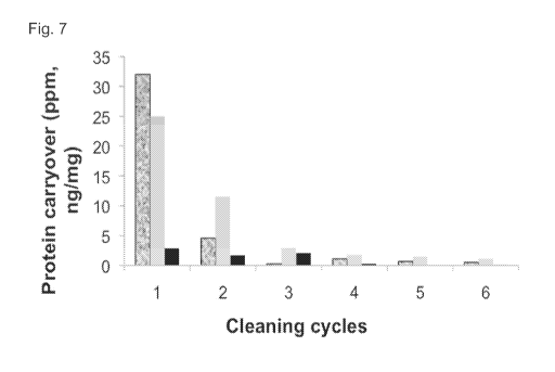
(ng/mg protein) as a function of the stage in the "mock run". Intact IgG is
shown in black, Fc
fragments are shown in grey.
[0026] Figure 7 shows a plot of total protein carryover as a function of
cleaning cycle
following conditions outlined in Method 8, Table 2 with Storage buffer (100 mM
sodium acetate
and 2% Benzyl alcohol (pH 5)). Legend: Elution cycles (black & grey),
Regeneration cycles
(grey), Storage buffer cycles (black).
[0027] Figure 8 is a plot of protein carryover as a function of sample from a
3.23 L pilot scale
purification of mAbZ on MabSelectTM SuRe column followed by column cleaning
with
optimized 6 cycle cleaning procedure (Entry 7, Table 2). Intact IgG is shown
in black, Fc
fragments are shown in grey.
[0028] Figure 9 shows a schematic outline of the optimized cleaning protocol
using 15-minute
static hold time (A) and a "mock run" (B). The Equilibration buffer is 25 mM
Tris, 25 mM
7
CA 02922832 2016-02-29
WO 2015/035180 PCT/US2014/054313
NaC1 (pH 7.1). The Elution buffer is 0.15 M sodium acetate (pH 2.9). The
Regeneration buffer
is 0.1 N NaOH (pH 13).
[0029] Figure 10 shows the results for a lab scale purification of mAbC using
the cleaning
procedure (Figure 9) before "mock run" to determine protein carryover. A.
Protein carryover
(ng/mg protein) as a function of Elution buffer cycle wash; B. protein
carryover (ng/mg protein)
as a function of Regeneration buffer cycle wash; C. protein carryover (ng/mg
protein) as a
function of the stage in the "mock run".
[0030] Figure 11 shows a 10% Tris-HC1 gel taken at different stages of an
optimized cleaning
protocol of a MabSelectTM SuRe column (Figure 9). Samples were taken after the
purification
of mAbC. Regeneration samples are concentrated (25 fold) and lanes 2, 4, 6, 8,
10 and 12
contain samples after the 15 min static holds.
[0031] Figure 12 shows the results for a pilot scale column (3 L) purification
of mAbC using
the optimized cleaning procedure (Entry 7, Table 2) before "mock run" to
determine protein
carryover. A. Protein carryover (ng/mg protein) as a function of Elution
buffer cycle wash; B.
protein carryover (ng/mg protein) as a function of Regeneration buffer cycle
wash. Intact IgG is
shown in black, Fc fragments are shown in grey.
[0032] Figure 13 shows the results intact IgG carryover detected following
mock elutions of a
cation exchange column (POROS) and an anion exchange column (QSFF). MAbA or
MAbB
had previously been loaded and eluted from the columns.
[0033] Figure 14A shows the results intact IgG carryover detected following
mock elutions of
a cation exchange column (POROS) and an anion exchange column (QSFF) before
and after a
clean-in-place procedure. MAbA had previously been loaded and eluted from the
columns.
[0034] Figure 14B shows the results intact IgG carryover detected following
mock elutions of
a cation exchange column (POROS) and an anion exchange column (QSFF) before
and after a
clean-in-place procedure. MAbB had previously been loaded and eluted from the
columns.
[0035] Figure 15 shows the amount of intact IgG (MAbC) eluting from a POROS or
QSFF
column at the end of selected steps of the cleaning protocol.
[0036] Figure 16 shows MAbD carryover at different steps of the cleaning
protocol on pilot
scale columns.
[0037] Figure 17 shows a plot of intact human IgG carryovers as a function of
different wash
conditions.
[0038] Figure 18 shows a plot of protein carryover as a function of CV washes
(resin cleaning
efforts) with different buffer solutions on a ProSeplOvA column. Legend: 6 M
guanidine HC1:
magenta); 19% ethanol: red; 2 M arginine HC1: brown; 20% hexene glycol: grey;
8 M urea/1 M
8
CA 02922832 2016-02-29
WO 2015/035180 PCT/US2014/054313
NaCl: orange; equilibration buffer: blue; 1% v/v phosphoric acid: yellow; 0.1
M Imidazole/19%
Ethanol: black; 0.1 M Acetic acid: green; 2 M potassium phosphate: turquoise.
[0039] Figure 19 is a chromatogram showing five consecutive mock runs of a 0.1
M acetic
acid cleaning solution.
[0040] Figure 20 is a chart showing carryover following consecutive mock runs
with various
cleaning solutions.
[0041] Figure 21 is a chart showing carryover following consecutive mock runs
with cleaning
solutions 6 M guanidine hydrochloride, 2 M arginine hydrochloride, or 20%
hexylene glycol.
[0042] Figure 22 shows a chromatogram of product elution without column
cleaning (top
panel). Carryover in each fraction is shown in the bottom panel.
[0043] Figure 23 shows product elution throughout pulse cleaning.
[0044] Figure 24 is a graph showing carryover over consecutive mock runs with
or without
pulse cleaning.
[0045] Figure 25 is a graph showing carryover over consecutive mock runs using
either
downflow or upflow conditions.
[0046] Figure 26 shows a chromatogram of product elution throughout pulse
cleaning. Black
line shows downflow conditions, gray line shows upflow conditions, light gray
shows pH.
[0047] Figure 27 shows carryover over consecutive mock runs at flow rates of
30 CV/hr and
15 CV/hr.
[0048] Figure 28 shows carryover over consecutive mock runs with single static
holds with
equilibration buffer or regeneration buffer. Normal pulses represent samples
with no static hold.
[0049] Figure 29 shows carryover over consecutive mock runs with multiple
static holds with
equilibration buffer. Normal pulses represent samples with no static hold.
[0050] Figure 30 shows a chromatogram showing product elution throughout the
pulse
cleaning. Black line shows 30 CV/hr conditions, gray line shows 15 CV/hr
conditions, light
gray shows pH.
[0051] Figure 31 shows a chromatogram showing product elution throughout the
pulse
cleaning with static holds. Black line shows normal pulses, medium gray line
shows
equilibration buffer hold conditions, dark gray shows regeneration buffer hold
conditions, and
light gray line shows pH.
[0052] Figure 32 shows a chromatogram showing product elution throughout the
pulse
cleaning with single or multiple static holds. Black line shows normal pulses,
medium gray line
shows 1X equilibration buffer hold conditions, dark gray shows 4X
equilibration buffer hold
conditions, and light gray line shows pH.
9
CA 02922832 2016-02-29
WO 2015/035180 PCT/US2014/054313
[0053] Figure 33 shows reduced cycle duration effect on carryover under pulse
cleaning
conditions.
[0054] Figure 34 shows a chromatogram showing product elution throughout the
pulse
cleaning with reduced cycle duration. Black line shows IgG and light gray line
shows pH.
DETAILED DESCRIPTION OF THE INVENTION
[0055] Provided herein are methods to clean or regenerate a chromatography
material, e.g., a
chromatography resin, for reuse. Chromatography reuse is a changeover
procedure where the
chromatography material is cleaned and/or regenerated for use with the same
product or a
different product. The methods of the invention can be used for the
regeneration of large-scale;
e.g. manufacturing-scale, chromatography material. Significant cost savings
can be achieved if
a resin; e.g., a Protein A resin, is reused for multiple products. In some
embodiments, the
cleaning procedure results in less than 1 ppm carryover of intact protein;
e.g., an IgG, into
subsequent purification samples. In some embodiments, this low protein
carryover is 103 fold
less protein carryover than that set in safety margins, and demonstrates that
the same resins can
be used to purify multiple products. In some embodiments, the chromatography
material is in a
chromatography column.
I. Definitions
[0056] The term "polypeptide" or "protein" are used interchangeably herein to
refer to
polymers of amino acids of any length. The polymer may be linear or branched,
it may
comprise modified amino acids, and it may be interrupted by non-amino acids.
The terms also
encompass an amino acid polymer that has been modified naturally or by
intervention; for
example, disulfide bond formation, glycosylation, lipidation, acetylation,
phosphorylation, or
any other manipulation or modification, such as conjugation with a labeling
component. Also
included within the definition are, for example, polypeptides containing one
or more analogs of
an amino acid (including, for example, unnatural amino acids, etc.), as well
as other
modifications known in the art. The terms "polypeptide" and "protein" as used
herein
specifically encompass antibodies.
[0057] "Purified" polypeptide (e.g., antibody or immunoadhesin) means that the
polypeptide
has been increased in purity, such that it exists in a form that is more pure
than it exists in its
natural environment and/or when initially synthesized and/or amplified under
laboratory
conditions. Purity is a relative term and does not necessarily mean absolute
purity.
CA 02922832 2016-02-29
WO 2015/035180 PCT/US2014/054313
[0058] A polypeptide "which binds" an antigen of interest, e.g. a tumor-
associated
polypeptide antigen target, is one that binds the antigen with sufficient
affinity such that the
polypeptide is useful as a diagnostic and/or therapeutic agent in targeting a
cell or tissue
expressing the antigen, and does not significantly cross-react with other
polypeptides. In such
embodiments, the extent of binding of the polypeptide to a "non-target"
polypeptide will be less
than about 10% of the binding of the polypeptide to its particular target
polypeptide as
determined by fluorescence activated cell sorting (FACS) analysis or
radioimmunoprecipitation
(RIA).
[0059] With regard to the binding of a polypeptide to a target molecule, the
term "specific
binding" or "specifically binds to" or is "specific for" a particular
polypeptide or an epitope on a
particular polypeptide target means binding that is measurably different from
a non-specific
interaction. Specific binding can be measured, for example, by determining
binding of a
molecule compared to binding of a control molecule, which generally is a
molecule of similar
structure that does not have binding activity. For example, specific binding
can be determined
by competition with a control molecule that is similar to the target, for
example, an excess of
non-labeled target. In this case, specific binding is indicated if the binding
of the labeled target
to a probe is competitively inhibited by excess unlabeled target.
[0060] The term "antibody" herein is used in the broadest sense and
specifically covers
monoclonal antibodies, polyclonal antibodies, multispecific antibodies (e.g.
bispecific
antibodies) formed from at least two intact antibodies, and antibody fragments
so long as they
exhibit the desired biological activity. The term "immunoglobulin" (Ig) is
used interchangeable
with antibody herein.
[0061] Antibodies are naturally occurring immunoglobulin molecules which have
varying
structures, all based upon the immunoglobulin fold. For example, IgG
antibodies have two
"heavy" chains and two "light" chains that are disulphide-bonded to form a
functional antibody.
Each heavy and light chain itself comprises a "constant" (C) and a "variable"
(V) region. The V
regions determine the antigen binding specificity of the antibody, whilst the
C regions provide
structural support and function in non-antigen-specific interactions with
immune effectors. The
antigen binding specificity of an antibody or antigen-binding fragment of an
antibody is the
ability of an antibody to specifically bind to a particular antigen.
[0062] The antigen binding specificity of an antibody is determined by the
structural
characteristics of the V region. The variability is not evenly distributed
across the 110-amino
acid span of the variable domains. Instead, the V regions consist of
relatively invariant stretches
called framework regions (FRs) of 15-30 amino acids separated by shorter
regions of extreme
11
CA 02922832 2016-02-29
WO 2015/035180 PCT/US2014/054313
variability called "hypervariable regions" that are each 9-12 amino acids
long. The variable
domains of native heavy and light chains each comprise four FRs, largely
adopting a I3-sheet
configuration, connected by three hypervariable regions ("HVRs"), which form
loops
connecting, and in some cases forming part of, the I3-sheet structure. The
hypervariable regions
in each chain are held together in close proximity by the FRs and, with the
hypervariable regions
from the other chain, contribute to the formation of the antigen-binding site
of antibodies (see
Kabat et al., Sequences of Proteins of Immunological Interest, 5th Ed. Public
Health Service,
National Institutes of Health, Bethesda, Md. (1991)). The constant domains are
not involved
directly in binding an antibody to an antigen, but exhibit various effector
functions, such as
participation of the antibody in antibody dependent cellular cytotoxicity
(ADCC).
[0063] Each V region typically comprises three hypervariable regions, for
example,
complementarity determining regions ("CDRs"), each of which contains a
"hypervariable loop",
and four framework regions. An antibody binding site, the minimal structural
unit required to
bind with substantial affinity to a particular desired antigen, will therefore
typically include the
three CDRs, and at least three, preferably four, framework regions
interspersed there between to
hold and present the CDRs in the appropriate conformation. Classical four
chain antibodies have
antigen binding sites which are defined by VH and VL domains in cooperation.
Certain
antibodies, such as camel and shark antibodies, lack light chains and rely on
binding sites
formed by heavy chains only. Single domain engineered immunoglobulins can be
prepared in
which the binding sites are formed by heavy chains or light chains alone, in
absence of
cooperation between VH and VL.
[0064] The term "variable" refers to the fact that certain portions of the
variable domains
differ extensively in sequence among antibodies and are used in the binding
and specificity of
each particular antibody for its particular antigen. However, the variability
is not evenly
distributed throughout the variable domains of antibodies. It is concentrated
in three segments
called hypervariable regions both in the light chain and the heavy chain
variable domains. The
more highly conserved portions of variable domains are called the framework
regions (FRs).
The variable domains of native heavy and light chains each comprise four FRs,
largely adopting
a I3-sheet configuration, connected by three hypervariable regions, which form
loops connecting,
and in some cases forming part of, the I3-sheet structure. The hypervariable
regions in each chain
are held together in close proximity by the FRs and, with the hypervariable
regions from the
other chain, contribute to the formation of the antigen-binding site of
antibodies (see Kabat et
al., Sequences of Proteins of Immunological Interest, 5th Ed. Public Health
Service, National
Institutes of Health, Bethesda, MD. (1991)). The constant domains are not
involved directly in
12
CA 02922832 2016-02-29
WO 2015/035180 PCT/US2014/054313
binding an antibody to an antigen, but exhibit various effector functions,
such as participation of
the antibody in antibody dependent cellular cytotoxicity (ADCC).
[0065] The term "hypervariable region" when used herein refers to the amino
acid residues of
an antibody that are responsible for antigen binding. The hypervariable region
may comprise
amino acid residues from a "complementarity determining region" or "CDR"
(e.g., around about
residues 24-34 (L1), 50-56 (L2) and 89-97 (L3) in the VL, and around about 31-
35B (H1), 50-65
(H2) and 95-102 (H3) in the VH (Kabat et al., Sequences of Proteins of
Immunological Interest,
5th Ed. Public Health Service, National Institutes of Health, Bethesda, Md.
(1991)) and/or those
residues from a "hypervariable loop" (e.g. residues 26-32 (L1), 50-52 (L2) and
91-96 (L3) in the
VL, and 26-32 (H1), 52A-55 (H2) and 96-101 (H3) in the VH (Chothia and Lesk J.
Mol. Biol.
196:901-917 (1987)).
[0066] "Framework" or "FR" residues are those variable domain residues other
than the
hypervariable region residues as herein defined.
[0067] "Antibody fragments" comprise a portion of an intact antibody,
preferably comprising
the antigen binding region thereof. Examples of antibody fragments include
Fab, Fab', F(aN)2,
and Fv fragments; diabodies; tandem diabodies (taDb), linear antibodies(e.g.,
U.S. Patent No.
5,641,870, Example 2; Zapata et al., Protein Eng. 8(10):1057-1062 (1995)); one-
armed
antibodies, single variable domain antibodies, minibodies, single-chain
antibody molecules;
multispecific antibodies formed from antibody fragments (e.g., including but
not limited to, Db-
Fc, taDb-Fc, taDb-CH3, (scFV)4-Fc, di-scFv, bi-scFv, or tandem (di,tri)-scFv);
and Bi-specific
T-cell engagers (BiTEs).
[0068] Papain digestion of antibodies produces two identical antigen-binding
fragments,
called "Fab" fragments, each with a single antigen-binding site, and a
residual "Fc" fragment,
whose name reflects its ability to crystallize readily. Pepsin treatment
yields an F(aN)2 fragment
that has two antigen-binding sites and is still capable of cross-linking
antigen.
[0069] "Fv" is the minimum antibody fragment that contains a complete antigen-
recognition
and antigen-binding site. This region consists of a dimer of one heavy chain
and one light chain
variable domain in tight, non-covalent association. It is in this
configuration that the three
hypervariable regions of each variable domain interact to define an antigen-
binding site on the
surface of the VH-VL dimer. Collectively, the six hypervariable regions confer
antigen-binding
specificity to the antibody. However, even a single variable domain (or half
of an Fv comprising
only three hypervariable regions specific for an antigen) has the ability to
recognize and bind
antigen, although at a lower affinity than the entire binding site.
13
CA 02922832 2016-02-29
WO 2015/035180 PCT/US2014/054313
[0070] The Fab fragment also contains the constant domain of the light chain
and the first
constant domain (CHO of the heavy chain. Fab' fragments differ from Fab
fragments by the
addition of a few residues at the carboxy terminus of the heavy chain CH1
domain including one
or more cysteines from the antibody hinge region. Fab'-SH is the designation
herein for Fab' in
which the cysteine residue(s) of the constant domains bear at least one free
thiol group. F(a1702
antibody fragments originally were produced as pairs of Fab' fragments that
have hinge
cysteines between them. Other chemical couplings of antibody fragments are
also known.
[0071] The "light chains" of antibodies (immunoglobulins) from any vertebrate
species can be
assigned to one of two clearly distinct types, called kappa (x) and lambda
(X), based on the
amino acid sequences of their constant domains.
[0072] Depending on the amino acid sequence of the constant domain of their
heavy chains,
antibodies can be assigned to different classes. There are five major classes
of intact antibodies:
IgA, IgD, IgE, IgG, and IgM, and several of these may be further divided into
subclasses
(isotypes), e.g., IgGl, IgG2, IgG3, IgG4, IgA, and IgA2. The heavy chain
constant domains that
correspond to the different classes of antibodies are called a, 6, 8, y, and
IA, respectively. The
subunit structures and three-dimensional configurations of different classes
of immunoglobulins
are well known.
[0073] "Single-chain Fv" or "scFv" antibody fragments comprise the VH and VL
domains of
antibody, wherein these domains are present in a single polypeptide chain. In
some
embodiments, the Fv polypeptide further comprises a polypeptide linker between
the VH and VL
domains that enables the scFv to form the desired structure for antigen
binding. For a review of
scFv see Pliickthun in The Pharmacology of Monoclonal Antibodies, vol. 113,
Rosenburg and
Moore eds., Springer-Verlag, New York, pp. 269-315 (1994).
[0074] The term "diabodies" refers to small antibody fragments with two
antigen-binding
sites, which fragments comprise a heavy chain variable domain (VH) connected
to a light chain
variable domain (VL) in the same polypeptide chain (VH - VL). By using a
linker that is too short
to allow pairing between the two domains on the same chain, the domains are
forced to pair with
the complementary domains of another chain and create two antigen-binding
sites. Diabodies are
described more fully in, for example, EP 404,097; WO 93/11161; and Hollinger
et al., Proc.
Natl. Acad. Sci. USA, 90:6444-6448 (1993).
[0075] The term "multispecific antibody" is used in the broadest sense and
specifically covers
an antibody that has polyepitopic specificity. Such multispecific antibodies
include, but are not
limited to, an antibody comprising a heavy chain variable domain (VH) and a
light chain variable
domain (VD, where the VHVL unit has polyepitopic specificity, antibodies
having two or more
14
CA 02922832 2016-02-29
WO 2015/035180 PCT/US2014/054313
VL and VH domains with each VHVL unit binding to a different epitope,
antibodies having two or
more single variable domains with each single variable domain binding to a
different epitope,
full length antibodies, antibody fragments such as Fab, Fv, dsFv, scFv,
diabodies, bispecific
diabodies, triabodies, tri-functional antibodies, antibody fragments that have
been linked
covalently or non-covalently. "Polyepitopic specificity" refers to the ability
to specifically bind
to two or more different epitopes on the same or different target(s). "Mono
specific" refers to the
ability to bind only one epitope. According to one embodiment the
multispecific antibody is an
IgG antibody that binds to each epitope with an affinity of 51.1M to 0.001 pM,
31.1M to 0.001
pM, 11.1M to 0.001 pM, 0.51.1M to 0.001 pM, or 0.11.1M to 0.001 pM.
[0076] The expression "single domain antibodies" (sdAbs) or "single variable
domain (SVD)
antibodies" generally refers to antibodies in which a single variable domain
(VH or VL) can
confer antigen binding. In other words, the single variable domain does not
need to interact with
another variable domain in order to recognize the target antigen. Examples of
single domain
antibodies include those derived from camelids (lamas and camels) and
cartilaginous fish (e.g.,
nurse sharks) and those derived from recombinant methods from humans and mouse
antibodies
(Nature (1989) 341:544-546; Dev Comp Immunol (2006) 30:43-56; Trend Biochem
Sci (2001)
26:230-235; Trends Biotechnol (2003):21:484-490; WO 2005/035572; WO 03/035694;
Febs
Lett (1994) 339:285-290; W000/29004; WO 02/051870).
[0077] The term "monoclonal antibody" as used herein refers to an antibody
obtained from a
population of substantially homogeneous antibodies, i.e., the individual
antibodies comprising
the population are identical and/or bind the same epitope, except for possible
variants that may
arise during production of the monoclonal antibody, such variants generally
being present in
minor amounts. In contrast to polyclonal antibody preparations that typically
include different
antibodies directed against different determinants (epitopes), each monoclonal
antibody is
directed against a single determinant on the antigen. In addition to their
specificity, the
monoclonal antibodies are advantageous in that they are uncontaminated by
other
immunoglobulins. The modifier "monoclonal" indicates the character of the
antibody as being
obtained from a substantially homogeneous population of antibodies, and is not
to be construed
as requiring production of the antibody by any particular method. For example,
the monoclonal
antibodies to be used in accordance with the methods provided herein may be
made by the
hybridoma method first described by Kohler et al., Nature 256:495 (1975), or
may be made by
recombinant DNA methods (see, e.g., U.S. Patent No. 4,816,567). The
"monoclonal antibodies"
may also be isolated from phage antibody libraries using the techniques
described in Clackson et
al., Nature 352:624-628 (1991) and Marks et al., J. Mol. Biol. 222:581-597
(1991), for example.
CA 02922832 2016-02-29
WO 2015/035180 PCT/US2014/054313
[0078] The monoclonal antibodies herein specifically include "chimeric"
antibodies
(immunoglobulins) in which a portion of the heavy and/or light chain is
identical with or
homologous to corresponding sequences in antibodies derived from a particular
species or
belonging to a particular antibody class or subclass, while the remainder of
the chain(s) is
identical with or homologous to corresponding sequences in antibodies derived
from another
species or belonging to another antibody class or subclass, as well as
fragments of such
antibodies, so long as they exhibit the desired biological activity (U.S.
Patent No. 4,816,567;
Morrison et al., Proc. Natl. Acad. Sci. USA 81:6851-6855 (1984)). Chimeric
antibodies of
interest herein include "primatized" antibodies comprising variable domain
antigen-binding
sequences derived from a non-human primate (e.g. Old World Monkey, such as
baboon, rhesus
or cynomolgus monkey) and human constant region sequences (US Pat No.
5,693,780).
[0079] "Humanized" forms of non-human (e.g., murine) antibodies are chimeric
antibodies
that contain minimal sequence derived from non-human immunoglobulin. For the
most part,
humanized antibodies are human immunoglobulins (recipient antibody) in which
residues from a
hypervariable region of the recipient are replaced by residues from a
hypervariable region of a
non-human species (donor antibody) such as mouse, rat, rabbit or nonhuman
primate having the
desired specificity, affinity, and capacity. In some instances, framework
region (FR) residues of
the human immunoglobulin are replaced by corresponding non-human residues.
Furthermore,
humanized antibodies may comprise residues that are not found in the recipient
antibody or in
the donor antibody. These modifications are made to further refine antibody
performance. In
general, the humanized antibody will comprise substantially all of at least
one, and typically
two, variable domains, in which all or substantially all of the hypervariable
loops correspond to
those of a non-human immunoglobulin and all or substantially all of the FRs
are those of a
human immunoglobulin sequence, except for FR substitution(s) as noted above.
The humanized
antibody optionally also will comprise at least a portion of an immunoglobulin
constant region,
typically that of a human immunoglobulin. For further details, see Jones et
al., Nature 321:522-
525 (1986); Riechmann et al., Nature 332:323-329 (1988); and Presta, Curr. Op.
Struct. Biol.
2:593-596 (1992).
[0080] For the purposes herein, an "intact antibody" is one comprising heavy
and light
variable domains as well as an Fc region. The constant domains may be native
sequence
constant domains (e.g. human native sequence constant domains) or amino acid
sequence variant
thereof. Preferably, the intact antibody has one or more effector functions.
[0081] "Native antibodies" are usually heterotetrameric glycoproteins of about
150,000
daltons, composed of two identical light (L) chains and two identical heavy
(H) chains. Each
16
CA 02922832 2016-02-29
WO 2015/035180 PCT/US2014/054313
light chain is linked to a heavy chain by one covalent disulfide bond, while
the number of
disulfide linkages varies among the heavy chains of different immunoglobulin
isotypes. Each
heavy and light chain also has regularly spaced intrachain disulfide bridges.
Each heavy chain
has at one end a variable domain (VH) followed by a number of constant
domains. Each light
chain has a variable domain at one end (VL) and a constant domain at its other
end; the constant
domain of the light chain is aligned with the first constant domain of the
heavy chain, and the
light chain variable domain is aligned with the variable domain of the heavy
chain. Particular
amino acid residues are believed to form an interface between the light chain
and heavy chain
variable domains.
[0082] A "naked antibody" is an antibody (as herein defined) that is not
conjugated to a
heterologous molecule, such as a cytotoxic moiety or radiolabel.
[0083] In some embodiments, antibody "effector functions" refer to those
biological activities
attributable to the Fc region (a native sequence Fc region or amino acid
sequence variant Fc
region) of an antibody, and vary with the antibody isotype. Examples of
antibody effector
functions include: Clq binding and complement dependent cytotoxicity; Fc
receptor binding;
antibody-dependent cell-mediated cytotoxicity (ADCC); phagocytosis; down
regulation of cell
surface receptors.
[0084] "Antibody-dependent cell-mediated cytotoxicity" and "ADCC" refer to a
cell-mediated
reaction in which nonspecific cytotoxic cells that express Fc receptors (FcRs)
(e.g. Natural
Killer (NK) cells, neutrophils, and macrophages) recognize bound antibody on a
target cell and
subsequently cause lysis of the target cell. The primary cells for mediating
ADCC, NK cells,
express FcyRIII only, whereas monocytes express FcyRI, FcyRII and FcyRIII. FcR
expression
on hematopoietic cells in summarized is Table 3 on page 464 of Ravetch and
Kinet, Annu. Rev.
Immunol 9:457-92 (1991). To assess ADCC activity of a molecule of interest, an
in vitro ADCC
assay, such as that described in US Patent No. 5,500,362 or 5,821,337 may be
performed. Useful
effector cells for such assays include peripheral blood mononuclear cells
(PBMC) and Natural
Killer (NK) cells. Alternatively, or additionally, ADCC activity of the
molecule of interest may
be assessed in vivo, e.g., in an animal model such as that disclosed in Clynes
et al., Proc. Natl.
Acad. Sci. (USA) 95:652-656 (1998).
[0085] "Human effector cells" are leukocytes that express one or more FcRs and
perform
effector functions. In some embodiments, the cells express at least FcyRIII
and carry out ADCC
effector function. Examples of human leukocytes that mediate ADCC include
peripheral blood
mononuclear cells (PBMC), natural killer (NK) cells, monocytes, cytotoxic T
cells and
neutrophils; with PBMCs and NK cells being preferred.
17
CA 02922832 2016-02-29
WO 2015/035180 PCT/US2014/054313
[0086] The terms "Fe receptor" or "FcR" are used to describe a receptor that
binds to the Fe
region of an antibody. In some embodiments, the FcR is a native sequence human
FcR.
Moreover, a preferred FcR is one that binds an IgG antibody (a gamma receptor)
and includes
receptors of the FcyRI, FcyRII, and Fey RIII subclasses, including allelic
variants and
alternatively spliced forms of these receptors. FcyRII receptors include
FcyRIIA (an "activating
receptor") and FcyRIIB (an "inhibiting receptor"), which have similar amino
acid sequences that
differ primarily in the cytoplasmic domains thereof. Activating receptor
FcyRIIA contains an
immunoreceptor tyrosine-based activation motif (ITAM) in its cytoplasmic
domain. Inhibiting
receptor FcyRIIB contains an immunoreceptor tyrosine-based inhibition motif
(ITIM) in its
cytoplasmic domain. (see Daeron, Annu. Rev. Immunol. 15:203-234 (1997)). FcRs
are reviewed
in Ravetch and Kinet, Annu. Rev. Immunol 9:457-92 (1991); Capel et al.,
Immunomethods 4:25-
34 (1994); and de Haas et al., J. Lab. Clin. Med. 126:330-41 (1995). Other
FcRs, including
those to be identified in the future, are encompassed by the term "FcR"
herein. The term also
includes the neonatal receptor, FcRn, which is responsible for the transfer of
maternal IgGs to
the fetus (Guyer et al., J. Immunol. 117:587 (1976) and Kim et al., J.
Immunol. 24:249 (1994)).
[0087] The term "sequential" as used herein with regard to chromatography
refers to having a
first chromatography followed by a second chromatography. Additional steps may
be included
between the first chromatography and the second chromatography.
[0088] The term "continuous" as used herein with regard to chromatography
refers to having a
first chromatography material and a second chromatography material either
directly connected
or some other mechanism which allows for continuous flow between the two
chromatography
materials.
[0089] The term "isolated" as used herein refers to a molecule that has been
separated from at
least some of the components with which it is typically found in nature or
produced. For
example, a polypeptide is referred to as "isolated" when it is separated from
at least some of the
components of the cell in which it was produced. Where a polypeptide is
secreted by a cell after
expression, physically separating the supernatant containing the polypeptide
from the cell that
produced it is considered to be "isolating" the polypeptide. Similarly, a
polynucleotide is
referred to as "isolated" when it is not part of the larger polynucleotide
(such as, for example,
genomic DNA or mitochondrial DNA, in the case of a DNA polynucleotide) in
which it is
typically found in nature, or is separated from at least some of the
components of the cell in
which it was produced, e.g., in the case of an RNA polynucleotide. Thus, a DNA
polynucleotide
that is contained in a vector inside a host cell may be referred to as
"isolated".
18
CA 02922832 2016-02-29
WO 2015/035180 PCT/US2014/054313
[0090] An "Acceptable Daily Exposure (ADE)" as used herein is a substance-
specific dose
that is unlikely to cause an adverse health event or undesirable physiological
effect if an
individual is exposed to this does or to a lower dose over a lifetime
(Teschner, W., et al., Vox
Sang. 2007, 92:42-55; Food and Drug Administration, HHS. Guidance for Industry
Estimating
the Maximum Safe Starting Dose in Initial Clinical Trials for Therapeutics in
Adult Healthy
Volunteers. Rockville, MD. July 2005 on the world wide web at
google.com/url?sa=t&rct=
j&q=&esrc=s&source=web&cd=l&ved=OCE8QFjAA&url=http%3A%2F%2Fwww.fda.gov%2
Fdownloads%2FDrugs%2F...%2FGuidances%2FUCM078932.pdf&ei=f4QhUJv4K90v6gGQ-
4DgAg&usg=AFQjCNFbTE75U0nDbFpfdpxK85uWXT8frg as accessed on August 7, 2012;
European Medicines Agency. Impurities: Residual Solvents, Note for Guidance on
Impurities:
Residual Solvents (CPMP/ICH/283/95). London, UK Sept. 1997 on the world wide
web at
ema.europa.eu/ema/index.jsp?curl=pages/regulation/general/general_content_00043
1.jsp&mid=
WC0b0 1 ac0580029593 as accessed on August 7, 2012). In addition to an ADE, an
"Estimated
Daily Intake (EDT)" for IgG is determined based on the amount of IgG
administered per dose.
[0091] "Contaminants" refer to materials that are different from the desired
polypeptide
product. The contaminant includes, without limitation: host cell materials,
such as CHOP;
leached Protein A; nucleic acid; a variant, fragment, aggregate or derivative
of the desired
polypeptide; another polypeptide; endotoxin; viral contaminant; cell culture
media component,
etc. In some examples, the contaminant may be a host cell protein (HCP) from,
for example but
not limited to, a bacterial cell such as an E. coli cell, an insect cell, a
prokaryotic cell, a
eukaryotic cell, a yeast cell, a mammalian cell, an avian cell, a fungal cell.
[0092] Reference to "about" a value or parameter herein includes (and
describes) variations
that are directed to that value or parameter per se. For example, description
referring to "about
X" includes description of "X".
[0093] As used herein and in the appended claims, the singular forms "a,"
"or," and "the"
include plural referents unless the context clearly dictates otherwise. It is
understood that aspects
and variations of the invention described herein include "consisting" and/or
"consisting
essentially of' aspects and variations.
H. Methods of Column Cleaning
(A) Chromatography
[0094] The invention provides methods to clean or regenerate chromatography
materials for
reuse. In some embodiments, the chromatography materials are used for large-
scale; e.g.,
manufacturing-scale, production of polypeptide products.
19
CA 02922832 2016-02-29
WO 2015/035180 PCT/US2014/054313
[0095] In some embodiments, the method comprises the steps of a) passing about
two or more
material volumes of elution buffer through the material, wherein the elution
buffer comprises
about 0.15 M acetic acid, at about pH 2.9; b) statically holding the material
in elution buffer for
a time ranging from about 10 min to about 30 min; c) passing about two or more
material
volumes of elution buffer through the material; and d) passing about two or
more material
volumes of regeneration buffer through the material, wherein the regeneration
buffer is about 0.1
N NaOH, pH 13. In some embodiments, the chromatography material is in a
chromatography
column. In some embodiments, the chromatography column is used for large-
scale, e.g.
manufacturing-scale, production of a polypeptide product such as an antibody
product. In some
embodiments, the chromatography material is a Protein A chromatography
material. In some
embodiments, the chromatography material is used to purify multiple antibody
products. In
some embodiments, the carryover after the cleaning methods comprises one or
more of <0.25
mg/mL total protein, < 1 ppm IgG fragments, < 1 ppm leached protein A, <1
[t.g/mL CZE LIF,
<1 ppm CHOP, and <1 pg/mL CHO DNA.
[0096] In some embodiments, the method comprising the steps of a) passing
about two
material volumes of elution buffer through the material, wherein the elution
buffer comprises
about 0.15 M acetic acid, at about pH 2.9; b) statically holding the material
in elution buffer for
about 30 min; c) passing about two material volumes of elution buffer through
the material; and
d) passing about four material volumes of regeneration buffer through the
material, wherein the
regeneration buffer is about 0.1 N NaOH, at about pH 13. In some embodiments,
the
chromatography material is in a chromatography column. In some embodiments,
the
chromatography column is used for large-scale, e.g. manufacturing-scale,
production of a
polypeptide product such as an antibody product. In some embodiments, the
chromatography
material is a Protein A chromatography material. In some embodiments, the
chromatography
material is used to purify multiple antibody products. In some embodiments,
the carryover after
the cleaning methods comprises one or more of <0.25 mg/mL total protein, < 1
ppm IgG
fragments, < 1 ppm leached protein A, <1 [t.g/mL CZE LIF, <1 ppm CHOP, and <1
pg/mL CHO
DNA.
[0097] In some embodiments, the method comprising the steps of a) passing
about two
material volumes of elution buffer through the material, wherein the elution
buffer comprises
about 0.15 M acetic acid, at about pH 2.9, b) statically holding the material
in elution buffer for
about 30 min, c) passing about two material volumes of elution buffer through
the material, d)
passing about two and one-half material volumes of regeneration buffer through
the material,
wherein the regeneration buffer is about 0.1 N NaOH, at about pH 13, e)
statically holding the
CA 02922832 2016-02-29
WO 2015/035180 PCT/US2014/054313
material in regeneration buffer for about 30 min, f) passing about two and one-
half material
volumes of regeneration buffer through the material. In some embodiments, the
chromatography material is in a chromatography column. In some embodiments,
the
chromatography column is used for large-scale, e.g. manufacturing-scale,
production of a
polypeptide product such as an antibody product. In some embodiments, the
chromatography
material is a Protein A chromatography material. In some embodiments, the
chromatography
material is used to purify multiple antibody products. In some embodiments,
the carryover after
the cleaning methods comprises one or more of <0.25 mg/mL total protein, < 1
ppm IgG
fragments, < 1 ppm leached protein A, <1 [t.g/mL CZE LIF, <1 ppm CHOP, and <1
pg/mL CHO
DNA.
[0098] In some embodiments, the method comprising the steps of a) passing
about two
material volumes of equilibration buffer through the material, wherein the
equilibration buffer
comprises about 25 mM Tris, about 25 mM NaC1, at about pH 7.1; b) statically
holding the
material in equilibration buffer for about 30 min; c) passing about two
material volumes of
equilibration buffer through the material; d) passing about two material
volumes of elution
buffer through the material, wherein the elution buffer is about 0.15 M Acetic
acid, at about pH
2.8; e) statically holding the material in elution buffer for about 30 min; f)
passing about two
material volumes of elution buffer through the material; g) passing about two
material volumes
of regeneration buffer through the material, wherein the regeneration buffer
is about 0.1 N
NaOH, at about pH 13; h) statically holding the material in regeneration
buffer for about 30 min;
i) passing about two material volumes of regeneration buffer through the
material. In some
embodiments, the chromatography material is in a chromatography column. In
some
embodiments, the chromatography column is used for large-scale, e.g.
manufacturing-scale,
production of a polypeptide product such as an antibody product. In some
embodiments, the
chromatography material is a Protein A chromatography material. In some
embodiments, the
chromatography material is used to purify multiple antibody products. In some
embodiments,
the carryover after the cleaning methods comprises one or more of <0.25 mg/mL
total protein, <
1 ppm IgG fragments, < 1 ppm leached protein A, <1 [t.g/mL CZE LIF, <1 ppm
CHOP, and <1
pg/mL CHO DNA.
[0099] In some embodiments, the method comprising the steps of a) passing
about four
material volumes of equilibration buffer through the material, wherein the
equilibration buffer
comprises about 25 mM Tris, about 25 mM NaC1, at about pH 7.1; b) performing
six cycles of
the steps comprising i) passing about three material volumes of elution buffer
through the
material, wherein the elution buffer is about 0.15 M Acetic acid, pH 2.8; ii)
statically holding the
21
CA 02922832 2016-02-29
WO 2015/035180 PCT/US2014/054313
material in elution buffer for about 10 min; iii) passing about one material
volume of elution
buffer through the material; iv) passing about three material volumes of
regeneration buffer
through the material, wherein the regeneration buffer is about 0.1 N NaOH, at
about pH 13; v)
statically holding the material in regeneration buffer for about 10 min; vi)
passing about one
material volume of regeneration buffer through the material In some
embodiments, the
chromatography material is in a chromatography column. In some embodiments,
the
chromatography column is used for large-scale, e.g. manufacturing-scale,
production of a
polypeptide product such as an antibody product. In some embodiments, the
chromatography
material is a Protein A chromatography material. In some embodiments, the
chromatography
material is used to purify multiple antibody products. In some embodiments,
the carryover after
the cleaning methods comprises one or more of <0.25 mg/mL total protein, < 1
ppm IgG
fragments, < 1 ppm leached protein A, <1 [t.g/mL CZE LIF, <1 ppm CHOP, and <1
pg/mL CHO
DNA.
[0100] In some embodiments, the method comprising six cycles of the steps of
a) passing
about three material volumes of elution buffer through the material, wherein
the elution buffer is
about 0.15 M Acetic acid, at about pH 2.8; b) statically holding the material
in elution buffer for
about 15 min; c) passing about one material volume of elution buffer through
the material; d)
passing about three material volumes of regeneration buffer through the
material, wherein the
regeneration buffer is about 0.1 N NaOH, at about pH 13; e) statically holding
the material in
regeneration buffer for about 15 min; f) passing about one material volume of
regeneration
buffer through the material; g) passing about three material volumes of
storage buffer through
the material, wherein the storage buffer is about 100 mM sodium acetate, about
2% benzyl
alcohol, at about pH 5.0; e) statically holding the material in storage buffer
for about 15 min; f)
passing about one material volume of storage buffer through the material. In
some
embodiments, the chromatography material is in a chromatography column. In
some
embodiments, the chromatography column is used for large-scale, e.g.
manufacturing-scale,
production of a polypeptide product such as an antibody product. In some
embodiments, the
chromatography material is a Protein A chromatography material. In some
embodiments, the
chromatography material is used to purify multiple antibody products. In some
embodiments,
the carryover after the cleaning methods comprises one or more of <0.25 mg/mL
total protein, <
1 ppm IgG fragments, < 1 ppm leached protein A, <1 [t.g/mL CZE LIF, <1 ppm
CHOP, and <1
pg/mL CHO DNA.
[0101] In some aspects of the invention, the chromatography material is an
affinity
chromatography material. Examples of affinity chromatography materials
include, but are not
22
CA 02922832 2016-02-29
WO 2015/035180 PCT/US2014/054313
limited to chromatography materials derivatized with protein A or protein G.
Examples of
affinity chromatography material include, but are not limited to, Prosep-VA,
Prosep-VA Ultra
Plus, Protein A sepharose fast flow, Tyopearl Protein A, MAbSelectTM,
MAbSelectTM SuRe and
MAbSelectTM SuRe LX. In some embodiments of the above, the affinity
chromatography
material is an affinity chromatography material. In some embodiments of the
above, the affinity
chromatography material is an affinity chromatography membrane. In some
embodiments, the
affinity chromatography material is a Protein G chromatography material. In
some
embodiments, the chromatography column is used for large-scale, e.g.
manufacturing-scale,
production of a polypeptide product such as an antibody product.
[0102] In some aspects, the invention provides methods to clean an ion-
exchange
chromatography material for reuse. In some embodiments, the method comprising
the steps of
a) passing about three material volumes of equilibration buffer through the
material, wherein the
equilibration buffer comprises about 25 mM Tris, about 25 mM NaC1, at about pH
7.1; b)
passing about two material volumes of about 0.5 N NaOH through the material c)
statically
holding the material in about 0.5 N NaOH for about 10 min; d) passing about
one material
volume of about 0.5 N NaOH through the material; e) statically holding the
material in about 0.5
N NaOH for about 10 min; and f) passing about one material volume of about 0.5
N NaOH
through the material. In some embodiments, the ion exchange material is in a
chromatography
column. In some embodiments, the chromatography column is used for large-
scale, e.g.
manufacturing-scale, production of a polypeptide product such as an antibody
product. In some
embodiments, the chromatography material is used to purify multiple antibody
products. In
some embodiments, the carryover after the cleaning methods comprises one or
more of <0.25
mg/mL total protein, < 1 ppm IgG fragments, < 1 ppm leached protein A, <1
[t.g/mL CZE LIF,
<1 ppm CHOP, and <1 pg/mL CHO DNA.
[0103] In some embodiments of any of the methods described herein, the
chromatography
material is an ion exchange chromatography material; for example, an anion
exchange
chromatography material or a cation exchange chromatography material. In some
embodiments
of any of the methods described herein, the chromatography material is an
anion exchange
material. In some embodiments, the anion exchange material is in a
chromatography column.
In some embodiments, the anion exchange chromatography material is a solid
phase that is
positively charged and has free anions for exchange with anions in an aqueous
solution passed
over or through the solid phase. In some embodiments of any of the methods
described herein,
the anion exchange material may be a membrane, a monolith, or resin. In an
embodiment, the
anion exchange material may be a resin. In some embodiments, the anion
exchange material
23
CA 02922832 2016-02-29
WO 2015/035180 PCT/US2014/054313
may comprise a primary amine, a secondary amine, a tertiary amine or a
quarternary ammonium
ion functional group, a polyamine functional group, or a diethylaminoaethyl
functional group.
In some embodiments of the above, the anion exchange chromatography material
is an anion
exchange chromatography material. In some embodiments of the above, the anion
exchange
chromatography material is an anion exchange chromatography membrane. In some
embodiments, the anion exchange chromatography material is used for large-
scale, e.g.
manufacturing-scale, production of a polypeptide product such as an antibody
product.
[0104] In some embodiments of any of the methods described herein, the
chromatography
material is a cation exchange material. In some embodiments, the cation
exchange material is in
a chromatography column. In some embodiments, the cation exchange material is
a solid phase
that is negatively charged and has free cations for exchange with cations in
an aqueous solution
passed over or through the solid phase. In some embodiments of any of the
methods described
herein, the cation exchange material may be a membrane, a monolith, or resin.
In some
embodiments, the cation exchange material may be a resin. The cation exchange
material may
comprise a carboxylic acid functional group or a sulfonic acid functional
group such as, but not
limited to, sulfonate, carboxylic, carboxymethyl sulfonic acid, sulfoisobutyl,
sulfoethyl,
carboxyl, sulphopropyl, sulphonyl, sulphoxyethyl, or orthophosphate. In some
embodiments of
the above, the cation exchange chromatography material is a cation exchange
chromatography
material. In some embodiments of the above, the cation exchange chromatography
material is a
cation exchange chromatography membrane. In some embodiments of the invention,
the
chromatography material is not a cation exchange chromatography material. In
some
embodiments, the cation exchange chromatography material is used for large-
scale, e.g.
manufacturing-scale, production of a polypeptide product such as an antibody
product.
[0105] In some embodiments of any of the methods described herein, the ion
exchange
material may utilize a conventional chromatography material or a convective
chromatography
material. The conventional chromatography materials include, for example,
perfusive materials
(e.g., poly (styrene-divinylbenzene) resin) and diffusive materials (e.g.,
cross-linked agarose
resin). In some embodiments, the poly (styrene-divinylbenzene) resin can be
Poros resin. In
some embodiments, the cross-linked agarose resin may be sulphopropyl-Sepharose
Fast Flow
("SPSFF") resin. The convective chromatography material may be a membrane
(e.g.,
polyethersulfone) or monolith material (e.g. cross-linked polymer). The
polyethersulfone
membrane may be Mustang. The cross-linked polymer monolith material may be
cross-linked
poly(glycidyl methacrylate-co-ethylene dimethacrylate).
24
CA 02922832 2016-02-29
WO 2015/035180 PCT/US2014/054313
[0106] Examples of anion exchange materials are known in the art and include,
but are not
limited to Poros HQ 50, Poros PI 50, Poros D, Mustang Q, Q Sepharose FF, and
DEAE
Sepharose.
[0107] Examples of cation exchange materials are known in the art include, but
are not limited
to Mustang S, Sartobind S, S03 Monolith, S Ceramic HyperD, Poros XS, Poros
HS50, Poros
HS20, SPSFF, SP-Sepharose XL (SPXL), CM Sepharose Fast Flow, Capto S,
Fractogel Se
HiCap, Fractogel S03, or Fractogel COO. In some embodiments of any of the
methods
described herein, the cation exchange material is Poros HS50. In some
embodiments, the Poros
HS resin may be Poros HS 50 lam or Poros HS 20 lam particles.
[0108] In some embodiments of any of the methods described herein, the
chromatography
material is a mixed mode material comprising functional groups capable of one
of more of the
following functionalities: anionic exchange, cation exchange, hydrogen
bonding, and
hydrophobic interactions. In some embodiments, the mixed mode material
comprises functional
groups capable of anionic exchange and hydrophobic interactions. The mixed
mode material
may contain N-benzyl-N-methyl ethanol amine, 4-mercapto-ethyl-pyridine,
hexylamine, or
phenylpropylamine as ligand or contain cross-linked polyallylamine. Examples
of the mixed
mode materials include Capto Adhere resin, QMA resin, Capto MMC resin, MEP
HyperCel
resin, HEA HyperCel resin, PPA HyperCel resin, or ChromaSorb membrane or
Sartobind STIC.
In some embodiments, the mixed mode material is Capto Adhere resin. In some
embodiments
of the above, the mixed mode material is a mixed mode chromatography material.
In some
embodiments of the above, the mixed mode material is a mixed mode
chromatography column.
In some embodiments of the above, the mixed mode material is a mixed mode
membrane. In
some embodiments, the mixed mode chromatography column is a large-scale; e.g.
manufacturing-scale, chromatography column.
[0109] In some aspects of the invention, the chromatography material is a
hydrophobic
interaction chromatography material. Hydrophobic interaction chromatography
(HIC) is a liquid
chromatography technique that separates biomolecules according to
hydrophobicity. Examples
of HIC chromatography materials include, but are not limited to, Toyopearl
hexyl 650, Toyopear
butyl 650, Toyopearl phenyl 650, Toyopearl ether 650, Source, Resource,
Sepharose Hi-Trap,
Octyl sepharose, and Phenyl sepharose. In some embodiments of the above, the
HIC
chromatography material is a HIC chromatography column. In some embodiments of
the above,
the HIC chromatography material is a HIC chromatography membrane. In some
embodiments,
the HUC chromatography column is a large-scale; e.g. manufacturing-scale,
chromatography
column.
CA 02922832 2016-02-29
WO 2015/035180 PCT/US2014/054313
[0110] In some aspects of the invention, the chromatography material is a
hydroxyapatite
(HAP) chromatography material. Examples of hydroxyapatite chromatography
material include
but are limited to HA Ultrogel, and CHT hydroxyapatite. In some embodiments of
the above,
the HAP chromatography material is a HAP chromatography column. In some
embodiments of
the above, the HAP chromatography material is a HAP chromatography membrane.
In some
embodiments, the HAP chromatography column is a large-scale; e.g.
manufacturing-scale,
chromatography column.
[0111] In some embodiments, the invention provides methods to clean or
regenerate an alkali
stable chromatography material; e.g. an alkali stable chromatography column.
[0112] The invention provides buffers for use in the methods of the invention.
Elution buffers
are generally used to remove a material from a chromatography material; e.g. a
desired material
or an undesired material such as a contaminant. Examples of elution buffers
include but are not
limited to about 0.15 M acetic acid at about pH 2.8-2.9. A regeneration buffer
is generally used
to recharge a column following a chromatography procedure. For example, a
regeneration
buffer for an anion chromatography may be about 0.1 N NaOH at about pH 13. An
equilibration
buffer may be used to make put the chromatography material under the same
conditions (salt
concentration, pH, etc.) as the sample. A nonlimiting example of an
equilibration buffer is about
25 mM Tris, and about 25 mM NaC1 at about pH 7.1. A storage buffer is
generally used to
maintain a chromatography material when not in use; for example, with a
microcode to prevent
contamination. A nonlimiting example of a strorage buffer is about 100 mM
sodium acetate,
about 2% benzyl alcohol, and at about pH 5Ø
[0113] In some embodiments of any of the methods described herein, the flow
rate is less than
about any of 50 material volumes/hr, 40 material volumes/hr, or 30 material
volumes/hr. The
flow rate may be between about any of 5 material volumes/hr and 50 material
volumes/hr, 10
material volumes/hr and 40 material volumes/hr, or 18 material volumes/hr and
36 material
volumes/hr. In some embodiments, the flow rate is about any of 9 material
volumes/hr, 18
material volumes/hr, 25 material volumes/hr, 30 material volumes/hr, 36
material volumes/hr, or
40 material volumes/hr.
[0114] In some embodiments, the chromatography material is in a chromatography
column.
In some embodiments of any of the methods described herein, the flow rate is
less than about
any of 50 column volumes (CV)/hr, 40 CV/hr, or 30 CV/hr. The flow rate may be
between about
any of 5 CV/hr and 50 CV/hr, 10 CV/hr and 40 CV/hr, or 18 CV/hr and 36 CV/hr.
In some
embodiments, the flow rate is about any of 9 CV/hr, 18 CV/hr, 25 CV/hr, 30
CV/hr, 36 CV/hr,
or 40 CV/hr. In some embodiments of any of the methods described herein, the
flow rate is less
26
CA 02922832 2016-02-29
WO 2015/035180 PCT/US2014/054313
than about any of 100 cm/hr, 75 cm/hr, or 50 cm/hr. The flow rate may be
between about any of
25 cm/hr and 150 cm/hr, 25 cm/hr and 100 cm/hr, 50 cm/hr and 100 cm/hr, or 65
cm/hr and 85
cm/hr.
[0115] Bed height is the height of chromatography material used. In some
embodiments of
any of the method described herein, the bed height is greater than about any
of 3 cm, 10 cm, or
15 cm. The bed height may be between about any of 3 cm and 35 cm, 5 cm and 15
cm, 3 cm and
cm, or 5 cm and 8 cm. In some embodiments, the bed height is about any of 3
cm, 5 cm, 10
cm, 15 cm, 20 cm, 25 cm, or 30 cm. In some embodiments, bed height is
determined based on
the amount of polypeptide or contaminants in the load. In some embodiments,
the
chromatography material is in a column used for large-scale; e.g.,
manufacturing-scale,
production of a polypeptide. In some embodiments, the manufacturing-scale
chromatography
material has a bed height of about any of 10 cm, 15, cm, 20 cm, 25 cm or 30
cm.
[0116] Bed diameter is the diameter of chromatography material used. In some
embodiments
of any of the method described herein, the bed diameter is greater than about
any of 80 cm, 100
cm, or 120 cm. In some embodiments, the bed diameter is about any of 50 cm, 60
cm, 70 cm,
80 cm, 90 cm, 100 cm, 110 cm, 120 cm, 130 cm, 140 cm, 150 cm, 160 cm, 170 cm,
180 cm,
190, or 200 cm. In some embodiments, bed diameter is determined based on the
amount of
polypeptide or contaminants in the load. In some embodiments, the
chromatography material is
in a column used for large-scale; e.g., manufacturing-scale, production of a
polypeptide. In some
embodiments, the manufacturing-scale chromatography material had a bed
diameter of about
any 50 cm, 60 cm, 70 cm, 80 cm, 90 cm, 100 cm, 110 cm, 120 cm, 130 cm, 140 cm,
150 cm,
160 cm, 170 cm, 180 cm, 190, or 200 cm.
[0117] In some embodiments, the chromatography is in a material of vessel with
a volume of
greater than about 1 mL, 2 mL, 3 mL, 4 mL, 5 mL, 6 mL, 7 mL, 8 mL, 9 mL, 10
mL, 15 mL, 20
mL, 25 mL, 30 mL, 40 mL, 50 mL, 75 mL, 100 mL, 200 mL, 300 mL, 400 mL, 500 mL,
600
mL, 700 mL, 800 mL, 900 mL, 1 L, 2 L, 3 L, 4 L, 5 L, 6 L, 7 L, 8 L, 9 L, 10 L,
25 L, 50 L, 100
L, 200 L, 300 L, 400 L, 500 L, 600 L, 700 L, 800 L, 900 L or 1000 L. In some
embodiments,
the vessel has a bed height of 14 cm and a bed volume of 80 cm; e.g. a large-
scale protein A
column. In some embodiments, the vessel has a bed height of 19 cm and a bed
volume of 100
cm, e.g. a large-scale anion exchange column. In some embodiments, the vessel
has a bed
height of 30 cm and a bed volume of 120 cm, e.g. a large-scale cation exchange
column.
[0118] Load, as used herein, is the composition loaded onto a chromatography
material. In
some embodiments, the load is a polypeptide that is loaded onto a
chromatography material that
had been previously used to isolate a different polypeptide. Loading buffer is
the buffer used to
27
CA 02922832 2016-02-29
WO 2015/035180 PCT/US2014/054313
load the composition comprising the product of interest onto a chromatography
material. The
chromatography material may be equilibrated with an equilibration buffer prior
to loading the
composition which is to be purified. In some examples, the wash buffer is used
after loading the
composition onto a chromatography material and before elution of the
polypeptide of interest
from the solid phase. However, some of the product of interest, e.g. a
polypeptide, may be
removed from the chromatography material by the wash buffer (e.g. similar to a
flow-through
mode).
[0119] Elution, as used herein, is the removal of the product, e.g.
polypeptide, from the
chromatography material. In some embodiments of the invention, the elution is
a "mock
elution" where an elution procedure is applied to a chromatography material
for which a protein
was not loaded subsequent of the last cleaning procedure. In some embodiments
of the
invention, the mock elution procedure is applied to a chromatography material
following any
one of the cleaning procedure described herein. In some embodiments, the mock
elution mimics
the elution that will be used to elute a protein that will be applied to the
material in an effort to
determine if there may be carryover material (e.g., contaminants) during the
actual production
run. A mock elution can be used as a means to evaluate the efficacy of the
cleaning procedure.
[0120] Elution buffer is the buffer used to elute the polypeptide or other
product of interest
from a chromatography material. In many cases, an elution buffer has a
different physical
characteristic than the load buffer. For example, the elution buffer may have
a different
conductivity than load buffer or a different pH than the load buffer. In some
embodiments, the
elution buffer has a lower conductivity than the load buffer. In some
embodiments, the elution
buffer has a higher conductivity than the load buffer. In some embodiments,
the elution buffer
has a lower pH than the load buffer. In some embodiments, the elution buffer
has a higher pH
than the load buffer. In some embodiments the elution buffer has a different
conductivity and a
different pH than the load buffer. The elution buffer can have any combination
of higher or
lower conductivity and higher or lower pH.
[0121] Conductivity refers to the ability of an aqueous solution to conduct an
electric current
between two electrodes. In solution, the current flows by ion transport.
Therefore, with an
increasing amount of ions present in the aqueous solution, the solution will
have a higher
conductivity. The basic unit of measure for conductivity is the Siemen (or
mho), mho (mS/cm),
and can be measured using a conductivity meter, such as various models of
Orion conductivity
meters. Since electrolytic conductivity is the capacity of ions in a solution
to carry electrical
current, the conductivity of a solution may be altered by changing the
concentration of ions
therein. For example, the concentration of a buffering agent and/or the
concentration of a salt
28
CA 02922832 2016-02-29
WO 2015/035180 PCT/US2014/054313
(e.g. sodium chloride, sodium acetate, or potassium chloride) in the solution
may be altered in
order to achieve the desired conductivity. Preferably, the salt concentration
of the various
buffers is modified to achieve the desired conductivity.
(B) Contaminants
[0122] The invention provides methods for the reuse of chromatography
materials for use on a
large-scale such as a manufacturing-scale. The method provides for the
multiple use of
chromatography materials for multiple polypeptide products. For example, using
the methods of
the invention, a first antibody can be purified at an industrial scale on a
chromatography
material, followed by the methods of cleaning/regenerating the chromatography
material
described herein, and then followed by the industrial scale purification of a
second antibody
product. In some embodiments, the methods of the invention are used to reduce
the "carryover"
of previous products that were purified using the chromatography material. In
some
embodiments, the carryover contaminants include but are not limited to whole
antibodies, IgG
fragments, Fc, and Fc fragments.
[0123] In some embodiments of any of the methods described herein, the at
least one
contaminant is any one or more of host cell material, such as CHOP; leached
Protein A; nucleic
acid; a variant, fragment, aggregate or derivative of the desired polypeptide;
another
polypeptide; endotoxin; viral contaminant; cell culture media component,
carboxypeptidase B,
gentamicin, etc. In some examples, the contaminant may be a host cell protein
(HCP) from, for
example but not limited to, a bacterial cell such as an E. coli cell, an
insect cell, a prokaryotic
cell, a eukaryotic cell, a yeast cell, a mammalian cell, an avian cell, a
fungal cell.
[0124] Leached Protein A is Protein A detached or washed from a solid phase to
which it is
bound. For example, leached Protein A can be leached from Protein A
chromatography material.
The amount of Protein A may be measured, for example, by ELISA. In some
embodiments of
any of the methods described herein, the amount of leached Protein A is
reduced by greater than
about any of 10 %, 20 %, 30 %, 40 %, 50 %, 60 %, 70 %, 80 %, or 90 %. The
amount of leached
Protein A may be reduced by between about any of 10 % and 99 %, 30% and 95%,
30 % and 99
%, 50% and 95%, 50 % and 99 %, 75 % and 99 %, or 85 % and 99 %. In some
embodiments,
the amount of leached Protein A is reduced by about any of 10 %, 20 %, 30 %,
40 %, 50 %, 60
%, 70 %, 80 %, 90 %, or 95 %. In some embodiments, the reduction is determined
by comparing
the amount of leached Protein A in the composition recovered from a
purification step(s) to the
amount of leached Protein A in the composition before the purification
step(s).
[0125] Host cell proteins (HCP) are proteins from the cells in which the
polypeptide was
produced. For example, CHOP are proteins from host cells, i.e., Chinese
Hamster Ovary
29
CA 02922832 2016-02-29
WO 2015/035180 PCT/US2014/054313
Proteins. The amount of CHOP may be measured by enzyme-linked immunosorbent
assay
("ELISA") or Meso Scale Discovery ("MSO"). In some embodiments of any of the
methods
described herein, the amount of HCP (e.g. CHOP) in the eluate is at a minimum
in a mock
elution. In some embodiments, the level of host cell protein in an eluate from
a mock elution is
compared with and without cleaning method or before and after cleaning method.
[0126] Methods of measuring DNA such as host cell DNA are known in the art and
described
in the examples section. In some embodiments of any of the methods described
herein, the
amount of DNA is reduced by greater than about any of 10 %, 20 %, 30 %, 40 %,
50 %, 60 %,
70 %, 80 %, or 90 %. The amount of DNA may be reduced by between about any of
10 % and
99 %, 30% and 95%, 30% and 99 %,50% and 95%, 50 % and 99 %,75 % and 99 %, or
85 %
and 99 %. The amount of DNA may be reduced by about any of 10 %, 20 %, 30 %,
40 %, 50 %,
60 %, 70 %, 80 %, 90 %, 95 %, or 99 %. In some embodiments, the reduction is
determined by
comparing the amount of DNA in the composition recovered from a purification
step(s) to the
amount of DNA in the composition before the purification step(s).
[0127] Fragment polypeptide can be low molecular weight (LMW) protein. In some
embodiments, the fragmented polypeptide is a fragment of the polypeptide of
interest. Examples
of LMW protein include, but not limited to, a Fab (Fragment antigen binding),
Fc (fragment,
crystallizable) regions or combination of both or any random fragmented part
of an antibody of
interest. Methods of measuring fragmented protein (e.g., LMW protein) are
known in the art and
described in the examples section. In some embodiments of any of the methods
described
herein, the amount of LMW protein is reduced by greater than about any of 5%,
10 %, 20 %, 30
%, 40 %, 50 %, 60 %, 70 %, 80 %, 90 %, or 95 %. The amount of LMW protein may
be reduced
by between about any of 10 % and 99 %, 30% and 95%, 30 % and 99 %, 50% and
95%, 50 %
and 99 %, 75 % and 99 %, or 85 % and 99 %. The amount of LMW protein may be
reduced by
about any of 5 %, 10 %, 20 %, 30 %, 40 %, 50 %, 60 %, 70 %, 80 %, 90 %, or 95
%. In some
embodiments, the reduction is determined by comparing the amount of fragmented
protein (e.g.,
LMW protein) in the composition recovered from a purification step(s) to the
amount of
fragmented protein (e.g., LMW protein) in the composition before the
purification step(s).
[0128] Aggregated polypeptide can be high molecular weight (HMW) protein. In
some
embodiments, the aggregated polypeptide is multimers of the polypeptide of
interest. The HMW
protein may be a dimer, up to 8x monomer, or larger of the polypeptide of
interest. Methods of
measuring aggregated protein (e.g., HMW protein) are known in the art. In some
embodiments,
the level of HMW in a mock elution is at a minimum; e.g., less than about 5
ppm, less than
about 4 ppm, less than about 3 ppm, less than about 2 ppm or less than about 1
ppm. In some
CA 02922832 2016-02-29
WO 2015/035180 PCT/US2014/054313
embodiments of any of the methods described herein, the amount of aggregated
protein is
reduced by greater than about any of 5%, 10 %, 20 %,30 %,40 %,50 %, 60 %, 70
%, 80 %, 90
%, or 95 %. The amount of aggregated protein may be reduced by between about
any of 10 %
and 99 %,30% and 95%, 30% and 99 %,50% and 95%, 50% and 99 %,75 % and 99 %, or
85
% and 99 %. The amount of aggregated protein may be reduced by about any of 5
%, 10 %, 20
%, 30 %, 40 %, 50 %, 60 %, 70 %, 80 %, 90 %, or 95 %. In some embodiments, the
reduction is
determined by comparing the amount of aggregated protein (e.g., HMW protein)
in the
composition recovered from a purification step(s) to the amount of aggregated
protein (e.g.,
HMW protein) in the composition before the purification step(s).
[0129] Cell culture media component refers to a component present in a cell
culture media. A
cell culture media may be a cell culture media at the time of harvesting
cells. In some
embodiments, the cell culture media component is gentamicin. The amount of
gentamicin may
be measured by ELISA. In some embodiments of any of the methods described
herein, the
amount of cell culture media component is reduced by greater than about any of
10 %, 20 %, 30
%, 40 %, 50 %, 60 %, 70 %, 80 %, or 90 %. The amount of cell culture media
component may
be reduced by between about any of 10 % and 99 %, 30% and 95%, 30 % and 99 %,
50% and
95%, 50 % and 99 %, 75 % and 99 %, or 85 % and 99 %. In some embodiments, the
amount of
cell culture media component is reduced by about any of 10 %, 20 %, 30 %, 40
%, 50 %, 60 %,
70 %, 80 %, 90 %, 95 %, or 98 %. In some embodiments, the reduction is
determined by
comparing the amount of cell culture media component in the composition
recovered from a
purification step(s) to the amount of cell culture media component in the
composition before the
purification step(s).
(C) Methods to detect contaminants
[0130] The invention provides methods to evaluate the effectiveness of the
cleaning of the
reusable chromatography material. For example, a chromatography material in
which a
polypeptide had been previously loaded and eluted at least one time is cleaned
by one of the
methods of the invention described above. A mock elution is then run on the
material where no
additional polypeptide had been loaded on the material after the cleaning
procedure. The mock
elution may follow the elution procedure that was used on the polypeptide that
was previously
loaded on the material or the elution procedure may follow the elution
procedure for the
polypeptide that will be purified after the cleaning procedure. In some
embodiments, a mock
loading is run on the material prior to the mock elution. A mock loading uses
the same
procedure to load a polypeptide on the material with the exception that the
polypeptide is not
included in the loading. In some embodiments, the eluent from the mock elution
is collected in
31
CA 02922832 2016-02-29
WO 2015/035180 PCT/US2014/054313
one or more fractions. In some embodiments, the eluent of the mock elution is
collected in a
single fraction. In some embodiments, the eluent, or a sample of the eluent,
is analyzed for
contaminants including carryover polypeptides from the previous loading of the
chromatography
material, IgG fragments, leached protein A, CHOP and CHO DNA.
Polypeptide quantification
[0131] The concentration of polypeptide such as an antibody can be determined
via
absorbance at 280 and 320 nm using a UV-visible spectrophotometer (8453 model
G1103A;
Agilent Technologies; Santa Clara, CA, U.S.A.) or NanoDrop 1000 model ND-1000
(Thermo
Fisher Scientific; Waltham, MA, U.S.A.). Species other than the polypeptide
previously loaded
on the reusable chromatography material or the polypeptide loaded onto a
material cleaned by
the methods of the invention (i.e. impurities) may be too low in concentration
to have an
appreciable effect on UV absorbance. As needed, samples may be diluted with an
appropriate
non-interfering diluent in the range of 0.1-1.0 absorbance unit. Sample
preparation and UV
measurement are performed in duplicate and the average value is recorded. MAb
absorption
coefficients may range from 1.42 to 1.645/mg=ml= cm.
[0132] Total protein can be determined by a capillary zone
electrophoresis/Laser-induced
fluorescence detection assay.
IgG detection
[0133] Intact human IgG and human IgG fragments may be detected using an
intact human
IgG-specific or IgG fragment-specific ELISA. Human Fc may be detected using a
human Fc-
specific ELISA.
CHO host cell protein (CHOP) quantification
[0134] An ELISA may be used to quantify the levels of the host cell protein
called CHOP.
Anti-CHOP antibodies are immobilized on microtiter plate wells. Dilutions of
the samples
containing CHOP, standards and controls are incubated in the wells, followed
by incubation
with anti-CHOP antibodies conjugated with horseradish peroxidase (HRP). The
HRP enzymatic
activity can be detected with o-phenylenediamine, and the CHOP is quantified
by reading
absorbance at 490 nm in a microtiter plate reader. Based on the principles of
sandwich ELISA,
the concentration of peroxidase corresponds to the CHOP concentration. The
assay range for
the ELISA is typically 5-320 ng/ml with intra-assay variability <10 %. CHOP
values may be
reported in units of ng/ml. Alternatively, CHOP values may be divided by the
MAb
concentration and the results may be reported in PPM (parts per million; e.g.
ng of CHOP/mg of
MAb). The CHOP ELISA may be used to quantify total CHOP levels in a sample but
does not
quantify the concentration of individual proteins.
32
CA 02922832 2016-02-29
WO 2015/035180 PCT/US2014/054313
CHO DNA Quantification
[0135] CHO DNA in product samples may be quantified using real-time PCR
(TaqMan PCR).
DNA from samples and controls may first be extracted using Qiagen's Virus
Biorobot kit. The
extracted samples, controls, and standard DNA, are subject to TaqMan real time
Polymerase
chain reaction (PCR) using PCR primers and probe in a 96-well plate with ABI's
sequence
detection system. The primers are defined by a 110 base pair segment of a
repetitive DNA
sequence in the Cricetulus griseus genome. The probe is labeled with a
fluorescent reporter dye
at 5' end and a quencher dye at the 3' end. When the probe is intact, the
emission spectrum of
the reporter is suppressed by the quencher. The 5' nuclease activity of
polymerase hydrolyzes
the probe and releases the report, which results in an increase in
fluorescence emission. The
sequence detector quantifies the amplified product in direct proportion to the
increase in
fluorescence emission measured continuously during the DNA amplification.
Cycle numbers at
which DNA had amplifies past the threshold (CT) are calculated for the
standard curve. A
standard curve ranging 1 pg/mL-10,000 pg/mL may be generated, which is used
for quantifying
DNA in samples.
Leached Protein A quantification
[0136] The level of leached Protein-A in the Protein A pools may be determined
by a
sandwich Protein-A ELISA. Chicken anti-staphylococcal protein A antibodies are
immobilized
on microtiter plate wells. The sample treatment procedure may include sample
dilution and
dissociation of the Protein A/IgG complex using microwave assisted heating as
a pretreatment
step before running the samples on a sandwich ELISA. Protein A, if present in
the sample, may
bind to the coated antibody. Bound protein A is detected using horseradish
peroxidase
conjugated anti-protein antibodies. Horseradish peroxidase enzymatic activity
is quantified with
a two component TMB substrate solution which produces a colorimetric signal.
HI. Polyp eptides
[0137] The methods of the invention may be used to clean chromatography
material used in
the purification of multiple polypeptides. In some embodiments, the
chromatography material is
used in large-scale; e.g., manufacturing-scale production of polypeptides such
as antibodies or
fragments thereof. In some embodiments, a chromatography material is used in
the purification
of a first polypeptide, such as a first antibody, the material is then cleaned
by the methods of the
invention, and then the chromatography material can be used to purify a second
polypeptide,
such as a second antibody. In some embodiments, the cleaning is effective such
that the
preparation comprising the second purified polypeptide is essentially free of
the first
33
CA 02922832 2016-02-29
WO 2015/035180 PCT/US2014/054313
polypeptide. In some embodiments, the preparation comprising the second
purified polypeptide
(e.g. a second antibody) comprises less than 1 ppm of the first polypeptide
(e.g. a first antibody).
In some embodiments, the second purified polypeptide comprises less than any
one of 1 ppm, 2
ppm, 3 ppm, 4 ppm, 5 ppm, 10 ppm, 20 ppm, 30 ppm, 40 ppm, 50 ppm or 100 ppm of
the first
polypeptide.
[0138] In some embodiments, the methods of the invention are used to reuse
chromatography
material used to purify therapeutic polypeptides. In some embodiments, the
polypeptide is an
antagonist. In some embodiments, the polypeptide is an agonist. In some
embodiments, the
polypeptide is an antibody. In some embodiments, the polypeptide is epitope
tagged. In some
embodiments, the polypeptide retains a biological and/or immunological
activity. In some
embodiments, the polypeptide is an antagonist. In some embodiments, the
polypeptide initiates
complement dependent cytotoxicity. In some embodiments the polypeptide is an
antibody or
immunoadhesin.
[0139] In some embodiments, the polypeptide, the first polypeptide and/or the
second
polypeptide, has a molecular weight of greater than about any of 5,000
Daltons, 10,000 Daltons,
15,000 Daltons, 25,000 Daltons, 50,000 Daltons, 75,000 Daltons, 100,000
Dalton, 125,000
Daltons, or 150,000 Daltons. The polypeptide may have a molecular weight
between about any
of 50,000 Daltons to 200,000 Daltons or 100,000 Daltons to 200,000 Daltons.
Alternatively, the
polypeptide for use herein may have a molecular weight of about 120,000
Daltons or about
25,000 Daltons.
[0140] pI is the isoelectric point and is the pH at which a particular
molecule or surface carries
no net electrical charge. In some embodiments of any of the methods described
herein, the pI of
the polypeptide, e.g. the first polypeptide and/or the second polypeptide, may
be between about
any of 6 to 10, 7 to 9, or 8 to 9. In some embodiments, the polypeptide has a
pI of about any of
6, 7, 7.5, 8, 8.5, 9, 9.5, or 10.
[0141] The polypeptides to be purified using reusable chromatography material
cleaned by the
methods described herein are generally produced using recombinant techniques.
Methods for
producing recombinant proteins are described, e.g., in U.S. Pat Nos. 5,534,615
and 4,816,567,
specifically incorporated herein by reference. In some embodiments, the
protein of interest is
produced in a CHO cell (see, e.g. WO 94/11026). When using recombinant
techniques, the
polypeptides can be produced intracellularly, in the periplasmic space, or
directly secreted into
the medium.
[0142] The polypeptides to be purified using reusable chromatography material
cleaned by the
methods described herein may be recovered from culture medium or from host
cell lysates. Cells
34
CA 02922832 2016-02-29
WO 2015/035180 PCT/US2014/054313
employed in expression of the polypeptides can be disrupted by various
physical or chemical
means, such as freeze-thaw cycling, sonication, mechanical disruption, or cell
lysing agents. If
the polypeptide is produced intracellularly, as a first step, the particulate
debris, either host cells
or lysed fragments, are removed, for example, by centrifugation or
ultrafiltration. Carter et al.,
Bio/Technology 10: 163-167 (1992) describe a procedure for isolating
polypeptides which are
secreted to the periplasmic space of E. coll. Briefly, cell paste is thawed in
the presence of
sodium acetate (pH 3.5), EDTA, and phenylmethylsulfonylfluoride (PMSF) over
about 30 min.
Cell debris can be removed by centrifugation. Where the polypeptide is
secreted into the
medium, supernatants from such expression systems are generally first
concentrated using a
commercially available polypeptide concentration filter, for example, an
Amicon or Millipore
Pellicon ultrafiltration unit. A protease inhibitor such as PMSF may be
included in any of the
foregoing steps to inhibit proteolysis and antibiotics may be included to
prevent the growth of
adventitious contaminants.
[0143] Examples of polypeptides that may be purified using reusable
chromatography
material cleaned by the methods described herein include but are not limited
to
immunoglobulins, immunoadhesins, antibodies, enzymes, hormones, fusion
proteins, Fc-
containing proteins, immunoconjugates, cytokines and interleukins. Examples of
polypeptide
include, but are not limited to, mammalian proteins, such as, e.g., renin; a
hormone; a growth
hormone, including human growth hormone and bovine growth hormone; growth
hormone
releasing factor; parathyroid hormone; thyroid stimulating hormone;
lipoproteins; alpha-1-
antitrypsin; insulin A-chain; insulin B-chain; proinsulin; follicle
stimulating hormone;
calcitonin; luteinizing hormone; glucagon; clotting factors such as factor
VIIIC, factor IX, tissue
factor, and von Willebrands factor; anti-clotting factors such as Protein C;
atrial natriuretic
factor; lung surfactant; a plasminogen activator, such as urokinase or human
urine or tissue-type
plasminogen activator (t-PA); bombesin; thrombin; hemopoietic growth factor;
tumor necrosis
factor-alpha and -beta; enkephalinase; RANTES (regulated on activation
normally T-cell
expressed and secreted); human macrophage inflammatory protein (MIP-1-alpha);
a serum
albumin such as human serum albumin; Muellerian-inhibiting substance; relaxin
A-chain;
relaxin B-chain; prorelaxin; mouse gonadotropin-associated peptide; an enzyme;
a microbial
protein, such as beta-lactamase; DNase; IgE; a cytotoxic T-lymphocyte
associated antigen
(CTLA), such as CTLA-4; inhibin; activin; vascular endothelial growth factor
(VEGF);
receptors for hormones or growth factors; protein A or D; rheumatoid factors;
a neurotrophic
factor such as bone-derived neurotrophic factor (BDNF), neurotrophin-3, -4, -
5, or -6 (NT-3,
NT-4, NT-5, or NT-6), or a nerve growth factor such as NGF-b; platelet-derived
growth factor
CA 02922832 2016-02-29
WO 2015/035180 PCT/US2014/054313
(PDGF); fibroblast growth factor such as aFGF and bFGF; epidermal growth
factor (EGF);
transforming growth factor (TGF) such as TGF-alpha and TGF-beta, including TGF-
I31, TGF-
132, TGF-I33, TGF-I34, or TGF-I35; insulin-like growth factor-I and -II (IGF-I
and IGF-II); des(1-
3)-IGF-I (brain IGF-I), insulin-like growth factor binding proteins (IGFBPs);
a cytokine; CD
proteins such as CD3, CD4, CD8, CD19 and CD20; erythropoietin; osteoinductive
factors;
immunotoxins; a fusion polypeptide, i.e. a polypeptide comprised on two or
more heterologous
polypeptides or fragments thereof and encoded by a recombinant nucleic acid;
an Fc-containing
polypeptide, for example, a fusion protein comprising an immunoglobulin Fc
region, or
fragment thereof, fused to a second polypeptide; an immunoconjugate; a bone
morphogenetic
protein (BMP); an interferon such as interferon-alpha, -beta, and -gamma;
colony stimulating
factors (CSFs), e.g., M-CSF, GM-CSF, and G-CSF; interleukins (ILs), e.g., IL-1
to IL-10;
superoxide dismutase; T-cell receptors; surface membrane proteins; decay
accelerating factor;
viral antigen such as, for example, a portion of the AIDS envelope; transport
proteins; homing
receptors; addressins; regulatory proteins; integrins such as CD11a, CD11b,
CD11c, CD18, an
ICAM, VLA-4 and VCAM; a tumor associated antigen such as CA125 (ovarian cancer
antigen)
or HER2, HER3 or HER4 receptor; immunoadhesins; and fragments and/or variants
of any of
the above-listed proteins as well as antibodies, including antibody fragments,
binding to a
protein, including, for example, any of the above-listed proteins.
(A) Antibodies
[0144] In some embodiments of any of the methods described herein, the
polypeptide that
may be purified using reusable chromatography material cleaned by the methods
described
herein, e.g. the first polypeptide, the second polypeptide or any subsequent
polypeptides, is an
antibody.
[0145] Molecular targets for antibodies include CD proteins and their ligands,
such as, but not
limited to: (i) CD3, CD4, CD8, CD19, CD11a, CD20, CD22, CD34, CD40, CD79a
(CD79a),
and CD79I3 (CD79b); (ii) members of the ErbB receptor family such as the EGF
receptor,
HER2, HER3 or HER4 receptor; (iii) cell adhesion molecules such as LFA-1, Mac
1, p150,95,
VLA-4, ICAM-1, VCAM and av/I33 integrin, including either alpha or beta
subunits thereof
(e.g., anti-CD11a, anti-CD18 or anti-CD 1 lb antibodies); (iv) growth factors
such as VEGF; IgE;
blood group antigens; flk2/flt3 receptor; obesity (OB) receptor; mpl receptor;
CTLA-4; protein
C, BR3, c-met, tissue factor, P7 etc; and (v) cell surface and transmembrane
tumor-associated
antigens (TAA), such as those described in U.S. Patent No. 7,521,541.
[0146] Other exemplary antibodies include those selected from, and without
limitation, anti-
estrogen receptor antibody, anti-progesterone receptor antibody, anti-p53
antibody, anti-HER-
36
CA 02922832 2016-02-29
WO 2015/035180 PCT/US2014/054313
2/neu antibody, anti-EGFR antibody, anti-cathepsin D antibody, anti-Bc1-2
antibody, anti-E-
cadherin antibody, anti-CA125 antibody, anti-CA15-3 antibody, anti-CA19-9
antibody, anti-c-
erbB-2 antibody, anti-P-glycoprotein antibody, anti-CEA antibody, anti-
retinoblastoma protein
antibody, anti-ras oncoprotein antibody, anti-Lewis X antibody, anti-Ki-67
antibody, anti-PCNA
antibody, anti-CD3 antibody, anti-CD4 antibody, anti-CD5 antibody, anti-CD7
antibody, anti-
CD8 antibody, anti-CD9/p24 antibody, anti-CD10 antibody, anti-CD1la antibody,
anti-CD11c
antibody, anti-CD13 antibody, anti-CD14 antibody, anti-CD15 antibody, anti-
CD19 antibody,
anti-CD20 antibody, anti-CD22 antibody, anti-CD23 antibody, anti-CD30
antibody, anti-CD31
antibody, anti-CD33 antibody, anti-CD34 antibody, anti-CD35 antibody, anti-
CD38 antibody,
anti-CD41 antibody, anti-LCA/CD45 antibody, anti-CD45R0 antibody, anti-CD45RA
antibody,
anti-CD39 antibody, anti-CD100 antibody, anti-CD95/Fas antibody, anti-CD99
antibody, anti-
CD106 antibody, anti-ubiquitin antibody, anti-CD71 antibody, anti-c-myc
antibody, anti-
cytokeratins antibody, anti-vimentins antibody, anti-HPV proteins antibody,
anti-kappa light
chains antibody, anti-lambda light chains antibody, anti-melanosomes antibody,
anti-prostate
specific antigen antibody, anti-S-100 antibody, anti-tau antigen antibody,
anti-fibrin antibody,
anti-keratins antibody and anti-Tn-antigen antibody.
(i) Polyclonal antibodies
[0147] In some embodiments, the antibodies are polyclonal antibodies.
Polyclonal antibodies
are preferably raised in animals by multiple subcutaneous (sc) or
intraperitoneal (ip) injections
of the relevant antigen and an adjuvant. It may be useful to conjugate the
relevant antigen to a
polypeptide that is immunogenic in the species to be immunized, e.g., keyhole
limpet
hemocyanin, serum albumin, bovine thyroglobulin, or soybean trypsin inhibitor
using a
bifunctional or derivatizing agent, for example, maleimidobenzoyl
sulfosuccinimide ester
(conjugation through cysteine residues), N-hydroxysuccinimide (through lysine
residues),
glutaraldehyde, succinic anhydride, SOC12, or RiN,C=NR, where R and R1 are
different alkyl
groups.
[0148] Animals are immunized against the antigen, immunogenic conjugates, or
derivatives
by combining, e.g., 100 i.ig or 51..tg of the polypeptide or conjugate (for
rabbits or mice,
respectively) with 3 volumes of Freund's complete adjuvant and injecting the
solution
intradermally at multiple sites. One month later the animals are boosted with
1/5 to 1/10 the
original amount of peptide or conjugate in Freund's complete adjuvant by
subcutaneous injection
at multiple sites. Seven to 14 days later the animals are bled and the serum
is assayed for
antibody titer. Animals are boosted until the titer plateaus. In some
embodiments, the animal is
boosted with the conjugate of the same antigen, but conjugated to a different
polypeptide and/or
37
CA 02922832 2016-02-29
WO 2015/035180 PCT/US2014/054313
through a different cross-linking reagent. Conjugates also can be made in
recombinant cell
culture as polypeptide fusions. Also, aggregating agents such as alum are
suitably used to
enhance the immune response.
(ii) Monoclonal antibodies
[0149] In some embodiments, the antibodies purified on reusable chromatography
material
cleaned by the methods of the invention are monoclonal antibodies. Monoclonal
antibodies are
obtained from a population of substantially homogeneous antibodies, i.e., the
individual
antibodies comprising the population are identical and/or bind the same
epitope except for
possible variants that arise during production of the monoclonal antibody,
such variants
generally being present in minor amounts. Thus, the modifier "monoclonal"
indicates the
character of the antibody as not being a mixture of discrete or polyclonal
antibodies.
[0150] For example, the monoclonal antibodies may be made using the hybridoma
method
first described by Kohler et al., Nature 256:495 (1975), or may be made by
recombinant DNA
methods (U.S. Patent No. 4,816,567).
[0151] In the hybridoma method, a mouse or other appropriate host animal, such
as a hamster,
is immunized as herein described to elicit lymphocytes that produce or are
capable of producing
antibodies that will specifically bind to the polypeptide used for
immunization. Alternatively,
lymphocytes may be immunized in vitro. Lymphocytes then are fused with myeloma
cells using
a suitable fusing agent, such as polyethylene glycol, to form a hybridoma cell
(Goding,
Monoclonal Antibodies: Principles and Practice, pp. 59-103 (Academic Press,
1986)).
[0152] The hybridoma cells thus prepared are seeded and grown in a suitable
culture medium
that preferably contains one or more substances that inhibit the growth or
survival of the
unfused, parental myeloma cells. For example, if the parental myeloma cells
lack the enzyme
hypoxanthine guanine phosphoribosyl transferase (HGPRT or HPRT), the culture
medium for
the hybridomas typically will include hypoxanthine, aminopterin, and thymidine
(HAT
medium), which substances prevent the growth of HGPRT-deficient cells.
[0153] In some embodiments, the myeloma cells are those that fuse efficiently,
support stable
high-level production of antibody by the selected antibody-producing cells,
and are sensitive to a
medium such as HAT medium. Among these, in some embodiments, the myeloma cell
lines are
murine myeloma lines, such as those derived from MOPC-21 and MPC-11 mouse
tumors
available from the Salk Institute Cell Distribution Center, San Diego,
California USA, and SP-2
or X63-Ag8-653 cells available from the American Type Culture Collection,
Rockville,
Maryland USA. Human myeloma and mouse-human heteromyeloma cell lines also have
been
described for the production of human monoclonal antibodies (Kozbor, J.
Immunol. 133:3001
38
CA 02922832 2016-02-29
WO 2015/035180 PCT/US2014/054313
(1984); Brodeur et al., Monoclonal Antibody Production Techniques and
Applications pp. 51-63
(Marcel Dekker, Inc., New York, 1987)).
[0154] Culture medium in which hybridoma cells are growing is assayed for
production of
monoclonal antibodies directed against the antigen. In some embodiments, the
binding
specificity of monoclonal antibodies produced by hybridoma cells is determined
by
immunoprecipitation or by an in vitro binding assay, such as radioimmunoassay
(RIA) or
enzyme-linked immunoabsorbent assay (ELIS A).
[0155] The binding affinity of the monoclonal antibody can, for example, be
determined by
the Scatchard analysis of Munson et al., Anal. Biochem. 107:220 (1980).
[0156] After hybridoma cells are identified that produce antibodies of the
desired specificity,
affinity, and/or activity, the clones may be subcloned by limiting dilution
procedures and grown
by standard methods (Goding, Monoclonal Antibodies: Principles and Practice
pp. 59-103
(Academic Press, 1986)). Suitable culture media for this purpose include, for
example, D-MEM
or RPMI-1640 medium. In addition, the hybridoma cells may be grown in vivo as
ascites tumors
in an animal.
[0157] The monoclonal antibodies secreted by the subclones are suitably
separated from the
culture medium, ascites fluid, or serum by conventional immunoglobulin
purification procedures
such as, for example, polypeptide A-Sepharose, hydroxylapatite chromatography,
gel
electrophoresis, dialysis, or affinity chromatography.
[0158] DNA encoding the monoclonal antibodies is readily isolated and
sequenced using
conventional procedures (e.g., by using oligonucleotide probes that are
capable of binding
specifically to genes encoding the heavy and light chains of murine
antibodies). In some
embodiments, the hybridoma cells serve as a source of such DNA. Once isolated,
the DNA may
be placed into expression vectors, which are then transfected into host cells
such as E. coli cells,
simian COS cells, Chinese Hamster Ovary (CHO) cells, or myeloma cells that do
not otherwise
produce immunoglobulin polypeptide, to obtain the synthesis of monoclonal
antibodies in the
recombinant host cells. Review articles on recombinant expression in bacteria
of DNA encoding
the antibody include Skerra et al., Curr. Opinion in Immunol. 5:256-262 (1993)
and Pliickthun,
Immunol. Revs., 130:151-188 (1992).
[0159] In a further embodiment, antibodies or antibody fragments can be
isolated from
antibody phage libraries generated using the techniques described in
McCafferty et al., Nature
348:552-554 (1990). Clackson et al., Nature 352:624-628 (1991) and Marks et
al., J. Mol. Biol.
222:581-597 (1991) describe the isolation of murine and human antibodies,
respectively, using
phage libraries. Subsequent publications describe the production of high
affinity (nM range)
39
CA 02922832 2016-02-29
WO 2015/035180 PCT/US2014/054313
human antibodies by chain shuffling (Marks et al., Bio/Technology 10:779-783
(1992)), as well
as combinatorial infection and in vivo recombination as a strategy for
constructing very large
phage libraries (Waterhouse et al., Nuc. Acids. Res. 21:2265-2266 (1993)).
Thus, these
techniques are viable alternatives to traditional monoclonal antibody
hybridoma techniques for
isolation of monoclonal antibodies.
[0160] The DNA also may be modified, for example, by substituting the coding
sequence for
human heavy- and light chain constant domains in place of the homologous
murine sequences
(U.S. Patent No. 4,816,567; Morrison et al., Proc. Natl Acad. Sci. USA 81:6851
(1984)), or by
covalently joining to the immunoglobulin coding sequence all or part of the
coding sequence for
a non-immunoglobulin polypeptide.
[0161] Typically such non-immunoglobulin polypeptides are substituted for the
constant
domains of an antibody, or they are substituted for the variable domains of
one antigen-
combining site of an antibody to create a chimeric bivalent antibody
comprising one antigen-
combining site having specificity for an antigen and another antigen-combining
site having
specificity for a different antigen.
[0162] In some embodiments of any of the methods described herein, the
antibody is IgA,
IgD, IgE, IgG, or IgM. In some embodiments, the antibody is an IgG monoclonal
antibody.
(iii) Humanized antibodies
[0163] In some embodiments, the antibody is a humanized antibody. Methods for
humanizing
non-human antibodies have been described in the art. In some embodiments, a
humanized
antibody has one or more amino acid residues introduced into it from a source
that is non-
human. These non-human amino acid residues are often referred to as "import"
residues, which
are typically taken from an "import" variable domain. Humanization can be
essentially
performed following the method of Winter and co-workers (Jones et al., Nature
321:522-525
(1986); Riechmann et al., Nature 332:323-327 (1988); Verhoeyen et al., Science
239:1534-1536
(1988)), by substituting hypervariable region sequences for the corresponding
sequences of a
human antibody. Accordingly, such "humanized" antibodies are chimeric
antibodies (U.S. Patent
No. 4,816,567) wherein substantially less than an intact human variable domain
has been
substituted by the corresponding sequence from a non-human species. In
practice, humanized
antibodies are typically human antibodies in which some hypervariable region
residues and
possibly some FR residues are substituted by residues from analogous sites in
rodent antibodies.
[0164] The choice of human variable domains, both light and heavy, to be used
in making the
humanized antibodies is very important to reduce antigenicity. According to
the so-called "best-
fit" method, the sequence of the variable domain of a rodent antibody is
screened against the
CA 02922832 2016-02-29
WO 2015/035180 PCT/US2014/054313
entire library of known human variable-domain sequences. The human sequence
that is closest
to that of the rodent is then accepted as the human framework region (FR) for
the humanized
antibody (Sims et al., J. Immunol. 151:2296 (1993); Chothia et al., J. Mol.
Biol. 196:901
(1987)). Another method uses a particular framework region derived from the
consensus
sequence of all human antibodies of a particular subgroup of light or heavy
chain variable
regions. The same framework may be used for several different humanized
antibodies (Carter et
al., Proc. Natl. Acad. Sci. USA 89:4285 (1992); Presta et al., J. Immunol.
151:2623 (1993)).
[0165] It is further important that antibodies be humanized with retention of
high affinity for
the antigen and other favorable biological properties. To achieve this goal,
in some embodiments
of the methods, humanized antibodies are prepared by a process of analysis of
the parental
sequences and various conceptual humanized products using three-dimensional
models of the
parental and humanized sequences. Three-dimensional immunoglobulin models are
commonly
available and are familiar to those skilled in the art. Computer programs are
available that
illustrate and display probable three-dimensional conformational structures of
selected candidate
immunoglobulin sequences. Inspection of these displays permits analysis of the
likely role of the
residues in the functioning of the candidate immunoglobulin sequence, i.e.,
the analysis of
residues that influence the ability of the candidate immunoglobulin to bind
its antigen. In this
way, FR residues can be selected and combined from the recipient and import
sequences so that
the desired antibody characteristic, such as increased affinity for the target
antigen(s), is
achieved. In general, the hypervariable region residues are directly and most
substantially
involved in influencing antigen binding.
(v) Human antibodies
[0166] In some embodiments, the antibody is a human antibody. As an
alternative to
humanization, human antibodies can be generated. For example, it is now
possible to produce
transgenic animals (e.g., mice) that are capable, upon immunization, of
producing a full
repertoire of human antibodies in the absence of endogenous immunoglobulin
production. For
example, it has been described that the homozygous deletion of the antibody
heavy chain joining
region (JH) gene in chimeric and germ-line mutant mice results in complete
inhibition of
endogenous antibody production. Transfer of the human germ-line immunoglobulin
gene array
in such germ-line mutant mice will result in the production of human
antibodies upon antigen
challenge. See, e.g., Jakobovits et al., Proc. Natl. Acad. Sci. USA 90:2551
(1993); Jakobovits et
al., Nature 362:255-258 (1993); Bruggermann et al., Year in Immuno. 7:33
(1993); and US
Patent Nos. 5,591,669; 5,589,369; and 5,545,807.
41
CA 02922832 2016-02-29
WO 2015/035180 PCT/US2014/054313
[0167] Alternatively, phage display technology (McCafferty et al., Nature
348:552-553
(1990)) can be used to produce human antibodies and antibody fragments in
vitro, from
immunoglobulin variable (V) domain gene repertoires from unimmunized donors.
According to
this technique, antibody V domain genes are cloned in-frame into either a
major or minor coat
polypeptide gene of a filamentous bacteriophage, such as M13 or fd, and
displayed as functional
antibody fragments on the surface of the phage particle. Because the
filamentous particle
contains a single-stranded DNA copy of the phage genome, selections based on
the functional
properties of the antibody also result in selection of the gene encoding the
antibody exhibiting
those properties. Thus, the phage mimics some of the properties of the B cell.
Phage display can
be performed in a variety of formats; for their review see, e.g., Johnson,
Kevin S. and Chiswell,
David J., Current Opinion in Structural Biology 3:564-571 (1993). Several
sources of V-gene
segments can be used for phage display. Clackson et al., Nature 352:624-628
(1991) isolated a
diverse array of anti-oxazolone antibodies from a small random combinatorial
library of V genes
derived from the spleens of immunized mice. A repertoire of V genes from
unimmunized human
donors can be constructed and antibodies to a diverse array of antigens
(including self-antigens)
can be isolated essentially following the techniques described by Marks et
al., J. Mol. Biol.
222:581-597 (1991), or Griffith et al., EMBO J. 12:725-734 (1993). See also,
US Patent Nos.
5,565,332 and 5,573,905.
[0168] Human antibodies may also be generated by in vitro activated B cells
(see US Patents
5,567,610 and 5,229,275).
(v) Antibody fragments
[0169] In some embodiments, the antibody is an antibody fragment. Various
techniques have
been developed for the production of antibody fragments. Traditionally, these
fragments were
derived via proteolytic digestion of intact antibodies (see, e.g., Morimoto et
al., Journal of
Biochemical and Biophysical Methods 24:107-117 (1992) and Brennan et al.,
Science 229:81
(1985)). However, these fragments can now be produced directly by recombinant
host cells. For
example, the antibody fragments can be isolated from the antibody phage
libraries discussed
above. Alternatively, Fab'-SH fragments can be directly recovered from E. coli
and chemically
coupled to form F(aN)2 fragments (Carter et al., Bio/Technology 10:163-167
(1992)). According
to another approach, F(aN)2 fragments can be isolated directly from
recombinant host cell
culture. Other techniques for the production of antibody fragments will be
apparent to the skilled
practitioner. In other embodiments, the antibody of choice is a single chain
Fv fragment (scFv).
See WO 93/16185; US Patent No. 5,571,894; and US Patent No. 5,587,458. The
antibody
42
CA 02922832 2016-02-29
WO 2015/035180 PCT/US2014/054313
fragment may also be a "linear antibody," e.g., as described in US Patent
5,641,870 for example.
Such linear antibody fragments may be monospecific or bispecific.
[0170] In some embodiments, fragments of the antibodies described herein are
provided. In
some embodiments, the antibody fragment is an antigen binding fragment. In
some
embodiments, the antigen binding fragment is selected from the group
consisting of a Fab
fragment, a Fab' fragment, a F(ab')2 fragment, a scFv, a Fv, and a diabody.
(vi) Bispecific antibodies
[0171] In some embodiments, the antibody is a bispecific antibody. Bispecific
antibodies are
antibodies that have binding specificities for at least two different
epitopes. Exemplary
bispecific antibodies may bind to two different epitopes. Alternatively, a
bispecific antibody
binding arm may be combined with an arm that binds to a triggering molecule on
a leukocyte
such as a T-cell receptor molecule (e.g. CD2 or CD3), or Fc receptors for IgG
(FcyR), such as
FcyRI (CD64), FcyRII (CD32) and FcyRIII (CD16) so as to focus cellular defense
mechanisms
to the cell. Bispecific antibodies can be prepared as full length antibodies
or antibody fragments
(e.g. F(abt)2 bispecific antibodies).
[0172] Methods for making bispecific antibodies are known in the art.
Traditional production
of full length bispecific antibodies is based on the coexpression of two
immunoglobulin heavy
chain-light chain pairs, where the two chains have different specificities
(Millstein et al., Nature
305:537-539 (1983)). Because of the random assortment of immunoglobulin heavy
and light
chains, these hybridomas (quadromas) produce a potential mixture of 10
different antibody
molecules, of which only one has the correct bispecific structure.
Purification of the correct
molecule, which is usually done by affinity chromatography steps, is rather
cumbersome, and
the product yields are low. Similar procedures are disclosed in WO 93/08829,
and in Traunecker
et al., EMBO J., 10:3655-3659 (1991).
[0173] According to a different approach, antibody variable domains with the
desired binding
specificities (antibody-antigen combining sites) are fused to immunoglobulin
constant domain
sequences. In some embodiments, the fusion is with an immunoglobulin heavy
chain constant
domain, comprising at least part of the hinge, CH2, and CH3 regions. In some
embodiments, the
first heavy chain constant region (CH1) containing the site necessary for
light chain binding,
present in at least one of the fusions. DNAs encoding the immunoglobulin heavy
chain fusions
and, if desired, the immunoglobulin light chain, are inserted into separate
expression vectors,
and are co-transfected into a suitable host organism. This provides for great
flexibility in
adjusting the mutual proportions of the three polypeptide fragments in
embodiments when
unequal ratios of the three polypeptide chains used in the construction
provide the optimum
43
CA 02922832 2016-02-29
WO 2015/035180 PCT/US2014/054313
yields. It is, however, possible to insert the coding sequences for two or all
three polypeptide
chains in one expression vector when the expression of at least two
polypeptide chains in equal
ratios results in high yields or when the ratios are of no particular
significance.
[0174] In some embodiments of this approach, the bispecific antibodies are
composed of a
hybrid immunoglobulin heavy chain with a first binding specificity in one arm,
and a hybrid
immunoglobulin heavy chain-light chain pair (providing a second binding
specificity) in the
other arm. It was found that this asymmetric structure facilitates the
separation of the desired
bispecific compound from unwanted immunoglobulin chain combinations, as the
presence of an
immunoglobulin light chain in only one half of the bispecific molecule
provides for a facile way
of separation. This approach is disclosed in WO 94/04690. For further details
of generating
bispecific antibodies see, for example, Suresh et al., Methods in Enzymology
121:210 (1986).
[0175] According to another approach described in US Patent No. 5,731,168, the
interface
between a pair of antibody molecules can be engineered to maximize the
percentage of
heterodimers that are recovered from recombinant cell culture. In some
embodiments, the
interface comprises at least a part of the CH3 domain of an antibody constant
domain. In this
method, one or more small amino acid side chains from the interface of the
first antibody
molecule are replaced with larger side chains (e.g. tyrosine or tryptophan).
Compensatory
"cavities" of identical or similar size to the large side chain(s) are created
on the interface of the
second antibody molecule by replacing large amino acid side chains with
smaller ones (e.g.
alanine or threonine). This provides a mechanism for increasing the yield of
the heterodimer
over other unwanted end-products such as homodimers.
[0176] Bispecific antibodies include cross-linked or "heteroconjugate"
antibodies. For
example, one of the antibodies in the heteroconjugate can be coupled to
avidin, the other to
biotin. Such antibodies have, for example, been proposed to target immune
system cells to
unwanted cells (US Patent No. 4,676,980), and for treatment of HIV infection
(WO 91/00360,
WO 92/200373, and EP 03089). Heteroconjugate antibodies may be made using any
convenient
cross-linking methods. Suitable cross-linking agents are well known in the
art, and are disclosed
in US Patent No. 4,676,980, along with a number of cross-linking techniques.
[0177] Techniques for generating bispecific antibodies from antibody fragments
have also
been described in the literature. For example, bispecific antibodies can be
prepared using
chemical linkage. Brennan et al., Science 229: 81(1985) describe a procedure
wherein intact
antibodies are proteolytically cleaved to generate F(aN)2 fragments. These
fragments are
reduced in the presence of the dithiol complexing agent sodium arsenite to
stabilize vicinal
dithiols and prevent intermolecular disulfide formation. The Fab' fragments
generated are then
44
CA 02922832 2016-02-29
WO 2015/035180 PCT/US2014/054313
converted to thionitrobenzoate (TNB) derivatives. One of the Fab'-TNB
derivatives is then
reconverted to the Fab'-thiol by reduction with mercaptoethylamine and is
mixed with an
equimolar amount of the other Fab'-TNB derivative to form the bispecific
antibody. The
bispecific antibodies produced can be used as agents for the selective
immobilization of
enzymes.
[0178] Various techniques for making and isolating bispecific antibody
fragments directly
from recombinant cell culture have also been described. For example,
bispecific antibodies have
been produced using leucine zippers. Kostelny et al., J. Immunol. 148(5):1547-
1553 (1992). The
leucine zipper peptides from the Fos and Jun proteins were linked to the Fab'
portions of two
different antibodies by gene fusion. The antibody homodimers were reduced at
the hinge region
to form monomers and then re-oxidized to form the antibody heterodimers. This
method can
also be utilized for the production of antibody homodimers. The "diabody"
technology described
by Hollinger et al., Proc. Natl. Acad. Sci. USA 90:6444-6448 (1993) has
provided an alternative
mechanism for making bispecific antibody fragments. The fragments comprise a
heavy chain
variable domain (VH) connected to a light chain variable domain (VL) by a
linker that is too
short to allow pairing between the two domains on the same chain. Accordingly,
the VH and VL
domains of one fragment are forced to pair with the complementary VL and VH
domains of
another fragment, thereby forming two antigen-binding sites. Another strategy
for making
bispecific antibody fragments by the use of single-chain Fv (sFv) dimers has
also been reported.
See Gruber et al., J. Immunol. 152:5368 (1994).
[0179] Antibodies with more than two valencies are contemplated. For example,
trispecific
antibodies can be prepared. Tutt et al., J. Immunol. 147: 60 (1991).
(vii) Multivalent Antibodies
[0180] In some embodiments, the antibodies are multivalent antibodies. A
multivalent
antibody may be internalized (and/or catabolized) faster than a bivalent
antibody by a cell
expressing an antigen to which the antibodies bind. The antibodies provided
herein can be
multivalent antibodies (which are other than of the IgM class) with three or
more antigen
binding sites (e.g., tetravalent antibodies), which can be readily produced by
recombinant
expression of nucleic acid encoding the polypeptide chains of the antibody.
The multivalent
antibody can comprise a dimerization domain and three or more antigen binding
sites. The
preferred dimerization domain comprises (or consists of) an Fc region or a
hinge region. In this
scenario, the antibody will comprise an Fc region and three or more antigen
binding sites amino-
terminal to the Fc region. The preferred multivalent antibody herein comprises
(or consists of)
three to about eight, but preferably four, antigen binding sites. The
multivalent antibody
CA 02922832 2016-02-29
WO 2015/035180 PCT/US2014/054313
comprises at least one polypeptide chain (and preferably two polypeptide
chains), wherein the
polypeptide chain(s) comprise two or more variable domains. For instance, the
polypeptide
chain(s) may comprise VD1-(X1)n-VD2-(X2) n-Fc, wherein VD1 is a first variable
domain,
VD2 is a second variable domain, Fc is one polypeptide chain of an Fc region,
X1 and X2
represent an amino acid or polypeptide, and n is 0 or 1. For instance, the
polypeptide chain(s)
may comprise: VH-CH1-flexible linker-VH-CH1-Fc region chain; or VH-CH1-VH-CH1-
Fc
region chain. The multivalent antibody herein preferably further comprises at
least two (and
preferably four) light chain variable domain polypeptides. The multivalent
antibody herein may,
for instance, comprise from about two to about eight light chain variable
domain polypeptides.
The light chain variable domain polypeptides contemplated here comprise a
light chain variable
domain and, optionally, further comprise a CL domain.
[0181] In some embodiments, the antibody is a multispecific antibody. Example
of
multispecific antibodies include, but are not limited to, an antibody
comprising a heavy chain
variable domain (VH) and a light chain variable domain (VL), where the VHVL
unit has
polyepitopic specificity, antibodies having two or more VL and VH domains with
each VHVL unit
binding to a different epitope, antibodies having two or more single variable
domains with each
single variable domain binding to a different epitope, full length antibodies,
antibody fragments
such as Fab, Fv, dsFv, scFv, diabodies, bispecific diabodies, triabodies, tri-
functional antibodies,
antibody fragments that have been linked covalently or non-covalently. In some
embodiment
that antibody has polyepitopic specificity; for example, the ability to
specifically bind to two or
more different epitopes on the same or different target(s). In some
embodiments, the antibodies
are monospecific; for example, an antibody that binds only one epitope.
According to one
embodiment the multispecific antibody is an IgG antibody that binds to each
epitope with an
affinity of 51.1M to 0.001 pM, 31.1M to 0.001 pM, 11.1M to 0.001 pM, 0.51.1M
to 0.001 pM, or
0.11.1M to 0.001 pM.
(viii) Other Antibody Modifications
[0182] It may be desirable to modify the antibody provided herein with respect
to effector
function, e.g., so as to enhance antigen-dependent cell-mediated cyotoxicity
(ADCC) and/or
complement dependent cytotoxicity (CDC) of the antibody. This may be achieved
by
introducing one or more amino acid substitutions in an Fc region of the
antibody. Alternatively
or additionally, cysteine residue(s) may be introduced in the Fc region,
thereby allowing
interchain disulfide bond formation in this region. The homodimeric antibody
thus generated
may have improved internalization capability and/or increased complement-
mediated cell killing
and antibody-dependent cellular cytotoxicity (ADCC). See Caron et al., J. Exp
Med. 176:1191-
46
CA 02922832 2016-02-29
WO 2015/035180 PCT/US2014/054313
1195 (1992) and Shopes, B. J., Immunol. 148:2918-2922 (1992). Homodimeric
antibodies with
enhanced anti-tumor activity may also be prepared using heterobifunctional
cross-linkers as
described in Wolff et al., Cancer Research 53:2560-2565 (1993). Alternatively,
an antibody can
be engineered which has dual Fc regions and may thereby have enhanced
complement mediated
lysis and ADCC capabilities. See Stevenson et al., Anti-Cancer Drug Design
3:219-230 (1989).
[0183] For increasing serum half the serum half-life of the antibody, amino
acid alterations
can be made in the antibody as described in US 2006/0067930, which is hereby
incorporated by
reference in its entirety.
(B) Polypeptide Variants and Modifications
[0184] Amino acid sequence modification(s) of the polypeptides, including
antibodies,
described herein may be used in reusable chromatography material cleaned by
the methods of
described herein.
(i) Variant Polypeptides
[0185] "Polypeptide variant" means a polypeptide, preferably an active
polypeptide, as
defined herein having at least about 80% amino acid sequence identity with a
full-length native
sequence of the polypeptide, a polypeptide sequence lacking the signal
peptide, an extracellular
domain of a polypeptide, with or without the signal peptide. Such polypeptide
variants include,
for instance, polypeptides wherein one or more amino acid residues are added,
or deleted, at the
N or C-terminus of the full-length native amino acid sequence. Ordinarily, a
TAT polypeptide
variant will have at least about 80% amino acid sequence identity,
alternatively at least about
any of 85%, 90%, 95%, 96%, 97%, 98%, or 99% amino acid sequence identity, to a
full-length
native sequence polypeptide sequence, a polypeptide sequence lacking the
signal peptide, an
extracellular domain of a polypeptide, with or without the signal peptide.
Optionally, variant
polypeptides will have no more than one conservative amino acid substitution
as compared to
the native polypeptide sequence, alternatively no more than about any of 2, 3,
4, 5, 6, 7, 8, 9, or
conservative amino acid substitution as compared to the native polypeptide
sequence.
[0186] The variant polypeptide may be truncated at the N-terminus or C-
terminus, or may lack
internal residues, for example, when compared with a full length native
polypeptide. Certain
variant polypeptides may lack amino acid residues that are not essential for a
desired biological
activity. These variant polypeptides with truncations, deletions, and
insertions may be prepared
by any of a number of conventional techniques. Desired variant polypeptides
may be chemically
synthesized. Another suitable technique involves isolating and amplifying a
nucleic acid
fragment encoding a desired variant polypeptide, by polymerase chain reaction
(PCR).
Oligonucleotides that define the desired termini of the nucleic acid fragment
are employed at the
47
CA 02922832 2016-02-29
WO 2015/035180 PCT/US2014/054313
5' and 3' primers in the PCR. Preferably, variant polypeptides share at least
one biological and/or
immunological activity with the native polypeptide disclosed herein.
[0187] Amino acid sequence insertions include amino- and/or carboxyl-terminal
fusions
ranging in length from one residue to polypeptides containing a hundred or
more residues, as
well as intrasequence insertions of single or multiple amino acid residues.
Examples of terminal
insertions include an antibody with an N-terminal methionyl residue or the
antibody fused to a
cytotoxic polypeptide. Other insertional variants of the antibody molecule
include the fusion to
the N- or C-terminus of the antibody to an enzyme or a polypeptide which
increases the serum
half-life of the antibody.
[0188] For example, it may be desirable to improve the binding affinity and/or
other
biological properties of the polypeptide. Amino acid sequence variants of the
polypeptide are
prepared by introducing appropriate nucleotide changes into the antibody
nucleic acid, or by
peptide synthesis. Such modifications include, for example, deletions from,
and/or insertions
into and/or substitutions of, residues within the amino acid sequences of the
polypeptide. Any
combination of deletion, insertion, and substitution is made to arrive at the
final construct,
provided that the final construct possesses the desired characteristics. The
amino acid changes
also may alter post-translational processes of the polypeptide (e.g.,
antibody), such as changing
the number or position of glycosylation sites.
[0189] Guidance in determining which amino acid residue may be inserted,
substituted or
deleted without adversely affecting the desired activity may be found by
comparing the
sequence of the polypeptide with that of homologous known polypeptide
molecules and
minimizing the number of amino acid sequence changes made in regions of high
homology.
[0190] A useful method for identification of certain residues or regions of
the polypeptide
(e.g., antibody) that are preferred locations for mutagenesis is called
"alanine scanning
mutagenesis" as described by Cunningham and Wells, Science 244:1081-1085
(1989). Here, a
residue or group of target residues are identified (e.g., charged residues
such as Arg, Asp, His,
Lys, and Glu) and replaced by a neutral or negatively charged amino acid (most
preferably
Alanine or Polyalanine) to affect the interaction of the amino acids with
antigen. Those amino
acid locations demonstrating functional sensitivity to the substitutions then
are refined by
introducing further or other variants at, or for, the sites of substitution.
Thus, while the site for
introducing an amino acid sequence variation is predetermined, the nature of
the mutation per se
need not be predetermined. For example, to analyze the performance of a
mutation at a given
site, ala scanning or random mutagenesis is conducted at the target codon or
region and the
expressed antibody variants are screened for the desired activity.
48
CA 02922832 2016-02-29
WO 2015/035180 PCT/US2014/054313
[0191] Another type of variant is an amino acid substitution variant. These
variants have at
least one amino acid residue in the antibody molecule replaced by a different
residue. The sites
of greatest interest for substitutional mutagenesis include the hypervariable
regions, but FR
alterations are also contemplated. Conservative substitutions are shown in the
Table 1 below
under the heading of "preferred substitutions." If such substitutions result
in a change in
biological activity, then more substantial changes, denominated "exemplary
substitutions" in the
Table 1, or as further described below in reference to amino acid classes, may
be introduced and
the products screened.
Table 1. Exemplary amino acid substitutions
Original Exemplary Preferred
Residue Substitutions Substitutions
Ala (A) Val; Leu; Ile Val
Arg (R) Lys; Gln; Asn Lys
Asn (N) Gln; His; Asp, Lys; Arg Gln
Asp (D) Glu; Asn Glu
Cys (C) Ser; Ala Ser
Gln (Q) Asn; Glu Asn
Glu (E) Asp; Gln Asp
Gly (G) Ala Ala
His (H) Asn; Gln; Lys; Arg Arg
Ile (I) Leu; Val; Met; Ala; Phe; Norleucine Leu
Leu (L) Norleucine; Ile; Val; Met; Ala; Phe Ile
Lys (K) Arg; Gln; Asn Arg
Met (M) Leu; Phe; Ile Leu
Phe (F) Trp; Leu; Val; Ile; Ala; Tyr Tyr
Pro (P) Ala Ala
Ser (S) Thr Thr
Thr (T) Val; Ser Ser
Trp (W) Tyr; Phe Tyr
Tyr (Y) Trp; Phe; Thr; Ser Phe
Val (V) Ile; Leu; Met; Phe; Ala; Norleucine Leu
49
CA 02922832 2016-02-29
WO 2015/035180 PCT/US2014/054313
[0192] Substantial modifications in the biological properties of the
polypeptide are
accomplished by selecting substitutions that differ significantly in their
effect on maintaining (a)
the structure of the polypeptide backbone in the area of the substitution, for
example, as a sheet
or helical conformation, (b) the charge or hydrophobicity of the molecule at
the target site, or (c)
the bulk of the side chain. Amino acids may be grouped according to
similarities in the
properties of their side chains (in A. L. Lehninger, Biochemistry second ed.,
pp. 73-75, Worth
Publishers, New York (1975)):
(1) non-polar: Ala (A), Val (V), Leu (L), Ile (I), Pro (P), Phe (F), Trp (W),
Met (M)
(2) uncharged polar: Gly (G), Ser (S), Thr (T), Cys (C), Tyr (Y), Asn (N), Gln
(Q)
(3) acidic: Asp (D), Glu (E)
(4) basic: Lys (K), Arg (R), His(H)
[0193] Alternatively, naturally occurring residues may be divided into groups
based on
common side-chain properties:
(1) hydrophobic: Norleucine, Met, Ala, Val, Leu, Ile;
(2) neutral hydrophilic: Cys, Ser, Thr, Asn, Gln;
(3) acidic: Asp, Glu;
(4) basic: His, Lys, Arg;
(5) residues that influence chain orientation: Gly, Pro;
(6) aromatic: Trp, Tyr, Phe.
[0194] Non-conservative substitutions will entail exchanging a member of one
of these classes
for another class.
[0195] Any cysteine residue not involved in maintaining the proper
conformation of the
antibody also may be substituted, generally with serine, to improve the
oxidative stability of the
molecule and prevent aberrant crosslinking. Conversely, cysteine bond(s) may
be added to the
polypeptide to improve its stability (particularly where the antibody is an
antibody fragment
such as an Fv fragment).
[0196] A particularly preferred type of substitutional variant involves
substituting one or more
hypervariable region residues of a parent antibody (e.g., a humanized
antibody). Generally, the
resulting variant(s) selected for further development will have improved
biological properties
relative to the parent antibody from which they are generated. A convenient
way for generating
such substitutional variants involves affinity maturation using phage display.
Briefly, several
hypervariable region sites (e.g., 6-7 sites) are mutated to generate all
possible amino
substitutions at each site. The antibody variants thus generated are displayed
in a monovalent
fashion from filamentous phage particles as fusions to the gene III product of
M13 packaged
CA 02922832 2016-02-29
WO 2015/035180 PCT/US2014/054313
within each particle. The phage-displayed variants are then screened for their
biological activity
(e.g., binding affinity) as herein disclosed. In order to identify candidate
hypervariable region
sites for modification, alanine scanning mutagenesis can be performed to
identify hypervariable
region residues contributing significantly to antigen binding. Alternatively,
or additionally, it
may be beneficial to analyze a crystal structure of the antigen-antibody
complex to identify
contact points between the antibody and target. Such contact residues and
neighboring residues
are candidates for substitution according to the techniques elaborated herein.
Once such variants
are generated, the panel of variants is subjected to screening as described
herein and antibodies
with superior properties in one or more relevant assays may be selected for
further development.
[0197] Another type of amino acid variant of the polypeptide alters the
original glycosylation
pattern of the antibody. The polypeptide may comprise non-amino acid moieties.
For example,
the polypeptide may be glycosylated. Such glycosylation may occur naturally
during expression
of the polypeptide in the host cell or host organism, or may be a deliberate
modification arising
from human intervention. By altering is meant deleting one or more
carbohydrate moieties
found in the polypeptide, and/or adding one or more glycosylation sites that
are not present in
the polypeptide.
[0198] Glycosylation of polypeptide is typically either N-linked or 0-linked.
N-linked refers
to the attachment of the carbohydrate moiety to the side chain of an
asparagine residue. The
tripeptide sequences asparagine-X-serine and asparagine-X-threonine, where X
is any amino
acid except proline, are the recognition sequences for enzymatic attachment of
the carbohydrate
moiety to the asparagine side chain. Thus, the presence of either of these
tripeptide sequences in
a polypeptide creates a potential glycosylation site. 0-linked glycosylation
refers to the
attachment of one of the sugars N-aceylgalactosamine, galactose, or xylose to
a hydroxyamino
acid, most commonly serine or threonine, although 5-hydroxyproline or 5-
hydroxylysine may
also be used.
[0199] Addition of glycosylation sites to the polypeptide is conveniently
accomplished by
altering the amino acid sequence such that it contains one or more of the
above-described
tripeptide sequences (for N-linked glycosylation sites). The alteration may
also be made by the
addition of, or substitution by, one or more serine or threonine residues to
the sequence of the
original antibody (for 0-linked glycosylation sites).
[0200] Removal of carbohydrate moieties present on the polypeptide may be
accomplished
chemically or enzymatically or by mutational substitution of codons encoding
for amino acid
residues that serve as targets for glycosylation. Enzymatic cleavage of
carbohydrate moieties on
polypeptides can be achieved by the use of a variety of endo- and exo-
glycosidases.
51
CA 02922832 2016-02-29
WO 2015/035180 PCT/US2014/054313
[0201] Other modifications include deamidation of glutaminyl and asparaginyl
residues to the
corresponding glutamyl and aspartyl residues, respectively, hydroxylation of
proline and lysine,
phosphorylation of hydroxyl groups of seryl or threonyl residues, methylation
of the a-amino
groups of lysine, arginine, and histidine side chains, acetylation of the N-
terminal amine, and
amidation of any C-terminal carboxyl group.
(ii) Chimeric Polypeptides
[0202] The polypeptide described herein may be modified in a way to form
chimeric
molecules comprising the polypeptide fused to another, heterologous
polypeptide or amino acid
sequence. In some embodiments, a chimeric molecule comprises a fusion of the
polypeptide
with a tag polypeptide which provides an epitope to which an anti-tag antibody
can selectively
bind. The epitope tag is generally placed at the amino- or carboxyl-terminus
of the polypeptide.
The presence of such epitope-tagged forms of the polypeptide can be detected
using an antibody
against the tag polypeptide. Also, provision of the epitope tag enables the
polypeptide to be
readily purified by affinity purification using an anti-tag antibody or
another type of affinity
matrix that binds to the epitope tag.
[0203] In an alternative embodiment, the chimeric molecule may comprise a
fusion of the
polypeptide with an immunoglobulin or a particular region of an
immunoglobulin. A bivalent
form of the chimeric molecule is referred to as an "immunoadhesin."
[0204] As used herein, the term "immunoadhesin" designates antibody-like
molecules which
combine the binding specificity of a heterologous polypeptide with the
effector functions of
immunoglobulin constant domains. Structurally, the immunoadhesins comprise a
fusion of an
amino acid sequence with the desired binding specificity which is other than
the antigen
recognition and binding site of an antibody (i.e., is "heterologous"), and an
immunoglobulin
constant domain sequence. The adhesin part of an immunoadhesin molecule
typically is a
contiguous amino acid sequence comprising at least the binding site of a
receptor or a ligand.
The immunoglobulin constant domain sequence in the immunoadhesin may be
obtained from
any immunoglobulin, such as IgG-1, IgG-2, IgG-3, or IgG-4 subtypes, IgA
(including IgA-1 and
IgA-2), IgE, IgD or IgM.
[0205] The Ig fusions preferably include the substitution of a soluble
(transmembrane domain
deleted or inactivated) form of a polypeptide in place of at least one
variable region within an Ig
molecule. In a particularly preferred embodiment, the immunoglobulin fusion
includes the
hinge, CH2 and CH3, or the hinge, CHi, CH2 and CH3 regions of an IgG1
molecule.
(iii) Polyp eptide Conjugates
52
CA 02922832 2016-02-29
WO 2015/035180 PCT/US2014/054313
[0206] The polypeptide for use in polypeptide formulations may be conjugated
to a cytotoxic
agent such as a chemotherapeutic agent, a growth inhibitory agent, a toxin
(e.g., an
enzymatically active toxin of bacterial, fungal, plant, or animal origin, or
fragments thereof), or
a radioactive isotope (i.e., a radioconjugate).
[0207] Chemotherapeutic agents useful in the generation of such conjugates can
be used. In
addition, enzymatically active toxins and fragments thereof that can be used
include diphtheria
A chain, nonbinding active fragments of diphtheria toxin, exotoxin A chain
(from Pseudomonas
aeruginosa), ricin A chain, abrin A chain, modeccin A chain, alpha-sarcin,
Aleurites fordii
proteins, dianthin proteins, Phytolaca americana proteins (PAPI, PAPII, and
PAP-S), momordica
charantia inhibitor, curcin, crotin, sapaonaria officinalis inhibitor,
gelonin, mitogellin,
restrictocin, phenomycin, enomycin, and the tricothecenes. A variety of
radionuclides are
, ,
available for the production of radioconjugated polypeptides. Examples include
212Bi, 1311 131in
90Y, and 186Re. Conjugates of the polypeptide and cytotoxic agent are made
using a variety of
bifunctional protein-coupling agents such as N-succinimidy1-3-(2-
pyridyldithiol) propionate
(SPDP), iminothiolane (IT), bifunctional derivatives of imidoesters (such as
dimethyl
adipimidate HCL), active esters (such as disuccinimidyl suberate), aldehydes
(such as
glutareldehyde), bis-azido compounds (such as bis(p-azidobenzoyl)
hexanediamine), bis-
diazonium derivatives (such as bis-(p-diazoniumbenzoy1)-ethylenediamine),
diisocyanates (such
as tolyene 2,6-diisocyanate), and bis-active fluorine compounds (such as 1,5-
difluoro-2,4-
dinitrobenzene). For example, a ricin immunotoxin can be prepared as described
in Vitetta et al.,
Science 238: 1098 (1987). Carbon-14-labeled 1-isothiocyanatobenzy1-3-
methyldiethylene
triaminepentaacetic acid (MX-DTPA) is an exemplary chelating agent for
conjugation of
radionucleotide to the polypeptide.
[0208] Conjugates of a polypeptide and one or more small molecule toxins, such
as a
calicheamicin, maytansinoids, a trichothene, and CC1065, and the derivatives
of these toxins
that have toxin activity, are also contemplated herein.
[0209] Maytansinoids are mitototic inhibitors which act by inhibiting tubulin
polymerization.
Maytansine was first isolated from the east African shrub Maytenus serrata.
Subsequently, it
was discovered that certain microbes also produce maytansinoids, such as
maytansinol and C-3
maytansinol esters. Synthetic maytansinol and derivatives and analogues
thereof are also
contemplated. There are many linking groups known in the art for making
polypeptide-
maytansinoid conjugates, including, for example, those disclosed in U.S. Pat.
No. 5,208,020.
The linking groups include disufide groups, thioether groups, acid labile
groups, photolabile
53
CA 02922832 2016-02-29
WO 2015/035180 PCT/US2014/054313
groups, peptidase labile groups, or esterase labile groups, as disclosed in
the above-identified
patents, disulfide and thioether groups being preferred.
[0210] The linker may be attached to the maytansinoid molecule at various
positions,
depending on the type of the link. For example, an ester linkage may be formed
by reaction with
a hydroxyl group using conventional coupling techniques. The reaction may
occur at the C-3
position having a hydroxyl group, the C-14 position modified with
hyrdoxymethyl, the C-15
position modified with a hydroxyl group, and the C-20 position having a
hydroxyl group. In a
preferred embodiment, the linkage is formed at the C-3 position of maytansinol
or a maytansinol
analogue.
[0211] Another conjugate of interest comprises a polypeptide conjugated to one
or more
calicheamicin molecules. The calicheamicin family of antibiotics are capable
of producing
double-stranded DNA breaks at sub-picomolar concentrations. For the
preparation of conjugates
of the calicheamicin family, see, e.g.,U U.S. Pat. No. 5,712,374. Structural
analogues of
calicheamicin which may be used include, but are not limited to, yii, a21,
a3I, N-acetyl-yii, PSAG
and Ali. Another anti-tumor drug that the antibody can be conjugated is QFA
which is an
antifolate. Both calicheamicin and QFA have intracellular sites of action and
do not readily cross
the plasma membrane. Therefore, cellular uptake of these agents through
polypeptide (e.g.,
antibody) mediated internalization greatly enhances their cytotoxic effects.
[0212] Other antitumor agents that can be conjugated to the polypeptides
described herein
include BCNU, streptozoicin, vincristine and 5-fluorouracil, the family of
agents known
collectively LL-E33288 complex, as well as esperamicins.
[0213] In some embodiments, the polypeptide may be a conjugate between a
polypeptide and
a compound with nucleolytic activity (e.g., a ribonuclease or a DNA
endonuclease such as a
deoxyribonuclease; DNase).
[0214] In yet another embodiment, the polypeptide (e.g., antibody) may be
conjugated to a
"receptor" (such streptavidin) for utilization in tumor pre-targeting wherein
the polypeptide
receptor conjugate is administered to the patient, followed by removal of
unbound conjugate
from the circulation using a clearing agent and then administration of a
"ligand" (e.g., avidin)
which is conjugated to a cytotoxic agent (e.g., a radionucleotide).
[0215] In some embodiments, the polypeptide may be conjugated to a prodrug-
activating
enzyme which converts a prodrug (e.g., a peptidyl chemotherapeutic agent) to
an active anti-
cancer drug. The enzyme component of the immunoconjugate includes any enzyme
capable of
acting on a prodrug in such a way so as to covert it into its more active,
cytotoxic form.
54
CA 02922832 2016-02-29
WO 2015/035180 PCT/US2014/054313
[0216] Enzymes that are useful include, but are not limited to, alkaline
phosphatase useful for
converting phosphate-containing prodrugs into free drugs; arylsulfatase useful
for converting
sulfate-containing prodrugs into free drugs; cytosine deaminase useful for
converting non-toxic
5-fluorocytosine into the anti-cancer drug, 5-fluorouracil; proteases, such as
serratia protease,
thermolysin, subtilisin, carboxypeptidases and cathepsins (such as cathepsins
B and L), that are
useful for converting peptide-containing prodrugs into free drugs; D-
alanylcarboxypeptidases,
useful for converting prodrugs that contain D-amino acid substituents;
carbohydrate-cleaving
enzymes such as 13-galactosidase and neuraminidase useful for converting
glycosylated prodrugs
into free drugs; 13-lactamase useful for converting drugs derivatized with 13-
lactams into free
drugs; and penicillin amidases, such as penicillin V amidase or penicillin G
amidase, useful for
converting drugs derivatized at their amine nitrogens with phenoxyacetyl or
phenylacetyl
groups, respectively, into free drugs. Alternatively, antibodies with
enzymatic activity, also
known in the art as "abzymes", can be used to convert the prodrugs into free
active drugs.
(iv) Other
[0217] Another type of covalent modification of the polypeptide comprises
linking the
polypeptide to one of a variety of nonproteinaceous polymers, e.g.,
polyethylene glycol,
polypropylene glycol, polyoxyalkylenes, or copolymers of polyethylene glycol
and
polypropylene glycol. The polypeptide also may be entrapped in microcapsules
prepared, for
example, by coacervation techniques or by interfacial polymerization (for
example,
hydroxymethylcellulose or gelatin-microcapsules and poly-(methylmethacylate)
microcapsules,
respectively), in colloidal drug delivery systems (for example, liposomes,
albumin microspheres,
microemulsions, nano-particles and nanocapsules), or in macroemulsions. Such
techniques are
disclosed in Remington's Pharmaceutical Sciences, 18th edition, Gennaro, A.R.,
Ed., (1990).
IV. Obtaining Polyp eptides for Use in the Formulations and Methods
[0218] The polypeptides to be purified using reusable chromatography material
cleaned by the
methods described herein may be obtained using methods well-known in the art,
including the
recombination methods. The following sections provide guidance regarding these
methods.
(A) Polynucleotides
[0219] "Polynucleotide," or "nucleic acid," as used interchangeably herein,
refer to polymers
of nucleotides of any length, and include DNA and RNA.
[0220] Polynucleotides encoding polypeptides may be obtained from any source
including,
but not limited to, a cDNA library prepared from tissue believed to possess
the polypeptide
mRNA and to express it at a detectable level. Accordingly, polynucleotides
encoding
CA 02922832 2016-02-29
WO 2015/035180 PCT/US2014/054313
polypeptide can be conveniently obtained from a cDNA library prepared from
human tissue. The
polypeptide-encoding gene may also be obtained from a genomic library or by
known synthetic
procedures (e.g., automated nucleic acid synthesis).
[0221] For example, the polynucleotide may encode an entire immunoglobulin
molecule
chain, such as a light chain or a heavy chain. A complete heavy chain includes
not only a heavy
chain variable region (VH) but also a heavy chain constant region (CH), which
typically will
comprise three constant domains: CH1, CH2 and CH3; and a "hinge" region. In
some situations,
the presence of a constant region is desirable.
[0222] Other polypeptides which may be encoded by the polynucleotide include
antigen-
binding antibody fragments such as single domain antibodies ("dAbs"), Fv,
scFv, Fab' and
F(aN)2 and "minibodies." Minibodies are (typically) bivalent antibody
fragments from which the
CH1 and CK or CL domain has been excised. As minibodies are smaller than
conventional
antibodies they should achieve better tissue penetration in
clinical/diagnostic use, but being
bivalent they should retain higher binding affinity than monovalent antibody
fragments, such as
dAbs. Accordingly, unless the context dictates otherwise, the term "antibody"
as used herein
encompasses not only whole antibody molecules but also antigen-binding
antibody fragments of
the type discussed above. Preferably each framework region present in the
encoded polypeptide
will comprise at least one amino acid substitution relative to the
corresponding human acceptor
framework. Thus, for example, the framework regions may comprise, in total,
three, four, five,
six, seven, eight, nine, ten, eleven, twelve, thirteen, fourteen, or fifteen
amino acid substitutions
relative to the acceptor framework regions.
[0223] Suitably, the polynucleotides described herein may be isolated and/or
purified. In some
embodiments, the polynucleotides are isolated polynucleotides.
[0224] The term "isolated polynucleotide" is intended to indicate that the
molecule is removed
or separated from its normal or natural environment or has been produced in
such a way that it is
not present in its normal or natural environment. In some embodiments, the
polynucleotides are
purified polynucleotides. The term purified is intended to indicate that at
least some
contaminating molecules or substances have been removed.
[0225] Suitably, the polynucleotides are substantially purified, such that the
relevant
polynucleotides constitutes the dominant (i.e., most abundant) polynucleotides
present in a
composition.
(B) Expression of Polynucleotides
[0226] The description below relates primarily to production of polypeptides
by culturing cells
transformed or transfected with a vector containing polypeptide-encoding
polynucleotides. It is,
56
CA 02922832 2016-02-29
WO 2015/035180 PCT/US2014/054313
of course, contemplated that alternative methods, which are well known in the
art, may be
employed to prepare polypeptides. For instance, the appropriate amino acid
sequence, or
portions thereof, may be produced by direct peptide synthesis using solid-
phase techniques (see,
e.g., Stewart et al., Solid-Phase Peptide Synthesis W.H. Freeman Co., San
Francisco, Calif.
(1969); Merrifield, J. Am. Chem. Soc. 85:2149-2154 (1963)). In vitro protein
synthesis may be
performed using manual techniques or by automation. Automated synthesis may be
accomplished, for instance, using an Applied Biosystems Peptide Synthesizer
(Foster City,
Calif.) using manufacturer's instructions. Various portions of the polypeptide
may be chemically
synthesized separately and combined using chemical or enzymatic methods to
produce the
desired polypeptide.
[0227] Polynucleotides as described herein are inserted into an expression
vector(s) for
production of the polypeptides. The term "control sequences" refers to DNA
sequences
necessary for the expression of an operably linked coding sequence in a
particular host
organism. The control sequences include, but are not limited to, promoters
(e.g., naturally-
associated or heterologous promoters), signal sequences, enhancer elements,
and transcription
termination sequences.
[0228] A polynucleotide is "operably linked" when it is placed into a
functional relationship
with another polynucleotide sequence. For example, nucleic acids for a
presequence or secretory
leader is operably linked to nucleic acids for a polypeptide if it is
expressed as a preprotein that
participates in the secretion of the polypeptide; a promoter or enhancer is
operably linked to a
coding sequence if it affects the transcription of the sequence; or a ribosome
binding site is
operably linked to a coding sequence if it is positioned so as to facilitate
translation. Generally,
"operably linked" means that the nucleic acid sequences being linked are
contiguous, and, in the
case of a secretory leader, contiguous and in reading phase. However,
enhancers do not have to
be contiguous. Linking is accomplished by ligation at convenient restriction
sites. If such sites
do not exist, the synthetic oligonucleotide adaptors or linkers are used in
accordance with
conventional practice.
[0229] For antibodies, the light and heavy chains can be cloned in the same or
different
expression vectors. The nucleic acid segments encoding immunoglobulin chains
are operably
linked to control sequences in the expression vector(s) that ensure the
expression of
immunoglobulin polypeptides.
[0230] The vectors containing the polynucleotide sequences (e.g., the variable
heavy and/or
variable light chain encoding sequences and optional expression control
sequences) can be
transferred into a host cell by well-known methods, which vary depending on
the type of cellular
57
CA 02922832 2016-02-29
WO 2015/035180 PCT/US2014/054313
host. For example, calcium chloride transfection is commonly utilized for
prokaryotic cells,
whereas calcium phosphate treatment, electroporation, lipofection, biolistics
or viral-based
transfection may be used for other cellular hosts. (See generally Sambrook et
al., Molecular
Cloning: A Laboratory Manual (Cold Spring Harbor Press, 2nd ed., 1989). Other
methods used
to transform mammalian cells include the use of polybrene, protoplast fusion,
liposomes,
electroporation, and microinjection. For production of transgenic animals,
transgenes can be
microinjected into fertilized oocytes, or can be incorporated into the genome
of embryonic stem
cells, and the nuclei of such cells transferred into enucleated oocytes.
(C) Vectors
[0231] The term "vector" includes expression vectors and transformation
vectors and shuttle
vectors.
[0232] The term "expression vector" means a construct capable of in vivo or in
vitro
expression.
[0233] The term "transformation vector" means a construct capable of being
transferred from
one entity to another entity - which may be of the species or may be of a
different species. If the
construct is capable of being transferred from one species to another - such
as from an
Escherichia coli plasmid to a bacterium, such as of the genus Bacillus, then
the transformation
vector is sometimes called a "shuttle vector". It may even be a construct
capable of being
transferred from an E. coli plasmid to an Agrobacterium to a plant.
[0234] Vectors may be transformed into a suitable host cell as described below
to provide for
expression of a polypeptide. Various vectors are publicly available. The
vector may, for
example, be in the form of a plasmid, cosmid, viral particle, or phage. The
appropriate nucleic
acid sequence may be inserted into the vector by a variety of procedures. In
general, DNA is
inserted into an appropriate restriction endonuclease site(s) using techniques
known in the art.
Construction of suitable vectors containing one or more of these components
employs standard
ligation techniques which are known to the skilled artisan.
[0235] The vectors may be for example, plasmid, virus or phage vectors
provided with an
origin of replication, optionally a promoter for the expression of the said
polynucleotide and
optionally a regulator of the promoter. Vectors may contain one or more
selectable marker genes
which are well known in the art.
[0236] These expression vectors are typically replicable in the host organisms
either as
episomes or as an integral part of the host chromosomal DNA.
(D) Host Cells
58
CA 02922832 2016-02-29
WO 2015/035180 PCT/US2014/054313
[0237] The host cell may be a bacterium, a yeast or other fungal cell, insect
cell, a plant cell,
or a mammalian cell, for example.
[0238] A transgenic multicellular host organism which has been genetically
manipulated may
be used to produce a polypeptide. The organism may be, for example, a
transgenic mammalian
organism (e.g., a transgenic goat or mouse line).
[0239] Suitable prokaryotes include but are not limited to eubacteria, such as
Gram-negative
or Gram-positive organisms, for example, Enterobacteriaceae such as E. coli.
Various E. coli
strains are publicly available, such as E. coli K12 strain MM294 (ATCC
31,446); E. coli X1776
(ATCC 31,537); E. coli strain W3110 (ATCC 27,325) and K5 772 (ATCC 53,635).
Other
suitable prokaryotic host cells include Enterobacteriaceae such as
Escherichia, e.g., E. coli,
Enterobacter, Erwinia, Klebsiella, Proteus, Salmonella, e.g., Salmonella
typhimurium, Serratia,
e.g., Serratia marcescans, and Shigella, as well as Bacilli such as B.
subtilis and B. licheniformis
(e.g., B. lichenifonnis 41P), Pseudomonas such as P. aeruginosa, and
Streptomyces. These
examples are illustrative rather than limiting. Strain W3110 is one
particularly preferred host or
parent host because it is a common host strain for recombinant polynucleotide
product
fermentations. Preferably, the host cell secretes minimal amounts of
proteolytic enzymes. For
example, strain W3110 may be modified to effect a genetic mutation in the
genes encoding
polypeptides endogenous to the host, with examples of such hosts including E.
coli W3110
strain 1A2, which has the complete genotype tonA; E. coli W3110 strain 9E4,
which has the
complete genotype tonA ptr3; E. coli W3110 strain 27C7 (ATCC 55,244), which
has the
complete genotype tonA ptr3 phoA El 5 (argF-lac)169 degP ompT kan'; E. coli
W3110 strain
37D6, which has the complete genotype tonA ptr3 phoA E15 (argF-lac)169 degP
ompT rbs7
ilvG kan'; E. coli W3110 strain 40B4, which is strain 37D6 with a non-
kanamycin resistant degP
deletion mutation; and an E. coli strain having mutant periplasmic protease.
Alternatively, in
vitro methods of cloning, e.g., PCR or other nucleic acid polymerase
reactions, are suitable.
[0240] In these prokaryotic hosts, one can make expression vectors, which will
typically
contain expression control sequences compatible with the host cell (e.g., an
origin of
replication). In addition, any number of a variety of well-known promoters
will be present, such
as the lactose promoter system, a tryptophan (trp) promoter system, a beta-
lactamase promoter
system, or a promoter system from phage lambda. The promoters will typically
control
expression, optionally with an operator sequence, and have ribosome binding
site sequences and
the like, for initiating and completing transcription and translation.
[0241] Eukaryotic microbes may be used for expression. Eukaryotic microbes
such as
filamentous fungi or yeast are suitable cloning or expression hosts for
polypeptide-encoding
59
CA 02922832 2016-02-29
WO 2015/035180 PCT/US2014/054313
vectors. Saccharomyces cerevisiae is a commonly used lower eukaryotic host
microorganism.
Others include Schizosaccharomyces pombe; Kluyveromyces hosts such as, e.g.,
K. lactis
(MW98-8C, CBS683, CBS4574), K. fragilis (ATCC 12,424), K bulgaricus (ATCC
16,045), K
wickeramii (ATCC 24,178), K waltii (ATCC 56,500), K. drosophilarum (ATCC
36,906), K.
thennotolerans, and K marxianus; yarrowia (EP 402,226); Pichia pastoris;
Candida;
Trichoderma reesia; Neurospora crassa; Schwanniomyces such as Schwanniomyces
occidentalis; and filamentous fungi such as, e.g., Neurospora, Penicillium,
Tolypocladium, and
Aspergillus hosts such as A. nidulans, and A. niger. Methylotropic yeasts are
suitable herein and
include, but are not limited to, yeast capable of growth on methanol selected
from the genera
consisting of Hansenula, Candida, Kloeckera, Pichia, Saccharomyces,
Torulopsis, and
Rhodotorula. Saccharomyces is a preferred yeast host, with suitable vectors
having expression
control sequences (e.g., promoters), an origin of replication, termination
sequences and the like
as desired. Typical promoters include 3-phosphoglycerate kinase and other
glycolytic enzymes.
Inducible yeast promoters include, among others, promoters from alcohol
dehydrogenase,
isocytochrome C, and enzymes responsible for maltose and galactose
utilization.
[0242] In addition to microorganisms, mammalian tissue cell culture may also
be used to
express and produce the polypeptides as described herein and in some instances
are preferred
(See Winnacker, From Genes to Clones VCH Publishers, N.Y., N.Y. (1987). For
some
embodiments, eukaryotic cells may be preferred, because a number of suitable
host cell lines
capable of secreting heterologous polypeptides (e.g., intact immunoglobulins)
have been
developed in the art, and include CHO cell lines, various Cos cell lines, HeLa
cells, preferably,
myeloma cell lines, or transformed B-cells or hybridomas. In some embodiments,
the
mammalian host cell is a CHO cell.
[0243] In some embodiments, the host cell is a vertebrate host cell. Examples
of useful
mammalian host cell lines are monkey kidney CV1 line transformed by SV40 (COS-
7, ATCC
CRL 1651); human embryonic kidney line (293 or 293 cells subcloned for growth
in suspension
culture); baby hamster kidney cells (BHK, ATCC CCL 10); Chinese hamster ovary
cells/-
DHFR(CHO or CHO-DP-12 line); mouse sertoli cells; monkey kidney cells (CV1
ATCC CCL
70); African green monkey kidney cells (VERO-76, ATCC CRL-1587); human
cervical
carcinoma cells (HELA, ATCC CCL 2); canine kidney cells (MDCK, ATCC CCL 34);
buffalo
rat liver cells (BRL 3A, ATCC CRL 1442); human lung cells (W138, ATCC CCL 75);
human
liver cells (Hep G2, HB 8065); mouse mammary tumor (MMT 060562, ATCC CCL51);
TRI
cells; MRC 5 cells; FS4 cells; and a human hepatoma line (Hep G2).
CA 02922832 2016-02-29
WO 2015/035180 PCT/US2014/054313
V. Exemplary embodiments
[0244] In some embodiments, the invention provides the following:
[0245] 1. A method to clean a chromatography material for reuse, the method
comprising
the steps of a) passing two or more material volumes of elution buffer through
the material,
wherein the elution buffer comprises about 0.15 M acetic acid and is about pH
2.9; b) statically
holding the material in elution buffer for a time ranging from about 10
minutes to about 30
minutes; c) passing about two or more material volumes of elution buffer
through the material;
and d) passing about two or more material volumes of regeneration buffer
through the material,
wherein the regeneration buffer comprises about 0.1 N NaOH and is about pH 13.
[0246] 2. A method to clean a chromatography material for reuse, the method
comprising
the steps of a) passing about two material volumes of elution buffer through
the material,
wherein the elution buffer comprises about 0.15 M acetic acid and is about pH
2.9; b) statically
holding the material in elution buffer for about 30 minutes; c) passing about
two material
volumes of elution buffer through the material; and d) passing about four
material volumes of
regeneration buffer through the material, wherein the regeneration buffer
comprises about 0.1 N
NaOH and is about pH 13.
[0247] 3. A method to clean a chromatography material for reuse, the method
comprising
the steps of a) passing about two material volumes of elution buffer through
the material,
wherein the elution buffer comprises about 0.15 M acetic acid and is about pH
2.9, b) statically
holding the material in elution buffer for about 30 minutes, c) passing about
two material
volumes of elution buffer through the material, and d) passing about two and
one-half material
volumes of regeneration buffer through the material, wherein the regeneration
buffer comprises
about 0.1 N NaOH and is about pH 13, e) statically holding the material in
regeneration buffer
for about 30 minutes, f) passing about two and one-half material volumes of
regeneration buffer
through the material.
[0248] 4. A method to clean a chromatography material for reuse, the method
comprising
the steps of a) passing about two material volumes of equilibration buffer
through the material,
wherein the equilibration buffer comprises about 25 mM Tris and about 25 mM
NaC1 and is
about pH 7.1; b) statically holding the material in equilibration buffer for
about 30 minutes; c)
passing about two material volumes of equilibration buffer through the
material; d) passing
about two material volumes of elution buffer through the material, wherein the
elution buffer
comprises about 0.15 M Acetic acid and is about pH 2.8; e) statically holding
the material in
elution buffer for about 30 minutes; f) passing about two material volumes of
elution buffer
through the material; g) passing about two material volumes of regeneration
buffer through the
61
CA 02922832 2016-02-29
WO 2015/035180 PCT/US2014/054313
material, wherein the regeneration buffer comprises 0.1 N NaOH, pH 13; h)
statically holding
the material in regeneration buffer for about 30 minutes; i) passing about two
material volumes
of regeneration buffer through the material.
[0249] 5. A method to clean a chromatography material for reuse, the method
comprising
the steps of a) passing about four material volumes of equilibration buffer
through the material,
wherein the equilibration buffer comprises about 25 mM Tris and about 25 mM
NaC1 and is pH
7.1; b) performing six cycles of the steps comprising i) passing about three
material volumes of
elution buffer through the material, wherein the elution buffer comprises
about 0.15 M Acetic
acid and is about pH 2.8; ii) statically holding the material in elution
buffer for about 10
minutes; iii) passing about one material volume of elution buffer through the
material; iv)
passing about three material volumes of regeneration buffer through the
material, wherein the
regeneration buffer comprises about 0.1 N NaOH and is about pH 13; v)
statically holding the
material in regeneration buffer for about 10 minutes; vi) passing about one
material volume of
regeneration buffer through the material.
[0250] 6. A method to clean a chromatography material for reuse, the method
comprising
six cycles of the steps of a) passing about three material volumes of elution
buffer through the
material, wherein the elution buffer comprises about 0.15 M Acetic acid and is
about pH 2.8; b)
statically holding the material in elution buffer for about 15 minutes; c)
passing about one
material volume of elution buffer through the material; d) passing about three
material volumes
of regeneration buffer through the material, wherein the regeneration buffer
comprises about 0.1
N NaOH and is about pH 13; e) statically holding the material in regeneration
buffer for about
15 minutes; f) passing about one material volume of regeneration buffer
through the material; g)
passing about three material volumes of storage buffer through the material,
wherein the storage
buffer comprises about 100 mM sodium acetate, about 2% benzyl alcohol, and is
about pH 5.0;
e) statically holding the material in storage buffer for about 15 minutes; f)
passing about one
material volume of storage buffer through the material.
[0251] 7. The method of any one of embodiments 1-6, wherein the chromatography
material is in a chromatography column.
[0252] 8. The method of any one of embodiments 1-7, wherein the chromatography
material is an affinity material.
[0253] 9. The method of embodiment 8, wherein the affinity material is a
protein A affinity
material.
[0254] 10. The method of embodiment 9, wherein the protein A affinity material
is a
MAbSelect material, a MAbSelect SuRe material or a MAbSelect SuRe LX material.
62
CA 02922832 2016-02-29
WO 2015/035180 PCT/US2014/054313
[0255] 11. The method of any one of embodiments 1-10, wherein the
chromatography
material is used for large-scale production of a polypeptide.
[0256] 12. A method to clean a chromatography material for reuse, the method
comprising
the steps of a) passing about three material volumes of equilibration buffer
through the material,
wherein the equilibration buffer comprises about 40 mM sodium acetate and is
about pH 5.5; b)
passing about two material volumes of about 0.5 N NaOH through the material c)
statically
holding the material in about 0.5 N NaOH for about 10 minutes; d) passing
about one material
volume of about 0.5 N NaOH through the material; and e) statically holding the
material in
about 0.5 N NaOH for about 10 minutes; f) passing about one material volume of
about 0.5 N
NaOH through the material.
[0257] 13. The method of embodiment 12, wherein the chromatography material is
in a
chromatography column.
[0258] 14. The method of embodiment 12 or 13, wherein the chromatography
material is an
ion exchange material.
[0259] 15. The method of embodiment 14 herein the ion exchange material is a
cation
exchange material.
[0260] 16. The method of embodiment 15, wherein the cation exchange material
is a
POROS HS50 material.
[0261] 17. The method of any one of embodiments 12-16, wherein the
chromatography
material is used for large-scale production of an antibody.
[0262] 18. A method to clean a chromatography material for reuse, the method
comprising
the steps of a) passing about three material volumes of equilibration buffer
through the material,
wherein the equilibration buffer comprises about 50 mM Tris, 85 mM sodium
acetate and is
about pH 8.8 and about 8.6 mS/cm; b) passing about two material volumes of
about 0.5 N
NaOH through the material; c) statically holding the material in about 0.5 N
NaOH for about 10
minutes; d) passing about one material volume of about 0.5 N NaOH through the
material; and
e) statically holding the material in about 0.5 N NaOH for about 10 minutes;
f) passing about
one material volume of about 0.5 N NaOH through the material.
[0263] 19. The method of embodiment 18, wherein the chromatography material is
in a
chromatography column.
[0264] 20. The method of embodiment 18 or 19, wherein the chromatography
material is an
ion exchange material.
[0265] 21. The method of embodiment 20, wherein the ion exchange material is
an anion
exchange material.
63
CA 02922832 2016-02-29
WO 2015/035180 PCT/US2014/054313
[0266] 22. The method of embodiment 21, wherein the anion exchange material is
a QSFF
material.
[0267] 23. The method of any one of embodiments 18-22, wherein the
chromatography
material is used for large-scale production of an antibody.
[0268] 24. The method of any one of embodiments 1-23, wherein the buffers are
passed
through the material at about 30 material volumes/hour, about 20 material
volumes/hour or
about 15 material volumes/hour.
[0269] 25. The method of any one of embodiments 1-24, wherein the buffer is
passed
through the material in a downflow direction or an upflow direction.
[0270] 26. The method of any one of embodiments 1-25, wherein the cleaning of
the
chromatography material is measured by running a mock elution after cleaning
the
chromatography material.
[0271] 27. The method of embodiment 26, wherein an eluent of the mock elution
comprising
one or more of <0.25 mg/mL total protein, < 1 ppm IgG fragments, < 1 ppm
leached protein A,
<1 [t.g/mL CZE LIF, <1 ppm CHOP, and <1 pg/mL CHO DNA indicates effective
cleaning of
the material for multiproduct use.
[0272] 28. The method of any one of embodiments 1-27, wherein the
chromatography
material is stable in alkali.
[0273] 29. The method of any one of embodiments 1-28, wherein the
chromatography
material is used to purify a polypeptide.
[0274] 30. The method of any one of embodiments 1-29, wherein the
chromatography
material is cleaned following purification of a first polypeptide and wherein
the chromatography
material is used to purify a second polypeptide following the cleaning.
[0275] 31. The method of embodiment 30, wherein the polypeptide is an antibody
or
immunoadhesin.
[0276] 32. The method of embodiment 31, wherein the polypeptide is an
immunoadhesin.
[0277] 33. The method of embodiment 31, wherein the polypeptide is an
antibody.
[0278] 34. The method of embodiment 33, wherein the antibody is a monoclonal
antibody.
[0279] 35. The method of embodiment 34, wherein the monoclonal antibody is a
chimeric
antibody, humanized antibody, or human antibody.
[0280] 36. The method of embodiment 35, wherein the monoclonal antibody is an
IgG
monoclonal antibody.
[0281] 37. The method of embodiment 36, wherein the antibody is an antigen
binding
fragment.
64
CA 02922832 2016-02-29
WO 2015/035180 PCT/US2014/054313
[0282] 38. The method of embodiment 37, wherein the antigen binding fragment
is a Fab
fragment, a Fab' fragment, a F(ab')2 fragment, a scFv, a di-scFv, a bi-scFv, a
tandem (di, tri)-
scFv, a Fv, a sdAb, a tri-functional antibody, a BiTE, a diabody or a
triabody.
[0283] 39. The method of embodiment 38, wherein the polypeptide is an enzyme,
a
hormone, a fusion protein, an Fe-containing protein, an immunoconjugate, a
cytokine or an
interleukin.
[0284] 40. The method of embodiment 30, wherein the first polypeptide is a
first antibody or
a first immunoadhesin and the second polypeptide is a second antibody or
second
immunoadhesin.
[0285] All of the features disclosed in this specification may be combined in
any combination.
Each feature disclosed in this specification may be replaced by an alternative
feature serving the
same, equivalent, or similar purpose. Thus, unless expressly stated otherwise,
each feature
disclosed is only an example of a generic series of equivalent or similar
features.
[0286] Further details of the invention are illustrated by the following non-
limiting Examples.
The disclosures of all references in the specification are expressly
incorporated herein by
reference.
EXAMPLES
[0287] The examples below are intended to be purely exemplary of the invention
and should
therefore not be considered to limit the invention in any way. The following
examples and
detailed description are offered by way of illustration and not by way of
limitation.
Example 1. Protein Carryover
[0288] This example describes an effort to quantify protein carryover from
sample to sample a
pre-cleaning test run was performed on lab scale using a standard Protein A
affinity material
(0.66 x 20 cm). This run was called a "mock run" as the process was run
according to a
standard purification procedure except the load cycle was simulated with the
equilibration
buffer. The elution pool was collected, as per a typical Protein A process,
and analyzed to
determine the presence of protein. The analysis revealed that 20-30 ppm of
protein carryover
was present in the "mock elution" in the absence of any additional column
cleaning. The result
was confirmed with a second run.
[0289] In order to determine safe carryover levels, a risk assessment was
conducted to
determine acceptable immunoglobulins (IgG) and protein carryover levels in
mAbs, and a
CA 02922832 2016-02-29
WO 2015/035180 PCT/US2014/054313
substance-specific Acceptable Daily Exposure (ADE) for IgG was calculated. A
comparison of
the ADE to EDT, resulted in a "worst case" x-fold safety margin (e.g., see
OCTAGAM ;
Product Approval Information Summary Basis of Approval OCTAGAM 5%.
OCTAPHARMA Pharmazeutika: Vienna, Austria. August, 2002 on the world wide web
at
fda.gov/downloads/BiologicsBloodVaccines/BloodBloodProducts/ApprovedProducts/Li
censedP
roductsBLAs/FractionatedPlasmaProducts/ucm064955.pdf as accessed on August 7,
2012. The
µ`worst-case" safety margin is the highest value of IgG carryover allowed from
a previous
sample, and this value is set at 10 lug mAb Al ml mAb B or 1000 ppm. Where mAb
A is the
carried over mAb and mAb B is the desired mAb of interest (Teschner, W., et
al., Vox Sang.
2007, 92:42-55; Food and Drug Administration, HHS. Guidance for Industry
Estimating the
Maximum Safe Starting Dose in Initial Clinical Trials for Therapeutics in
Adult Healthy
Volunteers. Rockville, MD. July 2005 on the world wide web at
google.com/url?sa=t&rct=j&q=
&esrc=s&source=web&cd=l&ved=OCE8QFjAA&url=http%3A%2F%2Fwww.fda.gov%2Fdow
nloads%2FDrugs%2F...%2FGuidances%2FUCM078932.pdf&ei=f4QhUJv4K90v6gGQ-
4DgAg&usg=AFQjCNFbTE75U0nDbFpfdpxK85uWXT8frg as accessed on August 7, 2012;
European Medicines Agency. Impurities: Residual Solvents, Note for Guidance on
Impurities:
Residual Solvents (CPMP/ICH/283/95). London, UK Sept. 1997 on the world wide
web at
ema.europa.eu/ema/index.jsp?curl=pages/regulation/general/general_content_00043
1.jsp&mid
=WC0b0 1 ac0580029593 as accessed on August 7, 2012; OCTAGAM ; Product
Approval
Information Summary Basis of Approval OCTAGAM 5%. OCTAPHARMA Pharmazeutika:
Vienna, Austria. August, 2002 on the world wide web at fda.gov/downloads
/BiologicsBloodVaccines/BloodBloodProducts/ApprovedProducts/LicensedProductsBLA
s/Fract
ionatedPlasmaProducts/ucm064955.pdf as accessed on August 7, 2012.
Materials and methods
Equipment
[0290] AKTA explorer 100 system: A standard AKTA explorer 100 chromatography
system
from GE Healthcare (Uppsala, Sweden) was used for experimentation. 0.66 cm
diameter x 20
cm bed height columns (Omnifit) packed with MabSelectTM SuRe (GE Healthcare)
Protein A
media were used for system evaluation. The system was controlled using UNICORN
software
(v 5.11). Affinity resins: MabSelectTM SuRe resin (GE Healthcare, Uppsala,
Sweden) was used
in this project as resin of choice because it is composed of a rigid, high-
flow agarose matrix and
alkali-stabilized protein A-derived ligand. This ligand provides greater
stability than
conventional protein A-based media under the alkaline conditions used in
cleaning-in-place
66
CA 02922832 2016-02-29
WO 2015/035180 PCT/US2014/054313
(CIP) protocols. Cleaning can be performed with cost-effective reagents such
as sodium
hydroxide that may help improve process economy.
[0291] Standard Purification Procedure ("mock elution"). Protein A cycles were
run using the
following parameters (a) MabSelectTM SuRe resin with a load capacity of 30 g/L
resin, (b) The
harvested cell culture fluid (HCCF) was loaded at 15 C (12-18 C) (all other
phases at room
temp.) on a 0.66 x 20 cm column, (c) The pool pH was adjusted to pH 5.0 by the
addition of 1.5
M Tris base. The buffers used were similar to those used for the batch
process. The columns
were equilibrated with 25 mM Tris and 25 mM sodium chloride, washed 0.4 M
potassium
phosphate, eluted with 0.1 N acetic acid (pH 2.9), and regenerated with 0.1 N
sodium hydroxide
for MabSelectTM SuRe (Fahrner, R. L., et al., Biotechnol. Genet. Eng. Rev.
2001, 18:301-327;
Fahrner, R. L., et al., Biotechnol. Appl. Biochem. 1999, 30:121-128; B.
Kelley, Biotechnol.
Prog. 2007, 23:995-1008; Trexlar-Schmidt, M. et al., Biopharm. Intl. March 2,
2009).
Buffers
[0292] The following buffers were used:
Elution Buffer: 0.15 M Acetic Acid, pH 2.9
Regeneration Buffer: 0.1 M NaOH, pH 13
Equilibration Buffer: 25 mM Tris, 25 mM NaC1, pH 7.1
Storage Buffer: 100 mM sodium acetate, 2% benzyl alcohol, pH 5.0
Cleaning Strategy.
[0293] The cleaning procedure is performed at 20 CV/hr flow rate. The cleaning
procedure
was developed based on two factors (a) pH cycling and (b) static hold times
[0294] The pre-cleaning carryover results (20-30 ppm) obtained in the "mock
run" without
additional cleaning was well below the established safety limit of 1000 ppm,
set by risk
assessment. However, it was decided to err on the side of caution, as the
limit for clinical
manufacturing would be lower than 1000 ppm. The goal of the project was to
develop cleaning
procedure that can be transferred to clinical manufacturing; hence, a cleaning
procedure was
identified to minimize carryover to less than 1 ppm. After careful
optimization the optimum
cleaning strategy was based on (a) a static hold and (b) pH cycling. The
addition of a static hold
procedure in the cleaning process allowed for extra residence time without
using extra buffer.
Increased residence time likely aides with mass transfer, and effectively
serves to extract any
remaining protein on the column into the buffer. Alternation between an acidic
and basic buffer
called pH cycling, enhances protein extraction and thus effectively washes the
column. The
optimal cleaning conditions included those buffers that were already used as
elution and
regeneration buffers. The 'Elution buffer' was 0.15 M AcOH (pH 2.9) and
'Regeneration
67
CA 02922832 2016-02-29
WO 2015/035180 PCT/US2014/054313
buffer' for cleaning was 0.1 N NaOH (pH 13). The choice of buffers was based
on their
respective properties. For example, the 'Elution buffer' (0.15 M AcOH, pH 2.9)
was used to
wash bound IgG from the Protein A resin. Sodium hydroxide solubilizes proteins
and nucleic
acids (all components of the production process) by denaturation and cleavage
of the proteins
into small fragments. In addition, sodium hydroxide destroys endotoxins and
regenerates the
resin. As neither of these conditions is incompatible with the resin, and was
already used for
purification of in house mAbs, their use was also economical.
Resin selection
[0295] MabSelectTM SuRe Protein A affinity resin was chosen for the
optimization as it has a
large working pH range (pH 3-12), and is stable under basic conditions,
without loss of binding
capacity. Thus it was compatible with current Elution (0.15 M AcOH) and
Regeneration (0.1 N
NaOH) buffers for cleaning.
[0296] Other resins, such as ProSep vA, were previously investigated.
Briefly, several
different cleaning agents were investigated to clean the ProSep vA columns.
However the
majority of the conditions showed similar performance. Screening of a variety
of buffers
followed by consecutive "mock runs," as outlined in the Standard Purification
Procedure,
resulted in decreased protein carryover from sample to sample. Increased
elution flow rate also
effectively cleaned the column. The major findings of this study were that
static holds and pH
cycling contributed more significantly to the reduction of protein carryover
compared to other
variables tested. Although some of the cleaning procedures did reduce protein
carryover on
ProSep vA, the reduction was not sufficient to warrant its usage on pilot, or
larger scale.
Nevertheless, the results from the pH cycling and static hold experiments
proved useful in the
optimization of the cleaning procedure on MabSelectTM SuRe resin.
Analytical methods
[0297] The antibody concentration of HCCF was determined using a 2.1 x 30 cm
POROS
column (Applied Biosystems, Foster City, CA) on an Agilent 1100 HPLC (Agilent
Technologies, Santa Clara, CA). Buffer A (100 mM sodium phosphate, 250 mM
sodium
chloride, pH 6.3), Buffer B (2% acetic acid, 100 mM glycine), and Buffer C
(0.1 M phosphoric
acid, 20% CAN (20% acetonitrile)) were used, and the total run time was 4.5
min. The protein
concentration in the purified pool was measured using the Agilent 8453
(Agilent Technologies,
Santa Clara, CA) spectrophotometer at 280 nm. Multi-product enzyme-linked
immunosorbent
assay methods were used for CHOP, and leached ProA analysis. TaqMan polymerase
chain
reaction was used for CHO DNA analysis. Total protein was measured using a
Capillary Zone
Electrophoresis/Laser-Induced Fluorescence Detection (CZE-LIF) assay. Intact
antibody and
68
CA 02922832 2016-02-29
WO 2015/035180 PCT/US2014/054313
fragmented antibody parts were measured using a generic ELISA assay. SDS/PAGE
was
performed on 18% Tris-HC1 gel.
Quantification of protein carryover without cleaning
[0298] The experimental protocol for the determination of mAb carryover is as
follows: First,
18 load cycles of a mAb were loaded onto a Protein A affinity column (0.66 x
20 cm, Volume =
6.8 mL) at 30 g/L, and the sample was eluted. The Protein A affinity column
was subsequently
cleaned, following one of the column cleaning procedures outlined in Table 2.
After cleaning, a
"mock run" was performed. To determine levels of protein or impurity
carryover, analytical
samples were taken either during the "mock run" at specific time points or
during the column
wash procedure. Analytical samples collected were adjusted to pH 5 - 5.5 (1.5
M Tris base
buffer) and then treated with a detergent (0.1% polysorbate, 0.05% sodium
azide) to prevent
protein surface adhesion (as this would give a false negative result).
[0299] In a first experiment the carryover in the elution pool was first
determined for three
different mAbs (mAbA, mAbB, and mAbC) purified sequentially on a MabSelectTM
SuRe
Protein A column without intermittent cleaning. The three purification cycles
were loaded at 30
g/L on a Protein A affinity column (0.66 x 20 cm, Volume = 6.8 mL), and the
results are shown
in (Figure 1). The data are graphed as the amount of intact IgG protein (ng
carryover/mg
product) carried over from the previous run as a function of elution.
According to the graph
without intermittent cleaning the highest carryover of the three load cycles
was 30-40 ppm
(Figure 1). The results clearly showed that in order to stay below 1 ppm of
protein carryover,
additional cleaning cycles are needed to recycle the column.
Optimization of the MabSelectTM SuRe cleaning procedure (CP)
[0300] In an attempt to simplify cleaning procedures by reducing buffer
consumption and
cleaning times, different combinations of buffers and run times were
investigated (Table 2,
Entries 1-3). As the levels of carryover never fell below the limit of 1 ppm,
it was clear that
more rigorous cleaning procedures needed to be identified for lab-scale
recycling on the
MabSelectTM SuRe columns. More rigorous cleaning conditions included the
addition of static
holds, where the column was held for a defined period of time in a buffer and
run at zero flow
(Table 2, Entry 4-5). It was found that static holds effectively washed more
protein off the resin
than flushing with a buffer. Static holds effectively increased the amount of
intact IgG washed
off the column 5-fold after an elution buffer static hold, and intact IgG was
not detected after a
Regeneration buffer static hold (Figure 2). Further, the amount of carryover
was significantly
reduced in a "mock elution" to less than 10 ppm of intact IgG for Entry 4
(Table 2) and less than
1 ppm of intact IgG is carried over for Entry 5 (Table 2).
69
CA 02922832 2016-02-29
WO 2015/035180
PCT/US2014/054313
Table 2. Cleaning procedures investigated to reuse a MabSelectTM SuRe columna.
Entry Condition Intact IgG carryover
(ng carryover/mg product)f
1 Regeneration bufferb (6 CV) 0.98 ppm
Equilibration buffer' (5 CV)
2 Regeneration bufferb (5 CV) 1.80 ppm
Elution bufferd (3 CV)
Regeneration bufferb (5 CV)
Equilibration buffer' (5 CV)
3 Elution bufferd (3 CV) <1 ppm
Regeneration bufferb (5 CV)
4 Elution bufferd (2 CV) <10 ppm
30-minute static hold
Elution bufferd (2 CV)
Regeneration bufferb (4 CV)
Elution bufferd (2 CV) 30-minute <1 ppm
static hold'
Elution bufferd (2 CV)
Regeneration bufferb (2.5 CV)
30-minute static hold'
Regeneration bufferb (2.5 CV)
6 Equilibration buffer' (2 CV) <3 ppm
30-minute static hold
Equilibration buffer' (2 CV)
Elution bufferd (2 CV, pH 2.8)
30-minute static hold
Elution bufferd (2 CV)
Regeneration bufferb (2 CV)
30-minute static hold
Regeneration bufferb (2 CV)
7 Equilibration buffer' (4 CV) <0.3 ppm
6 cycles of the following:
CA 02922832 2016-02-29
WO 2015/035180 PCT/US2014/054313
Elution bufferd (3 CV)
10-minute static hold
Elution bufferd (1 CV)
Regeneration bufferb (3 CV)
10-minute static hold
Regeneration bufferb (1 CV)
8 6 cycles of the following: <0.5 ppm
Elution bufferd (3CV)
15-minute static hold
Elution bufferd (1 CV)
Regeneration bufferb (3 CV)
15-minute static hold
Regeneration bufferb (1 CV)
Storage bufferg (3 CV)
15-minute static hold
Storage bufferb (1 CV)
CV = column volume. a0.66 x 20 cm. bRegeneration Buffer = 0.1 N NaOH (pH 13).
'Equilibration buffer = 25 mM Tris, 25 mM NaC1 (pH 7.1). dElution buffer =
0.15 M
Acetic acid (pH 2.9). 'static hold = holding the buffer in the column at 0
mL/min flow
rate. fCarryover determined in a "mock run." gStorage buffer = 100 mM sodium
acetate and 2% Benzyl alcohol (pH 5).
[0301] Increasing the number of cleanings with static hold time investigated
with Entries 6-7
(Table 2). Clearly, static hold times with additional cleaning cycles more
effectively cleaned the
resin than all other previously investigated conditions. Such that after "mock
runs" were carried
out, less than 3 ppm of carryover for Entry 6 (Table 2) and less than 0.3 ppm
of carryover for
Entry 7 (Table 2) were detected (Table 2, Figure 3). The increase in pH cycles
eluted more
protein during the sharp pH transitions. However, aggregate time with 0.1N
NaOH is increased
by increasing number of cycles with long static hold which could be
detrimental to the resin
binding capacity. Since the majority of protein is eluted after first cycle
(Figure 3) for both 30-
minute and 10-minute static hold times, the extra static hold time had limited
additional benefit.
Hence, shorter static hold times with increased cycles was preferred over
longer hold times. In
addition to ELISA assays, capillary electrophoresis-Sodium Dodecyl Sulfate
analysis (CE-SDS)
71
CA 02922832 2016-02-29
WO 2015/035180 PCT/US2014/054313
was performed to ensure fragment clearance through the cleaning cycles. CE-SDS
analysis of a
"mock elution" after cleaning of a MabSelectTM SuRe column using the procedure
outlined in
Entry 7 (Table 2) for a 94 ng/mL "mock elution" sample revealed that the
isolated mAb was
over 90% fully intact (Figure 4).
[0302] As a "proof of concept" using the cleaning procedure in Entry 7 (Table
2) an Akta
chromatogram (generated during purification run) of a "mock elution" suggested
efficient
cleaning of the column was achieved when a shift from Elution buffer (0.15 M
acetic acid) to
Regeneration buffer (0.1 N NaOH) was made. This was evidenced by successive
spikes in the
UV-intensity during this pH cycling for each of the 6 cycles (Figure 5). Taken
together, the pH
cycling and static holds provide an ideal cleaning procedures.
[0303] Scaling of the optimized cleaning procedure (Entry 7, Table 2) on lab
scale for the
purification of mAbA on a MabSelectTM SuRe Omni fit column (0.66 x 20 cm,
Volume = 6.7
mL, 18 cycles of HCCF at 30 g/L) did in fact minimize protein carryover
(Figure 6). The results
clearly demonstrate that after cleaning the column using the six cycles of
Elution buffer (0.15 M
Acetic acid), significantly less intact IgG or Fc fragment was detected after
each cycle, such that
by cycle 6 less than 5 ppm was detected (Figure 6). Similarly, even less
intact IgG and Fc
fragments (<10 ppm) were detected after each of the six cycle washes with
Regeneration buffer
(0.1 M NaOH, Figure 6). Further, by the time the "mock elution" was carried
out (after pre-
elution, pre-regeneration, and pre-equilibration) less than 1 ppm of protein
carryover was
detected in the "mock elution" sample (Figure 6).
[0304] During the course of the cleaning procedure optimization, small amounts
of protein
came off the column after periods of storage in storage buffer (100 mM Sodium
Acetate, 2%
benzyl alcohol at pH 5.0). This observation suggested that perhaps the storage
buffer could also
serve as an efficient cleaning buffer for the MabSelectTM SuRe resin. However,
subsequent
"mock runs" after intermittent column cleaning with the storage buffer did not
result in more
efficient column cleaning than previously optimized conditions (Entry 8, Table
2; Figure 7).
Further, the addition of this cleaning buffer in the process would make the
overall process
longer, without additional benefit. Hence, it was decided to proceed with the
existing optimized
cleaning procedure.
[0305] The optimized cleaning procedure (Entry 7, Table 2) was then
implemented on a pilot
scale for the purification of mAbZ (14 x 20, Volume = 3.23 L) as a final test
prior to extending
the procedure onto mAbs of interest. The results were promising as expected
with decreasing
intact IgG and Fc detected after each cleaning such that less than 1 ppm
protein carryover was
detected in the "mock elution" of a "mock run." This particular pilot run was
performed with
72
CA 02922832 2016-02-29
WO 2015/035180 PCT/US2014/054313
mAbZ on a MabSelectTM SuRe column that had previously been used in nine
purification cycles
(Figure 8).
[0306] As this cleaning procedure was so effective a total of 5 pilot scale
columns were
cleaned after implementation. Further analysis of all these pilot scale
samples to determine the
amount of other impurities such as: leached Protein A (Leached Protein A
assay), (Zhu-Shimoni,
J., et al., J Immunol. Methods. 2009, 341:59-67) other proteins (CZE LIF-
Total protein, assay;
D. Michels, (in preparation). Chinese Hamster Ovary Proteins (CHOP assay;
Fahrner, R. L. et
al., Biotechnol. Appl. Biochem. 1999, 30:121-128) and DNA (CHO DNA assay; a
TaqMan
polymerase chain reaction used for CHO DNA analysis), Fc fragments of an
antibody (Human
Fc ELISA; intact antibody and fragmented antibody parts were measured using a
generic in
house developed sandwich ELISAs) and total antibody (Intact Human IgG ELISA;
intact
antibody and fragmented antibody parts were measured using a generic in house
developed
sandwich ELISAs) were also performed to verify that the process performed
similarly at pilot
scale (Table 3). As expected, all detected impurities were well below
acceptable limits, and the
cleaning procedure can be used for purification of mAbs (Entry 6, Table 3).
Table 3. Analysis of all samples from pilot scale purification of mAbZ using
optimized
cleaning.
Entry Sample Leached CZE-LIF CHOP CHO
Protein A ( g/mL) DNA DNA
(1)Pm) (PPm) (pg/mL)
1 Elution 05 2.817 <0.25 0.51 <1.00
2 Elution 06 3.06 <0.25 <0.5 <1.00
3 Regeneration 05 17.7 >2.5 <0.5 <1.00
4 Regeneration 06 17.6 >2.5 <0.5 67.93
Pre-Elution <1 >2.5 <0.5 <1.0
6 Mock Elution <1 <0.25 0.74 <1.0
Results
[0307] The previously optimized extended cleaning condition (Entry 7, Table 2)
was
modified slightly for future studies and incorporated a 15 minute static hold
time, instead of a 10
minute one (Figure 8). The overall process of cleaning the resin took 4.5
hours at 20 column
volume (CV)/hour flow rate, and it was run for 6 cycles (6 times). These
conditions included
73
CA 02922832 2016-02-29
WO 2015/035180 PCT/US2014/054313
pH cycling, between the Elution and Regeneration buffer, and static holds to
effectively wash
the column. Briefly, the procedure is detailed in Figure 9. The entire process
is run a total of 6
cycles in order to thoroughly clean the resin. Finally, the resin was washed
with Equilibration
buffer (3CV) before storage in storage buffer (5CV, Storage buffer). In order
to effectively
monitor resin-cleaning, samples were collected after the 15-minute hold times
to analyze
carryover at each cycle and determine how much protein was removed from the
resin at each
step and each cycle (Figure 9).
[0308] After the resin was cleaned, a "mock run" was performed to verify the
protein
carryover (Figure 9). The "mock elution" was collected and assayed to
determine the amount of
carryover and the presence of other impurities (Zhu-Shimoni, J., et al., J
Immunol. Methods.
2009, 341:59-67; Fahrner, R. L., et al., Biotechnol. Appl. Biochem. 1999,
30:121-128). Total
protein was measured using a Capillary Zone Electrophoresis/Laser-Induced
Fluorescence
Detection (CZE-LIF) assay (D. Michaels et al., in preparation). A TaqMan
polymerase chain
reaction was used for CHO DNA analysis. Intact antibody and fragmented
antibody parts were
measured using generic sandwich ELISAs. These other impurities include host
cell
components, proteins, viruses, or DNA. These assays include a test for intact
human
immunoglobulin (IgG) using an ELISA, human Fc fragment in another ELISA, any
other
protein using a Capillary Zone Electrophoresis/Laser-Induced Fluorescence
Detection assay
(CZE/LIF), Chinese Hamster Ovary Proteins in a CHOP assay (Fahrner, R. L., et
al.,
Biotechnol. Appl. Biochem. 1999, 30:121-128), and leached Protein A assay (Zhu-
Shimoni, J., et
al., J Immunol. Methods. 2009, 341:59-67). In the "Intact Human IgG ELISA" and
the "human
Fc ELISA", the amount of entire antibody or antibody fragment on the column is
quantified;
where the former binds to both the fragment antigen-binding region (Fab) and
the Fragment
crystallizable (Fc) regions, and the latter binds only to human Fc region. The
CZE-LIF assay
can confirm those results by quantifying the total amount of protein in a
sample. Finally, it is
known that Protein A can leach off the resin during runs or during harsh
cleaning, negatively
impacting binding capacity, thus it is important to determine the amount of
leached Protein A
(Fahrner, R. L., et al., Biotechnol. Appl. Biochem. 1999, 30:121-128; Kelley,
B., Biotechnol.
Prog. 2007, 23995-1008; D. Michaels, in preparation; Fahrner, R. L., et al.,
Biotechnol. Genet.
Eng. Rev. 2001, 18:301-327).
[0309] To test the efficiency of the cleaning procedure a mAb of interest,
mAbC was purified
on lab scale on the MabSelectTM SuRe column (0.66 x 20 cm, Volume = 6.8 mL)
with an AKTA
Explorer 100 (as described in 2.2). The protein carryover during and after
several cleaning
cycles was measured and the results demonstrated that protein carryover
decreased after each
74
CA 02922832 2016-02-29
WO 2015/035180 PCT/US2014/054313
cleaning cycle (Figure 10). The intact IgG protein and Fc fragment carryover
of mAbC in a
subsequent "mock run" decreased from 25 ng/mg intact IgG and 35 ng/mg Fc
fragments in the
first cycle with Elution buffer to less than 5 ng/mg for both after the sixth
cycle with Elution
buffer (Figure 10). During and after Regeneration (which followed Elution),
significantly more
intact IgG and Fc fragments washed off the column until the sixth cycle was
reached where
levels were reduced cumulatively to less than 5 ng/mg carryover. To test
carryover a "mock run"
(run after the entire cleaning cycle) was performed and additional IgG and Fc
fragments washed
off the column in the pre-regeneration, but by the time the "mock elution"
process began (where
a second mAb would be expected to come off in the reuse process) less than 1
ppm of IgG and
Fc fragments were detected (Figure 10). Taken together these results confirm
that these
conditions are effective cleaning conditions and the total amount of protein
carried over from
previous runs will be less than 1 ppm when reusing the resin, after using this
cleaning procedure.
[0310] In order to get a better idea of what type of protein fragments are
present per cycle
wash, a sample from each cycle was run on a 10% Tris-HC1 gel (mAbC) (Figure
11) (Trexlar-
Schmidt, M., et al., Biopharm. Intl. March 2 2009). From cycle 1 to cycle 6
less protein was
observed in each successive cycle (decreased band intensity in each lane,
Figure 11). Further,
lanes with samples from the static hold cycles were more concentrated than
during cycle 1-6,
with more protein removed after each static hold cycle. These results
demonstrate that extended
residence times help remove residual protein off the column.
[0311] This optimized cleaning process was extended to the purification of
mAbX at pilot
scale on a MabSelectTM SuRe column (13.8 x 20, Volume = 3 L). The results were
similar to
the results previously seen at lab scale (Figure 6). As seen on the lab scale
runs, protein
impurities are removed from the resin in the initial cleaning stages, and
their overall
concentration decreases after each elution and regeneration cycle, until the
sixth cycle is reached
(Figure 12). During the resin regeneration the amount of protein that is
initially washed off the
column is much greater than after the sixth cycle, such that by the time the
sixth regeneration
cycle is reached, less than 1 ppm of protein impurities are detected (Figure
12).
[0312] Purification of mAbY on a MabSelectTM SuRe column (20 x 20, Volume =
6.28 L) and
subsequent column cleaning with the cleaning protocol described above (Figure
9), followed by
a "mock run" resulted in less than 1 ppm of leached Protein A, less than 0.25
mg/mL (limit of
quantification) CZE-LIF, less than 0.5 ppm CHOP, and less that 1.0 pg/mL CHO
DNA after the
sixth regeneration cycle in the mock elution (Table 4). All impurities were
comparable to
historical data, and within acceptable limits.
CA 02922832 2016-02-29
WO 2015/035180 PCT/US2014/054313
Table 4. Analysis of all samples from large-scale column (6.28 L) purification
of
mAbY on a MabSelectTM SuRe column using the optimized cleaning procedure
MSSCCP to
verify minimal carryover.a
Entry Sample Leached CZE- LIF CHOP CHO DNA
Protein A ( g/mL) (ppm) (pg/mL)
(1)Pm)
1 Elution 02 6.24 0.35 0.74 <1.00
2 Elution 03 2.72 0.28 0.63 <1.00
3 Elution 04 3.08 0.35 <0.5 <1.00
4 Elution 05 3.01 0.34 <0.5 <1.00
Elution 06 3.03 <0.25 <0.5 <1.00
6 Regeneration 06 23.72 >2.5 1.1 27.05
7 Pre-Elution 1.4 <0.25 <0.5 1.12
8 Mock Elution <1 <0.25 <0.5 <1.0
[0313] In a final test of the robustness of the optimized cleaning protocol
(Figure 9), a 6.28 L
MabSelectTM SuRe resin that had previously undergone 153 multiproduct load
cycles was used
to purify a mAb of interest. The carryover from the previously used resin into
a "mock run"
(Entry 1, Table 5) was compared to the carryover observed from three other
different
MabSelectTM SuRe resins. The results are outlined in Table 5. Briefly, the old
MabSelectTM
SuRe large-scale multiproduct resin (Entry 1, and 2, Table 5), behaved
comparably to a new
mAb specific (not a multiproduct) MabSelectTM SuRe resin (Entry 3, Table 5)
and a new mAb
specific lab scale MabSelectTM SuRe resin (Entry 4, Table 5). According to the
results all the
Protein A resins provided mAbs in greater than 90% yield, with comparable
CHOP, percentage
aggregate and leached Protein A (ng/mg). Taken together a multiproduct resin
has no negative
impact on product impurity profile or the step yield. Pilot scale and lab
scale results are also
comparable (Entry 4, Table 5).
[0314] In addition to the work presented, several more mAbs have been purified
on
multiproduct resins that were cleaned using the procedure. The results have
all been very
similar and reproducible with total protein levels below 0.25 ppm (assay
detection limit) (Table
6). The results suggest the optimized cleaning procedure is an efficient,
reproducible and robust
way to clean, regenerate, reuse and recycle multiproduct MabSelectTM SuRe
Protein A resins.
Use of the MSSCCP cleaning procedure for intermittent Protein A resin cleaning
reduces protein
carryover to well below established safety margins.
76
CA 02922832 2016-02-29
WO 2015/035180 PCT/US2014/054313
Table 5. Comparison of mAb product yields and impurity profiles of different
MabSelectTM
SuRe columns after using the optimized cleaning procedurea.
Entry Column/ Step Yield Leached % CHOP CHO
Scale Op. (%) Protein A ag.f (1)Pm) DNA
(PPm) (pg/mL)
1 A/400 Lb HCCF - - - 1,161,000 59,700
Protein A >90 8 3.17 <12000 62
2 B/100 Lc HCCF - - - 2,115,520 -
Protein A >90 12 2.6 <12000 80
3 C/400 Ld HCCF - - 2.2 1,115,320 -
Protein A >90 10 1.6 <12000 -
4 Dg Protein A >95 15 N/A <12000 960
aFor information on a particular assay see the Supporting Information. bColumn
A was an old
MabSelectTM SuRe multiproduct column used previously in 153 load cycles.
'Column B was a
lab scale run on a newer MabSelectTM SuRe column. dColumn C was a pilot scale
purification
on a MabSelectTM SuRe mAb-specific (not multiproduct) column. 'Exchg. =
Exchange. fag. =
aggregate. gColumn D was a newer lab scale mAb specific MabSelectTM SuRe
column.
[0315] A highly effective MabSelectTM SuRe cleaning method has been developed
that allows
MabSelectTM SuRe Protein A resin to be used for multi-product purification
with no impact on
product purity and or loss of resin binding capacity. Data from lab as well as
pilot scale
experiments suggest that a cleaning protocol that includes 6 cycles of 0.15 M
Acetic Acid
(Elution buffer) and 0.1 N Sodium Hydroxide (Regeneration buffer) washes and
15 min hold
times clean the MabSelectTM SuRe resin to less than 5 ppm of protein carryover
in this first mAb
cleaning step. The process was successfully implemented on a multi-product
Protein A resin
(MabSelectTm SuRe) on pilot scale, giving further credence of the usefulness
of the strategy.
Table 6. Analysis of in-house mAbs that have been purified on Protein A resin
that
was cleaned using the optimized cleaning procedure on a multiproduct column.
mAb scale CVb (mL) Total Protein (mg/mL)
1 Lab 6.8 <0.25
2 Lab 6.8 0.46
3 Pilot 3000 <0.25
77
CA 02922832 2016-02-29
WO 2015/035180 PCT/US2014/054313
4 Pilot 6280 <0.25
Pilot 6280 <0.25
6 Pilot 3230 <0.25
7 Pilot 6280 <0.25
8 Pilot 1730 <0.25
Example 2. Evaluation of ion exchange columns for multi-product use
[0316] Studies were performed to determine if a similar cleaning process could
be developed
for ion exchange chromatographies. MAbA and MAbB from ProA pools were loaded
onto
cation exchange columns (POROS) or anion exchange columns (QSFF). Following
normal
elution, the columns were then subject to a "mock elution". Fractions were
analyzed for the
presence of MAbA and/or MabB using a MabSelectSure assay, limit of detection
was 0.82
ng/mL. As seen in Figure 13, mock elution results indicated the need for
additional cleaning of
the columns.
[0317] The following clean-in-place (CIP) procedure was tested.
I. 3 CV Equilibration Buffer
II. 2 CV 0.5 N NaOH
minute static hold
III. 1 CV 0.5 N NaOH
10 minute static hold
IV. 1 CV 0.5 N NaOH
V. Post-cleaning mock run
[0318] Samples were conditioned with low concentration of detergent (0.1%
polysorbate 20,
0.05% sodium azide) to prevent sample from sticking to the wall of the
container. Samples were
adjusted to neutral pH prior to loading on the column. MAbA and MAbB were
loaded onto
cation exchange columns (POROS) or anion exchange columns (QSFF). Following
normal
elution, the columns were cleaned using the protocol described above. A second
set of columns
were loaded with MAbA or MAb B but following elution, were not cleaned using
the above
protocol. All columns were then subject to a "mock elution". Mock eluates were
analyzed for
the presence of intact IgG were analyzed by ELISA.
[0319] As shown in Figure 14, the cleaning method significantly reduced the
protein carryover
of MAbA in the POROS column (panel A) and significantly reduced the protein
carryover of
MAbB in the QSFF column (panel B). Little MAbA carryover was seen, even
without a CIP
78
CA 02922832 2016-02-29
WO 2015/035180 PCT/US2014/054313
step, with the QSFF column (panel A) and little MAbB carryover was seen, even
without a CIP
step, with the POROS column (panel B).
[0320] A third antibody, MAbC was applied to POROS and QSFF columns and the
amount of
intact IgG eluting from the column at the end of selected steps of the
cleaning protocol was
measured (Figure 15) such that by the end of the CIP cycle, the amount of
protein carryover was
less than 0.1 ppm. Protein carryover in a mock elution pool was also less than
0.1 ppm.
[0321] To determine if the cleaning protocol would be effective on ion
exchange
chromatography columns at scale, the cleaning protocol was performed on pilot
scale columns
that had been previously loaded with MAbD. The columns were a 7.22 L POROS
HS50 column
and a 1.57 L QSFF column. Following loading and elution of MAbD, the columns
were mock
equilibrated without cleaning and then mock eluted. The columns were then
cleaned according
to the CIP protocol described above followed by additional mock equilibration
and mock
elution. Samples from each step were removed and analyzed for intact IgG as
described above.
Results are shown in Figure 16. For both the POROS HS50 column and the QSFF
column, the
protein carryover was less than about 0.1 ppm.
Example 3. Evaluation of ProSep A column for multi-product use
[0322] Different cleaning solutions were evaluated at small scale in order to
assess which
solutions were most effective at reducing product carryover. Several different
categories of
solutions were tested, including acids, chaotropes, salts, and organic
solvents. This study was
designed to follow the standard protein A antibody process in order to best
mimic generic
process conditions, although actual processing conditions may differ depending
on the specific
product used.
[0323] Flow was directed in a downflow direction through the column for all
processes with
the exception of the cleaning cycle. Throughout the cleaning cycle, flow was
directed upwards
through the column in hopes of creating a best case cleaning scenario. Since
feedstock is
directed through the column in a downward direction during the loading cycle,
the top of the
Protein A column would theoretically foul the most. By directing flow of the
cleaning cycle
upwards, the theory is that the carryover and other impurities built up at the
top of the column
would not have to traverse through the entire column length before eluting
out. At the time of
this study, it was not clear if upflow was any more beneficial at cleaning the
column than
downflow. For the purposes of this section, since all experiments were
consistent in using
upflow, comparisons can still be made between the different cleaning solutions
despite this
finding.
79
CA 02922832 2016-02-29
WO 2015/035180 PCT/US2014/054313
Materials and Methods
Protein A chromatography processing
[0324] Protein A chromatography was performed using an AKTAexplorer 100
chromatography system (Amersham Pharmacia Biotech) and Unicorn 5.10 control
software (GE
Healthcare). ProSep A resin was used in substitute of ProSep vA resin.
Performances of the
two resins have previously been shown to be equivalent. The resin was packed
in a 0.66 cm
diameter Omnifit glass column to a bed height of 14 cm. Naive resin was packed
for every run.
All experiments were performed at room temperature (20-30 C). MAbl and mAb2
harvested
cell culture fluid (HCCF) were used. The standard protein A antibody process
was maintained
with flow rates at 30 CV/hr, load capacity at 14g/L resin, and pooling from
0.5 OD to a final
volume of 2 CV based on UV absorbance at 280 nm.
[0325] A precycle of elution and regeneration was followed by loading cycles
consisting of
equilibration, antibody load, three washes, elution/pooling, and regeneration.
Nine loading
cycles were run sequentially to sufficiently foul the column. A cleaning cycle
followed
consisting of an extension of regeneration, equilibration, 10 CVs of the
cleaning agent to be
tested, and regeneration. This cleaning cycle was run in an upflow direction
using a slower flow
rate of 10 CV/hr. 1 CV fractions were collected throughout the cleaning
solution block. The
column was then stored before running a series of mock runs (carryover cycles)
to evaluate the
product carryover. An integrity check consisting of a precycle, normal loading
cycle, and
storage followed the mock run to ensure that protein yields did not decrease.
Each experiment
was performed in duplicate.
[0326] A mock run is defined as a run in which the usual phases of each
process step are
followed with the exception of the load phase, during which no protein is
loaded onto the
column. Instead, Phosphate Buffered Saline (PBS) is loaded onto the column
(mock load) to
simulate the volume, pH, and conductivity of a normal load pool with protein.
Carryover
sample pools (mock pools) were collected from the mock run at the same
starting volume where
a normal protein elution pool would have been collected.
[0327] The overall process flow is summarized as follows:
Precycle
Elution 3 CV
Regeneration 3 CV
Loading Cycle (x9)
Equilibration 4 CV
CA 02922832 2016-02-29
WO 2015/035180
PCT/US2014/054313
Load with HCCF 14 g/L
Wash 1 3 CV
Wash 2 3 CV
Wash 3 3 CV
Elution/Pool 3 CV
Regeneration 3 CV
Cleaning Cycle
Regeneration 7 CV
Equilibration 5 CV
Cleaning Agent 10 CV
Regeneration 10 CV
Storage 5 CV
Precycle
Mock Run
Loading cycle (PBS load)
Integrity Check
Loading cycle (HCCF load)
Storage
[0328] Buffer components are shown in Table 7.
Table 7. Compositions of buffers used in Protein A chromatography processing.
Buffer Name Buffer Components
Equilibration 25 mM Tris, 25 mM NaC1, 5 mM EDTA, pH 7.1
Elution 0.1 M Acetic Acid, pH 2.9
Regeneration 0.1 M Phosphoric Acid
Wash 2 0.4 M Potassium Phosphate, pH 7.0
Cleaning Varies
Mock Load Phosphate Buffered Saline (PBS)
Storage 2% Benzyl Alcohol, 0.1 M Na Acetate, pH 5.0
[0329] Cleaning agent compositions are shown in Table 8.
Table 8. Composition of cleaning agents
81
CA 02922832 2016-02-29
WO 2015/035180
PCT/US2014/054313
Cleaning Agent pH
Conductivity (mS/cm)
25 mM Tris, 25 mM NaC1, 5 7.1
5.5
mM EDTA (Equilibration
Buffer)
1% v/v Phosphoric Acid 1.5
15.5
0.1 M Acetic Acid (Elution 2.9 .5
Buffer)
19% Ethanol 5.2
33,200.0
0.1 M Imidazole / 19% 7.5
1,061,000.0
Ethanol
2 M Arginine HC1 3.8
50.1
20% Hexylene Glycol 5.4
1,937.5
2M Potassium Phosphate 7.0
128.5
6M Guanidine HC1 5.0
284.0
8M Urea / 1M NaC1 8.2
52.8
Post-processing of protein A pools
[0330] Within 3 hours of elution, the cleaning fractions and carryover pools
were conditioned
with polysorbate 20 and sodium azide to a final concentration of 0.1%
polysorbate 20 and 0.05%
sodium azide. Polysorbate 20 is a detergent which prevents low levels of
protein from
adsorbing onto sample container walls and sodium azide is a preservative which
prevents
bacterial growth. The protein A pools (both protein and mock) were adjusted to
pH 5.0 and the
cleaning fractions were adjusted to between pH 5.0 - 7.0 using 1.5 M TRIS
base. All samples
were stored at 4 C until analyzed for product (intact IgG).
Analytics
[0331] Protein pool concentrations were found using a UV spectrophotometer
(Shimadzu) at
an absorbance of 280 nm. Cleaning and mock pool samples were submitted in
either duplicate
or triplicate and analyzed for product using an Intact Human IgG ELISA.
[0332] The results from the Intact Human IgG ELISA were converted to carryover
values
using the following calculation:
reported ELISA value (ng/mL) x dilution factor
potential carryover (ng IgG/mg Product) =
lowest product concentration (mg/ml)
[0333] To represent the worst-case carryover, the lowest product
concentrations from each
experiment's protein pool samples were used for the carryover calculations
(Table 9).
Table 9. Product Concentrations Used in the Carryover Calculations
Cleaning Agent Run Product Concentration
Product Concentration
# Source (Cycle #) (mg/mL)
Equilibration Buffer 1 9 6.98
82
CA 02922832 2016-02-29
WO 2015/035180 PCT/US2014/054313
2 4 6.05
1%v/v Phosphoric Acid 1 2 5.86
2 8 5.94
0.1M Acetic Acid 1 1 6.27
2 3 6.18
19% Ethanol 1 8 6.99
2 9 6.06
0.1M Imidazole / 19% 1 8 6.84
Ethanol 2 2 6.00
2M Arginine HC1 1 2 6.22
2 8 5.27
20% Hexylene Glycol 1 9 6.24
2 9 5.37
2M Potassium 1 5 5.85
Phosphate 2 1 7.53
6M Guanidine HC1 1 9 6.88
2 8 6.30
8M Urea / 1M NaC1 1 4 5.81
2 5 6.86
Results
[0334] The carryover results from the cleaning solution screenings are
summarized in Figure
17. For comparison, two runs were performed where all of the processing steps
were the same
but no cleaning cycles were performed. When compared to a column that has not
been cleaned,
all of the columns exposed to cleaning solutions showed marked reduction of
carryover. The
average carryover decreased between 65-93% when compared to a column not
exposed to the
cleaning cycle.
[0335] Equilibration buffer was originally included as a negative control for
the cleaning
solutions since protein normally does not elute from a Protein A column when
exposed to
equilibration buffer. However, the equilibration buffer performed just as well
as any of the other
solutions in reducing carryover. This suggested that additional flowthrough of
buffer through
the column helped remove carryover, regardless of the actual solution
composition.
[0336] A high variability between runs was seen for many of the different
cleaning solutions.
This variability could be due in part to the use of two different feedstocks
throughout the
experiments. MAbl HCCF was used for one of the two runs from each of the
following
samples: No Cleaning, Equilibration Buffer, 6M Guanidine HC1, 1% v/v
Phosphoric Acid, 19%
Ethanol, 0.1M Imidazole/19% Ethanol; mAb2 HCCF was used for all other runs.
Limited
feedstock availability prevented the use of a consistent feedstock throughout
all of the runs.
However, inconsistent feedstock may not explain all of the variability seen
since the 2 M
83
CA 02922832 2016-02-29
WO 2015/035180 PCT/US2014/054313
Potassium Phosphate samples showed high variability despite the use of mAb2
HCCF for both
runs (56.5% RSD).
[0337] An additional load cycle was performed after the carryover cycles of
each run to assess
performance of the column after cleaning. Product yields were consistent with
the yields
obtained during load cycles before exposure to cleaning agents (results not
shown).
[0338] Fractions were collected across the cleaning agent block of the
cleaning cycles in order
to assess the amount of antibody coming out in the cleanings (Figure 18).
Because some of the
cleaning agents may have had a denaturing effect on the proteins, the results
were used to
establish a trend rather than to find absolute quantities. All of the cleaning
agents released
antibody quantities that stabilized to low, nearly constant levels well within
10 CVs. In contrast,
the carryovers that eluted out during the mock cycle remained at much higher
concentrations.
Even with buffers such as equilibration buffer where no protein degradation
was expected, a
higher level of carryover was observed despite low levels of antibody released
from cleaning.
These findings suggest that a simple reduction or extension of cleaning
duration will not
significantly affect carryover levels when this cleaning procedure is used.
[0339] Several consecutive mock runs were performed following the cleaning
cycle for each
solution tested. The UV280nm signals from five consecutive mock runs of a 0.1
M Acetic Acid
cleaning are shown overlaid in the chromatogram in Figure 19. With every
consecutive mock
run performed, the sharp peak in the pooling region gradually decreased. This
reduction of
carryover with each consecutive cycle was not restricted to this particular
column. As can be
seen in Figure 20, the same trend occurred with almost every cleaning solution
that was
screened. Even when no cleaning was done on the column, carryovers decreased
significantly
with each extra mock run performed. In Figure 21, the 6M Guanidine HC1 samples
are missing
data from the 2nd carryover, but the same trend was seen. The 20% Hexylene
Glycol samples
showed the same trend as well up until the 5th carryover. It has not been
determined whether
the high value of the 5th carryover reflects actual carryover results or human
error in sampling.
[0340] The decrease in carryover with each consecutive mock run suggested that
carryover
could be reduced using standard Protein A buffers instead of more specialized
cleaning agents
such as those being screened. The use of existing buffers translates to easier
implementation of
these cleaning procedures in the purification pilot plant because less
preparation time would be
required for buffer batching and fewer uncertainties would exist about the
effects of the
chemicals on the column.
[0341] More importantly, these results also suggested pulsing of the mock run
buffers as an
alternative cleaning procedure for Protein A columns. Since the carryover was
reduced more
84
CA 02922832 2016-02-29
WO 2015/035180 PCT/US2014/054313
from consecutive mock runs than from continuous exposure to 0.1 M Acetic Acid
or 1% v/v
Phosphoric Acid, the low pH of the elution and regeneration buffers during the
mock runs
cannot wholly explain the improved cleaning. Instead, it is possible that the
high-to-low pH
transitions during the mock runs were responsible for the carryover
reductions.
[0342] Figure 21 shows that the 2 M Arginine HC1 sample did not follow the
carryover
reduction trend. Power to the Akta system had been interrupted causing the
column to be held
in buffers after and possibly during the 3rd mock run. The increased carryover
following the
extended duration of buffer exposure indicated that a single carryover result
may not be entirely
indicative of how much protein remains on the column. The possibility of
further carryover
elution even after an initial result suggests a column is "clean" presents an
important obstacle.
[0343] Ten different cleaning agents were screened in order to identify a
suitable cleaning
strategy that would reduce carryover in a Protein A column packed with ProSep
A. All of the
cleaning solutions helped reduce carryover; however, due to issues with
variability, the majority
of the agents showed similar performance. Carryover pools analyzed from
consecutive mock
runs showed a trend of decreased carryover with each additional mock run
performed.
Example 4. Pulsing of high to low pH buffers
[0344] Studies were conducted to investigate pulsing of mock run buffers as a
means of
reducing product carryover. Once a basic strategy was identified, optimization
was performed
using larger small scale columns. Optimization parameters included analysis of
flow
directionality, flow rates, and static soaking.
Materials and Methods
Preliminary analysis of product elution
[0345] Protein A chromatography was performed according to the methods
described in
Example 2 using mAb3 HCCF loaded at 15 C. Nine loading cycles were performed
followed
by storage, precycle, and two mock runs. No cleaning was performed. One CV
fractions were
collected across each of the mock runs and conditioned with TRIS base,
polysorbate 20, and
sodium azide as described previously. Fractions were analyzed by Intact Human
IgG ELISA.
pH pulsing and optimization
[0346] All experiments were carried out using mAb3 HCCF on 1.6 cm diameter
columns.
Bed height remained at 14 cm. Nine loading cycles were performed followed by
cleaning cycles
consisting of 3 CVs of equilibration buffer and 3 CVs of regeneration buffer.
Ten cleaning
cycles were performed followed by storage, precycle, and a series of mock
runs. One CV
CA 02922832 2016-02-29
WO 2015/035180 PCT/US2014/054313
fractions were collected across the entire cleaning process. Cleaning
fractions and mock pools
were conditioned and analyzed as described previously.
[0347] Default cleaning procedures were run in a downflow direction at 30
CV/hr for ten
cycles. Optimization studies involved modification of flow direction and flow
rates,
examination of static soaking conditions, and reduction of cleaning duration.
For flow direction
optimization, cleaning cycles were run in an upflow direction. For flow rate
optimization,
cleaning cycles were run at a flow rate of 15 CV/hr. Static soaking was
examined by holding
the column for three hours during the 4th cleaning cycle in either
equilibration buffer or
regeneration buffer. For optimization of static soaking, the column was held
in equilibration
buffer for three hours during four of the ten cleaning cycles. For
optimization of cleaning
duration, cleaning cycles were reduced to 2CVs of equilibration buffer and
2CVs of regeneration
buffer. Additionally, static soaking in equilibration buffer was performed for
five of the ten
cleaning cycles. Optimization parameters are summarized in Table 10.
Table 10. Pulse Cleaning Optimization
Optimization Flow Rate Flow Direction Cycle Duration Static Soaking
Parameter
Preliminary pH 30 CV/hr Downflow 3 CV/buffer N/A
Pulsing
Flow Direction 30 CV/hr Upflow 3 CV/buffer N/A
Flow Rate 15 CV/hr Downflow 3 CV/buffer N/A
Static Soaking 30 CV/hr Downflow 3 CV/buffer 3 hrs in Equil
Buffer
Static Soaking 30 CV/hr Downflow 3 CV/buffer 3 hrs in Regen
Buffer
Static Soaking 30 CV/hr Downflow 3 CV/buffer 4 Cycles ¨ 3
hrs
in Equil Buffer
Cycle Duration 30 CV/hr Downflow 2 CV/buffer 5 Cycles ¨3 hrs
in Equil Buffer
Results
[0348] Fractions were collected across the first two mock run cycles of a
fouled column to
investigate when during the mock run cycle product actually elutes out. The
chromatogram and
carryover results are shown in Figure 22. The majority of the product eluted
at the high to low
pH transition when the equilibration buffer was replaced by elution buffer.
Product also eluted
out to a lesser degree at the next pH drop when regeneration buffer replaced
the elution buffer.
These observations were repeated with the second carryover cycle. The
carryover results
confirmed that just holding the column at a low pH would not effectively clean
the column. If
the only requirement was low pH, the peaks during the 2nd carryover cycle
should have been
86
CA 02922832 2016-02-29
WO 2015/035180 PCT/US2014/054313
smaller and more protein should have come off during the entire elution and
regeneration blocks
of the first carryover cycle.
[0349] The combination of elution from the pH drops and additional elution
during the second
mock run strongly supported pulses of high-to-low pH as a potential cleaning
strategy. Since
protein elution peaked within three CVs of elution buffer, the buffer
durations were set to three
CVs for each of the high and low pH buffers. Equilibration buffer (pH 7.1) was
chosen as the
high pH buffer and regeneration buffer (pH 1.7) was chosen as the low pH
buffer.
pH pulsing
[0350] Product elution throughout ten pulsing cycles of equilibration and
regeneration buffers
is shown in Figure 23. Peaks of IgG elution from the column were present at
every high-to-low
pH transition of the cleaning. With each subsequent cycle, the quantity of
protein eluting off
decreased.
[0351] Several consecutive mock runs were performed following the pulse
cleaning. The
carryover quantities from five consecutive mock runs are shown in Figure 24.
For comparison,
consecutive carryovers are also shown from a column that had not been cleaned.
The ten cycles
of pH pulse cleaning resulted in an 86% reduction of carryover from the
column, from 484 ng
IgG/mg Product to 68 ng IgG/mg Product. Although cleaning resulted in marked
reduction of
carryover, additional mock runs still produced carryover. Optimization was
needed to further
reduce carryover.
Optimization of pulse cleaning
[0352] Upflow and downflow of buffers through the column during the cleaning
cycles were
compared. The carryovers for five consecutive mock runs are shown in Figure
25. Upflow
resulted in a 54% increase in initial carryover when compared to downflow (105
ng IgG/mg
Product vs. 68 ng IgG/mg Product). All subsequent carryovers showed an
increase in carryover
as well. The carryover during the fourth mock run was invalid because
polysorbate 20 and
sodium azide were mistakenly not added to the sample. Figure 26 shows product
elution
throughout the entire cleaning duration for both upflow and downflow columns.
While upflow
resulted in more mock run carryover, it also caused more protein to elute off
of the column
throughout the ten cleaning cycles, effectively cleaning the column more
thoroughly. Since a
notable quantity of protein was still eluting off the column during the last
cleaning cycle, the
upflow run could arguably be extended with more cycles to remove more of the
product eluting
off as carryover.
[0353] The effect of buffer flow rate during cleaning on mock run carryover
levels is shown in
Figure 27. Reducing the flow rate in half from 30 CV/hr to 15 CV/hr reduced
the initial
87
CA 02922832 2016-02-29
WO 2015/035180 PCT/US2014/054313
carryover by nearly 50% (from 68 ng IgG/mg Product to 35 ng IgG/mg Product).
While the
slower flow rate effectively cut the carryover in half, the disadvantage was a
doubling of
cleaning time.
[0354] Results from static soaking of the column in buffer during the cleaning
cycle are
shown in Figure 28. The column was held for three hours in either
equilibration buffer or
regeneration buffer and multiple carryovers were assessed. Soaking the column
in equilibration
buffer outperformed soaking of the column in regeneration buffer (19 ng IgG/mg
Product vs. 28
ng IgG/mg Product). Both static soakings were a noticeable improvement to the
68 ng IgG/mg
Product from normal pulse cleaning of the column. As with flow rate reduction,
the
disadvantage of increased cleaning time needed to be weighed against the
advantage of
carryover reduction.
[0355] Static soaking of the column in buffer was assessed further by
comparing a single three
hour hold in equilibration buffer to multiple three hour holds. The column was
held in a static
soak for four of the ten pulsing cycles and evaluated for carryover (Figure
29). With the
multiple static soaks, carryover dropped from 19 ng IgG/mg Product to 8 ng
IgG/mg Product.
[0356] Fractions collected throughout the cleaning cycles for each of the
optimization runs
were assessed for product elution in order to determine optimal cleaning
durations (Figures 23,
26, 30-32). The majority of the cleaning cycles showed little or no product
elution during the
third CVs of both equilibration and regeneration buffers. Consequently, the
duration of each
cleaning cycle was reduced to two CVs equilibration buffer and two CVs
regeneration buffer.
This reduction of cleaning duration translated to less buffer and shorter
cleaning times; however,
product carryover would either remain the same or increase slightly. To reduce
carryover
further while maintaining smaller buffer volumes, the number of static
soakings was increased
from four to five. Figure 33 shows that these changes slightly reduced
carryover from 7.8 ng
IgG/mg Product to 6.7 ng IgG/mg Product. The product elution throughout column
cleaning is
shown in Figure 34. By the end of the 10th cleaning cycle, 5.0 ng IgG/mg
Product eluted out
with the last column volume of regeneration buffer.
Example 5. Large-scale performance of cleaning.
[0357] The most promising pH pulse cleaning procedure identified during
optimization
studies was applied to previously used pilot scale columns. Columns for the
study were chosen
based upon molecule, dimensions, and number of previous protein contacts.
Since only the
Pharmacia skids allowed for automatic pause durations, columns were chosen
that would not
exceed the skids' flow rate cap of 2 L/min.
88
CA 02922832 2016-02-29
WO 2015/035180 PCT/US2014/054313
Materials and Methods
[0358] Previously used mAb4 and mAb5 Protein A columns were obtained from a
pilot plant
cold room storage. The mAb4 column measured 20 cm in diameter with a bed
height of 13.5
cm and had been previously used for 28 cycles. Bioprocess skid 1538 (Amersham
Biosciences
Pharmacia) was used for cleaning and skid 1050 (Millipore) was used for the
mock run. The
mAb5 column (Pharmacia Index) measured 14 cm in diameter with a bed height of
15 cm and
had been previously used for 20 cycles. Bioprocess skid 1076 (Amersham
Pharmacia Biotech)
was used for all mAb5 processes.
[0359] Ten cleaning cycles consisting of two CVs of equilibration buffer and
two CVs of
regeneration buffer were performed. The column was held in a 3 hour
equilibration buffer static
soak for five of the ten pulsing cycles. Cleaning was followed by storage of
the column,
sanitization of the skid, and a mock run. For the mAb5 column, an additional
mock run without
the column immediately followed sanitization to obtain a value for system
carryover.
[0360] Mock run parameters were based on parameters previously used in Protein
A
processing of each specific molecule. For the mAb5 column, the wash 3 step was
extended to
four CVs per typical mAb5 Protein A chromatography. A summary of mock run
parameters and
the original runs used for reference are listed in Table 11.
Table 11. Parameters used in pilot plant scale mock runs
Column ID / Parameter Mock Load Pool Collection Product
Molecule Source (Run Volume (L) Start Volume Concentration
Date) (L) (g/L)
mAb4 12/19/07 41.2 4.9 7.06
mAb5 11/16/07 56.9 3.6 4.95
[0361] Product carryovers are shown in Table 12. After cleaning, the aIGF1R
column had a
carryover of 48.2 ng IgG/mg Product while the antiAbeta column had a carryover
of 8.6 ng
IgG/mg Product. Previous studies have indicated that carryover values increase
with increased
protein contacts. Although the mAb4 column was exposed to eight more protein
contacting
cycles than the mAb5 column, the increased number of contacts alone may not be
significant
enough to explain the large disparity in carryover values.
Table 12. Results from pilot scale carryover studies
Column Carryover (ng Elution from Final System Carryover
IgG/mg Product) CV of Cleaning (ng (ng IgG/mg Product)
IgG/mg Product)
mAb4 48.2 12.7 Not Assessed
89
CA 02922832 2016-02-29
WO 2015/035180 PCT/US2014/054313
mAb5 8.6 62.9 0.97
[0362] By the end of the final cleaning cycle, product was still eluting out
with the cleaning
buffer for both columns. Compared to the 5.0 ng IgG/mg Product from the final
cleaning CV
observed during small scale optimization, the product elutions at pilot scale
were higher than
expected (12.7 and 62.9 ng IgG/mg Product for aIGF1R and antiAbeta columns,
respectively).
[0363] System carryover was analyzed by performing a mock run with no column
in place
following skid sanitization. System carryover represents the amount of
carryover attributable to
the skid rather than to the Prosep A resin. While the cleaning procedure and
sanitization of the
skid should have eliminated all system carryover, 0.97 ng IgG/mg Product was
still detected in
the system mock pool during the mAb5 run. These results indicate that while
system
contribution to carryover was minimal, further cleaning of the skid itself may
be necessary in
order to ensure that absolutely no product carries over from one run to the
next.
[0364] Application of the cleaning procedure used during small scale
optimization to the pilot
scale resulted in higher carryover levels with significant variability.
Product continued to elute
throughout the end of the cleaning cycle at higher levels than previously
observed at the small
scale as well. The system was found to contribute to small levels of carryover
even when no
column was in place.
[0365] Pulsing of buffers from high to low pH was found to be an effective
means of reducing
carryover at small scale. Using 1.6 cm diameter columns, carryover was reduced
to 6.7 ng
IgG/mg Product. However, application of the cleaning procedure to actual
columns used during
previous pilot runs resulted in much higher carryover values with the highest
value so far
measured at 48.2 ng IgG/mg Product. Although it is expected that carryover
levels will differ
somewhat between columns because of differences in column usage, the finalized
cleaning
procedure will need to ubiquitously eliminate carryover, regardless of the
specifics of any given
column. Additionally, it will be important to evaluate additional parameters
such as long term
column performance and minimum carryover detection limits.