Note: Descriptions are shown in the official language in which they were submitted.
CA 02923592 2016-03-07
CA Application
Blokes Ref. 13206/00001
1 New Method for Inducing Dopamine-Producing Neural Precursor Cells
2
3 TECHNICAL FIELD
4 [0001]
The present invention relates to a method for producing dopaminergic neuron
progenitor
6 cells.
7
8 BACKGROUND ART
9 [0002]
Parkinson's disease is a neurodegenerative disease caused by loss of
dopaminergic
11 neural cells in the mesencephalic substantia nigra, and about 4 million
people in the world are
12 currently suffering from this disease. For treatment of Parkinson's
disease, pharmacotherapy
13 with L-DOPA or a dopamine agonist; coagulation or deep brain stimulation
by stereotaxy; fetal
14 mesencephalic grafting; or the like has been carried out.
16 [0003]
17 Fetal mesencephalic grafting is problematic from an ethical point of
view because of its
18 source of supply, and the risk of infection is high in this treatment.
Thus, a therapeutic method
19 using neural cells prepared by differentiation induction from
pluripotent stem cells such as
embryonic stem cells (ES cells) or induced pluripotent stem cells (iPS cells)
has been proposed
21 (Non-patent Document 1). However, it has been pointed out that
transplantation of neural cells
22 prepared by differentiation induction may cause formation of a benign
tumor, and dyskinesia
23 which is thought to be due to cells other than the dopaminergic neural
cells of interest.
24 Therefore, selection of safe cells that can survive has been demanded
for the transplantation.
26 [0004]
27 Under such circumstances, selection of cells suitable for
transplantation using marker
28 genes for dopaminergic neural cells or dopaminergic neuron progenitor
cells has been proposed
29 (Patent Documents 1 to 4). However, these methods still need improvement
in the process of
selection of markers. Moreover, whether administration of cells immediately
after the selection
31 is preferred, or administration of cells induced from these intermediate
cells is preferred, has not
32 been discussed in these documents.
33
34
1
22885537.1
CA 02923592 2016-03-07
CA Application
Blakes Ref. 13206/00001
1 [0005]
2 The methods for producing dopaminergic neural cells may still need to be
improved also
3 from the viewpoint of reducing the influence of lot-to-lot variability
due to biological components
4 contained therein, and suppressing increases in the prices.
6 PRIOR ART DOCUMENTS
7 [Patent Documents]
8 [0006]
9 Patent Document 1: WO 2005/052190
Patent Document 2: WO 2006/009241
11 Patent Document 3: WO 2007/119759
12 Patent Document 4: WO 2013/015457
13
14 [Non-patent Document]
[0007]
16 Non-patent Document 1: Wernig M, et al., Proc Natl Acad Sci U S A. 2008,
105: 5856-5861
17
18 SUMMARY OF THE INVENTION
19 [0008]
An object of the present invention is to produce dopaminergic neuron
progenitor cells
21 which are preferred as a therapeutic agent for Parkinson's disease.
Thus, the present invention
22 aims to provide a production process for dopaminergic neuron progenitor
cells, or a kit
23 necessary for the production.
24
[0009]
26 In order to solve the above-described problems, the present inventors
focused attention
27 on cell surface membrane proteins Corin and Lrtm1, which are thought to
be marker genes for
28 dopaminergic neuron progenitor cells. The present inventors discovered
that, by extracting cells
29 using Corin and/or Lrtm1 as an index/indices and culturing the cells
followed by their
transplantation, the dopamine-producing cells can survive after the
transplantation. The present
31 inventors thus found that dopaminergic neuron progenitor cells as a
therapeutic agent for
32 Parkinson's disease can be obtained by this production process, thereby
completed the present
33 invention.
34
2
22885537.1
CA 02923592 2016-03-07
CA Application
Blakes Ref. 13206/00001
1 [0010]
2 The present invention relates to the followings:
3 [1] A method for producing dopaminergic neuron progenitor cells from
pluripotent stem
4 cells, said method comprising the steps of:
(i) performing adherent culture of pluripotent stem cells on an extracellular
matrix in a
6 medium containing a reagent(s) selected from the group consisting of BMP
inhibitor, TGFI3
7 inhibitor, SHH signal-stimulating agent, FGF8, and GSK3(3 inhibitor;
8 (ii) collecting Corin- and/or Lrtm1-positive cells from the cells
obtained in Step (i); and
9 (iii) performing suspension culture of the cells obtained in Step (ii)
in a medium
containing a neurotrophic factor.
11 [2] The method according to [1], wherein said extracellular matrix is
laminin 511 or a
12 fragment thereof.
13 [3] The method according to [2], wherein said laminin 511 is laminin
511E8.
14 [4] The method according to any one of [1] to [3], wherein Step (i)
comprises the steps
of:
16 (a) performing adherent culture of pluripotent stem cells on an
extracellular matrix in a
17 medium containing BMP inhibitor and TGFI3 inhibitor;
18 (b) performing adherent culture of the cells obtained in Step (a) on an
extracellular
19 matrix in a medium containing BMP inhibitor, TGFI3 inhibitor, SHH signal-
stimulating agent, and
FGF8;
21 (c) performing adherent culture of the cells obtained in Step (b) on an
extracellular matrix
22 in a medium containing BMP inhibitor, TGFI3 inhibitor, SHH signal-
stimulating agent, FGF8, and
23 GSK313 inhibitor; and
24 (d) performing adherent culture of the cells obtained in Step (c) on an
extracellular matrix
in a medium containing BMP inhibitor and GSK3f3 inhibitor.
26 [5] The method according to any one of [1] to [4], wherein said BMP
inhibitor is
27 LDN193189.
28 [6] The method according to any one of [1] to [4], wherein said TGFI3
inhibitor is A83-01.
29 [7] The method according to any one of [1] to [4], wherein said SHH
signal-stimulating
agent is Purmorphamine.
31 [8] The method according to any one of [1] to [4], wherein said GSK313
inhibitor is
32 CHIR99021.
33 [9] The method according to any one of [1] to [8], wherein said
neurotrophic factor is
34 BDNF and GDNF.
3
22885537.1
CA 02923592 2016-03-07
CA Application
Blakes Ref. 13206/00001
1 [10] The method according to any one of [1] to [9], wherein the medium
in Step (iii)
2 further comprises B27 supplement, ascorbic acid, and dibutyryl cyclic
AMP.
3 [11] The method according to any one of [1] to [10], wherein the medium
in Step (i)
4 and/or Step (iii) further comprises ROCK inhibitor.
[12] The method according to [11], wherein the ROCK inhibitor is Y-27632.
6 [13] The method according to any one of [1] to [12], wherein said Step
(i) is carried out
7 for at least 10 days.
8 [14] The method according to any one of [1] to [13], wherein said Step
(i) is carried out
9 for 12 days to 21 days.
[15] The method according to any one of [1] to [13], wherein said Step (i) is
carried out
11 for 12 days to 14 days.
12 [16] The method according to any one of [1] to [15], wherein said Step
(iii) is carried out
13 for at least 7 days.
14 [17] The method according to any one of [1] to [16], wherein said Step
(iii) is carried out
for 14 days to 30 days.
16 [18] The method according to any one of [1] to [17], wherein said Step
(iii) is carried out
17 for 14 days to 16 days.
18 [19] The method according to any one of [1] to [18], wherein said
substance which binds
19 to Corin or said substance which binds to Lrtm1 is an antibody or an
aptamer which binds to
Corin or Lrtm1.
21 [20] Dopaminergic neuron progenitor cells obtained by the method
according to any one
22 of [1] to [19].
23 [21] A therapeutic agent for Parkinson's disease, comprising
dopaminergic neuron
24 progenitor cells obtained by the method according to any one of [1] to
[19].
[22] A kit for preparing dopaminergic neuron progenitor cells from pluripotent
stem cells,
26 said kit comprising BMP inhibitor, TGFI3 inhibitor, SHH signal-
stimulating agent, FGF8, GSK313
27 inhibitor, extracellular matrix, and neurotrophic factor.
28 [23] The kit according to [22], further comprising an anti-Corin
antibody and/or anti-Lrtm1
29 antibody.
[24] The kit according to [22] or [23], wherein said extracellular matrix is
laminin 511E8.
31 [25] The kit according to any one of [22] to [24], wherein said BMP
inhibitor is
32 LDN193189.
33 [26] The kit according to any one of [22] to [25], wherein said TGFI3
inhibitor is A83-01.
4
22885537.1
CA 02923592 2016-03-07
CA Application
Blakes Ref. 13206/00001
1 [27] The kit according to any one of [22] to [26], wherein said SHH
signal-stimulating
2 agent is Purmorphamine.
3 [28] The kit according to any one of [22] to [27], wherein said GSK313
inhibitor is
4 CHIR99021.
[29] The kit according to any one of [22] or [28], wherein said neurotrophic
factor is
6 BDNF and GDNF.
7
8 EFFECT OF THE INVENTION
9 [0011]
According to the present invention, dopaminergic neuron progenitor cells which
are
11 useful for therapeutic agents for Parkinson's disease and the like and
suitable for
12 transplantation, and have a high survival rate, can be efficiently
obtained.
13
14 BRIEF DESCRIPTION OF THE DRAWINGS
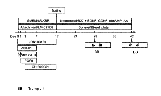
[0012]
16 Fig. 1 shows an example of the protocol for producing dopaminergic
cells. "Y"
17 represents Y-27632, and "AA" represents ascorbic acid.
18 Fig. 2 shows a graph showing the proportion of Tra-1-60-positive cells,
proportion of
19 PSA-NCAM-positive cells, and proportion of Corin-positive cells on Day
12 when differentiation
induction was carried out using MG (Matrigel), CS (CELLStart), LM111 (Laminin
111E8), or
21 LM511 (Laminin 511E8) as a coating agent.
22 Fig. 3 shows phase-contrast images (photographs) of human iPS cells
(83663) and the
23 cells during a differentiation induction process. An image obtained
before the differentiation
24 induction (left panel), an image obtained immediately after the
differentiation induction (day0)
(middle panel), and an image obtained 12 days after the induction (day12)
(right panel) are
26 shown.
27 Fig. 4 shows changes in the proportions of Corin-positive cells
(circle), PSA-NCAM-
28 positive cells (square), and Tra-1-60-positive cells (triangle). On Day
12 and later, the results
29 were obtained under conditions without sorting.
Fig. 5 shows changes in the expression levels of undifferentiation markers and
31 differentiation markers. Each value is represented as a relative value
with respect to the value
32 on Day 0, which is taken as 1. On Day 12 in Fig. 5A, values for Sox1,
hGSC, Sox17,
33 Brachyury, Nanog, and Oct4 are shown. On Day 42 in Fig. 5B, values for
Lmx1a, TH, Foxa2,
34 Nurr1, Map2ab, En1, and Oct4 are shown.
5
22885537.1
CA 02923592 2016-03-07
CA Application
Blakes Ref. 13206/00001
1 Fig. 6 shows immunostaining images (photographs) of cells on Day 42
during culture on
2 a poly-L-ornithine /laminin/fibronectin coating.
3 Fig. 7 shows results of analysis of gene expression on Day 12 (day12) in
Corin-positive
4 cells and Corin-negative cells obtained by sorting using an anti-Corin
antibody, and unsorted
cells. Fig. 7A shows the expression levels of Lmx1a, En1, Foxa2, Otx2, Gbx2,
and Six3 in each
6 type of cells. Each expression level is represented as a relative value
with respect to the value
7 observed for the unsorted cells (unsorted), which is taken as 1. Fig. 7B
shows results of
8 microarray analysis for comparison of expression between unsorted cells
(unsorted) and Corin-
9 positive cells (Corin+) on Day 12.
Fig. 8 shows results of analysis of gene expression on Day 12 in Corin-
positive cells
11 (Corin+) and Corin-negative cells (Corin-) obtained by sorting using an
anti-Corin antibody, and
12 unsorted cells (Unsorted). Fig. 8A shows trichrome staining images
(photographs) for
13 Foxa2/Lmx1a (upper panels) and Otx2/Lmx1a/DAPI (lower panels) in each
type of cells. Fig.
14 8B shows the proportion of Foxa2-positive/Lmx1a-positive cells in each
type of cells. Fig. 8C
shows the proportion of 01x2-positive/Lmx1a-negative cells in each type of
cells. Fig. 8D shows
16 the expression levels of Oct4 and Nanog in each type of cells, and in
the cells before the
17 differentiation induction.
18 Fig. 9 shows results of analysis of gene expression on Day 21 in Corin-
positive cells
19 (Corin+) and Corin-negative cells (Corin-) obtained by sorting using an
anti-Corin antibody, and
unsorted cells (unsorted). Fig. 9A shows the expression levels of Lmx1a, En1,
Foxa2, Otx2,
21 Gbx2, and Six3 in each type of cells. Each expression level is
represented as a relative value
22 with respect to the value observed for the unsorted cells (unsorted),
which is taken as 1. Fig.
23 9B shows double-staining images (photographs) for Corin/Lmx1a (upper
panel) and
24 Foxa2/Lmx1a (lower panels) in each type of cells. Fig. 9C shows the
proportion of Foxa2-
positive/Lmx1a-positive cells in each type of cells.
26 Fig. 10 shows results of analysis of the sizes of cell clusters
(spheres) on Day 28
27 (day28) and Day 42 (day42) after the differentiation induction. Fig. 10A
shows phase-contrast
28 images (photographs) of the cell clusters. Fig. 10B shows a graph
showing the diameters of the
29 cell clusters.
Fig. 11 shows results of analysis of gene expression on Day 28 (day28) and Day
42
31 (day42) after the differentiation induction. Fig. 11A shows
immunostaining images
32 (photographs) for Foxa2/DAPI and Nurr1/TH (tyrosine hydroxylase). Fig.
11B shows the
33 proportions of Nurr1-positive cells, Foxa2-positive cells, and TH-
positive cells on Day 28 (left
34 panel) and Day 42 (right panel). Fig. 11C shows the amounts of dopamine
(DA), 3,4-
6
22885537.1
CA 02923592 2016-03-07
CA Application
Blakes Ref. 13206/00001
1 dihydroxyphenyl acetic acid (DOPAC), and serotonin (5-HT) released from
1x10⁶ Corin+ cells or
2 unsorted cells on Day 42.
3 Fig. 12 shows the states of transplants at Week 16 after intracerebral
transplantation of
4 cells obtained by culturing Corin-positive cells collected by sorting
(Day 12) (Corin+), or cells
induced without sorting (Unsorted), to rats (to which 6-hydroxydopamine (6-
0HDA) was
6 administered) on Day 28 after differentiation induction. Fig. 12A shows
immunostaining images
7 (photographs) of GFAP and Ki67, and human nuclei in the transplants.
Magnified images are
8 separately provided for the areas surrounded by frames. Figs. 12B, 12C,
and 12D show graphs
9 prepared by plotting the sizes of the transplants in cases of
transplantation of cells on Day 28,
Day 42, and Day 19, respectively. Fig. 12E shows the proportions of Ki67-
positive cells in the
11 transplants in the cases of transplantation of cells on Day 28.
12 Fig. 13 shows results of intracerebral transplantation of cells obtained
by culturing Corin-
13 positive cells collected by sorting (day 12) (Corin+), cells induced
without sorting (Unsorted), or a
14 negative control (Medium), to rats (to which 6-0HDA was administered) on
Day 28 (day28) or
Day 42 (day42) after the differentiation induction. Fig. 13A shows the number
of times of
16 circling behavior per unit time in each period after the transplantation
of the cells on Day 28.
17 Fig. 13B shows a graph prepared by plotting the number of TH-positive
cells per transplant in
18 the cases of administration of the cells on Day 28. Fig. 13C shows
immunostaining images
19 (photographs) of TH (red) and human nuclei (green) in brain in the cases
of administration of
the cells on Day 28. Fig. 13D shows the number of times of circling behavior
per unit time in
21 each period after the transplantation of the cells on Day 42. Fig. 13E
shows a graph prepared
22 by plotting the number of TH-positive cells per transplant in the cases
of administration of the
23 cells on Day 42. Fig. 13F shows immunostaining images (photographs) of
TH (red) and human
24 nuclei (green) in brain in the cases of administration of the cells on
Day 42.
Fig. 14 shows results of analysis of transplants in the cases of
transplantation of the
26 cells on Day 28. Fig. 14A shows a graph prepared by plotting the number
of TH-positive cells
27 per neural cell (NauN+). Fig. 14B shows a graph prepared by plotting the
number of TH-positive
28 cells per donor cell (human nuc+). Fig. 14C shows double-staining images
(photographs) for
29 Foxa2/TH, Pitx3/TH, Nurr1/TH, and Girk2/TH in transplants.
Fig. 15 shows results of intracerebral transplantation of cells obtained by
culturing Corin-
31 positive cells collected by sorting (day 12) (Corin+), or cells induced
without sorting (Unsorted),
32 to rats (to which 6-0HDA was administered) on Day 28 (day28) after the
differentiation
33 induction. Fig. 15A shows a double-staining image (photograph) for
serotonin (green)/TH(red)
34 in a transplant of each type of cells at Week 16. Fig. 15B shows the
proportion of serotonin-
7
22885537.1
CA 02923592 2016-03-07
CA Application
Blakes Ref. 13206/00001
1 positive cells among surviving cells (NeuN-positive cells) at Week 16,
which proportion was
2 investigated for each type of cells.
3 Fig. 16 shows results of analysis of gene expression in cells on Day 28,
cells on Day 42,
4 and fetal ventral mesencephalic cells. Fig. 16A shows a result of
comparison of the expression
between cells subjected to sorting for Corin-positive cells on Day 12 (d28
Corin+) and unsorted
6 cells (d28 Unsorted) (left panel), and a result of comparison of the
expression between cells on
7 Day 28 after the induction (d28 Corin+) and cells on Day 42 after the
induction, which cells on
8 Day 28 and Day 42 had been sorted for Corin-positive cells (right panel).
Each comparison was
9 made using a microarray. Fig. 16B shows results of measurement of CORIN,
LMX1A, FOXA2,
NURR1, TH, and TPH2 in the cells on Day 28 (d28 Corin+), the cells on Day 42
(d42 Corin+),
11 and the fetal ventral mesencephalic cells (VM) using PCR. Each value is
shown as a relative
12 value with respect to the value for the fetal ventral mesencephalic
cells (VM), which is taken as
13 1. Fig. 16C shows a result of cluster analysis of the microarray data
for the cells on Day 28
14 which had been sorted for Corin-positive cells (d28 Corin+), unsorted
cells on Day 28 (d28
Unsorted), sorted cells on Day 42 (d42 Corin+), unsorted cells on Day 42 (d42
Unsorted), and
16 fetal ventral mesencephalic cells (VM).
17 Fig. 17 shows staining images of cells obtained by culturing Lrtm1-
positive cells
18 collected by sorting (Day 14). Fig. 17A shows images (photographs)
obtained by staining with
19 Foxa2 and/or Lmx1a, and DAPI seven days after the sorting. The number in
each panel
represents the proportion of positive cells. In the case of double staining,
the number
21 represents the proportion of double-positive cells. Fig. 17B shows
staining images
22 (photographs) for markers (Foxa2, Nurr1, and TH) taken after 21 days of
culture of cells sorted
23 for Lrtm1-positive cells (Lrtm1+) or unsorted cells (Unsort). The number
in each panel
24 represents the proportion of positive cells. In the cases of double
staining, each number
represents the proportion of double-positive cells.
26 Fig. 18 shows immunostaining images (photographs) of transplants for
markers (Foxa2,
27 TH, and Nurr1), which images were taken after transplantation of the
cells obtained by one-day
28 culture of Lrtm1-positive cells collected by sorting (Day 14) to SD rats
of 10 weeks old.
29 Fig. 19-1 shows staining images of cells obtained by collecting Lrtm1-
positive cells by
sorting and further culturing the collected cells (the images are arranged to
show, from left to
31 right, Foxa2/DAPI, Nurr1/DAPI, TH/DAPI, Foxa2+TH+/DAPI, Nurr1+TW/DAPI,
and
32 Foxa2+Nurr1+TH+/DAPI). The number in each panel represents the
proportion of positive cells
33 in the staining image. This figure shows staining images of cells which
were prepared by
8
22885537.1
CA 02923592 2016-03-07
CA Application
Blakes Ref. 13206/00001
1 sorting on Day 14 of the differentiation induction and culturing of the
sorted cells for 7 days
2 thereafter.
3 Fig. 19-2 shows staining images of cells obtained by collecting Lrtm1-
positive cells by
4 sorting and further culturing the collected cells (the images are
arranged to show, from left to
right, Foxa2/DAPI, Nurr1/DAPI, TH/DAPI, Foxa2+TH+/DAPI, Nurr1+TH+/DAPI, and
6 Foxa2+Nurr1+TW/DAPI). The number in each panel represents the proportion
of positive cells
7 in the staining image. This figure shows staining images of cells which
were prepared by
8 sorting on Day 14 of the differentiation induction and culturing of the
sorted cells for 14 days
9 thereafter.
Fig. 19-3 shows staining images of cells obtained by collecting Lrtm1-positive
cells by
11 sorting and further culturing the collected cells (the images are
arranged to show, from left to
12 right, Foxa2/DAPI, Nurr1/DAPI, TH/DAPI, Foxa2+TRIDAPI, Nurr1+TH+/DAPI,
and
13 Foxa2+Nurr1+TH+/DAPI). The number in each panel represents the
proportion of positive cells
14 in the staining image. This figure shows staining images of cells which
were prepared by
sorting on Day 21 of the differentiation induction and culturing of the sorted
cells for 7 days
16 thereafter.
17
18 EMBODIMENTS FOR CARRYING OUT THE INVENTION
19 [0013]
The present invention provides a method for producing dopaminergic neuron
progenitor
21 cells from pluripotent stem cells, which method comprises the steps of:
22 (i) performing adherent culture of pluripotent stem cells on an
extracellular matrix in a
23 medium containing a reagent(s) selected from the group consisting of BMP
inhibitor, TGFI3
24 inhibitor, SHH signal-stimulating agent, FGF8, and GSK313 inhibitor;
(ii) collecting Corin- and/or Lrtm1-positive cells from the cells obtained in
the Step (i);
26 and
27 (iii) performing suspension culture of the cells obtained in Step (ii)
in a medium
28 containing a neurotrophic factor.
29
[0014]
31 <Pluripotent Stem Cells>
32 The pluripotent stem cells which may be used in the present invention
are stem cells
33 having pluripotency which enables the cells to differentiate into any
cells existing in the living
34 body, which pluripotent stem cells also have growth ability. Examples of
the pluripotent stem
9
22885537.1
CA 02923592 2016-03-07
CA Application
Blakes Ref. 13206/00001
1 cells include, but are not limited to, embryonic stem (ES) cells,
embryonic stem cells derived
2 from a cloned embryo obtained by nuclear transfer ("ntES cells"),
germline stem cells ("GS
3 cells"), embryonic germ cells ("EG cells"), induced pluripotent stem
(iPS) cells, and pluripotent
4 cells derived from cultured fibroblasts and bone marrow stem cells (Muse
cells). The pluripotent
stem cells are preferably ES cells, ntES cells or iPS cells.
6
7 [0015]
8 (A) Embryonic Stem Cells
9 ES cells are stem cells established from the inner cell mass of an early
embryo (for
example, blastocyst) of a mammal such as human or mouse, which cells have
pluripotency and
11 growth ability by self-renewal.
12 ES cells are embryo-derived stem cells originated from the inner cell
mass of a
13 blastocyst, which is the embryo formed following the 8-cell stage and
the morula stage of a
14 fertilized egg. ES cells have ability to differentiate into any cells
constituting an adult, that is, the
so-called pluripotency of differentiation, and growth ability by self-renewal.
ES cells were
16 discovered in mouse in 1981 (M. J. Evans and M. H. Kaufman (1981),
Nature 292:154-156),
17 and this was followed by establishment of ES cell lines of primates such
as human and monkey
18 (J. A. Thomson et al. (1998), Science 282:1145-1147; J. A. Thomson et
al. (1995), Proc. Natl.
19 Acad. Sci. USA, 92:7844-7848; J. A. Thomson et al. (1996), Biol.
Reprod., 55:254-259; J. A.
Thomson and V. S. Marshall (1998), Curr. Top. Dev. Biol., 38:133-165).
21
22 [0016]
23 ES cells can be established by removing the inner cell mass from the
blastocyst of a
24 fertilized egg of a subject animal, followed by culturing the inner cell
mass on feeder fibroblasts.
The cells can be maintained by subculture using a medium supplemented with a
substance(s)
26 such as leukemia inhibitory factor (LIF) and/or basic fibroblast growth
factor (bFGF). Methods
27 of establishment and maintenance of human and monkey ES cells are
described in, for
28 example, US 5,843,780 B; Thomson JA, et al. (1995), Proc Natl. Acad.
Sci. U S A. 92:7844-
29 7848; Thomson JA, et al. (1998), Science. 282:1145-1147; H. Suemori et
al. (2006), Biochem.
Biophys. Res. Commun., 345:926-932; M. Ueno et al. (2006), Proc. Natl. Acad.
Sci. USA,
31 103:9554-9559; H. Suemori et al. (2001), Dev. Dyn., 222:273-279; H.
Kawasaki et al. (2002),
32 Proc. Natl. Acad. Sci. USA, 99:1580-1585; and Klimanskaya I, et al.
(2006), Nature. 444:481-
33 485.
34
22885537.1
CA 02923592 2016-03-07
CA Application
Blakes Ref. 13206/00001
1 [0017]
2 In terms of the medium for preparation of ES cells, human ES cells can
be maintained,
3 for example, using DMEM/F-12 medium supplemented with 0.1 mM 2-
mercaptoethanol, 0.1 mM
4 non-essential amino acids, 2 mM L-glutamic acid, 20% KSR, and 4 ng/ml
bFGF at 37 C under a
moist atmosphere of 2% CO2/98% air (0. Fumitaka et al. (2008), Nat.
Biotechnol., 26:215-224).
6 ES cells need to be subcultured every 3 to 4 days, and the subculture can
be carried out using
7 0.25% trypsin and 0.1 mg/ml collagenase IV in PBS supplemented with 1 mM
CaCl2 and 20%
8 KSR.
9
[0018]
11 Selection of ES cells can be generally carried out by the Real-Time PCR
method using
12 as an index/indices expression of a gene marker(s) such as alkaline
phosphatase, Oct-3/4,
13 and/or Nanog. In particular, for selection of human ES cells, expression
of a gene marker(s)
14 such as OCT-3/4, NANOG, and/or ECAD can be used as an index/indices (E.
Kroon et al.
(2008), Nat. Biotechnol., 26:443-452).
16 In terms of human ES cell lines, for example, WA01(H1) and WA09(H9) can
be obtained
17 from WiCell Research Institute, and KhES-1, KhES-2, and KhES-3 can be
obtained from
18 Institute for Frontier Medical Sciences, Kyoto University (Kyoto,
Japan).
19
[0019]
21 (B) Germline Stem Cells
22 Germline stem cells are pluripotent stem cells derived from testis, and
play a role as the
23 origin for spermatogenesis. Similarly to ES cells, these cells can be
induced to differentiate into
24 various series of cells, and, for example, have a property to enable
preparation of a chimeric
mouse by transplantation of the cells to a mouse blastocyst (M. Kanatsu-
Shinohara et al. (2003)
26 Biol. Reprod., 69:612-616; K. Shinohara et al. (2004), Cell, 119:1001-
1012). Germline stem
27 cells are capable of self-renewal in a medium containing glial cell line-
derived neurotrophic
28 factor (GDNF), and, by repeating subculture under the same culture
conditions as those for ES
29 cells, germline stem cells can be obtained (Masanori Takehashi et al.
(2008), Experimental
Medicine, 26(5) (extra edition):41-46, Yodosha (Tokyo, Japan)).
31
32 [0020]
33 (C) Embryonic Germ Cells
1
22885537.1
CA 02923592 2016-03-07
CA Application
Blakes Ref. 13206/00001
1 Embryonic germ cells are established from fetal primordial germ cells
and have
2 pluripotency similarly to ES cells. They can be established by culturing
primordial germ cells in
3 the presence of substances such as LIF, bFGF, and stem cell factor (Y.
Matsui et al. (1992),
4 Cell, 70:841-847; J. L. Resnick et al. (1992), Nature, 359:550-551).
6 [0021]
7 (D) Induced Pluripotent Stem Cells
8 Induced pluripotent stem (iPS) cells can be prepared by introducing
specific
9 reprogramming factors to somatic cells, which reprogramming factors are
in the form of DNA or
protein. iPS cells are somatic cell-derived artificial stem cells having
properties almost
11 equivalent to those of ES cells, such as pluripotency of differentiation
and growth ability by self-
12 renewal (K. Takahashi and S. Yamanaka (2006) Cell, 126:663-676; K.
Takahashi et al. (2007),
13 Cell, 131:861-872; J. Yu et al. (2007), Science, 318:1917-1920;
Nakagawa, M. et al., Nat.
14 Biotechnol. 26:101-106 (2008); WO 2007/069666). The reprogramming
factors may be
constituted by genes or gene products thereof, or non-coding RNAs, which are
expressed
16 specifically in ES cells; or genes or gene products thereof, non-coding
RNAs, or low molecular
17 weight compounds, which play important roles in maintenance of the
undifferentiated state of
18 ES cells. Examples of the genes included in the reprogramming factors
include 0ct3/4, Sox2,
19 Sox1, Sox3, Sox15, Sox17, Klf4, K1f2, c-Myc, N-Myc, L-Myc, Nanog, Lin28,
Fbx15, ERas,
ECAT15-2, Tc11, beta-catenin, Lin28b, Sa111, Sa114, Esrrb, Nr5a2, Tbx3, and
Glis1, and these
21 reprogramming factors may be used either individually or as a
combination of two or more of
22 these. Examples of the combinations of the reprogramming factors include
those described in
23 WO 2007/069666; WO 2008/118820; WO 2009/007852; WO 2009/032194; WO
2009/058413;
24 WO 2009/057831; WO 2009/075119; WO 2009/079007; WO 2009/091659; WO
2009/101084;
WO 2009/101407; WO 2009/102983; WO 2009/114949; WO 2009/117439; WO
2009/126250;
26 WO 2009/126251; WO 2009/126655; WO 2009/157593; WO 2010/009015; WO
2010/033906;
27 WO 2010/033920; WO 2010/042800; WO 2010/050626; WO 2010/056831; WO
2010/068955;
28 WO 2010/098419; WO 2010/102267; WO 2010/111409; WO 2010/111422; WO
2010/115050;
29 WO 2010/124290; WO 2010/147395; WO 2010/147612; Huangfu D, et al.
(2008), Nat.
Biotechnol., 26: 795-797; Shi Y, et al. (2008), Cell Stem Cell, 2: 525-528;
Eminli S, et al. (2008),
31 Stem Cells. 26:2467-2474; Huangfu D, et al. (2008), Nat Biotechnol.
26:1269-1275; Shi Y, et al.
32 (2008), Cell Stem Cell, 3, 568-574; Zhao Y, et al. (2008), Cell Stem
Cell, 3:475-479; Marson A,
33 (2008), Cell Stem Cell, 3, 132-135; Feng B, et al. (2009), Nat Cell
Biol. 11:197-203; R.L. Judson
34 et al., (2009), Nat. Biotech., 27:459-461; Lyssiotis CA, et al. (2009),
Proc Natl Acad Sci U S A.
12
22885537.1
CA 02923592 2016-03-07
CA Application
Blakes Ref. 13206/00001
1 106:8912-8917; Kim JB, et al. (2009), Nature. 461:649-643; lchida JK, et
al. (2009), Cell Stem
2 Cell. 5:491-503; Heng JC, et al. (2010), Cell Stem Cell. 6:167-74; Han J,
et al. (2010), Nature.
3 463:1096-100; Mali P, et al. (2010), Stem Cells. 28:713-720; and Maekawa
M, et al. (2011),
4 Nature. 474:225-9.
6 [0022]
7 Examples of the above-described reprogramming factors also include
histone
8 deacetylase (HDAC) inhibitors [for example, low molecular weight
inhibitors such as valproic
9 acid (VPA), trichostatin A, sodium butyrate, MC 1293, and M344; and
nucleic acid-type
expression inhibitors such as siRNAs and shRNAs against HDAC (e.g., HDAC1
siRNA
11 Smartpool (Millipore) and HuSH 29mer shRNA Constructs against HDAC1
(OriGene))], MEK
12 inhibitors (for example, PD184352, PD98059, U0126, SL327, and
PD0325901), glycogen
13 synthase kinase-3 inhibitors (for example, Bio and CHIR99021), DNA
methyltransferase
14 inhibitors (for example, 5'-azacytidine), histone methyltransferase
inhibitors (for example, low
molecular weight inhibitors such as BIX-01294; and nucleic acid-type
expression inhibitors such
16 as siRNAs and shRNAs against Suv39h1, Suv39h2, SetDBI, and G9a), L-
channel calcium
17 agonists (for example, Bayk8644), butyric acid, TGFI3 inhibitors or ALK5
inhibitors (for example,
18 LY364947, SB431542, 616453, and A83-01), p53 inhibitors (for example,
siRNAs and shRNAs
19 against p53), ARID3A inhibitors (for example, siRNAs and shRNAs against
ARID3A), miRNAs
such as miR-291-3p, miR-294, miR-295, and mir-302, Wnt Signaling (for example,
soluble
21 Wnt3a), neuropeptide Y, prostaglandins (for example, prostaglandin E2
and prostaglandin J2),
22 hTERT, SV4OLT, UTF1, IRX6, GLISI, PITX2, and DMRTBI, which are employed
for enhancing
23 the establishment efficiency, and, in the present description, these
factors employed for the
24 purpose of enhancement of the establishment efficiency are not
particularly distinguished from
reprogramming factors.
26
27 [0023]
28 In cases where the reprogramming factors are in the form of protein, the
reprogramming
29 factors may be introduced into somatic cells by a method such as
lipofection, fusion with a cell
membrane-permeable peptide (e.g., HIV-derived TAT or polyarginine), or
microinjection.
31
32 [0024]
33 In cases where the reprogramming factors are in the form of DNA, the
reprogramming
34 factors may be introduced into somatic cells by a method such as use of
a vector including
13
22885537.1
CA 02923592 2016-03-07
CA Application
Blakes Ref. 13206/00001
1 virus, plasmid, and artificial chromosome vectors; lipofection; use of
liposome; or microinjection.
2 Examples of the virus vectors include retrovirus vectors, lentivirus
vectors (these are described
3 in Cell, 126, pp. 663-676, 2006; Cell, 131, pp. 861-872, 2007; and
Science, 318, pp. 1917-1920,
4 2007), adenovirus vectors (Science, 322, 945-949, 2008), adeno-associated
virus vectors, and
Sendai virus vectors (WO 2010/008054). Examples of the artificial chromosome
vectors include
6 human artificial chromosomes (HACs), yeast artificial chromosomes (YACs),
and bacterial
7 artificial chromosomes (BACs and PACs). Examples of the plasmids which
may be used
8 include plasmids for mammalian cells (Science, 322:949-953, 2008). The
vectors may contain
9 a regulatory sequence(s) such as a promoter, enhancer, ribosome binding
sequence,
terminator, and/or polyadenylation site for allowing expression of the nuclear
reprogramming
11 substances; and, as required, a sequence of a selection marker such as a
drug resistance gene
12 (e.g., kanamycin-resistant gene, ampicillin-resistant gene, or puromycin-
resistant gene),
13 thymidine kinase gene, or diphtheria toxin gene; a gene sequence of a
reporter such as the
14 green-fluorescent protein (GFP), p-glucuronidase (GUS), or FLAG; and/or
the like. Further, in
order to remove, after introduction of the above vector into somatic cells,
the genes encoding
16 the reprogramming factors, or both the promoters and the genes encoding
the reprogramming
17 factors linked thereto, the vector may have LoxP sequences upstream and
downstream of these
18 sequences.
19
[0025]
21 In cases where the reprogramming factors are in the form of RNA, each
reprogramming
22 factor may be introduced into somatic cells by a method such as
lipofection or microinjection,
23 and RNA in which 5-methylcytidine and pseudouridine (TriLink
Biotechnologies) are
24 incorporated may be used in order to suppress degradation (Warren L,
(2010) Cell Stem Cell.
7:618-630).
26
27 [0026]
28 Examples of the medium for induction of the iPS cells include DMEM,
DMEM/F12, and
29 DME media supplemented with 10 to 15% FBS (these media may further
contain LIE,
penicillin/streptomycin, puromycin, L-glutamine, non-essential amino acids, 2-
mercaptoethanol,
31 and/or the like, as appropriate); and commercially available media [for
example, a medium for
32 culturing mouse ES cells (TX-WES medium, Thromb-X), medium for culturing
primate ES cells
33 (medium for primate ES/iPS cells, ReproCELL), and serum-free medium
(mTeSR, Stemcell
34 Technology)].
14
22885537.1
CA 02923592 2016-03-07
CA Application
Blakes Ref. 13206/00001
1 [0027]
2 Examples of the culture method include a method wherein somatic cells
and
3 reprogramming factors are brought into contact with each other at 37 C in
the presence of 5%
4 CO2 on DMEM or DMEM/F12 medium supplemented with 10% FBS, and the cells
are cultured
for about 4 to 7 days, followed by plating the cells on feeder cells (e.g.,
mitomycin C-treated
6 STO cells or SNL cells) and starting culture in a bFGF-containing medium
for culturing primate
7 ES cells about 10 days after the contact between the somatic cells and
the reprogramming
8 factors, thereby allowing iPS-like colonies to appear about 30 to about
45 days after the contact,
9 or later.
11 [0028]
12 Alternatively, the cells may be cultured at 37 C in the presence of 5%
CO2 on feeder
13 cells (e.g., mitomycin C-treated STO cells or SNL cells) in DMEM medium
supplemented with
14 10% FBS (this medium may further contain LIF, penicillin/streptomycin,
puromycin, L-glutamine,
non-essential amino acids, 2-mercaptoethanol, and/or the like, as appropriate)
for about 25 to
16 about 30 days or longer, to allow ES-like colonies to appear. Preferred
examples of the culture
17 method include a method wherein the somatic cells themselves to be
reprogrammed are used
18 instead of the feeder cells (Takahashi K, et al. (2009), PLoS One.
4:e8067 or WO
19 2010/137746), and a method wherein an extracellular matrix (e.g.,
Laminin-5 (WO
2009/123349) or Matrigel (BD)) is used instead.
21
22 [0029]
23 Other examples of the culture method include a method wherein culture is
carried out
24 using a serum-free medium (Sun N, et al. (2009), Proc Natl Acad Sci U
SA. 106:15720-15725).
Further, in order to enhance the establishment efficiency, iPS cells may be
established under
26 low oxygen conditions (at an oxygen concentration of 0.1% to 15%)
(Yoshida Y, et al. (2009),
27 Cell Stem Cell. 5:237-241 or WO 2010/013845).
28
29 [0030]
During the culture, the medium is replaced with a fresh medium once every day
from
31 Day 2 of the culture. The number of the somatic cells used for nuclear
reprogramming is not
32 restricted, and usually within the range of about 5x10³ to about 5x10⁶
cells per 100-cm2 area on
33 the culture dish.
34
22885537.1
CA 02923592 2016-03-07
CA Application
Blakes Ref. 13206/00001
1 [0031]
2 iPS cells can be selected based on the shape of each formed colony. In
cases where a
3 drug resistance gene expressed in conjunction with a gene that is
expressed upon
4 reprogramming of a somatic cell (e.g., Oct3/4 or Nanog) is introduced as
a marker gene,
established iPS cells can be selected by culturing the cells in a medium
containing the
6 corresponding drug (selection medium). iPS cells can be selected by
observation under a
7 fluorescence microscope in cases where the marker gene is the gene of a
fluorescent protein;
8 by adding a luminescent substrate in cases where the marker gene is the
gene of luciferase; or
9 by adding a coloring substrate in cases where the marker gene is the gene
of a coloring
enzyme.
11
12 [0032]
13 The term "somatic cells" used in the present description means any
animal cells
14 (preferably cells of mammals including human) excluding germ-line cells
and totipotent cells
such as eggs, oocytes, and ES cells. Examples of the somatic cells include,
but are not limited
16 to, any of fetal somatic cells, neonatal somatic cells, and mature,
healthy and diseased somatic
17 cells, as well as any of primary cultured cells, subcultured cells, and
established cell lines.
18 Specific examples of the somatic cells include (1) tissue stem cells
(somatic stem cells) such as
19 neural stem cells, hematopoietic stem cells, mesenchymal stem cells, and
dental pulp stem
cells; (2) tissue progenitor cells; and (3) differentiated cells such as
lymphocytes, epithelial cells,
21 endothelial cells, muscle cells, fibroblasts (skin cells and the like),
hair cells, hepatic cells,
22 gastric mucosal cells, enterocytes, spleen cells, pancreatic cells
(pancreatic exocrine cells and
23 the like), brain cells, lung cells, kidney cells, and adipocytes.
24
[0033]
26 In cases where iPS cells are used as a material for the cells to be
transplanted, somatic
27 cells whose HLA genotype is the same or substantially the same as that
of the individual to
28 which the cells are to be transplanted are preferably used in view of
prevention of the rejection
29 reaction. The term "substantially the same" herein means that the HLA
genotype is matching to
an extent at which the immune reaction against the transplanted cells can be
suppressed with
31 an immunosuppressive agent. For example, the somatic cells have matched
HLA types at 3
32 loci HLA-A, HLA-B, and HLA-DR, or at the 4 loci further including HLA-C.
33
34
16
22885537.1
CA 02923592 2016-03-07
CA Application
Blakes Ref. 13206/00001
1 [0034]
2 (E) ES Cells Derived from Cloned Embryo Obtained by Nuclear Transfer
3 ntES cells are ES cells derived from a cloned embryo prepared by the
nuclear transfer
4 technique, and have almost the same properties as those of ES cells
derived from fertilized
eggs (T. Wakayama et al. (2001), Science, 292:740-743; S. Wakayama et al.
(2005), Biol.
6 Reprod., 72:932-936; J. Byrne et al. (2007), Nature, 450:497-502). That
is, an ntES (nuclear
7 transfer ES) cell is an ES cell established from the inner cell mass of a
blastocyst derived from a
8 cloned embryo obtained by replacement of the nucleus of an unfertilized
egg with the nucleus of
9 a somatic cell. For preparation of an ntES cell, the combination of the
nuclear transfer
technique (J.B. Cibelli et al. (1998), Nature Biotechnol., 16:642-646) and the
ES cell preparation
11 technique (described above) is employed (Sayaka Wakayama et al. (2008),
Experimental
12 Medicine 26(5) (extra edition), pp. 47-52). In nuclear transfer,
reprogramming can be achieved
13 by injecting the nucleus of a somatic cell into a mammalian enucleated
unfertilized egg and
14 culturing the resultant for several hours.
16 [0035]
17 (F) Multilineage-differentiating Stress Enduring Cells (Muse Cells)
18 Muse cells are pluripotent stem cells produced by the method described
in WO
19 2011/007900. More specifically, Muse cells are cells having pluripotency
obtained by subjecting
fibroblasts or bone marrow stromal cells to trypsin treatment for a long
period, preferably to
21 trypsin treatment for 8 hours or 16 hours, followed by suspension
culture of the treated cells.
22 Muse cells are positive for SSEA-3 and CD105.
23
24 [0036]
<Dopaminergic Neuron Progenitor Cells>
26 In the present invention, "dopaminergic neuron progenitor cells" also
includes
27 dopaminergic neural cells, dopaminergic neurons, and the like. The
dopaminergic neuron
28 progenitor cells may be a cell population containing other types of
cells. The cell population is
29 preferably a cell population which does not contain a serotonin neural
cell. The dopaminergic
neuron progenitor cells are preferably a cell population containing Foxa2Nurr1-
and/or TH-
31 positive cells. In the present invention, examples of human Foxa2
include the polynucleotides
32 of NCBI accession Nos. NM 021784 and NM 153675, and proteins encoded by
these
33 polynucleotides. In the present invention, examples of human Nurr1
include the polynucleotide
34 of NCBI accession No. NM 006186, and proteins encoded by this
polynucleotide. In the
17
22885537.1
CA 02923592 2016-03-07
CA Application
Blakes Ref. 13206/00001
1 present invention, examples of human TH include the polynucleotides of
NCB! accession Nos.
2 NM 000360, NM_199292, and NM_199293, and proteins encoded by these
polynucleotides.
3
4 [0037]
<Extracellular Matrix>
6 In the present invention, the extracellular matrix is a supramolecular
structure present
7 outside the cell, and may be either a naturally-occurring substance or an
artificial (recombinant)
8 substance. Examples of the extracellular matrix include substances such
as collagen,
9 proteoglycan, fibronectin, hyaluronic acid, tenascin, entactin, elastin,
fibrillin, and laminin, and
fragments thereof. Two or more of these extracellular matrices may be used in
combination.
11 For example, the extracellular matrix may be a product prepared from
cells, such as BD Matrigel
12 (trademark). The extracellular matrix is preferably laminin or a
fragment thereof. The laminin in
13 the present invention is not limited as long as it has a heterotrimeric
structure composed of an
14 a-chain, a 13-chain, and a y-chain. Examples of the a-chain include al,
a2, a3, a4, and a5;
examples of the 13-chain include 131, 132, and 133; and examples of the y-
chain include yl, y2, and
16 y3. The laminin is more preferably laminin 511, which is composed of
a5,131, and yl. The
17 laminin in the present invention may also be a fragment of laminin, and
the fragment is not
18 limited as long as it has an integrin-binding activity. For example, the
fragment of laminin may
19 be E8 fragment, which is a fragment obtained by digestion with elastase.
Accordingly, an
example of the laminin in the present invention is laminin 511E8 (preferably
human laminin
21 511E8), which is described in WO 2011/043405.
22
23 [0038]
24 <BMP Inhibitor>
Examples of the BMP inhibitor in the present invention include protein-based
inhibitors
26 such as Chordin, Noggin and Follistatin; Dorsomorphin (that is, 6-[4-(2-
piperidin-l-yl-
27 ethoxy)phenyI]-3-pyridin-4-yl-pyrazolo[1,5-a]pyrimidine) and its
derivatives (P. B. Yu et al.
28 (2007), Circulation, 116:11_60; P. B. Yu et al. (2008), Nat. Chem.
Biol., 4:33-41; J. Hao et al.
29 (2008), PLoS ONE, 3(8):e2904); and LDN193189 (that is, 4-(6-(4-
(piperazin-1-
yl)phenyl)pyrazolo[1,5-a]pyrimidin-3-yl)quinoline). Dorsomorphin and LDN193189
are
31 commercially available, and can be obtained from Sigma-Aldrich and
Stemgent, respectively.
32 The BMP inhibitor to be used in the present invention may be preferably
LDN193189.
33
34
18
22885537.1
CA 02923592 2016-03-07
CA Application
Blakes Ref. 13206/00001
1 [0039]
2 The concentration of LDN193189 in the medium is not limited as long as
BMP can be
3 inhibited at the concentration. Examples of the concentration of
LDN193189 include, but are
4 not limited to, 1 nM, 10 nM, 50 nM, 100 nM, 500 nM, 750 nM, 1 pM, 2 pM, 3
pM, 4 pM, 5 pM, 6
pM, 7 pM, 8 pM, 9 pM, 10 pM, 15 pM, 20 pM, 25 pM, 30 pM, 40 pM, and 50 pM. The
6 concentration is preferably 100 nM.
7
8 [0040]
9 <TGF[3 Inhibitor>
The TGFp inhibitor in the present invention is a substance which inhibits
signal
11 transduction that proceeds from binding of TGF[3 to its receptor to
SMAD. Examples of the
12 TGFP inhibitor include substances that inhibit binding to the ALK
family, which is a receptor, and
13 substances that inhibit phosphorylation of SMAD by the ALK family.
Specific examples of the
14 TGFP inhibitor include Lefty-1 (e.g., NCBI Accession Nos. NM_010094
(mouse) and
NM 020997 (human)); SB431542 and SB202190 (these are described in R. K.
Lindemann et
16 al., Mol. Cancer, 2003, 2:20); SB505124 (GlaxoSmithKline); NPC30345,
SD093, SD908, and
17 SD208 (Scios); LY2109761, LY364947, and LY580276 (Lilly Research
Laboratories); A83-01
18 (WO 2009146408); and derivatives thereof. The TGFP inhibitor to be used
in the present
19 invention may be preferably A83-01.
21 [0041]
22 The concentration of A83-01 in the medium is not limited as long as ALK5
can be
23 inhibited at the concentration. Examples of the concentration of A83-01
include, but are not
24 limited to, 1 nM, 10 nM, 50 nM, 100 nM, 500 nM, 750 nM, 1 pM, 2 pM, 3
pM, 4 pM, 5 pM, 6 pM,
7 pM, 8 pM, 9 pM, 10 pM, 15 pM, 20 pM, 25 pM, 30 pM, 40 pM, and 50 pM. The
concentration
26 is preferably 500 nM to 5 pM, more preferably 500 nM.
27
28 [0042]
29 <SHH Signal-stimulating Agent>
The SHH (Sonic hedgehog) signal-stimulating agent in the present invention is
defined
31 as a substance that causes disinhibition of Smoothened (Smo) due to
binding of SHH to its
32 receptor, Patched (Ptch1), and also causes activation of G1i2, which
follows the disinhibition.
33 Examples of the SHH signal-stimulating agent include SHH, Hh-Ag1.5 (Li,
X., et al., Nature
34 Biotechnology, 23, 215-221(2005)), Smoothened Agonist, SAG (N-Methyl-N'-
(3-
19
22885537.1
CA 02923592 2016-03-07
CA Application
Blakes Ref. 13206/00001
1 pyridinylbenzy1)-N'43-chlorobenzo[b]thiophene-2-carbony1)-1,4-
diaminocyclohexane), 20a-
2 hydroxycholesterol, purmorphamine, and derivatives thereof (Stanton BZ,
Peng LF., Mol
3 Biosyst. 6:44-54, 2010). The SHH signal-stimulating agent to be used in
the present invention
4 may be preferably purmorphamine.
6 [0043]
7 The concentration of purmorphamine in the medium is not limited as long
as G1i2 can be
8 activated at the concentration. Examples of the concentration of
purmorphamine include, but
9 are not limited to, 1 nM, 10 nM, 50 nM, 100 nM, 500 nM, 750 nM, 1 pM, 2
pM, 3 pM, 4 pM, 5
pM, 6 pM, 7 pM, 8 pM, 9 pM, 10 pM, 15 pM, 20 pM, 25 pM, 30 pM, 40 pM, and 50
pM. The
11 concentration is preferably 2 pM.
12
13 [0044]
14 <GSK313 Inhibitor>
In the present invention, GSK3p inhibitor is defined as a substance which
inhibits kinase
16 activity (for example, ability to phosphorylate p-catenin) of GSK-313
protein. A number of GSK3P
17 inhibitors are known, and examples of the GSK311 inhibitors include BIO
(another name, GSK-
18 33 inhibitor IX; 6-bromoindirubin-3'-oxime), which is an indirubin
derivative; SB216763 (3-(2,4-
19 dichloropheny1)-4-(1-methy1-1H-indol-3-y1)-1H-pyrrole-2,5-dione), which
is a maleimide
derivative; GSK-313 inhibitor VII (4-dibromoacetophenone), which is a phenyl a-
bromomethyl
21 ketone compound; L803-mts (another name, GSK-30 peptide inhibitor; Myr-N-
22 GKEAPPAPPOpSP-NH2 (SEQ ID NO:11), which is a cell membrane-permeable
phosphorylated
23 peptide; and CHI R99021 (64244-(2,4-dichloropheny1)-5-(4-methy1-1H-
imidazol-2-y1)pyrimidin-2-
24 ylamino]ethylamino]pyridine-3-carbonitrile), which has high selectivity.
These compounds are
commercially available from, for example, Calbiochem and Biomol, and can be
easily employed.
26 The compounds may also be obtained from other sources, or may be
prepared. The GSK3P
27 inhibitor to be used in the present invention may be preferably
CHIR99021.
28
29 [0045]
Examples of the concentration of CHIR99021 in the medium include, but are not
limited
31 to, 1 nM, 10 nM, 50 nM, 100 nM, 500 nM, 750 nM, 1 pM, 2 pM, 3 pM, 4 pM,
5 pM, 6 pM, 7 pM,
32 8 pM, 9 pM, 10 pM, 15 pM, 20 pM, 25 pM, 30 pM, 40 pM, and 50 pM. The
concentration is
33 preferably 1 pM.
34
22885537.1
CA 02923592 2016-03-07
CA Application
Blakes Ref. 13206/00001
1 [0046]
2 <FGF8>
3 In the present invention, the FGF8 is not limited, and, in cases of
human FGF8,
4 examples of the FGF8 include the following four splicing forms: FGF8a,
FGF8b, FGF8e, and
FGF8f. The FGF8 in the present invention is more preferably FGF8b. FGF8 is
commercially
6 available from, for example, Wako Pure Chemical Industries, Ltd. and R&D
Systems, Inc., and
7 can be easily employed. The FGF8 may also be obtained by forced
expression in cells by a
8 method known to those skilled in the art.
9
[0047]
11 Examples of the concentration of FGF8 in the medium include, but are not
limited to, 1
12 ng/mL, 5 ng/mL, 10 ng/mL, 50 ng/mL, 100 ng/mL, 150 ng/mL, 200 ng/mL, 250
ng/mL, 500
13 ng/mL, 1000 ng/mL, 2000 ng/mL, and 5000 ng/mL. The concentration is
preferably 100 ng/mL.
14
[0048]
16 <Method for Selecting Cells>
17 In the present invention, the selection of Corin-positive cells and/or
Lrtm1-positive cells
18 from a cell population may be carried out using a substance(s) that
specifically bind(s) to Corin
19 and/or Lrtm1. As a substance that specifically binds to Corin or Lrtm1,
an antibody or an
aptamer may be used. The substance is preferably an antibody or an antigen-
binding fragment
21 thereof.
22
23 [0049]
24 In the present invention, the antibody may be either a polyclonal or
monoclonal antibody.
These antibodies can be prepared using techniques well known to those skilled
in the art
26 (Current protocols in Molecular Biology edit. Ausubel et al. (1987)
Publish. John Wiley and
27 Sons. Sections 11.12-11.13). More specifically, in cases where the
antibody is a polyclonal
28 antibody, the polyclonal antibody can be obtained by allowing E. coli, a
mammalian cell line, or
29 the like to express a protein encoded by Corin or Lrtm1, or an
oligopeptide or a glycolipid having
a partial amino acid sequence thereof, according to a conventional method, and
purifying the
31 resulting expression product, followed by immunization of a non-human
mammal such as a
32 rabbit therewith and isolating the polyclonal antibody from the serum of
the immunized animal
33 according to a conventional method. In cases where the antibody is a
monoclonal antibody, the
34 monoclonal antibody can be obtained from hybridoma cells prepared by
cell fusion of spleen
21
22885537.1
CA 02923592 2016-03-07
CA Application
Blakes Ref. 13206/00001
1 cells obtained from the above-described immunized non-human mammal with
myeloma cells
2 (Current protocols in Molecular Biology edit. Ausubel et al. (1987)
Publish. John Wiley and
3 Sons. Sections 11.4-11.11). Examples of the antigen-binding fragment of
the antibody include
4 fragments of the antibody (e.g., Fab fragment) and synthetic antibody
fragments (e.g., single-
chain Fv fragment "ScFv"). Antibody fragments such as the Fab and F(ab)2
fragments can also
6 be prepared by well known methods in genetic engineering. For example, an
antibody against
7 Corin can be obtained by the preparation methods described in WO
2004/065599 and WO
8 2006/009241, and an antibody against Lrtm1 can be obtained by the
preparation method
9 described in WO 2013/015457.
11 [0050]
12 A sequence of human Corin can be obtained from NCBI accession No.
NM_006587.
13 Similarly, a sequence of human Lrtm1 can be obtained from NM_020678.
14
[0051]
16 For the purpose of recognition or separation of cells expressing Corin
or Lrtm1, the
17 binding substance may be bound or conjugated, for example, to a
detectable substance such as
18 a fluorescent label, radioactive label, chemiluminescent label, enzyme,
biotin, or streptavidin, or
19 to a substance that allows isolation/extraction of the cells, such as
protein A, protein G, beads,
or magnetic beads.
21
22 [0052]
23 Alternatively, the binding substance may be indirectly labeled. The
labeling may be
24 carried out by various methods known to those skilled in the art, and
examples of the methods
include a method in which a preliminarily labeled antibody (secondary
antibody) that specifically
26 binds to the antibody is used.
27
28 [0053]
29 Examples of the method for detecting the dopaminergic neuron progenitor
cells include
use of a flow cytometer, protein chip, or the like.
31
32 [0054]
33 Examples of the method for extracting the dopaminergic neuron progenitor
cells include
34 a method in which the binding substance is conjugated to particles to
cause precipitation of the
22
22885537.1
CA 02923592 2016-03-07
CA Application
Blakes Ref. 13206/00001
1 resulting conjugate, a method in which the cells are sorted using
magnetic beads having
2 magnetism (e.g., MACS), a method in which a fluorescent label and a cell
sorter are used, and
3 a method in which a carrier (e.g., cell-concentrating column) to which an
antibody or the like is
4 immobilized is used.
6 [0055]
7 In the present invention, the aptamer which specifically binds to Corin
or Lrtm1 can be
8 prepared using a technique well known to those skilled in the art (SELEX
(systematic evolution
9 of ligand by exponential enrichment) method: Ellington, A.D. & Szostak,
J. W. (1990) Nature,
346, 818-822; Tuerk, C. & Gold, L. (1990) Science, 249, 505-510).
11
12 [0056]
13 <Neurotrophic Factor>
14 In the present invention, the neurotrophic factor means a ligand for a
membrane
receptor playing an important role in survival and maintenance of the function
of motor neurons.
16 Examples of the neurotrophic factor include Nerve Growth Factor (NGF),
Brain-derived
17 Neurotrophic Factor (BDNF), Neurotrophin 3 (NT-3), Neurotrophin 4/5 (NT-
4/5), Neurotrophin 6
18 (NT-6), basic FGF, acidic FGF, FGF-5, Epidermal Growth Factor (EGF),
Hepatocyte Growth
19 Factor (HGF), Insulin, Insulin Like Growth Factor 1 (IGF 1), Insulin
Like Growth Factor 2 (IGF 2),
Glia cell line-derived Neurotrophic Factor (GDNF), TGF-b2, TGF-b3, Interleukin
6 (IL-6), Ciliary
21 Neurotrophic Factor (CNTF), and LIF. In the present invention, the
neurotrophic factor is
22 preferably a factor selected from the group consisting of GDNF and BDNF.
Neurotrophic
23 factors are commercially available from, for example, Wako Pure Chemical
Industries, Ltd. and
24 R&D Systems, Inc., and can be easily employed. The neurotrophic factor
may also be obtained
by forced expression in cells by a method known to those skilled in the art.
26
27 [0057]
28 Examples of the concentration of GDNF1 in the medium include, but are
not limited to,
29 0.1 ng/mL, 0.5 ng/mL, 1 ng/mL, 5 ng/mL, 10 ng/mL, 15 ng/mL, 20 ng/mL, 25
ng/mL, 30 ng/mL,
40 ng/mL, 50 ng/mL, 100 ng/mL, 200 ng/mL, and 500 ng/mL. The concentration is
preferably
31 10 ng/mL.
32
33
34
23
22885537.1
CA 02923592 2016-03-07
CA Application
Blakes Ref. 13206/00001
1 [0058]
2 Examples of the concentration of BDNF1 in the medium include, but are
not limited to,
3 0.1 ng/mL, 0.5 ng/mL, 1 ng/mL, 5 ng/mL, 10 ng/mL, 15 ng/mL, 20 ng/mL, 25
ng/mL, 30 ng/mL,
4 40 ng/mL, 50 ng/mL, 100 ng/mL, 200 ng/mL, and 500 ng/mL. The
concentration is preferably
20 ng/mL.
6
7 [0059]
8 <Step (i)>
9 In the present invention, the Step (i) is preferably carried out by the
following multistep
process comprising the steps of:
11 (a) performing adherent culture of pluripotent stem cells on an
extracellular matrix in a
12 medium containing BMP inhibitor and a TGFI3 inhibitor;
13 (b) performing adherent culture of the cells obtained in Step (a) on an
extracellular
14 matrix in a medium containing BMP inhibitor, TGFI3 inhibitor, SHH signal-
stimulating agent, and
FGF8;
16 (c) performing adherent culture of the cells obtained in Step (b) on an
extracellular matrix
17 in a medium containing BMP inhibitor, TGFI3 inhibitor, SHH signal-
stimulating agent, FGF8, and
18 GSK313 inhibitor; and
19 (d) performing adherent culture of the cells obtained in Step (c) on an
extracellular matrix
in a medium containing BMP inhibitor and GSK3I3 inhibitor.
21
22 [0060]
23 In the present invention, the medium to be used in Step (i) may be
prepared using a
24 medium for animal cell culture as a basal medium. Examples of the basal
medium include
Glasgow's Minimal Essential Medium (GMEM), IMDM, Medium 199, Eagle's Minimum
Essential
26 Medium (EMEM), aMEM, Dulbecco's modified Eagle's Medium (DMEM), Ham's
F12 medium,
27 RPM! 1640 medium, Fischer's medium, Neurobasal Medium (Life
Technologies), and mixtures
28 of two or more of these media. The medium is preferably GMEM. The medium
may contain
29 serum, or may be serum-free. If necessary, the medium may contain one or
more of serum
replacements such as albumin, transferrin, Knockout Serum Replacement (KSR)
(serum
31 replacement for FBS in ES cell culture), N2 supplement (Invitrogen), B27
supplement
32 (Invitrogen), fatty acids, insulin, collagen precursor, trace elements,
2-mercaptoethanol, and 3'-
33 thiolglycerol, and may also contain one or more of substances such as
lipids, amino acids, L-
34 glutamine, Glutamax (Invitrogen), non-essential amino acids, vitamins,
growth factors, low-
24
22885537.1
CA 02923592 2016-03-07
CA Application
Blakes Ref. 13206/00001
1 molecular-weight compounds, antibiotics, antioxidants, pyruvic acid,
buffers, and inorganic salts.
2 A preferred medium is GMEM, which contains KSR, 2-mercaptoethanol, non-
essential amino
3 acids, and pyruvic acid. The medium may be supplemented, if necessary,
with a reagent(s)
4 selected from the group consisting of BMP inhibitor, TGFp inhibitor, SHH
signal-stimulating
agent, FGF8, and GSK3r3 inhibitor, and used for the culture.
6
7 [0061]
8 The "adherent culture on an extracellular matrix" in Step (i) may be
carried out by
9 culturing the cells using a culture vessel coated with an extracellular
matrix. The coating
treatment may be carried out by placing a solution containing the
extracellular matrix in the
11 culture vessel, and then removing the solution as appropriate.
12
13 [0062]
14 In terms of the culture conditions, the culture temperature is not
limited, and may be
about 30 to 40 C, preferably about 37 C. The culture is carried out under an
atmosphere of
16 CO2-containing air, wherein the CO2 concentration is preferably about 2
to 5%.
17
18 [0063]
19 The culture period is not limited as long as Corin- and/or Lrtm1-
positive cells appear
during the period. Step (i) is preferably carried out for at least 10 days.
The period of Step (i) is
21 more preferably 12 days to 21 days, still more preferably 12 days to 14
days.
22
23 [0064]
24 In Step (i), examples of the period of Step (a) include not less than 1
day, not less than 2
days, not less than 3 days, not less than 4 days, not less than 5 days, not
less than 6 days, not
26 less than 7 days, and periods longer than these. The period of Step (a)
is preferably 1 day.
27 Similarly, examples of the period of Step (b) include not less than 1
day, not less than 2 days,
28 not less than 3 days, not less than 4 days, not less than 5 days, not
less than 6 days, not less
29 than 7 days, and periods longer than these. The period of Step (b) is
preferably 2 days.
Similarly, examples of the period of Step (c) include not less than 1 day, not
less than 2 days,
31 not less than 3 days, not less than 4 days, not less than 5 days, not
less than 6 days, not less
32 than 7 days, and periods longer than these. The period of Step (c) is
preferably 4 days.
33 Similarly, examples of the period of Step (d) include not less than 1
day, not less than 2 days,
34 not less than 3 days, not less than 4 days, not less than 5 days, not
less than 6 days, not less
22885537.1
CA 02923592 2016-03-07
CA Application
Blakes Ref. 13206/00001
1 than 7 days, and periods longer than these. The period of Step (d) is
preferably not less than 5
2 days.
3
4 [0065]
The pluripotent stem cells may be dissociated. Examples of the method for
dissociating
6 the pluripotent stem cells include a method in which the cells are
mechanically dissociated, and
7 a method in which a dissociation solution having protease activity and
collagenase activity (e.g.,
8 Accutase (trademark) or Accumax (trademark)) or a dissociation solution
having only
9 collagenase activity is used. The method is preferably a method in which
human pluripotent
stem cells are dissociated using trypsin or an alternative thereto (e.g.,
TrypLE CTS (Life
11 Technologies)). In cases where the cells are dissociated, it is
preferred to add a ROCK inhibitor
12 as appropriate after the dissociation, followed by performing culture of
the dissociated cells. In
13 cases where a ROCK inhibitor is added, the culture in the presence of
the ROCK inhibitor may
14 be carried out for at least one day. The culture is more preferably
carried out for one day.
16 [0066]
17 <ROCK Inhibitor>
18 In the present invention, the ROCK inhibitor is not limited as long as
the ROCK inhibitor
19 can suppress the function of Rho kinase (ROCK). Examples of the ROCK
inhibitor include Y-
27632 (see, for example, Ishizaki et al., Mol. Pharmacol. 57, 976-983 (2000)
or Narumiya et al.,
21 Methods Enzymol. 325,273-284 (2000)), Fasudil/HA1077 (see, for example,
Uenata et al.,
22 Nature 389: 990-994 (1997)), H-1152 (see, for example, Sasaki et al.,
Pharmacol. Ther. 93:
23 225-232 (2002)), Wf-536 (see, for example, Nakajima et al., Cancer
Chemother Pharmacol.
24 52(4): 319-324 (2003)), and derivatives thereof; antisense nucleic
acids, RNA interference-
inducing nucleic acids (e.g., siRNAs), and dominant negative mutants against
ROCK, and
26 expression vectors therefor. Other low-molecular-weight compounds are
also known as ROCK
27 inhibitors, and these compounds and derivatives thereof may also be used
in the present
28 invention (see, for example, US 20050209261 A, US 20050192304 A, US
20040014755 A, US
29 20040002508 A, US 20040002507 A, US 20030125344 A, US 20030087919 A, WO
2003/062227, WO 2003/059913, WO 2003/062225, WO 2002/076976, and WO
2004/039796).
31 In the present invention, one or more ROCK inhibitors may be used. The
ROCK inhibitor to be
32 used in the present invention may be preferably Y-27632.
33
34
26
22885537.1
CA 02923592 2016-03-07
CA Application
Blakes Ref. 13206/00001
1 [0067]
2 Examples of the concentration of Y-27632 include, but are not limited
to, 100 nM, 500
3 nM, 750 nM, 1pM, 2 pM, 3 pM, 4 pM, 5 pM, 6 pM, 7 pM, 8 pM, 9 pM, 10 pM,
15 pM, 20 pM, 25
4 pM, 30 pM, 40 pM, and 50 pM. The concentration of Y-27632 is preferably
10 pM.
6 [0068]
7 <Step (ii)>
8 The step (ii) of collecting Corin- and/or Lrtm1-positive cells may be
carried out based on
9 the <Method for Selecting Cells> described above.
11 [0069]
12 <Step (iii)>
13 The medium to be used in Step (iii) may be prepared using a medium for
animal cell
14 culture as a basal medium. Examples of the basal medium include
Glasgow's Minimal
Essential Medium(GMEM), IMDM, Medium 199, Eagle's Minimum Essential Medium
(EMEM),
16 aMEM, Dulbecco's modified Eagle's Medium (DMEM), Ham's F12 medium, RPM!
1640
17 medium, Fischer's medium, Neurobasal Medium (Life Technologies), and
mixtures of two or
18 more of these media. The medium is preferably Neurobasal Medium. The
medium may contain
19 serum, or may be serum-free. If necessary, the medium may contain one or
more of serum
replacements such as albumin, transferrin, Knockout Serum Replacement (KSR)
(serum
21 replacement for FBS in ES cell culture), N2 supplement (Invitrogen), B27
supplement
22 (Invitrogen), fatty acids, insulin, collagen precursor, trace elements,
2-mercaptoethanol, and 3'-
23 thiolglycerol, and may also contain one or more of substances such as
lipids, amino acids, L-
24 glutamine, Glutamax (Invitrogen), non-essential amino acids, vitamins,
growth factors, low-
molecular-weight compounds, antibiotics, antioxidants, pyruvic acid, buffers,
inorganic salts, and
26 nucleic acids (for example, dibutyryl cyclic AMP (dbcAMP)). A preferred
medium is Neurobasal
27 Medium supplemented with B27 supplement, ascorbic acid, and dbcAMP. The
medium may be
28 supplemented, if necessary, with a neurotrophic factor(s), and used for
the culture.
29
[0070]
31 The suspension culture in Step (iii) means culturing of the cells in a
state where the cells
32 are not adhering to the culture vessel. The culture vessel that may be
used is not limited, and
33 examples of the culture vessel include culture vessels that are not
artificially treated for the
34 purpose of enhancing adhesiveness to cells (for example, by coating
treatment with an
27
22885537.1
CA 02923592 2016-03-07
CA Application
Blakes Ref. 13206/00001
1 extracellular matrix or the like), and culture vessels that are
artificially treated such that
2 adhesion is suppressed (for example, by coating treatment with a
polyhydroxyethylmethacrylate
3 (poly-HEMA), a nonionic surface-active polyol (e.g., Pluronic F-127), or
a phospholipid analogue
4 (e.g., a water-soluble polymer containing 2-methacryloyloxyethyl
phosphorylcholine as a
constituent (Lipidure)).
6
7 [0071]
8 In terms of the culture conditions, the culture temperature is not
limited, and may be
9 about 30 to 40 C, preferably about 37 C. The culture is carried out under
an atmosphere of
CO2-containing air, wherein the CO2 concentration is preferably about 2 to 5%.
11
12 [0072]
13 The culture period is not limited as long as Nurr1- and/or Foxa2-
positive cells appear
14 during the period. Step (iii) is preferably carried out for at least 7
days. The period of Step (iii)
is more preferably 7 days to 30 days, still more preferably 14 days to 21
days, or 14 days to 16
16 days. The period of Step (iii) is most preferably 16 days.
17
18 [0073]
19 In cases where Step (iii) is carried out after Step (ii), it is
preferred to add a ROCK
inhibitor as appropriate to carry out the culture. In cases where a ROCK
inhibitor is added, the
21 culture in the presence of the ROCK inhibitor may be carried out for at
least one day. The
22 culture is more preferably carried out for one day.
23
24 [0074]
<Therapeutic Agent for Parkinson's Disease>
26 The dopaminergic neuron progenitor cells obtained by the present
invention may be
27 prepared as a formulation for administration to patients with
Parkinson's disease. The
28 administration is carried out by suspending the obtained dopaminergic
neuron progenitor cells in
29 physiological saline or the like and transplanting the resulting
suspension to the striate body
area of the patient. Accordingly, the present invention provides a therapeutic
agent for
31 Parkinson's disease comprising dopaminergic neuron progenitor cells
obtained from pluripotent
32 stem cells by the above-described method.
33
34
28
22885537.1
CA 02923592 2016-03-07
CA Application
Blakes Ref. 13206/00001
1 [0075]
2 In the present invention, the number of dopaminergic neuron progenitor
cells contained
3 in the therapeutic agent for Parkinson's disease is not limited as long
as the transplant can
4 survive after the administration. For example, not less than 15x104 cells
may be contained.
The number of the cells may be increased or decreased as appropriate depending
on
6 symptoms and/or the size of the body.
7
8 [0076]
9 The transplantation of the dopaminergic neuron progenitor cells to the
affected area may
be carried out by a method described in, for example, Nature Neuroscience, 2,
1137 (1999) or N
11 Engl J Med. 344: 710-9 (2001).
12
13 [0077]
14 <Kit>
Other embodiments of the present invention include a kit for preparation of
dopaminergic
16 neuron progenitor cells from pluripotent stem cells. The kit comprises a
medium, additives,
17 culture vessel, and/or the like to be used for the above-described steps
of preparation of
18 dopaminergic neuron progenitor cells. For example, the kit may contain a
reagent(s) selected
19 from the group consisting of anti-Corin antibodies, anti-Lrtm1
antibodies, BMP inhibitor, TGF8
inhibitor, SHH signal-stimulating agent, FGF8, GSK3r3 inhibitor, extracellular
matrices, and
21 neurotrophic factors. The kit may further contain a document or an
instruction in which a
22 procedure for the production process is described.
23
24 [0078]
The present invention is described below more concretely by way of Examples.
26 However, needless to say, the present invention is not limited to these.
27
28 EXAMPLES
29 [0079]
Example 1
31 Cells and Culture
32 Human ES cells (KhES-1) were obtained from Institute for Frontier
Medical Sciences,
33 Kyoto University (Suemori H, et al. Biochem Biophys Res Commun. 345:926-
32, 2006). 404C2
34 and 83663, which are human iPS cells produced by introducing Oct3/4,
Sox2, K1f4, L-MYC,
29
22885537.1
CA 02923592 2016-03-07
CA Application
Blakes Ref. 13206/00001
1 LIN28, and p53shRNA to human fibroblasts using an episomal vector, were
received from Prof.
2 Yamanaka at Kyoto University (Okita et al., Nat Methods. 8: 409-412,
2011).
3
4 [0080]
The ES cells and the iPS cells were cultured by the method according to the
method
6 described in Miyazaki T et al. (Nat Commun. 3:1236, 2012). Briefly, these
cells were cultured
7 in 6-well plates coated with Laminin 511E8.
8
9 [0081]
The thus obtained ES cells or iPS cells were dissociated using TrypLE CTS
(Life
11 Technologies), and transferred to a 6-well plate coated with Laminin
511E8 (iMatrix-511, Nippi)
12 in an amount of 4x10⁴ cells per well. The cells were then cultured by
the above-described
13 culture method for four days, and, after confirming confluence of the
cells, the medium was
14 replaced with Basal Medium A (GMEM (lnvitrogen) supplemented with 8% KSR
(Invitrogen), 1
mM sodium pyruvate (Invitrogen), 0.1 mM MEM non-essential amino acid
(Invitrogen), and 0.1
16 mM 2-Mercaptoethanol (WAKO)) supplemented with 10 pM Y-27632 (WAKO), 0.1
pM
17 LDN193189 (STEMGENT), and 0.5 pM A83-01(WAK0). On the next day (Day 1),
the medium
18 was replaced with Basal Medium A supplemented with 0.1 pM LDN193189, 0.5
pM A83-01, 2
19 pM purmorphamine (WAKO), and 100 ng/mL FGF8 (WAKO). Two days later (Day
3), the
medium was replaced with Basal Medium A supplemented with 0.1 pM LDN193189,
0.5 pM
21 A83-01, 2 pM purmorphamine, 100 ng/mL FGF8, and 3 pM CHIR99021 (WAKO).
Four days
22 later (Day 7), the medium was replaced with Basal Medium A supplemented
with 0.1 pM
23 LDN193189 and 3 pM CHIR99021. Then, replacement of the medium was
carried out once
24 every day. The same experiment was carried out for 12 days except that
Matrigel (BD),
CELLstart (Invitrogen), or Laminin 111E8 was used instead of the coating agent
described
26 above, Laminin 511E8. As a result, PSA-NCAM-positive cells and Corin-
positive cells were
27 obtained with each of the coating agents, but the use of Laminin 511E8
resulted in a smaller
28 number of remaining undifferentiated cells (Tra-1-60-positive cells) and
the highest proportion of
29 Corin-positive cells (Fig. 2). Thus, it was found that, by performing
adherent culture with
Laminin 511E8 coating, a larger number of cells can be handled at one time,
and a higher
31 proportion of desired cells can be achieved. Laminin 511E8 was used as
the coating agent in
32 the following experiments.
33
34
22885537.1
CA 02923592 2016-03-07
CA Application
Blakes Ref. 13206/00001
1 [0082]
2 An anti-Corin antibody was prepared by the following method. First, from
the Corin gene
3 of Macaca fascicularis, a gene sequence encoding a part of the
extracellular domain (79th-
4 453rd amino acids) was introduced into 293E cells, and the cells were
then allowed to express
the extracellular-domain fragment of Corin protein, followed by recovery the
protein. A mouse
6 was immunized with the recovered protein, and lymphocytes were then
extracted and fused with
7 myeloma cells. From the cell population after the fusion, a clone having
reactivity with Corin
8 was selected. The culture supernatant of the clone was used as the anti-
Corin monoclonal
9 antibody.
11 [0083]
12 Five days after the culture in Basal Medium A supplemented with 0.1 pM
LDN193189
13 and 3 pM CHIR99021 (Day 12), the cells were dissociated using TrypLE
CTS, and suspended
14 in Ca2+Mg2+-free HBSS (Invitrogen) supplemented with 2% FBS, 10 pM Y-
27632 (WAKO), 20
mM D-glucose, and 50 pg/ml penicillin/streptomycin. The anti-Corin antibody
was added to the
16 suspension, and the resulting mixture was incubated at 4 C for 20
minutes, followed by sorting
17 the cells using FACSAria II (BD) to collect Corin-positive cells.
18
19 [0084]
The collected Corin-positive cells were transferred to a Lipidure-coat 96 well
plate (NOF
21 Corporation) in an amount of 20,000 cells/well, and subjected to
suspension culture in Basal
22 Medium B (Neurobasal medium (Invitrogen) supplemented with B27
Supplement (without
23 vitamin A: Invitrogen), 20 ng/mL BDNF, 10 ng/mL GDNF, 200 mM ascorbic
acid, and 0.4 mM
24 dbcAMP (Sigma)). In this suspension culture, the initial medium was
supplemented with 30 pM
Y-27632, but a half volume of the initial medium was replaced with Y-27632-
free medium once
26 every three days. Cells obtained 16 days after the sorting (Day 28), and
cells obtained by
27 continuing the suspension culture for additional 14 days (Day 42), were
evaluated for whether
28 they are suitable for transplantation. During the suspension culture, a
half volume of the
29 medium was replaced once every three days. The scheme of this culture
method is shown in
Fig. 1.
31
32 [0085]
33 In addition, suspension culture was carried out in the same manner as
described above
34 except that the sorting for Corin on Day 12 was not carried out, to
provide a control.
31
22885537.1
CA 02923592 2016-03-07
CA Application
Blakes Ref. 13206/00001
1 [0086]
2 Study of Schedule of Sortino
3 Fig. 3 shows images of cells obtained from iPS cells (836B3) by the
method described
4 above, which images were taken at different times during the
differentiation induction. Fig. 4
shows the proportions of Corin-positive cells, polysialylated (PSA)-NCAM-
positive cells, and
6 Tra-1-60-positive cells obtained by performing no sorting on Day 12,
transferring cell clusters
7 obtained on Day 28 to a dish coated with poly-L-ornithine, fibronectin,
and laminin, and
8 performing culture in Basal Medium B for additional 14 days (Day 42). In
this differentiation
9 induction method, the proportion of cells positive for PSA-NCAM, which is
a neural cell marker,
increased from Day 7, and these cells became the majority on Day 12 or near
Day 12. Cells
11 positive for Corin, which is a floor plate marker, appeared on Day 10,
and reached the peak
12 between Days 21 to 28. The same trend was observed also in the case
where 404C2 was
13 used. Thus, it was suggested that, in cases where Corin-positive cells
are obtained by sorting,
14 the sorting is preferably carried out on Day 10 or later.
16 [0087]
17 Subsequently, expression of Oct4, Nanog, Sox1, Sox17, Brachyury, hGSC,
and the like
18 until Day 12 was measured by quantitative PCR. Their changes with time
were as shown in Fig.
19 5A. The expression of SOX1 increased in an early phase, and was
maintained thereafter. The
expression of undifferentiation markers Oct4 and Nanog showed decreases. The
expression of
21 Brachyury, which is a mesodermal marker, transiently increased, and then
decreased. The
22 expression of SOX17, which is an endodermal marker, was constantly low.
Fig. 5B shows
23 changes in the expression of Oct4, Map2ab, Lmx1a, En1, Nurr1, TH, and
Foxa2 measured with
24 time in the cells obtained by performing no sorting on Day 12,
transferring cell clusters obtained
on Day 28 to a dish coated with poly-L-ornithine, fibronectin, and laminin,
and performing culture
26 in Basal Medium B for additional 14 days (Day 42). The expression levels
of the markers
27 described above other than TH were found to have reached the plateau on
Day 12. The same
28 trend was observed also in the case where 404C2 was used.
29
[0088]
31 The cells on Day 42 obtained without performing sorting were subjected
to
32 immunostaining against TH (marker for dopaminergic neural cells) and
against Foxa2 and Nurr1
33 (both are markers for midbrain). As a result, 40% of the cells were
positive for TH, and its
34 coexpression with Foxa2 and Nurr1 was found. The TH-positive cells were
also found to exhibit
32
22885537.1
CA 02923592 2016-03-07
CA Application
Blakes Ref. 13206/00001
1 coexpression of AADC, Pitx3, and Girk2, which are other markers for
dopaminergic neural cells
2 (Fig. 6).
3
4 [0089]
Effect of Sorting Using Corin as Index
6 In the same manner as described above, expression of each marker gene in
the cells
7 immediately after the sorting on Day 12 was measured by PCR (Fig. 7A). As
a result, the
8 Day12-Corin-positive cells showed higher expression of not only Corin,
but also midbrain
9 markers Lmx1a and En1 as well as a floor plate marker Foxa2, compared to
the unsorted cells.
On the other hand, those cells showed lower expression of a hindbrain marker
Gbx2 and a
11 forebrain marker Six3, compared to the unsorted cells. A similar trend
was observed also in the
12 case of 404C2. Comparison by comprehensive expression analysis was made
between the
13 case where the sorting was carried out on Day 12 and the case where the
sorting was not
14 carried out. As a result, higher expression of a rostra! marker Pax6,
caudal marker Foxa2, early
nerve marker Neurog2, postmitotic neural cell marker NEFM (not shown in the
figure), and
16 nonneuronal cell markers DLK1 and CYP1B1, was observed in the unsorted
cells (Fig. 7B). As
17 a result of additional immunostaining, an increased proportion of
Lmx1a/Foxa2-co-positive cells
18 was found among Corin-positive cells (Figs. 8A and 8B; 75.52 8.255%
vs. 47.37 6.624%).
19 On the other hand, cells positive for 01x2, which is expressed in the
rostral side of the
midbrain/hindbrain boundary, and negative for Lmx1a, were found to be
decreased in the sorted
21 cells (Fig. 8C). No large difference was found in the expression of
undifferentiation markers
22 Oct4 and Nanog (Fig. 8D).
23
24 [0090]
Subsequently, a similar study was carried out for the cells immediately after
the sorting
26 on Day 21. Although the proportion of Corin-positive cells was higher in
the case of sorting on
27 Day 21 (45.47 47.61% (day21) vs. 18.97 15.49% (day12)), the
expression levels of Lmx1a
28 and Foxa2 did not show any change due to the sorting on Day 21 (Fig.
9A). Similarly, in the
29 immunostaining, the proportion of Lmx1a/Foxa2-co-positive cells among
Corin-positive cells did
not show a significant difference between the cell population obtained by
carrying out the sorting
31 on Day 21 and the unsorted cell population (Figs. 9B and 9C).
32
33
34
33
22885537.1
CA 02923592 2016-03-07
CA Application
Blakes Ref. 13206/00001
1 [0091]
2 From these results, it was found that sorting on Day 12 is more
preferred than sorting on
3 Day 21.
4
[0092]
6 Culture Period after Sorting
7 Cells on Day 28 and Day 42 induced by the culture method described above
were
8 studied on the difference in the degree of maturation of dopaminergic
neural cells depending on
9 whether the sorting for Corin-positive cells on Day 12 was carried out or
not. On Day 28 and
Day 42, the sphere size of the Corin-positive cells was found to be smaller
compared to that of
11 unsorted cells (Figs. 10A and 10B). As a result of immunostaining on Day
28 and Day 42, the
12 proportion of Foxa2-positive cells in the case where the Day-12 sorting
was carried out was
13 found to be 70 to 75%, and higher than that in the case where no sorting
was carried out (Figs.
14 11A and 11B). The proportions of Nurr1-positive cells and TH-positive
cells were 27.34
5.511% and 2.098 1.243%, respectively, on day 28, and 19.91 6.966% and
42.04 4.481%,
16 respectively, on day 42. Thus, the proportion of TH-positive cells was
higher on Day 42 than on
17 Day 21. As a result of HPLC quantification of dopamine (DA), 3,4-
dihydroxyphenyl acetic acid
18 (DOPAC), and serotonin (5-HT) released into the medium due to
stimulation by high potassium
19 chloride on Day 42, the amounts of DA and DOPAC released were found to
be significantly
larger in the case where the cells were subjected to the Corin sorting,
compared to the case
21 where the Corin sorting was not carried out (Fig. 11C).
22
23 [0093]
24 Subsequently, suspension culture was carried out using the cells that
were subjected to
the Corin sorting on Day 12 or the cells that were not subjected to the Corin
sorting. On Day 28
26 or Day 42, 4x10⁵ cells were suspended in 2 pl of physiological saline,
and administered into the
27 striate body of the cerebrum of model rats for Parkinson's disease (rats
to which 6-0HDA was
28 administered) using a 22-gauge injection needle. At Week 16, observation
of the transplants
29 was carried out (Fig. 12A). As a result of measurement of the sizes of
the transplants derived
from the Day-28 cells, the transplants were found to have almost the same size
(8.5 to 1.5 mm3)
31 in the case where the sorting for Corin-positive cells was carried out
(Fig. 12B). On the other
32 hand, the transplants were found to have large variation in size (88.4
to 0.5mm3) in the case
33 where the Corin sorting was not carried out. A significant difference
was found in the mean size
34 between these cases (34.96 37.52 mm3 (unsorted) vs 3.45 2.932mm3
(sorted)) (Fig. 12B).
34
22885537.1
CA 02923592 2016-03-07
CA Application
Blakes Ref. 13206/00001
1 In addition, suspension culture was carried out using the cells that were
subjected to the Corin
2 sorting on Day 12 or the cells that were not subjected to the Corin
sorting, and the cells were
3 then similarly administered to model rats for Parkinson's disease after
seven days of the culture
4 (Day 19). As a result, the sizes of the transplants were found to be
significantly smaller in the
sorted group (Fig. 12D). As a result of quantification of the proportion of
Ki67-positive cells in
6 the transplants, the proportion was found to be as low as not more than
1% irrespective of
7 whether the sorting was carried out or not. However, the proportion was
significantly lower in
8 the cells that were subjected to the Corin sorting (Fig. 12E). On the
other hand, in the cases of
9 Day 42, the sizes of the transplants were not more than 1 mrna
irrespective of whether the
sorting was carried out or not (Fig. 12C), and no Ki67-positive cells were
found.
11
12 [0094]
13 Subsequently, circling due to amphetamine induction, which is caused by
left-right
14 imbalance of the dopamine level, was studied. As a result, the group to
which the cells sorted
for Corin-positive cells on Day 12 were administered (Day 28) and the group to
which the
16 unsorted cells were administered (Day 28) both showed significantly
lower numbers of times of
17 circling compared to a group to which the cells were not administered
(administration of only the
18 medium). At Week 16 after the administration of the cells, the changes
in the behavior were not
19 significantly different between the case where the sorting was carried
out and the case where
the sorting was not carried out (Fig. 13A). As a result of observation of TH-
positive cells in the
21 transplants at this time, it was found that a significantly larger
number of TH-positive cells were
22 remaining in the case where the cells sorted for Corin were transplanted
(Figs. 13B and 13C;
23 6747 2341 cells/graft (sorted) vs. 3436 2384 cells/graft
(unsorted)). At this time, the number
24 of TH-positive cells per number of NeuN-positive cells (total nerve
cells), as well as the number
of TH-positive cells per number of hNc-positive cells (total surviving cells),
were significantly
26 higher in the case where the sorting was carried out (Figs. 14A and
14B). Coexpression of
27 other markers for dopaminergic cells (Foxa2, Nurr1, and Pilx3) was also
found. Girk2, which is
28 a marker for A9 dopaminergic cells in the substantia nigra pars
compacta, was also positive in a
29 half of the cells (Fig. 14C). As a result of observation of the number
of serotonin cells, the
proportion of these cells was found to be low in both cases (Figs. 15A and
15B).
31
32 [0095]
33 On the other hand, when the cells that were sorted for Corin-positive
cells on Day 12
34 and the unsorted cells were similarly administered (Day 42), no
significant difference in
22885537.1
CA 02923592 2016-03-07
CA Application
Blakes Ref. 13206/00001
1 amelioration of the abnormal circling was found between these cells (Fig.
13D). Similarly, these
2 cells showed no significant difference in the number and concentration of
TH-positive cells in
3 the transplant (Figs. 13E and 13F). In correlation with these results,
the mean number of
4 surviving TH-positive cells was 6747 2341 cells/graft in the case of
transplantation on Day 28
(Fig. 13B), while the mean number was 1431 753.7 cells/graft in the case of
transplantation on
6 Day 42 (Fig. 13E). Thus, the number of surviving cells tended to be
larger in the case of
7 transplantation on Day 28.
8
9 [0096]
Subsequently, comparisons were made by microarray analysis between the cells
on Day
11 28 subjected to the Day-12 sorting and the unsorted cells on Day 28, and
between the cells on
12 Day 28 subjected to the Day-12 sorting and the cells on Day 42 subjected
to the Day-12 sorting
13 (Fig. 16A). The unsorted cells on Day 28 were found to show higher
expression of PAX6, which
14 is a forebrain marker. On the other hand, the sorted cells were found to
show high expression
of ALCAM. In the comparison between the cells on Day 28 and the cells on Day
42, the cells on
16 Day 28 showed higher expression of SHH, WNT5A, and CORIN. On the other
hand, the cells
17 on Day 42 showed higher expression of TH.
18
19 [0097]
A comparison was made between fetal ventral mesencephalic cells (7 weeks old),
which
21 have been clinically used for Parkinson's disease so far, and cells on
Day 28 and Day 42 which
22 were subjected to sorting for Corin-positive cells on Day 12. The fetal
ventral mesencephalic
23 cells showed higher expression of TH and PITX3, which are markers for
dopaminergic neural
24 cells (not shown in the figure), relative to the cells on Day 28. The
fetal ventral mesencephalic
cells also showed higher expression of TPH2, which is a marker for
serotonergic nerve cells
26 (Fig. 16B). As a result of hierarchical cluster analysis, the fetal
ventral mesencephalic cells
27 were found to show higher similarity to the cells on Day 42 (Fig. 16C).
Since the cells on Day
28 28 show expression of genes associated with axon guidance (SPON1 and
SLIT2), it was
29 suggested that the cells are at an earlier developmental stage than
fetal ventral mesencephalic
cells.
31
32 [0098]
33 From the above results, it was suggested that, by using cells (Day 28)
prepared by
34 selecting Corin-positive cells and subjecting the selected cells to
suspension culture for 16 days,
36
22885537.1
CA 02923592 2016-03-07
CA Application
Blakes Ref. 13206/00001
1 the growth of the transplant and the survival of dopaminergic cells can
be increased, so that
2 such cells are suitable for use in treatment of Parkinson's disease.
3
4 [0099]
Example 2
6 Cell Culture
7 ES cells (Kh-ES1) were dissociated using TrypLE CTS (Life
Technologies), and the
8 whole cells were transferred to a 6-well plate coated with Laminin 511E8.
The cells were then
9 cultured in Basal Medium A (GMEM supplemented with 8% KSR, 1 mM sodium
pyruvate, 0.1
mM MEM non-essential amino acid, and 0.1 mM 2-mercaptoethanol) supplemented
with 10 pM
11 Y-27632, 0.1 pM LDN193189, and 0.5 pM A83-01. One day after the
beginning of the culture
12 (Day 1), the medium was replaced with Basal Medium A supplemented with
0.1 pM
13 LDN193189, 0.5 pM A83-01, 2 pM purmorphamine, and 100 ng/mL FGF8. Three
days after the
14 beginning of the culture (Day 3), the medium was replaced with Basal
Medium A supplemented
with 0.1 pM LDN193189, 0.5 pM A83-01, 2 pM purmorphamine, 100 ng/mL FGF8, and
3 pM
16 CHIR99021. Seven days after the beginning of the culture (Day 7), the
medium was replaced
17 with Basal Medium A supplemented with 0.1 pM LDN193189 and 3 pM
CHIR99021. On Day 12
18 (12 days after the beginning of the culture), the medium was replaced
with Neurobasal medium
19 (Invitrogen) supplemented with B27 Supplement (without vitamin A), 2 mM
L-glutamine, 20
ng/mL recombinant human (rh) BDNF, 10 ng/mL rhGDNF, 400 pM dbcAMP (Sigma), and
200
21 pM ascorbic acid. Unless otherwise specified, medium replacement was
carried out every day
22 using the same composition as that used on the previous day.
23
24 [0100]
On Day 14(14 days after the beginning of the culture), the cells were
dissociated using
26 TrypLE CTS, and subjected to sorting using a FACS with an anti-Lrtm1
antibody (WO
27 2013/015457), to recover Lrtm1-positive cells.
. 28 The recovered Lrtm1-positive cells were transferred to a
Lipidure-coat 96 well plate
29 (Thermo) in an amount of 20,000 cells/well, and subjected to suspension
culture using
Neurobasal medium supplemented with 30 pM Y-27632, B27 Supplement (without
vitamin A), 2
31 mM L-glutamine, 20 ng/mL recombinant human (rh) BDNF, 10 ng/mL rhGDNF,
400 pM
32 dbcAMP, 1% KSR, penicillin/streptomycin (Gibco), and 200 pM ascorbic
acid. Thereafter, a half
33 volume of the medium was replaced with Y-27632-free medium once every
three days. The
37
22885537.1
CA 02923592 2016-03-07
CA Application
Blakes Ref. 13206/00001
1 cells were used in experiments 7 days after the sorting (Day 21) or 21
days after the sorting
2 (Day 35).
3
4 [0101]
Effect of Sorting Using Lrtm1 as Index (Immunostaining)
6 Based on investigation of the cells on Day 21 by immunostaining, the
proportion of
7 Foxa2-positive cells was 87.4%; the proportion of Lmx1a-positive cells
was 87.5%; and the
8 proportion of Foxa2-positive/Lmx1a-positive cells was 82.7% (Fig. 17A).
Based on comparison
9 on Day 35 between the cells subjected to the sorting and the unsorted
cells, it was found that
the proportions of Foxa2-, Nurr1-, and TH-positive cells were higher in the
cells subjected to the
11 sorting for Lrtm1(Fig. 17B).
12
13 [0102]
14 Effect of Sorting Using Lrtm1 as Index (Cell Transplantation)
On the next day of the sorting for Lrtm1 (Day 15), 10 to 15x104 cells/tract
were
16 administered into the brain of SD rats of 10 weeks old, followed by
observation at Week 4 after
17 the administration. As a result, survival of Foxa2-, TH-, and Nurr1-
positive cells derived from
18 the transplanted (human-derived) cells was observed (Fig. 18).
19
[0103]
21 Example 3
22 Cell Culture
23 ES cells (Kh-ES1) were dissociated using TrypLE CTS (Life Technologies),
and 4x10⁵
24 cells were then transferred to a 6-well plate coated with Laminin 511E8.
The cells were then
cultured in StemFit medium (Ajinomoto) supplemented with 10 pM Y-27632. Four
days later,
26 the medium was replaced with the above-described Basal Medium A
supplemented with 0.1 pM
27 LDN193189 and 0.5 pM A83-01 (Day 0). One day after the beginning of the
culture (Day 1), the
28 medium was replaced with Basal Medium A supplemented with 0.1 pM
LDN193189, 0.5 pM
29 A83-01, 2 pM purmorphamine, and 100 ng/mL FGF8. Three days after the
beginning of the
culture (Day 3), the medium was replaced with Basal Medium A supplemented with
0.1 pM
31 LDN193189, 0.5 pM A83-01, 2 pM purmorphamine, 100 ng/mL FGF8, and 3 pM
CHIR99021.
32 Seven days after the beginning of the culture (Day 7), the medium was
replaced with Basal
33 Medium A supplemented with 0.1 pM LDN193189 and 3 pM CHIR99021. On Day
14 (14 days
34 after the beginning of the culture), the cells were sorted, or the
medium was replaced with
38
22885537.1
CA 02923592 2016-03-07
CA Application
Blakes Ref. 13206/00001
1 Neurobasal medium (lnvitrogen) supplemented with B27 Supplement (without
vitamin A), 2 mM
2 L-glutamine, 20 ng/mL recombinant human (rh) BDNF, 10 ng/mL rhGDNF, 400
pM dbcAMP
3 (Sigma), and 200 pM ascorbic acid. Unless otherwise specified, medium
replacement was
4 carried out every day using the same composition as that used on the
previous day.
6 [0104]
7 Sorting
8 The cells were cultured by the method described above, and dissociated
on Day 14 (14
9 days after the beginning of the culture) or Day 21(21 days after the
beginning of the culture)
using TrypLE CTS. The cells were then sorted using a FACS with an anti-Lrtm1
antibody (WO
11 2013/015457), to recover Lrtm1-positive cells.
12
13 [0105]
14 The recovered Lrtm1-positive cells were transferred to a Lipidure-coat
96 well plate
(Thermo) in an amount of 2x10⁴ cells/well, and subjected to suspension culture
using
16 Neurobasal medium supplemented with 30 pM Y-27632, B27 Supplement
(without vitamin A), 2
17 mM L-glutamine, 20 ng/mL recombinant human (rh) BDNF, 10 ng/mL rhGDNF,
400 pM
18 dbcAMP, 1% KSR, penicillin/streptomycin (Gibco), and 200 pM ascorbic
acid. Thereafter, a half
19 volume of the medium was replaced with Y-27632-free medium once every
three days. The
cells were used in experiments 7 days or 14 days after the sorting.
21
22 [0106]
23 Effect of Sorting Using Lrtm1 as Index (Immunostaining)
24 Investigation by immunostaining (Foxa2, Nurr1, and TH) was carried out
for the cells of
14 day-7 day (sorting on Day 14, followed by 7 days of culture), 14 day-14 day
(sorting on Day
26 14, followed by 14 days of culture), and 21 day-7 day (sorting on Day
21, followed by 7 days of
27 culture). As a result, the proportion of Foxa2-positive cells was found
to be not less than 90%
28 under any of the above conditions, but the proportion of Nurr1-
positive/TH-positive cells was
29 13.1%, 24.0%, and 10.2% in the cases of 14 day-7 day, 14 day-14 day, and
21 day-7 day,
respectively (Fig. 19). Thus, the proportion of Foxa2, Nurr1, and TH-positive
cells was highest
31 in the case of 14 day-14 day.
32
33
34
39
22885537.1
CA 02923592 2016-03-07
CA Application
Blakes Ref. 13206/00001
1 [0107]
2 Based on the results described above, it was shown that the timing
of the sorting is
3 preferably Day 14 rather than Day 21 after the beginning of the
differentiation induction, and that
4 the culture period after the sorting is preferably 14 days rather than 7
days.
INDUSTRIAL APPLICABILITY
6
7 [0108]
8 The present invention is useful for regenerative medicine,
especially for treatment of
9 Parkinson's disease.
40
22885537.1