Note: Descriptions are shown in the official language in which they were submitted.
81795222
DEVICE AND METHOD FOR REINFORCEMENT OF A FACET
INCORPORATION BY REFERENCE TO ANY PRIORITY APPLICATIONS
[0001] The present application is related to U.S. Application No.
14/274575 filed May 9, 2014, which claims priority to U.S. Provisional Patent
Application No. 61/883960 filed September 27, 2013.
BACKGROUND OF THE INVENTION
[0002] Some embodiments described herein relate generally to methods
and
implants for fusing bone, for example, fusing vertebrae by securing the
articular processes
of the vertebrae.
[0003] Traumatic, inflammatory, and degenerative disorders of the
spine can
lead to severe pain and loss of mobility. One source of back and spine pain is
related to
degeneration of the facets of the spine or facet arthritis. Bony contact or
grinding of
degenerated facet joint surfaces can play a role in some pain syndromes. While
many
technological advances have focused on the intervertebral disc and artificial
replacement
or repair of the intervertebral disc, little advancement in facet repair has
been made. Facet
joint and disc degeneration frequently occur together. Thus, a need exists to
address the
clinical concerns raised by degenerative facet joints.
[0004] The current standard of care to address the degenerative
problems with
the facet joints is to fuse the two adjacent vertebrae. By performing this
surgical
procedure, the relative motion between the two adjacent vertebrae is stopped,
thus
stopping motion of the facets and any potential pain generated as a result
thereof.
Procedures to fuse two adjacent vertebrae often involve fixation and/or
stabilization of the
two adjacent vertebrae until the two adjacent vertebrae fuse.
[0005] Injuries and/or surgical procedure on and/or effecting other
bones can
also result in the desire to fixate and/or stabilize a bone until the bone, or
bone portions,
can fuse, for example, to stabilize a sternum after heart surgery, to
stabilize a rib after a
break, etc. Current procedures to fixate and/or stabilize adjacent vertebrae
and/or other
bones can be slow and/or complex
-1 -
Date Recue/Date Received 2021-04-08
CA 02923623 2016-03-07
WO 2015/047909
PCT/US2014/056598
100061 Accordingly,
a need exists for an apparatus and a procedure to quickly
and/or easily stabilize and/or fixate a bone.
SUMMARY OF THE INVENTION
100071 In some
embodiments, a device for reinforcing a facet joint implant is
provided. The device comprises a First securing segment comprising a proximal
surface
and a distal surface. The first securing segment comprises a first lumen
disposed between
the proximal surface and the distal surface. The first lumen is adapted for
receiving a
fastener member. The device comprises a second securing segment comprising a
proximal
surface and a distal surface. The second securing segment comprises a second
lumen. The
device comprises a central portion between the first securing segment and the
second
securing segment.
100081 In some
embodiments a longitudinal axis of the first securing segment
is disposed at an angle relative to a longitudinal axis of the second securing
segment. In
some embodiments, a plane of the distal surface of the first securing segment
is not
parallel to a plane of the distal surface of the second securing segment. In
some
embodiments, the distal surface of the facet reinforcement device is
configured for
engaging a bony surface of a facet. In some embodiments, the distal surface of
the facet
reinforcement device comprises sharp engagement members.
100091 In some
embodiments, a kit for treating a spine is provided. The kit
comprises a fastener member. The kit comprises a facet reinforcement device.
The facet
reinforcement device comprises a proximal surface and a distal surface. The
facet
reinforcement device comprises a lumen disposed between the proximal surface
and the
distal surface. The lumen is adapted for receiving the fastener member.
100101 In some
embodiments, the facet reinforcement device further
comprises a second portion adapted to attach to a spinous process of a
vertebra. In
embodiments, the second portion of the facet reinforcement device comprises at
least one
lumen. Some embodiments of the kit, further comprise a fastener for securing
the facet
reinforcement device to the vertebra. In some embodiments, the fastener
secures the
facet reinforcement device to the spinous process of the superior vertebra. In
some
embodiments, the fastener is a screw or bolt.
-2-
CA 02923623 2016-03-07
WO 2015/047909
PCT/US2014/056598
100111 In some
embodiments, a method for treating a spine is provided. The
method may include placing a facet reinforcement device comprising a lumen
adjacent to
a first vertebra. The method may include passing a fastener member through the
lumen.
The method includes passing the fastener member through a first articular
process of a
facet joint. The method may include passing the fastener member through a
second
articular process of the facet joint. The method may include securing one end
of the
fastener member to the other end of the fastener member, thereby retaining the
facet
reinforcement device.
[0012] In some
embodiments, a method for treating a spine is provided. The
method may include the step of preparing a facet joint for fixation. The
method may
include passing a fastener member through a first articular process of a facet
joint. The
method may include passing a fastener member through a second articular
process of the
facet joint. The method may include placing a facet reinforcement device with
a lumen for
receiving the flexible fastening band against a surface of the first articular
process. The
method may include passing a fastener member through the lumen. The method may
include securing the fastener member. The method may include securing the
facet
reinforcement device to a spinous process with a fastener. The methods may
further
comprise inserting a facet implant with an interface configured to receive the
fastener
member into the facet joint. The methods may further comprise passing the
fastener
member through the interface of the facet implant.
[0013] In some
embodiments, a method for treating a spine is provided.
Methods may further comprise preparing a second facet joint at a same level of
the spine
for fixation. The method may include placing a second facet reinforcement
device against
a first articular process of the second facet joint. The method may include
passing a
second fastener member through a first articular process of the second facet
joint. The
method may include passing a second fastener member through a second articular
process
of the second facet joint. The method may include securing the second fastener
member.
The method may include securing the second facet reinforcement device to a
spinous
process with a fastener. The methods may further comprise inserting a second
facet
implant with an interface configured to receive the fastener member into the
facet joint.
The methods may further comprise passing the second fastener member through
the
interface of the second facet implant.
-3-
81795222
[0013a] In some embodiments, there is provided a kit for treating a spine,
comprising a
fastener member comprising a proximal end portion and a distal end portion,
the distal end
portion comprising a fastening mechanism configured to accept the proximal end
portion
forming a loop, wherein the fastening mechanism is configured to allow the
proximal end
portion to travel through the distal end portion in only one direction; an
implant configured to
be inserted between a superior articular process and an inferior articular
process, wherein the
implant is configured to be retained within the loop of the fastener member;
and a facet
reinforcement device comprising a proximal surface, a distal surface, and a
lumen disposed
between the proximal and distal surface, the lumen adapted for receiving the
fastener member,
wherein the facet reinforcement device is configured to be retained within the
loop of the
fastener member, wherein the facet reinforcement device is configured to be
disposed on an
outer facing surface of the superior articular process or an outer facing
surface of the inferior
articular process, wherein the facet reinforcement device comprises a
roughened surface,
wherein the proximal surface of the facet reinforcement device comprises a
recess to
mechanically interfit with a portion of the fastener member.
[0013b] In some embodiments, there is provided a kit for treating a spine,
comprising a
fastener member comprising a proximal end portion and a distal end portion,
the distal end
portion comprising a fastening mechanism configured to accept the proximal end
portion,
wherein the fastening mechanism is configured to allow the proximal end
portion to travel
through the distal end portion in only one direction; an implant having an
interface configured
for receiving the fastener member; and a facet reinforcement device configured
to be disposed
on an outer facing surface of a facet, the facet reinforcement device
comprising a proximal
surface, a distal surface, and a lumen disposed between the proximal and
distal surface, the
lumen adapted for receiving the fastener member, wherein at least a portion of
the facet
reinforcement device is malleable to conform to the shape of the facet,
wherein the proximal
surface comprises a first material and the distal surface comprises a second
material, different
than the first material.
[0013c] In some embodiments, there is provided a kit for treating a spine,
comprising
an implant with a first face and a second face adapted to contact the bony or
cartilaginous
articular surfaces of the facets of adjacent vertebrae, wherein the implant
has at least a
- 3a -
Date Recue/Date Received 2022-12-29
81795222
plurality of apertures extending from the first face to the second face; a
fastener member
comprising a proximal end portion and a distal end portion, the fastener
member configured
for generally maintaining the location of the implant with respect to the
facet joint, the
fastener member configured to extend through an aperture in the implant; and a
first enlarged
structure adapted to engage an outer surface of a facet of the facet joint,
wherein the first
enlarged structure has a cylindrical abluminal surface and a centrally located
lumen, wherein
the kit has a first configuration wherein the first enlarged structure is
movable along the
fastener member, and a second configuration wherein the first enlarged
structure is
substantially fixed relative to the fastener member and the first enlarged
structure is
mechanically interfit with the fastener member to increase stability of the
first enlarged
structure and the fastener member; wherein the fastener member is configured
to allow the
proximal end portion to travel through the distal end portion in only one
direction to transition
from the first configuration to the second configuration.
- 3b -
Date Recue/Date Received 2022-12-29
CA 02923623 2016-03-07
WO 2015/047909
PCT/US2014/056598
100141 In some embodiments, a device for placement on a facet joint is
provided, the purpose of the device being to provide reinforcement to the bone
when a
fastener member is used to secure the joint. The device may include sharp
engagement
members on a bone contact side to prevent migration. The device may include a
through-
opening to accept a primary facet fixation device. In some embodiments, the
device for
placement on a facet joint has a second through-opening for accepting at least
one
additional fastener. In some embodiments, a screw may be provided for
placement
through the second through-opening.
BRIEF DESCRIPTION OF THE DRAWINGS
100151 FIG. I is a lateral elevational view of a portion of the
vertebral column.
100161 FIG. 2A is a schematic superior view of an isolated thoracic
vertebra.
100171 FIG. 213 are schematic side view of an isolated thoracic
vertebra.
100181 FIG. 3A is a schematic posterior elevational view of a portion
of the
vertebral column.
[0019] FIG. 3B is a posterior-oblique elevational view of a portion of
the
vertebral column.
100201 FIG. 4A is a schematic side view of a facet joint in the
cervical
vertebrae.
100211 FIG. 4B is a schematic superior view of a facet joint in the
cervical
vertebrae.
[0022] FIG. 5A is a schematic side view of a facet joint in the
thoracic
vertebrae.
[0023] FIG. 5B is a schematic superior view of a facet joint in the
thoracic
vertebrae.
100241 FIG. 6A is a schematic side view of a facet joint in the lumbar
vertebrae.
100251 FIG. 6B is a schematic superior view of a facet joint in the
lumbar
vertebrae.
[0026] FIG. 7 is a block diagram of an implant according to an
embodiment.
[0027] FIGS. 8A and 8B are schematic views of one embodiment of a
facet
joint implant comprising a circular disc,
-4-
CA 02923623 2016-03-07
WO 2015/047909
PCT/US2014/056598
[0028] FIG. 8C is a
schematic view of the implant from FIG. 7A implanted in
a facet joint.
[0029] FIGS. 9A and
9B are schematic views of one embodiment of a facet
joint implant comprising an octagonal disc.
[0030] FIGS. 10A
and 10B are schematic views of one embodiment of a facet
joint implant comprising a biconcave disc.
[0031] FIGS. l IA
and I IB are schematic views of one embodiment of a facet
joint implant comprising a single-face variable thickness disc.
[0032] FIGS. 12A
and 12B are schematic views of one embodiment of a facet
joint implant comprising a curved disc.
[0033] HG. 13 is a
schematic view of the implant from FIG. 12A implanted in
a facet joint.
[0034] FIGS. 14A
and 14B are schematic views of one embodiment of a facet
joint implant comprising a disc with a roughened surface on one face.
[0035] FIGS. 15A
and 15B are schematic views of one embodiment of a facet
joint implant comprising a disc with a porous surface on one face.
[0036] FIGS. 16A
and 16B are schematic views of one embodiment of a facet
joint implant comprising a bent disc with a roughened surface on the greater
face.
[0037] FIG. 17 is a
schematic view of the implant from FIG. 16A implanted in
a facet joint.
[0038] FIGS. 18A
and 18B are schematic views Ione embodiment of a facet
joint implant comprising two discs, each with a roughened surface on one face.
[0039] FIG. 19 is a
schematic view of the implant from FIG. 18A implanted in
a facet joint.
[0040] FIG. 20 is a
schematic view of a fastener member comprising a braided
cable.
100411 FIGS. 21A
and 21B are schematic views of one embodiment of a facet
joint implant with a fastener interface comprising a centrally located hole.
[0042] FIGS. 22A
and 22B are schematic views of one embodiment of a facet
joint implant with a fastener interface comprising an eccentrically located
hole.
[0043] FIGS. 23A
and 23B are schematic views of one embodiment of a facet
joint implant with a fastener interface comprising an edge contiguous hole.
-5-
CA 02923623 2016-03-07
WO 2015/047909
PCT/US2014/056598
[0044] FIGS. 24A
and 24B are schematic views of one embodiment of a facet
joint implant comprising two discs, each with an eccentrically located hole.
[0045] FIGS. 25A
and 25B are schematic views of one embodiment of a facet
joint implant comprising a curved disc with a fastener interface.
[0046] FIG. 26
depicts one embodiment where the cable is engaged to the
articular processes using knots in the cable.
[0047] FIGS. 27A
and 27B depict another embodiment of the fastener member
comprising a braided cable with threaded ends adapted to accept threaded nuts.
[0048] FIG. 28
depicts one embodiment where a cable is engaged to the
articular processes using nuts threaded onto the cable.
[0049] HG. 29
depicts a preferred embodiment comprising a curved implant,
cable and two set-screw fastener rings.
[0050] FIGS. 30A
and 30B are elevational and cross-sectional views of one
embodiment of the set-screw fastener rings, respectively.
[0051] FIGS. 31
through 33 are elevational views of various embodiments of
the screw in the set-screw fastener rings.
[0052] FIGS. 34A to
35B are one embodiment comprising friction fit fastener
rings. FIGS. 34A and 34B depict the fastener rings in their reduced state and
FIGS. 35A
and 35B depict the fastener rings in their expanded state.
[0053] FIGS. 36A to
36C illustrate embodiments comprising a implant with a
close-ended threaded fastener interface and a threaded fastener member.
[0054] FIGS. 3613
and 36C depict a threaded fastener member with a pivotable
washer.
[0055] FIG. 37A is
a cross sectional view of the implant in FIG. 36A
implanted in a facet joint; FIG. 37B is a cross sectional view of the implant
in FIG. 36B
implanted in a facet joint.
100561 FIG. 38 is a
cross sectional view of a two-part implant comprising flat
discs implanted into a facet joint.
[0057] FIG. 39 is a
cross sectional view of a two-part implant comprising
curved discs implanted into a facet joint.
[0058] FIGS. 40A
and 40B are schematic views of one embodiment of a facet
joint implant with an integral fastener member comprising a centrally located
barbed
spike.
-6-
CA 02923623 2016-03-07
WO 2015/047909
PCT/US2014/056598
[0059] FIGS. 41A and 41B are schematic views of one embodiment of a
facet
joint implant with an integral fastener member comprising an eccentrically
located barbed
spike.
[0060] FIG. 42 depicts the implant of FIG. 41A implanted into a facet
joint.
[0061] FIG. 43 illustrates a two-part implant implanted into a facet
joint.
[0062] FIG. 44 shows one embodiment comprising a implant with multiple
anchoring projections.
[0063] FIG. 45 shows the implant of FIG. 44 implanted into a facet
joint.
100641 FIGS. 46A and 46B depict one embodiment comprising a implant
with
a rigid soft tissue side anchor.
[0065] FIGS. 47A and 47B depict one embodiment comprising a implant
with
an embedded flexible soft tissue side anchor.
[0066] FIG. 48A is a perspective view of an implant according to an
embodiment.
[0067] FIG. 48B is a side view of the implant of FIG. 48A.
[0068] FIG. 48C is a cross-sectional side view of the implant of FIG.
48A.
[0069] FIGS. 49-51 are posterior perspective views of a portion of the
vertebral column depicting a method of stabilizing a vertebra using an implant
and
fastener member according to an embodiment.
[0070] FIG. 52 is a flow chart illustrating a method of using the
implant and
fastener member depicted FIGS. 49-51.
[0071] FIG. 53 is a perspective view of a flexible fastening band
according to
an embodiment.
[0072] FIG. 54 is a perspective view of a portion of the flexible
fastening band
depicted in FIG. 53.
[0073] FIG. 55 is a side view of a flexible fastening band according
to an
embodiment.
100741 FIG. 56 is a top view the flexible fastening band depicted in
FIG. 55.
[0075] FIG. 57 is a side view of a flexible fastening band according
to an
embodiment.
[0076] FIG. 58 is a perspective view of a flexible fastening band
according to
an embodiment.
-7-
CA 02923623 2016-03-07
WO 2015/047909
PCT/US2014/056598
[0077] FIG. 59 is a cross-sectional side view of the flexible
fastening band
depicted in FIG. 58.
[0078] FIG. 60 is a cross-sectional view taken along line XXIII of the
flexible
fastening band depicted in FIG. 58.
[0079] FIG. 61 is a cross-sectional top view of the flexible fastening
band
depicted in FIG. 58 in a first configuration.
[0080] FIG. 62 is a cross-sectional top view of the flexible fastening
band
depicted in FIG. 58 in a second configuration.
[0081] FIG. 63 is an exploded view of a flexible fastening band
according to
an embodiment.
[0082] FIG. 64 is a perspective view of the flexible fastening band
depicted in
FIG. 63.
[0083] FIG. 65 is a cross-sectional view of the flexible fastening
band
depicted in FIG. 64.
[0084] FIG. 66 is a front perspective view of implant according to an
embodiment.
[0085] FIG. 67 is a rear perspective view of the implant of FIG. 66.
[0086] FIG. 68 is a side view of the implant of FIG. 66.
[0087] FIG. 69 is a cross-sectional side view of the implant of FIG.
66.
[0088] FIG. 70 is a front perspective view of implant according to an
embodiment.
[0089] FIG. 71 is a rear perspective view of the implant of FIG. 70.
[0090] FIG. 72 is a side view of the implant of FIG. 70.
[0091] FIG. 73 is a cross-sectional side view of the implant of FIG.
70.
[0092] FIG. 74 is a front perspective view of implant according to an
ern bodiment.
[0093] FIG. 75 is a rear perspective view of the implant of FIG. 74.
[0094] FIG. 76 is a side view of the implant of ric. 74.
[0095] FIG. 77 is a cross-sectional side view of the implant of FIG.
74.
[0096] FIG. 78 is a front perspective view of implant according to an
embodiment.
[0097] FIG. 79 is a rear perspective view of the implant of FIG. 78.
-8-
CA 02923623 2016-03-07
WO 2015/047909
PCT/US2014/056598
[0098] FIG. 80 is a side view of the implant of FIG. 78.
[0099] FIG. 81 is a cross-sectional side view of the implant of FIG.
78.
101001 FIG. 82 is a front perspective view of a facet reinforcement
device
according to an embodiment.
101011 FIGS. 83-84 are posterior perspective views of a portion of the
vertebral column depicting a method of stabilizing a vertebra using the facet
reinforcement device of FIG. 82 and a fastener member according to an
embodiment.
[0102] FIG. 85 is a front perspective view of a facet reinforcement
device
according to an embodiment.
[0103] FIGS. 86-87 are posterior perspective views of a portion of the
vertebral column depicting a method of stabilizing a vertebra using the facet
reinforcement device of FIG. 85 and a fastener member according to an
embodiment.
[0104] FIG. 88 is a front perspective view of a facet reinforcement
device
according to an embodiment.
[0105] FIGS. 89-91 are perspective views of a portion of the vertebral
column
depicting a method of stabilizing a vertebra using a first facet reinforcement
device of
FIG. 88, a second facet reinforcement device, and one or more fastener members
according to an embodiment.
DETAILED DESCRIPTION
[0106] As used in this specification, the singular forms "a," "an" and
"the"
include plural referents unless the context clearly dictates otherwise. Thus,
for example,
the term "a ratchet" is intended to mean a single ratchet or a combination of
ratchets. As
used in this specification, a substance can include any biologic and/or
chemical substance,
including, but not limited to, medicine, adhesives, etc, and/or a bone graft,
including, but
not limited to, autograft, allograft, xenograft, alloplastic graft, a
synthetic graft, and/or
combinations of grafts, medicines, and/or adhesives. While exemplary
references are
made with respect to vertebra, in some embodiments another bone can be
involved.
While specific reference may be made to a specific vertebra and/or subset
and/or grouping
of vertebrae, it is understood that any vertebra and/or subset and/or
grouping, or
combination of vertebrae can be used.
-9-
CA 02923623 2016-03-07
WO 2015/047909
PCT/US2014/056598
101071 As shown in
FIG. 1, the vertebral column 2 comprises a series of
alternating vertebrae 4 and fibrous discs 6 that provide axial support and
movement to the
upper portions of the body. The vertebral column 2 typically comprises thirty-
three
vertebrae 4, with seven cervical (C1-C7), twelve thoracic (T1-T12), five
lumbar (L1-15),
five fused sacral (S1-S5) and four fused coccygeal vertebrae. FIGS. 2A and 2B
depict a
typical thoracic vertebra. Each vertebra includes an anterior body 8 with a
posterior arch
10. The posterior arch 10 comprises two pedicles 12 and two laminae 14 that
join
posteriorly to form a spinous process 16. Projecting from each side of the
posterior arch
is a transverse 18, superior 20 and inferior articular process 22. The facets
24, 26 of
the superior 20 and inferior articular processes 22 form facet joints 28 with
the articular
processes of the adjacent vertebrae (see FIGS. 3A and 3B). The facet joints
are true
synovial joints with cartilaginous surfaces and a joint capsule.
101081 The
orientation of the facet joints vary, depending on the level of the
vertebral column. In the Cl and C2 vertebrae, for example the facet joints are
parallel to
the transverse plane. FIGS. 4A to 6B depict examples of the orientations of
the facet
joints at different levels of the vertebral column. In the C3 to C7 vertebrae
examples
shown in FIGS. 4A and 4B, the facets are oriented at a 45-degree angle to the
transverse
plane 30 and parallel to the frontal plane 32, respectively. This orientation
allows the
facet joints of the cervical vertebrae to flex, extend, lateral flex and
rotate. At a 45-degree
angle in the transverse plane 30, the facet joints of the cervical spine can
guide, but do not
limit, the movement of the cervical vertebrae. FIGS. 5A and 5B depict examples
of the
thoracic vertebrae, where the facets are oriented at a 60-degree angle to the
transverse
plane 30 and a 20-degree angle to the frontal plane 32, respectively. This
orientation is
capable of providing lateral flexion and rotation, but only limited flexion
and extension.
FIGS. 6A and 6B illustrate examples of the lumbar region, where the facet
joints are
oriented at 90-degree angles to the transverse plane 30 and a 45-degree angle
to the frontal
plane 32, respectively. The lumbar vertebrae are capable of flexion, extension
and lateral
flexion, but little, if any, rotation because of the 90-degree orientation of
the facet joints in
the transverse plane. The actual range of motion along the vertebral column
can vary
considerably with each individual vertebra.
101091 In addition
to guiding movement of the vertebrae, the facet joints also
contribute to the load-bearing ability of the vertebral column. One study by
King et al.
Mechanism of Spinal Injury Due to Caudocephalad Acceleration, Orthop. Clin.
North
-10-
CA 02923623 2016-03-07
WO 2015/047909
PCT/US2014/056598
Am., 6:19 1975, found facet joint load-bearing as high as 30% in some
positions of the
vertebral column. The facet joints may also play a role in resisting shear
stresses between
the vertebrae. Over time, these forces acting on the facet joints can cause
degeneration
and arthritis.
[0110] In some
embodiments described herein, a vertebral facet joint implant
can be used to stabilize, fixate, and/or fuse a first vertebra to a second
vertebra to reduce
pain, to reduce further degradation of a spine, or of a specific vertebra of a
spine, and/or
until the first vertebra and the second vertebra have fused. In some
embodiments, the
vertebral facet joint implant can be implanted and deployed to restore the
space between
facets of a superior articular process of a first vertebra and an inferior
articular process of
an adjacent vertebra. In some embodiments, the vertebral facet joint implant
can be
implanted and deployed to help stabilize adjacent vertebrae with adhesives,
and/or can be
implanted and deployed to deliver a medication. FIG. 7 depicts a block diagram
of a
vertebral facet joint implant ("implant") 160. Implant 160 includes a first
side 162, a
second side 164, a fastener interface 166, and a substance interface 168.
FIGS. 8A-47B
depict implants and fasteners according to different embodiments.
[0111] As shown in
FIG. 7, implant 160 can be, for example, substantially
disc shaped. In other embodiments, the spacer can be other shapes, e.g.,
square, elliptical,
or any other shape. First side 162 and/or second side 164 can be, for example,
convex,
concave, or flat. Said another way, first side 162 can be concave, convex, or
flat, and
second side 164 can be concave, convex, or flat: for example, first side 162
can be
concave and second side 164 can be concave, first side 162 can be concave and
second
side 164 can be convex, etc. In such embodiments, the shape can be determined
based on
a shape of a bone portion that the first side 162 and/or the second side 164
is configured
to contact. Said another way, the first side 162 and/or the second side 164
can be shaped
to substantially compliment the shape of a bone portion. On other words, the
first side
162 or the second side 164 need not exactly match the shape of the
corresponding bone
portion, but instead can have a concave shape for a bone portion with a
generally convex
shape where the contact with the implant is to occur or can have a convex
shape for a
bone portion with a generally concave shape where the contact with the implant
is to
occur. Implant 160 can include any biocompatiblc material, e.g., stainless
steel, titanium,
PEEK, nylon. etc.
-11-
CA 02923623 2016-03-07
WO 2015/047909
PCT/US2014/056598
101121 Implant 160
includes fastener interface 166. Fastener interface 166 can
be configured to retain implant 160 in substantially the same position.
Specifically,
fastener interface 166 can be configured to accept a fastener member (not
shown) to
substantially prevent movement of implant 160. Fastener interface 166 can
include an
aperture and/or other opening. Fastener interface 166 can extend through
implant 160,
e.g. can extend from first side 162 and through to second side 164. Tn some
embodiments, fastener interface 166 can extend through only a portion of
implant 160,
e.g. can extend from first side 162 and through less than half of a width (not
shown) of
implant 160. Fastener interface 166 can be disposed on and/or through first
side 162,
second side 164, and/or both first side 162 and second side 164. Fastener
interface 166
can be disposed through a center (not shown) of implant 160. In other
embodiments,
fastener interface 166 can be disposed anywhere on and/or through implant 160,
e.g.,
offset from center. Fastener interface 166 can be substantially circular
(cylindrical). In
other embodiments, fastener interface 166 can be other shapes and/or can be
shaped based
on a shape of the fastener member, for example, rectangular (cuboid). In some
embodiments, fastener interface 166 can be a irregular shape, based at least
in part in the
location of fastener interface 166, see, e.g., FIG. 48, and/or partial shapes,
see, e.g., FIG.
23B. Fastener interface 166 can include a substantially smooth inner surface
(not shown)
to allow the fastener member to easily pass through and/or into fastener
interface 166,
and/or can include a threaded inner surface to allow the fastener member to
thread into
fastener interface 166. While depicted in FIG. 7 as including one fastener
interface,
implant 160 can include more than one fastener interface 160.
101131 Implant 160
includes substance interface 168. Substance interface can
be configured to retain, carry and/or otherwise deliver a substance to aid in
fusion, such
as, for example, medicines, adhesives, bone grail, and/or combinations of
substances.
Substance interface 168 can include an aperture and/or other opening.
Substance
interface 168 can extend through implant 160, e.g. can extend from first side
162 and
through to second side 164. In some embodiments, fastener interface can extend
through
only a portion of implant 160, e.g. can extend from first side 162 and through
less than
half of a width (not shown) of implant 160. Substance interface 168 can be
disposed on
and/or through first side 162, second side 164, and/or both first side 162 and
second side
164. Substance interface 168 can be disposed through a center (not shown) of
implant
160. In other embodiments, substance interface 168 can be disposed anywhere on
and/or
-12-
CA 02923623 2016-03-07
WO 2015/047909
PCT/US2014/056598
through implant 160, e.g., offset from center. Substance interface 168 can be
substantially
circular (cylindrical). In other embodiments, substance interface 168 can be
other shapes
and/or can be shaped based on a shape of the fastener member, for example,
rectangular
(cuboid). In some embodiments, substance interface 168 can be an irregular
shape, based
at least in part in the location of substance interface 168. While depicted in
FIG. 7 as
including one substance interface, implant 160 can include more than one
substance
interface 160. The location, size, shape, and/or number of substance
interface(s) 168 can
be determined based on the location, size, shape, and/or number of fastener
interface(s)
166.
[0114] In one
embodiment, a device for restoring the spacing between two
facets of a facet joint is provided. As shown in FIGS. 8A and 8B, the device
comprises a
implant 34 with a least two faces, a first face 36 adapted to contact the
articular surface of
one facet of the facet joint and a second face 38 adapted to contact the
articular surface of
the other facet. In one embodiment, the implant 34 has a generally circular
profile and is
sized to fit generally within the joint capsule of the facet joint 28. FIG. 8C
illustrates the
implant 34 of FIGS. 8A and 8B positioned in a facet joint. In other
embodiments, the
implant can have any of a variety of profiles, including but not limited to
square,
rectangle, oval, star, polygon or combination thereof. An octagonal implant is
shown in
FIGS. 9A and 9B. In one embodiment, a implant having the desired shape is
selected
from an array of prostheses after radiographic visualization of the articular
processes
and/or by radio-contrast injection into the facet joint to visualize the joint
capsule. In one
embodiment, the implant has a diameter of about 4 mm to about 30 mm. In
another
embodiment, the implant has a diameter of about 5 mm to about 25 mm. In still
another
embodiment, the implant has a diameter of about 10 mm to about 20 mm. In one
embodiment, the implant has a cross-sectional area of about 10 mm2 to about
700 mm2.
In another embodiment, the implant has a cross-sectional area of about 25 mm2
to about
500 mm2. In still another embodiment, the implant has a cross-sectional area
of about 20
mm2 to about 400 mm2, or about 25 mm2 to about 100 mm2.
[0115] The implant
has a thickness generally equal to about the anatomic
spacing between two facets of a facet joint. The implant generally has a
thickness within
the range of about 0.5 mm to about 3.0 mm. In certain embodiments, the implant
has a
thickness of about 1 mm to about 2 mm. In one preferred embodiment, the
implant has a
thickness of about 0.5 mm to about 1.5 mm. In one embodiment, the thickness of
the
-13-
CA 02923623 2016-03-07
WO 2015/047909
PCT/US2014/056598
implant is nonuniform within the same implant. For example, in FIGS. 10A and
10B, the
thickness of the implant 42 is increased around the entire outer edge 44,
along at least one
and, as illustrated, both faces 46, 48. In FIGS. 11A and 11B, only a portion
of the edge 44
on one face 46 of the implant 42 has a thickness that is greater than the
thickness of a
central region, and, optionally, also thicker than the typical anatomic
spacing between two
facets of a facet joint. An increased edge thickness may resist lateral
displacement of the
implant out of the facet joint.
101161 In some
embodiments, the implant is configured to provide an
improved fit with the articular process and/or joint capsule. For example, in
FIGS. 12A
and 12B, the implant 49 has a bend, angle or curve 50 to generally match the
natural
shape of an articular facet. FIG. 13 depicts the implant of FIGS. 12A and 12B
positioned
in a facet joint. The implant may be rigid with a preformed bend.
Alternatively, the
implant may be sufficiently malleable that it will conform post implantation
to the unique
configuration of the adjacent facet face. Certain embodiments, such as those
depicted in
FIG. 8C and FIG. 13, the implant is configured to be implanted between the
articular
processes and/or within the joint capsule of the facet joint, without securing
of the implant
to any bony structures. Such embodiments can thus be used without invasion or
disruption of the vertebral bone and/or structure, thereby maintaining the
integrity of the
vertebral bone and/or structure.
101171 In one
embodiment, at least a portion of one surface of the implant is
highly polished. A highly polished portion of the implant may reduce the
surface friction
and/or wear in that portion of the implant as it contacts bone, cartilage or
another surface
of the implant. A highly polished surface on the implant may also decrease the
risk of the
implant wedging between the articular surfaces of the facet joint, which can
cause pain
and locking of the facet joint.
101181 In one
embodiment, shown in FIGS. 14A and 14B, at least a portion of
one surface of the implant 50 has a roughened surface 52. A roughened surface
may be
advantageous when in contact with a bone or tissue surface because it may
prevent
slippage of the implant 50 against the bone and aid in maintaining the implant
50 in the
joint. In one embodiment, shown in FIGS. 15A and 15B, at least a portion of
one surface
of the implant 50 has a porous surface 54. A porous surface 54 can be created
in any a
variety of ways known in the art, such as by applying sintered beads or
spraying plasma
onto the implant surface. A porous surface 54 can allow bone to grow into or
attach to
-14-
CA 02923623 2016-03-07
WO 2015/047909
PCT/US2014/056598
the surface of the implant 50, thus securing the implant 50 to the bone. In
one
embodiment, an adhesive or sealant, such as a cyanoacrylate,
polymethylmethacrylate, or
other adhesive known in the art, is used to bond one face of the implant to an
articular
surface.
[0119] In one
embodiment, one surface of the implant is roughened or porous
and a second surface that is highly polished. The first surface contacts or
engages one
facet of the facet joint and aids in maintaining the implant between the
articular surfaces.
The second surface of the implant is highly polished and contacts the other
facet of the
facet joint to provide movement at that facet joint. FIGS. 16A and I 6B
represent one
embodiment of the implant comprising a curved or bent disc 56 with a roughened
surface
52 on the greater face 58 of the disc and a highly polished surface 60 on the
lesser face 62.
FIG. 17 depicts the implant of FIGS. 16A and 16B positioned in a facet joint.
The
implant generally maintains a fixed position relative to the facet contacting
the roughened
surface while the movement of the facet joint is preserved between the other
facet and the
highly polished lesser face of the implant.
[0120] FIGS. 18A
and 18B show one embodiment, where the implant 64
comprises two separate discs 66, each disc comprising a first face 68 that
articulates with
the complementary first face 68 of the other disc, and a second face 70
adapted to secure
the disc to the adjacent bone or cartilage of one facet of the facet joint 28.
In one
embodiment, the thickness of one disc will generally be about half of the
anatomic
spacing between two facets of the facet joint. In other embodiments, the
implant
comprises three or more discs. In one embodiment the total thickness of all
the discs is
generally about 25% to about 300% of the anatomic spacing between the two
facets. In
another embodiment, the total thickness of the discs is generally about 50% to
about
150% of the anatomic spacing. In still another embodiment, the total thickness
of the
discs is about 75% to about 125% of the anatomic spacing. Each disc of the two-
part
implant can otherwise also have features similar to those of a single-disc
implant,
including but not limited to curved or bent configurations, highly polished or
roughened
surfaces, and other feature mentioned below. The two discs need not have the
same size,
thickness, configuration or features. FIG. 19 depicts one embodiment of a two-
part
implant 64 positioned within a facet joint 28.
[0121] The implant
can be manufactured from any of a variety of materials
known in the art, including but not limited to a polymer such as
polyetheretherketone
-15-
CA 02923623 2016-03-07
WO 2015/047909
PCT/US2014/056598
(PEEK), polyetherketoneketone (PEKK), polyethylene, fluoropolymer, hydrogel,
or
elastomer; a ceramic such as zirconia, alumina, or silicon nitride; a metal
such as
titanium, titanium alloy, cobalt chromium or stainless steel; or any
combination of the
above materials.
[0122] In one
embodiment, the implant is maintained between the two facets
of the facet joint by taking advantage of the joint capsule and/or other body
tissue
surrounding the facet joint to limit the migration of the implant out of the
facet joint. In
some embodiments, the shape of the implant itself is capable of resisting
displacement of
the implant from its position generally between the facet joint surfaces. In
one
embodiment, a concave or biconcave configuration resists displacement of the
implant by
providing an increased thickness at the periphery of the implant that requires
a larger
force and/or greater distraction of facet joint surfaces in order to cause
displacement. In
other embodiments, surface treatments or texturing are used to maintain the
implant
against a facet of the facet joint, as described previously. In some
embodiments, a
combination of disc configuration, surface texturing and existing body tissue
or structures
are used to maintain the position of the implant.
[0123] Bone growth
facilitators, electrical current, or other known techniques
may be used to accelerate osteoincorporation of textured or microporous
anchoring
surfaces.
[0124] The implant
may be configured with a fastener interface to engage
("secure") a fastener member that facilitates retention of the implant within
the joint
capsule of the facet joint. Use of a fastener member may be advantageous for
preventing
migration of the implant over time use or with the extreme ranges of vertebral
movement
that may distract the articular surfaces sufficiently to allow the implant to
slip out.
[0125] In one
embodiment, shown in FIGS. 20 to 21B, the fastener member
comprises a wire or cable 72 with a portion 74 that engages the implant 76 at
a fastener
interface 78, and at least one other portion 80 that engages or anchors to the
bone or soft
tissue surrounding the facet joint. The wire or cable may be solid, braided or
multi-
filamented. The fastener member in this embodiment will be described primarily
as a
cable or wire, but it is to be understood that any of a variety of elongate
structures capable
of extending through a central aperture will also work, including pins,
screws, and single
strand or multistrand polymeric strings or weaves, polymeric meshes and fabric
and other
structures that will be apparent to those of skill in the art in view of the
disclosure herein.
-16-
CA 02923623 2016-03-07
WO 2015/047909
PCT/US2014/056598
[0126] The cross-
sectional shape of the fastener member can be any of a
variety of shapes, including but not limited to circles, ovals, squares,
rectangles, other
polygons or any other shape. The wire or cable generally has a diameter of
about 0.5 mm
to about 2 mm and a length of about 5 mm to about 60 mm. In other embodiments,
wire
or cable has a diameter of about 0.25 mm to about 1 mm, or about 0.75 mm to
about 1.25
mm. The diameter of the wire or cable may vary along the length of the wire or
cable. In
one embodiment, the wire or cable has a length of about 10 mm to about 40 mm.
In
another embodiment, the wire or cable has a length of about 20 mm to about 30
mm.
[0127] In one
embodiment, shown in FIGS. 21A and 21B, the fastener
interface 78 of the implant 76 is a conduit between the two faces 82, 84 of
the implant 76,
forming an aperture 78. In one embodiment, the aperture 78 has a diameter
larger than
the diameter of the wire or cable 72, to provide the implant 76 with a range
of motion as
the facet joint moves. The aperture 78 inside diameter may be at least about
110%, often
at least about 150% and in certain embodiments at least about 200% or 300% or
greater of
the outside diameter or corresponding dimension of the fastener member in the
vicinity of
the engagement portion 78. The cross-sectional shape of the aperture 78 can
match or not
match the cross sectional shape of the wire or cable used.
[0128] In another
embodiment, the fastener interface 78 extends only partially
through the implant 72. The fastener interface 78 may be located generally in
the center
of the implant, or it may be located eccentrically, as depicted in FIGS. 22A
and 22B. In
one embodiment, shown in FIGS. 23A and 23B, the fastener interface 78 is
located at the
edge 86 of the implant 76 such that the interior surface of the hole 78 is
contiguous with
the outer edge of the implant. This configuration of the fastener interface 78
does not
require the cable 72 to be threaded through the fastener interface 78 and may
facilitate
engagement of the fastener member with the implant. FIGS. 24A and 24B depict
an
embodiment comprising a two-part implant 88. Either a single cable or two
separate
cables may be used retain both discs within the facet joint. FIGS. 25A and 25B
depict
another embodiment comprising a curved implant 90 with a fastener interface 78
adapted
to accept a cable.
[0129] In FIG. 26,
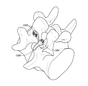
the wire or cable 72 is secured to the articular processes 20,
22 by tying one or more knots 92 in the cable 72 that can resist pulling of
the wire or
cable through the articular process. In another embodiment, one or both ends
of the wire
or cable are provided with an anchor to resist migration of the implants. As
shown in
-17-
CA 02923623 2016-03-07
WO 2015/047909
PCT/US2014/056598
FIGS. 27A and 27B, one or both ends of the wire or cable 72 may be threaded
such that a
nut 94 can be tightened on the wire or cable 72 to secure the wire or cable to
the articular
processes 20, 22. FIG. 28 depicts the attachment of a nut onto a threaded end
of a cable.
The threaded portion 96 of the wire or cable can be secured to the cable by
pressing,
crimping or twisting the threaded 96 portion onto the cable 72. In one
embodiment, the
threaded portion 96 is made from titanium, titanium alloy, cobalt chromium,
stainless
steel, or any combination thereof.
[0130] In one
embodiment, the wire or cable has two threaded ends 96 for
engaging the bony or cartilaginous tissue, one portion for each facet of the
facet joint.
[0131] In another
embodiment, shown in FIG. 29, the wire or cable is secured
to the articular process with fastener rings 98. As depicted in FIGS. 30A and
30B, the
fastener rings 98 comprise a ring 100 with a central lumen 102 and a locking
element to
facilitate locking the ring 100 to a fastener member. The central lumen 102 is
adapted to
accept insertion of a wire or cable through it. The illustrated locking
element is in the
form of a side lumen 104 which is threaded and configured to accept a
rotatable screw
106 with a proximal end 108, a threaded body 110 and a distal end 112. The
threaded
body 110 is complementary to the threads of the side lumen 104 so that when
the screw
106 is rotated at its distal end 112, the proximal end 108 of the screw 106
moves further
into the central lumen 102 and is capable of applying increasing force to a
wire or cable
inserted through the central lumen 102. In one embodiment, the force on the
wire or
cable is capable of creating a friction fit or a mechanical interfit to resist
movement
between the wire or cable and the fastener ring 98, thereby securing the wire
or cable to
the articular process 20 or 22. As shown in FIGS. 31 to 33, the distal end 112
of the
screw 106 can be configured to engage the wire or cable in any of a variety
designs,
including but no limited to a blunt tip 114, curved tip 116 and piercing tip
118.
[0132] In another
embodiment, depicted in FIGS. 34A and 34B, the wire or
cable is securable to the articular process with a fastener ring 120 have
radially inward
biased projections 122 defining a central lumen 124. The central lumen has a
cross-
sectional shape smaller than that of the wire or cable but is capable of
enlargement when
the inward projections 122 are bent away, as shown in FIGS. 35A and 35B. The
inward
projections 122 apply increasing force to the wire or cable within the central
lumen 124 as
the projections 122 are bent, thereby creating a friction fit.
-18-
CA 02923623 2016-03-07
WO 2015/047909
PCT/US2014/056598
101331 In one
embodiment, one end of the wire or cable fastener member is
preformed with a retainer for engaging the articular process. The retainer may
be a
preformed ring, bulb, flared end, T-bar end, or any of a variety of shapes
having a greater
cross sectional area than the other portions of the wire or cable fastener
member. This
configuration of the wire or cable fastener member is adapted to engage an
articular
process by passing the free end of a wire or cable fastener member through an
articular
process such that the end with the preformed retainer can engage the articular
process.
101341 In one
embodiment, the wire or cable fastener member is secured to the
articular processes with sufficient laxity or length between the secured ends
or between
the implant and one secured end so that the two articular processes are not
fixed in
position relative to each other and remain capable of performing movements
such as
flexion, extension, lateral flexion and/or rotation. In one embodiment, the
fastener
member comprises a cable of braided polymer, including but not limited to a
braided
polymer such as PEEK or PEKK, or a braided metal, such as braided cobalt
chromium or
titanium. The cable can be selected with different degrees of flexibility to
provide
different degrees of movement at that facet joint. The cable has a first
segment capable of
engaging the implant at its fastener interface to limit the movement.
101351 In one
embodiment, shown in FIG. 36A, the fastener member
comprises a screw or bolt 126 with a proximal end 128, body 130 and distal end
132. The
distal end 132 of the screw or bolt is capable of forming a mechanical
interfit with a
complementary fastener interface 134 on the implant or spacer 136. The distal
end 132
typically comprises threads, but other configurations may be used to form a
mechanical
interfit. The complementary fastener interface 134 on the implant 136 could be
a
threaded through hole or, a close-ended hole. The proximal end 128 of the
screw or bolt
126 has a hex or other type of interface known in the art, capable of engaging
a rotating
tool to manipulate the screw or bolt 126. The body of the screw or bolt 126
has a length
sufficient to at least span the length of the hole or conduit created through
the articular
process for securing the implant. In FIG. 36B, the fastener member further
comprises a
pivotable washer 127 with a pivot surface 129 that articulates with the
proximal end 128
of the screw 126. In one embodiment, the pivotable washer 127 is capable of a
range of
positions relative to the screw 126 and provides the screw 126 with a better
surface area
contact with the bone.
-19-
CA 02923623 2016-03-07
WO 2015/047909
PCT/US2014/056598
101361 FIG. 37 is a
cross-sectional view of a facet joint 28 with a spacer 136
bolted to one articular process 20 of a facet joint 28. The spacer 136
position is fixed
relative to one facet 24 of the joint 28, but provides for spacing and
movement of the
other facet 26 with respect to the spacer 136. In embodiments comprising a two-
part
implant, shown in FIGS. 38 and 39, each disc may have its own screw or bolt
fastener
member. FIG. 38 depicts a flat two-part implant 138 and FIG. 39 depicts a
curved two-
part implant 140.
101371 In some
embodiments, shown in FIGS. 40A through 41B, the fastener
member is integral with or attached to the implant and comprises a projection
142 from
the implant 144 that is adapted to engage the adjacent articular process or
surrounding
tissue. In one embodiment, the projection comprises at least one spike 142 or
hook
projecting from one face of the implant 144. In one embodiment, the spike 142
or hook
can be ribbed, barbed or threaded to resist separation after insertion into
bone or tissue.
FIG. 42 depicts the implant 144 of FIG. 40A engaged to a facet 24 of the facet
joint 28.
In one embodiment comprising a two-part implant 146, shown in FIG. 43, each
disc 148
may have its own projection-fastener member 142. In some embodiments, as
depicted in
FIG. 44, more than one projection 150 is provided on the implant 152. FIG. 45
illustrates
the implant of FIG. 44 placed in a facet joint 28. The projections 150 may be
angled with
respect to the implant 152 to resist dislodgement by the movement at the
joint.
101381 FIGS. 46A to
47B illustrate embodiments where the fastener member
comprises a projection 154 extending laterally such as from the side of the
implant 156,
and adapted to engage the soft tissue surrounding the facet joint, rather than
a bony or
cartilaginous articular process. In one example, the implant of FIG. 46 could
be inserted
into a facet joint through an incision made in the joint capsule, but the
integrity of the
joint capsule opposite the incision site is maintained and used as an
anchoring site for the
implant. The orientation of the projection can be fixed as in FIG. 44, or
flexible. FIG. 47
depicts a flexible tether such as a wire 158 with its proximal end 160
embedded in or
otherwise attached to the implant and one or more barbs which may be attached
to its
distal end 162. A flexible projection may provide greater selection of soft
tissue
anchoring sites for the implant.
101391 In one
embodiment, the joint capsule is closed after placement of the
implant. Closure may be performed using adhesives, suturing, stapling or any
of a variety
of closure mechanisms known in the art.
-20-
CA 02923623 2016-03-07
WO 2015/047909
PCT/US2014/056598
[0140] FIGS. 48A-
48C depict an implant 260 according to an embodiment.
Specifically, FIG. 48A is a front perspective view of implant 260, FIG. 48B is
a side view
of implant 260, and FIG. 48C is a cross-sectional side view of implant 260.
Implant 260
can be similar to, and have similar elements and uses as implant 160 described
above. By
way of example, a fastener interface 266 of implant 260 can be similar to
fastener
interface 166 of implant 160. Implant 260 includes a concave first face 262, a
convex
second face 264, a centrally disposed circular fastener interface 266, and
four irregular
shaped substance interfaces 268.
[0141] FIGS. 49-51
show posterior perspective views of a portion of the
vertebral column during a method for fusing adjacent vertebrae using an
implant 260
according to an embodiment. As shown in FIG. 49, implant 260 and a fastener
member
280 can be used to fuse a vertebra V1 and vertebra V2 via the inferior
articular process
IAP IA of vertebra V1 and the superior articular process SAP2A of vertebra V2.
Any
fastener member can include any biocompatible material, e.g., stainless steel,
titanium,
PEEK, nylon, etc. Also as shown in FIG. 49, an implant 360 and a fastener
member 380
are used to fuse a vertebra VI and vertebra V2 via the inferior articular
process IAMB of
vertebra VI and the superior articular process SAP2B of vertebra V2. In some
embodiments, vertebra VI and/or vertebra V2 are fused using only one of
implant 260 or
implant 360. In some such embodiments, one of implant 260 and fastener member
280 or
implant 360 and fastener member 380 can be used to stabilize vertebra VI
and/or vertebra
V2 via one of via the inferior articular process IAP IA of vertebra VI and the
superior
articular process SAP2A of vertebra V2, or, via the inferior articular process
IA13113 of
vertebra V1 and the superior articular process SAP2B of vertebra V2. In other
such
embodiments, one of fastener member 280 or fastener member 380 can be used to
stabilize vertebra VI and/or vertebra V2 via both of the inferior articular
process IAPI A
of vertebra VI and the superior articular process SAP2A of vertebra V2 (for
example, in
combination with implant 260), and, the inferior articular process IAP1B of
vertebra VI
and the superior articular process SAP2B of vertebra V2 (for example, in
combination
with implant 360).
[0142] FIG. 52
depicts a flow chart illustrating a method 6000 of using
implant 260 with fastener member 280 and/or implant 360 with fastener member
380.
Prior to use of implant 260 and/or implant 360, a patient can be prepared for
surgery, at
6002. Some examples of preparations for surgery are described in U.S. patent
application
-21-
81795222
Ser. No. 12/859,009; filed Aug. 18, 2010, and titled "Vertebral Facet Joint
Drill and
Method of Use" (referred to as "the '009 application"). In addition to those
procedures
described in the '009 application, in some embodiments, the surgical procedure
can
include direct visualization of the vertebra(e)
to be stabilized. Said another way,
the medical practitioner can perform the operation without the use of
fluoroscopy. This
direct visualization can be possible due to the small incision necessary for
implantation of
the implant, for example, less than about 25 mm, and due to the ease of
implanting
and deploying the implant. In some
embodiments, the surgical procedure used can
include forming an opening in body tissue substantially equidistant between a
first articular
process of the first vertebra and a second articular process of the first
vertebra. A cannula
(not shown) can be inserted through the opening and a proximal end of the
cannula can
be positioned near the superior articular process SAP2A of vertebra V2. In
some
embodiments, the surgical procedure can include preparing the area near and/or
around
the vertebra V2 by, for example, removing all or a portion of ligaments,
cartilage, and/or
other tissue. For example, the area near and/or around a facet joint can be
prepared by
removing all or a portion of the facet joint capsule.
101431 A drill or
other device can be used to form a lumen in superior articular
process SAP2A of vertebra V2 and inferior articular process IA PI A of
vertebra V1, at
6004. Specifically, the drill can be used to form the lumen in a facet of
superior articular
process SAP2A of vertebra V2 and to form the lumen in a facet of inferior
articular
process IAP1A of vertebra Vi. Methods and devices for forming lumens in
vertebra are
described in the '009 application. A portion of the surface of the facet of
SAP2A and
'APIA can be prepared for fusion, at 6006. Specifically, a portion of the
surface of the
facet can be ground, scored, roughened, sanded, etc, such that the surface of
the facet can
better adhere to any substances to aid in fusion and/or otherwise fuse more
readily to the
implant. The fastener member 280 can be positioned within the cannula and can
be
advanced through the cannula until a proximal end portion 282 of fastener
member 280 is
positioned near the lumen of superior articular process SAP2A of vertebra V2.
In some
embodiments, the proximal end of the cannula can have a bend to direct the
proximal end
portion 282 of fastener member 280 into the lumen of superior articular
process SAP2A
of vertebra V2. The proximal end portion 282 of fastener member 280 is
inserted into the
lumen of superior articular process SAP2A of vertebra V2, at 6008. A substance
can be
disposed in a substance interface 268 of implant 260, at 6010. In some
embodiments,
-22-
Date Recue/Date Received 2021-04-08
CA 02923623 2016-03-07
WO 2015/047909
PCT/US2014/056598
implant 260 can have a substance disposed in substance interface 268 prior to
a surgical
procedure, for example, during manufacturing of implant 260, post-
manufacturing, and/or
as part of a kit. Implant 260 is inserted between the superior articular
process SAP2A of
vertebra V2 and inferior articular process 'APIA of vertebra V1, at 6012.
[0144] The proximal
end portion 282 of fastener member 280 is inserted into
the lumen of inferior articular process IAPI A of vertebra V1, at 6014. The
fastener
member can be secured, at 6016. Securing the fastener member 280 can be based
on the
type of fastener member used. By way of example, securing a fastener member
similar to
a flexible fastener band as depicted in FIGS. 49-51, can include inserting the
proximal
end portion 282 into a fastening mechanism of a distal end portion 284 of the
fastener
member 280, and advancing the proximal end portion 282 through the fastening
mechanism to secure the fastening mechanism. In other embodiments, fastener
member
can be secured by tying a first portion the fastener member to a second
portion of the
fastener member, by screwing the fastener member into a threaded fastener
interface,
threading a fastener onto a threaded end of a fastener member disposed through
a fastener
interface, combinations of above, etc. In some embodiments, implant 260 can be
disposed prior to inserting the proximal end portion of the fastener member
280 into the
lumen of superior articular process SAP2A of vertebra V2. The cannula can be
removed
and/or reinserted at various points during the method 6000, including, for
example, after
the proximal end portion 282 of fastener member 280 is inserted into the lumen
formed
within the superior articular process SAP2A of vertebra V2, after vertebra V1
and/or
Vertebra V2 has been stabilized, or at other points during method 6000.
101451 After the
fastener member is secured, superior articular process SAP2A
of vertebra V2 can fuse to inferior articular process [APIA of vertebra V 1 .
Fusing can
include one or more of bone material from superior articular process SAP2A of
vertebra
V2, bone material from inferior articular process TAPIA of vertebra VI, and
the substance
that fuses articular process SAP2A of vertebra V2 to inferior articular
process TANA of
vertebra VI through substance interface 268. In some embodiments, after
superior
articular process SAP2A of vertebra V2 is fused to inferior articular process
1API A of
vertebra V1, the fastener member 280 is not removed. In some other
embodiments, after
superior articular process SAP2A of vertebra V2 is fused to inferior articular
process
IAP1A of vertebra VI, all or a portion of the fastener member 280 can be
removed. In
other embodiments, fastener member 280 can be removed after fusion of superior
-23-
CA 02923623 2016-03-07
WO 2015/047909
PCT/US2014/056598
articular process SAP2A of vertebra V2 to inferior articular process IAP1A of
vertebra
VI has started, but has not finished.
101461 In addition
to the fastener members shown above, such as, for example,
fastener member 260, FIGS. 53-65 show fastener members according to other
embodiments.
101471 FIG. 53
depicts views of a fastener member 480. Fastener member 480
can be a flexible fastening band ("band") 480. FIG. 54 depicts a view of a
portion of band
480 can be similar to band 280 described above and can include similar
components. By
way of example, band 480 includes a proximal end portion 482, a first portion
484, a
second portion 486, and a distal end portion 488 including a fastening
mechanism 490. In
contrast to band 280, band 480 includes a cylindrical second portion 486 and
each
includes a third portion 489. As depicted in FIGS. 53-54, third portion 489 is
substantially the same shape as first portion 482. As shown in FIGS. 53 and
54, band 480
includes a gear rack 487 and gears 494. Each of gears 494 can be wedge shaped
to allow
each of gears 494 to displace the ratchet of fastening mechanism 490 in only
one
direction. In some embodiments, gears 494 can be other shapes, such as blocks,
etc.
101481 FIG. 55 is a
side view and FIG. 56 is a top view of a fastener member
840. fastener member 840 can be a flexible fastening band ("band") 580
according to
another embodiment. Band 840 can be similar to band 280 and band 480 described
above
and can include similar components. By way of example, band 840 includes a
proximal
end portion 842, a first portion 844 including a gear rack 847, a second
portion 846, and a
distal end portion 848 including a fastening mechanism 850 and a ratchet 862.
In contrast
to gear rack 487, a cross sectional area of each gear 864 of gear rack 847 is
rectangular in
shape instead of wedge shaped. Furthermore, in contrast to first portion 282,
first portion
844 is cylindrical in shape instead of cuboidal in shape. In this manner, the
lumen 866 of
the fastening mechanism 850 is cylindrical in shape. A band according to this
embodiment may be particularly useful in deployments where a single band in
used to
stabilize adjacent vertebrae. In this manner, the second portion can be
disposed within the
lumen of the first articular process of the first vertebra and a portion of
the first portion
can be disposed within the lumen of the second articular process of the first
vertebra. In
these embodiments the portion of the band within the first articular process
of the first
vertebra and the portion of the band within in the second articular process of
the first
vertebra can both have substantially the same shape as the lumen in the first
articular
-24-
CA 02923623 2016-03-07
WO 2015/047909
PCT/US2014/056598
process of the first vertebra and the lumen in the second articular process of
the first
vertebra. In this manner, and as described above regarding band 480, the
amount of open
space within the lumens can be minimized, the amount of surface area of the
first portion
and/or second portion of the band in contact with the lumens can increase, and
subsequently the movement of the first vertebra and/or the second vertebra can
be reduced
or minimized. furthermore, when movement of the first vertebra and/or the
second
vertebra does occur, forces acting against the band can be more equally
distributed
throughout the first portion and/or the second portion, due at least to the
increased surface
area of the band in contact with the lumens.
[0149] FIG. 57 is a
side view a fastener member 940. Fastener member 940
can be a flexible fastening band ("band") 940 according to an embodiment. Band
940 can
be similar to band 280, band 480, and band 840 described above and can include
similar
components. By way of example, band 840 includes a proximal end portion 942, a
first
portion 944 including a gear rack 947, a second portion 946, and a distal end
portion 948
including a fastening mechanism 950. Similar to gear rack 847, a cross
sectional area of
each gear 964 of gear rack 947 is rectangular in shape. In contrast to gear
rack 847, each
of gears 964 extend the entire circumference of first portion 944 instead of
only a portion
of the circumference of first portion 944. Furthermore, in contrast to first
portion 282, but
similar to first portion 844, first portion 944 is cylindrical in shape
instead of cuboidal in
shape. In this manner, the lumen 966 of the fastening mechanism 950 is
cylindrical in
shape. A band according to this embodiment may be particularly useful in
deployments
where the movement and repositioning of the band after implantation may be
difficult. In
this manner, because each of the gears can be the entire circumference of the
first portion
and/or the second portion, the first portion and/or the second portion can
enter the
fastening mechanism in any radial orientation and still engage the ratchet.
[0150] FIGS. 58-62
are views of a fastener member 780. Fastener member
780 can be a flexible fastening band ("band") 780 according to another
embodiment.
FIG. 58 is a perspective view and FIG. 59 is a cross-sectional side view of
band 780.
FIG. 60 is a cross-sectional view of band 780 taken along line XXIII. FIG. 61
is a cross-
sectional top view of band 780 in a first configuration and FIG. 62 is a cross-
sectional top
view of band 780 in a second configuration. Band 780 can be similar to band
280 and
band 480 described above and can include similar components. By way of
example, band
780 includes a proximal end portion (not shown), a first portion 784 including
a gear rack
-25-
CA 02923623 2016-03-07
WO 2015/047909
PCT/US2014/056598
787 (see FIG. 59), a second portion 786, and a distal end portion 788
including a fastening
mechanism 790 and a ratchet 792. In contrast to band 280 and band 480, band
780
includes a reinforcement piece 772.
[0151]
Reinforcement piece 772 can include any of the materials described
above for a fastener member. In some embodiments, reinforcement piece 772 can
include
a material stronger than second portion 786 and/or first portion 784, for
example, first
portion 784 and second portion 786 can include PEEK and reinforcement piece
772 can
include titanium. As shown in HG. 59, reinforcement piece 772 can be disposed
within
band 780 approximately along the entire length of second portion 786, and a
portion of
reinforcement piece 772 can be disposed within the distal end portion 788. In
some
embodiments, reinforcement piece can include a length along at least a portion
of the
length of second portion 786 and/or first portion 784 but not the distal end
portion. In
some embodiments, reinforcement piece 772 can be disposed only within second
portion
786. Reinforcement piece 772 can have a length in first dimension (length), a
length in a
second dimension (width), and a length in a third dimension (height). As
described
herein, a reinforcement piece be different shapes that can include more or
fewer
dimensions.
[0152] The
reinforcement piece can be molded within the band. Said another
way, in embodiments where the first portion, the second portion, and or the
distal end
portion are moldable materials, the reinforcement piece can be placed in the
mold and the
moldable materials can be injected or otherwise put in the mold around the
reinforcement
piece. In other embodiments, each portion of the band (for example, the
proximal end
portion, the first portion, the second portion, the third portion, and/or the
distal end
portion) around the reinforcement piece can have a top half and a bottom half,
and each of
the top half and the bottom half can be placed around the reinforcement piece,
and sealed.
As shown in FIG. 61, reinforcement piece 772 includes support members 774.
While
FIG. 61 shows reinforcement piece 772 including four support members 774, in
some
embodiments, more or fewer support members 774 can be used. Support members
774
can maintain the position of reinforcement piece 772 during the molding and/or
assembly
process of band 780. As shown in FIG. 62, support members 774 are removed
before
band 780 is used.
[0153] As shown in
FIG. 60, reinforcement piece 772 can has a substantially
uniform cuboidal shape. In other embodiments, reinforcement piece 772 can have
other
-26-
CA 02923623 2016-03-07
WO 2015/047909
PCT/US2014/056598
shapes. The shape of the reinforcement piece can be selected depending on the
desired
bending and/or torsion characteristics of the material chosen. By way of
example, a
substantially planar cuboidal shape can provide a greater increase in bending
strength
while providing a lesser increase in torsion strength, a cylindrical shape can
provide an
increase in bending strength while providing very little increase in torsion
strength, a
substantially square and/or tubular cuboidal shape can provide similar bending
and torsion
increases. Any shape can be selected to achieve the desired bending and
torsion strength.
Combinations of materials and shapes can also be considered. For example, a
material
having higher torsion strength may be combined with a shape having a lower
torsion
strength to combine for the desired torsion strength. As shown in FIGS. 61 and
62,
reinforcement piece 772 includes holes 776 distributed along the length of the
first
dimension. While FIGS. 61 and 62 shows band 780 including many holes 776, in
some
embodiments, more or fewer holes 776 can be used. FIGS. 61 and 62 depict holes
776
distributed substantially equally along the length of the first dimension, in
some
embodiments, the holes can be distributed differently or along different
dimensions
depending on the shape and/or material chosen, and/or whether the
reinforcement piece is
solid or hollow. Holes 776 can be configured to reduce the weight of
reinforcement piece
772 while still provided band 780 additional strength. Holes 776 can be round,
oval,
square, or any other shape.
101541 FIG. 63 is
an exploded view, FIG. 64 is a perspective view, and FIG.
65 is a cross-sectional view of a fastener member 880. Fastener member 880 can
be a
flexible fastening band ("band") 880 according to another embodiment. Band 880
can be
similar to band 280 and band 480 described above and can include similar
components.
By way of example, band 880 includes a proximal end portion 882, a first
portion 884, a
second portion 886 including a gear rack 887, a distal end portion 888, a
fastening
mechanism 890 and a ratchet 892. In contrast to band 280 and band 480, the
fastening
mechanism 890 of band 880 is separately formed from distal portion 888 of band
880.
While second portion 886 of band 880 is shown in FIGS. 63-65 as having a
substantially
cuboidal shape, in some embodiments, second portion 886 can be substantially
cylindrical
in shape or any other appropriate shape discussed herein. As shown in FIGS. 64
and 65,
band 880 includes a gear rack 887 and gears 894. Each of gears 894 can be
wedge shaped
to allow each of gears 894 to displace a ratchet 892 of fastening mechanism
890 in only
one direction. In some embodiments, gears 894 can be other shapes, such as
blocks, or
-27-
CA 02923623 2016-03-07
WO 2015/047909
PCT/US2014/056598
any other appropriate shape discussed herein. As shown in FIGS. 63-65, distal
end
portion 888 can be substantially circular in shape and can have a diameter
greater than a
width of second portion 886. In other embodiments, distal portion 888 can have
other
shapes, for example, oval, rectangular, square. etc.
[0155] In addition
to the implants shown above, such as, for example, implant
160, FIGS. 66-81 show implants according to other embodiments.
[0156] FIGS. 66-69
depict an implant 1060 according to an embodiment.
Specifically, FIG. 66 is a front perspective view of implant 1060, FIG. 67 is
a rear
perspective view of implant 1060, FIG. 68 is a side view of implant 1060, and
FIG. 69 is
a cross-sectional side view of implant 1060. Implant 1060 can be similar to,
and have
similar elements and uses as implant 160 and implant 260 described above. By
way of
example, a fastener interface 1066 of implant 1060 can be similar to fastener
interface 166
of implant 160, and similar to fastener interface 266 of implant 260 Implant
1060 includes
a concave first face 1062, a convex second face 1064, a centrally-disposed
substantially-
circular fastener interface 1066, and six substantially-circular shaped
substance interfaces
1068.
[0157] FIGS. 70-73
depict an implant 1160 according to an embodiment.
Specifically, FIG. 70 is a front perspective view of implant 1160. FIG. 71 is
a rear
perspective view of implant 1160, FIG. 72 is a side view of implant 1160, and
FIG. 73 is
a cross-sectional side view of implant 1160. Implant 1160 can be similar to,
and have
similar elements and uses as implant 160 and implant 260 described above. By
way of
example, a fastener interface 1166 of implant 1160 can be similar to fastener
interface 166
of implant 160, and similar to fastener interface 266 of implant 260. Implant
1160
includes a concave first face 1162, a convex second face 1164, a centrally-
disposed
substantially-circular fastener interface 1166, and five rounded rectangular
shaped
substance interfaces 1168.
[0158] FIGS. 74-77
depict an implant 1260 according to an embodiment.
Specifically, FIG. 74 is a front perspective view of implant 1260, FIG. 75 is
a rear
perspective view of implant 1260, FIG. 76 is a side view of implant 1260, and
FIG. 77 is
a cross-sectional side view of implant 1260. Implant 1260 can be similar to,
and have
similar elements and uses as implant 160 and implant 260 described above. By
way of
example, a fastener interface 1266 of implant 1260 can be similar to fastener
interface 166
of implant 160, and similar to fastener interface 266 of implant 260. Implant
1260
-28-
CA 02923623 2016-03-07
WO 2015/047909
PCT/US2014/056598
includes a concave first face 1262, a convex second face 1264, a centrally-
disposed
substantially-circular fastener interface 1266, and several substantially-
circular shaped
and variably-sized substance interfaces 1268.
[0159] FIGS. 78-81
depict an implant 1360 according to an embodiment.
Specifically, FIG. 78 is a front perspective view of implant 1360, FIG. 79 is
a rear
perspective view of implant 1360, FIG. 80 is a side view of implant 1360, and
FIG. 81 is
a cross-sectional side view of implant 1360. Implant 1360 can be similar to,
and have
similar elements and uses as implant 160 and implant 260 described above. By
way of
example, a fastener interface 1366 of implant 1360 can be similar to fastener
interface 166
of implant 160. and similar to fastener interface 266 of implant 260. Implant
1360
includes a concave first face 1362, a convex second face 1364, a centrally-
disposed
substantially-circular fastener interface 1166, four irregular shaped
substance interfaces
1368, and four projections 1369. Each of the four projections 1369 can engage,
or other
wise dig, latch, lock, or hook into or onto, a bone portion to prevent or
reduce movement
of the implant 1360, such as, for example, rotation of implant 1360,
longitudinal
movement of implant 1360, and/or lateral movement of implant 1360. In this
manner, the
projections 1369 can secure implant 1360 to a bone portion during a fusion
procedure. In
some embodiments, projections 1369 can substantially maintain a position of
implant
1369 after a fastener member is removed.
[0160] While
various embodiments have been described above, it should be
understood that they have been presented by way of example only, not
limitation, and
various changes in form and details may be made. For example, while the
descriptions
given are with reference to stabilizing vertebra, another bone(s), such as,
for example, a
sternum and/or a rib(s) could be stabilized using the fastener members and
implants
described herein. In another example, a fastener member can be used to
stabilize and/or
fixate an intramedullary (IM) rod or nail. For example, the fastener member
can be used
at different longitudinal locations along an IM rod or nail, and used to
couple adjacent
bone portions to the IM rod or nail. In such situations, a given fastener
member can fix a
first bone portion, the 1M rod or nail, and a second bone portion, all of
which are
positioned between the distal portion and the proximal portion of the fastener
member. In
yet another example, a fastener member can be used to stabilize and/or fixate
a bone
fragment. While various embodiments have been described above with regard to
natural
bone spaces, (e.g., the space between an inferior articulate process and a
superior
-29-
CA 02923623 2016-03-07
WO 2015/047909
PCT/US2014/056598
articulate process), in other embodiments, the bone spacing can be man-made
(e.g.,
sternum split during a heart procedure), and/or due to an injury (e.g., broken
bone).
[0161] Where methods described above indicate certain events occurring
in
certain order, the ordering of certain events can be modified. Additionally,
certain of the
events can be performed concurrently in a parallel process when possible, as
well as
performed sequentially as described above. Any portion of the apparatus and/or
methods
described herein may be combined in any combination, except mutually exclusive
combinations. '1 he embodiments described herein can include various
combinations
and/or sub-combinations of the functions, components and/or features of the
different
embodiments described. For example, FIGS. 54 and 56 depict band 580 including
a
single ratchet 592, and FIG. 57 depicts band 680 including a single ratchet
692, however,
in some embodiments, any of the fastener members can include any number of
ratchets.
Similarly, any of fastener members can include a reinforcement piece and/or a
implant.
Furthermore, while one embodiment of an implant may be shown in use with one
embodiment of a fastener member, in other embodiments, implants and fastener
member
can be used with other implants and fastener members. For example, while FIG.
28
depicts an implant being secured with a threaded wire, in some embodiments, a
flexible
fastening band can be used.
101621 Facet reinforcement device
101631 Although the flexible fastening band may be used alone or with
an
embodiment of facet implant as described above, in some applications it may be
desirable
to reinforce the fixation of the band as it exits the bone of the articular
process. This may
prevent cut out by relieving pressure on the surface of the articular process
and hold in the
bone from the fastener band and/or fastening mechanism. The facet
reinforcement may
also anchor the flexible facet band to the vertebra using a fastener. This may
prevent
migration of the band and restrict motion at the facet joint to improve
fusion.
[0164] FIG. 82 depicts one arrangement of facet reinforcement device
1400.
The facet reinforcement device 1400 of the illustrated arrangement can include
a proximal
surface 1402, a distal surface 1406, an ablumenal surface 1404 extending from
the
proximal surface 1402 to the distal surface 1406. In certain arrangements, the
proximal
surface 1402 and/or distal surface 1406 can be curved and/or malleable to
conform to the
shape of the facet. The facet reinforcement device 1400 can include a luminal
surface
1410 surrounding a central lumen 1420. The luminal surface 1410 can extend
from the
-30-
CA 02923623 2016-03-07
WO 2015/047909
PCT/US2014/056598
proximal surface 1402 to the distal surface 1406. The central lumen 1420 can
be centrally
disposed within the device 1400. As described below, the luminal surface 1410
can
include a fastener interface (not illustrated) in certain embodiments.
[0165] As will be
explained below, the facet reinforcement devices described
herein can be used in combination with the implants depicted in FIGS. 8A-81A
and/or
other implants described herein. The facet reinforcement device 1400 can also
be used in
combination with the fastener members depicted in FIGS. 20-65, and/or other
fastener
members described herein. Accordingly, the fastener member 1480 depicted in
FIG. 84
can refer to any fastener member described herein; and the fastener member
1580
depicted in FIG. 87 can refer to any fastener member described herein; and the
fastener
members 1680, 1680A depicted in FIG. 89 can refer to any fastener member
described
herein.
[0166] As shown in
FIG. 82, at least a portion of one surface of the facet
reinforcement device 1400 can include a roughened surface. A roughened surface
may be
advantageous when in contact with a bone or tissue surface because it may
prevent
slippage or migration of the facet reinforcement device 1400 against the bone.
A
roughened surface may aid in maintaining the facet reinforcement device 1400
and the
fastener member 1480 (see Figure 83) engaged with tissue or bone.
[0167] The
roughened surface can include at least one projection 1416. As
shown in FIG. 82, the facet reinforcement device 1400 can comprise a plurality
of
projections 1416. The projections 1416 can extend from the distal surface 1406
and can
include a sharp edge or tip. The projections 1416 can also extend between the
ablumenal
surface 1404 and the luminal surface 1410 or in certain embodiments only
extend along a
portion of said area. In some embodiments, the projections 1416 comprise at
least one
spike, barb, wedge, or hook projecting from at least a portion of one surface
of the facet
reinforcement device 1400. In some embodiments, the projections 1416 can be
ribbed,
barbed, or threaded to resist separation after insertion into bone or tissue.
The projections
1416 may have different shapes from one another or they may have a uniform
shape. A
portion of the surface of the projections 1416 can be porous. A porous surface
can be
created in any a variety of ways known in the art, such as by applying
sintered beads or
spraying plasma onto the surface of the projection 1416. A porous surface can
allow bone
to grow into or attach to the surface of the projection 1416, thus securing
the projection
1416 and the facet reinforcement device 1400 to the bone. In certain
embodiments, other
-31-
CA 02923623 2016-03-07
WO 2015/047909
PCT/US2014/056598
surfaces of the facet reinforcement device 1400 can be porous. Ti one
embodiment, an
adhesive or sealant, such as a cyanoacrylate, polymethylmethacrylate, or other
adhesive
known in the art, is used to bond at least one surface of the facet
reinforcement device
1400 to a bone or tissue surface. In some embodiments, an adhesive or sealant
is used to
bond the distal surface 1406 of the facet reinforcement device 1400 to the
surface of the
facet.
[0168] The facet
reinforcement device 1400 may include one row of
projections 1416 or may include multiple rows of projections 1416. The facet
reinforcement device 1400 may include projections 1416 arranged in a random
order or
orientation.
[0169] The
ablumenal surface 1404 of the facet reinforcement device 1400
can include a substantially circular cross-section (cylindrical), as shown in
FIG. 82. The
ablumenal surface 1404 can have other cross-sectional shapes including, but
not limited
to, circular (cylindrical), hexagonal, rectangular (cuboid), square,
elliptical, and/or have a
combination of curved, flat surfaces and/or partial shapes. In certain
embodiments, the
ablumenal surface 1404 may conform to the shape of an insertion tool.
[0170] In the
illustrated embodiment of Fig. 82, the central lumen 1420 and
the luminal surface 1410 can be circular (cylindrical). The central lumen 1420
and the
luminal surface 1410 can have other cross-sectional shapes including, but not
limited to,
hexagonal, rectangular (cuboid), square, elliptical, and/or have a combination
of curved,
flat surfaces and/or partial shapes. The central lumen 1420 and the luminal
surface 1410
may conform to the shape of an insertion tool. In certain embodiments, the
central lumen
1420 and the luminal surface 1410 can be shaped based on a shape of the
fastener
member (not shown) (e.g., the central lumen and lumina] surface can have a
similar cross-
sectional shape as the fastener member extending through the central lumen).
In certain
embodiments, the central lumen 1420 and the luminal surface 1410 can include a
substantially smooth inner surface to allow the fastener member 1480 to easily
pass
through. In other embodiments, the central lumen 1420 and the luminal surface
1410 can
include a threaded inner surface to allow the fastener member 1480 to thread
into central
lumen 1420.
[0171] The central
lumen 1420 and the lumina] surface 1410 may be
configured to match the shape of a lumen formed in the articular process,
during a method
of use. The central lumen 1420 and the luminal surface 1410 may be smaller
than a
-32-
CA 02923623 2016-03-07
WO 2015/047909
PCT/US2014/056598
lumen formed in the articular process, during a method of use. In this
configuration, the
facet reinforcement device 1400 may reduce stress at the outer aspect of the
lumen in the
bone. The central lumen 1420 and the luminal surface 1410 may be larger than a
lumen
formed in the articular process, during a method of use. In this
configuration, the facet
reinforcement device 1400 may be in contact with a larger surface area of the
facet,
thereby distributing the forces of the fastener member.
01721 The proximal
surface 1402 may have a feature to mechanically interfit
with an insertion tool, including grooves and/or protrusions configured to
mate with a
corresponding groove and/or protrusion of the insertion tool. The proximal
surface 1402
may have a feature (e.g., a groove or recess) to mechanically interfit with a
portion of the
fastener member 1480 (shown in Figure 84). The feature to mechanically
interfit with a
portion of the fastener member may increase stability of the system and
resistance to
migration of components of the system.
101731 The diameter
of the facet reinforcement device 1400 may be in the
range of 2 mm-20mm or in the range of 4 mm-15 mm. The diameter of the central
lumen
1420 may be in the range of .5 mm-10 mm or range of 1-7 mm.
101741 FIGS. 83-84
show posterior perspective views of a portion of the
vertebral column during a method for fusing adjacent vertebrae using the
embodiment of
the facet reinforcement device 1400 shown in Figure 82. The method can include
using
an implant deployed to restore the space between facets of a superior
articular process of a
first vertebra and an inferior articular process of an adjacent vertebra.
101751 In one
method of use, a drill or other device can be used to form a
lumen in superior articular process SAP2A of vertebra V2 and inferior
articular process
IAP IA of vertebra VI. A portion of the surface of the facet of SAP2A and a
portion of the
surface of the facet of IAP IA can be prepared for fusion. For example, a
portion of the
surface of the facet can be ground, scored, roughened, sanded, etc., such that
the surface
of the facet can better adhere to any substances to aid in fusion and/or
otherwise fuse
more readily to an implant positioned within the facet joint.
101761 FIG. 83
illustrates the facet reinforcement device 1400 placed on the
outer, posterior facing surface of the left inferior articular process IAP1A
of the superior
vertebra VI. In other embodiments and/or in addition, the facet reinforcement
device can
be placed on the surface of the facet of SAP2A. A lumen is formed in the
articular
process. The facet reinforcement device 1400 can be placed after a lumen is
formed in the
-33-
CA 02923623 2016-03-07
WO 2015/047909
PCT/US2014/056598
articular process. In another arrangement, the facet reinforcement device 1400
can be
placed prior to forming a lumen in the articular process. In this method, the
facet
reinforcement device 1400 may serve as a guide for drilling the lumen. The
facet
reinforcement device 1400 can be placed after or prior preparation for fusion.
An
insertion tool may remain on the facet reinforcement device 1400 during the
steps of
forming the lumen and/or during the step of preparing for fusion.
101771 As shown in
FIG. 84, a facet reinforcement device 1400 and a fastener
member 1480 can be used to fuse a vertebra VI and vertebra V2 via the inferior
articular
process IAP IA of vertebra VI and the superior articular process SAP2A of
vertebra V2.
In some embodiments. at least one implant (not shown in FIG. 84) is used with
the
fastener member 1480 to fuse a vertebra VI and vertebra V2. FIG. 84 depicts
fusing the
inferior articular process TAP IA of vertebra VI and the superior articular
process SAP2A
of vertebra V2. However, the inferior articular process IAP113 of vertebra VI
can be fused
to the superior articular process SAP2B of vertebra V2.
101781 In one
method of use, the fastener member 1480 can be positioned
within a cannula and can be advanced through the cannula. The proximal end
portion of
fastener member 1480 can then be inserted into the central lumen 1420 of the
facet
reinforcement device 1400. The proximal end portion of the fastener member
1480 can be
adjacent and/or abut the luminal surface 1410. The proximal end portion of
fastener
member 1480 can then be inserted into the lumen of inferior articular process
'APIA of
vertebra VI. The proximal end portion of fastener member 1480 can be advanced
until a
proximal end portion of fastener member is positioned near the lumen of
superior
articular process SAP2A of vertebra V2. In some embodiments, the proximal end
of the
cannula can have a bend to direct the proximal end portion of fastener member
1480 into
the lumen of superior articular process SAP2A of vertebra V2. The proximal end
portion
of fastener member 1480 can be inserted into the lumen of superior articular
process
SAP2A of vertebra V2. An implant can be inserted between the superior
articular process
SAP2A of vertebra V2 and inferior articular process IAP IA of vertebra Vi. In
some
embodiments, the implant can be disposed prior to inserting the proximal end
portion of
the fastener member 1480 into the lumen of superior articular process SAP2A of
vertebra
V2. The eannula can be removed and/or reinserted at various points during the
method,
including, for example, after the proximal end portion of fastener member 1480
is
inserted into the lumen formed within the superior articular process SAP2A of
vertebra
-34-
CA 02923623 2016-03-07
WO 2015/047909
PCT/US2014/056598
V2, after vertebra VI and/or vertebra V2 have been stabilized, or at any other
point during
the method.
101791 The fastener
member 1480 can be secured. Securing the fastener
member 1480 can be based on the type of fastener member used. By way of
example,
securing a fastener member 1480 having the characteristics of the fastener
member
depicted in FIGS. 49-51, can include the following steps: inserting the
proximal end
portion of the fastener member 1480 into a fastening mechanism 1484; the
fastener
mechanism located at a distal end portion of the fastener member 1480;
securing an end
of the fastener member 1480 to the opposite end of the fastener member 1480;
securing
the proximal end portion of the fastener member 1480 to the distal end portion
of the
fastener member 1480; and/or advancing the proximal end portion of the
fastener member
1480 through the fastening mechanism 1484. In other embodiments, fastener
member
1480 can be secured by tying a first portion the fastener member to a second
portion of the
fastener member, by forming a knot in a first end and second end; by screwing
the
fastener member into a threaded central lumen, by threading a fastener onto a
threaded
end of a fastener member disposed through a threaded central lumen, by
including
enlarged portion at the end of the fastener member, and/or combinations of
above. The
fastener member 1480 can be secured in order to retain the facet reinforcement
device
1400. The facet reinforcement device 1400 is retained within a loop or other
defined
segment of the fastener member 1480. The reinforcement device 1400 can remain
freely
movable along a portion of the defined segment after the fastener member 1480
is
secured. In some embodiments, the reinforcement device 1400 is immobile or
otherwise
secured along a portion of the defined segment after the fastener member 1480
is secured.
101801 FIG. 84
illustrates the assembled system, including the facet
reinforcement device 1400 and the fastener member 1480. The assembled system
is
implanted on the left facet joint 1490 between the superior vertebra V1 and
the inferior
vertebra V2. The left facet joint 1490 may be compressed by the assembled
system,
thereby bringing the two facet surfaces in close apposition. This compression
is in
contrast with the unsecured right facet joint 1492.
[0181] A second
facet reinforcement device 1400, a second fastener member
1480 with or without a second implant may be implanted in the right facet
joint 1492,
according to the method described above with respect to the left facet joint
1490. The
implantation of a second facet reinforcement device 1400 and a second fastener
may
-35-
CA 02923623 2016-03-07
WO 2015/047909
PCT/US2014/056598
improve stabilization. A second facet reinforcement device 1400 and a second
fastener
member 1480 with or without a second implant may be implanted on other levels
of the
spine. A plurality of reinforcement devices 1400 and a plurality of fastener
members 1480
with or without a plurality of implants may be implanted on other levels of
the spine, and
at various locations on the spine. In some embodiments, the same and/or
similar method
of fixation, the same fastener members 1480, the same implants and/or the same
facet
reinforcement devices 1400 may be used at different locations. In other
embodiments, a
different method of fixation, different fastener members 1480, different
implants and/or
different facet reinforcement devices 1 400 may be used at different
locations.
101821 FIG. 85
shows another embodiment of a facet reinforcement device
1500. In the illustrated arrangement, the facet reinforcement device 1500 has
an inferior
end 1502 and a superior end 1504. The facet reinforcement device 1500 has a
first
securing portion 1530 toward the inferior end 1502 and a second securing
portion 1540
toward the superior end 1504. The first securing portion 1530 and second
securing portion
1540 can be connected to each other by a central portion 1510.
101831 The first
securing portion 1530 can be configured for placement on an
outer facet surface of a facet. The first securing portion 1530 can include a
lumen 1506
surrounded by a luminal surface 1511. The lumen 1506 and the luminal surface
1511 can
be substantially circular (cylindrical) as shown in the illustrated
embodiment. The lumen
1506 can have other cross-sectional shapes including, but not limited to,
circular
(cylindrical), hexagonal, rectangular (cuboid), square, elliptical, and/or
have a
combination of curved, flat surfaces and/or partial shapes. The lumen 1506 and
the
luminal surface 1511 can be shaped based on a shape of a fastener member 1580
(e.g.,
having a shape complimentary or similar to the outer shape of the portion of
the fastener
member 1580 extending there-through). The lumen 1506 and the lumina] surface
1511
may conform to the shape of an insertion tool. The lumen 1506 and the luminal
surface
1511 can include a substantially smooth inner surface to allow the fastener
member 1580
to easily pass through. In other embodiments, the lumen 1506 and the luminal
surface
1511 can include a threaded surface to allow the fastener member to thread
into the lumen
1506.
101841 The first
securing portion 1530 can include one (as illustrated), two,
three or a plurality of lumens 1506. The one or more lumens 1506 can have the
same
shape or different shape. The first securing portion 1530 may include one row
of lumen
-36-
CA 02923623 2016-03-07
WO 2015/047909
PCT/US2014/056598
1506, for example, the lumens 1506 can be aligned along an axis. In other
arrangements,
the first securing portion 1530 may include multiple rows of lumens 1506. The
first
securing portion 1530 may include lumens 1506 arranged in a random order or
orientation. As will be described below, the one or more lumens 1506 can be
configured
to accept one or more fastener members 1580 and/or one or more fasteners 1590.
101851 The second
securing portion 1540 can be configured for placement on
a vertebral structure. The vertebral structure can be remote or distanced from
the outer
facet surface of a facet. For example, in one arrangement, the second securing
portion
1540 can be configured for placement on an outer surface or base of the
spinous process
1570: a translaminar position, and/or for placement on an outer surface of the
lamina
(e.g., base of spinous process).
101861 The second
securing portion 1540 can include a lumen 1508
surrounded by a luminal surface 1521. The lumen 1508 and the luminal surface
1521 can
be circular (cylindrical). The lumen 1508 can have other cross-sectional
shapes including,
but not limited to, hexagonal, rectangular (cuboid), square, elliptical,
and/or have a
combination of curved, flat surfaces and/or partial shapes. The lumen 1508 can
be shaped
based on a shape of the fastener member 1580 and/or the fastener 1590. The
lumen 1508
and the lumina' surface 1521 may be circular, hexagonal, rectangular (cuboid),
square,
elliptical, and/or have a combination of curved, flat surfaces and/or partial
shapes. The
lumen 1508 and the luminal surface 1521 may conform to the shape of an
insertion tool.
The lumen 1508 and the luminal surface 1521 can include a substantially smooth
surface
to allow the fastener member 1580 and/or the fastener 1590 to easily pass
through, or the
lumen 1508 can include a threaded surface to allow the fastener member 1580
and/or the
fastener 1590 to thread into the lumen 1508.
101871 The second
securing portion 1540 can include one, two (as illustrated),
three or a plurality of lumens 1508. Additional lumens 1508 may increase
fixation
security and reduce torsional forces.
101881 The one or
more lumens 1508 can have the same shape or different
shape. The two lumens 1508 depicted in FIG. 85 have substantially the same
shape. The
second securing portion 1540 may include one row of lumens 1508, for example,
the
lumens 1508 can be aligned along an axis. The row of lumens 1508 may be
aligned along
a longitudinal axis of the second securing portion 1540. The second securing
portion 1540
may include multiple rows of lumens 1508. The second securing portion 1540 may
-37-
CA 02923623 2016-03-07
WO 2015/047909
PCT/US2014/056598
include lumens 1508 arranged in a random order or orientation. The one or more
lumens
1508 can be configured to accept one or more fastener members 1580 and/or one
or more
fasteners 1590. The one or more lumens 1508 can be oriented in order to
facilitate
placement of the fastener members 1580 and/or the fasteners 1590. The fastener
1590
may be placed in a translaminar position. The fastener 1590 may be placed in
the spinous
process, or the base of the spinous process. The fastener 1590 may be placed
across the
spinous process of a vertebra.
[0189] At least one
surface of the facet reinforcement device 1500 may have a
feature to mechanically interfit with an insertion tool (not shown), including
grooves
and/or protrusions configured to mate with a corresponding groove and/or
protrusion of
the insertion tool. At least one surface of the facet reinforcement device
1500 can have a
feature (e.g., a recess or groove) to mechanically interfit with the fastener
member 1580
and/or fastener 1590. The feature to mechanically interfit with the fastener
member 1580
and/or fastener 1590 member may increase stability of the system and
resistance to
migration of components of the system.
[0190] The diameter
of the first securing portion 1530 may be in the range of
2 mm-20mm or the diameter may be in the range of 4 mm-15 mm. The diameter of
the
lumen 1506 may be in the range of .5 mm-10 mm or in the range of 1-7 mm. The
diameter of the lumen 1508 may be in the range of .5 mm-10 mm or 1-7 mm.
[0191] In some
embodiments, at least a portion of one surface of the facet
reinforcement device 1500 has a roughened surface and/or a porous surface, as
described
above with respect to Figure 82. The roughened surface can comprise at least
one
projection 1516. In one embodiment, the projection 1516 can comprise at least
one spike,
barb, wedge, or hook projecting from one surface of the facet reinforcement
device 1500.
The first securing portion 1530, the second securing portion 1540, the central
portion
1510, and/or any combination of these portions may include a roughened surface
and/or a
porous surface. In some embodiment, an adhesive or sealant, such as a
cyanoaerylate,
polymethylmethaerylate, or other adhesive known in the art, is used to bond
one surface
of the facet reinforcement device 1500 to a bone or tissue surface. In some
embodiments,
at least a portion of one surface of the facet reinforcement device 1500 may
be curved or
malleable. The portion of one surface of the facet reinforcement device 1500
may be
shaped to conform to a shape of an anatomic structure, such as a facet or
spinous process.
-38-
CA 02923623 2016-03-07
WO 2015/047909
PCT/US2014/056598
[0192] As shown the
first securing portion 1530 and the second securing
portion 1540 may lie on different planes as shown in FIG. 85. The first
securing portion
1530 can lie on a plane P1, as shown in FIG. 85. The first securing portion
1530 can
include the lumen 1506. The lumen 1506 has a central, longitudinal axis, Axis
1,
extending through the lumen 1506. The plane PI can lie perpendicular to the
Axis 1 of the
lumen 1506 of the first securing portion 1530. The plane P1 can lie adjacent
to a distal
surface 1531 of the first securing portion 1530. The plane PI can lie adjacent
to a surface
of the first securing portion 1530 configured to engage a bone or tissue.
[0193] The second
securing portion 1540 can lie on a plane P2. The second
securing portion 1540 can include at least one lumen 1508. The lumen 1508 has
a central,
longitudinal axis, Axis 2, extending through the lumen 1508. The plane P2 can
lie parallel
to the Axis 2 of the lumen 1508 of the second securing portion 1540. The plane
P2 can
lie adjacent to a distal surface 1541 of the second securing portion 1540. The
plane P2
can lie adjacent to a surface of the second securing portion 1540 configured
to engage a
bone or tissue.
[0194] The plane P1
may be angled relative to the plane P2 to form an angle A
(see FIG. 85) between the plane PI and the plane P2. The angle A may be
between 30-150
degrees. The angle A may be 60-105 degrees in one arrangement. The Axis 1 and
the
Axis 2 may not be parallel in such arrangements. In one embodiment, the Axis 1
and the
Axis 2 may be perpendicular. In other embodiments, the Axis 1 may be angled
relative to
the Axis 2.
[0195] In certain
arrangements, the first securing portion 1530 may be offset
in multiple dimensions from the second securing portion 1540. The first
securing portion
1530 may be offset along a longitudinal axis of the facet replacement device
1500 from
the second securing portion 1540. The longitudinal axis of the first securing
portion 1530
may be offset, angled, or otherwise not aligned with the longitudinal axis of
the second
securing portion 1540.
[0196] For example,
as illustrated in FIG. 86, the second securing portion
1540 may lie medial and/or posterior to the first securing portion 1530. The
second
securing portion 1540 may lie superior to the first securing portion 1530.
Proximal or
posterior, as referred to here, refers to the part of the vertebra or the
facet reinforcement
device that is configured to be implanted in a vertebra toward the tip of the
spinous
process. Distal and anterior refer to the part of the vertebra or the facet
reinforcement
-39-
CA 02923623 2016-03-07
WO 2015/047909
PCT/US2014/056598
device that is configured to be implanted in a vertebra toward the vertebral
body. Medial
refers to toward the midline (center of spinous process), lateral refers to a
direction away
from the midline (toward the tip of the transverse processes). Superior refers
to a
direction toward the head, or to a part of the facet reinforcement device that
configured to
face toward the head on placement, and inferior to a structure or part of the
facet
reinforcement device which faces or is positioned toward the feet. The central
portion
1510 may be bent and/or twisted to provide the offset and/or angulation of the
first
securing portion 1530 relative to the second securing portion 1540.
101971 FIGS. 86-87
show perspective views of a portion of the vertebral
column during a method for fusing adjacent vertebrae using the facet
reinforcement
device 1500. The method may include using an implant deployed to restore the
space
between facets of a superior articular process of a first vertebra and an
inferior articular
process of an adjacent vertebra.
[0198] In one
method of use, a drill or other device can be used to form a
lumen in superior articular process SAP2A of vertebra V2 and inferior
articular process
TAP IA of vertebra VI. A portion of the surface of the facet of SAP2A and a
portion of the
surface of the facet of IAP1A can be prepared for fusion. Specifically, a
portion of the
surface of the facet can be ground, scored, roughened, sanded, etc., such that
the surface
of the facet can better adhere to any substances to aid in fusion and/or
otherwise fuse
more readily to the implant if used.
[0199] FIG. 86
illustrates the facet reinforcement device 1500 placed on an
outer surface of the superior vertebra VI. The first securing portion 1530 is
placed on
outer, posterior facing surface of the left inferior articular process IAP1A
of the superior
vertebra Vi. The second securing portion 1540 is placed on outer surface of
the spinous
process, near the base of the spinous process of Vl.
[0200] In one
method of use, a lumen is formed in the articular process. The
facet reinforcement device 1500 can be placed after a lumen is formed in the
articular
process. The facet reinforcement device 1500 can be placed prior to forming a
lumen in
the articular process. In this method, the facet reinforcement device 1500 may
serve as a
guide for drilling the lumen. The facet reinforcement device 1500 can be
placed after
preparation for fusion. The facet reinforcement device 1500 can be placed
before
preparation for fusion. An insertion tool may remain on the facet
reinforcement device
1500 during the steps of forming the lumen and/or during the step of preparing
for fusion.
-40-
CA 02923623 2016-03-07
WO 2015/047909
PCT/US2014/056598
102011 As shown in
FIG. 87, a facet reinforcement device 1500 and a fastener
member 1580 can be used to fuse a vertebra V1 and vertebra V2 via the inferior
articular
process IAP I A of vertebra VI and the superior articular process SAP2A of
vertebra V2.
In some embodiments, at least one implant is used with the fastener member
1580 to fuse
a vertebra V1 and vertebra V2. FIG. 87 depicts fusing the inferior articular
process TAPIA
of vertebra VI and the superior articular process SAP2A of vertebra V2.
However, the
inferior articular process IAP1B of vertebra V1 can be fused to the superior
articular
process SAP2B of vertebra V2.
102021 The fastener
member 1580 can be secured. Securing the fastener
member 1580 can be based on the type of fastener member used. By way of
example,
securing a fastener member 1580 having the characteristics of the fastener
member
depicted in FIGS. 49-51, can include the following steps: inserting the
proximal end
portion of the fastener member 1580 into a fastening mechanism 1584; the
fastener
mechanism located at a distal end portion of the fastener member 1580;
securing an end
of the fastener member 1580 to the opposite end of the fastener member 1580;
securing
the proximal end portion of the fastener member 1580 to the distal end portion
of the
fastener member 1580; and/or advancing the proximal end portion of the
fastener member
1580 through the fastening mechanism 1584. In other embodiments, fastener
member
1580 can be secured by tying a first portion the fastener member to a second
portion of the
fastener member, by forming a knot in a first end and second end; by screwing
the
fastener member into a threaded central lumen, by threading a fastener onto a
threaded
end of a fastener member disposed through a threaded central lumen, by includ
at the end
of the fastener member, and/or combinations of above. The fastener member 1580
can be
secured in order to retain the facet reinforcement device 1500. The facet
reinforcement
device 1500 is retained within a loop or other defined segment of the fastener
member
1580. The reinforcement device 1500 can remain freely movable along a portion
of the
defined segment after the fastener member 1580 is secured. In some
embodiments, the
reinforcement device 1500 is immobile or otherwise secured along a portion of
the
defined segment after the fastener member 1580 is secured. The fastener member
1580
can be secured in order to retain the first securing portion 1530.
102031 A lumen is
formed in the spinous process. The facet reinforcement
device 1500 can be placed after a lumen is formed in the spinous process. The
facet
reinforcement device 1500 can be placed prior to forming a lumen in the
spinous process.
-41-
CA 02923623 2016-03-07
WO 2015/047909
PCT/US2014/056598
In this method, the facet reinforcement device 1500 may serve as a guide for
drilling the
lumen. The second securing portion 1540 can be secured to V1 using fastener
member
1580 and/or other fastener 1590. The fastener 1590 may be a screw, a bolt, a
dual headed
screw, a pedicle screw, a transpedicular screw, a post, a plug, a tether,
artificial ligament,
a rod or any other device of securing a plate to bone, which would be known to
one
skilled in the art. The fasteners 1590 may pass translaminally or through the
base of the
spinous process 1570. In some embodiments, the fastener 1590 is threaded and
the lumen
1508 is threaded. The corresponding threading between lumen 1508 and fastener
1590
may facilitate the securing and/or locking of the fastener 1590 to the facet
reinforcement
device 1500. FIG. 86 depicts two lumens 1508 in the second securing portion
1540. The
fasteners 1590 associated with the lumens 1508 may be the same or different,
or a
combination of similar and different fasteners for three or more lumens 1508.
[0204] FIG. 87
illustrates the assembled system, including the facet
reinforcement device 1500 and the fastener member 1580. The assembled system
is
implanted on the left facet joint between a superior vertebra V1 and an
inferior vertebra
V2. The left facet joint may be compressed by the assembled system, thereby
bringing the
two facet surfaces in close apposition. This compression is in contrasts with
the unsecured
right facet joint.
102051 FIG. 88
shows an embodiment of a facet reinforcement device 1600.
The facet reinforcement device 1600 has similar features to the facet
reinforcement device
1500. described herein. The facet reinforcement device 1600 includes a first
securing
portion 1630 toward an inferior end 1602 and a second securing portion 1640
toward a
superior end 1604. A central portion 1610 connects the first securing portion
1630 and the
second securing portion 1640. The first securing portion 1630 can include at
least one
lumen 1606 surrounded by a luminal surface 1611. The first securing portion
1630 can be
configured for placement on an outer facet surface of a facet. In some
embodiments, at
least a portion of one surface of the facet reinforcement device 1600 has a
roughened
surface and/or a porous surface, which may include at least one projection
1616.
[0206] The second
securing portion 1640 can be configured for placement on
a vertebral structure. The vertebral structure can be remote from the outer
facet surface of
a facet. The second securing portion 1640 can be configured for placement on
an outer
surface of the spinous process 1670.
-42-
CA 02923623 2016-03-07
WO 2015/047909
PCT/US2014/056598
102071 The second
securing portion 1640 can include at least one lumen 1608
surrounded by a lumina] surface 1621. The second securing portion 1640 can
include two
lumens 1608. The two lumens 1608 may be in a different configuration than
lumens 1508,
shown in FIG. 85. For example, the two lumens 1608 are oriented vertically
along the
spinous process in the second securing portion 1640. The two lumens 1508 are
oriented
horizontally along the base of the spinous process in the second securing
portion 1540.
The orientation of the two lumens 1608 of the facet reinforcement device 1600
is
generally perpendicular to the lumens 1508 of the facet reinforcement device
1500. The
orientation shown in FIG. 89 may be advantageous in countering forces on the
facet
reinforcement device 1600. The orientation shown in FIG. 89 may permit
increased
visualization of posterior vertebral structures such as the VI lamina. The
orientation of
the lumens 1608 permits placement of fasteners 1690A across the spinous
process 1670.
102081 The first
securing portion 1630 and the second securing portion 1640
may lie on different planes. The first securing portion 1630 lies on a plane
P3, as shown in
FIG. 88. The lumen 1606 has a central, longitudinal axis, Axis 1, extending
through the
lumen 1606. The plane P3 lies perpendicular to the Axis 1 of the lumen 1606 of
the first
securing portion 1630. The plane P3 can lie adjacent to a distal surface 1631
of the first
securing portion 1630.
102091 The second
securing portion 1640 lies on a plane P4. The second
securing portion 1640 can include at least one lumen 1608. The lumen 1608 has
a central,
longitudinal axis, Axis 2, extending through the lumen 1608. The plane P4 lies
parallel to
the Axis 2 of the lumen 1608 of the second securing portion 1640. The plane P4
can lie
adjacent to a distal surface 1641 of the second securing portion 1640. The
plane P3 may
be angled relative to the plane P4. An angle A may be formed between the plane
P3 and
the plane P4. The angle A may be between 30-150 degrees. The angle A may be 60-
105
degrees. The first securing portion 1630 may be offset in multiple dimensions
from the
second securing portion 1640.
102101 FIGS. 89-91
show perspective views of a portion of the vertebral
column during a method for fusing adjacent vertebrae using a facet
reinforcement device
1600 and a facet reinforcement device 1600A. The implantation of the facet
reinforcement devices 1600, 1600A can be substantially similar to the
implantation of the
facet reinforcement device 1500.
-43-
CA 02923623 2016-03-07
WO 2015/047909
PCT/US2014/056598
102111 FIG. 90
illustrates the facet reinforcement devices 1600 and 1600A
placed on an outer surface of the superior vertebra V 1 . A first securing
portion 1630 is
placed on outer, posterior facing surface of the lefl inferior articular
process IAP1A of the
superior vertebra VI. A second securing portion 1640 is placed on outer
surface of the
spinous process 1 670. A first securing portion 1630A is placed on outer,
posterior facing
surface of the right inferior articular process IAP1B of the superior vertebra
VI . A second
securing portion 1640A is placed on outer surface of the spinous process 1670.
102121 The second
securing portion 1640 can include two lumens 1608. The
two lumens 1608 are oriented vertically along the spinous process in the
second securing
portion 1640. The second securing portion 1640A can include two lumens 1608A.
The
two lumens 1608A are oriented vertically along the spinous process in the
second
securing portion 1640A. The orientation of the lumens 1608, 1608A permits
placement of
fasteners 1690A across the spinous process 1670.
102131 A lumen can
be formed in superior articular process SAP2A of
vertebra V2 and inferior articular process IAP1A of vertebra VI. A lumen can
be formed
in superior articular process SAP2B of vertebra V2 and inferior articular
process 1AP IB
of vertebra VI. A fastener member 1680 and a fastener member 1680A can be
inserted in
a manner as described above. The proximal end portion of a fastener member
1680 can be
inserted into the lumen 1606 of the first securing portion 1630, the lumen of
interior
articular process 1AP IA of vertebra V1, and the lumen of superior articular
process
SAP2A of vertebra V2. The proximal end portion of a fastener member 1680A can
be
inserted into a lumen in the first securing portion 1630A, the lumen of
inferior articular
process IAP1B of vertebra V1, and the lumen of superior articular process
SAP2B of
vertebra V2. An implant can be inserted between the superior articular process
and the
inferior articular process.
102141 The fastener
members 1680, 1680A can have the characteristics of the
fastener member 1580 and can be secured in a similar manner to securing
fastener
member 1580. The proximal end portion of the fastener member 1680, 1680A can
be
inserted into a fastening mechanism 1684, 1684A. The fastener mechanism 1684,
1684A
can be located at a distal end portion of the fastener member 1680, 1680A. As
shown in
FIG. 90, the facet reinforcement device 1600 and the fastener member 1680 can
be used
to fuse a vertebra VI and vertebra V2 via the inferior articular process IAPIA
of vertebra
VI and the superior articular process SAP2A of vertebra V2. The facet
reinforcement
-44-
CA 02923623 2016-03-07
WO 2015/047909
PCT/US2014/056598
device 1600A and a fastener member 1680A can be used to fuse a vertebra V1 and
vertebra V2 via the inferior articular process IAP1 B of vertebra V1 and the
superior
articular process SAP2B of vertebra V2. The facet reinforcement device 1600
can be
substantially similar to the facet reinforcement device 1600A. The facet
reinforcement
device 1600 can he a mirror image of the facet reinforcement device 1600A.
102151 A lumen is
formed in the spinous process 1670. The second securing
portions 1640, 1640A can be secured to V1 using the fastener members 1680, the
fastener
1690, and/or the fastener 1690A. The fastener members 1680 and the fastener
1690 can
be inserted in a manner as describe above with respect to fastener members
1580 and the
fastener 1590. The fastener 1690 may be a screw, a bolt, a dual headed screw,
a pedicle
screw, a transpedicular screw, a post, a plug, a tether, artificial ligament,
a rod or any
other means of securing a plate to bone, which would be known to one skilled
in the art.
In some embodiments, the fastener 1690 is threaded and the lumen 1608 is
threaded. The
corresponding threading between the lumen 1608 and the fastener 1690 may
facilitate the
securing and/or locking of the fastener 1690 to the facet reinforcement device
1600. FIG.
89 depicts two lumens 1608 in the second securing portion 1640. The fasteners
1690
associated with the lumens 1608 may be the same or different, or a combination
of similar
and different fasteners for three or more lumens 1608.
102161 The fastener
1690A may pass translaminally or through the spinous
process 1670. The fastener 1690A is located between the lumen 1608 and the
lumen
1608A. The fastener 1690A is located between the second securing portion 1640
and the
second securing portion 1640A. Referring to FIGS. 90 and 91, the fastener
1690A is
illustrated with head 1694, shaft 1696, and nut 1692. The shaft 1696 may be
threaded or
smooth. The fastener 1690A can take the form of a screw, a bolt, a dual headed
screw, a
pedicle screw, a transpedicular screw, a post, a plug, a tether, artificial
ligament, a rod or
any other form known in the art. Additional security may be provided by
securing the two
facet reinforcement devices 1600, 1600A together through the spinous process.
102171 FIGS. 89-91
illustrate the assembled system, including the facet
reinforcement device 1600 and 1600A. FIG. 91 illustrates the system, viewed
from
superior to Vl. The fasteners 1690A may be seen passing through lumen 1608 in
facet
reinforcement device 1600, through spinous process 1670 and through lumen
1608A in
facet reinforcement device 1600A in FIG.90.
-45-
CA 02923623 2016-03-07
WO 2015/047909
PCT/US2014/056598
102181 The
assembled system is implanted on the left facet joint and the right
facet joint between a superior vertebra VI and an inferior vertebra V2. The
facet joints
may be compressed by the assembled system, thereby bringing the two facet
surfaces in
close apposition. The implantation of the second facet reinforcement device
1600A and
the second fastener member 1680A may improve stabilization.
102191 The facet
reinforcement device 1400, 1500, 1600, 1600A and fasteners
1580, 1590, 1680, 1690, 1690A may be made of any of a variety of materials
known in
the art, including but not limited to a polymer such as polyetheretherketone
(PEEK),
polyetherketoneketone (PEKK), polyethylene, fluoropolymer, hydrogel, or
elastomer; a
ceramic such as zirconia, alumina, or silicon nitride; a metal such as
titanium, titanium
alloy, cobalt chromium or stainless steel; or any combination of the above
materials. The
facet reinforcement device 1400, 1500, 1600, 1600A may be made of multiple
materials
in combination. For example, the ablumenal surface 1404 can comprise a
polymer, such
as PEEK or polyethylene, and the lumina! surface 1410 can comprise a metal or
ceramic.
For example, the proximal surface 1402 can comprise a polymer and the distal
surface
1406 and/or the projections 1416 can comprise a metal or ceramic. The material
of the
facet reinforcement device 1400 can be the same as the material of the
fastener member
and/or the implant. The material of the facet reinforcement device 1400 can be
different
from the material of the fastener member and/or the implant.
102201 Kits may be
provided to facilitate spine fixation procedures. Kits may
include one or more facet reinforcement devices, such as any of those
described herein.
Different sizes and configurations of facet reinforcement devices may be
provided in a
single kit. Different kits may be available that have different sizes and
configurations of
facet reinforcement devices. Kits may include one or more fastener members,
such as any
of those described herein. The kits may include one or more fasteners, such as
any of
those described herein. These fasteners may be screws, bolts and nuts,
tethers, plugs,
posts or other configurations of fastener that would be known to one of skill
in the art.
Kits may include one or more facet implants, such as any of those described
herein.
102211 Kits may
include drills or drill bits for creating lumens in the articular
processes of a facet joint. The kits may include drills or drill bits for
creating lumens in
the spinous process. Kits may include drills or drill bits for creating a
fastener lumen in
bone. The kits may include tools for preparing the facet joint surface. Kits
may include
one or more tools for implantation.
-46-
CA 02923623 2016-03-07
WO 2015/047909
PCT/US2014/056598
102221 Methods of
use may include any of the following steps. Method steps
may include: using an implant deployed to restore the space between facets of
a superior
articular process of a first vertebra and an inferior articular process of an
adjacent
vertebra; forming a lumen in a superior articular process of a vertebra;
forming a lumen in
a inferior articular process of a vertebra; forming a lumen in a second
superior articular
process of a vertebra; forming a lumen in a second interior articular process
of a vertebra;
and/or preparing the surface of the facet for fusion.
102231 Method steps
may include: placing a facet reinforcement device on an
outer surface of the superior vertebra; placing the first securing portion on
the inferior
articular process: and/or placing second securing portion on outer surface of
the spinous
process.
102241 Method steps
may include: fusing the vertebra with a fastener
member; positioning the fastener member in a first securing portion; inserting
the fastener
member into a lumen of a first securing portion; inserting the fastener member
into the
lumen or an inferior articular process of a vertebra; advancing the fastener
member;
and/or inserting the fastener member into the lumen of a superior articular
process of a
vertebra.
102251 Method steps
may include: securing the fastener member; inserting an
end of the fastener member into a fastening mechanism; advancing the fastener
member
through the fastening mechanism; retaining the facet reinforcement device;
and/or
retaining the first securing portion.
102261 Method steps
may include: forming a lumen in the spinous process;
positioning a fastener member in a second securing portion; positioning a
fastener in a
second securing portion; positioning a fastener in a second securing portion
that passes
tranlaminally; and/or positioning a fastener between the second securing
portion of a first
facet reinforcement device and the second securing portion of a second facet
reinforcement device.
102271 Method steps
may include: preparing a facet joint; positioning a facet
reinforcement device, placing a fastener member through a facet reinforcement
device;
placing a fastener member through a first articular process of a facet joint;
placing a
fastener member through a second articular process of the facet joint; and/or
securing the
fastener member over the facet reinforcement device. The positioning of the
facet
reinforcement device may be performed prior to preparing the facet joint or
after
-47-
CA 02923623 2016-03-07
WO 2015/047909
PCT/US2014/056598
preparing the facet joint. The step of preparing the facet joint may involve
drilling a
lumen through both articular processes of the facet joint. The step of
preparing the facet
joint may involve roughening up, drilling, burring, or otherwise preparing the
articular
surfaces of the facet joint.
[0228] The method
may include passing the fastener member through an
aperture in an implant. The method may include any of the following steps from
the
following order: placing a fastener member through the facet reinforcement
device; then
placing the fastener member through a first articular process of a facet
joint; then placing
the fastener member through an implant; then placing the fastener member
through a
second articular process of the facet joint.
[0229] The method
may include securing the fastener member. The method
may include passing an end of the fastener member through a fastening
mechanism;
inserting the proximal end portion of the fastener member 1480 into a
fastening
mechanism 1484; the fastener mechanism located at a distal end portion of the
fastener
member 1480; securing an end of the fastener member 1480 to the opposite end
of the
fastener member 1480; securing the proximal end portion of the fastener member
1480 to
the distal end portion of the fastener member 1480; and/or advancing the
proximal end
portion of the fastener member 1480 through the fastening mechanism 1484. The
method
may include tying a first portion the fastener member to a second portion of
the fastener
member; forming a knot in a first end and second end; screwing the fastener
member into
a threaded central lumen; threading a fastener onto a threaded end of a
fastener member
disposed through a threaded central lumen; and/or including enlarged portions
at the end
of the fastener member. The method may include using the fastener member to
secure the
facet reinforcement device; and/or using the fastener member to secure the
facet first
securing portion.
102301 The method
may include securing the fastener. The method may
include passing an end of the fastener through the second securing portion;
passing an end
of the fastener through the second securing portion of a first facet
reinforcement device;
and/or passing an end of the fastener through the second securing portion of a
second
facet reinforcement device. The step of positioning the facet reinforcement
device may be
tbllowed by the step of using a fastener. The method may include using a
fastener to
secure the facet reinforcement device to a vertebral structure. This vertebral
structure
-48-
CA 02923623 2016-03-07
WO 2015/047909
PCT/US2014/056598
may be a spinous process, the base of a spinous process, or other posterior
structure. The
step of using a fastener may include placing one or more fasteners.
[0231] The method
may include repeating steps to place a second facet
reinforcement device at the contralateral facet joint. The method may include
repeating
steps to place a second facet reinforcement device at another facet joint. The
method may
include repeating steps to place a second facet reinforcement device at
another vertebral
level. The method may include using a fastener to secure the facet
reinforcement device to
a vertebral structure. The method may include by placing one or more fasteners
through a
lumen in the first facet reinforcement device and through a lumen in the
second facet
reinforcement device. The fastener may be placed through a vertebral
structure. The
fastener may be placed through a spinous process. The fastener may be secured
with a nut
or other securing element. The fastener may be threaded into a lumen in the
first facet
reinforcement device and/or a lumen in the second facet reinforcement device.
[0232] In the above
embodiments, it should be understood that fastener
member similar to any of those described above, for example, at 72, 280, 380,
480, 580,
680, 780, and 880, may be used with any of the embodiments of the facet
reinforcement
device described herein. Furthermore, the facet reinforcement devices may be
used with
various of the facet implants described herein, such as those with a wire or
cable retaining
device. Though the facet reinforcement devices are primarily described in
relation to
reinforcing the inferior articular process, it is to be understood that
embodiments may also
be used to reinforce the superior articular process. For example, for use on a
surface of a
superior facet, embodiments of a facet reinforcement device similar to 1500
may be
shaped and sized such that the second securing portion 1540 meets the
vertebra. The facet
reinforcement device 1500 could permit placement of one or more fasteners 1590
through, for example, the pedicle or into the vertebral body.
[0233] Similarly,
through the illustrations of the facet reinforcement device
show the facet reinforcement device applied to lumbar vertebrae, it will be
understood
that multiple sizes and shapes may be provided adapted for placement on facet
surfaces in
the cervical or thoracic region.
[0234] The terms
"generally" "approximately", "about", and "substantially" as
used herein represent an amount or characteristic close to the stated amount
or
characteristic that still performs a desired function or achieves a desired
result. For
example, the terms "approximately", "generally" "about", and -substantially"
may refer to
-49-
81795222
an amount that is within less than 10% of, within less than 5% of, within less
than 1% of,
within less than 0.1% of, and within less than 0.01% of the stated amount
or characteristic.
[0235] The term "up to about" as used herein has its ordinary
meaning as
known to those skilled in the art and may include 0 wt. %, minimum or trace
wt. %, the
given wt. %, and all wt. % in between.
[0236] Although the present invention has been described in relation
to
various exemplary embodiments, various additional embodiments and alterations
to the
described embodiments are contemplated within the scope of the invention.
Thus, no part
of the foregoing description should be interpreted to limit the scope of the
invention as set
forth in the following claims. For all of the embodiments described above, the
steps of
the methods need not be performed sequentially.
Further features of the invention are given in the following numbered clauses:
I. A device for reinforcing a facet joint implant, comprising:
a first securing segment;
a central portion; and
a second securing segment;
wherein the first securing segment comprises a proximal surface, a distal
surface,
and a lumen disposed between the proximal and distal surface, the lumen
adapted for
receiving a flexible fastening band;
wherein the second securing segment comprises a proximal surface, a distal
surface. and at least one fastener lumen.
2. The device of clause 1, wherein a longitudinal axis of the first
securing
segment is disposed at an angle to a longitudinal axis of the second securing
segment.
3. The device of clause 1, wherein a plane of the lumen of the first
securing segment is not parallel to a plane of the lumen of the second
securing segment.
4. The device of clause 1, wherein the second securing segment
comprises at least a second fastener lumen.
5. The device of clause 4, wherein the second securing segment is adapted
to be secured to the spinous process of the vertebra.
6. A kit for treating a spine, comprising:
a flexible fastening band; and
-50-
Date Recue/Date Received 2021-04-08
81795222
a facet reinforcement device comprising a proximal surface, a distal surface,
an
abluminal surface, a lumenal surface, and a lumen for receiving the flexible
fastening
band.
7. The kit of clause 6, wherein the distal surface of the facet
reinforcement device is configured for engaging a bony surface of a facet.
8. The kit of clause 6, wherein the distal surface of the facet
reinforcement device comprises sharp engagement members.
9. The kit of clause 6, wherein the facet reinforcement device further
comprises a second portion adapted to attach to a vertebra.
10. The kit of clause 9, wherein the second portion of the facet
reinforcement device is adapted to attach to a spinous process.
11. The kit of clause 9, wherein the second portion of the facet
reinforcement device is adapted to attach to a lamina.
12. The kit of clause 9, wherein the second portion of the facet
reinforcement device comprises at least one fastener lumen.
13. The kit of clause 9, further comprising:
a fastener for securing the facet reinforcement device to the vertebra.
14. The kit of clause13, wherein the fastener is a screw or bolt.
15. The kit of clause 6, further comprising an implant having an interface
configured for receiving the flexible facet band.
16. A method of treating a spine, comprising:
preparing a facet joint for fixation;
passing a flexible fastening band through a first articular process and second
articular process of the facet joint;
placing a facet reinforcement device with a lumen for receiving the flexible
fastening band against a surface of the first articular process such that the
flexible
fastening band passes through the lumen;
securing the flexible fastening band.
17. The method of clause 16, further comprising:
securing the facet reinforcement device to a vertebra with a fastener.
18. The method of clause 17, wherein securing the facet reinforcement
device to a vertebra with a fastener comprises securing the facet
reinforcement device to a
spinous process, the base of a spinous process, or the lam ma of a vertebra.
-51-
Date Recue/Date Received 2021-04-08
81795222
19. The method of clause 16, further comprising:
preparing a second facet joint at a same level of the spine for fixation;
placing a second facet reinforcement device against a first articular process
of the
second facet joint;
passing a second flexible fastening band through the first articular process
and a
second articular process of the second facet joint;
securing the second flexible fastening band.
20. The method of clause 16, further comprising:
inserting a facet implant with an interface configured to receive the flexible
fastening band into the facet joint; and wherein
passing the flexible fastening band through the first articular process and
second
articular process of the facet joint also comprises passing the flexible
fastening band
through the facet implant interface.
21. A device for placement on a facet joint, the purpose of the device
being
to provide reinforcement to the bone when a fixation member is used to secure
the joint,
the device having sharp engagement members on a bone contact side to prevent
migration, and a through-opening to accept a primary facet fixation device.
22. The device of clause 20, further comprising a second through-opening
for accepting at least one additional fixation member.
23. The device of clause 21, further comprising a screw for placement
through the second through-opening.
-52-
Date Recue/Date Received 2021-04-08