Note: Descriptions are shown in the official language in which they were submitted.
CA 02924021 2016-03-10
WO 2015/038972
PCT/US2014/055512
QUANTUM MOLECULAR SEQUENCING (QM-SEQ): IDENTIFICATION OF UNIQUE
NANOELECTRONIC TUNNELING SPECTROSCOPY FINGERPRINTS FOR DNA, RNA,
AND SINGLE NUCLEOTIDE MODIFICATIONS
CROSS-REFERENCE TO RELATED APPLICATIONS
[0001] This application claims benefit of priority pursuant to 35 U.S.C.
119(e) of U.S.
provisional patent application no. 61/877,634, filed September 13, 2013, which
is hereby
incorporated by reference in its entirety.
FIELD
[0002] The disclosed methods, devices, compositions, and systems are
directed to
identifying and sequencing of nucleic acids.
BACKGROUND
[0003] New diagnostic tools for personalized medicine and the rapidly
evolving field of
genetics requires inexpensive, fast, reliable, enzyme-free, and high-
throughput sequencing
techniques. While several DNA sequencing techniques developed recently have
tried to
reduce the sequencing costs and time, the reported nucleic acid sequences are
statistically
significant ensemble averages. While these ensemble averages can be used to
derive some
correlation between nucleotide sequences and physiological behavior, trace
levels of genetic
variations or mutations can dominate the biological functions. This is
exemplified by the rapid
emergence of multi-drug resistant strains of bacteria, or superbugs, and fast
mutating
pathogens which nominally exist in trace quantities before drug treatments.
Recent studies
involving fast identification of drug-resistance encoding DNA sequences, such
as [3 -
lactamases, which cause resistance against penicillin-based antibiotics, have
shown that
these techniques are essential for providing timely, targeted medical
intervention, thus
underscoring the need for reliable single molecule sequencing tools for rapid
and high-
throughput sequencing. Current second generation sequencing technologies are
capable of
detecting single nucleotide polymorphisms (SNP) using deep and ultra-deep
(about 100
reads per polynucleotide) sequencing methods, and single copy PCR (polymerase
chain
reaction) amplification. However, these methods are expensive and technically
complex,
making them difficult to apply in clinical settings. While recent studies have
outlined the
potential use of single-cell genomics for medicine and non-invasive clinical
applications,
1
CA 02924021 2016-03-10
WO 2015/038972
PCT/US2014/055512
these studies involve enzymatic amplification of DNA from single molecules,
and DNA
sequencing using traditional sequencing tools (optical markers). Thus, the
present
techniques for identification of DNA rely on enzyme based DNA amplification
which can
introduce sequence bias and can potentially lead to errors in DNA sequence
detection for
trace or single-cell samples. Other new techniques have tried to improve the
sequencing
errors in de novo sequencing, with the use of nucleic acid markers and
specific enzymes
that allow sequencing of DNA molecules only.
[0004] Electronic identification of DNA sequences is a candidate for
next-generation
sequencing technology, as it may offer an enzyme-free technique without DNA
amplification.
This method may offer the possibility of reducing processing time and errors
associated with
other techniques. Several groups have been exploring using nanopore
conductance of DNA
nucleotides based on either ionic current change along the pore, or tunneling
current decay
when a base is traversing the pore. In these experiments, DNA is made to
travel through a
very small hole, where its structure is probed. However, this method lacks
single molecule
resolution capability and suffers from insufficient change in conductance due
to nucleotide
modifications, thus limiting its potential use for diagnostics and epigenomics
identifications.
Other studies have explored scanning tunneling microscopy for single molecule
detection
and identification. Although imaging of single DNA molecules, using scanning
tunneling
microscopy has been accomplished, none have offered a reliable method or
device for
accurate, reproducible, and efficient identification and discrimination of
individual
nucleotides, nucleosides, and nucleobases or the ability to sequence
nucleotides,
nucleosides, and nucleobases in a molecule with multiple nucleotides,
nucleosides,
nucleobases, and combinations thereof.
[0005] RNA sequencing presents unique challenges. In the recent years,
massively
parallel RNA sequencing, has allowed high-throughput quantification of gene
expression and
identification of rare transcripts, including small RNA characterization,
transcription start site
identification among others . However, most RNA sequencing methods rely on
cDNA
synthesis as well as a number of manipulations which introduce bias at
multiple levels
including priming with random hexamers, ligation, amplification and
sequencing. Moreover, a
number of common natural (5-methylcytosine, pseudouridine) and chemical
modifications
(N7-methylguanine) do not stop reverse transcriptase during cDNA synthesis and
therefore
are not detected using high throughput DNA sequencing methods. Commonly used
reverse
transcriptases are also known to introduce artifacts into the cDNA, e.g.
tendency to delete
nucleotides in regions of RNA secondary structure. This leads to a "blurring"
of the
sequencing pattern in the resultant cDNA. Further, DNA methylation, which is
not detected
2
CA 02924021 2016-03-10
WO 2015/038972
PCT/US2014/055512
by present sequencing techniques, has been found to be a dominant marker for
cancer cells,
and can been used to distinguish the somatic changes that occur between
cancerous cells
and non-cancerous cells.
SUMMARY
[0006] Techniques, methods, devices, and compositions disclosed herein may
be used
to determine the identity of an unknown nucleotide, nucleoside, or nucleobase
wherein the
method comprises, analyzing the unknown nucleotide, nucleoside, and nucleobase
by
quantum tunneling, determining one or more electronic parameters for the
unknown
nucleotide, nucleoside, and nucleobase, using the electronic parameters to
determine a
signature for the nucleotide, nucleoside, and nucleobase, comparing the
electronic signature
of the unknown base to electronic fingerprints for one or more known
nucleotides,
nucleosides, and nucleobases, matching the unknown nucleotides', nucleosides',
and
nucleobases' electronic signature to an electronic fingerprint of a known base
(for example,
modified and unmodified DNA nucleotides Adenine, A, Thymine, T, Guanine, G,
Cytosine, C,
RNA nucleotides A, G, C, Uracyl, U, Peptide Nucleic Acids (PNA) and other
artificial nucleic
acid macromolecules, nucleotide modifications like methylation, 5-carboxy, 5-
formyl, 5-
hydroxymethyl, 5-methyl deoxy, 5-methyl, 5-hydroxymethyl, N6-methyl-
deoxyadenosine, and
other modifications used to determine RNA secondary/tertiary structure like N-
methyl isatoic
anhydride (NMIA) or dimethyl sulfate (DMS)), and thereby identifying the
unknown
nucleobase, nucleobase modifications or nucleic acid macromolecule
secondary/tertiary
structure. In many embodiments, the electronic signature of the unknown
nucleobase may
be determined while the nucleobase is in a specific biochemical condition or
environment, for
example a pH environment selected from acidic, neutral, or basic pH. In many
embodiments, a nucleobase's electronic signature is altered by the biochemical
condition,
e.g., the pH environment. In some embodiments, the unknown nucleobase's
identity is
determined in an acidic environment, where the various modified and unmodified
nucleobases can be differentiated. In many embodiments, the disclosed method
of
identifying an unknown nucleobase may involve a computing device that
comprises one or
more standard electronic fingerprints and matches an electronic signature of
an unknown
nucleobase to the one or more standard electronic fingerprints.
[0007] The disclosed technique can be used to determine the 3'->5' order
of a
polynucleotide (or other macromolecule having one or more nucleotide,
nucleoside,
nucleobase or combinations thereof) by tagging the 5' end of the
polynucleotide. In many
cases, polynucleotide refers to a macromolecule comprising one or more
nucleotides,
3
CA 02924021 2016-03-10
WO 2015/038972
PCT/US2014/055512
nucleosides, nucleobases, or combinations thereof. This is achieved, in some
embodiments, by ligation of a specific 5' or 3' end specific primer tag (in
some cases by
using T4 ligase) to create templates with 5'- and 3'-ends of known sequences.
Using the
disclosed methods, devices, and compositions, the sequence of the
polynucleotides (or
other polymeric molecule comprising one or more nucleotide, nucleoside,
nucleobase, or
combinations thereof) will be identified which will reveal the directionality
of the unknown
DNA/RNA/PNA sample.
[0008] Microfluidic devices described here can be used to change the pH
for
simultaneous or near simultaneous determination of an electronic signature of
a nucleobase
in two or more different environmental conditions. Using the microfluidic
channels can feed
DNA (for example single stranded DNA) from single DNA wells, as shown in Fig.
26, wherein
channels are coated with different polyelectrolytes (polyanions and
polycations) to alter and
maintain the pH of an environment to desired value. Then a single metal tip,
or plurality of
tips (e.g. as described below for parallel sequencing), can be used to
sequence nucleobases
in different pH environments and other biochemical conditions.
[0009] Also disclosed, is a that may be used to identify multiple
unknown
nucleotides/nucleobases using the unique electronic fingerprints described
herein, wherein
the electronic fingerprints comprise one or more biophysical electronic
parameters such as
values for HOMO level, LUMO level, bandgap, Fowler-Nordheim transition voltage
for
electrons and holes, slope of the tunneling curve, tunneling barrier height
for electron and
holes, the difference in barrier heights for electrons and holes, effective
masses of electrons
and holes, ratio of effective masses of electron and holes in different
biochemical conditions,
etc. These biophysical electronic parameters may be used in various
combinations in order
to identify the unknown, modified or unmodified nucleotides/nucleobases. In
many cases,
the identity of the unknown nucleotide/nucleobase may be determined with a
high-degree of
confidence. The disclosed methods may include the use of a clustering method
wherein one
or more biophysical electronic parameters for a number of known
nucleobase/nucleotides
are used to create electronic fingerprints, which can be compared to an
electronic signature
determined for an unknown nucleobase/nucleotide. In many cases, the electronic
parameters are stored as electronic data in a computer program which can be
used to select
the electronic parameters determined for the unknown nucleobase/nucleotide and
compare
with a similarly configured fingerprint (comprising values for the same
parameters as were
selected for the electronic signature) of a known nucleotide/nucleobase. The
disclosed
methods can be used for automated sequencing and calling the nucleobases for a
robust
sequencing technique and software analysis.
4
CA 02924021 2016-03-10
WO 2015/038972
PCT/US2014/055512
[0010] Compositions useful in determining the identity of unknown
nucleobases are also
disclosed. In some embodiments, a substrate for determining the identity of a
nucleobase is
disclosed wherein the substrate may be a smooth highly ordered gold substrate,
for example
Au(111). In some embodiments, the substrate is charged and treated with a
solution
comprising one or more ionic molecules, for example poly-L-lysine, wherein the
ionic
molecule may aid in linking a negatively charged polymer, such as single
stranded DNA, to
the gold substrate.
[0011] Chemical modifications of the nucleotide/nucleobases are also
determined using
the disclosed methods. In some cases, chemical modifications may be useful in
determining
the secondary/tertiary nucleic acid macromolecular structure of a
polynucleotide or other
polymeric molecule comprising one or more nucleotides, nucleosides,
nucleobases, or
combinations thereof. In some cases, polynucleotides may be modified using N-
methyl
isatoic anhydride (NMIA), dimethyl sulfate (DMS) and the like. Chemical
modifications of
DNA/RNA/PNA may also be useful in determining epigenetic markers and nucleic
acid
damage. In some cases the chemical modification may be 5-carboxy, 5-formyl, 5-
hydroxymethyl, 5-methyl deoxy, 5-methyl, 5-hydroxymethyl, N6-methyl-
deoxyadenosine, and
the like. The chemical modification may be determined simultaneously with
unmodified
DNA/RNA/PNA nucleotides using the disclosed electronic fingerprints.
[0012] While multiple embodiments are disclosed, still other embodiments
of the present
invention will become apparent to those skilled in the art from the following
detailed
description. As will be apparent, the invention may be practiced through
modifications of
various described aspects, all without departing from the spirit and scope of
the present
invention. Accordingly, the detailed description is to be regarded as
illustrative in nature and
not restrictive.
BRIEF DESCRIPTION OF THE DRAWINGS
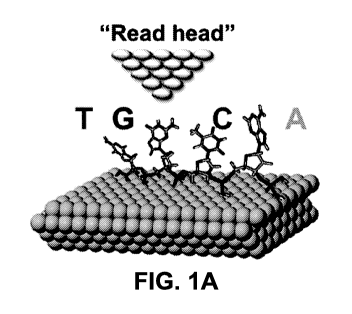
[0013] Figures la-g Sequencing nucleic acid macromolecules like DNA,
RNA, PNA,
using Quantum Molecular Sequencing (QM-Seq). (a) Illustration of QuanT -Seq
showing
single stranded (ss) DNA deposited on clean Au (111) surface. A three-step
extrusion
deposition scheme is used to reproducibly obtain stretched, linearized DNA and
RNA
molecules, with reduced configurational entropy. The metal tip used to obtain
QM-Seq
electronic spectra (tunneling data) acts as a "read head". (b) QM-Seq utilizes
nanoelectronic
tunneling of electrons and holes through nucleotides to provide unique
electronic
fingerprints. Schematic of frontier band structure, HOMO and LUMO molecular
orbitals is
shown for purines and pyrimidines at acidic conditions where significant
differences can be
5
CA 02924021 2016-03-10
WO 2015/038972
PCT/US2014/055512
observed between both nucleobases (not drawn to scale). Different degrees of
conjugation
and chemically distinct nucleobases (adenine and thymine here) lead to
different electronic
states and energy gaps. (c-g) Representative QM-Seq spectra (tunneling data)
for each
(deoxy)ribonucleotide with its corresponding chemical structures. R- can be
either H or OH
for deoxyribonucleotides (DNA) and ribonucleotides (RNA) respectively.
Spectral data was
measured at acidic conditions. Spectra shown here correspond to DNA
nucleotides
(A,C,G,T) and RNA nucleotide (U). Structures shown are (c) (deoxy)adenosine 5'-
monophosphate, (d) (deoxy)guanosine 5'-monophosphate, (e) (deoxy)cytidine 5'-
monophosphate, (f) thymidine 5'-monophosphate and (g) uridine 5'-
monophosphate. A, G,
C, T/U nucleotides are always denoted with green, black, blue and red colors,
respectively.
[0014] Figures 2a-b Frontier Molecular Orbitals of nucleobases,
deoxynucleosides and
ribonucleosides: HOMO, LUMO molecular orbitals structures using density
functional
theoretical (DFT) calculations with B3LYP functional and 6-311G (2d,2p) basis
set for (a)
adenine, deoxyadenosine and adenosine as a purine example; and for (b)
cytosine,
deoxycytidine and cytidine as example of pyrimidine. Shading indicates the
different phases
of the wave function.
[0015] Figures 3a-f Sequencing single DNA molecule using scanning
tunneling
microscopy - scanning tunneling spectroscopy (STM-STS). (a) Illustration
showing the DNA
processing scheme. Denatured single stranded (ss) DNA are deposited on clean
Au (111)
surface modified with poly-L-lysine using an extrusion deposition technique to
reproducibly
obtain elongated linearized DNA template for sequencing. (b) Schematic
illustration of STM-
STS to obtain topographic image, I-V and dl/dV or Density of states (DOS)
spectra of ssDNA
nucleotides, deposited on positively charged Au (111) surface. Electron or
holes tunnel
through single nucleotides to provide the tunneling probability using
electrical tunneling
current data. A, G, C, T nucleotides are, where possible, differentiated by
different shading.
(c-f) Chemical structure of DNA nucleotides (monophosphates), Adenosine 5'-
monophosphate (c), Deoxyguanosine 5'-monophosphate (d), Deoxycytidine 5'-
monophosphate (e), and Deoxythymidine 5'- monophosphate (f), at neutral pH.
[0016] Figures 4a-f Electronic fingerprints obtained using STM-STS for
DNA
nucleotides. (a) Distribution of HOMO (negative) and LUMO (positive) levels
for A, G, C and
T, under acidic conditions (surface washed with 0.1 M NCI). A clear separation
of LUMO
levels (positive voltage peaks) was used to identify pyrimidines (C, T) from
purines (A, G),
and differences in HOMO levels was used to separate pyrimidines (C from T).
(b) Energy
gap between LUMO and HOMO energy levels under acidic conditions. (c) HOMO/LUMO
levels of Thymine at acidic (NCI), neutral (H20) and basic (NaOH) pH
conditions. Arrows
6
CA 02924021 2016-03-10
WO 2015/038972
PCT/US2014/055512
indicate shifts of the LUMO levels between acid, neutral and basic pH
conditions. (d)
Biochemical structures of Thymine at different pH conditions including keto-
enol
tautomerization at acidic conditions, and acid-base behavior between neutral
and basic
conditions. (e) Electron Fowler-Nordheim plot of Thymine at acidic conditions,
characterized
by its transition voltage (Vt 1 and the slope of triangular tunneling
(proportional to the
rans,
tunneling energy barrier). At very small voltages, the tunneling becomes
trapezoidal/rectangular and hence shows deviation from a linear slope(the
slope becomes
logarithmic). (f) Probability density function of transition voltage for
electron (Vtrans ) and hole
(Vtrans h+) at acidic conditions for all four nucleotides. Vtrans e- Vtrans h+
and slope (S) of the
Fowler-Nordheim tunneling show the same behavior as HOMO/LUMO levels and their
energy bandgap ("Band Gap"), respectively.
[0017] Figures 5a-f Electronic fingerprints for DNA nucleotides. (a)
Boxplot of measured
HOMO (negative) and LUMO (positive) levels for A, G, C and T, under acidic
conditions
poly-L-lysine-modified surface (washed with 0.1 M HCI) . Boxplot contains
second and third
quartiles (25-75%) while whiskers show the data from 5-95%. A clear separation
of LUMO
levels (positive voltage peaks) was used to identify pyrimidines (C, T) from
purines (A, G),
and differences in HOMO levels was used to separate pyrimidines (C from T) ,
in protonated
molecules. (b) Energy gap between LUMO and HOMO energy levels under acidic
conditions. This energy gap can be different from a neutral molecule. (c)
HOMO/LUMO
levels of Thymine at acidic (NCI), neutral (H20) and basic (NaOH) pH
conditions. (d)
Biochemical structures of Thymine at different pH conditions including keto-
enol
tautomerization at acidic conditions, and acid-base behavior between neutral
and basic
conditions. (e) Distribution of transition voltage for electron (Vtrans,e )
and hole ( Vtrans,h+ ) at
acidic conditions for all four nucleotides. Vtrans,e - Vtrans,h+ show the same
behavior as
HOMO-LUMO levels and their energy bandgap, respectively. (f) Electron Fowler-
Nordheim
plot of Thymine at acidic conditions, characterized by its transition voltage
( Vtrans,e ) and the
slope of triangular tunneling (proportional to the tunneling energy barrier).
The schematic
shows transition from direct tunneling at low voltages to triangular tunneling
at high bias
voltage. At very low voltages (zero-bias limit), the barrier becomes
rectangular and the
tunneling current shows a logarithmic slope with applied bias voltage.
[0018] Figure 6a-d Sequencing of beta-lactamase gene ampR using STM-STS.
(a)
Characterization of Adenine at acidic conditions on poly-L-lysine modified
gold. Solid green
line shows dl/dV or density of states, dashed grey line is the I-V data, and
dotted green line
shows the distribution of the HOMO and LUMO energy levels. (b) STM image of
single
7
CA 02924021 2016-03-10
WO 2015/038972
PCT/US2014/055512
ssDNA molecule of 1091 nt ampR gene. Image shows DNA is linearized on top of
poly-L-
Lysine modified gold substrate, allowing easy STS identification. (c)
Identification of DNA
nucleotides in the highlighted region shown in (b), using electronic
fingerprint of A, G, C and
T under acidic conditions, measured using STM-STS. Identified nucleotides are
color coded
(black: A or G, blue: C and red: T). (d) Identified ampR sequence based on
primary
(highlighted) and secondary identifications using STS data from (c).
[0019] Figures 7a-d Electronic fingerprints for RNA nucleotides and
comparison to
DNA: (a) Boxplot of HOMO and LUMO energy of the ensemble of single molecule
measurements of RNA nucleotides at acidic conditions, box comprises 25-75%
while
whiskers show the 5% to 95% of the values. (b) Boxplot of measured energy band
gap of
RNA nucleotides at acidic conditions showing two distinct energy levels for
purines and
pyrimidines. (c-d) Comparison of distribution of HOMO/LUMO energy levels for
same
nucleobases on DNA and RNA, (c) deoxyadenosine and adenosine comparison, (d)
deoxycytidine and cytidine comparison.
[0020] Figures 8a-e Identification of single nucleotide modifications using
STM-STS. (a)
STM image of adenine oligomer treated with dimethyl sulfate (DMS), deposited
on poly-L-
lysine coated Au(111) substrate, under acidic conditions. Facile
identification of methylated
and unmethylated adenine on adjoining nucleotides (as shown) highlights the
potential for
detecting single nucleotide modifications, using this new sequencing
technique. (b) Reaction
products of adenine methylation with DMS, (c) Reaction scheme of guanine with
DMS to
produce 7-methyl guanine and its hydrolyzed product with an opened-ring, (d)
Distribution of
HOMO/LUMO levels under acidic conditions for unmethylated (solid line) and
methylated
(dashed line) for adenine, (e) Distribution of HOMO/LUMO levels under acidic
conditions for
guanine (solid line), methylated guanine (dotted line) and ring-opened
methylated guanine
(dashed line).
[0021] Figures 9a-d Identification of single nucleotide modifications
using QM-Seq. (a)
Reaction products of cytosine methylation with DMS. (b) Boxplot (25-75%
quartiles) of
HOMO and LUMO positions under acidic conditions for unmethylated (blue)
cytosine and
methylated cytosine (purple). Whiskers show the 5%-95% percentiles, central
line is the
median. (c-d) Tunneling spectra (I-V, dotted curve) and (dl/dV, solid curve)
of unmethylated
cytosine (c) and methylated cytosine (d). Both have the same vertical axis
(Voltage).
Superimposed blue and purple lines are visual aid to show the difference on
the peak
position with respect to each distribution.
8
CA 02924021 2016-03-10
WO 2015/038972
PCT/US2014/055512
[0022] Figures 10a-b Measurement of I-V and density of electronic states
(dl/dV)
spectra. (a) STS Current (I)-Voltage (V) curve for Cytosine at neutral pH, (b)
its derivative
showing the peaks positions (HOMO and LUMO energy levels) and its energy gap.
The
tunneling signatures shown in other figures are probability density functions
representing
ensembles of at least 20 independent spectroscopy data, measured for the
respective
nucleobases. For each the independent measurement of I-V spectra, the
derivative dl/dV
was used to identify the HOMO and LUMO levels, and the energy band gap. These
were
then used to generate the probability density functions which represents the
normal
distributions from the energy positions of both HOMO and LUMO levels, and the
energy
band gap. The polydispersity of electronic signatures is likely caused by the
configurational
entropy, or charge tunneling through different molecular conformations aided
by the thermal
energy at room temperature.
[0023] Figures 11a-d Chemical structure of nucleotides under different
pH conditions
with their respective pKa. From top to bottom, (a) Adenine (A), (b) Guanine
(G), (c) Cytosine
(C), and (d) Thymine (T). Thymine has a single pKa at 9.9 under acidic
conditions and can
undergo enolization and protonation.
[0024] Figure 12 Effect of pH on guanine LUMO/HOMO levels. Distribution
of LUMO
(positive peak) and HOMO (negative peak) levels for Guanine deposited on Au
(111)
surface, at acidic (washed with 0.1 M NCI), neutral (H20) and basic (0.1 M
NaOH) pH.
Arrows indicate the shift of LUMO and HOMO levels between acidic, neutral and
basic
conditions. Guanine exhibits three biochemical structures at acidic (pH is
below first
pKa-3.2-3.3), neutral and basic conditions (above its second pKa-9.2-9.6).
Likely hole
trapping in isomers results in a steady increase of the HOMO level (harder to
tunnel holes)
as the pH increases (from acidic, to neutral to basic condition). However,
multiple resonance
structures at the acidic and basic conditions (Fig.11) results in easier
electron tunneling (and
lower LUMO levels), compared to neutral condition. Moreover, further
electrostatic repulsion
at basic condition (due to pKa2) improves electron tunneling probability, and
results in a
further decrease of LUMO level for basic pH.
[0025] Figures 13a-e Raw data and statistics of guanine: (a) Raw current-
voltage (I-V)
curves for Guanine at acidic conditions. (b) Raw spectra or dl/dV of (a),
arrows indicate
identified HOMO/LUMO levels as the first significant negative/positive peak on
each spectra.
(c-e). Histograms of the positions of HOMO (c), LUMO (d) and Energy Gap (e)
for guanine,
superimposed by a normal probability density function (indicated by curve,
also shown in
Fig.4a,b) fitted to the data set. The shaded box indicates the area of the
curve comprising
the mean standard deviation.
9
CA 02924021 2016-03-10
WO 2015/038972
PCT/US2014/055512
[0026] Figure 14 Effect of pH on adenine LUMO/HOMO levels. Distribution
of LUMO
(positive peak) and HOMO (negative peak) levels for Adenine deposited on Au
(111)
surface, at acidic (washed with 0.1 M NCI), neutral (H20) and basic (0.1 M
NaOH) pH. While
Adenine has multiple resonance structures at any pH conditions (both charged
and
uncharged), significant effect of pH on its tunneling probability is not
observed (due to
dissipation of the charge amongst the resonance structures). Minor increase in
HOMO level
with increase in pH can be attributed to easier hole tunneling at acidic pH
(due to the positive
charge).
[0027] Figures 15a-e Raw data and statistics of adenine: (a) Raw current-
voltage (I-V)
curves for Adenine at acidic conditions. (b) Raw spectra or dl/dV of (a),
arrows indicate
identified HOMO/LUMO levels as the first significant negative/positive peak on
each spectra.
(c-e). Histograms of the positions of HOMO (c), LUMO (d) and Energy Gap (e)
for adenine,
superimposed by a normal probability density function (indicated by curve,
also shown in
Fig.4a,b) fitted to the data set. The shaded box indicates the area of the
curve comprising
the mean standard deviation.
[0028] Figure 16 Effect of pH on cytosine LUMO/HOMO levels. Distribution
of LUMO
(positive peak) and HOMO (negative peak) levels for Cytosine, deposited on Au
(111)
surface at acidic (washed with 0.1 M NCI), neutral (H20) and basic (0.1 M
NaOH) pH.
Cytosine has a clear pH effect with two main structures: above its pKa-4.4, no
difference
appears between neutral and basic conditions. However, its protonated form at
acidic
conditions show likely electron trapping effect, increasing the LUMO energy
level.
[0029] Figures 17a-e Raw data and statistics of cytosine: (a) Raw
current-voltage (I-V)
curves for Cytosine at acidic conditions. (b) Raw spectra or dl/dV of (a),
arrows indicate
identified HOMO/LUMO levels as the first significant negative/positive peak on
each spectra.
(c-e). Histograms of the positions of HOMO (c), LUMO (d) and Energy Gap (e)
for Cytosine,
superimposed by a normal probability density function (indicated by curve,
also shown in
Fig.4a,b) fitted to the data set. The shaded box indicates the area of the
curve comprising
the mean standard deviation.
[0030] Figures 18a-d Identification of single nucleotide modifications
using QuanT -Seq.
(a) Reaction products of methylation of Adenine with DMS. (b) Reaction
products of
methylation of Guanine with DMS. (c) Boxplot of HOMO and LUMO energy levels
distribution for adenine and methylated adenine deposited on poly-lysine
modified Au (111)
surface, under acidic conditions. Addition of a methyl group shifts the HOMO
level by
reducing the hole tunneling probability. (d) Boxplot of HOMO and LUMO energy
levels
CA 02924021 2016-03-10
WO 2015/038972
PCT/US2014/055512
distribution for guanine and methylated guanine deposited on poly-lysine
modified Au (111)
surface, under acidic conditions.
[0031] Figures 19a-e Raw data and statistics of Thymine: (a) Raw current-
voltage (l-V)
curves for Thymine at acidic conditions. (b) Raw spectra or dl/dV of (a),
arrows indicate
identified HOMO/LUMO levels as the first significant negative/positive peak on
each spectra.
(c-e). Histograms of the positions of HOMO (c), LUMO (d) and Energy Gap (e)
for Thymine
(bars), superimposed by a normal probability density function (indicated by
curve, also
shown in Fig.4a,b) fitted to the data set. The shaded box indicates the area
of the curve
comprising the mean standard deviation.
[0032] Figure 20 Configurational energy contribution to HOMO, LUMO and
Energy gap
dispersion for adenine (nucleobase) adsorbed on graphene ¨ Adapted from Ahmed
et al.
which describes DFT simulation of a nucleobase at different configurations
positioned on top
of a conductive substrate and its contribution to the local density of states
based on DFT
theory. Lines are local density of states (LDOS) of nitrogen atom adsorbed on
graphene at
different angles (conformation superimposed in the center). Yellow-shaded
regions
correspond to dominant peak near Fermi level. Grey-shadow boxes represent the
distribution of predominant peak (positive and negative) near the Fermi level
considering all
possible conformations (from 0 to 90 ).
[0033] Figures 21a-d Effect of pH on electron and hole transition
voltage (between
tunneling and field emission regimes), from Fowler-Nordheim plot. Vtrans for
electron (Vtrans e_)
and hole (Vtrans h+) is shown for (a) Adenine (A), (b) Guanine (G), (c)
Cytosine (C), and (d)
Thymine (T). Arrows indicate the shift of Vtrans e- and Vtrans ,h+ between
acidic (NCI), neutral
(H20) and basic (NaOH) conditions. All these transitions mimic the respective
changes in
LUMO and HOMO levels, thereby confirming the role of Vtrans as one potential
biophysical
figure of merit.
[0034] Figures 22a-c Tunneling properties of DNA nucleotides Guanine,
Cytosine and
Thymine. l-V (dashed line), dl/dV or density of states (solid line) and
probability distribution
of LUMO and HOMO levels (dotted line) for Guanine (a), Cytosine (b) and
Thymine (c). The
dotted lines are the normal probability distribution functions fitted for both
LUMO and HOMO
energy levels.
[0035] Figures 23a-b Linearization of ssDNA using the extrusion
deposition technique.
STM images of ssDNA deposited on bare gold without extrusion (a) and on poly-L-
lysine
modified gold with extrusion (b). The role of poly-L-lysine coating and our
extrusion
11
CA 02924021 2016-03-10
WO 2015/038972
PCT/US2014/055512
deposition scheme is clearly visible in this STM data, where linearized DNA
allows clear STS
identification of single nucleotides (Fig.25).
[0036] Figures 24a-b Identification of single nucleotide modifications
using STM-STS.
(a) Reaction products of methylation of Cytosine with DMS. (b) HOMO and LUMO
energy
levels distribution for cytosine and methylated cytosine deposited on poly-
lysine modified Au
(111) surface, under acidic conditions. Addition of a methyl group shifts the
HOMO level by
reducing the hole tunneling probability.
[0037] Figure 25 Single molecule DNA detection capability. Using a low
concentration
of ssDNA (1-5 nM in doubly distilled water or TE buffer
(Tris(hydroxymethyl)aminomethane-
Ethylenediaminetetraacetic acid (or EDTA) buffer) to mimic physiological
concentration,
using the disclosed technique several DNA linearized strands can be detected
using STM-
STS sequencing. In a sample scan shown here, DNA molecules were found in a
small scan
area (1pm x1pm) on ultrasmooth Au(111) substrate. This demonstrates the
capability of this
sequencing technique to detect and sequence very low concentrations of DNA
molecules.
[0038] Figure 26 Depicts a substrates forming channels in a microfluidic
device. The
channel dimensions (width) can vary between 100 nanometers (nm=10-9 m) to 50
micrometers pm.
[0039] Figures 27a-c (a) is a picture of centimeter scale optically
created tip patterns,
using a simple optical lithography, followed by anisotropic KOH etching. (b)
SEM image
showing high fidelity and periodically patterned STM tips made from gold.
Using a large area
(cmXcm) scale STM chip on an ultraflat/ultrasmooth substrate, a 2 pm x2 pm
surface can be
scanned, and create an entire sequence over cm scale, by massively parallel
scanning and
simple readout from a chip, similar to the ones shown in the figure. (c) is a
lmegapixel (or
one megatip) 2cmX2cm chip is shown. Voltage can be simultaneously applied to a
plurality
of tips, the current is collected and stored, and all current values from the
plurality of tips
may be read simultaneously (similar to a CCD camera). After the current is
read, another
bias voltage can be applied, and so on, to recreate the entire current-voltage
curve over a
massive 2cmX2cm substrate. Several thousand genomes can be placed, linearized
and
read simultaneously in the microfluidic channels. Piezos may be used to move a
sample a
few angstroms, to allow for sequencing the next nucleobases ¨ and the process
repeated to
analyze additional nucleobases. Therefore, in a single 2 micrometer scan
movement (or
piezo scan), of the massively parallel sequencer can sequence all possible
nucleobases on
a relatively large sample biochip, patterned using a simple microfluidic
device.
12
CA 02924021 2016-03-10
WO 2015/038972
PCT/US2014/055512
[0040] Figure 28 Schematic diagram showing method of base calling by
automatic
method.
[0041] Figure 29 Structure determination based on the reactivity. The
secondary/tertiary
nucleic acid structure, RNA here, was obtained using electronic fingerprints
of chemical
modification with RNA SHAPE and/or DMS molecule, and using RNA Structure
software
with constrained single-stranded regions where SHAPE or DMS had reacted.
[0042] Figure 30 Assignment of reacted vs. unreacted nucleotides during
RNA structure
determination.
[0043] Figure 31 The Clustering method assigns the RNA nucleotides with
high
confidence. The diagonal indicates accurate base calling. Letters in uppercase
are the
unmodified RNA nucleotides, letters in lower case are the modified RNA
nucleotides.
[0044] Figure 32 RNA structure of HIV-RNase measured experimentally with
QM-Seq
(upper panel). Lower panel shows an in silico unconstrained RNA structure
predicted using
RNA folding software.
[0045] Figure 33 Comparison between using (top) 3 parameter electronic
states
(HOMO-LUMO-Energy gap), and (bottom) multidimensional biophysical parameters
(>9
parameters, including but not limited to HOMO, LUMO, Energy gap, tunneling
barrier heights
for electron and holes, difference in tunneling barrier heights, voltages
corresponding to
change in tunneling barrier profile from direct tunneling to Fowler-Nordheim
tunneling for
electron and holes, effective masses of electrons and holes in nucleotide
tunneling, ratio of
effective electron and hole masses, slopes of corresponding Fowler-Nordheim
plots), all
calculated from quantum tunneling spectroscopy scans and used as electronic
fingerprints,
obtained by QM-Seq on HIV-1 RNAse.. The electronic states can help in
identification
between RNA purines and pyrimidines, but the multi-variable electronic
fingerprints allow
unique identification of all four nucleobases with high precision, as shown in
this figure
(bottom).
[0046] Figures 34a-h Different Biophysical parameters used as electronic
fingerprints for
DNA nucleotide (A,T,G,C) identification determined on a poly-lysine coated
ultraflat Au(111)
substrate in acidic conditions. a) LUMO-level b) HOMO-level c) Barrier height
for electrons
d) Barrier height for holes e) Total tunneling barrier height for molecule f)
ratio of effective
electron and hole masses for charge tunneling through individual nucleotides.
Transition
voltage from direct to Fowler-Nordheim tunneling for g) electrons and h)
holes.
13
CA 02924021 2016-03-10
WO 2015/038972
PCT/US2014/055512
[0047] Figures 35a-h Different Biophysical parameters used as electronic
fingerprints
for RNA nucleotide (A,U,G,C) identification on modified Au(111) substrate in
neutral
conditions. a) LUMO-level b) HOMO-level c) Barrier height for electrons d)
Barrier height for
holes e) Total tunneling barrier height for molecule f) ratio of effective
electron and hole
masses for charge tunneling through individual nucleotides. Transition voltage
from direct to
Fowler-Nordheim tunneling for g) electrons and h) holes.
[0048] Figure 36 Schematic diagram showing method of base calling by
automatic
method.
[0049] Figure 37 Flowchart showing an embodiment of a method for
determining the
identity of a nucleobase, its position on a substrate, and its sequence in a
polynucleotide.
DETAILED DESCRIPTION
[0050] Before the present disclosure, the challenge for DNA sequencing
using tunneling
spectroscopy has been to identify a unique tunneling spectrum for each
nucleotide.
Quantum tunneling spectroscopy of DNA nucleotides represents the electronic
density of
states of the individual nucleobase, nucleoside, and nucleotide. Disclosed
herein are
methods, devices, and compositions that are used to determine unique
fingerprints for
modified and unmodified DNA and RNA nucleobases, nucleosides, and nucleotides
for use
in comparison with electronic signatures of a nucleotide whose identity is
unknown (an
unknown nucleoside, nucleotide or nucleobase) to aid in identification of the
unknown
nucleotide. Previous attempts to identify nucleotides from both single
stranded (ss) DNA and
double stranded (ds) DNA have been generally unsuccessful in determining
unique
tunneling spectra for the four DNA nucleobases, nucleosides, and nucleotides.
[0051] The disclosed methods, devices, and compositions also aid in
alleviating
limitations of existing methods of sequencing RNA. The disclosed methods,
devices, and
compositions may be used in the direct sequencing of RNA, with non-amplified
templates at
a single molecule level. In many cases, the present disclosure may aid in
determining the
identity and abundance of RNA molecules obtained from a cell or tissue.
Further, the
present disclosure's identification of unique electronic tunneling spectra
(tunneling data) for
nucleotide (DNA/RNA) modifications of single molecules can provide a useful
epigenomics
technique for early detection of diseases. Epigenomic studies can provide
insights into
dynamic states of genomes, especially their role in determining disease states
and
developmental biology.
14
CA 02924021 2016-03-10
WO 2015/038972
PCT/US2014/055512
[0052] The disclosed methods, devices, and compositions provide for
collection of
tunneling data or I-V data that is highly reproducible with little noise.
Previous methods
suffered from a lack of reproducibility and low signal to noise ratios. The
presently disclosed
methods, devices, and compositions provide for enhanced data collection in
various ways.
For example, the disclosed methods, devices, and compositions use an
ultrasmooth charged
surface that is coated with an ionic polymer. In one embodiment, an Au(111)
charged
surface may be coated with poly-lysine. The use of an ionic polymer may aid in
orienting the
nucleic acid backbone, which may provide for tunneling data with greater
reproducibility and
higher signal to noise ratios than previous methods. In addition, the
disclosed methods,
devices, and compositions may use a defined environment to collect fingerprint
data. For
example, the disclosed methods, devices, and compositions may perform quantum
tunneling
in a high or low pH environment to aid in differentiating various modified and
unmodified
nucleobases, nucleotides, and nucleosides. The use of a defined environment
may also aid
in enhancing the tunneling data obtained.
[0053] Nanoelectronic tunneling is a quantum-physical process that occurs
at the
nanoscale. Nanoelectronic tunneling takes advantage of the tendency of the
wavefunctions
of separate atoms or molecules to overlap. If a voltage bias, or bias, is
applied (by increasing
or decreasing a potential of a metal tip positioned near the atoms of a
substrate in contact
with the atoms), tunneling of either electrons or holes between the tip and
the atom/molecule
can occur, even over a potential barrier. While classical charge conduction
nominally occurs
from a region of high potential to a region of low potential, where the two
regions are in
separated by downstream potential bias (current flows from high to low
potential), quantum
tunneling occurs without physical contact (and hence the density of molecular
states is
unperturbed by measurement) over a potential barrier height, and where the
tunneling
probability is reduced with increase in barrier height. Electrons can be
injected (electron
tunneling) or extracted (hole tunneling) to/from one of the molecules due to
the wavefunction
overlap.
[0054] Tunneling current spectra of a nucleotide represents the
electronic density of
states. Disclosed herein is the use of tunneling current data to create unique
fingerprints for
use in nucleotide identification. Several attempts have been made by modeling
and by
experiments to identify and differentiate different nucleotides from both
single stranded (ss)
DNA and double stranded (ds) DNA, RNA, PNA, other nucleic acid macromolecules,
DNA/RNA/PNA nucleotide modifications, nucleic acid structures. However, until
the present
disclosure, only guanine (G) bases has been only partially successfully
identified using
tunneling microscopy on ssDNA.
CA 02924021 2016-03-10
WO 2015/038972
PCT/US2014/055512
[0055] Presented herein is a first demonstration of determining unique
electronic
fingerprints of nucleotides, nucleosides, and nucleobases A, G, T, C and U
performed using
single-molecule DNA/RNA/PNA sequencing. In addition, unique fingerprints of
modified
nucleotides/nucleobases are also disclosed. Nucleobase may refer to cytosine
(abbreviated
as "C"), guanine (abbreviated as "G"), adenine (abbreviated as "A"), thymine
(abbreviated as
"T"), and uracil (abbreviated as "U"). C, G, A, and T may be found in
deoxyribonucleic acid
(DNA) and C, G, A, and U may be found in ribonucleic acid (RNA). Fig. 1 shows
electronic
fingerprints determined by quantum tunneling spectroscopy for nucleotides A,
G, C, T and U.
The terms nucleoside, nucleotide, and nucleobase are used interchangeably and
refer to
natural and synthetic, and modified and unmodified nucleosides, nucleotides,
and
nucleobases.
[0056] The disclosed technique uses quantum tunneling data to create an
electronic
signature for unknown nucleotides, nucleoside, and nucleobases to aid in
determining their
identity, and may be performed at room temperature (i.e. about 20-25 C), or at
cryogenic
temperatures between 1K to 300K. In some cases, the electronic state of the
nucleotides,
nucleoside, and nucleobases may shift depending on the biophysical condition,
or
environment, for example the pH at which the nucleotide, nucleoside, or
nucleobase is
analyzed. In some cases, distinct states of the nucleotide, nucleoside, or
nucleobase may be
identified at acidic pH (i.e. pH less than about 7). In many embodiments, the
pH of the
environment used to determine the electronic parameters is less than about 3.
[0057] Fingerprints of modified and unmodified nucleotides, nucleoside,
and
nucleobases may be determined in various biophysical conditions or
environments, which
may shift their electronic state. This may aid in differentiating nucleobases
that may have
similar or overlapping parameter values under some biophysical conditions.
This may aid in
identifying the nucleobase by comparing it to signatures of known nucleobases
determined
in the same environment. As described above, the fingerprint of a nucleobase
may be
determined at a given pH and compared to fingerprints of known nucleobases
obtained in
the same pH. In other environments, the fingerprint may be determined in an
environment
having specific characteristics other than pH, for example molarity, polarity,
hydrophobicity,
etc. In various embodiments, the nucleobase may be determined in an
environment
comprising a given amount of an alcohol, salt, or non-polar solvent or solute.
[0058] As disclosed herein, "tunneling current data" or "current data"
or "I-V data" refers
to current and voltage (bias voltage) data measured in quantum tunneling at
various bias
voltages. Tunneling current data may refer to I-V, dl/dV and/or I/V2 data
acquired from the
tunneling current measurement. In most cases, various parameters or values are
derived
16
CA 02924021 2016-03-10
WO 2015/038972
PCT/US2014/055512
from tunneling current data. Parameters may include values for LUMO, HOMO,
Bandgap,
Vtrans+ (V), Vtrans- (V), 1)e- (eV), h+ (eV), melmh+ and A(I) (eV) (described
below).
[0059] As disclosed herein, "signature" or "electronic signature" refers
three or more
values for parameters derived from I-V data collected for a nucleotide of
unknown identity.
Parameters for use in creating a signature include LUMO, HOMO, Bandgap,
Vtrans+(V)
- Vtrans-
(V), (Pe_ (eV), (Ph+ (eV), melmh+ and A(I) (eV), any three or more of which
may be used to
create the signature. For example, in some embodiments, an electronic
signature of an
unknown nucleotide may comprise values for LUMO, HOMO, and Bandgap. In other
embodiments, an electronic signature may comprise values for LUMO, HOMO,
Bandgap,
Vtrans+ (V), Vtrans- (V), (eV), (eV), m /m and A(I) (eV).
rans+ - , - = e- ,, = h+ - ¨e- ¨h+
[0060] As disclosed herein, "fingerprint" or "electronic fingerprint"
refers to three or more
values for parameters derived from I-V data collected for a nucleotide of
known identity. The
parameters selected for creating a fingerprint for a known nucleotide are the
same as those
selected for creating a signature for the unknown nucleotide, to which the
known nucleotide
is being compared. Values for a givent parameter used in creating an
electronic signature
may be represented as a value +/- a standard deviation, or as a range of
values.
Parameters for use in creating a fingerprint include LUMO, HOMO, Bandgap,
Vtrans+(V),
Vtrans- (V), 1)e- (eV), h+ (eV), melmh+ and A(I) (eV). In some embodiments, an
electronic
signature for an unknown nucleobase may comprise values for LUMO, HOMO, and
Bandgap, and this signature may be compared to electronic fingerprints of
known
nucleobases, wherein the fingerprints comprise values for the same parameters -
LUMO,
HOMO, and Bandgap. In other embodiments, the signature may comprise values for
LUMO,
HOMO, Bandgap, Vtrans+ (V), Vtrans- (V), (De- (eV), Oh+ (eV), Me-/Mh+ and A(I)
(eV), and may be
compared to a fingerprint comprising values for LUMO, HOMO, Bandgap, Vtrans+
,V,, Vtrans-
(V), (Pe- (eV), (Ph+ (eV), me-/mh+ and A(I) (eV).
[0061] The disclosed techniques may be used to sequence polynucleic
acids,
polynucleotides, and other polymeric molecules comprising one or more
nucleotide,
nucleoside, or nucleobase.
[0062] In many cases, a flame-annealed flat, template-stripped
ultrasmooth gold (111)
crystal facet substrate may be used. Designation (111) here indicates the
crystal structure of
the exposed top surface of the gold atoms. Other orientations can also be used
for this
purpose (e.g. 100). Ultrasmooth substrates have very low surface roughness,
for example
less than about 1.0 nm variation from a planar surface. Described herein are
methods for
obtaining ultrasmooth substrates using a flame annealing and template
stripping process as
17
CA 02924021 2016-03-10
WO 2015/038972
PCT/US2014/055512
described below. In some embodiments, other substrates may be used. In some
embodiments, other conductive substrates may be used, for example graphene,
highly
ordered pyrolytic graphite (HOPG), atomically-flat freshly cleaved mica with
gold (or other
metal) coating, other ultrasmooth metals like copper (111), silver etc. In
many cases, the
substrate should be conductive for the purposes of scanning and quantum
tunneling
spectroscopy, and smooth for easy identification of single molecules.
[0063] In some embodiments, a polynucleotide may be linearized DNA and
the
polynucleotides may be drawn-out on the disclosed ultrasmooth substrate. This
may aid in
separating individual nucleotides and reducing their configurational entropy
for scanning.
This may aid in the study of charge tunneling through the nucleobases, instead
of the sugar
backbone. In some cases, the substrate may be a charged substrate. For
example, where
the substrate is gold, a positively charged gold (111) surface may be
prepared.
[0064] In some embodiments, a positively charged gold substrate is
produced for use
with an extrusion deposition technique. First, freshly prepared ultrasmooth
gold (111)
surface is treated in a plasma cleaner (e.g. ozone plasma cleaner), to prepare
a uniformly
negatively charged surface. In many embodiments the gold may then be treated
with an
ionic solution, for example a positively charged molecule such as poly-L-
lysine, to produce a
uniformly coated positively charged gold surface. In some embodiments, the
extrusion-
deposition technique involves a three step process to disperse elongated
linear ssDNA on a
gold substrate. In a first step, a gold (111) surface may be charged by
treating it with a
chemical solution. In some cases, the gold surface may be positively charged
by coating it
with poly-L-lysine, for example 10ppm poly-L-lysine solution. Other molecules,
for use in
coating an ultrasmooth surface, can include any polycationic polymer, for
example
polyallylamine hydrochloride, catecholamine polymer, amino silane like
aminopropylethoxysilane, or epoxide modified silanes like 3' glycidoxy
propyltrimethoxysilane. In other embodiments, electrostatic fixing of the
negative charge of
the sugar-backbone can be performed by applying a voltage to electrically bond
the
backbone to the substrate. In some cases, the chemical solution may aid in
linking the
negatively charged phosphate backbone via electrostatic interaction to a
substrate that is
positively charged. In embodiments used to sequence a polynucleotide, acidic
conditions
may aid in de-convoluting nucleotides, for example pyrimidines C or T, and
purines ¨ G or A.
[0065] A second step in the extrusion-deposition technique may involve
melting single-
stranded DNA (ssDNA). For example, ssDNA may be melted by heating the ssDNA,
for
example at 95 C for 5min. In most embodiments the melted ssDNA is rapidly
cooled, which
may aid in preventing the formation or re-formation of secondary and/or
tertiary structure in
18
CA 02924021 2016-03-10
WO 2015/038972
PCT/US2014/055512
the ssDNA. In some embodiments, rapid cooling may involve flash cooling on ice
for 5 min.
In many embodiments, dsDNA and short mononucleotide ssDNA may not contain
tertiary
structures; ssDNA longer than about 1 kb may form secondary structures. In
many cases, a
positively charged surface may help to disrupt or prevent formation of
secondary structures.
[0066] A third step in the extrusion-deposition process may include
extruding the ssDNA
onto the gold substrate. In some cases, a translational motion may be used to
deposit and
draw out a linearized DNA chain on the charged substrate from a DNA dispensing
device,
for example a pipette.
[0067] In some embodiments, a chemically-etched tip may be used for
nanoelectronic
tunneling. In some embodiments, a platinum-iridium tip (80:20 Pt-lr) may be
used. In other
embodiments, other suitable STM tips can also be used. Some other commonly
used tips,
that may be used are tungsten, gold, carbon and platinum metal. Other tips
commonly used
are Pt, I, W, Au, Ag, Cu, Carbon nanotubes and combinations thereof.
[0068] Known and unknown nucleotides are studied by tunneling electrons
and holes
through the nucleotides. In some cases, the nucleotides studied are
linearized, single
stranded polynucleotides, as depicted in Fig.1a,b.
[0069] The tunneling current spectroscopy (current (I)-voltage (V)) may
be a direct
measure of the local electronic density of states (dl/dV spectra, Fig.10 and
described in
more detail below) of the molecule, and may serve to provide a unique
electronic fingerprint
based on the nucleotide's biochemical structure (Fig.1).
[0070] An electronic signature is obtained for a nucleotide using
quantum tunneling, at
molecular resolution (Fig.10a). In some cases, an electronic density of states
(DOS) may be
obtained from a first derivative of the current-voltage (I-V) spectrum, and a
first significant
positive and a first significant negative peak assigned as a Lowest Unoccupied
Molecular
Orbital (LUMO) energy level and a Highest Occupied Molecular Orbital (HOMO)
energy
level, respectively. In many cases, a first significant peak is a peak that is
at least about 30%
of the maximum dl/dV, or the first derivative of the current-voltage spectrum
(wherein the
first derivative represents the density of states for the biomolecule for
electron and hole
tunneling and greater than about 1.0 V. In some cases, a peak that occurs at
less than
about 1.0V (between 0 and +1.0 V or 0 and -1.0 V) may indicate a conductive
substrate or
a minor contamination from the environment. The difference between these first
peaks may
be assigned (designated) as the LUMO/HOMO energy gap or "band gap" (Fig.10b).
The
electron tunneling peak (on application of positive bias voltage here)
corresponds to the
LUMO levels, and the hole tunneling peak (on application of negative bias
voltage here)
19
CA 02924021 2016-03-10
WO 2015/038972
PCT/US2014/055512
corresponds to the HOMO levels of the molecule. The difference between the
LUMO and
HOMO levels is the energy bandgap of the molecule.
[0071] Additional biophysical parameters which are intrinsic to each
nucleobase can
also be calculated using the two distinct tunneling regimes (direct tunneling
and Fowler-
Nordheim tunneling) separated by a transition voltage (V, 1 at the inflection
point. Two
rans,
main models for quantum tunneling were developed based on the WKB
approximation
applied to the SchrOdinger equation. Simmons model for tunneling between
electrodes
separated by an insulator (eq. 1) describes the tunneling current at both
regimes, its
dependence on the applied bias voltage and the effect of the original
tunneling barrier.
A com*i) ) (2com*4)-Hiv)i
I = 4nq2hd 1:130 e (2
h ¨ qV) e h (eq. 1)
[0072] Where (13, is the average barrier height which is proportional to
the applied voltage
as the shape of the tunneling barrier changes from rectangular to trapezoidal
and triangular,
m* is the effective electron mass, h the reduced Plank's constant, d is the
mean tunneling
distance, A is the effective tunneling area, q is the elementary charge and V
is the applied
bias voltage. The model is generic for any shape of tunneling barrier as only
the average
barrier height is required (.13.).
[0073] The other analytical approach used for quantum tunneling is based
on Stratton
model (eq. 2), also derived from WKB approximation. While both Simmons and
Stratton
model starts from the same current density description, they took different
approximations
for solving the tunneling probability integral which yields to different
equation sets. Stratton
equation for describing quantum tunneling is:
4nmqA r n c(V)kT [1. e-c(v)qvie-b(v) (eq. 2)
I = h3 C2 (V) c(V)1cT)
[0074] Where m is the electron mass, k is the Boltzmann constant, T is
the temperature
and b(V) and c(V) are two parameters resultant from the Taylor expansion of
the tunneling
probability and defined as:
b= a fx2(0 ¨ 07dx and c = la fx2(0 ¨ 0-7dx
xi 2 xi
Where a = /h and x, and x2 are the positions where gb ¨ = 0 for each
side of the
tunneling gap, is the Fermi energy of the electrode and gb is the energy
barrier (x and V
dependent).
[0075] While these parameters can be fitted experimentally with temperature
dependence of tunneling current, the model was simplified to the form of I a
sinh(q171-/h),
CA 02924021 2016-03-10
WO 2015/038972
PCT/US2014/055512
as it describes the sequencing conditions used here. Using this relationship,
we derived the
minimum (Vtrans) on the In(I/V2) vs. V-1 plot as the following equation within
a few percent
error:
2h
V ¨ ¨ (eq.3)
trans q d
[0076] Using Simmons model, a simplified Fowler-Nordheim equation is
derived for high
bias voltages (qV > 4)0). This takes the following form:
I *
in (L) 4d,\2m(13.8 3hq (1)
(eq.4)
v2 V
[0077] Combining both models, one can derive expressions for the direct
calculation of
the original barrier height (4)0) and the "effective" tunneling distance (d-
r\/re) using
experimental data extracted directly from the FN plot:
Vtrans.3=S d 3=S=hq
(I)0 = 7¨\/r2*
16
16,\127/10003
[0078] Where S is the slope of the In(I/V2) vs. V-1 corresponding at
high bias voltages
(qV > 4)0). Note that both Stratton and Simmons use the same approximation of
the
SchrOdinger (WKB) and the only difference come on the treatment of tunneling
probability
integrals. Hartman made a comparison of both models against the exact solution
of WKB
approximations and both Stratton and Simmons model are within a few percentage
of error
from the exact solution. With this approximation, using both models,
experimental
spectroscopic data can be fit on either model that would be impossible
otherwise due to
intractability of the non-linearity of both models.
[0079] This method allows the quantitative comparison of nucleotides by
examining up
to 9 parameters (HOMO Voltage, LUMO Voltage, Energy Bandgap Vtrans, Vtrans,
h+,
4)0,h+ , Anat, and eff e- ¨eff m /mh+/ 1= In many embodiments, the
signatures may be determined by
¨
analyzing values for at least three parameters. In most embodiments, more than
three
parameters are used to determine a signature. For example, four, five, six,
seven, eight, or
nine parameter values may be used to determine a signature for comparison to a
fingerprint
comprising the same parameter values.
[0080] Nucleotide fingerprints and signatures are determined by
submitting the
nucleotide to quantum tunneling and then collecting and analyzing the
tunneling current
data. In many cases, in order to create a quantum tunneling nucleotide
fingerprint, tunneling
current data is collected from about 15 to about 50 points on an individual
nucleotide
molecule (for example a single molecule of adenine). In addition, quantum
tunneling data is
21
CA 02924021 2016-03-10
WO 2015/038972
PCT/US2014/055512
collected for about 20 different individual molecules, which may aid in
creating a statistically
accurate fingerprint of the nucleotide.
[0081] Probability density curves (Voltage, V, or Energy, eV, versus
probability density
function (dl/dV)) of DNA several known nucleotides have been determined.
Several
probability density curves are shown in Figs. 4a, 4b, 4c, 4f, 8d,8e, 12, 14,
16, 21, 22, and
24b. These curves are statistical distributions of independent measurements,
which have
been fitted to a normalized sum of Gaussian curves (equation S1, below. Ni:
normalization
constant, V: applied bias voltage, mean, o-i: standard deviation).
P (V) = E, INiexp [ __________ (v IL,l)21) Equation S1
2oT
1 0 [0082] These parameters may be used to create an electronic
fingerprint for a given
nucleotide consisting of HOMO level, LUMO level, and energy gap (Band Gap). In
many
embodiments, nucleobase fingerprints of known nucleobases may be used to
analyze the
quantum tunneling signature collected from an unknown nucleotide or
polynucleotide DNA
molecule to determine the nucleotide's identity and the polynucleotide's
sequence.
[0083] Nucleic acids biochemistry may be defined by the environment where
the nucleic
acid is found. In some cases, the surrounding pH may affect the structure of a
nucleic acid,
for example a nucleobase/nucleotide. In some embodiments altering the pH may
result in
the nucleobase having different structures. This effect may occur above and/or
below a
nucleobase's pKa, as shown in Fig.11. Additionally, besides acid-base
behavior, other
biochemical changes can occur at extreme pH (either acidic or basic). For
instance, thymine
can form tautomers at acidic pH where enolized-T is predominant over the keto
form.
[0084] The relative charge of DNA nucleotides can facilitate either
electron or hole
tunneling depending on the system pH. For example, in some embodiments a
positively
charged DNA nucleotide species may facilitate hole tunneling and increase the
energy level
for electron tunneling (LUMO), and a negatively charged species may exhibit
the opposite
behavior (Fig.12,14). This effect can be observed on the spectra shift for a
guanine
nucleotide along its two pKa (Fig.12) where the nucleotide transitions between
positively
charged structure under acidic pH, to a negatively charged structure at basic
pH. In some
embodiments, electrostatic interactions may, therefore, change the probability
of the charge
tunneling (increases on charge repulsion), resulting in different (lower)
respective LUMO and
HOMO levels.
[0085] Tunneling signatures (or fingerprints) for individual nucleotides
may differ under
different environmental conditions, for example under different pH conditions.
In many
22
CA 02924021 2016-03-10
WO 2015/038972
PCT/US2014/055512
cases, electron/hole tunneling current through a nucleotide is collected under
different
environmental conditions. Differences in quantum tunneling signatures under
different
environmental conditions, may in some cases be due to the presence of keto-
enol tautomers
of the nucleobases, which may differ under different pH conditions (Fig.11 and
as discussed
below). The presence or absence of a specific keto-enol tautomer may lead to
separation of
electron/hole tunneling probability between different nucleobases, for example
between
purines (A,G) and pyrimidines (C,T).
[0086] The charge density of a nucleotide may aid in determining the
energy
increase/decrease for these effects. In some cases, purines, which may have
several
conjugated structures, may have a local charge on any atom that is
significantly reduced in
comparison with pyrimidines, which may have the charge localized on a single
atom
(Fig.11). In some embodiments, the conjugation effect may have a significant
impact on the
tunneling energy shifts and may be readily observed in acidic conditions
(Fig.4c, 12, 14, 16),
for example, where purines may exhibit a significantly smaller effect than
pyrimidines (e.g.
adenine data in Fig. 14).
[0087] In many cases, the use of HOMO-LUMO and energy gap parameters may
aid in
distinguishing purines (A,G) from pyrimidines (C,T) under acidic conditions
based on the
energy gap (there is about a 1.7-2 eV difference between the purines A, 2.73
eV and G 2.58
eV and the pyrimidines C, 4.43 eV and T, 4.82 eV) and LUMO level (about 1.5 eV
difference
between the purines A, 1.61 V and G 1.49 V and the pyrimidines C, 3.13 V and
T, 3.08 V). In
some embodiments, C and T may be distinguished or de-convoluted based on their
HOMO
energy level difference (about 0.45 eV difference between C, -1.30 V and T, -
1.74 V). In
further embodiments A and G can be distinguished/differentiated/de-convoluted
using their
LUMO levels at basic pH (about 0.40 eV difference between A, 1.72 V and T,
1.33 V).
Characteristic LUMO, HOMO, and Band Gap values for the nucleobases A, T, G,
and C are
presented in Table I. Table I shows these values determined at neutral, acidic
and basic pH
environments. Thus, in some embodiments, the identity of an unknown nucleotide
may be
determined by collecting quantum tunneling data on the nucleotide at one or
more pH values
(acid, basic, and neutral), determining the LUMO, HOMO, and Band Gap values
for that
nucleotide, and comparing those values to values previously determined for
nucleotides of
known identity.
Table I: Summary of LUMO, HOMO and band gap energy levels for A, C, G, and T
on bare
Au(111) surface under different pH conditions. Values correspond to mean
standard
deviation.
23
CA 02924021 2016-03-10
WO 2015/038972
PCT/US2014/055512
Voltage (V) / Energy (eV) HCI (acidic) H20 (neutral) NaOH
(basic)
PM LUMO (V) 1.61 020 1.74 0.28 1.72 0.19
11111111111111111111111111111 HOMO (V) -1.12 0.13 -1.51 0.24 -
1.28 0.17
11111111111111111111111111111 Band Gap (eV) 2.73 0.20 3.25
0.22 3.00 0.22
!pi, LUMO (V) 3.13 0.26 1.61 029 1.41 021
HOMO (V) -1.30 0.17 -1.53 0.19 -1.40 0.19
11111111111111111111111111111 Band Gap (eV) 4.43 0.29 3.11
0.24 2.82 0.24
i.1.1G LUMO (V) 1.49 0.28 1.89 0.25 1.33
0.17
PNiin HOMO (V) -1.09 0.11 -1.53 0.13 -1.60 0.34
11111111111111111111111111111 Band Gap (eV) 2.58 0.32 3.43
0.24 2.94 0.42
ii-.F.M: LUMO (V) 3.08 0.45 2.31 0.20 1.58 0.23
11,1 HOMO (V) -1.74 0.29 -1.30 0.22 -1.46 0.39
Band Gap (eV) 4.82 0.48 3.70 0.25 3.04 0.43
Table II: Summary of LUMO, HOMO and band clap energy levels for A, C, G, and U
on
modified Au(111) surface under different pH conditions. Values correspond to
mean
standard deviation.
Voltage (V) / Energy (eV) HCI (acidic) H20 (neutral) NaOH
(basic)
AWiil LUMO (V) 1.46 0.21 1.49 0.28 1.43 0.22
111, HOMO (V) -1.46 0.23 -1.40 0.28 -1.40 0.26
11111111111111111111111111111 Band Gap (eV) 2.93 0.29 2.89
0.38 2.83 0.32
pi, LUMO (V) 2.21 0.22 1.59 0.15 1.76 0.24
HOMO (V) -1.37 0.26 -1.70 0.31 -1.68 0.26
11111111111111111111111111111 Band Gap (eV) 3.57 0.25 3.29
0.37 3.44 0.40
24
CA 02924021 2016-03-10
WO 2015/038972
PCT/US2014/055512
Kgq
LUMO (V) 1.50 0.18 1.36 0.32 1.53 0.27
HOMO (V) -1.33 0.16 -1.73 0.24 -1.31 0.34
Band Gap (eV) 2.83 0.21 2.73 0.33 2.83 0.36
LUMO (V) 2.03 0.25 2.59 0.67 1.62 0.37
Eliffi HOMO (V) -1.49 0.25 -1.23 0.23 -1.51 0.33
iii1111111111111 Band Gap (eV) 3.53 0.32 3.82 0.73 3.13
0.43
[0088] Guanine: In many cases, guanine may exhibit three distinct
biochemical
structures at acid conditions (acidic pH is below first pK,-3.2-3.3), neutral
conditions and
basic conditions (above its second pK,-9.2-9.6). In some cases, hole trapping
in isomers
may result in a steady increase of the HOMO level (i.e. harder to tunnel
holes) as the pH
increases (from acidic, to neutral to basic condition). In some embodiments,
multiple
resonance structures at the acidic and basic conditions (Fig.11) may result in
easier electron
tunneling (and lower LUMO levels), compared to neutral condition. In some
cases, further
electrostatic repulsion at basic condition (due to pKa2) can improve electron
tunneling
probability, and may result in a further decrease of LUMO level for basic pH.
[0089] Adenine: In many cases, adenine may exhibit multiple resonance
structures at
any pH condition (both charged and uncharged). In most cases, pH changes do
not
significantly affect adenine's tunneling probability. In some cases, this lack
of pH effect may
be due to dissipation of the charge amongst the resonance structures. In some
cases,
adenine may exhibit an increase in HOMO level with increase in pH, which in
some cases
may be attributed to easier hole tunneling at acidic pH (due to the positive
charge).
[0090] Cytosine: In many embodiments, cytosine may display distinct pH
effects with
two main structures. For example, in some embodiments above its pK, -4.4,
cytosine may
exhibit no difference between neutral and basic conditions. In other cases,
where cytosine is
in its protonated form at acidic conditions, it may exhibit an electron
trapping effect, which
may result in increased LUMO energy level.
[0091] Tunneling current data may be analyzed in other ways in order to
differentiate/distinguish various nucleobases. In some embodiments, tunneling
current may
be analyzed using a Fowler- Nordheim (F-N) plot. These plots may aid in
identifying
underlying biophysical parameters governing charge tunneling through the
single
CA 02924021 2016-03-10
WO 2015/038972
PCT/US2014/055512
nucleotides or through individual nucleotides of a polynucleotide. Tunneling
current (1)-
voltage (V) data may be plotted as In(I/V2) vs. (1/V). In some embodiments,
this plot may aid
in extracting the transition voltage (Vtrans) and the slope of the tunneling
regime (for triangular
barrier). Vtrans is determined as the minimum (equivalent to the transition
point between
different regimes) on the F-N plot. S is the slope of the F-N plot at high
bias (small values of
1/V). This value takes a negative slope for electron tunneling and positive
slope for hole
tunneling. Fig. 4e is an example of a F-N plot for the nucleotide T. In some
cases, the
transition voltage, Vtrans,e-, may represent the transition from tunneling to
field emission
regime, and the slope, S, may be a measure of tunneling barrier (for electrons
here). In
some cases, these biophysical parameters for electron (Vtrans,e-) and hole
(Vtrans,h+) tunneling
through the nucleotide sequences represent identifying components of
electronic signatures,
and may be used similarly to HOMO-LUMO and Band Gap values to characterize and
identify unknown nucleotides and polynucleotide sequences.
[0092] In some cases, Vtrans,e- and Vtrans,h+ values may be used to
distinguish different
nucleobases under different environmental conditions, for example pH. In some
cases,
Vtrans,e- and Vtrans,h+ values, determined under acidic, neutral, and basic
conditions may be
used to differentiate among 2 or more nucleobases. In many embodiments, one or
more
parameters may be used to aid in differentiating 2 or more nucleobases. In
some cases, the
parameters may be selected from, Vtrans,e-, Vtrans,h+, S, HOMO, LUMO, or Band
energy (Band
Gap) values. In many embodiments, the parameters may be determined under one
or more
different conditions, for example acidic, neutral, or basic conditions.
[0093] In many cases, additional parameters may be extracted from
analysis of
tunneling data, such as transition voltage from tunneling to field emission,
and the slope
indicating the barrier for charge tunneling. These tunneling constants,
Vtrans,h+, Vtrans,e-,
S=Se+Sh (where Se = S electron tunneling and Sh = hole tunneling), may be
characteristic of
the molecule through which charges are tunneled. In some cases, these
parameters may be
determined for individual nucleotides to aid in their differentiation. In some
embodiments,
these parameters may be combined with HOMO-LUMO and Band Gap values to aid in
determining nucleobase identity and creating a nucleotide fingerprint. In some
embodiments,
determination of the change in hole tunneling probabilities using Vtrans,h+,
can be used like a
HOMO level to determine the identity of nucleotides under different pH
conditions.
[0094] Additionally, Fowler-Nordheim plots can be used to identify the
tunneling
transition voltage for both electron and hole ( Vtrans, e- and Vtrans, h+) and
energy barrier (S)
(Fig.4e and Table III). Together, up to six parameters (VHOMO, VLUMO, Energy
gap, S, Vtrans,e-,
Vtrans, h+) can be used to identify and validate the identity of a single
nucleotide.
26
CA 02924021 2016-03-10
WO 2015/038972 PCT/US2014/055512
Table III: Summary of values of Vtrans from FN plots for both electron (Vn
t 1 and hole
ras e-,
(Vtrans h+) at different pH conditions on bare Au(111) surface. Values
correspond to mean
standard deviation.
Transition voltage, HCI (acidic) H20 (neutral) NaOH (basic)
Vtrans (V)
v 1.11 0.23 1.10 0.19 1.23 0.29
trans e
111111111F-
V -0.58 0.30 -0.61 0.25 -0.56 0.16
trans, h.'
V 1.55 0.33 1.03 0.18 0.98 0.28
tr e
,
V -0.58 0.17 -0.66 0.25 -0.67 0.24
trans,h
V 1.10 0.26 1.27 0.12 0.91 0.16
tr e
,
V-0.57 0.23 -0.62 0.22 -0.72 0.18
trans,h'
1.52 0.29 1.34 0.14 1.12 0.31
tr e
,
V -0.91 0.35 -0.60 0.17 -0.68 0.28
trans ,h
[0095] In many embodiments, an acidic environment may aid in the formation
of
distinguishable nucleotide isomers. The pKa for A, G, T, and C are about 4.1,
3.3, 9.9, and
4.4 respectively). In many cases, an acidic environment can be used to
reproducibly
sequence single nucleotides using Band Gap, HOMO, LUMO, Vtrans and S values
(Fig.4a,b,e,f). In some embodiments, a single STM-STS measurement, performed
under
acidic pH, may be used to sequence single stranded DNA (using STM) and single
nucleotides (using STS data, shown for A in Fig.5a and T, G, C, in Fig.22). In
other
embodiments, multiple STM-STS measurements, performed under multiple pH
environments, may be used to sequence single stranded DNA and single
nucleotides. In
some embodiments, the time scale for determining DNA and/or nucleotide
identity with the
disclosed method may be on the order of seconds or minutes.
[0096] In many embodiments, the disclosed technique may be able to
sequence a
polynucleotide with over about 85%, 90%, 95%, 96%, 97%, or 99% accuracy. In
some
27
CA 02924021 2016-03-10
WO 2015/038972
PCT/US2014/055512
embodiments, the presently claimed technique may be used to sequence
polynucleotides of
greater than about 30 nt, 40 nt, 50 nt, 60 nt, 70 nt, 80 nt, 90 nt, 100 nt,
200 nt, 300 nt, 400 nt,
500 nt, lk nt, 2k nt, 3k nt, 4k nt, 5k nt, or 10k nt. In many cases, the
disclosed technique can
be used to determine 3'->5' order of a polynucleotide. In some cases, 3'->5'
directionality
may be determined by tagging the end of a single stranded DNA, in some
embodiments the
3' or 5' end is tagged. For example, tagging may be accomplished by using a
ligase with
specific 5' or 3' end specific primer tags, for example T4 ligase. The
ligation step may create
templates with marked 5'- or 3'-ends. In some cases, the sequence near the
tagged end may
be known. Using the disclosed sequencing method, the known sequences will be
identified
by the tag, which will reveal the directionality of the unknown DNA sample.
[0097] The disclosed method may be used to differentiate and identify
modified
nucleobases. In some embodiments, the presently disclosed technique may be
used to
differentiate and identify nucleotides and nucleobases, including naturally
occurring,
synthetic, and/or modified nucleotides and nucleobases. Naturally occurring
nucleotides may
include modified and unmodified nucleobases, including adenine, guanine,
cytosine,
thymine, uracil, and inosine. In some embodiments, the disclosed method may be
used to
determine the identity of other A,U,G,C RNA bases containing ribose sugar with
2'0H group.
Nucleobases may, in some cases be modified, for example by methylation. In
some
embodiments, various additional chemical modifications used with RNA, DNA,
and/or sugar
backbones can be detected. In some embodiments, the disclosed method may be
used to
detect 1-methy1-7-nitroisatoic anyhydride, or benzoylcyanide, or other
electrophiles),
Dihydroxy-3-ethoxy-2-butanone (Kethoxal), CMCT (1-cyclohexyl-(2-
morpholinoethyl)carbodiimide metho-p-toluene sulfonate), or deaminated bases,
for example
deamination with bisulfite. Methylated nucleobases, may include
methylcytosine,
methyladenine, methylguanine, methyluridine, methylinosine, 5-methylcytosine,
5-
hydroxymethylcytosine, 7-methylguanosine, N6-methyladenosine, and 06-
methylguanine.
[0098] The disclosed compositions, methods, and techniques may be used
to determine
electronic signatures for a variety of molecules. In some case, the molecule
may be a
nucleotide or nucleobase. In many embodiments, the disclosed techniques and
compositions may identify and differentiate molecules based on their
electronic density of
states. In some embodiments, the electronic density of states may be
determined using
tunneling spectroscopy (correlated STM-STS). In some embodiments, different
electronic
signatures may be identifiable and distinct for each molecule depending on the
pH
environment. In many cases, nucleotides may be analyzed in acidic, basic,
and/or neutral
28
CA 02924021 2016-03-10
WO 2015/038972
PCT/US2014/055512
conditions. In some embodiments, the acid- base behavior of nucleotides and
their
corresponding tautomeric structures may aid in identification of unknown
nucleotides.
[0099] The presently disclosed technique may be automated to aid in the
detection and
sequencing of polymer chains, especially polynucleotides. In some embodiments,
single
chains may be sequenced using high resolution STS to provide for fast single-
molecule
sequencing with single nucleotide resolution. The disclosed technique can be
developed for
fast, inexpensive, accurate, enzyme-free, and high-throughput identification
of single
nucleotides and modifications, and can provide an alternative for next-
generation
sequencing technology in biomedical applications.
[00100] The presently claimed techniques, methods, devices, and compositions
may be
used to sequence a polynucleotide on a substrate. In some cases, the substrate
is gold
(111). In some embodiments, the substrate forms a microfluidic channel or a
well. In some
embodiments a microfluidic channel or well is coated with a ultrasmooth
substrate, for
example gold (Au (111). In many embodiments, a plurality of polynucleotides
may be
sequenced simultaneously in separate channels or wells, using the disclosed
technique. In
many cases, a microfluidic well may feed a polynucleotide, for example a
single stranded
polynucleotide, into a microfluidic channel where the polynucleotide is
sequenced using the
disclosed technique.
[00101] Since a single STM tip and a single Au(111) substrate may be used
for
sequencing low concentrations of DNA or RNA, multiple microfluidic channels
and wells and
multiple STM tips can be used to extrude and sequence multiple polynucleotides
(RNA or
DNA molecules) simultaneously on the disclosed substrate. The operating costs
for this fast,
high-throughput, enzyme-free, single molecule DNA sequencing technique may be
very low.
For a simple gold substrate, entire genome sequences can be made on a single
substrate,
significantly reducing the cost of operation (to tens of dollars) and time
(few hours or
minutes) for entire sequence. In some embodiments, wherein many individual
single
polynucleotides are sequenced simultaneously, the time may be reduced to less
than a few
hours.
[00102] The present disclosure further provides for a method for
identifying a nucleobase,
nucleoside and/or a nucleotide comprising: acquiring tunneling current data
for the a
nucleobase, nucleoside and/or a nucleotide; deriving at least three, at least
four, at least five,
at least six, at least seven, at least eight or at least nine electronic
signatures from the
tunneling current data, wherein the electronic signatures are selected from
the group
consisting of a HOMO(eV) value, a LUM0(eV) value, a Bandgap(eV) value, a
Vtrans,(V)
29
CA 02924021 2016-03-10
WO 2015/038972
PCT/US2014/055512
value, a Vtrans_(V) value, a (1)e-(eV) value, a cl)h+(eV) value, a melmh,
value and a L(eV)
value; matching the at least three, at least four, at least five, at least
six, at least seven, at
least eight or at least nine electronic signatures to a set of corresponding
electronic
fingerprint reference values, thereby identifying the a nucleobase, nucleoside
and/or a
nucleotide; wherein, deoxyadenosine comprises the set of corresponding
electronic
fingerprint reference values of HOMO(eV) value is -1.39 + 0.3; LUM0(eV) value
is 1.42 +
0.24; Bandgap(eV) value is 2.81 + 0.41; Vtrans,(V) value is 1.14 + 0.2;
Vtrans_(V) value is -
0.51 + 0.32; (1)e_(eV) value is 1.45 + 0.57; (1)11,(eV) value is 1.03 + 0.61;
melmh, value is 0.29
+ 0.23 and L(eV) value is 2.48 + 0.98; adenosine comprises the set of
corresponding
electronic fingerprint reference values of HOMO(eV) value is -1.44 + 0.2;
LUM0(eV) value is
1.47 + 0.21; Bandgap(eV) value is 2.9 + 0.27; Vtrans,(V) value is 1.26 + 0.26;
Vtrans_(V)
value is -0.63 + 0.23; e_(eV) value is 2.06 + 0.72; cph+(eV) value is 1.25 +
0.59; melmh, value
is 0.43 + 0.17 and L(eV) value is 3.3 + 0.93; methylated deoxyadenosine
comprises the set
of corresponding electronic fingerprint reference values of HOMO(eV) value is -
2.04 + 0.28;
LUM0(eV) value is 2.06 + 0.37; Bandgap(eV) value is 4.1 + 0.25; Vtrans,(V)
value is 1.47 +
0.37; Vtrans_(V) value is -0.91 + 0.27; (1)e_(eV) value is 1.6 + 0.36;
(1)11,(eV) value is 1.28 +
0.41; melmh, value is 1.21 + 0.98 and L(eV) value is 2.87 + 0.74;
deoxyguanosine
comprises the set of corresponding electronic fingerprint reference values of
HOMO(eV)
value is -1.36 + 0.19; the LUM0(eV) value is 1.48 + 0.24; the Bandgap(eV)
value is 2.84 +
0.27; the Vtrans,(V) value is 1.13 + 0.13; the Vtrans_(V) value is -0.48 +
0.29; the 4)e-(eV)
value is 1.33 + 0.3; the cph+(eV) value is 0.79 + 0.5; the melmh, value is
0.32 + 0.25 and the
L(eV) value is 2.12 + 0.65; guanosine comprises the set of corresponding
electronic
fingerprint reference values of HOMO(eV) value is -1.4 + 0.31; the LUM0(eV)
value is 1.47 +
0.19; the Bandgap(eV) value is 2.86 + 0.31; the Vtrans,(V) value is 1.13 +
0.17; the Vtrans_
(V) value is -0.59 + 0.15; the e_(eV) value is 1.97 + 0.44; the cph+(eV) value
is 1.07 + 0.44;
the melmh, value is 0.54 + 0.19 and the L(eV) value is 3.04 + 0.72; methylated
deoxyguanosine comprises the set of corresponding electronic fingerprint
reference values
of HOMO(eV) value is -2.24 + 0.42; the LUM0(eV) value is 2.3 + 0.64; the
Bandgap(eV)
value is 4.53 + 0.85; the Vtrans,(V) value is 1.5 + 0.46; the Vtrans_(V) value
is -1.33 + 0.55;
the e_(eV) value is 3.29 + 1.36; the cph+(eV) value is 3.25 + 1.69; the meimh,
value is 1.13 +
0.72 and the L(eV) value is 6.54 + 2.98; deoxycytidine comprises the set of
corresponding
electronic fingerprint reference values of HOMO(eV) value is -1.81 + 0.34; the
LUM0(eV)
value is 2.39 + 0.4; the Bandgap(eV) value is 4.2 + 0.49; the Vtrans,(V) value
is 1.34 + 0.31;
the Vtrans_(V) value is -0.8 + 0.26; the e_(eV) value is 2.62 + 0.89; the
cl)h+(eV) value is 1.57
+ 0.63; the melmh, value is 0.64 + 0.31 and the L(eV) value is 4.19 + 1.17;
cytidine
comprises the set of corresponding electronic fingerprint reference values of
HOMO(eV)
CA 02924021 2016-03-10
WO 2015/038972
PCT/US2014/055512
value is -1.4 + 0.24; the LUM0(eV) value is 2.2 + 0.22; the Bandgap(eV) value
is 3.6 + 0.25;
the Vtrans,(V) value is 1.59 + 0.28; the Vtrans_(V) value is -0.59 + 0.33; the
e_(eV) value is
3.17 + 0.63; the (1),-,+(eV) value is 1.23 + 0.68; the melmh, value is 0.39 +
0.25 and the
L(eV) value is 4.4 + 1; methylated doexycytidine comprises the set of
corresponding
electronic fingerprint reference values of HOMO(eV) value is -2.78 + 0.39; the
LUM0(eV)
value is 2.62 + 0.59; the Bandgap(eV) value is 5.4 + 0.36; the Vtrans,(V)
value is 1.62 +
0.37; the Vtrans_(V) value is -1.89 + 0.29; the e_(eV) value is 3.07 + 0.8;
the cph+(eV) value is
3.4 + 1.13; the melmh, value is 1.18 + 1.46 and the L(eV) value is 6.46 +
1.89; thymidine
comprises the set of corresponding electronic fingerprint reference values of
HOMO(eV)
value is -1.38 + 0.19; the LUM0(eV) value is 2.68 + 0.3; the Bandgap(eV) value
is 4.06 +
0.32; the Vtrans,(V) value is 1.43 + 0.37; the Vtrans_(V) value is -0.44 +
0.19; the cpe(eV)
value is 2.75 + 0.69; the (1),-,+(eV) value is 0.85 + 0.4; the melmh, value is
0.33 + 0.17 and the
L(eV) value is 3.61 + 0.73; and uracil comprises the set of corresponding
electronic
fingerprint reference values of HOMO(eV) value is -1.51 + 0.25; the LUM0(eV)
value is 2.04
+ 0.25; the Bandgap(eV) value is 3.54 + 0.31; the Vtrans,(V) value is 1.53 +
0.34; the Vtrans_
(V) value is -0.9 + 0.36; the e_(eV) value is 3.71 + 1.36; the cph+(eV) value
is 1.98 + 1.09; the
melmh, value is 0.68 + 0.29 and the L(eV) value is 5.68 + 1.61.
[00103] The present disclosure further provides for a method for
developing a set of
electronic fingerprint reference values for nucleobase, nucleoside and/or a
nucleotide
comprising: acquiring tunneling current data for the nucleoside, wherein the
identity of the
nucleobase, nucleoside and/or a nucleotide is known; deriving at least one, at
least two, at
least three, at least four, at least five, at least six, at least seven, at
least eight or at least
nine electronic signatures from the tunneling current data; developing the set
of electronic
fingerprint reference values from the electronic signatures, wherein the set
of electronic
fingerprint reference values are capable of identifying the nucleobase,
nucleoside and/or a
nucleotide.
[00104] In another aspect, the set of electronic fingerprint reference
values are capable of
distinguishing a first nucleobase, nucleoside and/or a nucleotide from a
second nucleobase,
nucleoside and/or a nucleotide, wherein the first nucleobase, nucleoside
and/or a nucleotide
and the second nucleobase, nucleoside and/or a nucleotide are different
nucleosides.
[00105] In another aspect, the electronic signatures are selected from
the group
consisting of a HOMO(eV) value, a LUM0(eV) value, a Bandgap(eV) value, a
Vtrans,(V)
value, a Vtrans_(V) value, a (1)e-(eV) value, a (1),-,+(eV) value, a melmh,
value and a L(eV)
value.
31
CA 02924021 2016-03-10
WO 2015/038972
PCT/US2014/055512
[00106] In another aspect, the set of electronic fingerprint reference
values are selected
from the group consisting of a HOMO(eV) value, a LUM0(eV) value, a Bandgap(eV)
value, a
Vtrans,(V) value, a Vtrans_(V) value, a (1)e-(eV) value, a (1),-,_,(eV) value,
a melmh, value and a
L(eV) value.
[00107] The present disclosure further provides for method for determining
a nucleic acid
sequence, wherein the nucleic acid sequence is selected from the group
consisting of DNA,
modified DNA, RNA, modified RNA, PNA, modified PNA and any combination
thereof, and
wherein the nucleic acid sequence comprises nucleobases and a charged
backbone.
[00108] The disclosed technique may be used to provide massively parallel
sequencing
-- using a stripped gold substrate. In one embodiment, template stripping may
be used to
prepare the substrate, and the massively parallel STM imaging may be performed
using
template stripped gold substrates. In one embodiment, the tips may be created
optically,
using optical lithography, followed by anisotropic etching, such as KOH
etching.
EXAMPLES
-- Example 1 ¨ LUMO, HOMO, and Band Gap values
[00109] Flame annealed flat, template-stripped ultrasmooth gold (111)
substrates (see
below). To prepare linearized DNA with nucleotides drawn out from the
substrate (to study
charge tunneling through the nucleobases, instead of the sugar backbone), a
positively
charged gold (111) surface was prepared and developed for use in a new
extrusion
-- deposition technique, detailed below (Fig.1a).
STM Substrate preparation
[00110] The flame-annealed Au(111) surface was obtained by template
stripping. In a
typical template stripping process, thermally evaporated gold (Au) films are
flame annealed
on silicon (100), or other index matched substrate (Au(111) is formed at 45
orientation to
-- Si(100)), to produce Au(111) orientation. Since the gold coating has no
adhesion to the
cleaned silicon substrate, they can be peeled off by using an epoxy,
electrodeposited metal,
or other polymer films wich can adhere to the gold. The peeled off films
reveal atomically flat
(mimicking the smoothness of flat silicon wafer) Au(111) substare (described
in Nagpal et
al., Science. 325, 594, 2009). Immediately after peeling, the surface was
treated with 03
-- plasma for 2min (Jelight Company INC UVO Cleaner Model No. 42), to
negatively charge
the surface uniformly (for adsorption of positiviely charged polyelectrolyte).
For bare gold
samples, first 500 L of 0.1M HCI, 0.1M Na2Sa4or 0.1M NaOH was added on the
surface
32
CA 02924021 2016-03-10
WO 2015/038972
PCT/US2014/055512
and dried with compressed air. Then 1 iL of DNA solution (either oligomers or
ampR) was
extended with translational motion on the surface and let it dry. For poly-I-
lysine samples, 25
of 10 ppm solution (MW 70,000-150,00 g/mol purchased from Sigma, USA) was
added
on clean gold substrate followed by 5 min incubation at room temperature, then
it was
washed with 500 L of double distilled H20 and dried with compressed air. The
DNA sample
was prepared for STM-STS, as described above. Additionally, the samples were
washed
with 500 L of water, acid or base at same concentration and dried under
compressed air.
ssDNA oligomers and ssDNA ampR DNA for STM
[00111] Single-stranded oligomers, (poly(dA)15, poly(dC)15, poly(dG)15,
poly(dT)i5) were
purchased from I nvitrogen, USA. The DNA oligomers were dissolved in 0.1M
Na2504
solution at a concentration of 20 M and stored at -20 C until used. DNA
concentrations
were measured using NanoDrop 2000 spectrophotometer (Thermo Scientific, USA).
Extrusion deposition technique for linearizing DNA strands for sequencing
[00112] To disperse elongated linear ssDNA on gold substrate, a three-step
procedure
was followed. First, the gold (111) surface was positively charged by coating
it with by
10ppm poly-L-lysine solution as described above. Second, ssDNA was melted at
95 C for
5min, followed by flash cooling on ice for 5 min. In some cases, dsDNA and
short
mononucleotide ssDNA strands do not contain tertiary structures, but 1 kb long
ssDNA can
form secondary structures. In general, melting may help remove secondary
structures on
DNA and the use of a positively charged surface may help disrupting secondary
structures.
Positive charge on the surface was provided by poly-L-lysine peptide which
links with the
phosphate backbone via electrostatic interaction. In most cases, for example
for sequencing
purposes, acidic conditions were used to de-
convolute/distinguish/differentiate four
nucleotides, C, T and purines ¨ G or A. Third, the ssDNA dispersion (1-5nM)
was extruded
on the modified Au(111) surface with a translational motion, to form
linearized DNA chains
(Fig.23, described below). Extrusion of the polynucleotide was done with
different setups.
As specific examples, we describe two embodiments: using a pipette tip (0.1-1
L) and
slowly applying a translational motion while depositing; and using
microfluidics, where the
polynucleotide is added on one side and the capillary forces extrudes the
polynucleotide
through the nano/micro-channel.
[00113] Depositing DNA on a positively charged gold surface, following an
extruding
motion, allowed the DNA to be immobilized on the gold surface due to
interactions of the
negatively charged phosphate backbone with positively charged surface. This
interaction
33
CA 02924021 2016-03-10
WO 2015/038972
PCT/US2014/055512
exposed the nucleotides on top of atomically flat gold, and allowed the
nucleotides to to be
sequenced using measurement of their STS spectrum. This method also reduced
secondary
structures, by linearizing the ssDNA, as well as reduces the noise and
background signals
from the ribose sugar and the phosphate backbone.
[00114] Surface modification with poly-L-lysine had a generalized effect
towards lowering
energy of LUMO level and increased the energy of the HOMO level while keeping
similar
energy gaps between both. This effect may be due to the slight basic component
of lysine
residues which increases the surface relative pH.
[00115] A chemically-etched platinum-iridium tip (80:20 Pt-lr) was used
and correlated
STM and STS studies were conducted, by tunneling electrons and holes through
the
linearized DNA nucleotides (Figs.1a and 3a,b). The tunneling current
spectroscopy data
(current (I)-voltage (V)) is a direct measure of the local electronic density
of states (dl/dV
spectra, Fig.10 and discussion above) of the molecule, and serves to help
create a unique
electronic fingerprint based on the nucleotides biochemical structure (Figs.1
and 3a,b). To
identify distinct tunneling signatures for the various DNA nucleotides, the
electron/hole
tunneling through the nucleotides, was investigated under different pH
conditions. The
presence of keto-enol tautomers of the nucleobases under different pH
conditions (Fig.11
and described below) can aid in separating electron/hole tunneling probability
between
purines (A,G) and pyrimidines (C,T) to aid in differentiating these two
groups.
Imaging and spectroscopy
[00116] Scanning Tunneling Microscope images were obtained with a modified
Molecular
Imaging PicoSPM II using chemically etched Pt-Ir tips (80:20) purchased from
Agilent
Technologies, USA. The instrument was operated at room temperature and under
atmospheric pressure. Tunneling junction parameters were set at tunneling
currents of 100
pA and sample bias voltage of 0.1V. Spectroscopy measurements were obtained at
a scan
rate of 90V/s with previous junction parameters in order to avoid degradation
of the DNA
sample due to high current/voltage. Scanning tunneling spectroscopy data
containing
information on current-voltage (I-V) spectra was used to obtain its derivative
dl/dV using
Matlab. dl/dV is proportional to the electronic local density of states as
discussed below.
Energy band assignment of LUMO and HOMO levels was done by assigning the first
significant positive and negative peaks on the spectra, respectively (Fig.10).
The energy
difference between LUMO and HOMO values defines the electronic LUMO-HOMO
energy
band gap. Each nucleotide was assigned based on its HOMO/LUMO and energy gap
for
34
CA 02924021 2016-03-10
WO 2015/038972
PCT/US2014/055512
primary identification between purines and pyrimidines. Identification of C
and T was based
on their LUMO and HOMO level differences.
[00117] X-Y positions corresponding to each pixel were used to calculate the
distances
between data points. This information was also used to assign sequence, as
each nucleotide
has a size of about 0.65 nm. Based on spatial measurements of nucleotide
sequences, the
distance between two adjacent measurements was computed in nm and divided by
0.65 .
Therefore, each measurement corresponds to a contiguous nucleotide and the
position is
only used for computing the order thereof. The sequences were therefore
identified using
the Quantum Molecular Sequencing scans First, for each nucleotide biophysical
parameters
were identified, for example, HOMO, LUMO, Band Gap, Transition voltage
(positive and
negative), ratio of electron/hole effective masses, To for electron and hole
and L40,.
Identified parameters from reference library (as determined on training sets
from well-
characterized, known sequences, such as homopolynucleotides lacking
modifications) were
used to construct a machine learning model as a reference. Then, unknown
spectra were
processed to extract the parameters and those were compared against the
training set to
identify the probability of each individual group from the training set. The
group with highest
probability is assigned to the original spectra and used for sequence
alignment. This
methodology allows identification of the sequence. For checking the accuracy
of the
identified sequencing against annotated sequences (e.g. ampR here) ,the
identified
sequence was compared against ampR sequence available at National Center for
Biotechnology information (Accession number EF680734.1, available at
www.ncbi.nlm.nih.gov/nuccore/EF680734.1), using Basic Local Alignment Search
Tool
(BLAST). BLAST is used in this case for aligning the measured sequence to a
reference. In
addition to sequence aligning, the data obtained can also be used for de novo
assembly into
a new sequence annotation
[00118] Density Functional Theory simulations: Electronic structure
calculations were
performed using density functional theory with B3LYP functional and 6-
311G(2d,2p) basis
set on GAMESS software package using restricted Hartree-Fock method and
depicted in
Fig. 2, and described in Phys. Rev. 140, A1133, C.C.J.Roothaan Rev.Mod.Phys.
23, 69-89,
and J.Comput.Chem. 14, 1347-1363 (1993). For neutral nucleobases comparison
with
deoxynucleotides and ribonucleotides a 6-311G(2d,2p) basis set, as described
at J. Chem.
Phys. 77, 3654 (1982) and J. Chem. Phys. 80, 3265 (1984), was used which
provides
accurate results as it is a split-valence triple zeta description of the
Gaussian orbitals. The
study case of the different tautomers with pH on the isolated nucleobases we
used a 6-
31++G(2d,2p) basis set as described at J. Chem. Phys. 77, 3654 (1982) and J.
Chem. Phys.
CA 02924021 2016-03-10
WO 2015/038972
PCT/US2014/055512
80, 3265 (1984). Addition of diffuse functions on both hydrogens and heavy
atoms provides
a better description for charged molecules. The structure of each nucleobase,
nucleotide, or
nucleoside was initially optimized using Jmol software integrated feature.
Further geometry
optimization was calculated during electronic calculation on GAMESS. Molecular
orbitals
were drawn using MacMolPlt.
Table IV: Summary of isolated nucleobases energy band gaps simulated from
density
function theoretical DFT calculations using 6-31++G(2d,2p) basis set and B3LYP
functional.
Band Gap (eV)
Nuctobase
iiimgmoggagmogn FICI (acidic) H 0 (neutral) NaOH (basic)
2
A 4.68 5.33
5.71 5.27
4.71 5.17 3.48
5.55 5.41 4.16
5.71 5.61 4.22
Table V: Comparison of energy band gaps from nucleobases, deoxyribonucleotides
and
ribonucleotides calculated with DFT using 6-311G(2d,2p) basis set and B3LYP
functional in
neutral conditions. Energy band gaps in eV.
Nucleobase Deoxynucleotide Nucleotide
A 5.43 5.42 5.39
5.39 5.36 5.39
5.51 5.42 5.44
5.52 5.39
5.69 5.50
36
CA 02924021 2016-03-10
WO 2015/038972
PCT/US2014/055512
[00119] STS measurements performed at acidic pH may facilitate formation of
keto/enol
isomers. Acid pH environments may be achieved by addition of a strong acid,
for example
HCI In many embodiments, the pH environment may be achieved by addition of any
acid,
base, or pH buffers, for example acids may include sulfuric, citric, nitric,
lactic, carbonic,
-- phosphoric, boric, oxalic, and acetic acid. In most embodiments, the acid
used to change the
pH environment. In many embodiments, the acid will have a pKa below 3, which
may aid in
ensuring that the desired nucleotide chemical modification can be achieved. In
the case of
deoxyribonucleotides, this may be seen in Fig.11. In many cases, STS performed
at acidic
pH may allow for separation of Lowest Unoccupied Molecular Orbital (LUMO) and
Highest
-- Occupied Molecular Orbital (HOMO) levels, which may indicate the
probability of tunneling
electron and holes, respectively. This separation may be seen in the V or eV
vs Probability
plots of Fig.4a. This separation may also be seen in the energy "Band Gap", or
the
difference between HOMO-LUMO levels depicted in Fig.4b. In some embodiments,
HOMO
levels (or hole tunneling probability) of nucleotides C (-1.30 0.17eV) and T (-
1.74 0.29eV)
-- may also exhibit a separation as seen in Fig.4a. The separation between C
and T HOMO
levels may be due to their keto and enolized structures (Fig.11).
[00120] Basic conditions may also be used to distinguish nucleobases. In
some cases,
basic pH may aid in distinguishing between Adenine and Guanine nucleotides (A
and G). In
these cases, LUMO levels may be about 1.72 0.19 eV for A and 1.33 0.17 eV for
G. In
-- some embodiments, basic pH may be achieved by addition of a strong base,
for example
NaOH. In many cases, the desired pH environment may be achieved by addition of
a variety
of acids, bases or buffers, including potassium, ammonium, calcium, magnesium,
barium,
aluminum, ferric, and zinc lithium hydroxide). In most cases, a base used to
achieve a basic
pH will have a pKa above 9, which may aid in ensuring that the desired
nucleotide chemical
-- modification can be achieved In some case, HOMO levels for A and G may also
differ under
basic conditions. Values for four nucleotides, A, T, G, and C, in three
different environments,
are reported in Table I.
[00121] In some cases, differences in biochemistry may be seen with other
isomers, and
detected using the STS of single nucleotides, under different pH conditions
-- (Fig.4c,12,14,16). For example, thymine nucleobase (T), unlike adenine,
guanine, and
cytosine, may tunnel charges (both electrons and holes) through the enol
isomers (formed
under acidic condition), (Fig.4c,d,11, Table l). This effect may be due to due
to conjugation.
STS spectroscopy through single T nucleotides under acidic, neutral and basic
pH
demonstrates these biochemical changes, which may be due to ease of tunneling
charges
-- through single molecules (Fig.4c,d). The LUMO level in single T nucleotides
decreases with
37
CA 02924021 2016-03-10
WO 2015/038972
PCT/US2014/055512
increase in pH due to easier electron tunneling (likely effect of
electrostatic repulsion,
Fig.4d,11, discussed above). Similar effect of pH on the LUMO and HOMO levels
is also
observed for other nucleotides (Fig.12,14,16). For example, the two pKa values
and
resulting isomers for guanine can be seen using STS data (Fig.12, Table l).
Therefore,
biochemical structure, nucleobase tautomers and other isomers formed under
different pH
conditions (determined by their pKa values), were tracked using probability of
electron and
hole tunneling, as monitored using LUMO and HOMO values respectively (along
with Band
Gap, Fig.4a,b,c,12,14,16, Table l).
[00122] It was hypothesized, using DFT studies, that the presence of
protonated and
deprotonated acid/base for the nucleotides and keto-enol tautomers of the
nucleobases
under different pH conditions (e.g. Fig.11 and as described above), could lead
to separation
of electron/hole tunneling probability between purines (A,G) and pyrimidines
(C,T) under
different pH conditions. The resulting quantum molecular sequencing (QM-Seq)
electronic
signatures would be distinct leading to the development of a robust
biochemical nucleotide
identification method.
Example 2 ¨ Biophysical parameters as new QM-Seq signatures.
[00123] To develop additional biophysical figures of merit or parameters
for facile
identification of nucleobases towards sequencing applications, detailed
analysis of tunneling
current was analyzed from single molecules (deoxynucleotides here). Tunneling
current was
analyzed using a Fowler-Nordheim (F-N) plot, to identify the underlying
biophysical
parameters governing charge tunneling through the single nucleotides. The
tunneling current
(I)-voltage (V) data was plotted as In(l/V2) vs. (1/V), to extract the
transition voltage (Vtrans) _ 1 of
the tunneling regime (for triangular barrier), as shown for F-N plot for T in
Fig. 4e. The
transition voltage, Vtrans,e-, represents the transition from tunneling to
field emission regime,
and it is a measure of the tunneling barrier (for electrons here). These
parameters for
electron (Vtrans,e-) and hole (Vtrans,h+) tunneling through the nucleotide
sequences represent
identifying components of electronic signatures, may be used similarly to HOMO-
LUMO and
bandgap values to characterize and identify sequences (discussion below). On
extracting
these parameters for individual nucleotides, as shown in Fig.4f, we observe
distinct
separation of Vtrans,e- and Vtrans,h+ values under acidic conditions (Table
III, discussion
previously and below). Similar shifts were also observed in electron and hole
transition
voltage under different pH conditions, as shown in Fig. 21 and Table III).
Therefore, using
HOMO-LUMO levels, energy bandgap, Vtrans,h+, and Vtrans,e-, as biophysical
parameters, we
can identify nucleotides using charge (electron and hole) tunneling data.
38
CA 02924021 2016-03-10
WO 2015/038972
PCT/US2014/055512
[00124] QM-Seq signatures for ribonucleotide identification: Using the
DFT investigation,
along with the experimental biophysical and biochemical studies, we identified
that acidic pH
ensures formation of distinguishable signatures (pKa for A, G, T, and C are
4.1, 3.3, 9.9, and
4.4 respectively) which can be used to reproducibly identify single
nucleotides (using energy
bandgap, HOMO-LUMO, Vtrans,h+, and Vtrans,e-,, Fig.4 a,b,e,f, QM-Seq data for
DNA in Tables I
and III, QM-Seq data for RNA in Table II), for fast and accurate electronic
identification.
Furthermore, DFT studies suggested that quantum signatures or electronic
fingerprints for
RNA pyrimidine nucleobases can be different from DNA. To evaluate the
potential of QM-
Seq for direct RNA sequencing and uniqueness of quantum signatures, we
measured the
QM-Seq biophysical parameters for RNA homo oligonucleotides under acidic
conditions
(Fig. 7a,b, Table II). Clear separation of QM-Seq signatures allows quick
identification of
RNA purines (A/G) and pyrimidines (C/U). However, dispersion of signatures due
to
molecule entropy and delocalization of charge cloud over the 2'hydroxylated
sugar backbone
prevents further distinction between nucleotides. Comparing the purines (Fig.
7c) and
pyrimidines (Fig. 7d) QM-Seq signatures between RNA and DNA shows clear
distinction
between fingerprints for pyrimidine nucleobases, as suggested by DFT
simulations. Since
the 2'hydroxylated sugar backbone distinguishes RNA and DNA nucleotides,
strong
localization of charges to the nucleobases prevents difference in signatures
for purine
nucleotides (Fig. 7c, Table II). These results outline a relationship between
biochemical
structure of nucleotides and their QM-Seq signatures, and demonstrate the
ability for fast
single-molecule sequencing using unique QM-Seq electronic fingerprints.
[00125] RNA production using in vitro transcription: RNA samples were
prepared using in
vitro transcription from extracted DNA genes using MAXIscript kit (Applied
Biosystems). We
mixed 500-1000 ng of DNA template, 1 iiL of ATP 10 mM, 1 iiL of CTP 10 mM, 1
iiL of GTP
10 mM, 1 1.1 of UTP 10 mM, 1 iiL of nuclease-free water in a PCR tube. Then, 2
iiL of 10X
transcription buffer was added and mixed thoroughly. Finally, 2 liL of 5P6
polymerase
enzyme was added to the reaction followed by vortex and spin. All the reagents
were kept at
room temperature for the assembly except the polymerase (Note that assembling
the
reaction in ice can precipitate the template DNA). The solution was then
incubated for lh at
room temperature. Following the incubation, 1 L of TURBO DNase was added to
degrade
the template DNA and it was incubated at 37 C for 30 minutes. Then, the
solution was
transferred to 1.5 mL centrifuge tube and preceded to ethanol precipitation.
We added 25 L
of nuclease free water, 5 L of sodium acetate 3M at pH=5.5 and 3 volumes of
chilled
absolute ethanol. The solution was incubated at -20 C for at least 30
minutes. Then, the
39
CA 02924021 2016-03-10
WO 2015/038972
PCT/US2014/055512
product was centrifuged at maximum speed for 15 minutes followed by two
washing with
ethanol (70%). Finally the RNA pellet was re-suspended on 15 L of 0.5x TE
buffer.
[00126] RNA modification with N-methyl isatoic anhydride: On 10 of
folded RNA add
iL of N-methyl isatoic anhydride (NMIA) solution (130 mM of NMIA in DMSO).
Incubate
5 at 37 C for 2.5 hours. Follow the reaction with ethanol precipitation as
described above.
Re-suspend RNA pellet in 10 of 0.5x TE buffer.
[00127] RNA Modification with Di-methyl Sulfate: On 10 of
folded RNA add 10 L of
DMS solution (0.8 mM of DMS (Dimethyl sulfate, SPEX CertiPrep, USA) in
methanol).
Incubate both tubes at 37 C for 2 hours. Follow the reaction with ethanol
precipitation as
10 described above. Re-suspend RNA pellet in 10 iiL of 0.5x TE buffer.
[00128] Data analysis: Several parameters were extracted from each the
tunneling
current data from each nucleobase (HOMO, LUMO, Band Gap, Transition voltage
(positive
and negative), ratio of electron/hole effective masses, To for electron and
hole and 40). We
have developed a sorting algorithm that can be used to identify both sequence
and structure
simultaneously (Fig .1).
[00129] First, parameters were identified, for example, HOMO, LUMO, Band
Gap,
Transition voltage (positive and negative), ratio of electron/hole effective
masses, To for
electron and hole and L*0, on either unmodified homo oligomers or modified
(either with
NMIA or DMS). Identified parameters from individual modified/unmodified oligos
(as
determined on training sets from well-characterized, known sequences, such as
homopolynucleotides containing or lacking modifications) were used to
construct a machine
learning model (for example a Naïve-Bayes model, which classifies previously
defined
groups based on Bayesian probability that the new data point belongs in a
specific group. In
this model, parameters are assumed (naively) that they are independent from
each other
and compared to the reference. Then, the overall score or probability to
pertain in each
group is computed and provided as output. The highest score/probability from
certain group
is defined as called group) as a reference. Then, unknown spectra were
processed to extract
the parameters and those were compared against the training set to identify
the probability
of each individual group from the training set. The group with highest
probability is assigned
to the original spectra and used for sequence alignment. This methodology
allows
identification of both sequence and structure simultaneously. Other machine
learning
processes or algorithms for data classifications (supervised machine learning)
that can be
used include: Analytical learning, Artificial neural network, Backpropagation,
Boosting (meta-
algorithm), Bayesian statistics, Case-based reasoning, Decision tree learning,
Inductive logic
CA 02924021 2016-03-10
WO 2015/038972 PCT/US2014/055512
programming, Gaussian process regression, Group method of data handling,
Kernel
estimators, Learning Automata, Minimum message length (decision trees,
decision graphs,
etc.), Multi-linear subspace learning, Naive bayes classifier, Nearest
Neighbor Algorithm,
Probably approximately correct learning (PAC) learning, Ripple down rules, a
knowledge
acquisition methodology, Symbolic machine learning algorithms, Sub-symbolic
machine
learning algorithms, Support vector machines, Random Forests, Ensembles of
Classifiers,
Ordinal classification, Data Pre-processing, Handling imbalanced datasets,
Statistical
relational learning, Proaftn, and multi-criteria classification algorithm.
[00130] In other embodiments, values for parameters derived from the
tunneling current
data were identified, for example, HOMO, LUMO, Band Gap, Transition voltage
(positive and
negative), ratio of electron/hole effective masses, To for electron and hole
and L,To These
values were identified for both unmodified homo oligomers or modified (either
with NMIA or
DMS) homo oligomers in various environments. These identified parameters ,
referred to as
"training sets" were obtained from well-characterized, known sequences, such
as
homopolynucleotides containing or lacking modifications. The parameter values
from the
training sets were then used to construct a machine learning model as a
reference. Various
machine learning models may be used, for example a Naïve-Bayes model, which
classifies
previously defined groups based on Bayesian probability that the new data
point belongs in
a specific group. In this model, parameters are assumed (naively) to be
independent from
each other and compared to the reference. Then, an overall score or
probability that the new
data point belongs in each group is computed and provided as output. The
highest
score/probability from a certain group is defined as a called group.
[00131] Next, tunneling current data is collected for unknown
nucleobases. This
tunneling current data was processed to determine values for the various
parameters:
HOMO, LUMO, Energy Bandgap Vtrans, e-, Vtrans, h+, 4)0,e, 4)0,h+ , A4) and m
/m
¨eff e- ¨eff h+= These
values were then compared against values obtained from the training sets in
order to identify
the probability that the unknown nucleobase belongs to an individual group
from the training
set. The called group (the group with highest probability of matching the
unknown
nucleobase's group) is assigned to that nucleobase and used for sequence
alignment. This
methodology allows identification of both sequence and structure
simultaneously. Other
machine learning processes for data classifications (supervised machine
learning) that can
be used include: Analytical learning, Artificial neural network,
Backpropagation, Boosting
(meta-algorithm), Bayesian statistics, Case-based reasoning, Decision tree
learning,
Inductive logic programming, Gaussian process regression, Group method of data
handling,
Kernel estimators, Learning Automata, Minimum message length (decision trees,
decision
41
CA 02924021 2016-03-10
WO 2015/038972
PCT/US2014/055512
graphs, etc.), Multi-linear subspace learning, Naive bayes classifier, Nearest
Neighbor
Algorithm, probably approximately correct (PAC) learning, Ripple down rules, a
knowledge
acquisition methodology, Symbolic machine learning algorithms, Sub-symbolic
machine
learning algorithms, Support vector machines, Random Forests, Ensembles of
Classifiers,
Ordinal classification, Data Pre-processing, Handling imbalanced datasets,
Statistical
relational learning, Proaftn, and multi-criteria classification algorithm.
Example 3 ¨ Transition Voltage values
[00132] Detailed analyses of tunneling current data from single molecules
(nucleotides
here) was also conducted to further aid in identification of nucleobases in
sequencing
applications. For these experiments, tunneling current was analyzed using a
Fowler-
Nordheim (F-N) plot. This analysis was performed to identify underlying
biophysical
parameters governing charge tunneling through the single nucleotides.
Tunneling current (1)-
voltage (V) data was plotted as In(I/V2) vs. (1/V), in order to extract the
transition voltage
(Vtrans) and the slope of the tunneling regime (for triangular barrier). An
example of this
analysis is shown in the F-N plot for T in Fig.4e. The transition voltage,
Vtrans e-, represents
the transition from tunneling to field emission regime, and the slope, S, is a
measure of
tunneling barrier (for electrons here).
[00133] On careful analysis of tunneling parameters, like transition
voltage from tunneling
to field emission, and the slope indicating the barrier for charge tunneling,
three biophysical
parameters/constants may be extracted. These tunneling constants (Vtrans h+,
Vtrans e-
,S=Se+Sh) were characteristic of the molecule through which charges are
tunneled
(nucleotides here), and were used to develop additional figure of merits to
HOMO-LUMO
and bandgaps, respectively. For example, on analyzing the change in hole
tunneling
probabilities using Vtrans h+, it was observed that it can be used like HOMO
level for
nucleotides under different pH conditions (Fig.21, Table III). Similarly,
Vtrans e- represents the
ease of electron tunneling (lower value shows easier electron tunneling), like
LUMO level.
Slope S mimics the bandgap observed in these biomolecules. On more careful
analysis,
similar behavior was observed for these Fowler-Nordheim (F-N) transition
voltages (Vtrans)
(Fig.21, Table III). Vtrans represents the shift from triangular tunneling to
field emission of
either electrons or holes. Vtrans show the same pattern with pH as the HOMO
(Vtrans h+) and
LUMO (Vtrans e-) level which confirms the biophysical theory behind F-N
tunneling applied for
biomolecules like DNA. Hence, these tunneling parameters can be used as
additional new
QM-Seq signatures/Figures of Merit developed in this work.
42
CA 02924021 2016-03-10
WO 2015/038972
PCT/US2014/055512
[00134] Using the transition from direct tunneling to Fowler-Nordheim
tunneling in
biomolecules by measuring the transition voltage (Vtrans), we estimate the
tunneling barrier
height (energy offset between the metal tip Fermi level (EF) and the frontier
molecular orbital,
i.e. either HOMO or LUMO). When the applied bias voltage (bias) is less than
the barrier
height, direct tunneling is assigned to the dominant transport mechanism. In
the zero-bias
limit, the barrier is assumed to be rectangular, and can be approximated as
where is the
effective electron mass, is the barrier height, d is the tunneling distance,
and h (h=h/2u) is
the Planck's constant. At high bias voltage, conduction mechanism is dominated
by Fowler-
Nordheim tunneling, or field emission, and the triangular barrier can be
approximated.
Therefore, the transition from direct tunneling (logarithmic on F-N plot) to
Fowler-Nordheim
tunneling (linear on F-N plot) exhibits an inflection point (Vtrans) on the F-
N plot (In(//V2) vs.
1/V). The transitions in shape of the tunneling curve from a rectangular ( V=
0 V) to a
trapezoidal ( V< (DBle) then to a triangular form ( V> (DBle) can be seen with
increasing bias.
Therefore, Vtrans provides an experimental method to measure the transition
from rectangular
to triangular barrier, thus measuring the height of the original rectangular
barrier associated
with the tunneling transport in biomolecules.
[00135] These experiments indicate that the parameters for electron
(Vtrans e-) and hole
(Vtrans [I+) tunneling through the nucleotide sequences represent signature
components, and
may be used similarly to HOMO-LUMO and Band Gap values to characterize and
identify
sequences. On extracting these parameters for individual nucleotides, as shown
in Fig.4f,
separation of Vtrans e- and Vt. h, values under acidic conditions can be
observed (Table III,
and discussions above). Similar shifts in electron and hole transition voltage
under different
pH conditions was also observed, as shown in Fig.21 and Table III. Therefore,
using HOMO-
LUMO levels, Vtrans and slope (S) as components of identifying signatures (or
parameters),
nucleotides can be separated using charge (electron and hole) tunneling data.
Example 4 ¨ AmpR sequencing
[00136] For example, and as describe more thoroughly below, the disclosed
technique
was used to determine electronic fingerprints (or tunneling data) on a
sequence of an 85 and
a 700 nt region of ampR gene, which encodes resistance to beta-lactam
antibiotics; and a
350 nt region of HIV-1 RNase sequence. The presently disclosed technique
succeeded in
these sequencing projects with over 95% success rate in a single Quantum
Molecular
Sequencing scan/read, where success is defined as matching the identity of the
unknown
nucleotide with the identity of the known sequence. In many embodiments, the
success rate
may be greater than about 96%, 97%, 98%, or 99%.
43
CA 02924021 2016-03-10
WO 2015/038972
PCT/US2014/055512
[00137] Using the biophysical and biochemical studies described above, it
was
determined that an acidic pH could be used to promote the formation of
distinguishable
isomers (pKa for A, G, T, and C are 4.1, 3.3, 9.9, and 4.4 respectively), and
that these
distinguishable isomers can be used to reproducibly sequence single
nucleotides (using
Band Gap, HOMO-LUMO, Vtrans and S, Fig.4a,b,e,f).
[00138] In these experiments, a single STM-STS measurement, under acidic pH,
was
used to sequence single molecule DNA (using STM) and single nucleotides (using
STS
data, shown for A in Fig.5a and T, G, C, in Fig.22). This was achievable
within a time scale
of minutes.
[00139] In order to demonstrate the simplicity of this method, and
potential applications to
study drug resistance and mutating pathogens, sequencing of bacterial
antibiotic resistance
gene ampR was performed. The ampR gene is useful for pathogenic treatment
because it
encodes [3-lactamase which inhibits penicillin derived antibiotics. A ssDNA
solution was
prepared, with low concentrations (1-5 nM) to mimic physiological levels (see
below, Fig.24).
[00140] Single stranded DNA of ampicillin resistance gene (ampR) gene was
obtained in
two steps. Firstly, double stranded ampR DNA was amplified from plasmid
pZ12LUC
plasmid (Expressys, Germany) by performing polymerase chain reaction (PCR)
using
Phusion High-Fidelity PCR Kit (Thermo Scientific, USA). Plasmid pZ12LUC was
extracted
from Escherichia co/istrain DH5a-Z1 using genejet plasmid miniprep kit (Thermo
Scientific,
USA). Forward (CGAGCTCGTAAACTTGGTCTGA) and reverse primers
(GTGAAGACGAAAGGGCCTCG) (Invitrogen, USA) were used to amplify 1091 bp of ampR
gene. Single stranded ampR DNA was obtained by second round of PCR using
double
stranded ampR as the template DNA and only the forward or reverse primer. The
products
of each reaction were purified using gel extraction with ZymoClean Gel DNA
recovery kit
(Zymo Research, USA) and diluted to 5 nM (1.7 ng/pL) in 0.1M Na2504 (to mimic
physiological concentrations, Fig .25). DNA concentrations were measured using
NanoDrop
2000 spectrophotometer (Thermo Scientific, USA).
[00141] Using the three-step extrusion deposition technique described
above, single
molecules of elongated linear strands of ssDNA were reproducibly deposited on
the
substrate (Fig.6b, and Fig.23). Simultaneous STM imaging and STS spectroscopy
of single
strands of ampR DNA was performed (as shown in Fig.6b,c,d). The STS scan
measurement
setup had a lateral resolution of 1 nm (limited by the resolution of our piezo
scanner and
setup, see below). Using the STS scans, nucleotides were correctly identify on
each
measurement, and adjacent nucleobases were also identified using secondary
identification
44
CA 02924021 2016-03-10
WO 2015/038972
PCT/US2014/055512
technique (see Methods), with over 95% accuracy (Fig.6c). Overall, a total of
40 nucleotides
were successfully identified within an 85 base region on ampR gene (Fig.6c,d).
[00142] Figure 36 illustrates one example of a sequencer 100
(polynucleotide sequence
determining device) according to some embodiments of the present invention. As
shown in
Figure 36, a read head 106 is positioned over a sample 108. Sample 108, as
discussed
previously, is a single-strand of DNA or RNA sample with one or more
nucleotides
positioned on a substrate, which may be flat (111) oriented gold. In some
embodiments,
sample 108 is positioned on a translation stage 110 and read head 106 is
fixed. In some
other embodiments, sample 108 may be fixed while read head 106 is mounted on a
translation stage. Read head 106 can be a single tip read head as discussed
above and as
is illustrated in Figures la and 3b or may be an array of tips as illustrated
in Figures 27(a)-
(c). Sample 108 can be prepared as discussed in, for example, Examples 1-3,
above, and
shown in Figures 3b and 27(c). The arrangement of read head 106 over sample
108 is
illustrated, for example, in Figures la, 3b, and 27a-c. Illustration of the
preparation of
sample 108 is illustrated in Figures 3a and discussed in detail above.
[00143] As is further shown in Figure 36, a bias voltage V is generated
between sample
108 and read head 106 by bias voltage generator 104 and a current I is
measured by current
sensor 116. Bias voltage generator 104 can be controlled by a processor 102 to
scan
across a range of bias voltages V and the current I at each bias voltage V is
read by current
sensor 116 and provided to processor 102. As such, processor 102 can collect
an I/V curve
(otherwise referred to as a spectra, tunneling data) for each x-y position of
read head 106
over sample 108. As is further shown in Figure 36, processor 102 is coupled to
control a
scanner 112 that is coupled to a translation stage 110. Translation stage 110
can, for
example, be a piezoelectric x-y-z stage capable of moving sample 108 relative
to read head
106 as directed by scanner 112. However, any translation stage that is capable
of moving
sample 108 in a precise fashion can be utilized.
[00144] Processor 102, therefore, can control both the position of sample
108 relative to
read head 106 and can further be coupled to a data backbone 104 and thereby to
data
storage 126, memory 124, interfaces 122, and user interface 120. Data storage
126 can be
fixed storage such as memory hard drives, FLASH drives, magnetic drives, etc.
Memory
124 can be volatile or non-volatile memory that can store data and software
instructions.
Interfaces 122 can be any interface that connects to external devices or
networks. Interface
122 can, for example, be used to couple sequencer 100 to an external computing
system
that performs analysis of the electronic signature data acquired by sequencer
100. User
CA 02924021 2016-03-10
WO 2015/038972
PCT/US2014/055512
interface 120 can be, for example, video screens, audio devices, keyboards,
pointer devices,
touchscreens, or other devices that allow processor 102 to communicate with a
user.
[00145] Figure 37 illustrates a process 200 that may be executed on a
sequencing device
such as sequencer 100 shown in Fig. 36 to provide sequencing of one or more
strands of
DNA or RNA. As shown in Figure 37, process 100 starts by positioning read head
106 in
step 202. As shown in Figure 36, positioning read head 106 can be accomplished
by
moving sample 108 with respect to read head 106. Scan positioning can be
performed by
positioning the tip at a start position, arbitrarily designated as (x,y) =
(0,0). Further iterations
can step through x,y positions according to a scan pattern. The z position
(the distance
between read head 106 and sample 108) can be adjusted and fixed by a
calibration step
using tunneling information for gold prior to execution of process 200. In
step 204, I/V data
is acquired for each read tip on read head 106 at the current (x,y) position.
In step 206, the
tunneling data or I/V data may be stored for later analysis. In some
embodiments, analysis
of the tunneling data or IN data may be performed concurrently with data
acquisition.
[00146] In step 208, processor 102 checks to see if the scan is finished. A
scan is
finished if tunneling data is collected at each x-y position on the substrate.
In some
embodiments the user may select a subset of x-y positions for analysis. If the
scan is not,
processor 102 returns to step 202 where read head 106 is positioned at the
next x-y location
over sample 108. If the scan is finished, then data analysis begins at step
210. In some
embodiments, data analysis may be performed by processor 102 on sequencer 100
and
sequencer 100 may transmit the acquired tunneling data for further analysis on
a separate
computer. Therefore, in some embodiments, processor 102 may provide data to an
analysis
computer (not shown) where the remainder of this process is accomplished.
[00147] In step 210, based on the acquired tunneling data or I/V data the
x-y location of
individual nucleotides can be obtained. This process is illustrated and
discussed above, for
example, with respect to figure 10a-b. In particular, dl/dV data can be
analyzed to identify
LUMO and HOMO peaks, which may indicate that read head 106 is positioned over
a
nucleotide in sample 108. If only the low voltage peak is acquired, then read
head 106 is
positioned over the gold substrate. In a multi-tip array, data from each tip
can be separately
analyzed to determine the location of individual nucleotides on sample 108.
[00148] In step 212, individual parameters are calculated using the
tunneling current data,
or I/V data, at each x-y location that is identified to be over a nucleotide.
Parameters, as
discussed throughout, may include dl/dV, I/V2, HOMO, LUMO, Energy Bandgap
Vtrans, e-,
Vtrans, h+, 4)0,e-, 4)0,h+ , Acp and m /m
elf e- eff h= (As discussed above, and illustrated in Figures 36
46
CA 02924021 2016-03-10
WO 2015/038972
PCT/US2014/055512
and 37). A collection of three or more parameter values for a nucleotide
comprise an
electronic signature for an unknown nucleotide.
[00149] In step 214, the unknown nucleotide is identified based on a
comparison of the
the nucleotide'ssignature obtained in step 212 with a database of parameter
values for
known nucleotides collected in the same environment. For the comparison,
values of the
parameters selected for determining the signature of the unknown nucleobase
(for example
HOMO, LUMO, Bandgap, Vtrans e-, and Vtrans h+) are compared against values for
the same
parameters (in this case HOMO, LUMO, Bandgap, Vtrans e-, and Vtrans h+) from
known
nucleobases (as described above in Example 2). For various embodiments, values
for
parameters of known nucleobases are provided in Tables VIII-X. In some
embodiments,
these values for known nucleobases (modified and unmodified) are referred to
as a
"reference library" of values and may be stored as electronic data in a
database.
[00150] Identified parameters from individual modified or unmodified
oligos (as
determined on training sets from well-characterized, known sequences, such as
homopolynucleotides containing or lacking modifications) are used to construct
a machine
learning model (for example a Naïve-Bayes model, which classifies previously
defined
groups based on Bayesian probability that the new data point belongs in a
specific group). In
this model, parameters are assumed (naively) that they are independent from
each other
and compared to the reference. Then, the overall score or probability that the
parameter
fingerprint is in each group is computed and provided as output. The highest
score or
probability that the parameter fingerprint is from a certain group is defined.
Then, unknown
parameter fingerprints, are compared against the model to identify the
probability of the
parameter fingerprint belonging to each individual group from the training set
in the model.
The group with the highest probability is assigned to the original spectra and
used for
sequence alignment. This methodology allows identification of both sequence
and structure
simultaneously. In some embodiments, the parameter fingerprint can be added to
the model
as the nucelobases are identified.
[00151] Other machine learning processes for data classifications
(supervised machine
learning) that can be used include: Analytical learning, Artificial neural
network,
Backpropagation, Boosting (meta-algorithm), Bayesian statistics, Case-based
reasoning,
Decision tree learning, Inductive logic programming, Gaussian process
regression, Group
method of data handling, Kernel estimators, Learning Automata, Minimum message
length
(decision trees, decision graphs, etc.), Multilinear subspace learning, Naive
bayes classifier,
Nearest Neighbor Algorithm, Probably approximately correct learning (PAC)
learning, Ripple
down rules, a knowledge acquisition methodology, Symbolic machine learning
algorithms,
47
CA 02924021 2016-03-10
WO 2015/038972 PCT/US2014/055512
Sub-symbolic machine learning algorithms, Support vector machines, Random
Forests,
Ensembles of Classifiers, Ordinal classification, Data Pre-processing,
Handling imbalanced
datasets, Statistical relational learning, Proaftn, and multi-criteria
classification algorithm.
[00152] As discussed above, values for parameters derived from the tunneling
current
data were identified, for example, HOMO, LUMO, Band Gap, Transition voltage
(positive and
negative), ratio of electron/hole effective masses, To for electron and hole
and L,To These
values were identified for both unmodified homo oligomers or modified (either
with NMIA or
DMS) homo oligomers in various environments. These identified parameters ,
referred to as
"training sets" were obtained from well-characterized, known sequences, such
as
homopolynucleotides containing or lacking modifications. The parameter values
from the
training sets were then used to construct a machine learning model as a
reference. Various
machine learning models may be used, for example a Naïve-Bayes model, which
classifies
previously defined groups based on Bayesian probability that the new data
point belongs in
a specific group. In this model, parameters are assumed (naively) to be
independent from
each other and compared to the reference. Then, an overall score or
probability that the new
data point belongs in each group is computed and provided as output. The
highest
score/probability from a certain group is defined as a called group.
[00153] Next, tunneling current data is collected for unknown
nucleobases. This
tunneling current data was processed to determine values for the various
parameters:
HOMO, LUMO, Energy Bandgap Vtrans, e-, Vtrans, h+, 4)0,e, 4)0,h+ , A4) and m
/m
¨eff e- ¨eff h+= These
values were then compared against values obtained from the training sets in
order to identify
the probability that the unknown nucleobase belongs to an individual group
from the training
set. The called group (the group with highest probability of matching the
unknown
nucleobase's group) is assigned to that nucleobase and used for sequence
alignment. This
methodology allows identification of both sequence and structure
simultaneously. Other
machine learning processes for data classifications (supervised machine
learning) that can
be used include: Analytical learning, Artificial neural network,
Backpropagation, Boosting
(meta-algorithm), Bayesian statistics, Case-based reasoning, Decision tree
learning,
Inductive logic programming, Gaussian process regression, Group method of data
handling,
Kernel estimators, Learning Automata, Minimum message length (decision trees,
decision
graphs, etc.), Multi-linear subspace learning, Naive bayes classifier, Nearest
Neighbor
Algorithm, probably approximately correct (PAC) learning, Ripple down rules, a
knowledge
acquisition methodology, Symbolic machine learning algorithms, Sub-symbolic
machine
learning algorithms, Support vector machines, Random Forests, Ensembles of
Classifiers,
48
CA 02924021 2016-03-10
WO 2015/038972
PCT/US2014/055512
Ordinal classification, Data Pre-processing, Handling imbalanced datasets,
Statistical
relational learning, Proaftn, and multi-criteria classification algorithm.
[00154] In step 216, if the data analysis is not complete (e.g., if all of
the data at each
identified nuecleobasis site is not analyzed) the process returns to step 212.
However, if all
of the data has been analyzed, the process displays the determined sequence in
step 218.
Table VII: A "reference library" for biophysical parameters used in
determining electronic
fingerprints for DNA nucleotides (A, T, G, C) for base calling. The values
were determined
on coated (poly lysine, as described above) or uncoated Au(111) substrates in
the pH
environments listed in the Table.
Biophysiezil parainetei=s for
determining electronic fingerprints g 4
coaled Au(111) Acidic
HOMO (eV) -1.39 0.30 -1.36 0.19 -1.81 0.34
-1.38 0.19
LUMO (eV) 1.42 0.24 1.48 0.24 2.39 0.40
2.68 0.30
Bandgap (eV) 2.81 0.41 2.84 0.27 4.20 0.49
4.06 0.32
Virans+ (V) 1.14 0.20 1.13 0.13 1.34 0.31
1.43 0.37
V (V) -0.51 0.32 -0.48 0.29 -0.80 0.26
-0.44 0.19
Oe(eV) 1.45 0.57 1.33 0.30 2.62 0.89
2.75 0.69
oh+ (eV) 1.03 0.61 0.79 0.50 1.57 0.63
0.85 0.40
me-huh+ 0.29 0.23 0.32 0.25 0.64 0.31
0.33 0.17
AO (eV) 2.48 0.98 2.12 0.65 4.19 1.17
3.61 0.73
Au( 111) Acidie,-:
HOMO (eV) -1.13 0.13 -1.14 0.11 -1.20 0.18
-1.74 0.29
LUMO (eV) 1.61 0.20 2.01 0.28 2.31 0.88
3.08 0.46
Bandgap (eV) 2.74 0.20 3.15 0.32 3.52 0.99
4.82 0.48
Virans+ (V) 1.28 0.20 1.49 0.24 1.57 0.42
1.62 0.40
Vtrans_ (V) -0.55 0.33 -0.53 0.27 -0.55 0.23
-0.91 0.49
0e_(eV) 1.72 0.51 2.98 0.77 3.36 1.70
4.49 1.97
0h+ (eV) 0.68 0.30 0.74 0.36 0.84 0.38
1.95 1.42
me-inah+ 0.56 0.51 0.60 0.70 0.57 0.52
0.78 0.37
AO (eV) 2.40 0.59 3.73 0.99 4.20 1.94
6.44 2.60
HOMO (eV) -1.50 0.24 -1.53 0.13 -1.50 0.19
-1.39 0.22
LUMO (eV) 1.72 0.28 1.90 0.25 1.61 0.29
2.31 0.20
Bandgap (eV) 3.22 0.20 3.44 0.24 3.11 0.24
3.70 0.25
Virans+ (V) 1.37 0.28 1.56 0.37 1.14 0.24
1.37 0.18
49
CA 02924021 2016-03-10
WO 2015/038972
PCT/US2014/055512
Vtrans_ (V) -0.58 0.43 -0.47 0.29 -0.47 0.28
-0.50 0.39
2.11 0.57 2.78 0.92 1.71 0.60 2.01 0.56
oh+ (eV) 1.22 1.02 0.93 0.32 0.91 0.48
0.59 0.24
naeinah+ 0.36 0.34 0.29 0.27 0.37 0.39
0.45 0.41
AO (eV) 3.33 1.08 3.71 0.93 2.63 0.61
2.60 0.49
...............................................................................
...............................................................................
..................
HOMO (eV) -1.28 0.17 -1.60 0.34 -1.39 0.20
-1.48 0.38
LUMO (eV) 1.72 0.19 1.33 0.17 1.46 0.15
1.56 0.23
Bandgap (eV) 3.00 0.22 2.94 0.42 2.85 0.22
3.05 0.44
Vtrar, (V) 1.36 0.28 1.06 0.09 1.16 0.15
1.33 0.33
Vtrans_ (V) -0.43 0.35 -0.72 0.19 -0.49 0.35
-0.57 0.36
(1),_(eV) 1.83 0.45 1.40 0.22 1.28 0.49
1.77 0.74
oh+ (eV) 0.76 0.36 1.41 0.42 0.79 0.29
1.01 0.88
naeinah+ 0.29 0.36 0.48 0.18 0.28 0.24
0.47 0.67
40 (eV) 2.59 0.58 2.81 0.52 2.07 0.56
2.78 1.41
Table VIII: A "reference library" for biophysical parameters used as
electronic fingerprints for
modified (methylated) DNA nucleotides (A, T, G,C) for base calling
p Biophysical ...
::k .::.:
.== .== it :.:
=
.
:: :0:: .:.:.:
:: :
: :: :T.:::::
....
i..parkimeters/fingerprints ..
: :
: : :::
===
===::::::::::::::::::::::::::::::::::::::::::::.:.:::::::::::::.:.:::::::::::::
::=
.. .Poly-Lysinc coaled A u(111) Acidic treated with DMS .
HOMO (eV) -2.04 0.28 -2.24 0.42 -2.78 0.39
N/A
LUMO (eV) 2.06 0.37 2.30 0.64 2.62 0.59
N/A
Bandgap (eV) 4.10 0.25 4.53 0.85 5.40 0.36
N/A
Vtrans+ (V) 1.47 0.37 1.50 0.46 1.62 0.37
N/A
Vtrans_ (V) -0.91 0.27 -1.33 0.55 -1.89 0.29
N/A
(1),_(eV) 1.60 0.36 3.29 1.36 3.07 0.80
N/A
h+ (eV) 1.28 0.41 3.25 1.69 3.40 1.13
N/A
naeinah+ 1.21 0.98 1.13 0.72 1.18 1.46
N/A
40 (eV) 2.87 0.74 6.54 2.98 6.46 1.89
N/A
Table IX: A "reference library" for biophysical parameters used as electronic
fingerprints for
modified RNA nucleotides (A, U, G, C) for base calling
;:::.:....
.:1.1iophysical
:V.:=ll'ametersiringSTP011*:.:.:.:.:.:.:.:.:.:.:.:.:.:.:.:.:.:.:.:.:.:.:.:.ii
ii:.:.:.:.:.:.:.:.:.:A:::.:.:.:.:.:.:.:.:.:.ii
iii:.:.:.:.:.:.:.:.:.:.:Miii:.:.:.:.:.:.:.:.:.:.:.
iii:.:.:.:.:.:.:.:.:.:.:.ieii:.:.:.:.:.:.:.:.:.:.:.iii
iii:.:.:.:.:.:.:.:.:.:.:.:.:.:.:A.:.:.:.:.:.:.:.:.:.:.:.:.:.:.:.ii
CA 02924021 2016-03-10
WO 2015/038972
PCT/US2014/055512
HOMO (eV) -1.44 0.20 -1.40 0.31 -1.40 0.24 -
1.51 0.25
LUMO (eV) 1.47 0.21 1.47 0.19 2.20 0.22 2.04
0.25
Bandgap (eV) 2.90 0.27 2.86 0.31 3.60 0.25 3.54
0.31
Vtrans+ (V) 1.26 0.26 1.13 0.17 1.59 0.28 1.53
0.34
Vtrans_ (V) -0.63 0.23 -0.59 0.15 -0.59 0.33 -
0.90 0.36
(1),_(eV) 2.06 0.72 1 .97 0.44 3.17 0.63 3.71
1.36
Oh+ (eV) 1.25 0.59 1.07 0.44 1.23 0.68 1.98
1.09
nadnah+ 0.43 0.17 0.54 0.19 0.39 0.25 0.68
0.29
AO (eV) 3.30 0.93 3.04 0.72 4.40 1.00 5.68
1.61
HOMO (eV) -1.45 0.36 -1.37 0.24 -1.53 0.35 -
1.18 0.21
LUMO (eV) 1.48 0.27 1.48 0.41 1.52 0.16 2.49
0.56
Bandgap (eV) 2.92 0.40 2.85 0.45 3.05 0.37 3.67
0.63
Vtrans+ (V) 1.31 0.34 1.39 0.28 1.21 0.23 1.93
0.37
Vtrans_ (V) -0.89 0.20 -0.70 0.32 -0.86 0.44 -
0.62 0.22
(1),_(eV) 2.57 1.03 2.67 1.12 2.14 0.65 4.50
1.06
Oh+ (eV) 1.85 0.67 1.44 0.93 2.09 1.30 1.08
0.36
nadnah+ 0.66 0.18 0.50 0.29 0.55 0.31 0.47
0.32
M) (eV) 4.42 0.91 4.12 1.69 4.23 1.70 5.58
1.06
0g)13, -1,ysine coated Au( 1 1 1) Basic::
HOMO (eV) -1.42 0.28 -1.31 0.34 -1.56 0.21 -
1.50 0.35
LUMO (eV) 1.45 0.23 1.52 0.27 1.66 0.25 1.62
0.37
Bandgap (eV) 2.87 0.36 2.83 0.37 3.21 0.34 3.11
0.45
Vtrans+ (V) 1.45 0.36 1.67 0.42 1.41 0.26 1.53
0.31
Vtrans_ (V) -0.63 0.30 -0.96 0.33 -0.94 0.38 -
1.14 0.48
(1),_(eV) 2.48 0.73 4.01 0.96 3.15 0.77 3.68
0.96
Oh+ (eV) 1.39 0.57 1.94 0.90 1.95 0.96 2.61
1.40
nadnah+ 0.40 0.26 0.78 0.36 0.80 0.38 0.90
0.53
40 (eV) 3.87 1.06 5.95 1.23 5.09 1.47 6.29
1.77
51
CA 02924021 2016-03-10
WO 2015/038972
PCT/US2014/055512
Table X: A "reference library" for biophysical parameters used as electronic
fingerprints for
modified RNA modifications (A, U, G,C) for base calling
Oiiophysical
n.
HOMO (eV) -1.92 0.25 -1.82 0.37 -1.59 0.28
-1.39 0.20
LUMO (eV) 1.95 0.38 1.92 0.49 1.46 0.30
1.51 0.29
Bandgap (eV) 3.88 0.42 3.74 0.60 3.05 0.47
2.90 0.34
Vtrans+ (V) 1.55 0.49 1.09 0.17 1.17 0.35
1.10 0.13
Vtrans_ (V) -1.07 0.55 -1.03 0.51 -0.55 0.17
-0.34 0.18
(1)e(eV) 3.10 1.40 1.85 0.90 1.82 1.04
1.72 0.34
Oh+ (eV) 2.46 1.65 1.41 0.61 0.94 0.40
0.60 0.34
me-huh+ 0.62 0.31 1.35 1.37 0.44 0.16
0.29 0.15
AO (eV) 5.56 2.40 3.26 0.79 2.76 1.08
2.33 0.54
HOMO (eV) -1.64 0.32 -1.81 0.29 -1.62 0.32
-1.62 0.34
LUMO (eV) 1.79 0.39 1.87 0.41 1.66 0.32
1.54 0.31
Bandgap (eV) 3.43 0.54 3.68 0.54 3.28 0.53
3.16 0.48
Vtrans+ (V) 1.41 0.44 1.40 0.42 1.43 0.36
1.13 0.20
Vtrans_ (V) -0.72 0.33 -0.87 0.36 -0.73 0.33
-0.61 0.33
0e_(eV) 3.25 1.53 2.93 1.46 3.11 1.39
1.74 0.62
0h+ (eV) 1.39 0.81 1.70 0.87 1.38 0.89
1.05 0.70
me-huh+ 0.69 0.49 0.72 0.43 0.67 0.45
0.82 2.40
4(1) (eV) 4.64 1.76 4.64 1.68 4.49 1.94
2.79 1.00
Example 5 ¨ Detection of modified nucleobases
[00155] For these experiments, DNA oligomers were methylated using
dimethyl sulfate
(DMS) (Fig.8a). Methylation is a particularly important modification for
epigenetic gene
silencing, and can potentially be used for detection of early onset of
diseases like cancer.
DNA methylation results in a change of the biochemical structure of the
methylated
nucleotide compared to the non-methylated nucleotide (Fig.8b,8c, 24a).
Dimethyl sulfate is
known to react with DNA to methylate guanine and adenine on single stranded
regions while
cytosine is known to react to a limited extent. In vivo, DNA may contain
methylated cytosine
52
CA 02924021 2016-03-10
WO 2015/038972
PCT/US2014/055512
bases, specifically, 5-methylcytosine. Other potential methylated bases
include, 5-
Hydroxymethylcytosine, 7-Methylguanosine, N6-Methyladenosine.
[00156] Methylation may change the probability of charge tunneling, STS
measurements
were conducted to investigate resultant changes in the spectrum. As observed
(Figs.8, 24,
Table VI), a chemical modification of the purine or pyrimidine rings affects
the conjugation
and reduces the tunneling probability of both electron and hole.
Table VI: Summary of LUMO, HOMO, band gap enemy levels for methylated and
unmethylated A, C and G on modified cold surface. Values correspond to mean
standard
deviation.
Voltage (V) / Energy (eV) Methylated Unmethylated
11)N-M1 LUMO (V) 2.19 0.52 1.43 0.18
HOMO (V) -2.01 0.28 -1.37 0.22
111111111111111111111111 Band Gap (eV) 4.15 0.42 2.79 0.32
LUMO (V) 2.62 0.59 2.17 0.28
111111, HOMO (V) -2.78 0.39 -1.86 0.39
HE Band Gap (eV) 5.40 0.36 4.03 0Ø37
LUMO (V) 2.32 0.58 1.48 0.22
HE HOMO (V) -2.15 0.48 -1.49 0.19
111111111111111111111111 Band Gap (eV) 4.47 0.78 2.96 0.25
Methylation of DNA
[00157] DNA methylation was performed using dimethyl sulfate (DMS) (SPEX
CertiPrep,
USA) after diluting to 800 jiM in methanol. 10 jiL of DNA oligomer (20 M) was
mixed with 10
jiL of 800 jiM DMS (equivalent to 2.6 excess with respect to DNA oligomers)
and incubated
for 24 hours at room temperature. Methylated DNA was precipitated using
standard ethanol
precipitation. Solution was diluted to 90 jiL with sterile double distilled
water, followed by
addition of 10 jiL of Sodium Acetate (3M, pH 5.5) and 200 jiL of chilled
absolute ethanol.
The solution was mixed and incubated for at least 20 min at -20 C. Afterwards,
it was
centrifuged at 13,000 rpm for 15 min and the supernatant was removed. The DNA
pellet
53
CA 02924021 2016-03-10
WO 2015/038972
PCT/US2014/055512
obtained was washed twice with 500 L and 1000 L of 70% ethanol followed by
centrifugation. Cleaned DNA was then re-suspended in sterile water and its
concentration
was determined using Nanodrop. The obtained methylated DNA was diluted to half
using
0.1M Na2Sa4for measurements in STM.
[00158] Methylation of Guanine and Adenine nucleotides (Fig.8b,c) resulted
in an
increase of both LUMO and HOMO energy levels, thereby also increasing the
respective
HOMO/LUMO energy gap (Fig.8d,e). The observed change in electronic energy
levels may
be due to the methylation of purines resulting in a loss of conjugation, as
shown in isomers
in Fig.8b,c. The loss of conjugation may result in a larger barrier for
tunneling of both
electrons and holes (Fig.8d,e, Table VI). Methylation was also studied in
pyrimidines
(Fig.9a,b, Table VI), and the corresponding electronic shifts were observed.
Following these
investigations, single strands of DNA were methylated. Results from these
studies
demonstrated that methylated and unmethylated nucleotides may be distinguished
at single
nucleobase resolution (Fig.8a). These results point towards the applicability
of this technique
for detecting single DNA molecules as well as single nucleotide modifications
within them.
Example 6 ¨ Massively Parallel Sequencing
[00159] Massively parallel sequencing using the disclosed method may be
achieved in
various ways. In one embodiment, a lmegapixel (or one megatip) 2cmX2cm chip is
used in
a process similar to CCD or camera chip. For example, voltage can be
simultaneously
applied to a plurality of tips, the current is collected and stored, and all
current values from
the plurality of tips may be read simultaneously (similar to a CCD). After the
current is read,
another bias voltage can be applied, and so on, to recreate the entire current-
voltage curve
over a massive 2cmX2cm substrate. Thus several thousand genomes can be placed
and
read simultaneously. Piezos may be used to move a sample a few angstroms, to
allow for
sequencing the next nucleobases ¨ and the process repeated to analyze
additional
nucleobases. Therefore, in a single 2micrometer scan movement (or piezo scan),
the
disclosed method, set up as a massively parallel sequencer, can sequence all
possible
nucleobases on a relatively large sample biochip, patterned using a simple
microfluidic
device. In various embodiments the polynucleotides may be extruded onto a
substrate
having various sizes for example less than about 1.0 cm,
[00160] Fig. 27a is a picture of centimeter scale optically created tip
patterns, using a
simple optical lithography, followed by anisotropic KOH etching. The multi-tip
sequencer will
be made using a megapixel tip array fabricated using modified template
stripping process
(Nagpal et. al., Science, 325, 594, 2009). By using optical lithography of
circular or square
54
CA 02924021 2016-03-10
WO 2015/038972
PCT/US2014/055512
holes in otherwise protected silicon (100) surface, we utilized self-limiting
anisotropic
potassium hydroxide etching (KOH etching) process to make patterned inverted
pyramid
divets on a smooth silicon wafer. The inverted pyramids tips are periodic, and
the periodicity,
packing, and patterning is easily changed using the optical lithography of
exposed silicon
wafer. These inverted pyramids are then coated with gold, silver, or copper
metal, followed
by back-filling with epoxy or thick electro-deposited metal-layer backing to
allow
mechanically stable film. Since these noble metals have no adhesion to the
silicon template,
these patterned megapixel tips arrays are peeled of, and this megapixel tip
array will be
used for making the patterned quantum sequencing reader, using a reader array
and CCD-
type megapixel reads. The microfluidic device dimensions is matched with the
periodicity of
the megapixel tip reader, to enable massively parallel data acquisition and
detection of
nucleotide sequence, modification and structure Fig. 27b is an SEM image
showing high
fidelity and periodically patterned STM tips made from gold. Using a large
area (cmXcm)
scale STM chip on an ultraflat substrate, a 2 pm x2 pm surface may be scanned,
and create
an entire sequence over cm scale, by massively parallel scanning and simple
readout from a
chip, similar to the ones shown in the figure.
[00161] All references disclosed herein, whether patent or non-patent,
are hereby
incorporated by reference as if each was included at its citation, in its
entirety.
[00162] Although the present disclosure has been described with a certain
degree of
particularity, it is understood the disclosure has been made by way of
example, and changes
in detail or structure may be made without departing from the spirit of the
disclosure as
defined in the appended claims.