Note: Descriptions are shown in the official language in which they were submitted.
81795609
BISPECIFIC ANTIBODIES THAT NEUTRALIZE BOTH TNF-ALPHA AND IL-6:
NOVEL THERAPEUTIC AGENT FOR AUTOIMMUNE DISEASE
Inventors: David M. Goldenberg, Rongxiu Li, and Chien-Hsing Chang
RELATED APPLICATIONS
101] This
application claims priority from provisional U.S. Patent Application Serial
Nos. 61/898,798, filed November 1,2013.
[02]
FIELD OF THE INVENTION
1031 The present invention relates to compositions and methods of use of
complexes
comprising at least one anti-INF-a antibody or antigen-binding fragment
thereof and at least
one anti-IL-6 antibody or antigen-binding fragment thereof. The complex may be
a bispecific
or multispecific antibody or fragment thereof. Preferably, the complex is a
DOCK-AND-
LOCK (DNLI ) complex, in which the components are joined using the binding
affinity
between a DDD (dimerization and docking domain) moiety of human protein kinase
A
(PKA) regulatory subunit Rla, R113, Erna or RI113, and an AD (anchoring
domain) moiety of
an A-kinase anchoring protein (AICAP), wherein a pair of DDD moieties forms a
dimer that
binds to a complementary sequence on the AD moiety. Although the basic DNL
complex is
trimeric, complexes with other stoichiometries are possible, such as
tetrameric, pentameric or
hexameric. The subject complexes are of use to treat autoimmune disease,
inflammatory
disease or other conditions in which TNF-a and IL-6 play a pathogenic role. In
particularly
preferred embodiments, the disease Or condition is selected from the group
consisting of
systemic lupus erythematosus (SLE), rheumatoid arthritis, inflammatory bowel
disease, type
II diabetes, obesity, atherosclerosis and cachexia related to cancer.
1
Date Recue/Date Received 2022-06-24
CA 02924520 2016-03-15
WO 2015/065987
PCT/US2014/062592
BACKGROUND OF THE INVENTION
[04] TNF-a and IL-6 are proinflammatory cytokines involved in the pathogenesis
of
various autoimmune diseases, such as rheumatoid arthritis (RA), systemic lupus
erythematosus (SLE), inflammatory bowel disease, and type 2 diabetes. Blocking
the
biological activities of TNF-a has demonstrated clinical benefits in patients
with RA and
Crohrt's disease, as exemplified by five antibody- or receptor-based
therapeutics currently on
the market. The promise of IL-6 blockade was also reinforced by the regulatory
approval of
one anti-TL-6R antibody for treating RA and juvenile idiopathic arthritis,
with additional
antibodies targeting either 1L-6R or IL-6 in advanced clinical trials. As
reported by Mori et
al. (hit Immunol 2011; 23: 701-12), IL-6 directly activates STAT3, whereas TNF-
a indirectly
activates STAT3 via stimulating the expression of IL-6, which then activates
STAT3 and
triggers a cytokine amplification loop of IL-6, resulting in sustained STAT3
activation and
chronic inflammation.
[05] Numerous antibodies against TNF-a are commercially available and/or
publicly
known, including infliximab (Jansenn Biotech, Inc.), adalimumab (Abbvie,
Inc.),
certolizumab pegol (UCB, Inc.) and golimumab (Centocor). Although these
therapeutic
agents have significantly improved the treatment of certain autoimmune
diseases, such as
rheumatoid arthritis (RA), it has been reported that about 30% of RA patients
treated with
TNF inhibitors (including anti-TNFa antibodies) show little to no effect of
the therapy, with
about two thirds demonstrating moderate to high disease activity at 1 year
after treatment
(Hirabara et al., 2014, Clin Rheumatol 33:1247-54). Further, loss of
therapeutic efficacy is
frequently observed with anti-TNF monoclonal antibodies (adalimumab,
infliximab) in
patients receiving concomitant low-dose methotrexate, due to immunogenicity-
related issues
(Hirabara et al, 2014). A need exists for more effective compositions and
methods for use of
anti-TNF antibodies in treating diseases and conditions related to INF-a.
[06] Dysregulated IL-6 production has been demonstrated to play a pathological
role in
various autoimmune and chronic inflammatory diseases. Therapies against 1L-6
pathways
have commonly targeted the 1L-6 receptor (1L-6R), including the anti-1L-6R
antibodies
tocilizumab, and sarilumab. Antibodies targeted directly against IL-6 have
also been
developed, such as olokizumab (UCB), siltuximab (Janssen), BMS-943429 (Bristol-
Myers
Squibb) and sirukumab (Centocor). The latter have been used against various
autoimmune
diseases and cancers. Following regulatory approval of tocilizumab for
rheumatoid arthritis,
Castleman's disease and systemic juvenile idiopathic arthritis, favorable
results of off-label
2
CA 02924520 2016-03-15
WO 2015/065987
PCT/US2014/062592
use have been reported in systemic lupus erythematosus, systemic sclerosis,
polymyositis,
vasculitis syndrome including giant cell arteritis, Takayasu arteritis,
cryoglobulinemia,
glomerulonephritis and rheumatoid vasculitis (see, e.g., Tanaka & Kishimoto,
2012, Int J Biol
Sci 8:1227-36). While these results are promising, no antibodies against IL-6
(as opposed to
IL-6R) have yet been approved for human use in any indication.
[07] A need exists in the field for more effective, well-tolerated therapeutic
agents targeted
against 'TNF and IL-6.
SUMMARY OF THE INVENTION
[08] The present invention concerns compositions and methods of use of
bispecific or
multispecific antibodies comprising at least one anti-INF-a antibody or
antigen-binding
fragment thereof and at least one anti- IL-6 antibody or antigen-binding
fragment thereof.
Preferably, the bispecific or multispecific antibody is in the form of a DNL
complex,
comprising AD and DDD moiety binding pairs as described below.
1091 The antibodies may be chimeric, humanized or human antibodies. In certain
preferred
embodiments, the antibodies are humanized, comprising the CDR sequences of,
e.g., a
murine anti-IL-6 or anti-TNF-a antibody and the framework (FR) and constant
region
sequences from one or more human antibodies. Methods of antibody humanization
are well
known in the art, as discussed in detail below. The antibody can be of various
isotypes,
preferably human IgGI, IgG2, IgG3 or IgG4, more preferably comprising human
IgG1 hinge
and constant region sequences. More preferably, the antibody or fragment
thereof may be
designed or selected to comprise human constant region sequences that belong
to specific
allotypes, which may result in reduced immunogenicity. Preferred allotypes for
administration include a non-Glml allotype (nG1m1), such as G1m3, G1m3,1,
Glm3,2 or
Glm3,1,2. More preferably, the allotype is selected from the group consisting
of the nGlml,
Glm3, nG1m1,2 and Km3 allotypes.
[010] Numerous anti-INF-a antibodies are commercially available and/or
publicly known,
including but not limited to CDP571 (Ofci et al., 2011, Diabetes 45:881-85);
MTNFA1,
M2INFAI, M3INFAI, M3INFABI, M302B and M303 (Thermo Scientific); 3H15L1,
D13H3, TN3, 17H1L4, MP9-20A4, and 68B6A3 LI (Life Technologies); NBP1-19532,
NB600-587, NBP2-27223, and NBP2-27224, (NOVUS BIOLOGICALS*); ab9635,
(ABCAMO); certolizumab pegol (UCB, Brussels, Belgium); adalimumab (Abbvie);
infliximab and golimumab (Centocor). These and many other known anti-TNF-a
antibodies
may be used in the claimed methods and compositions.
3
CA 02924520 2016-03-15
WO 2015/065987
PCT/US2014/062592
[011] Numerous anti-IL-6 antibodies are commercially available and/or publicly
known,
including but not limited to 51L6, 4HCLC, 4H16L21, 677B6A2, and 20F3 (Thermo
Scientific); NBP1-47810, NBP2025275, NBP1047355, and NBP2021624 (NOVUS
BIOLOGICALS ); olokizumab (UCB); siltuximab (Janssen); BMS-943429 (Bristol-
Myers
Squibb); and sirulcumab (Centocor). These and many other known anti-IL-6
antibodies may
be used in the claimed methods and compositions.
[012] The subject antibodies may be co-administered with one or more other
therapeutic
agents. The therapeutic agents may be conjugated to the antibodies or
administered
separately, either before, concomitantly with or after the antibody.
Therapeutic agents of use
for treating immune or inflammatory diseases are preferably selected from
drugs, anti-
angiogenic agents, pro-apoptotic agents, antibiotics, hormones, hormone
antagonists,
chemokines, prodrugs, enzymes, immunomodulators, cytokines or other known
agents of use
for immune or inflammatory diseases.
[013] Drugs of use may possess a pharmaceutical property selected from the
group
consisting of antimitotic, antikinase (e.g., anti-tyrosine kinase),
alkylating, antimetabolite,
antibiotic, alkaloid, anti-angiogenic, pro-apoptotic agents, immune
modulators, and
combinations thereof.
[014] Exemplary drugs of use may include 5-fluorouracil, aplidin, azaribinc,
anastrozolc,
anthracyclines, bendamustine, bleomycin, bortezomib, bryostatin-1, busulfan,
calicheamycin,
camptothecin, carboplatin, 10-hydroxycamptothecin, carmustine, celecoxib,
chlorambucil,
eisplatin (CDDP), Cox-2 inhibitors, irinotecan (CPT-11), SN-38, carboplatin,
eladribine,
camptothecans, cyclophosphamide, cytarabine, dacarbazine, docetaxel,
dactinomycin,
daunorubicin, doxorubicin, 2-pyrrolinodoxorubicine (2P-DOX), cyano-morpholino
doxorubicin, doxorubicin glucuronide, epirubicin glucuronide, estramustine,
epipodophyllotoxin, estrogen receptor binding agents, etoposide (VP16),
etoposide
glucuronide, etoposide phosphate, floxuridine (FUdR), 3',5'-0-dioleoyl-FudR
(FUdR-d0),
fludarabine, flutamide, famesyl-protein transferase inhibitors, gemcitabine,
hydroxyurea,
idarubicin, ifosfamide, L-asparaginase, lenolidamide, leucovorin, lomustine,
mechlorethamine, melphalan, mercaptopurine, 6-mercaptopurine, methotrexate,
mitoxantrone, mithramycin, mitomycin, mitotane, navelbine, nitrosourea,
plicomycin,
procarbazine, paclitaxel, pentostatin, PSI-341, raloxifene, semustine,
streptozocin,
tamoxifen, temazolomide (an aqueous form of DT1C), transplatinum, thalidomide,
thioguanine, thiotepa, teniposide, topotecan, uracil mustard, vinorelbine,
vinblastine,
vincristine and vinca alkaloids.
4
CA 02924520 2016-03-15
WO 2015/065987
PCT/US2014/062592
[015] Chemokines of use may include RANTES, MCAF, MIP1-alpha, MIP1-Beta and IP-
10.
[016] In certain embodiments, anti-angiogenic agents, such as angiostatin,
baculostatin,
canstatin, maspin, anti-VEGF antibodies, anti-P1GF peptides and antibodies,
anti-vascular
growth factor antibodies, anti-Flk-1 antibodies, anti-Flt-1 antibodies and
peptides, anti-Kras
antibodies, anti-cMET antibodies, anti-MIF (macrophage migration-inhibitory
factor)
antibodies, laminin peptides, fibronectin peptides, plasminogen activator
inhibitors, tissue
metalloproteinase inhibitors, interferons, interleukin-12, IP-10, Gro-B,
thrombospondin, 2-
methoxyoestradiol, proliferin-related protein, carboxiamidotriazole, CM101,
Marimastat,
pentosan polysulphate, angiopoietin-2, interferon-alpha, herbimycin A,
PNU145156E, 16K
prolactin fragment, Linomide (roquinimex), thalidomide, pentoxifylline,
genistein, TNP-470,
endostatin, paclitaxel, accutin, angiostatin, cidofovir, vincristine,
bleomycin, AGM-1470,
platelet factor 4 or minocycline may be of use.
[017] lmmunomodulators of use may be selected from a cytokine, a stem cell
growth factor, a
lymphotoxin, a hematopoietic factor, a colony stimulating factor (CSF), an
interferon (IFN),
erythropoietin, thrombopoietin and a combination thereof. Specifically useful
are
lymphotoxins such as tumor necrosis factor (TNF), hematopoietic factors, such
as interleukin
(IL), colony stimulating factor, such as granulocyte-colony stimulating factor
(G-CSF) or
granulocyte macrophage-colony stimulating factor (GM-CSF), interferon, such as
interferons-a, -13 or -7, and stem cell growth factor, such as that designated
"Si factor".
Included among the cytokines are growth hormones such as human growth hormone,
N-
methionyl human growth hormone, and bovine growth hormone; parathyroid
hormone;
thyroxine; insulin; proinsulin; relaxin; prorelaxin; glycoprotein hormones
such as follicle
stimulating hormone (FSH), thyroid stimulating hormone (TSH), and luteinizing
hormone
(LH); hepatic growth factor; prostaglandin, fibroblast growth factor;
prolactin; placental
lactogen, OB protein; tumor necrosis factor-a and -13; mullerian-inhibiting
substance; mouse
gonadotropin-associated peptide; inhibin; activin; vascular endothelial growth
factor;
integrin; thrombopoietin (TP0); nerve growth factors such as NGF-13; platelet-
growth factor;
transforming growth factors (TGFs) such as TGF- a and TGF- 13; insulin-like
growth factor-I
and -11; erythropoietin (EPO); osteoinductive factors; interferons such as
interferon-a, 43, and
-y; colony stimulating factors (CSFs) such as macrophage-CSF (M-CSF);
interleulcins (ILs)
such as IL-1, IL-la, IL-2, IL-3, IL-4, IL-5, IL-6, IL-7, IL-8, IL-9, IL-10, IL-
11, IL-12; IL-13,
IL-14, TL-15, IL-16, IL-17, IL-18, IL-21, IL-25, LIF, kit-ligand or FLT-3,
angiostatin,
thrombospondin, endostatin, tumor necrosis factor and LT. Lcnolidamide is yet
another
CA 02924520 2016-03-15
WO 2015/065987
PCT/US2014/062592
immunomodulator that has shown activity in controlling certain cancers, such
as multiple
myeloma and hematopoietic tumors.
[018] The antibodies or complexes may be used to treat a variety of diseases
or conditions
in which TNF-a and IL-6 play a pathogenic role, such as autoimmune, immune
dysfunction
or inflammatory diseases. Exemplary diseases or conditions may be selected
from the group
consisting of rheumatoid arthritis (RA), systemic lupus erythematosus, type 2
diabetes,
Crohn's disease, Castleman's disease, juvenile idiopathic arthritis, systemic
sclerosis,
polymyositis, vasculitis syndrome, Takayasu arteritis, cryoglobulinemia,
glomerulonephritis,
rheumatoid vasculitis, arthritis, sepsis, septic shock, inflammation, non-
septic
hyperinflammatory disorder, nephritis, inflammatory bowel disease,
inflammatory liver
injury, acute pancreatitis, acute respiratory distress syndrome, ischemia-
reperfusion injury,
ischemic stroke, graft-vs.-host disease and cachexia related to cancer.
BRIEF DESCRIPTION OF THE DRAWINGS
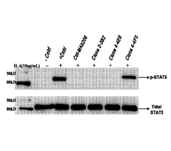
[019] FIG. 1. Assay for neutralizing anti-IL-6 antibodies. Supernatants from
clones were
incubated with human IL-6 at 37 C for 1 hour, prior to incubation with HT-29
cells. The cells
were incubated with rhIL-6 alone or in combination with serum for 15 min at 37
C and
phosphorylation of STAT3 was detected by Western blotting.
[020] FIG. 2A. Titration of neutralizing anti-IL-6 antibodies. The ability to
block IL-6
induced phosphorylation of STAT3 was determined by Western blot analysis using
the
indicated concentrations of the 2-3B2 anti-IL-6 antibody. A substantial
inhibition of IL-6
dependent phosphorylation was seen as low as 0.067 nM antibody.
[021] FIG. 2B. Titration of neutralizing anti-IL-6 antibodies. The ability to
block IL-6
induced phosphorylation of STAT3 was determined by Western blot analysis using
the
indicated concentrations of the 4-4E6 anti-IL-6 antibody. Approximately
equivalent effects
on phosphorylation were observed at 0.67 nM 4-4E6 vs. 0.0067 nM 2-34B2
antibody (FIG.
2A).
[022] FIG. 3. Neutralization activity of TNF-a mediated cytotoxicity by
immunized mouse
sera on WEHI 164 cells. Serum from mouse #3 was the most effective at
inhibiting TNF-a
mediated cytotoxicity.
[023] FIG. 4. Neutralization activity of TNF-a mediated cytotoxicity by
antibodies from
clones 4C9D11 and 4D3B11 in WEHI 164 cells.
[024] FIG. 5. Neutralization activity of TNF-a mediated cytotoxicity by
antibodies from
clones 4C9D11G11 and 4D3B11C4 in L929 cells.
6
CA 02924520 2016-03-15
WO 2015/065987
PCT/US2014/062592
[025] FIG. 6. Antibody-based neutralization of rhTNF-a-induced cell surface
expression of
ICAM-1 in ECV-304 cells (a derivative of T24 bladder cancer cell line).
[026] FIG. 7. Amino acid sequence of the anti-IL-6 antibody (2-3B2) heavy
chain (VH)
sequence (SEQ ID NO:94). The sequence of a homologous heavy chain of the
B34781
antibody (SEQ ID NO:95), obtained from the NCBI protein sequence database, is
shown for
comparison. Putative CDR sequences (underlined) were identified by comparison
with the
known sequence of the homologous B34781 antibody.
[027] FIG. 8. Amino acid sequence of the anti-IL-6 antibody (2-3B2) light
chain (VK)
sequence (SEQ ID NO:96). The sequence of a homologous light chain of
AAB53778.1 (SEQ
ID NO:97), obtained from the NCBI protein sequence database, is shown for
comparison.
Putative CDR sequences (underlined) were identified by comparison with the
known
sequence of the homologous AAB53778.1.
[028] FIG. 9. Activity of c1L6/TN Fa DVD construct for neutralizing 1L-6
induced
phosphorylation of STAT3 in HT-29 cells, compared to parent 2-3B2 anti-IL-6
antibody.
[029] FIG. 10. Amino acid sequence of the anti-TNF-a antibody (4C9) heavy
chain (VH)
sequence (SEQ ID NO:98). The sequence of a homologous heavy chain of the
AAS66033.1
antibody (SEQ ID NO:99), obtained from the NCBI protein sequence database, is
shown for
comparison. Putative CDR sequences (underlined) were identified by comparison
with the
known sequence of the homologous AAS66033.1 antibody.
[030] FIG. 11. Amino acid sequence of the anti-IL-6 antibody (4C9) light chain
(VK)
sequence (SEQ ID NO:100). The sequence of a homologous heavy chain of
AAS66032.1
(SEQ ID NO:101), obtained from the NCBI protein sequence database, is shown
for
comparison. Putative CDR sequences (underlined) were identified by comparison
with the
known sequence of the homologous AA566032.1.
[031] FIG. 12. Schematic illustration of the synthesis of CI(-AD2-cIgG-anti-
TNF-a-pdHL2.
[032] FIG. 13. Inhibition of IL-6 induced phosphorylation of STAT3 by cT*-(c6)-
(c6)
complex compared to Fab-DDD2-cIL-6 protein.
[033] FIG. 14. Inhibition of natural EL-6 induced phosphorylation of STAT3 by
cT*-(c6)-
(c6) complex compared to Fab-DDD2-cIL-6 protein.
[034] FIG. 15. Inhibition of rhTNF-a induced cell death in L929 cells by anti-
TNF-a
antibody constructs.
[035] FIG. 16. Inhibition of cell death induced by natural TNF-a in L929 cells
by anti-
TNF-a antibody constructs.
7
CA 02924520 2016-03-15
WO 2015/065987
PCT/US2014/062592
[036] FIG. 17. Relative affinities of cT*-(c6)-(c6), c-anti-TNF-a and c-anti-
IL-6 for IL-6
and TNF-a from different species.
[037] FIG. 18A. Role of STAT3 in IL-6 and TNF-a mediated pathways.
[038] FIG. 18B. Role of STAT3 in IL-6 and TNF-a mediated disease processes.
Definitions
[039] Unless otherwise specified, "a" or "an" means "one or more".
[040] As used herein, the terms "and" and "or" may be used to mean either the
conjunctive
or disjunctive. That is, both terms should be understood as equivalent to
"and/or" unless
otherwise stated.
[041] A "therapeutic agent" is an atom, molecule, or compound that is useful
in the
treatment of a disease. Examples of therapeutic agents include antibodies,
antibody
fragments, peptides, drugs, toxins, enzymes, nucleases, hormones,
immunomodulators,
antiscnsc oligonucleotides, small interfering RNA (siRNA), chclators, boron
compounds,
photoactive agents, dyes, and radioisotopes.
[042] A "diagnostic agent" is an atom, molecule, or compound that is useful in
diagnosing a
disease. Useful diagnostic agents include, but are not limited to,
radioisotopes, dyes (such as
with the biotin-streptavidin complex), contrast agents, fluorescent compounds
or molecules,
and enhancing agents (e.g., paramagnetic ions) for magnetic resonance imaging
(MRI).
[043] An "antibody" as used herein refers to a full-length (i.e., naturally
occurring or formed
by normal immunoglobulin gene fragment recombinatorial processes)
immunoglobulin
molecule (e.g., an IgG antibody) or an immunologically active (i.e.,
specifically binding)
portion of an immunoglobulin molecule, like an antibody fragment. An
"antibody" includes
monoclonal, polyclonal, bispecific, multispecific, murine, chimeric, humanized
and human
antibodies.
[044] A "naked antibody" is an antibody or antigen binding fragment thereof
that is not
attached to a therapeutic or diagnostic agent. The Fc portion of an intact
naked antibody can
provide effector functions, such as complement fixation and ADCC (see, e.g.,
Markrides,
Pharmacol Rev 50:59-87, 1998). Other mechanisms by which naked antibodies
induce cell
death may include apoptosis. (Vaswani and Hamilton, Ann Allergy Asthma Immunol
81: 105-
119, 1998.)
[045] An "antibody fragment" is a portion of an intact antibody such as
F(ab')2, F(ab)2, Fab',
Fab, Fv, sFv, scFv, dAb and the like. Regardless of structure, an antibody
fragment binds
with the same antigen that is recognized by the full-length antibody. For
example, antibody
8
81795609
fragments include isolated fragments consisting of the variable regions, such
as the "Fv"
fragments consisting of the variable regions of the heavy and light chains or
recombinant
single chain polypeptide molecules in which light and heavy variable regions
are connected
by a peptide linker ("scFv proteins"). "Single-chain antibodies", often
abbreviated as "scFv"
consist of a polypeptide chain that comprises both a VH and a VI, domain which
interact to
form an antigen- binding site. The VH and VL domains are usually linked by a
peptide of 1 to
25 amino acid residues. Antibody fragments also include diabodies, triabodies
and single
domain antibodies (dAb).
1046) An antibody or antibody complex preparation, or a composition described
herein, is
said to be administered in a "therapeutically effective amount" if the amount
administered is
physiologically significant. An agent is physiologically significant if its
presence results in a
detectable change in the physiology of a recipient subject. In particular
embodiments, an
antibody preparation is physiologically significant if its presence invokes an
antitumor
response or mitigates the signs and symptoms of an autoimmune disease state. A
physiologically significant effect could also be the evocation of a humoral
and/or cellular
immune response in the recipient subject leading to growth inhibition or death
of target cells.
DOCK-AND-LOCK (DNL )
[047] In preferred embodiments, a bivalent or multivalent antibody is formed
as a DOCK-
AND-LOCK (DNL ) complex (see, e.g., U.S. Patent Nos. 7,521,056; 7,527,787;
7,534,866; 7,550,143 and 7,666,400.
Generally, the technique takes advantage of the specific and high-
affinity binding interactions that occur between a dimerization and docking
domain (DDD)
sequence of the regulatory (R) subunits of cAMP-dependent protein kin ase
(PICA) and an
anchor domain (AD) sequence derived from any of a variety of AKAP proteins
(Baillie etal.,
FEBS Letters. 2005; 579: 3264. Wong and Scott, Nat. Rev. Mol. Cell Biol. 2004;
5: 959).
The DDD and AD peptides may be attached to any protein, peptide or other
molecule.
Because the DDD sequences spontaneously dimerize and bind to the AD sequence,
the
technique allows the formation of complexes between any selected molecules
that may be
attached to DDD or AD sequences.
10481 Although the standard DNL complex comprises a trimer with two DDD-
linked
molecules attached to one AD-linked molecule, variations in complex structure
allow the
formation of dimers, trimers, tetramers, pentamers, hexarners and other
multimers. In some
embodiments, the DNL complex may comprise two or more antibodies, antibody
fragments
9
Date Recue/Date Received 2020-10-26
CA 02924520 2016-03-15
WO 2015/065987
PCT/US2014/062592
or fusion proteins which bind to the same antigenic determinant or to two or
more different
antigens. The DNL complex may also comprise one or more other effectors, such
as
proteins, peptides, immunomodulators, cytokines, interleukins, interferons,
binding proteins,
peptide ligands, carrier proteins, toxins, ribonucleases such as onconase,
inhibitory
oligonucleotides such as siRNA, antigens or xenoantigens, polymers such as
PEG, enzymes,
therapeutic agents, hormones, cytotoxic agents, anti-angiogenic agents, pro-
apoptotic agents
or any other molecule or aggregate.
10491 PKA, which plays a central role in one of the best studied signal
transduction
pathways triggered by the binding of the second messenger cAMP to the R
subunits, was first
isolated from rabbit skeletal muscle in 1968 (Walsh etal., J. Biol. Chem.
1968;243:3763).
The structure of the holoenzyme consists of two catalytic subunits held in an
inactive form by
the R subunits (Taylor, J. Biol. Chem. 1989;264:8443). Tsozymes of PKA are
found with two
types of R subunits (RI and RII), and each type has a and f3 isoforms (Scott,
Pharmacol.
Ther. 1991;50:123). Thus, the four isoforms of PKA regulatory subunits are
RIa, R1t3, Ma
and RIIP. The R subunits have been isolated only as stable dimers and the
dimerization
domain has been shown to consist of the first 44 amino-terminal residues of
RlIa (Newlon et
al., Nat. Struct. Biol. 1999; 6:222). As discussed below, similar portions of
the amino acid
sequences of other regulatory subunits are involved in dimerization and
docking, each located
near the N-terminal end of the regulatory subunit. Binding of cAMP to the R
subunits leads
to the release of active catalytic subunits for a broad spectrum of
serine/threonine kinase
activities, which are oriented toward selected substrates through the
compartmentalization of
PKA via its docking with AKAPs (Scott et al., J. Biol. Chem. 1990;265;21561)
10501 Since the first AKAP, microtubule-associated protein-2, was
characterized in 1984
(Lohmann et al., Proc. Natl. Acad. Sci USA. 1984; 81:6723), more than 50 AKAPs
that
localize to various sub-cellular sites, including plasma membrane, actin
cytoskeleton,
nucleus, mitochondria, and endoplasmic reticulum, have been identified with
diverse
structures in species ranging from yeast to humans (Wong and Scott, Nat. Rev.
Mol. Cell
Biol. 2004;5:959). The AD of AKAPs for PKA is an amphipathic helix of 14-18
residues
(Carr et al., J. Biol. Chem. 1991;266:14188). The amino acid sequences of the
AD are quite
varied among individual AKAPs, with the binding affinities reported for RII
dimers ranging
from 2 to 90 nM (Alto et al., Proc. Natl. Acad. Sci. USA. 2003;100:4445).
AKAPs will only
bind to dimeric R subunits. For human RTIa, the AD binds to a hydrophobic
surface formed
by the 23 amino-terminal residues (Colledge and Scott, Trends Cell Biol. 1999;
6:216). Thus,
CA 02924520 2016-03-15
WO 2015/065987
PCT/US2014/062592
the dimerization domain and AKAP binding domain of human RIIa are both located
within
the same N-terminal 44 amino acid sequence (Newlon et al., Nat. Struct. Biol.
1999;6:222;
Newlon et al., EMBO J. 2001;20:1651), which is termed the DDD herein.
10511 We have developed a platform technology to utilize the DDD of human PKA
regulatory subunit Ma., RIt3, 1211ot or Rt113 and the AD of AKAP as an
excellent pair of linker
modules for docking any two entities, referred to hereafter as A and B, into a
noncovalent
complex, which could be further locked into a DNL complex through the
introduction of
cysteine residues into both the DDD and AD at strategic positions to
facilitate the formation
of disulfide bonds. The general methodology of the approach is as follows.
Entity A is
constructed by linking a DDD sequence to a precursor of A, resulting in a
first component
hereafter referred to as a. Because the DDD sequence would effect the
spontaneous formation
of a dimer, A would thus be composed of a2. Entity B is constructed by linking
an AD
sequence to a precursor of B, resulting in a second component hereafter
referred to as b. The
dimeric motif of DDD contained in a2 will create a docking site for binding to
the AD
sequence contained in b, thus facilitating a ready association of az and b to
form a binary,
trimeric complex composed of a2b. This binding event is made irreversible with
a subsequent
reaction to covalently secure the two entities via disulfide bridges, which
occurs very
efficiently based on the principle of effective local concentration because
the initial binding
interactions should bring the reactive thiol groups placed onto both the DDD
and AD into
proximity (Chmura et al., Proc. Natl. Acad. Sci. USA. 2001;98:8480) to ligate
site-
specifically. Using various combinations of linkers, adaptor modules and
precursors, a wide
variety of DNL constructs of different stoichiometry may be produced and used
(see, e.g.,
U.S. Nos. 7,550,143; 7,521,056; 7,534,866; 7,527,787 and 7,666,400.)
10521 By attaching the DDD and AD away from the functional groups of the two
precursors, such site-specific ligations are also expected to preserve the
original activities of
the two precursors. This approach is modular in nature and potentially can be
applied to link,
site-specifically and covalently, a wide range of substances, including
peptides, proteins,
antibodies, antibody fragments, and other effector moieties with a wide range
of activities.
Utilizing the fusion protein method of constructing AD and DDD conjugated
effectors
described in the Examples below, virtually any protein or peptide may be
incorporated into a
DNL construct. However, the technique is not limiting and other methods of
conjugation
may be utilized.
11
CA 02924520 2016-03-15
WO 2015/065987
PCT/US2014/062592
[0531 A variety of methods are known for making fusion proteins, including
nucleic acid
synthesis, hybridization and/or amplification to produce a synthetic double-
stranded nucleic
acid encoding a fusion protein of interest. Such double-stranded nucleic acids
may be
inserted into expression vectors for fusion protein production by standard
molecular biology
techniques (see, e.g. Sambrook et al., Molecular Cloning, A laboratory manual,
2nd Ed, 1989).
In such preferred embodiments, the AD and/or DDD moiety may be attached to
either the N-
terminal or C-terminal end of an effector protein or peptide. However, the
skilled artisan will
realize that the site of attachment of an AD or DDD moiety to an effector
moiety may vary,
depending on the chemical nature of the effector moiety and the part(s) of the
effector moiety
involved in its physiological activity. Site-specific attachment of a variety
of effector moieties
may be performed using techniques known in the art, such as the use of
bivalent cross-linking
reagents and/or other chemical conjugation techniques.
Structure-Function Relationships in AD and DDD Moieties
[054] For different types of DNLEz, constructs, different AD or DDD sequences
may be
utilized. Exemplary DDD and AD sequences are provided below.
DDD/
SHIQIPPGLTELLQGYTVEVLRQQPPDLVEFAVEYFTRLREARA (SEQ ID NO:1)
DDD2
CGHIQIPPGLTELLQGYTVEVLRQQPPDLVEFAVEYFTRLREARA (SEQ ID NO:2)
AD/
QIEYLAKQIVDNAIQQA (SEQ ID NO:3)
AD2
CGQIEYLAKQIVDNAIQQAGC (SEQ ID NO:4)
10551 The skilled artisan will realize that DDD1 and DDD2 are based on the DDD
sequence
of the human RIIct isoform of protein kinase A. However, in alternative
embodiments, the
DDD and AD moieties may be based on the DDD sequence of the human RIa form of
protein kinasc A and a corresponding AKAP sequence, as exemplified in DDD3,
DDD3C
and AD3 below.
DDD3
12
CA 02924520 2016-03-15
WO 2015/065987
PCT/US2014/062592
SLRECELYVQKHNIQALLKDSIVQLCTARPERPMAFLREYFERLEKEEAK (SEQ ID
NO:5)
DDD3C
MSCGGSLRECELYVQKHNIQALLKDSIVQLCTARPERPMAFLREYFERLEKEEAK
(SEQ ID NO:6)
AD3
CGFEELAWKIAKMIWSDVFQQGC (SEQ ID NO:7)
[056] In other alternative embodiments, other sequence variants of AD and/or
DDD
moieties may be utilized in construction of the DNLA) complexes. For example,
there are
only four variants of human PKA DDD sequences, corresponding to the DDD
moieties of
PKA RIa, RIIa, RIP and RIIP. The RIIa DDD sequence is the basis of DDD1 and
DDD2
disclosed above. The four human PKA DDD sequences are shown below. The DDD
sequence represents residues 1-44 of Rlla, 1-44 of REP, 12-61 of Rla and 13-66
of Rip.
(Note that the sequence of DDD1 is modified slightly from the human PKA RIIa
DDD
moiety.)
PKA Rla
SLRECELYVQKEINIQALLICDVSIVQLCTARPERPMAFLREYFEKLEKEEAK (SEQ ID
NO:8)
PKA RIfi
SLKGCELYVQLFIGIQQVLKDCIVHLCISKPERPMKFLREHFEKLEKEENRQILA (SEQ
ID NO:9)
PKA RIla
SHIQIPPGLTELLQGYTVEVGQQPPDLVDFAVEYFTRLREARRQ (SEQ ID NO:10)
PKA RIA6
SIEIPAGLTELLQGFTVEVLRHQPADLLEFALQHFTRLQQENER (SEQ ID NO:11)
[057] The structure-function relationships of the AD and DDD domains have been
the
subject of investigation. (See, e.g., Burns-Hamuro et al., 2005, Protein Sci
14:292-92; Carr
et al., 2001, J Biol Chem 276:17332-38; Alto etal., 2003, Proc Natl Acad Sci
USA 100:4445-
50; Hundsrucker et al., 2006, Biochem J 396:297-306; Stokka et al., 2006,
Biochem J
13
81795609
400:493-99; Gold et al., 2006, Mol Cell 24:383-95; Kinderman et al., 2006, Mol
Cell 24:397-
408.
[0581 For example, Kinderman et al. (2006, Mol Cell 24:397-408) examined the
crystal
structure of the AD-DDD binding interaction and concluded that the human DDD
sequence
contained a number of conserved amino acid residues that were important in
either dirtier
formation or AKAP binding, underlined in SEQ ID NO:1 below. (See Figure I of
Kinderman et al., 2006. The skilled artisan will realize
that in designing sequence variants of the DDD sequence, one would desirably
avoid
changing any of the underlined residues, while conservative amino acid
substitutions might
be made for residues that are less critical for dimerization and AKAP binding.
SHIQIPPGLTELLQGYTVEVLRQQPPDLVEFAVEYFTRLREARA (SEQ ID NO:1)
[059] As discussed in more detail below, conservative amino acid substitutions
have been
characterized for each of the twenty common L-amino acids. Thus, based on the
data of
Kinderman (2006) and conservative amino acid substitutions, potential
alternative DDD
sequences based on SEQ ID NO:1 are shown in Table 1. In devising Table 1, only
highly
conservative amino acid substitutions were considered. For example, charged
residues were
only substituted for residues of the same charge, residues with small side
chains were
substituted with residues of similar size, hydroxyl side chains were only
substituted with
other hydroxyls, etc. Because of the unique effect of proline on amino acid
secondary
structure, no other residues were substituted for proline. A limited number of
such potential
alternative DDD moiety sequences are shown in SEQ ID NO:12 to SEQ ID NO:31
below.
The skilled artisan will realize that an almost unlimited number of
alternative species within
the genus of DDD moieties can be constructed by standard techniques, for
example using a
commercial peptide synthesizer or well known site-directed mutagenesis
techniques. The
effect of the amino acid substitutions on AD moiety binding may also be
readily determined
by standard binding assays, for example as disclosed in Alto et al. (2003,
Proc Natl Acad Sci
USA 100:4445-50).
Table 1. Conservative Amino Acid Substitutions in DDD1 (SEQ ID NO:1).
Consensus
sequence disclosed as SEQ ID NO:87.
S HI QIPP GL TELLQGYTVE VL
T K N A SD NA
QQPP DLV,EF AVE YF T RL RE AR A
14
Date Recue/Date Received 2020-10-26
CA 02924520 2016-03-15
WO 2015/065987
PCT/US2014/062592
NN E D L D SK KDL KL
V V V
THIQIPPGLTELLQGYTVEVLRQQPPDLVEFAVEYFTRLREARA (SEQ ID NO:12)
SKIQIPPGLTELLQGYTVEVLRQQPPDLVEFAVEYFTRLREARA (SEQ ID NO:13)
SRIQIPPGLTELLQGYTVEVLRQQPPDLVEFAVEYFTRLREARA (SEQ ID NO:14)
SHNIPPGLTELLQGYTVEVLRQQPPDLVEFAVEYFTRLREARA (SEQ ID NO:15)
SHIQIPPALTELLQGYTVEVLRQQPPDLVEFAVEYFTRLREARA (SEQ ID NO:16)
SHIQIPPGLSELLQGYTVEVLRQQPPDLVEFAVEYFTRLREARA (SEQ ID NO:17)
SHIQIPPGLTDLLQGYTVEVLRQQPPDLVEFAVEYFTRLREARA (SEQ ID NO:18)
SH1QIPPGLTELLNGYTVEVLRQQPPDLVEFAVEYFTRLREARA (SEQ ID NO:19)
SHTQTPPGLTELLQAY'TVEVLRQQPPDLVEFAVEYFTRLREARA (SEQ ID NO:20)
SHIQIPPGLTELLQGYSVEVLRQQPPDLVEFAVEYFTRLREARA (SEQ ID NO:21)
SHIQIPPGLTELLQGYTVDVLRQQPPDLVEFAVEYFTRLREARA (SEQ ID NO:22)
SHIQIPPGLTELLQGYTVEVLKQQPPDLVEFAVEYFTRLREARA (SEQ ID NO:23)
SHIQIPPGLTELLQGYTVEVLRNQPPDLVEFAVEYFTRLREARA (SEQ ID NO: 24)
SHIQIPPGLTELLQGYTVEVLRQNPPDLVEFAVEYFTRLREARA (SEQ ID NO:25)
SHIQIPPGLTELLQGYTVEVLRQQPPELVEFAVEYFTRLREARA (SEQ ID NO:26)
SHIQIPPGLTELLQGYTVEVLRQQPPDLVDFAVEYFTRLREARA (SEQ ID NO:27)
SHIQIPPGLTELLQGYTVEVLRQQPPDLVEFLVEYFTRLREARA (SEQ ID NO:28)
SHIQIPPGLTELLQGYTVEVLRQQPPDLVEFIVEYFTRLREARA (SEQ ID NO:29)
SHIQIPPGLTELLQGYTVEVLRQQPPDLVEFVVEYFTRLREARA (SEQ ID NO:30)
SHIQIPPGLTELLQGYTVEVLRQQPPDLVEFAVDYFTRLREARA (SEQ ID NO: 31)
10601 Alto et al. (2003, Proc Natl Acad Sci USA 100:4445-50) performed a
bioinformatic
analysis of the AD sequence of various AKAP proteins to design an RII
selective AD
sequence called AKAP-IS (SEQ ID NO:3), with a binding constant for DDD of 0.4
nM. The
AKAP-IS sequence was designed as a peptide antagonist of AKAP binding to PKA.
Residues in the AKAP-IS sequence where substitutions tended to decrease
binding to DDD
CA 02924520 2016-03-15
WO 2015/065987
PCT/US2014/062592
are underlined in SEQ ID NO:3 below. The skilled artisan will realize that in
designing
sequence variants of the AD sequence, one would desirably avoid changing any
of the
underlined residues, while conservative amino acid substitutions might be made
for residues
that are less critical for DDD binding. Table 2 shows potential conservative
amino acid
substitutions in the sequence of AKAP-IS (AD1, SEQ ID NO:3), similar to that
shown for
DDD1 (SEQ ID NO:1) in Table 1 above.
[061] A limited number of such potential alternative AD moiety sequences are
shown in
SEQ ID NO:32 to SEQ ID NO:49 below. Again, a very large number of species
within the
genus of possible AD moiety sequences could be made, tested and used by the
skilled artisan,
based on the data of Alto et al. (2003). It is noted that Figure 2 of Alto
(2003) shows an even
large number of potential amino acid substitutions that may be made, while
retaining binding
activity to DDD moieties, based on actual binding experiments.
AKAP-IS
Q1EYLAKQIVDNAIQQA (SEQ ID NO:3)
Table 2. Conservative Amino Acid Substitutions in AD1 (SEQ ID NO:3). Consensus
sequence disclosed as SEQ ID NO:88.
QI E YL AK QI V DNA I QQ A
NL DF I RN E Q N N L
V T V
V
NIEYLAKQIVDNAIQQA (SEQ ID NO:32)
QLEYLAKQIVDNAIQQA (SEQ ID NO:33)
QVEYLAKQIVDNAIQQA (SEQ ID NO:34)
QIDYLAKQIVDNAIQQA (SEQ ID NO:35)
QIEFLAKQIVDNAIQQA (SEQ ID NO:36)
QIETLAKQIVDNAIQQA (SEQ ID NO:37)
QIESLAKQIVDNAIQQA (SEQ ID NO:38)
QIEYIAKQIVDNAIQQA (SEQ ID NO:39)
QIEYVAKQIVDNAIQQA (SEQ ID NO:40)
16
CA 02924520 2016-03-15
WO 2015/065987
PCT/US2014/062592
QIEYLARQIVDNAIQQA (SEQ ID NO:41)
QIEYLAKNIVDNAIQQA (SEQ ID NO:42)
QIEYLAKQIVENAIQQA (SEQ ID NO:43)
QIEYLAKQIVDQAIQQA (SEQ ID NO:44)
QIEYLAKQIVDNAINQA (SEQ ID NO:45)
QIEYLAKQIVDNAIQNA (SEQ ID NO:46)
QIEYLAKQIVDNAIQQL (SEQ ID NO:47)
QIEYLAKQIVDNAIQQI (SEQ ID NO:48)
Q1EYLAKQIVDNAIQQV (SEQ ID NO:49)
[062] Gold et al. (2006, Mol Cell 24:383-95) utilized crystallography and
peptide screening
to develop a SuperAKAP-1S sequence (SEQ 1D NO:50), exhibiting a five order of
magnitude
higher selectivity for the RII isoform of PKA compared with the RI isoform.
Underlined
residues indicate the positions of amino acid substitutions, relative to the
AKAP-IS sequence,
which increased binding to the DDD moiety of Rlla. In this sequence, the N-
terminal Q
residue is numbered as residue number 4 and the C-terminal A residue is
residue number 20.
Residues where substitutions could be made to affect the affinity for Rlla
were residues 8,
11, 15, 16, 18, 19 and 20 (Gold et al., 2006). It is contemplated that in
certain alternative
embodiments, the SuperAKAP-IS sequence may be substituted for the AKAP-IS AD
moiety
sequence to prepare DNLO) constructs. Other alternative sequences that might
be substituted
for the AKAP-1S AD sequence are shown in SEQ ID NO:51-53. Substitutions
relative to the
AKAP-IS sequence are underlined. It is anticipated that, as with the AD2
sequence shown in
SEQ ID NO:4, the AD moiety may also include the additional N-terminal residues
cysteine
and glycine and C-terminal residues glycine and cysteine.
SuperAKAP-IS
QIEYVAKQIVDYAIHQA (SEQ ID NO:50)
Alternative AKAP sequences
QIEYKAKQIVDHAIHQA (SEQ ID NO:51)
QIEYHAKQIVDHAIHQA (SEQ ID NO:52)
QIEYVAKQIVDHAIHQA (SEQ ID NO:53)
17
CA 02924520 2016-03-15
WO 2015/065987
PCT/US2014/062592
[063] Figure 2 of Gold et al. disclosed additional DDD-binding sequences from
a variety of
AKAP proteins, shown below.
RII-Specific AKAPs
AKAP-KL
PLEYQAGLLVQNAIQQAI (SEQ ID NO:54)
AKAP79
LLIETASSLVKNAIQLSI (SEQ ID NO:55)
AKAP-Lbc
LIEEAASRIVDAVIEQVK (SEQ ID NO:56)
RI-Specific AKAPs
AKAPce
ALYQFADRFSELVISEAL (SEQ ID NO:57)
RIAD
LEQVANQLADQIIKEAT (SEQ ID NO:58)
PV38
FEELAWKIAKMEWSDVF (SEQ TD NO:59)
Dual-Specificity AKAPs
AKAP7
ELVRLSKRLVENAVLKAV (SEQ ID NO:60)
MAP2D
TAEEVSARIVQVVTAEAV (SEQ ID NO:61)
DAKAP1
QIKQAAFQLISQVILEAT (SEQ ID NO:62)
DAKAP2
LAWKIAKMIVSDVMQQ (SEQ 1D NO:63)
18
CA 02924520 2016-03-15
WO 2015/065987
PCT/US2014/062592
[0641 Stoklca et al. (2006, Biochem J 400:493-99) also developed peptide
competitors of
AKAP binding to PKA, shown in SEQ ID NO:64-66. The peptide antagonists were
designated as Ht31 (SEQ ID NO:64), MAD (SEQ ID NO:65) and PV-38 (SEQ ID
NO:66).
The Ht-31 peptide exhibited a greater affinity for the MI isoform of PKA,
while the RIAD
and PV-38 showed higher affinity for RI.
Ht31
DLIEEAASRIVDAVIEQVKAAGAY (SEQ ID NO:64)
RIAD
LEQYANQLADQIIKEATE (SEQ ID NO:65)
PV-38
FEELAWKIAKMIWSDVFQQC (SEQ ID NO:66)
10651 Hundsruckcr et al. (2006, Biochem J 396:297-306) developed still other
peptide
competitors for AKAP binding to PKA, with a binding constant as low as 0.4 nM
to the DDD
of the MI form of PKA. The sequences of various AKAP antagonistic peptides are
provided
in Table 1 of Hundsrucker et al., reproduced in Table 3 below. AKAPIS
represents a
synthetic Rh subunit-binding peptide. All other peptides are derived from the
Rh-binding
domains of the indicated AKAPs.
Table 3. AKAP Peptide sequences
Peptide Sequence
AKAPIS QIEYLAKQIVDNAIQQA (SEQ ID NO:3)
AKAPIS-P QIEYLAKQIPDNAIQQA (SEQ ID NO:67)
Ht31 KGADLIEEAASRIVDAVIEQVKAAG (SEQ ID NO:68)
Ht31-P KGADLIEEAASRIPDAPIEQVKAAG (SEQ ID NO:69)
AKAP76-wt-pep PEDAELVRLSKRLVENAVLKAVQQY (SEQ ID NO:70)
AKAP7o-L304T-pep PEDAELVRTSKRLVENAVLKAVQQY (SEQ ID NO:71)
AKAP7o-L308D-pep PEDAELVRLSKRDVENAVLKAVQQY (SEQ ID NO:72)
AKAP7o-P-pep PEDAELVRLSKRLPENAVLKAVQQY (SEQ ID NO:73)
AKAP7o-PP-pep PEDAELVRLSKRLPENAPLKAVQQY (SEQ ID NO:74)
19
81795609
AICAP7S-L314E-pep PEDAELVRLSKRLVENAVEKAVQQY (SEQ ID NO:75)
AKAP1-p ep EEOLDRNEEIKRAAFQESQVISEA (SEQ ID NO:76)
AICAP2-pep LVDDPLEYQAGLLVQNAIQQAIAEQ (SEQ ID NO:77)
AKAP5-pep QYETLLIETASSLVKNAIQLSIEQL (SEQ ID NO:78)
AKAP9-pep LEKQYQEQLEEEVAKVIVSMSIAFA (SEQ ID NO:79)
AKAPIO-pep NTDEAQEELAWICIAKMIVSDIMQQA (SEQ ID NO:80)
AKAPII-pep VNLDICKAVLAEKIVAEAIEKAEREL (SEQ ID NO:81)
AKAP12-pep NGILELETKSSKLVQNIIQTAVDQF (SEQ ID NO:82)
AKAP14-pep TQDKNYEDELTQVALALVEDVINYA (SEQ ID NO:83)
Rab32-pep ETSAKDNINIEEAARFLVEKILVNH (SEQ ID NO:84)
[066] Residues that were highly conserved among the AD domains of different
A1CAP
proteins are indicated below by underlining with reference to the AKAP IS
sequence (SEQ
ID NO:3). The residues are the same as observed by Alto et at. (2003), with
the addition of
the C-terminal alanine residue. (See FIG. 4 of Hundsrucker et al. (2006).
The sequences of peptide antagonists with particularly high affinities for the
RH DDD sequence were those of AICAP-IS, AICAP7S-wt-pep, AKAP78-L304T-pep and
AICAP78-L308D-pep.
4/CAP-IS
QIEYLAKQLV_DNAIQQA (SEQ ID NO:3)
[067] Carr et al. (2001, J Biol Chem 276:17332-38) examined the degree of
sequence
homology between different AKAP-binding DDD sequences from human and non-human
proteins and identified residues in the DDD sequences that appeared to be the
most highly
conserved among different DDD moieties. These are indicated below by
underlining with
reference to the human PICA Rlla DDD sequence of SEQ ID NO:1. Residues that
were
particularly conserved are further indicated by italics. The residues overlap
with, but are not
identical to those suggested by Kinderman et al. (2006) to be important for
binding to AKAP
proteins. The skilled artisan will realize that in designing sequence variants
of DDD, it
would be most preferred to avoid changing the most conserved residues
(italicized), and it
would be preferred to also avoid changing the conserved residues (underlined),
while
Date Recue/Date Received 2020-10-26
CA 02924520 2016-03-15
WO 2015/065987
PCT/US2014/062592
conservative amino acid substitutions may be considered for residues that are
neither
underlined nor italicized..
SHIQ/PPGLTELLQGYTVEVLRQQPPDLVEFAVEYFTRLREARA (SEQ ID NO:1)
[068] A modified set of conservative amino acid substitutions for the DDD I
(SEQ ID
NO:!) sequence, based on the data of Carr et al. (2001) is shown in Table 4.
Even with this
reduced set of substituted sequences, there are over 65,000 possible
alternative DDD moiety
sequences that may be produced, tested and used by the skilled artisan without
undue
experimentation. The skilled artisan could readily derive such alternative DDD
amino acid
sequences as disclosed above for Table 1 and Table 2.
Table 4. Conservative Amino Acid Substitutions in DDD1 (SEQ ID NO:!).
Consensus
sequence disclosed as SEQ ID NO:89.
SHI QIPPGL TELLQGYTVEVLR
A
QQPPDLVEFAVEYFTRLRE,ARA
I D SK
A V V
[069] The skilled artisan will realize that these and other amino acid
substitutions in the
DDD or AD amino acid sequences may be utilized to produce alternative species
within the
genus of AD or DDD moieties, using techniques that are standard in the field
and only
routine experimentation.
Amino Acid Substitutions
[070] In alternative embodiments, the disclosed methods and compositions may
involve
production and use of proteins or peptides with one or more substituted amino
acid residues.
For example, the DDD and/or AD sequences used to make DNL constructs may be
modified as discussed above.
[071] The skilled artisan will be aware that, in general, amino acid
substitutions typically
involve the replacement of an amino acid with another amino acid of relatively
similar
properties (i.e., conservative amino acid substitutions). The properties of
the various amino
acids and effect of amino acid substitution on protein structure and function
have been the
subject of extensive study and knowledge in the art.
21
CA 02924520 2016-03-15
WO 2015/065987
PCT/US2014/062592
[0721 For example, the hydropathic index of amino acids may be considered
(Kyte &
Doolittle, 1982, J. Mol. Biol., 157:105-132). The relative hydropathic
character of the amino
acid contributes to the secondary structure of the resultant protein, which in
turn defines the
interaction of the protein with other molecules. Each amino acid has been
assigned a
hydropathic index on the basis of its hydrophobicity and charge
characteristics (Kyte &
Doolittle, 1982), these are: isoleucine (+4.5); valine (+4.2); leucine (+3.8);
phenylalanine
(+2.8); cysteine/cystine (+2.5); methionine (+1.9); alanine (+1.8); glycine (-
0.4); threonine (-
0.7); serine (-0.8); tryptophan (-0.9); tyrosine (-1.3); proline (-1.6);
histidine (-3.2); glutamate
(-3.5); glutamine (-3.5); aspartate (-3.5); asparagine (-3.5); lysine (-3.9);
and arginine (-4.5).
In making conservative substitutions, the use of amino acids whose hydropathic
indices are
within 2 is preferred, within + 1 are more preferred, and within 0.5 are
even more
preferred.
10731 Amino acid substitution may also take into account the hydrophilicity of
the amino
acid residue (e.g., U.S. Pat. No. 4,554,101). Hydrophilicity values have been
assigned to
amino acid residues: arginine (+3.0); lysine (+3.0); aspartate (+3.0);
glutamate (+3.0); serine
(+0.3); asparagine (+0.2); glutamine (+0.2); glycine (0); threonine (-0.4);
proline (-0.5 ±1);
alanine (-0.5); histidine (-0.5); cysteine (-1.0); methionine (-1.3); valine (-
1.5); leucine (-1.8);
isolcucinc (-1.8); tyrosine (-2.3); phenylalanine (-2.5); tryptophan (-3.4).
Replacement of
amino acids with others of similar hydrophilicity is preferred.
10741 Other considerations include the size of the amino acid side chain. For
example, it
would generally not be preferred to replace an amino acid with a compact side
chain, such as
glycine or serine, with an amino acid with a bulky side chain, e.g.,
tryptophan or tyrosine.
The effect of various amino acid residues on protein secondary structure is
also a
consideration. Through empirical study, the effect of different amino acid
residues on the
tendency of protein domains to adopt an alpha-helical, beta-sheet or reverse
turn secondary
structure has been determined and is known in the art (see, e.g., Chou &
Fasman, 1974,
Biochemistry, 13:222-245; 1978, Ann. Rev. Biochem., 47: 251-276; 1979,
Biophys. J.,
26:367-384).
[0751 Based on such considerations and extensive empirical study, tables of
conservative
amino acid substitutions have been constructed and are known in the art. For
example:
arginine and lysine; glutamate and aspartate; serine and threonine; glutamine
and asparagine;
and valine, leucine and isoleueine. Alternatively: Ala (A) len, ile, val; Arg
(R) gln, asn, lys;
Asn (N) his, asp, lys, arg, gln; Asp (D) asn, glu; Cys (C) ala, ser; Gin (Q)
glu, asn; Glu (E)
gln, asp; Gly (G) ala; His (H) asn, gln, lys, arg; Ile (I) val, met, ala, phe,
leu; Leu (L) val, met,
22
CA 02924520 2016-03-15
WO 2015/065987
PCT/US2014/062592
ala, phe, ile; Lys (K) gin, asn, arg; Met (M) phe, ile, leu; Phe (F) leu, val,
ile, ala, tyr; Pro (P)
ala; Ser (S), thr; Thr (T) ser; Trp (W) phe, tyr; Tyr (Y) trp, phe, thr, ser;
Val (V) ile, leu, met,
phe, ala.
10761 Other considerations for amino acid substitutions include whether or not
the residue is
located in the interior of a protein or is solvent exposed. For interior
residues, conservative
substitutions would include: Asp and Asn; Ser and Thr; Ser and Ala; Thr and
Ala; Ala and
Gly; Ile and Val; Val and Leu; Leu and Ile; Leu and Met; Phe and Tyr; Tyr and
Trp. (See,
e.g., PROWL website at rockefeller.edu) For solvent exposed residues,
conservative
substitutions would include: Asp and Asn; Asp and Glu; Glu and Gin; Glu and
Ala; Gly and
Asn; Ala and Pro; Ala and Gly; Ala and Ser; Ala and Lys; Ser and Thr; Lys and
Arg; Val and
Leu; Lea and Ile; Ile and Val; Phe and Tyr. (Id.) Various matrices have been
constructed to
assist in selection of amino acid substitutions, such as the PAM250 scoring
matrix, Dayboff
matrix, Grantham matrix, McLachlan matrix, Doolittle matrix, Henikoff matrix,
Miyata
matrix, Fitch matrix, Jones matrix, Rao matrix, Levin matrix and Risler matrix
(Idem.)
10771 In determining amino acid substitutions, one may also consider the
existence of
intermolecular or intramoleadar bonds, such as formation of ionic bonds (salt
bridges)
between positively charged residues (e.g., His, Arg, Lys) and negatively
charged residues
(e.g., Asp, Glu) or disulfide bonds between nearby cysteine residues.
10781 Methods of substituting any amino acid for any other amino acid in an
encoded
protein sequence are well known and a matter of routine experimentation for
the skilled
artisan, for example by the technique of site-directed mutagenesis or by
synthesis and
assembly of oligonucleotides encoding an amino acid substitution and splicing
into an
expression vector construct.
Antibodies and Antibody Fragments
10791 Techniques for preparing monoclonal antibodies against virtually any
target antigen,
such as IL-6 or INF-a, are well known in the art. See, for example, Kohler and
Milstein,
Nature 256: 495 (1975), and Coligan et al. (eds.), CURRENT PROTOCOLS IN
IMMUNOLOGY, VOL. 1, pages 2.5.1-2.6.7 (John Wiley & Sons 1991). Briefly,
monoclonal antibodies can be obtained by injecting mice with a composition
comprising an
antigen, removing the spleen to obtain B-lymphocytes, fusing the B-lymphocytes
with
myeloma cells to produce hybridomas, cloning the hybridomas, selecting
positive clones
which produce antibodies to the antigen, culturing the clones that produce
antibodies to the
antigen, and isolating the antibodies from the hybridoma cultures.
23
CA 02924520 2016-03-15
WO 2015/065987
PCT/US2014/062592
[080] MAbs can be isolated and purified from hybridoma cultures by a variety
of well-
established techniques. Such isolation techniques include affinity
chromatography with
Protein-A SEPHAROSE , size-exclusion chromatography, and ion-exchange
chromatography. See, for example, Coligan at pages 2.7.1-2.7.12 and pages
2.9.1-2.9.3.
Also, see Baines et al., "Purification of Immunoglobulin G (IgG)," in METHODS
IN
MOLECULAR BIOLOGY, VOL. 10, pages 79-104 (The Humana Press, Inc. 1992).
[081] After the initial raising of antibodies to the immunogen, the antibodies
can be
sequenced and subsequently prepared by recombinant techniques. Humanization
and
chimerization of murine antibodies and antibody fragments are well known to
those skilled in
the art. The use of antibody components derived from humanized, chimeric or
human
antibodies obviates potential problems associated with the immunogenicity of
murine constant
regions.
Chimeric Antibodies
[082] A chimeric antibody is a recombinant protein in which the variable
regions of a
human antibody have been replaced by the variable regions of, for example, a
mouse
antibody, including the complementarity-determining regions (CDRs) of the
mouse antibody.
Chimeric antibodies exhibit decreased immunogenicity and increased stability
when
administered to a subject. General techniques for cloning murine
immunoglobulin variable
domains are disclosed, for example, in Orlandi et al., Proc. Nat'l Acad. Sci.
USA 86:3833
(1989). Techniques for constructing chimeric antibodies are well known to
those of skill in
the art. As an example, Leung et al., Hybridoma /3:469 (1994), produced an LL2
chimera
by combining DNA sequences encoding the V, and VH domains of murine LL2, an
anti-
CD22 monoclonal antibody, with respective human K and IgGi constant region
domains.
Humanized Antibodies
[083] Techniques for producing humanized MAbs are well known in the art (see,
e.g., Jones
etal., Nature 321: 522 (1986), Riechmann et al., Nature 332: 323 (1988),
Verhoeyen et al.,
Science 239: 1534 (1988), Carter et al., Proc. Nat'l Acad. Sci. USA 89: 4285
(1992), Sandhu,
Crit. Rev. Biotech. 12: 437 (1992), and Singer etal., J. Immun. 150: 2844
(1993)). A
chimeric or murine monoclonal antibody may be humanized by transferring the
mouse CDRs
from the heavy and light variable chains of the mouse immunoglobulin into the
corresponding variable domains of a human antibody. The mouse framework
regions (FR) in
the chimeric monoclonal antibody are also replaced with human FR sequences. As
simply
transferring mouse CDRs into human FRs often results in a reduction or even
loss of antibody
24
CA 02924520 2016-03-15
WO 2015/065987
PCT/US2014/062592
affinity, additional modification might be required in order to restore the
original affmity of the
murine antibody. This can be accomplished by the replacement of one or more
human residues
in the FR regions with their murine counterparts to obtain an antibody that
possesses good
binding affinity to its epitope. See, for example, Tempest et al.,
Biotechnology 9:266 (1991) and
Verhoeyen et al., Science 239: 1534 (1988). Generally, those human FR amino
acid residues
that differ from their murine counterparts and are located close to or
touching one or more
CDR amino acid residues would be candidates for substitution.
Human Antibodies
[084] Methods for producing fully human antibodies using either combinatorial
approaches
or transgenic animals transformed with human immunoglobulin loci are known in
the art
(e.g., Mancini et al., 2004, New MicrobioL 27:315-28; Conrad and Scheller,
2005, Comb.
Chem. High Throughput Screen. 8:117-26; Brekke and Loset, 2003, Curr. Opin.
Phamacol.
3:544-50). A fully human antibody also can be constructed by genetic or
chromosomal
transfection methods, as well as phage display technology, all of which are
known in the art.
See for example, McCafferty etal., Nature 348:552-553 (1990). Such fully human
antibodies are expected to exhibit even fewer side effects than chimeric or
humanized
antibodies and to function in vivo as essentially endogenous human antibodies.
In certain
embodiments, the claimed methods and procedures may utilize human antibodies
produced
by such techniques.
[085] In one alternative, the phage display technique may be used to generate
human
antibodies (e.g., Dantas-Barbosa et al., 2005, Genet. MoL Res. 4:126-40).
Human antibodies
may be generated from normal humans or from humans that exhibit a particular
disease state,
such as cancer (Dantas-Barbosa et al., 2005). The advantage to constructing
human
antibodies from a diseased individual is that the circulating antibody
repertoire may be biased
towards antibodies against disease-associated antigens.
[086] In one non-limiting example of this methodology, Dantas-Barbosa et al.
(2005)
constructed a phage display library of human Fab antibody fragments from
osteosarcoma
patients. Generally, total RNA was obtained from circulating blood lymphocytes
(Id.).
Recombinant Fab were cloned from the , y and K chain antibody repertoires and
inserted
into a phage display library (Id.). RNAs were converted to cDNAs and used to
make Fab
cDNA libraries using specific primers against the heavy and light chain
immunoglobulin
sequences (Marks et al., 1991, .1 MoL Biol. 222:581-97). Library construction
was
performed according to Andris-Widhopf et al. (2000, In: Phage Display
Laboratory Manual,
81795609
Barbas et al. (eds), lst edition, Cold Spring Harbor Laboratory Press, Cold
Spring Harbor, NY
pp. 9.1 to 9.22). The final Fab fragments were digested with restriction
endonucleases and
inserted into the bacteriophage genome to make the phage display library. Such
libraries may
be screened by standard phage display methods, as known in the art (see, e.g.,
Pasqualini and
Ruoslahti, 1996, Nature 380:364-366; Pasqualini, 1999, The Quart. J. Nucl.
Med. 43:159-
162).
[087] Phage display can be performed in a variety of formats, for their
review, see e.g.
Johnson and Chiswell, Current Opinion in Structural Biology 3:5564-571(1993).
Human
antibodies may also be generated by in vitro activated B cells. See U.S.
Patent Nos.
5,567,610 and 5,229,275. The skilled
artisan will realize that these techniques are exemplary and any known method
for making
and screening human antibodies or antibody fragments may be utilized.
[088] In another alternative, transgenic animals that have been genetically
engineered to
produce human antibodies may be used to generate antibodies against
essentially any
immunogenic target, using standard immunization protocols. Methods for
obtaining human
antibodies from transgenic mice are disclosed by Green et al., Nature Genet.
7;13 (1994),
Lonberg et aL, Nature 368;856 (1994), and Taylor et al., Mt. hnmun. 6:579
(1994). A non-
limiting example of such a system is the XENOMOUSE (e.g., Green et al., 1999,
J.
Inimunol. Methods 231:11-23) from Abgenix (Fremont, CA). In the XENOMOUSE and
similar animals, the mouse antibody genes have been inactivated and replaced
by functional
human antibody genes, while the remainder of the mouse immune system remains
intact.
[089] The XENOMOUSE was transformed with germline-configured YACs (yeast
artificial chromosomes) that contained portions of the human IgH and Igkappa
loci, including
the majority of the variable region sequences, along accessory genes and
regulatory
sequences. The human variable region repertoire may be used to generate
antibody
producing B cells, which may be processed into hybridomas by known techniques.
A
XENOMOUSE immunized with a target antigen will produce human antibodies by
the
normal immune response, which may be harvested and/or produced by standard
techniques
discussed above. A variety of strains of XENOMOUSE are available, each of
which is
capable of producing a different class of antibody. Transgenically produced
human
antibodies have been shown to have therapeutic potential, while retaining the
pharmacoldnetic properties of normal human antibodies (Green et al., 1999).
The skilled
artisan will realize that the claimed compositions and methods are not limited
to use of the
26
Date Recue/Date Received 2020-10-26
81795609
XENOMOUSE system but may utilize any tmnsgenic animal that has been
genetically
engineered to produce human antibodies.
Antibody Fragments
[0901 Antibody fragments which recognize specific epitopes can be generated by
known
techniques. Antibody fragments are antigen binding portions of an antibody,
such as F(a1:02,
Fab', F(ab)2, Fab, Fv, sPv and the like. F(ab')2 fragments can be produced by
pepsin digestion
of the antibody molecule and Fab' fragments can be generated by reducing
disulfide bridges
of the F(ab')2 fragments. Alternatively, Fab' expression libraries can be
constructed (Huse et
aL, 1989, Science, 246:1274-1281) to allow rapid and easy identification of
monoclonal Fab'
fragments with the desired specificity. F(ab)2 fragments may be generated by
papain digestion
of an antibody.
1091] A single chain Fv molecule (scFv) comprises a VL domain and a VH domain.
The
VL and VH domains associate to form a target binding site. These two domains
are further
covalently linked by a peptide linker (L). Methods for making scFv molecules
and designing
suitable peptide linkers are described in US Patent No. 4,704,692, US Patent
No. 4,946,778,
R. Raag and M. Whitlow, "Single Chain Ft's." FASEB Vol 9:73-80 (1995) and R.E.
Bird and
B.W. Walker, "Single Chain Antibody Variable Regions," TIBTECH, Vol 9: 132-137
(1991).
[092] Techniques for producing single domain antibodies are also known in the
art, as
disclosed for example in Cossins et al. (2006, Prot Express Purif 5 I:253-
259).
Single domain antibodies (VHH) may be obtained, for example, from
camels, alpacas or llamas by standard immunization techniques. (See, e.g.,
Muyldermans et
al., TIBS 26:230-235, 2001; Yau et al., J Immunol Methods 281:161-75, 2003;
Maass et al., J
Immunol Methods 324:13-25, 2007). The VHH may have potent antigen-binding
capacity
and can interact with novel epitopes that are inaccessible to conventional VH-
VL pairs.
(Muyldermans et al., 2001). Alpaca serum IgG contains about 50% camelid heavy
chain
only IgG antibodies (HCAbs) (Maass et al., 2007). Alpacas may be immunized
with known
antigens, such as TNF-cm, and VHHs can be isolated that bind to and neutralize
the target
antigen (Maass et al., 2007). PCR primers that amplify virtually all alpaca
VHH coding
sequences have been identified and may be used to construct alpaca VHH phage
display
libraries, which can be used for antibody fragment isolation by standard
biopanning
techniques well known in the art (Maass et al., 2007). In certain embodiments,
anti-
pancreatic cancer VHH antibody fragments may be utilized in the claimed
compositions and
methods.
27
Date Recue/Date Received 2020-10-26
CA 02924520 2016-03-15
WO 2015/065987
PCT/US2014/062592
[0931 An antibody fragment can be prepared by proteolytic hydrolysis of the
full length
antibody or by expression in E. coil or another host of the DNA coding for the
fragment. An
antibody fragment can be obtained by pepsin or papain digestion of full length
antibodies by
conventional methods. These methods are described, for example, by Goldenberg,
U.S.
Patent Nos. 4,036,945 and 4,331,647 and references contained therein. Also,
see Nisonoff et
al., Arch Biochem. Biophys. 89: 230 (1960); Porter, Biochem. J. 73: 119
(1959), Edelman et
al., in METHODS IN ENZYMOLOGY VOL. 1, page 422 (Academic Press 1967), and
Coligan at pages 2.8.1-2.8.10 and 2.10.-2.10.4.
Known Antibodies
[094] Various embodiments, for example in combination therapy, may involve the
use of
antibodies binding to target antigens besides IL-6 or TNF-a. A variety of
antibodies are
commercially available and/or known in the art. Antibodies of use may be
commercially
obtained, for example, from the American Type Culture Collection (ATCC,
Manassas, VA).
A large number of antibodies against various disease targets, including but
not limited to
tumor-associated antigens, have been deposited at the ATCC and/or have
published variable
region sequences and are available for use in the claimed methods and
compositions. See,
e.g., U.S. Patent Nos. 7,312,318; 7,282,567; 7,151,164; 7,074,403; 7,060,802;
7,056,509;
7,049,060; 7,045,132; 7,041,803; 7,041,802; 7,041,293; 7,038,018; 7,037,498;
7,012,133;
7,001,598; 6,998,468; 6,994,976; 6,994,852; 6,989,241; 6,974,863; 6,965,018;
6,964,854;
6,962,981; 6,962,813; 6,956,107; 6,951,924; 6,949,244; 6,946,129; 6,943,020;
6,939,547;
6,921,645; 6,921,645; 6,921,533; 6,919,433; 6,919,078; 6,916,475; 6,905,681;
6,899,879;
6,893,625; 6,887,468; 6,887,466; 6,884,594; 6,881,405; 6,878,812; 6,875,580;
6,872,568;
6,867,006; 6,864,062; 6,861,511; 6,861,227; 6,861,226; 6,838,282; 6,835,549;
6,835,370;
6,824,780; 6,824,778; 6,812,206; 6,793,924; 6,783,758; 6,770,450; 6,767,711;
6,764,688;
6,764,681; 6,764,679; 6,743,898; 6,733,981; 6,730,307; 6,720,155; 6,716,966;
6,709,653;
6,693,176; 6,692,908; 6,689,607; 6,689,362; 6,689,355; 6,682,737; 6,682,736;
6,682,734;
6,673,344; 6,653,104; 6,652,852; 6,635,482; 6,630,144; 6,610,833; 6,610,294;
6,605,441;
6,605,279; 6,596,852; 6,592,868; 6,576,745; 6,572;856; 6,566,076; 6,562,618;
6,545,130;
6,544,749; 6,534,058; 6,528,625; 6,528,269; 6,521,227; 6,518,404; 6,511,665;
6,491,915;
6,488,930; 6,482,598; 6,482,408; 6,479,247; 6,468,531; 6,468,529; 6,465,173;
6,461,823;
6,458,356; 6,455,044; 6,455,040, 6,451,310; 6,444,206' 6,441,143; 6,432,404;
6,432,402;
6,419,928; 6,413,726; 6,406,694; 6,403,770; 6,403,091; 6,395,276; 6,395,274;
6,387,350;
6,383,759; 6,383,484; 6,376,654; 6,372,215; 6,359,126; 6,355,481; 6,355,444;
6,355,245;
6,355,244; 6,346,246; 6,344,198; 6,340,571; 6,340,459; 6,331,175; 6,306,393;
6,254,868;
28
81795609
6,187,287; 6,183,744; 6,129,914; 6,120,767; 6,096,289; 6,077,499; 5,922,302;
5,874,540;
5,814,440; 5,798,229; 5,789,554; 5,776,456; 5,736,119; 5,716,595; 5,677,136;
5,587,459;
5,443,953, 5,525,338.
These are exemplary only and a wide variety of other antibodies and their
hybridomas are known in the art. The skilled artisan will' realize that
antibody sequences or
antibody-secreting hybridomas against almost any disease-associated antigen
may be
obtained by a simple search of the ATCC, NCBI and/or USPTO databases for
antibodies
against a selected disease-associated target of interest. The antigen binding
domains of the
cloned antibodies may be amplified, excised, ligated into an expression
vector, transfected
into an adapted host cell and used for protein production, using standard
techniques well
known in the art (see, e.g., U.S. Patent Nos. 7,531,327; 7,537,930; 7,608,425
and 7,785,880,
[095] Particular antibodies that may be of use for therapy of cancer within
the scope of the
claimed methods and compositions include, but are not limited to, LL1 (anti-
CD74), LL2 and
RFB4 (anti-CD22), RS7 (anti-epithelial glycoprotein-1 (EGP-1)), PAM4 and KC4
(both anti-
mucin), MN-14 (anti-carcinoembryonic antigen (CEA, also known as CD66e), Mu-9
(anti-
colon-specific antigen-p), Irnmu 31 (an anti-alpha-fetoprotein), TAG-72 (e.g.,
CC49), Tn,
J591 or HuJ591 (anti-PSMA (prostate-specific membrane antigen)), AB-PG1-XG1-
026 (anti-
PSMA dimer), D2/B (anti-PSMA), G250 (anti-carbonic anbydrase IX), hL243 (anti-
HLA-
DR), alemtuzumab (anti-CD52), bevacizumab (anti-VEGF), cetuximab (anti-EGFR),
gemtuzumab (anti-CD33), ibritumomab tiuxetan (anti-CD20); paniturnumab (anti-
EGFR);
rituximab (anti-CD20); tositumomab (anti-CD20); GA101 (anti-CD20); and
trastuzumab
(anti-ErbB2). Such antibodies are known in the art (e.g., U.S. Patent Nos.
5,686,072;
5,874,540; 6,107,090; 6,183,744; 6,306,393; 6,653,104; 6,730.300; 6,899,864;
6,926,893;
6,962,702; 7,074,403; 7,230,084; 7,238,785; 7,238,786; 7,256,004; 7,282,567;
7,300,655;
7,312,318; 7,585,491; 7,612,180; 7,642,239; and U.S. Patent Application Publ.
No.
20040202666 (now abandoned); 20050271671; and 20060193865.
Specific known antibodies of use include 1iPAM4
(U.S. Patent No. 7,282,567), hA20 (U.S. Patent No. 7,251,164), hA19 (U.S.
Patent No.
7,109,304), 1ilMMU31 (U.S. Patent No. 7,300,655), hLL1 (U.S. Patent No.
7,312,318,),
hLL2 (U.S. Patent No. 7,074,403), hMu-9 (U.S. Patent No. 7,387;773), hL243
(U.S. Patent
No. 7,612,180), IIMN-14 (U.S. Patent No. 6,676,924), hMN-15 (U.S. Patent No.
7,541,440),
1112.1 (U.S. Patent Application 12/772,645), hRS7 (U.S. Patent No. 7,238,785),
IIMN-3 (U.S.
Patent No. 7,541,440), AB-PG1-XG1-026 (U.S. Patent Application 11/983,372,
deposited as
29
Date Recue/Date Received 2020-10-26
81795609
ATCC PTA-4405 and PTA-4406) and D2/13 (WO 2009/130575)
[096] Anti-TNF-a antibodies are known in the art and may be of use to treat
immune
diseases, such as autoimmune disease, immune dysfunction (e.g., graft-versus-
host disease,
organ transplant rejection) or diabetes. Known antibodies against TNF-a
include the human
antibody CDP571 (Ofei et al., 2011, Diabetes 45:881-85); murine antibodies
MTNFAI,
M2TNFAI, M3TNFAI, M3TNFABI, M302B and M303 (Thermo Scientific, Rockford, IL);
infliximab (Centocor, Malvern, PA); certolizumab pegol ((JCB, Brussels,
Belgium); and
adalimumab (Abbott, Abbott Park, IL). These and many other known anti-TNF-a
antibodies
may be used in the claimed methods and compositions. Other antibodies of use
for therapy
of immune dysregulatory or autoimmune disease include, but are not limited to,
anti-B-cell
antibodies such as veltuzumab, epratuzumab, milatuzumab or hL243; tocilizumab
(anti-IL-6
receptor); basiliximab (anti-CD25); daclizumab (anti-CD25); efalizumab (anti-
CD1 la);
muromonab-CD3 (anti-CD3 receptor); anti-CD4OL (UCB, Brussels, Belgium);
natalizumab
(anti-a4 integin) and omalizumab (anti-IgE).
[097] Type-2 diabetes may be treated using known antibodies against B-cell
antigens, such
as CD22 (epratuzumab), CD74 (milatuzumab), CD19 (hA19), CD20 (veltuzumab) or
HLA-
DR (hL243) (see, e.g., Winer et al., 2011, Nature Med 17:610-18). Anti-CD3
antibodies also
have been proposed for therapy of type 1 diabetes (Cemea et al., 2010,
Diabetes Metab Rev
26:602-05).
[098] Macrophage migration inhibitory factor (MIF) is an important regulator
of innate and
adaptive immunity and apoptosis. It has been reported that CD74 is the
endogenous receptor
for MIF (Leng et al., 2003, J Exp Med 197:1467-76). The therapeutic effect of
antagonistic
anti-CD74 antibodies on MW-mediated intracellular pathways may be of use for
treatment of
a broad range of disease states, such as cancers of the bladder, prostate,
breast, lung, colon
and chronic lymphocytic leukemia (e.g., Meyer-Siegler et al., 2004, BMC Cancer
12:34;
Shachar & Haran, 2011, Leuk Lymphoma 52:1446-54); autoimmune diseases such as
rheumatoid arthritis and systemic lupus erythematosus (Morand & Leech, 2005,
Front Biosci
10:12-22; Shachar & Haran, 2011, Leuk Lymphoma 52:1446-54); kidney diseases
such as
renal allograft rejection (Lan, 2008, Nephron Exp Nephrol. 109:e79-83); and
numerous
inflammatory diseases (Meyer-Siegler et al., 2009, Mediators Inflamm epub
March 22, 2009;
Takahashi et al., 2009, Respir Res 10:33; Milatuzumab (hLL1) is an exemplary
anti-CD74
antibody of therapeutic use for treatment of MIF-mediated diseases.
Date Recue/Date Received 2020-10-26
CA 02924520 2016-03-15
WO 2015/065987
PCT/US2014/062592
[099] Anti-CD3 antibodies have been reported to reduce development and
progression of
atherosclerosis (Steffens et al., 2006, Circulation 114:1977-84). Antibodies
against oxidized
LDL induced a regression of established atherosclerosis in a mouse model
(Ginsberg, 2007, J
Am Coll Cardiol 52:2319-21). Anti-ICAM-1 antibody was shown to reduce ischemic
cell
damage after cerebral artery occlusion in rats (Zhang et al., 1994, Neurology
44:1747-51).
Antibody Allotypes
[0100] Immunogenicity of therapeutic antibodies is associated with increased
risk of infusion
reactions and decreased duration of therapeutic response (Baert et al., 2003,
N Engl J Med
348:602-08). The extent to which therapeutic antibodies induce an immune
response in the
host may be determined in part by the allotype of the antibody (Stickler et
at., 2011, Genes
and Tmmunity 12:213-21). Antibody allotype is related to amino acid sequence
variations at
specific locations in the constant region sequences of the antibody. The
allotypcs of IgG
antibodies containing a heavy chain 7-type constant region are designated as
Gm allotypes
(1976, J Immunol 117:1056-59).
[0101] For the common IgG1 human antibodies, the most prevalent allotype is
Glml
(Stickler et al., 2011, Genes and Immunity 12:213-21). However, the G1m3
allotype also
occurs frequently in Caucasians (Id.). It has been reported that Glml
antibodies contain
allotypic sequences that tend to induce an immune response when administered
to non-Glml
(nGlml) recipients, such as G1m3 patients (Id.). Non-Glml allotype antibodies
are not as
immunogenic when administered to Glml patients (Id.).
[0102] The human Glml allotype comprises the amino acids aspartic acid at
Kabat position
356 and leucine at Kabat position 358 in the CH3 sequence of the heavy chain
IgGI. The
nGlml allotype comprises the amino acids glutamic acid at Kabat position 356
and
methionine at Kabat position 358. Both Glml and nGlml allotypes comprise a
glutamic acid
residue at Kabat position 357 and the allotypes are sometimes referred to as
DEL and EEM
allotypes. A non-limiting example of the heavy chain constant region sequences
for Glml
and nGlml allotype antibodies is shown for the exemplary antibodies rituximab
(SEQ ID
NO:85) and veltuzumab (SEQ ID NO:86).
Rituximab heavy chain variable region sequence (SEQ ID NO:85)
ASTKGPSVFPLAPSSKSTSG6TAALGCLVKDYFPEPVTVSWN SGALTSGVHTFPAVL
QSSGLYSLSSVVTVPSSSLGTQTYICNVNHKPSNTKVDKICAEPKSCDKTHTCPPCPAP
ELLGGPSVFLFPPKPKDTLMISRTPEVTCVVVDVSHEDPEVKFNWYVDGVEVHNAK
TKPREEQYNSTYRVVSVLTVLHQDWLNGKEYKCKVSNKALPAPIEKTISKAKGQPRE
31
CA 02924520 2016-03-15
WO 2015/065987
PCT/US2014/062592
PQVYTLPPSRDELTKNQVSLTCLVKGFYPSDIAVEWESNGQPENNYKTTPPVLDSDG
SFFLYSKLTVDKSRWQQGNVFSCSVMHEALHNHYTQKSLSLSPGK
Veltuzumab heavy chain variable region (SEQ ID NO:86
ASTKGPSVFPLAPSSKSTSGGTAALGCLVKDYFPEPVTVSWNSGALTSGVEITFPAVL
QSSGLYSLSSVVTVPSSSLGTQTYICNVNHKPSNTKVDKRVEPKSCDKTHTCPPCPAP
ELLGGPSVFLFPPKPKDTLMTSRTPEVICVVVDVSHEDPEVKFNWYVDGVEVHNAK
TKPREEQYNSTYRVVSVLTVLHQDWLNGKEYKCKVSNKALPAPIEKTISKAKGQPRE
PQVYTLPPSREEMTKNQVSLTCLVKGFYPSDIAVEWESNGQPENNYKTIPPVLDSDG
SFFLYSKLTVDKSRWQQGNVFSCSVMHEALHNHYTQKSLSLSPGK
101031 Jefferis and Lefranc (2009, mAbs 1:1-7) reviewed sequence variations
characteristic
of IgG allotypes and their effect on immunogenicity. They reported that the
G1m3 allotype is
characterized by an argininc residue at Kabat position 214, compared to a
lysine residue at
Kabat 214 in the G1m17 allotype. The nG1m1,2 allotype was characterized by
glutamic acid
at Kabat position 356, methionine at Kabat position 358 and alanine at Kabat
position 431.
The G1m1,2 allotype was characterized by aspartic acid at Kabat position 356,
leucine at
Kabat position 358 and glycine at Kabat position 431. In addition to heavy
chain constant
region sequence variants, Jefferis and Lefranc (2009) reported allotypic
variants in the kappa
light chain constant region, with the Km1 allotype characterized by valine at
Kabat position
153 and leucine at Kabat position 191, the Km1,2 allotype by alanine at Kabat
position 153
and leucine at Kabat position 191, and the IC.m3 allotype characterized by
alanine at Kabat
position 153 and valine at Kabat position 191.
101041 With regard to therapeutic antibodies, veltuzumab and rituximab are,
respectively,
humanized and chimeric IgG1 antibodies against CD20, of use for therapy of a
wide variety
of hematological malignancies and/or autoimmune diseases. Table 5 compares the
allotype
sequences of rituximab vs. veltuzumab. As shown in Table 5, rituximab
(G1m17,1) is a
DEL allotype IgGI, with an additional sequence variation at Kabat position 214
(heavy chain
CHI) of lysine in rituximab vs. arginine in veltuzumab. It has been reported
that veltuzumab
is less immunogenic in subjects than rituximab (see, e.g., Morchhauser et al.,
2009, J Clin
Oncol 27:3346-53; Goldenberg et al., 2009, Blood 113:1062-70; Robak & Robak,
2011,
BioDrugs 25:13-25), an effect that has been attributed to the difference
between humanized
and chimeric antibodies. However, the difference in allotypcs between the EEM
and DEL
allotypes likely also accounts for the lower immunogenicity of veltuzumab.
32
CA 02924520 2016-03-15
WO 2015/065987
PCT/US2014/062592
Table 5. Allotypes of Rituximab vs. Veltuzumab
Heavy chain position and associated allotypes
Complete 214 (allotype) 356/358 (allotype) 431 (allotype)
allotype
Rituximab G1m17,1 K 17 D/L 1 A
Veltuzumab G 1m3 R 3 E/M A
[0105] In order to reduce the immunogenicity of therapeutic antibodies in
individuals of
nGlml genotype, it is desirable to select the allotype of the antibody to
correspond to the
G1m3 allotype, characterized by arginine at Kabat 214, and the nG1m1,2 null-
allotype,
characterized by glutamic acid at Kabat position 356, methionine at Kabat
position 358 and
alanine at Kabat position 431. Surprisingly, it was found that repeated
subcutaneous
administration of G1m3 antibodies over a long period of time did not result in
a significant
immune response. In alternative embodiments, the human IgG4 heavy chain in
common with
the Glm3 allotype has arginine at Kabat 214, glutamic acid at Kabat 356,
methionine at
Kabat 359 and alanine at Kabat 431. Since immunogenicity appears to relate at
least in part
to the residues at those locations, use of the human IgG4 heavy chain constant
region
sequence for therapeutic antibodies is also a preferred embodiment.
Combinations of GI m3
IgG1 antibodies with IgG4 antibodies may also be of use for therapeutic
administration.
Immunoconjugates
[0106] In certain embodiments, the antibodies or complexes may be conjugated
to one or
more therapeutic or diagnostic agents. The therapeutic agents do not need to
be the same but
can be different, e.g. a drug and a radioisotopc. For example, 1311 can be
incorporated into a
tyrosine of an antibody or fusion protein and a drug attached to an epsilon
amino group of a
lysine residue. Therapeutic and diagnostic agents also can be attached, for
example to
reduced SH groups and/or to carbohydrate side chains. Many methods for making
covalent
or non-covalent conjugates of therapeutic or diagnostic agents with antibodies
or fusion
proteins are known in the art and any such known method may be utilized.
[0107] A therapeutic or diagnostic agent can be attached at the hinge region
of a reduced
antibody component via disulfide bond formation. Alternatively, such agents
can be attached
using a heterobifunctional cross-linker, such as N-succinyl 3-(2-
pyridyldithio)propionate
(SPDP). Yu etal., Int. .1. Cancer 56: 244 (1994). General techniques for such
conjugation
33
81795609
are well-known in the art. See, for example, Wong, CHEMISTRY OF PROTEIN
CONJUGATION AND CROSS-LINKING (CRC Press 1991); Upeslacis etal.,
"Modification of Antibodies by Chemical Methods," in MONOCLONAL ANTIBODIES:
PRINCIPLES AND APPLICATIONS, Birch et al. (eds.), pages 187-230 (Wiley-Liss,
Inc.
1995); Price, "Production and Characterization of Synthetic Peptide-Derived
Antibodies," in
MONOCLONAL ANTIBODIES: PRODUCTION, ENGINEERING AND CLINICAL
APPLICATION, Ritter et al. (eds.), pages 60-84 (Cambridge University Press
1995).
Alternatively, the therapeutic or diagnostic agent can be conjugated via a
carbohydrate moiety
in the Fc region of the antibody. The carbohydrate group can be used to
increase the loading
of the same agent that is bound to a thiol group, or the carbohydrate moiety
can be used to
bind a different therapeutic or diagnostic agent.
[0108] Methods for conjugating peptides to antibody components via an antibody
carbohydrate moiety are well-known to those of skill in the art. See, for
example, Shih et al.,
Int. J. Cancer 41: 832 (1988); Shih et aL, Int. J. Cancer 46: 1101(1990); and
Shih et al., U.S.
Patent No. 5,057,313. The general method
involves reacting an antibody component having an oxidized carbohydrate
portion with a
carrier polymer that has at least one free amine function. This reaction
results in an initial
Schiff base (imine) linkage, which can be stabilized by reduction to a
secondary amine to
form the final conjugate.
101091 The Pc region may be absent if the antibody used as the antibody
component is an
antibody fragment. However, it is possible to introduce a carbohydrate moiety
into the light
chain variable region of a full length antibody or antibody fragment. See, for
example, Leung
et al., J. Immunol. 154: 5919 (1995); Hansen et al., U.S. Patent No. 5,443,953
(1995), Leung
etal., U.S. patent No. 6,254,868. The
engineered carbohydrate moiety is used to attach the therapeutic or diagnostic
agent.
101101 In some embodiments, a chelating agent may be attached to an antibody,
antibody
fragment or fusion protein and used to chelate a therapeutic or diagnostic
agent, such as a
radionuclide. Exemplary chelators include but are not limited to DTPA (such as
Mx-DTPA),
DOTA, TETA, NETA or NOTA. Methods of conjugation and use of chelating agents
to attach
metals or other ligands to proteins are well known in the art (see, e.g., U.S.
Patent No.
7,563,433,
[0111] In certain embodiments, radioactive metals or paramagnetic ions may be
attached to
proteins or peptides by reaction with a reagent having a long tail, to which
may be attached a
multiplicity of chelating groups for binding ions_ Such a tail can be a
polymer such as a
34
Date Recue/Date Received 2020-10-26
81795609
polylysine, polysaccharide, or other derivatized or derivatizable chains
having pendant
groups to which can be bound chelating groups such as, e.g.,
ethylenediaminetetraacetic acid
(EDTA), diethylenetriarninepentaacetic acid (DTPA), porphyrins, polyarnines,
crown ethers,
bis-thiosemicarbazones, polyoximes, and like groups known to be useful for
this purpose.
[01121 Chelates may be directly linked to antibodies or peptides, for example
as disclosed in
U.S. Patent 4,824,659. Particularly useful
metal-chelate combinations include 2-benzyl-DTPA and its monornethyl and
cyclohexyl
analogs, used with diagnostic isotopes in the general energy range of 60 to
4,000 keV, such
as 1251, 1311, 1231, 1241, otu, Cu,64 isF, 67Ga, issGa,
¨r 94rnm, nc, IN, 150, 76Br, , for
radioimaging. The same chelates, when cornplexed with non-radioactive metals,
such as
manganese, iron and gadolinium are useful for MM. Macrocyclic chelates such as
NOTA,
DOTA, and TETA are of use with a variety of metals and radiometals, most
particularly with
radionuclides of gallium, yttrium and copper, respectively. Such metal-chelate
complexes
can be made very stable by tailoring the ring size to the metal of interest.
Other ring-type
chelates such as macrocyclic polyethers, which are of interest for stably
binding nuclides,
such as 223Ra for RAFT are encompassed.
[01131 More recently, methods of "F-labeling of use in PET scanning techniques
have been
disclosed, for example by reaction of F-18 with a metal or other atom, such as
aluminum.
The 18F-Al conjugate may be complexed with chelating groups, such as DOTA,
NOTA or
NETA that are attached directly to antibodies or used to label targetable
constructs in pre-
targeting methods. Such F-18 labeling techniques are disclosed in U.S. Patent
No. 7,563,433.
Therapeutic Agents
[01141 In alternative embodiments, therapeutic agents such as cytotoxic
agents, anti-
angiogenic agents, pro-apoptotic agents, antibiotics, hormones, hormone
antagonists,
chemokines, drugs, prodrugs, toxins, enzymes or other agents may be used,
either conjugated
to the subject antibody complexes or separately administered before,
simultaneously with, or
after the antibody complex. Drugs of use may possess a pharmaceutical property
selected from
the group consisting of antimitotic, ldnase inhibitor, Bruton kinase
inhibitor, alkylating,
antimetabolite, antibiotic, alkaloid, anti-angiogenic, pro-apoptotic agents
and combinations
thereof.
101151 Exemplary drugs of use include, but are not limited to, 5-fluorouracil,
afatinib,
aplidin, azaribine, anastrozole, anthracyclines, axitinib, AVL-101, AVL-291,
bendamustine,
bleomycin, bortezomib, bosutinib, bryostatin-1, busulfan, calicheamycin,
camptothecin,
Date Recue/Date Received 2020-10-26
CA 02924520 2016-03-15
WO 2015/065987
PCT/US2014/062592
carboplatin, 10-hydroxycamptothecin, carmustine, celecoxib, chlorambucil,
cisplatin
(CDDP), Cox-2 inhibitors, irinotecan (CPT-11), SN-38, carboplatin, cladribine,
camptothecans, crizotinib, cyclophosphamide, cytarabine, dacarbazine,
dasatinib, dinaciclib,
docetaxel, dactinomycin, daunorubicin, doxorubicin, 2-pyrrolinodoxorubicine
(2P-DOX),
cyano-moTholino doxorubicin, doxorubicin glucuronide, epirubicin glucuronide,
erlotinib,
estramustine, epidophyllotoxin, entinostat, estrogen receptor binding agents,
etoposide
(VP16), etoposide glucuronide, etoposide phosphate, exemestane, fingolimod,
floxuridine
(FUdR), 3',5'-0-dioleoy1-FudR (FUdR-d0), fludarabine, flutamide, farnesyl-
protein
transferase inhibitors, flavopiridol, fostamatinib, ganetespib, GDC-0834, GS-
1101, gefitinib,
gemcitabine, hydroxyurea, ibrutinib, idarubicin, idelalisib, ifosfamide,
imatinib, L-
asparaginase, lapatinib, lenolidamide, leucovorin, LFM-A13, lomustine,
mechlorethamine,
melphalan, mercaptopurine, 6-mercaptopurine, methotrexate, mitoxantrone,
mithramycin,
mitomycin, mitotanc, navelbinc, ncratinib, nilotinib, nitrosurca, olaparib,
plicomycin,
procarbazine, paclitaxel, PCI-32765, pentostatin, PSI-341, raloxifene,
semustine, sorafenib,
streptozocin, SU11248, sunitinib, tamoxifen, temazolomide (an aqueous form of
DTIC),
transplatinum, thalidomide, thioguanine, thiotepa, teniposide, topotecan,
uracil mustard,
vatalanib, vinorelbine, vinblastine, vincristine, vinca alkaloids and ZD1839.
[0116] Toxins of use may include ricin, abrin, alpha toxin, saporin,
ribonucleasc (RNasc),
e.g., onconase, DNase I, Staphylococcal enterotoxin-A, pokeweed antiviral
protein, gelonin,
diphtheria toxin, Pseudomonas exotoxin, and Pseudomonas endotoxin.
[0117] Chemokines of use may include RANTES, MCAF, MIP1-alpha, MIP1-Beta and
IP-10.
[0118] In certain embodiments, anti-angiogenic agents, such as angiostatin,
baculostatin,
canstatin, maspin, anti-VEGF antibodies, anti-P1GF peptides and antibodies,
anti-vascular
growth factor antibodies, anti-Flk-1 antibodies, anti-Flt-1 antibodies and
peptides, anti-Kras
antibodies, anti-cMET antibodies, anti-MIF (macrophage migration-inhibitory
factor)
antibodies, laminin peptides, fibronectin peptides, plasminogen activator
inhibitors, tissue
metalloproteinase inhibitors, interferons, interleukin-12, IP-10, Gro-13,
thrombospondin, 2-
methoxyoestradiol, proliferin-related protein, carboxiamidotriazole, CM! 01,
Marimastat,
pentosan polysulphate, angiopoietin-2, interferon-alpha, herbimycin A,
PNU145156E, 16K
prolactin fragment, Linomide (roquinimex), thalidomide, pentoxifylline,
genistein, TNP-470,
endostatin, paclitaxel, accutin, angiostatin, cidofovir, vincristine,
bleomycin, AGM-1470,
platelet factor 4 or minocycline may be of use.
[0119] Immunomodulators of use may be selected from a cytokine, a stem cell
growth factor, a
lymphotoxin, a hematopoietic factor, a colony stimulating factor (CSF), an
interferon (IFN),
36
CA 02924520 2016-03-15
WO 2015/065987
PCT/US2014/062592
erythropoietin, thrombopoietin and a combination thereof. Specifically useful
are
lymphotoxins such as tumor necrosis factor ('TNF), hematopoietic factors, such
as interleukin
(IL), colony stimulating factor, such as granulocyte-colony stimulating factor
(G-CSF) or
granulocyte macrophage-colony stimulating factor (GM-CSF), interferon, such as
interferons-a, -13 or -7, and stem cell growth factor, such as that designated
"Si factor".
Included among the cytokines are growth hormones such as human growth hormone,
N-
methionyl human growth hormone, and bovine growth hormone; parathyroid
hormone;
thyroxine; insulin; proinsulin; relaxin; prorelaxin; glycoprotein hormones
such as follicle
stimulating hormone (FSH), thyroid stimulating hormone (TSH), and luteinizing
hormone
(LH); hepatic growth factor; prostaglandin, fibroblast growth factor;
prolactin; placental
lactogen, OB protein; tumor necrosis factor-a and -13; mullerian-inhibiting
substance; mouse
gonadotropin-associated peptide; inhibin; activin; vascular endothelial growth
factor;
integrin; thrombopoietin (TP0); nerve growth factors such as NGF-B; platelet-
growth factor;
transforming growth factors (TGFs) such as TGF- a and TGF- B; insulin-like
growth factor-I
and -II; erythropoietin (EPO); osteoinductive factors; interferons such as
interferon-a, -13, and
-y; colony stimulating factors (CSFs) such as macrophage-CSF (M-CSF);
interleukins (ILs)
such as IL-1, IL-la, IL-2, IL-3, IL-4, IL-5, IL-6, IL-7, IL-8, IL-9, IL-10, IL-
11, IL-12; IL-13,
IL-14, 1L-16, TL-17, IL-18, 1L-21, 1L-25, LIF, kit-ligand or FLT-3,
angiostatin,
thrombospondin, endostatin, tumor necrosis factor and LT.
[0120] Radionuclides of use include, but are not limited to- 1111n
, 1771m, 212Bi, 213Bi, 211At,
62cti, 671cu, 90y, 1251, 131I, 32p, 33p, 4.7sc, 111Ag, 67Ga, 142pr, 1535m,
161Th, 166Dy, 166140,
186Re, 188Re, 189Re, 212ph, 223Ra, 225 Ac, A, 59Fe, 75Se, 77As, 89Sr, 991v1o,
105Rh, 109pd, 143pr,
149pm, 169Er, 1941r, 198Au, 199Aa, 227Th and 211
Pb. The therapeutic radionuclide preferably
has a decay-energy in the range of 20 to 6,000 keV, preferably in the ranges
60 to 200 keV
for an Auger emitter, 100-2,500 keV for a beta emitter, and 4,000-6,000 keV
for an alpha
emitter. Maximum decay energies of useful beta-particle-emitting nuclides are
preferably 20-
5,000 keV, more preferably 100-4,000 keV, and most preferably 500-2,500 keV.
Also
preferred are radionuclides that substantially decay with Auger-emitting
particles. For
example, Co-58, Ga-67, Br-80m, Tc-99m, Rh-103m, Pt-109, In-1 1 1, Sb-119, 1-
125, Ho-161,
Os-189m and Ir-192. Decay energies of useful beta-particle-emitting nuclides
are preferably
<1,000 keV, more preferably <100 keV, and most preferably <70 keV. Also
preferred are
radionuclides that substantially decay with generation of alpha-particles.
Such radionuclides
include, but arc not limited to: Dy-152, At-211, Bi-212, Ra-223, Rn-219, Po-
215, Bi-211,
37
CA 02924520 2016-03-15
WO 2015/065987
PCT/US2014/062592
Ac-225, Fr-221, At-217, Bi-213, Th-227 and Fm-255. Decay energies of useful
alpha-
particle-emitting radionuclides are preferably 2,000-10,000 keV, more
preferably 3,000-
8,000 keV, and most preferably 4,000-7,000 keV. Additional potential
radioisotopes of use
include "C, "N, 150, "Br, 198Au, 224Ac, 1261, 133/, 77Br, 113m-u,
1 "Ru, 97Ru, 1 3Ru,
w5Ru, mg, 203Hg, 121.-re, (22.Te, 125.Te, i65Tm, 167Tm, 168-fm, 197pt, 109pd,
105Rh,
142pr, 143pr, 161-rb, 166-0,
H 199Au, 57Co,
58Co, 51Cr, 59Fe, 75se, 201T1, 225Ac, 76Br, 169yb,
and the like. Some useful diagnostic nuclides may include 18F, 52Fe, 62c_u,
64cu, Cu,
67Ga,
68Ga,
Y 89Zr, 94Tc, 94mTC, 99mTc, or
Radionuclides and other metals may be delivered,
for example, using chelating groups attached to an antibody or conjugate.
Macrocyclic
chelates such as NOTA, DOTA, and TETA are of use with a variety of metals and
radiometals, most particularly with radionuclides of gallium, yttrium and
copper,
respectively. Such metal-chelate complexes can be made very stable by
tailoring the ring
size to the metal of interest. Other ring-type chelates, such as macrocyclic
polycthers for
complexing 223Ra, may be used.
[0121] Therapeutic agents may include a photoactive agent or dye. Fluorescent
compositions, such as fluorochrome, and other chromogens, or dyes, such as
porphyrins
sensitive to visible light, have been used to detect and to treat lesions by
directing the suitable
light to the lesion. In therapy, this has been termed photoradiation,
phototherapy, or
photodynamic therapy. See Joni et al. (eds.), PHOTODYNAMIC THERAPY OF TUMORS
AND OTHER DISEASES (Libreria Progetto 1985); van den Bergh, Chem. Britain
(1986),
22:430. Moreover, monoclonal antibodies have been coupled with photoactivated
dyes for
achieving phototherapy. See Mew et al., J. Immunol. (1983),130:1473; idem.,
Cancer Res.
(1985), 45:4380; Oseroff et al., Proc. Natl. Acad. Sci. USA (1986), 83:8744;
idem.,
Photochem. Photobiol. (1987), 46:83; Hasan et al., Prog. Clin. Biol. Res.
(1989), 288:471;
Tatsuta et al., Lasers Surg. Med. (1989), 9:422; Pelegrin et al., Cancer
(1991), 67:2529.
[0122] Other useful therapeutic agents may comprise oligonucleotides,
especially antisense
oligonucleotides that preferably are directed against oncogenes and oncogene
products, such
as bc1-2 or p53. A preferred form of therapeutic oligonucleotide is siRNA.
Diagnostic Agents
[0123] Diagnostic agents are preferably selected from the group consisting of
a radionuclide, a
radiological contrast agent, a paramagnetic ion, a metal, a fluorescent label,
a
chemiluminescent label, an ultrasound contrast agent and a photoactive agent.
Such
diagnostic agents are well known and any such known diagnostic agent may be
used. Non-
limiting examples of diagnostic agents may include a radionuclide such as "
In, I77Lu,
38
CA 02924520 2016-03-15
WO 2015/065987
PCT/US2014/062592
18F, "Fe, "Cu, 64Cu, "Cu, "Ga, 68Ga,"Y, 90Y, "Zr, 94"'Tc, "Tc, 99mTc, rzoi,
1231, 124T, 1251,
1311, 154-158Gd, 32p, tic, 13N, 150, 186Re, 188Re, 514n, 52m¨ ,
Mil 55CO, 72AS, 75Br, 76Br, 82mRb, 83sr,
or other gamma-, beta-, or positron-emitters. Paramagnetic ions of use may
include
chromium (III), manganese (II), iron (III), iron (II), cobalt (II), nickel
(II), copper (II),
neodymium (III), samarium (III), ytterbium (III), gadolinium (III), vanadium
(II), terbium
(III), dysprosium (III), holmium (III) or erbium (III). Metal contrast agents
may include
lanthanum (III), gold (III), lead (II) or bismuth (III). Ultrasound contrast
agents may
comprise liposomes, such as gas filled liposomes. Radiopaque diagnostic agents
may be
selected from compounds, barium compounds, gallium compounds, and thallium
compounds.
A wide variety of fluorescent labels are known in the art, including but not
limited to
fluorescein isothiocyanate, rhodamine, phycoerytherin, phycocyanin,
allophycocyanin, o-
phthaldehyde and fluorescamine. Chemiluminescent labels of use may include
luminol,
isoluminol, an aromatic acridinium ester, an imidazolc, an acridinium salt or
an oxalate ester.
Therapeutic Use
[0124] In another aspect, the invention relates to a method of treating a
subject, comprising
administering a therapeutically effective amount of an antibody complex as
described herein
to a subject. Diseases that may be treated with the antibody complexes
described herein
include, but are not limited to immune diseases (e.g., SLE, RA, juvenile
idiopathic arthritis,
Crohn's disease, type 2 diabetes, Castleman's disease) or inflammatory
diseases (e.g., sepsis,
septic shock, inflammation, inflammatory bowel disease, inflammatory liver
injury, acute
pancreatitis). Such therapeutics can be given once or repeatedly, depending on
the disease
state and tolerability of the conjugate, and can also be used optimally in
combination with
other therapeutic modalities, such as immunomodulator therapy, immunotherapy,
chemotherapy, antisense therapy, interference RNA therapy, gene therapy, and
the like. Each
combination will be adapted to patient condition and prior therapy, and other
factors
considered by the managing physician.
[0125] As used herein, the term "subject" refers to any animal (i.e.,
vertebrates and
invertebrates) including, but not limited to mammals, including humans. It is
not intended
that the term be limited to a particular age or sex. Thus, adult and newborn
subjects, as well
as fetuses, whether male or female, are encompassed by the term.
[0126] In preferred embodiments, the antibodies that are used in the treatment
of human
disease are human or humanized (CDR-grafted) versions of antibodies; although
murine and
chimeric versions of antibodies can be used. Same species IgG molecules are
mostly
preferred to minimize immune responses. This is particularly important when
considering
39
81795609
repeat treatments. For humans, a human or humanized IgG antibody is less
likely to generate
an anti-IgG immune response from patients.
[01271 In another preferred embodiment, diseases that may be treated using the
antibody
complexes include, but are not limited to immune dysregulation disease and
related
autoimmune diseases, including Class BI autoimmune diseases such as immune-
mediated
thrombocytopenias, such as acute idiopathic thromboeytopenic purpura and
chronic
idiopathic thrombocytopenic purpura, dermatomyositis, Sjogren's syndrome,
multiple
sclerosis, Sydenham's chorea, myasthenia gravis, systemic lupus erythematosus,
lupus
nephritis, rheumatic fever, polyglandular syndromes, bullous pemphigoid,
diabetes mellitus,
Henoch-Schonlein purpura, post-streptococcal nephritis, erythema nodosum,
Takayasu's
arteritis, Addison's disease, rheumatoid arthritis, sarcoidosis, ulcerative
colitis, erythema
multiforme, IgA nephropathy, polyarteritis nodosa, ankylosing spondylitis,
Goodpasture's
syndrome, thromboangitis obliterans, Sji5gren's syndrome, primary biliary
cirrhosis,
Hashimoto's thyroiditis, thyrotoxicosis, scleroderrna, chronic active
hepatitis, rheumatoid
arthritis, polymyositis/dermatomyositis, polychondritis, pemphigus vulgaris,
Wegener's
granulomatosis, membranous nephropathy, arnyotrophic lateral sclerosis, tabes
dorsalis, giant
cell arteritis/polyrnyalgia, pernicious anemia, rapidly progressive
glomerulonephritis and
fibrosing alveolitis, and also juvenile diabetes, as disclosed in U.S.
Provisional Application
Serial No. 60/360,259, filed March 1, 2002 (now expired). Antibodies that may
be of use for
combination therapy in these diseases include, but are not limited to, those
reactive with
FILA-DR antigens, B-cell and plasma-cell antigens (e.g., CD I9, CD20, CD21,
CD22, CD23,
CD4, CD5, CD8, CD14, CD15, CD19, CD20, CD21, CD22, CD23, CD25, CD33, CD37,
CD38, CD40, CD4OL, CD46, CD52, CD54, CD74, CD80, CD126, CD138, B7, MUC1, Ia,
HM1.24, and HLA-DR),11-6, IL-17. Since many of these autoimmune diseases are
affected
by autoantibodies made by aberrant B-cell populations, depletion of these B-
cells is a
preferred method of autoimmune disease therapy. In a preferred embodiment, the
anti-B-cell,
anti-T-cell, or anti-macrophage or other such antibodies of use in the co-
treatment of patients
with autoirrunune diseases also can be conjugated to result in more effective
Therapeutics to
control the host responses involved in said autoimmune diseases, and can be
given alone or in
combination with other therapeutic agents, such as TNF inhibitors or anti-IL-
6R antibodies
and the like.
101281 In a preferred embodiment, a more effective therapeutic agent can be
provided by
using multivalent, multispecific antibodies. Exemplary bivalent and bispecific
antibodies are
found in U.S. Patent Nos. 7,387,772; 7,300,655; 7,238,785; and 7,282,567,
Date Recue/Date Received 2020-10-26
81795609
These multivalent or
multispecific antibodies are particularly preferred in the targeting of
disease associated cells
which express multiple antigen targets and even multiple epitopes of the same
antigen target,
but which often evade antibody targeting and sufficient binding for
immunotherapy because
of insufficient expression or availability of a single antigen target on the
cell. By targeting
multiple antigens or epitopes, said antibodies show a higher binding and
residence time on
the target, thus affording a higher saturation with the drag being targeted in
this invention.
Formulation and Administration
101291 Suitable routes of administration of the conjugates include, without
limitation, oral,
parenteral, rectal, transmucosal, intestinal administration, intramuscular,
subcutaneous,
intramedullary, intrathecal, direct intraventricular, intravenous,
intravitreal, intraperitoneal,
intranasal, or intraocular injections. The preferred routes of administration
are parenteral.
Alternatively, one may administer the compound in a local rather than systemic
manner, for
example, via injection of the compound directly into a solid tumor.
101301 Antibody complexes or immunoconjugates can be formulated according to
known
methods to prepare pharmaceutically useful compositions, whereby the antibody
complex or
immunoconjugate is combined in a mixture with a pharmaceutically suitable
excipient.
Sterile phosphate-buffered saline is one example of a pharmaceutically
suitable excipient.
Other suitable excipients are well-known to those in the art. See, for
example, Ansel et al.,
PHARMACEUTICAL DOSAGE FORMS AND DRUG DELIVERY SYSTEMS, 5th
Edition (Lea & Febiger 1990), and Gennaro (ed.), REMINGTON'S PHARMACEUTICAL
SCIENCES, 18th Edition (Mack Publishing Company 1990), and revised editions
thereof.
101311 The antibody complex or immunoconjugate can be formulated for
intravenous
administration via, for example, bolus injection or continuous infusion.
Preferably, the
antibody of the present invention is infused over a period of less than about
4 hours, and more
preferably, over a period of less than about 3 hours. For example, the first
25-50 mg could be
infused within 30 minutes, preferably even 15 min, and the remainder infused
over the next
2-3 hrs. Formulations for injection can be presented in unit dosage form,
e.g_, in ampoules or
in multi-dose containers, with an added preservative. The compositions can
take such forms
as suspensions, solutions or emulsions in oily or aqueous vehicles, and can
contain
formulatory agents such as suspending, stabilizing and/or dispersing agents.
Alternatively,
the active ingredient can be in powder form for constitution with a suitable
vehicle, e.g.,
sterile pyrogen-free water, before use
41
Date Recue/Date Received 2020-10-26
CA 02924520 2016-03-15
WO 2015/065987
PCT/US2014/062592
[0132] Additional pharmaceutical methods may be employed to control the
duration of action
of the antibody complex. Control release preparations can be prepared through
the use of
polymers to complex or adsorb the antibody complex. For example, biocompatible
polymers
include matrices of poly(ethylene-co-vinyl acetate) and matrices of a
polyanhydride
copolymer of a stearic acid dimer and sebacic acid. Sherwood et al.,
Bio/Technology 10:
1446 (1992). The rate of release of an antibody complex or immunoconjugate
from such a
matrix depends upon the molecular weight, the amount of antibody complex or
immunoconjugate within the matrix, and the size of dispersed particles.
Saltzman et al.,
Biophys. J. 55: 163 (1989); Sherwood et al., supra. Other solid dosage forms
are described
in Ansel et al., PHARMACEUTICAL DOSAGE FORMS AND DRUG DELIVERY
SYSTEMS, 5th Edition (Lea & Febiger 1990), and Gennaro (ed.), REMINGTON'S
PHARMACEUTICAL SCIENCES, 18th Edition (Mack Publishing Company 1990), and
revised editions thereof.
[0133] Generally, the dosage of an administered antibody complex or
immunoconjugate for
humans will vary depending upon such factors as the patient's age, weight,
height, sex,
general medical condition and previous medical history. It may be desirable to
provide the
recipient with a dosage that is in the range of from about 1 mg/kg to 25 mg/kg
as a single
intravenous infusion, although a lower or higher dosage also may be
administered as
circumstances dictate. A dosage of 1-20 mg/kg for a 70 kg patient, for
example, is 70-1,400
mg, or 41-824 mg/m2 for a 1.7-m patient. The dosage may be repeated as needed,
for
example, once per week for 4-10 weeks, once per week for 8 weeks, or once per
week for 4
weeks. It may also be given less frequently, such as every other week for
several months, or
monthly or quarterly for many months, as needed in a maintenance therapy.
[0134] Alternatively, an antibody complex or immunoconjugate may be
administered as one
dosage every 2 or 3 weeks, repeated for a total of at least 3 dosages. Or,
twice per week for
4-6 weeks. If the dosage is lowered to approximately 200-300 mg/m2(340 mg per
dosage for
a 1.7-m patient, or 4.9 mg/kg for a 70 kg patient), it may be administered
once or even twice
weekly for 4 to 10 weeks. Alternatively, the dosage schedule may be decreased,
namely
every 2 or 3 weeks for 2-3 months. It has been determined, however, that even
higher doses,
such as 20 mg/kg once weekly or once every 2-3 weeks can be administered by
slow i.v.
infusion, for repeated dosing cycles. The dosing schedule can optionally be
repeated at other
intervals and dosage may be given through various parenteral routes, with
appropriate
adjustment of the dose and schedule.
42
81795609
Expression Vectors
101351 Still other embodiments may concern DNA sequences comprising a nucleic
acid
encoding an antibody, antibody fragment, toxin or constituent fusion protein
of an antibody
complex, such as a DNL construct. Fusion proteins may comprise an antibody or
fragment
or toxin attached to, for example, an AD or DDD moiety.
101361 Various embodiments relate to expression vectors comprising the coding
DNA
sequences. The vectors may contain sequences encoding the light and heavy
chain constant
regions and the hinge region of a human immunoglobulin to which may be
attached chimeric,
humanized or human variable region sequences. The vectors may additionally
contain
promoters that express the encoded protein(s) in a selected host cell,
enhancers and signal or
leader sequences. Vectors that are particularly useful are pdHL2 or GS. More
preferably, the
light and heavy chain constant regions and hinge region may be from a human EU
myeloma
immunoglobulin, where optionally at least one of the amino acid in the
alIotype positions is
changed to that found in a different IgG1 allotype, and wherein optionally
amino acid 253 of
the heavy chain of EU based on the EU number system may be replaced with
alanine. See
Edelman et aL, Proc. NatL Acad. Sc! USA 63: 78-85 (1969). In other
embodiments, an IgG1
sequence may be converted to an IgG4 sequence.
[0137] The skilled artisan will realize that methods of genetically
engineering expression
constructs and insertion into host cells to express engineered proteins are
well known in the
art and a matter of routine experimentation. Host cells and methods of
expression of cloned
antibodies or fragments have been described, for example, in U.S. Patent Nos.
7,531,327 and
7,537,930.
Kits
[0138] Various embodiments may concern kits containing components suitable for
treating or
diagnosing diseased tissue in a patient. Exemplary kits may contain one or
more antibody
complexes as described herein. If the composition containing components for
administration
is not formulated for delivery via the alimentary canal, such as by oral
delivery, a device
capable of delivering the kit components through some other route may be
included. One
type of device, for applications such as parenteral delivery, is a syringe
that is used to inject
the composition into the body of a subject. Inhalation devices may also be
used. In certain
embodiments, a therapeutic agent may be provided in the form of a prefilled
syringe or
autoinjection pen containing a sterile, liquid formulation or lyophilized
preparation.
[0139] The kit components may be packaged together or separated into two or
more
containers. In some embodiments, the containers may be vials that contain
sterile,
43
Date Recue/Date Received 2020-10-26
CA 02924520 2016-03-15
WO 2015/065987
PCT/US2014/062592
lyophilized formulations of a composition that are suitable for
reconstitution. A kit may also
contain one or more buffers suitable for reconstitution and/or dilution of
other reagents. Other
containers that may be used include, but are not limited to, a pouch, tray,
box, tube, or the
like. Kit components may be packaged and maintained sterilely within the
containers.
Another component that can be included is instructions to a person using a kit
for its use.
EXAMPLES
[0140] In the working Examples below, the DOCK-AND-LOCK (DM") technology was
used to generate the first bispecific antibody (bsAb) with high potency to
neutralize both
INF-a and IL-6. This prototype DNL construct, designated cT*-(c6)-(c6),
comprises a
chimeric anti-INF-a IgG linked at the carboxyl terminus of each light chain to
a pair of
dimerized Fab's derived from a chimeric anti-IL-6 antibody, thus featuring a
hexavalent
bsAb capable of blocking 2 and 4 molecules of INF-a and IL-6, respectively, as
well as a
fully functional Fc. As discussed below, the exemplary anti-INF-a/anti-1L-6
bispecific
antibody showed potent activity in in vitro assays designed to test efficacy
for immune
diseases such as SLE or RA. However, the person of ordinary skill will realize
that the
subject complexes of use are not limited to the specific DNL cT*-(c6)-(c6)
complex
discussed below, but more generally encompass bispecific antibodies and/or
antigen-binding
antibody fragments with at least one binding site for 1L-6 an at least one
binding site for
INF-a.
Example 1. Generation of Neutralizing Mouse Anti-Human IL-6 Monoclonal
Antibody
[0141] The 2-3B2 mouse monoclonal antibody against human IL-6 was produced
using
standard immunological techniques, discussed below, that may be used to make
anti-human
IL-6 antibodies in general.
[0142] Recombinant human IL-6 (rhIL-6) was obtained from ProSpec-Tany
TechnoGene
Ltd. (Rehovot, Israel). Multiple mice were initially immunized with 30 i.tg
rhIL-6
administered i.p., followed by booster injections of 30 or 10 [tg with or
without adjuvant,
according to a standard boosting schedule. Animals were tested for presence of
anti-IL-6
antibodies by ELISA assay using rhIL-6 coated microtiter plates and serial
dilutions of
serum. Prior to fusion, the presence of neutralizing anti-IL-6 antibodies was
detected by the
ability to block IL-6 stimulated protein phosphorylation (of STAT3) using
Western blotting
(data not shown).
[0143] Cells secreting neutralizing anti-1L-6 antibodies were fused with the
P3-X63.Ag8.653
myeloma cell line by PEG mediated cell fusion using standard techniques to
generate
44
CA 02924520 2016-03-15
WO 2015/065987
PCT/US2014/062592
antibody-secreting hybridomas cell lines. The 2-3B2, 4-4F5 and 4-4E6 anti-IL-6
clones were
obtained by selection on HAT medium, cloning and subcloning. Supernatants from
isolated
clones containing neutralizing anti-IL-6 were detected by the ability to block
IL-6 stimulated
STAT3 protein phosphorylation, determined by Western blotting (FIG. 1). Clones
2-3B2, 4-
4E6 and control anti-IL-6 MAb206, but not clone 4-4F5, were able to block rhIL-
6 induced
phosphorylation (FIG. 1). Size exclusion HPLC of antibodies purified by
protein A column
chromatography demonstrated the presence of homogeneous antibodies, which was
confirmed by SDS-PAGE (data not shown). Isotyping using an SBA CLONETYPINGTm
system showed that the anti-IL-6 antibodies were IgGl/k murine isotypes.
[0144] Binding to human IL-6 was determined Western blotting against rhIL-6
(FIG. 2).
Based on the intensity of labeling using identical concentrations of antibody,
it was
determined that the 2-3B2 clone (FIG. 2A) showed higher affinity for human IL-
6 than the 4-
4E6 clone (FIG. 2B) and 2-3B2 was selected for production of chimeric and
humanized anti-
IL-6 antibodies. Serial dilution demonstrated that the 2-3B2 antibody was
about 100-fold
more potent than 4-4E6 for inhibiting the IL-6 induced phosphorylation of
STAT3 (FIG. 2A-
B). Neither antibody bound to murine IL-6 (not shown).
Example 2. Generation of Neutralizing Mouse Anti-Human TNF-a Monoclonal
Antibody
[0145] Monoclonal antibodies against human TNF-a were prepared using standard
techniques, as discussed in Example 1 above for IL-6. Mice were immunized with
recombinant human TNF-a obtained from ProSpec-Tany TechnoGene Ltd. (Rehovot,
Israel).Testing of serum from immunized mice for anti-TNF-a antibodies was
performed by
ELISA (data not shown).
[0146] Neutralizing antibodies were also detected by cytotoxicity assay.
Briefly, WEHI 164
cells (mouse fibrosarcoma) were cultured in RPMI complete media. Cells were
plated at a
density of 1x10⁴ cells/well in 75 L of medium in 96-well plates and kept in a
37 C incubator
overnight before the assay. On the day of the assay, sera from the immunized
mice were
diluted 1:25, 1:125, 1:625, 1:3,125,1: 15,625, and 1: 78,125 in RPMI complete
medium
containing 8 g/mL of actinomycin-D and 0.4 ng/mL of rhINF-a. Twenty-five L of
the
diluted sera were added to the cells in the corresponding wells. The addition
of the sera to the
cells made the final dilutions of the sera as 1:100, 1:500, 1:2,500, 1:12,500,
1:62,500, and
1:312,500. The final concentration of actinomycin-D was 2 [tg/mL and rhTNF-a
was 0.1
ng/mL. Plates were incubated in a 37 C/5% CO2 incubator for 20 hours. After
this
incubation, 20 [iL of MTS reagent was added to all the wells and the
absorbance in each well
CA 02924520 2016-03-15
WO 2015/065987
PCT/US2014/062592
determined in a plate reader at 490nm after two hours. As a negative control,
serum from a
naive mouse (not immunized) was diluted in a like manner. One set of wells was
incubated
with only actinomycin-D and rhTNF-a to determine maximum growth inhibition.
Another
set of cells remained untreated (cells grown in media lacking actinomycin-D
and rhTNF-a).
Growth inhibition was measured as percent of untreated control cell growth.
[0147] The results of the cytotoxicity assay are shown in FIG. 3. Serum from
each of the
inoculated mice showed the ability to neutralize rhTNF-ct mediated
cytotoxicity. Serum from
mouse #3 showed the greatest ability to inhibit rhTNF-a mediated cytotoxicity.
[0148] Hybridomas were produced from splenocytes of mice showing the presence
of anti-
TNF-a antibodies by PEG fusion, essentially as discussed above. Selection of
fused
hybridomas was performed using HAT medium. Neutralizing clones 4C9 and 4D3
were
obtained from mouse #3. After further subcloning, antibodies were purified by
chromatography on protein G columns. Purified antibodies were determined to be
homogeneous by size separation HPLC and SDS-PAGE (data not shown). Isotype
analysis,
performed as discussed above, showed that 4C9 was IgGI/ic while 4D3 was
IgG2alic.
[0149] The ability of anti-TNF-a antibodies from clones 4C9D11 and 4D3B11 to
neutralize
TNF-a-mediated cytotoxicity was determined (FIG. 4). WEHI 164 cells were
seeded at 1 x
104 cells/well into 96-well plates and grown in 200 pL of RPM1 complete medium
overnight.
On the day of the assay, supernatants from clones were collected and diluted
1:2. A further
1:5 dilution was made thereafter. Each dilution was made in RPMI complete
medium
containing a final concentration of actinomycin-D at 2 ug/mL and rhTNF-o. at
0.1 ng/mL.
Before addition of the diluted supernatant, the medium in the plate for WEHI
164 cells
growth was removed, and replaced with the diluted supernatant in the
corresponding wells,
100 L/well. The plate was incubated for 20 hours at 37 C in a 5% CO2
incubator. After
this incubation, 20 !IL of MTS was added to all the wells and the absorbance
in each well
determined in a plate reader at 490nm after two hours. As a negative control,
supernatant
from a clone which stopped producing antibody (ELISA negative) was diluted in
a like
manner. One set of wells was incubated with only actinomycin-D and rhTNF- a to
determine
maximum growth inhibition. Another set of cells remained untreated (cells
grown in media
lacking actinomycin-D and rhTNF-a). Growth inhibition was measured as percent
of
untreated control cell growth. The antibody from clone 4D3B11 was more
effective at
blocking TNF-a mediated cytotoxicity in this assay (FIG. 4).
[0150] Antibody binding specificity was determined by Western blotting against
rhTNF-a.
Under reducing conditions, 4D3B11C4 and the anti-TNF-a antibody REMICADE
46
CA 02924520 2016-03-15
WO 2015/065987
PCT/US2014/062592
(infliximab) showed no or weak binding to human TNF-a, with no binding to
human TNF-P
or murine TNF-a (not shown). Under the same reducing conditions, antibody
4C9D11G11
showed strong binding to human TNF-a, with no binding to human TNF-P or murine
TNF-a
(not shown).
101511 Neutralization of rhTNF-a induced cytotoxicity by anti-TNF-a antibodies
was
determined in a different in vitro system (FIG. 5). L929 cells (mouse
fibroblasts) were
seeded at 2 x 10⁴ cells/well into 96-well plates and grown in 90 tit of MEM
medium
overnight (10% horse serum complete medium). On the following day, the
purified
antibodies were diluted 1:5 in MEM medium (containing a final concentration of
actinomycin-D at 20 g/mL and rhTNF-a at 1 ng/mL) for an antibody concentration
range of
10,000 to 3.2 ng/mL. The antibodies were pre-incubated with rhTNF-a at RT for
one hour.
After this pre-incubation, 10 uL of the diluted antibodies were then added to
the 90 pi, cells
in the corresponding wells, that made the final concentration of the
antibodies from 1000 to
0.32 ng/mL, with a final concentration of actinomycin-D and rhTNF-a at 2 g/mL
and 0.1
ng/mL, respectively. The plate was incubated for 20 hours at 37 C. Following
this
incubation, 20 uL of MTS was added to all the wells and the absorbance in each
well
determined in a plate reader at 490nm after two hours. As a negative control,
an anti-hTNF-a
antibody, 4C3 (non-neutralizing), was diluted in a like manner. REMICADE , the
commercial anti-TNF-a antibody was also diluted in a like manner as a positive
control. One
set of wells was incubated with only actinomycin-D and rhTNF-a to determine
maximum
growth inhibition.
[0152] Under these conditions, the 4C9D11G11 antibody (EC50 11.2 ng/mL) was
more
effective than 4D3B11C4 (EC50 22.1 ng/mL) at inhibiting TNF-a-induced
cytotoxicity (FIG.
5). Neither monoclonal antibody was as effective as REMICADE (EC50 3.6 ng/mL)
(FIG.
5).
[0153] An assay was performed for antibody based neutralization of rhTNF-a-
induced cell
surface expression of ICAM-1 (FIG. 6). ECV-304 cells (a derivative of T24,
bladder cancer
cell line) were seeded at 2x10⁵ cells/well into 6-well plates, grown in 10%
FBS Medium 199
for 6 hours for attaching. Varying doses of the mAbs or REMICADE (positive
control)
were mixed with constant amounts of rhTNF- a (10 ng/mL). The mixture of the
antibodies
and rhTNF-a was pre-incubated at 37 'V, for two hours, and then pipetted into
the appropriate
corresponding wells in duplicate. Cells were then grown for 72 hours in a 37
"C incubator.
After this incubation, supernatant was removed and cells were trypsinized and
transferred to
15 mL tubes. Cells were washed with cold PBS/0.5% BSA two times, supernatant
was
47
CA 02924520 2016-03-15
WO 2015/065987
PCT/US2014/062592
removed and the cell pellets were re-suspended in the residual wash buffer (-
100 [IL). An
aliquot of 25 JAL from the cell suspension from each sample was then
transferred to 4 mL
flow tubes. Cells were Fe-blocked by treatment with 1 jig of human IgG for 15
min at RT and
then incubated with fluorescent-conjugated anti-CD54 reagent for 45 min at 4
C. Cells were
then washed with 4 mL of PBS/5% BSA for two times and re-suspended in 4001.11,
of PBS
and then subjected to flow-cytometric analysis (FACS). One set of cells
remained untreated
as background fluorescent control. Another set of cells treated with only 10
ng/mL of
rhTNF-a served as the positive control for obtaining maximum fluorescent (i.e.
maximum
ICAM-1 up-regulation).
[0154] The 4C9 clone again showed higher neutralizing activity than the 4D3
clone and 4C9
was selected for chimerization.
Example 3. Production of Chimeric Anti-1L-6 Antibody from 2-382 Hybridoma
[0155] Total RNA was extracted from hybridomas 2-3B2 cells by standard
techniques and
mRNA was separated from the total RNA fraction. The mRNA was used as a
template for
VH and VK cDNA synthesis, using a QIAGENt) OneStep RT-PCR kit. Primers used
were as
shown below (restriction sites are underlined).
Vkl BACK (PNAS 86:3833-3837,1989)
GACATTCAGCTGACCCAGTCTCCA (SEQ ID NO:90)
CK3'-BH: (Biotechniques 15:286-291, 1993)
GCCGGATCCTCACTGGATGGTGGGAAGATGGATACA (SEQ ID NO: 91)
VH] BACK: (PNAS 86:3833-3837, 1989)
AGGTSMARCTGCAGSAGTCWGG (SEQ ID NO:92, S=GG, M--=A/C,
W=A/T)
CHI-C: (Clinical Cancer Res 5:3095s-3100s, 1999)
AGCTGGGAAGGTGTGCAC (SEQ ID NO:93)
[0156] The VH and Vk cDNA sequences were cloned into the pGEMT vector for
sequencing
by the Sanger dideoxy technique, using an automated DNA sequencer. The
putative VH
(SEQ ID NO:94) and Vk (SEQ ID NO:96) murine amino sequences are shown in FIG.
7 and
FIG. 8. The locations of the heavy and light chain CDR sequences are proposed,
based on
homology with the known heavy and light chain antibody sequences of B34871
(SEQ ID
NO:95) and AAB53778.1 (SEQ ID NO:97), respectively, from the NCBI protein
sequence
database. The indicated 2-3B2 heavy chain CDR sequences are CDR1 (GFTFSRFGMH,
48
CA 02924520 2016-03-15
WO 2015/065987
PCT/US2014/062592
SEQ ID NO:107), CDR2 (YIGRGSSTIYYADTVKG, SEQ ID NO:108) and CDR3
(SNWDGAMDY, SEQ ID NO:109). The 2-3B2 light chain CDR sequences are CDR1
(RASGNIHNFLA, SEQ ID NO:110), CDR2 (NAETLAD, SEQ ID NO:111) and CDR3
(QHFWSTPWT, SEQ ID NO:112).
101571 The VH and VK sequences from the 2-3B2 anti-IL-6 antibody and the VH
and VK
sequences from the 4C9 anti-INF-a antibody were used to make a cIL6/INFa DVD
(dual
variable domain) antibody construct. (See, e.g., Wu et al., 2009, MAbs 1:339-
47.) The
resulting bispecific DVD construct was compared with the parent 2-3B2 anti-IL-
6 antibody
for the ability to inhibit IL-6 induced phosphorylation of STAT3 on HT-29
cells (FIG. 9). As
shown in FIG. 9, the DVD construct showed the same efficacy as the parent anti-
IL-6
antibody for inhibition of 1L-6 mediated phosphorylation.
[0158] The sequences for restriction sites and leader peptides for cloning
into vector pdHL2
were added to the VH and VK sequences of 2-3B2. The complete sequences were
synthesized commercially (GenScript, Piscataway, NJ). The 2-3B2-VH-pUC57 and 2-
3B2-
VK-pUC57 vectors were produced by incorporating the VH sequence as a XhoI-
HindIII
insert and the VK sequence as a XbaI-BamH I insert into corresponding sites in
pUC57. A
vector expressing chimeric 2-3B2 antibody was produced starting with the hA20-
pdf1L2-IgG
vector (see, e.g., Goldenberg et al., 2002, Blood 100:11 Abstract 2260). The
hA20-VH
sequence was replaced with cIL6-VH and the hA20-VK sequence was replaced with
cIL6-
VK by restriction enzyme digestion and ligation. The resulting chimeric 2-3B2
antibody
comprised the murine VH and VK sequences of 2-3B2, attached to human antibody
constant
region sequences. After transfection, screening and antibody purification on a
protein A
column, a chimeric anti-IL6 clone 1B5 (c-IL6-1B5) was obtained as a
homogeneous antibody
preparation, as confirmed by HPLC and SDS-PAGE (not shown). The final clone is
identified as 1B5A9.
Example 4. Production of Chimeric Anti-TNF-u Antibody from 4C9 Hybridoma
[0159] Total RNA was extracted from hybridomas 4C9 cells by standard
techniques and
mRNA was separated from the total RNA fraction. The mRNA was used as a
template for
VH and VK cDNA synthesis, using a PH-USION High Fidelity PCR kit (Thermo
Scientific,
Pittsburgh, PA). Primers used were as disclosed in Example 3 above.
[0160] The VH and Vk cDNA sequences were cloned into the pGEMT vector for
sequencing
by the Sanger dideoxy technique, using an automated DNA sequencer. The
putative VH
(SEQ ID NO:98) and Vk (SEQ ID NO:100) murine amino sequences are shown in FIG.
10
49
CA 02924520 2016-03-15
WO 2015/065987
PCT/US2014/062592
and FIG. 11. The locations of the heavy and light chain CDR sequences are
proposed, based
on homology with the known heavy and light chain antibody sequences of
AAS66033.1
(SEQ ID NO:99) and AAS66032.1 (SEQ ID NO:101), respectively, from the NCBI
protein
sequence database. The indicated 4C9 heavy chain CDR sequences are CDR1 (GFWN,
SEQ
ID NO:113), CDR2 (YISYSGRTYYNPSLKS, SEQ ID NO:114) and CDR3 (DANYVLDY,
SEQ ID NO:115). The 4C9 light chain CDR sequences are CDR1
(KSSQSLLNSSTQKNYLA, SEQ ID NO:116), CDR2 (FASARES, SEQ ID NO:117) and
CDR3 (QQHYRTPFT, SEQ ID NO:118).
[0161] An optimized DNA sequence encoding the INF-a VF1, also comprising a 5'
leader
sequence and 3' flanking sequence, was designed as shown in SEQ ID NO:102
below and
cloned into pdHL2. The optimized 4C9-VH sequence is underlined. The DNA
sequence was
synthesized by GenScript (Piscataway, NJ).
CTCGAGCACACAGGACCTCACCATGGGATGGAGCTGTATCATCCTCTTCTTGGTA
GCAACAGCTACAGGTAAGGGGCTCACAGTAGCAGGCTTGAGGTCTGGACATATA
TATGGGTGACAATGACATCCACTTTGCC ri TCTCTCCACAGGTGTCCACTCCGTGC
AGCTGCAGGAGAGCGGACCCTCCCTGGTGAAGCCTAGICAGACCCTGAGCCTGA
CATGCTCCGTGACTGGGGACTCTATCACCAGTGGCTTCTGGAACTGGATTCGGAA
GTTCCCAGGAAACAAGTTTGAATACATGGGATATATCTCTTACAGTGGGCGCACA
TACTATAACCCCAGCCTGAAGTCCAGGCTGICTATTACAAGAGACACTTCTAAAA
ACCAGT __ 1"1 TATCTGCAGCTGAACAGCGTGACTGCCGAGGATACTGCTACCTACTA
TTGTGCCAGGGACGCTAATTATGTGCTGGATTACTGGGGCCAGGGAACCACACTG
ACCGTGAGCTCCGGTGAGTCCTTACAACCTCTCTCTTCTATTCAGCTTAAATAGAT
TTTACTGCATTTGTTGGGGGGGAAATGTGTGTATCTGAATTTCAGGTCATGAAGG
ACTAGGGACACCTTGGGAGTCAGAAAGGGTCATTGGGAAGCTT (SEQ ID NO:102)
[0162] An optimized DNA sequence encoding the INF-a VK, also comprising a 5'
leader
sequence and 3' flanking sequence, was designed as shown in SEQ ID NO:103
below and
cloned into pdHL2. The optimized 4C9-VK sequence is underlined. The DNA
sequence was
synthesized by GenScript (Piscataway, NJ).
TCTAGACACAGGACCTCACCATGGGATGGAGCTGTATCATCCTCTTCTTGGTAGC
AACAGCTACAGGTAAGGGGCTCACAGTAGCAGGCTTGAGGTCIGGACATATATA
TGGGTGACAATGACATCCACTTTGCCTTTCTCTCCACAGGIGTCCACTCCGACATC
CAGCTGACCCAGAGCCCCAGCTCCCTGGCTATGTCCGTGGGACAGAAGGTGACA
CA 02924520 2016-03-15
WO 2015/065987
PCT/US2014/062592
ATGAACTGCAAATCTAGTCAGTCTCTGCTGAACAGCTCCACTCAGAAGAATTACC
TGGCTTGGTTCCAGCAGAAGCCCGGGCAGAGTCCTAAACTGCTGGTGTATTTTGC
CTCTGCTAGGGAGAGTGGCGTGCCAGACAGATTCATCGGCAGCGGCAGCGGGAC
CGATTITACCCTGACAATTTCTAGTGTGCAGGCCGAGGACCTGGCTGATTAC'TTC
TGTCAGCAGCACTATCGGACTCCCTTCACCTTTGGCTCCGGAACAAAGCTGGAGA
TCAAGCGTGAGTAGAATTTAAACTTTGCTTCCTCAGTTGGATCC (SEQ ID NO:103)
101631 The VH and VK coding sequences were inserted into pUC57 and then pdHL2
for
expression of the chimeric 4C9 antibody as discussed in Example 3 above. The
chimeric 4C9
was produced by transfection of pdHL2, screening for transfectants and
antibody purification
on a protein A column. The selected clone was designated 6A9. The purified
antibody was
determined to be homogeneous by HPLC and SDS-PAGE (not shown). A binding
affinity
assay for chimeric anti-TNF-a showed a dissociation constant (KD) of 4.13 en
(not shown).
Example 5. Construction of CHI-DDD2-cFab-anti-IL-6-pGSILL
[0164] The hLL2-Fab-DDD2-pGSHL#2 plasmid (see, e.g., W02013 181087A2; Rossi et
al.,
2009, Blood 113:6161-71; U.S. Patent Publ. Nos. 20130323204, 20140212425) was
used as a
starting material for production of a DDD2 conjugated Fab anti-IL-6 antibody
fragment. The
hLL2-DDD2 plasmid was digested with Xbal/Xhol and the 6577 bp vector was
isolated.
cIL6-pdHL2 (Example 3) was digested with XbaI/XhoI and the 2604 bp cIL6 coding
insert
was isolated. The two were ligated to form the 9182 bp Vk-cIL6-Fab-DDD2-pGSHL
vector.
After screening by PstI digestion and electrophoresis, the 9182 bp vector was
digested with
XhoI/Hind3/Alkaline phosphatase and an 8536 vector was isolated. The cIL6-
pdHL2 vector,
comprising a 648 bp cIL6-VH coding insert was digested with Xhol/Hind3. The
648 bp VH
encoding insert was ligated with the 8536 bp vector and VK insert to generate
CH1-DDD2-
cFab-anti-IL-6-pGSHL. The final construct was then transfected, clones were
picked and
purified by Kappa-select (GE Healthcare Life Sciences, Piscataway, NJ). The
purified
antibody product of CH1-DDD2-cFab-anti-IL-6 appeared homogeneous on HPLC and
SDS-
PAGE (not shown). The DDD2-derivatized cIL6-Fab showed equivalent activity to
the
underivatized cIL6 or an hR1-(IL6)4construct when assayed for inhibition of IL-
6 induced
STAT3 phosphorylation (not shown).
Example 6. Construction of CK-AD2-cIgG-anti-TNIF-u-pdHL2
[0165] The Ck-AD2-IgG-hA20-pdHL2 plasmid (see, e.g., W0201262583A1; Chang et
al.,
2012, PLoS ONE 7(8): e44235; U.S. Patent Publ. Nos. 20130323204, 20070140966)
was
used as a starting material for production of an AD2 conjugated IgG anti-TNF-a
antibody.
The Ck-AD2-hA20 plasmid was digested with BamHI/XhoI to obtain the Ck-AD2
coding
51
CA 02924520 2016-03-15
WO 2015/065987
PCT/US2014/062592
portion. Plasmid cIgG-anti-TNF-a-pdHL2 (Example 4) was digested with
BamHI/XhoI to
obtain ACK-cIgG-anti-TNF-a-pdHL2. The two were ligated to form Ck-AD2-cIgG-
anti-TNF-
a-pdHL2 (see FIG. 12). The CK-AD2-cIgG-anti-TNF-a-pdHL2 vector was used to
transform
DriFa competent cells. Colonies were picked and purified by mini-Prep. Plasmid
DNA was
analyzed by restriction endonuclease digestion and agarose gel electrophoresis
(not shown).
The plasmid DNA was purified by Maxi-Prep and the insert was DNA sequenced.
The DNA
sequences encoding eTNF-a-VH, AD2 and cTNF-a-VK are shown in SEQ ID NOs 104-
106
below.
cTNF-a-VH
GTGCAGCTGCAGGAGAGCGGACCCTCCCTGGTGAAGCCTAGTCAGACCCTGAGC
CTGACATGCTCCGTGACTGGGGACTCTATCACCAGTGGCTTCTGGAACTGGATTC
GGAAGTTCCCAGGAA A CAA GTTTGAAT ACATGCrGATATATCTCTTACAGIGGGC
GCACATACTATAACCCCAGCCTGAAGTCCAG GCTGTCTATTACAAGAGACACTTC
TAAAAACCAGTTTTATCTGCAGCT GAACAGCGTGACTGCCGAGGATACTGCTACC
TACTATTGTGCCAGGGACGCTAATTATGTGCTGGATTACTGGG GCCAGGGAACCA
CACTGACCGTGAGCTCC (SEQ ID NO:104)
AD2
TGTGGCCAGATCGAGTACCTGGCCAAGCAGATCGTGGACAACGCCATCCAGCAG
GCCGGGTGC (SEQ ID NO:105)
cTNF-a-VK
GACATCCAGCTGACCCAGAGCCCCAGCTCCCTGGCTATGTCCGTGGGACAGAAG
GTGACAATGAACTGCAAATCTAGTCAGTCTCTGCTGAACAGCTCCACTCAGAAGA
ATTACCTGGCTTGGTTCCAGCAGAAGCCCGGGCAGAGTCCTAAACTGCTGGTGTA
TTFIGCCICTGCTAGGGAGAGTGGCGTGCCAGACAGATTCATCGGCAGCGGCAGC
GGGACCGAT __ fl TACCCTGACAATTTCTAGTGTGCAGGCCGAGGACCTGGCTGATT
ACTTCTGTCAGCAGCACTATCGGACTCC CTTCACCTTTGGCTCCGGAACAAAGCT
GGAGATCAAGCGTGAGTAGAATTTAAACTTTGCT (SEQ ID NO:106)
101661 After transfection, screening, expression and antibody purification,
clone 4A5
encoding CK-AD2-cIgG-4A5 was obtained.
52
81795609
Example 7. Construction of cT*-(c6)-(c6) Anti-IL-6/Anti-TNF-a Bispecific
DNL Complex
101671 The Ck-AD2-cIgG-4A5 and CH1-DDD2-cFab-anti-IL-6 fusion proteins were
used to
make a DOCK-AND-LOCK (DNL) complex, using techniques disclosed herein and in
issued U.S. Patent Nos. 7,550,143; 7,521,056; 7,534,866; 7,527,787; 7,666,400;
7858,070;
7,871,622; 7,906,121; 7,906,118; 8,163,291; 7,901,680; 7,981,398; 8,003,111;
8,034,352;
8,562,988; 8,211,440; 8,491,914; 8,282,934; 8,246,960; 8,349,332; 8,277,817;
8,158,129;
8,475,794; 8,597,659; 8,481,041; 8,435,540 and 8,551,480.
101681 The intact DNL complex was formed by mixing the AD2 and DDD2
components
together under reducing conditions and allowing the complementary sequences on
the DDD
moiety to form a dimer that binds to the AD moiety. Twenty five mg of CK-AD2-
cIgG-4A5
was mixed with 50 mg of CHI-DDD2-cFab-anti-11,-6. A I/10 volume of 1 M Tris,
pH 7.5, 1
mM EDTA, 2 mM reduced glutathione was added to the reaction and the proteins
were
reduced overnight at room temperature. The complexes were then oxidized with 4
mM
oxidized glutathione at room temperature for 3 hours to form disulfide bonds
between the
AD2 and DDD2 moieties to stabilize the complex.
101691 Chromatography of the complex on a MABSELECTrm column was performed.
After
loading, the column was washed with 0.04 M PBS, pH 7.4 + 1 mM oxidized
glutathione,
followed by a PBS wash and elution with 0.1 M citrate (pH 3.5). The elution
volume was 25
ml (2.5 ml of 3 M Tris, pH 8.6 + 22.5 ml of eluate). The concentration
measured by 0D280
was 2.3 mg/ml (57.5 mg total).
101701 The product was dialyzed against two 5-L changes of 0.04 M PBS, pH 7.4.
The final
concentration by 0D280 was 1.8 mg/m1 (52.5 mg total). Purified complex was
analyzed by
SE-HPLC, which confirmed the presence of cT*-(c6)-(c6) as an apparently
homogeneous
peak (not shown). The results were confirmed by SDS-PAGE. The activity of the
purified
cT*-(c6)-(c6) bispecifie antibody complex was then examined.
[01711 The cT*-(c6)-(c6) complex showed greater activity than the Fab-DDD2-cTL-
6 protein
for inhibiting 1L-6 induced phosphorylation of STAT3 (FIG. 13). HT-29 cells
were seeded at
2X10⁶ cells/well in 6-well plates, grown overnight. The indicated antibodies
were pre-
incubated with h1L6 at 37 C for 1 hour. Then media containing rhIL-6 alone or
in
combination with antibodies was added to the HT-29 cells for 30 mM at 37 C.
After the
incubation, the supernatant was removed, cells were washed and lysed.
53
Date Recue/Date Received 2020-10-26
CA 02924520 2016-03-15
WO 2015/065987
PCT/US2014/062592
[0172] Two SDS-PAGE gels were run, transferred to nitrocellulose membranes.
Membranes
were cut at 60 KDa, the upper portions was probed with either anti-p-STAT3 or
anti-t-
STAT3 (FIG. 13). The lower portions were probed with b-actin for loading
control (FIG.
13). This assay showed that TNF-(IL6)-(IL6) neutralized IL-6 with similar or
greater potency
compared to anti-IL-6 Fab-DDD2.
[0173] FIG. 14 shows that the cT*-(c6)-(c6) complex was able to neutralize
natural IL-6
induced phosphorylation of STAT3 in HT-29 cells. HT-29 cells were seeded at
2X10⁶
cells/well in 6-well plates and grown overnight. TNF-(IL6)-(IL6) or chimeric
anti-1L6 1B5A9
was pre-incubated with the supernatant containing 10 ng/mL of natural IL6
released from
collagen Type II stimulated RA patient PBMCs at 37 C for 1 hour. At the end of
the
incubation, the supernatant containing natural IL-6 alone or in combination
with antibodies
was added to the HT-29 cells for 30 min at 37 C. After incubation, the
supernatant was
removed and cells were washed and lysed. Two SDS-PAGE gels were run,
transferred to
nitrocellulose membranes. Membranes were cut at 60 KDa, the upper portion was
probed
with either anti-p-STAT3 or anti-t-STAT3 (FIG. 14). The lower portions were
probed with
b-actin for loading control (FIG. 14).
[0174] The ability to neutralize TNF-a induced cell death was also examined
for cT*-(c6)-
(c6) compared to other anti-TNF-a antibody constructs (FIG. 15). In the
presence of 2 pig/mL
of actinomycin-D, recombinant human TNF-a at 0.1 ngInaL induced about 70% cell
death in
L929 cells. As shown, the TNF-a-IL6-IL6, chimeric anti-TNF-a clone 6A9 and Ck-
AD2-
cTNF-a-IgG clone 4A5 were able to neutralize the activity of rhTNF-a, and
inhibit cell death
in a dose-response manner (FIG. 15), just like their parent antibody 4C9.
[0175] TNF-a-(1L6)-(1L6) and Ck-AD2-cTNF-a were also able to neutralize cell
death of
L929 cells induced by natural human TNF-a (released from RA PBMCs) (FIG. 16).
Upon
stimulation by type II collagen for 5 days, natural human TNF-a is released
from the cultured
PBMCs isolated from a rheumatoid arthritis patient (S22). In the presence of 2
mg/mL of
actinomycin-D, INF-a at 0.1 ng/mL induced about 76% cell death. As shown in
FIG. 16,
TNF-a-IL6-IL6 and Ck-AD2-cTNF-a-IgG clone 4A5 were able to neutralize the
activity of
the natural human TNF-a, and inhibit cell death in a dose-response manner.
[0176] The ability of cT*-(c6)-(c6) to bind to IL-6 or TNF-a from rat, monkey
or human was
determined by ELISA. The results are summarized in FIG. 17, which shows that
the affinity
of cT*-(c6)-(c6) for IL-6 or INF-a from different species was approximately
the same as the
54
CA 02924520 2016-03-15
WO 2015/065987
PCT/US2014/062592
individual antibodies, and that the antibodies showed approximately similar
dissociation
constants for human, Cynomolgus monkey and canine antigens.
[0177] As can be seen in FIG. 18, STAT3 plays a central role in both TNF-a and
IL-6
mediated pathways and disease processes and inhibition of STAT3
phosphorylation induced
by 1NF-a or IL-6 is a reasonable surrogate to determine the efficacy of anti-
TNF-a or anti-
IL-6 antibody complexes as moderators of such disease processes. Because TNF-a
and IL-6
play pathogenic roles in the development of a variety of autoimmune, immune
dysfunction or
inflammatory diseases, including but not limited to systemic lupus
erythematosus,
rheumatoid arthritis, inflammatory bowel disease, type II diabetes, obesity,
atherosclerosis
and cachexia related to cancer, the presence results show that bispecific anti-
IL-6/anti-TNF-a
antibodies, such as cT*-(c6)-(c6), are of use for treatment of such TNF-a/IL-6
mediated
diseases or conditions.