Note: Descriptions are shown in the official language in which they were submitted.
TITLE
APPARATUS AND METHOD FOR DETERMINING PHYSIOLOGIC PERTURBATIONS
OF A PATIENT
CROSS-REFERENCE TO RELATED APPLICATIONS
100011 This application is based upon and claims the benefit of priority under
35 U.S.C.
119(e) from U.S. Serial No. 61/879,707, filed September 19, 2013.
FIELD OF THE INVENTION
[0002] This disclosure is related to an image capture device configured to
capture a video
image of the eye (including the iris and pupil), method of detecting a pupil
and an iris of an
eye on the video image, method of detennining both static and dynamic
measurements of the
pupil and the iris, and a method of detennining drug usage or a medical
condition of a patient
based on the static and dynamic measurements of the pupil and the iris.
BACKGROUND
[0003] Control of the pupil is a complex physiology that involves multiple
neuronal
pathways, and pupillary behavior is the reflection of the integrity and
functionality of
neurological circuits. Measurement of pupil size and dynamic response to light
can reflect
alterations or abnormalities in the metabolism or the structure of the central
nervous system.
Such determinations are important in both experimental and clinical settings.
[0004] Pupil assessment is a routine practice in medical care, used in a
variety of settings,
ranging from first responders to intensive care units. Currently, pupil
assessment is most
commonly performed using a penlight. While this is an easy assessment method,
the results
remain subjective and variable with operator expertise. The information
generated by the
1
Date Recue/Date Received 2021-03-01
CA 02924546 2016-03-16
WO 2015/042413 PCT/US2014/056579
penlight is limited to gross pupil features, such as presence or absence of
light reflex and
estimation of pupil size and symmetry. Subtle changes cannot be assessed, and
these are
important tools to track clinical conditions such as brain trauma and
viability following
cardiac or pulmonary arrest. Accurate pupil measurement can also be used to
monitor drug
use and abuse, tolerance and opioid hyperalgesia.
[0005] The "background" description provided herein is for the purpose of
generally
presenting the context of the disclosure. Work of the presently named
inventor, to the extent
it is described in this background section, as well as aspects of the
description which may not
otherwise qualify as prior art at the time of filing, are neither expressly or
impliedly admitted
as prior art against the present invention.
SUMMARY
[0006] An exemplary embodiment of the present disclosure describes an
apparatus (a smart
phone based pupillometer device) that combines an infra-red camera (e.g.
PupilCam)
contained in a chamber attachment to a smart phone, with applications that
will enable
objective measurement of pupil size and dynamic behavior in the clinical
setting. The infra-
red camera attachment will be adaptable to fit the patients face to facilitate
accurate pupil
assessment by a ubiquitous device. The device will be a screening tool and
specific
applications will contain algorithms/methods developed to address different
clinical
situations.
[0007] A device according to an exemplary embodiment is both an application
for smart
phones and hardware (chamber to adapt the smart phone to the patient's face).
Pupillometers
have been used in ophthalmology and many other medical fields to evaluate
pupil's size and
reactivity. The devices currently available have not gained broader clinical
use because they
are expensive, stand-alone devices that provide raw data without
interpretation, so they
2
CA 02924546 2016-03-16
WO 2015/042413 PCT/US2014/056579
require a trained professional to evaluate the readings, synthesize the
information and guide
appropriate interventions.
[0008] A method and apparatus according to an exemplary embodiment will enable
clinicians and health care professionals to assess, precisely and objectively,
pupil dynamic
measurements and compare these parameters over time using different algorithms
specific to
different clinical situations. The application format on the smart phone will
also enable
objective generation of comparative information to facilitate the
understanding of the
generated data. The device also will permit certain, limited assessments by
laypersons to
determine the need for further medical intervention.
[0009] The apparatus and method according to an exemplary embodiment provide
an
objective measurement of pupil responsiveness in clinical situations. The
apparatus and
method according to an exemplary embodiment will replace both current
assessment tools:
the penlight which is imprecise and subjective, and existing clinical
pupillometer devices,
which are prohibitively expensive and whose objective measurements require an
expert
trained to synthesize and interpret the results.
[0010] The apparatus and method according to an exemplary embodiment provide
ready
access to data that are important tools in different clinical situations,
integrating the chamber
(described as mount interface below), adjustable to the patient's face, with
the smartphone as
a processor of the collected information. Specific algorithms will interpret
the data, adjusting
to different clinical situations, and allowing wide use and access by
different medical
professionals and laypersons.
[0011] Among multiple applications, the assessment of pupil dynamics applied
to opioid use
presents one of the greatest opportunities for broader use of pupillometry.
Opioids cause
pupillary constriction by excitation of the parasympathetic innervations of
the pupil. Thus,
opioid-related miosis is thought to be the most sensitive indicator of mu-
receptor-mediated
3
CA 02924546 2016-03-16
WO 2015/042413 PCT/US2014/056579
efficacy. Miosis has been shown to be strictly dose dependent with various
opioids, which
explains the common occurrence of 'pinpoint' pupils in opioid exposure. In
addition, a
relationship has been shown between opioid concentrations in plasma and pupil
diameter.
With these known correlations, the apparatus and method according to an
exemplary
embodiment will be an important tool to evaluate patients at the beginning of
the opioid
therapy and track the evolution of the treatment assessing compliance,
tolerance, abuse and
hyperalgesia status.
[0012] Another important application related to opioid use is the pupillary
assessment of
noxious stimulation and analgesia during surgery under general anesthesia.
Referring to
chronic use of opioids, the pupillary assessment can also be used to monitor
and diagnose,
among other clinical features, withdrawal abstinence syndrome in patients or
babies from
mothers that use opioids.
BRIEF DESCRIPTION OF THE DRAWINGS
[0013] A more complete appreciation of the disclosed embodiments and many of
the
attendant advantages thereof will be readily obtained as the same becomes
better understood
by reference to the following detailed description when considered in
connection with the
accompanying drawings, wherein:
[0014] Figure 1 illustrates an overview of a method of detecting the iris and
the pupil of an
eye according to an exemplary embodiment.
[0015] Figure 2 illustrates a method of detecting the pupil and iris according
to an exemplary
embodiment.
[0016] Figure 3 illustrates a design of the mount interface that is integrated
with a
smartphone device according to an exemplary embodiment.
4
CA 02924546 2016-03-16
WO 2015/042413 PCT/US2014/056579
[0017] Figure 4 illustrates an application design that acquires and processes
videos and
displays computer clinical parameters on a mobile device according to an
exemplary
embodiment.
[0018] Figure 5 illustrates a pupil and iris detection overlaid on an image
frame according to
an exemplary embodiment.
[0019] Figure 6 illustrates an exemplary computing system.
DETAILED DESCRIPTION
The present embodiments are related to a method of determining a physiologic
perturbation of a patient. The method includes the steps of acquiring a video
sequence,
including a plurality of video frames, of an eye of a patient, selecting at
least one parameter
of a plurality of parameters including a baseline pupil size, a maximum change
in size of a
pupil, an average velocity of constriction of the pupil, a maximum velocity of
constriction of
the pupil, latency of constriction of the pupil, and a velocity of re-dilation
of the pupil,
determining, using processing circuitry and based on the plurality of video
frames, the
selected at least one parameter of the plurality of parameters, and
determining the physiologic
perturbation of the patient based on the determined at least one parameter,
where the least one
parameter of the plurality of parameters is selected based on which
physiologic perturbation
of the patient is to be determined.
[0020] The method further comprises localizing, in a first frame among the
plurality of
frames, a center of the pupil and two points on a boundary of the pupil and
the iris,
generating, using the processing circuitry, a mask image corresponding to an
expected
location of the iris based on said localizing, said mask image include a
plurality of pixels, and
determining the at least one parameters based on the generated mask image.
[0021] The method further comprises determining, using the processing
circuitry, an
intracranial pressure to be less than 20 mmHg when the maximum change in the
size of the
CA 02924546 2016-03-16
WO 2015/042413 PCT/US2014/056579
pupil is determined to be greater than 50%, determining, using the processing
circuitry, an
intracranial pressure to be greater than 20mmHg when the maximum change in the
size of the
pupil is determined to be less than or equal to 10%, determining, using the
processing
circuitry, a midline shift when the maximum change in the size of the pupil is
determined to
be less than or equal to 10%, determining, using the processing circuitry, an
intracranial
pressure to be greater than 20mmHg when the average velocity of constriction
of the pupil is
less than .6 mm/sec.
The method further comprises acquiring a first video sequence of the eye of
the
patient while the patient is in a supine position, determining, using the
processing circuitry
and based on the first video sequence, a first version of the selected at
least one parameter of
the plurality of parameters, acquiring a second video sequence of the eye of
the patient while
the patient is in an upright position, determining, using the processing
circuitry and based on
the second video sequence, a second version of the selected at least one
parameter of the
plurality of parameters, and determining, using the processing circuitry,
whether the patient
has postural orthostatic tachycardia syndrome (POTS) based on a comparison of
the first and
the second version of the selected at least one parameter of the plurality of
parameters,
wherein the second video sequence is acquired a predetermined amount of time
after the
patient gets to the upright position from the supine position.
[0022] The method further comprises determining, using the processing
circuitry, that the
patient has POTS when the patient's maximum pupil diameter measured in the
supine
position is 2.5% greater than the patient's maximum pupil diameter measured in
the upright
position, or determining, using the processing circuitry, that the patient has
POTS when the
patient's minimum pupil diameter measured in the supine position is 6.7%
greater than the
patient's minimum pupil diameter measured in the upright position.
6
CA 02924546 2016-03-16
WO 2015/042413 PCT/US2014/056579
[0023] The method further comprises determining, using the processing
circuitry, that the
patient has POTS when the patient's first change in size of the pupil is 8.5%
less than the
second change in size of the pupil, or determining, using the processing
circuitry, that the
patient has POTS when the patient's first average velocity of constriction of
the pupil is 7.3%
less than the second average velocity of constriction of the pupil, wherein a
flash embedded
in a mobile device is used to stimulate the eye for measuring a degree of
dilation and
constriction.
The present embodiments are also related to an apparatus for determining a
physiologic perturbation of a patient. The apparatus includes circuitry that
is programmed or
configured to acquire a video sequence, including a plurality of video frames,
of an eye of a
patient, select at least one parameter of a plurality of parameters including
a baseline pupil
size, a maximum change in size of a pupil, an average velocity of constriction
of the pupil, a
maximum velocity of constriction of the pupil, latency of constriction of the
pupil, and a
velocity of re-dilation of the pupil, determine, using processing circuitry
and based on the
plurality of video frames, the selected at least one parameter of the
plurality of parameters,
and determine the physiologic perturbation of the patient based on the
determined at least one
parameter where the least one parameter of the plurality of parameters is
selected based on
which physiologic perturbation of the patient is to be determined.
[0024] The apparatus includes the circuitry configured to determine an
intracranial pressure
to be less than 20 mmHg when the maximum change in the size of the pupil is
determined to
be greater than 50%, determine an intracranial pressure to be greater than
20mmHg when the
maximum change in the size of the pupil is determined to be less than or equal
to 10%,
determine a midline shift when the maximum change in the size of the pupil is
determined to
be less than or equal to 10%, and determine an intracranial pressure to be
greater than
20mm11g when the average velocity of constriction of the pupil is less than .6
mm/sec.
7
CA 02924546 2016-03-16
WO 2015/042413 PCT/US2014/056579
The apparatus includes the circuitry is configured to acquire a first video
sequence of
the eye of the patient while the patient is in a supine position, determine,
using the processing
circuitry and based on the first video sequence, a first version of the
selected at least one
parameter of the plurality of parameters, acquire a second video sequence of
the eye of the
patient while the patient is in an upright position, determine, using the
processing circuitry
and based on the second video sequence, a second version of the selected at
least one
parameter of the plurality of parameters, and determine, using the processing
circuitry,
whether the patient has postural orthostatic tachycardia syndrome (POTS) based
on a
comparison of the first and the second version of the selected at least one
parameter of the
plurality of parameters, wherein the second video sequence is acquired a
predetermined
amount of time after the patient gets to the upright position from the supine
position.
[0025] The apparatus includes circuitry is configured to determine that the
patient has POTS
when the patient's maximum pupil diameter measured in the supine position is
2.5% greater
than the patient's maximum pupil diameter measured in the upright position, or
determine
that the patient has POTS when the patient's minimum pupil diameter measured
in the supine
position is 6.7% greater than the patient's minimum pupil diameter measured in
the upright
position.
[0026] The apparatus includes circuitry configured to determine that the
patient has POTS
when the patient's first change in size of the pupil is 8.5% less than the
second change in size
of the pupil, or determine that the patient has POTS when the patient's first
average velocity
of constriction of the pupil is 7.3% less than the second average velocity of
constriction of the
pupil.
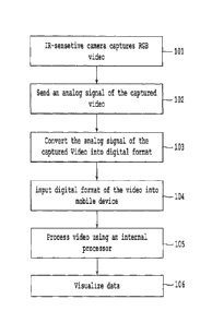
[0027] Figure 1 illustrates an overview of a method of detecting the iris and
the pupil of an
eye according to an exemplary embodiment. In order to image the constriction
and dilation of
a pupil with high contrast against the iris, it is necessary to use infra-red
(IR) light within the
8
CA 02924546 2016-03-16
WO 2015/042413
PCT/US2014/056579
safe level. Unfortunately, mobile cameras in current smartphones block IR
light while
passing visible light to improve image quality. Thus, the present embodiment
is designed to
include an easy mount interface to couple a low-cost IR-sensitive CMOS camera
module
onto existing mobile devices (the design of the mount interface onto existing
mobile devices
will be described in further detail with regard to Figure 3). The existing
mobile devices can
be sensitive to IR light or can be modified to be sensitive to IR light.
[0028] In Step 101, a high-quality RGB CMOS camera module, that has six high-
power
infrared LEDs to light up in the dark can be configured to capture vivid RGB
videos at
standard definition resolution (the high-quality RGB CMOS camera module can
capture
vivid RBG videos even in completely dark environments). In Step 102 the camera
module
sends an RCA analog signal of the captured video to a video capture device.
The video
capture device is part of the mount interface that receives an analog signal
from the camera
module. The camera module is easily mountable onto an existing mobile device
and should
be powered close to a 12V specification. The focal length of 4.3 mm and the
aperture of 2.0
of the camera module provide a field of view to 24-100 mm. The flash embedded
in a
smartphone device can be used for stimulating the eyes for measuring the
degree of dilation
and constriction of the pupil. Additionally, a modular source can also be used
for visual
stimulation. The method of the present embodiment can be used to combine
pupillary
changes to Glasgow Coma Scale (GCS).
[0029] In Step 103, the RCA analog signal that is output from the camera
module is
converted to an MPEG 4 format by the video capture device. It should be noted
that the
video capture device may convert the RCA analog signal to any audio/video
format and the
current embodiment is not restricted to the MPEG 4 format. In Step 104, the
MPEG 4 format
is input to a mobile device, including any of a smartphone device and/or a
tablet where an
application software is installed, via an embedded universal data I/O
interface such as
9
CA 02924546 2016-03-16
WO 2015/042413 PCT/US2014/056579
Bluetooth or micro/mini USB. Although the MPEG 4 format is described as being
input via
an embedded universal data I/O interface such as Bluetooth or micro/mini USB,
it should be
understood that any wireless/wired communication may be used and the present
embodiment
is not restricted to any particular wireless/wired communication.
[0030] Once data is transferred to the mobile device, the data can be
processed using the
internal processor (processing circuitry) of the mobile device in Step 105.
The processing of
the data is described with regard to Figure 2. Further, in Step 106, a user
may visualize data
that has been processed by the internal processor of the mobile device. The
user may also
visualize unprocessed data that is received from the camera module and the
video capture
device. The RGB image and an IR image captured by the camera module can be
overlaid to
be viewed by a user. The present embodiment is not limited to localized
processing. Sending
data to a remote server and returning a result from remote processing may also
be utilized.
[0031] It should be noted that the camera module is disposable and reusable.
Further, the
camera module can also be permanently mounted on the mobile device. Although
video
images are discussed above and below, it should be understood that the camera
module can
capture both live video streams and still images.
[0032] Figure 2 illustrates a method of detecting the pupil and iris. All the
steps described
below with regard to Figure 2 may be performed by a processor (processing
circuitry) of a
smartphone device or any external processing device. A video image, including
a plurality of
frames, is received in Step 201. As noted above with regard to Figure 1, the
video image is
captured by the camera module and converted into an MPEG 4 format by the video
capture
device. Given a video sequence with M frames, each frame of the video sequence
is
processed to detect the center and the radius of the pupil. In frame 1 (the
first frame among
the plurality of frames of the video sequence), a user is asked to localize
the center and any
two points on the boundary of the pupil and the iris as an initialization step
(Step 202). A
CA 02924546 2016-03-16
WO 2015/042413 PCT/US2014/056579
user may view the first frame on a display (such as display of a smartphone
device or any
display connected to the RGB CMOS camera) and can choose a center point and
two points
on the boundary of the pupil and the iris. Based on this information, a mask
image to cover
the expected location of the iris is generated in order to spare computation
in an unrelated
area (Step 202). The following steps are the same for processing other frames
of the video
sequence. In Step 203, if necessary, down-sampling of the image video frames
is performed
for saving computation time. In Step 203 an RGB image frame (video image
frame) is
converted to a gray scale with double data type ranging from 0 to 1. In Step
204, because
bright zones of specular reflection caused by the light source may exist, an
additional step
that removes the artifact by filling bright pixels with surrounding dark
pixels. Bright zones
may be removed from the mask image. All the pixels within the mask image are
considered
candidates for determining the center point of the pupil (Step 205). For
example, if the mask
image includes N pixels, then all N pixels are considered candidates in
determining the center
point of the pupil and the iris. A good-of-fit function fas defined below is
used to determine
the center point and the radius of the pupil and the iris.
lige,r11 ¨ I ge,r 90,r11 ge,r/8
6=1( (n ¨ 1) (p=o+1.
where n stands for the number of discrete values of the polar variable 9 that
are considered
and 90,r stands for the directional derivative of image intensity in the
radial direction. The
first term captures the weighted summed strength of the gradients across the
boundary, the
second term captures the uniformity of the gradients along the boundary, and
the last term
captures a slight preference for darker regions on the boundary interior. For
each n point in a
single video frame, the line integral of function fat distance of [Rmin, Rmax]
from the point
is computed (Step 206), and after mean filtering two local maxima
corresponding to the pupil
and the iris each are acquired. A goodness-of-fit measurement is defined as
the sum of two
11
CA 02924546 2016-03-16
WO 2015/042413 PCT/US2014/056579
peak values (Step 207). The goodness-of-fit measurement and corresponding Rs
are stored in
memory (Step 208). These procedures are repeated in remaining N-1 points in
the mask
image. The identified center and radius of the pupil are the ones that
maximize goodness-of-
fit measurement for all N points (Step 209). For the processing of the next
(k+l)th frame, the
mask image is updated using the values computed for the k'th frame. In other
words, the
center and radius of the pupil and the iris calculated for the k'th frame are
used to create an
updated mask image and steps 202 to 209 are repeated for each additional frame
of the video
sequence.
[0033] Figure 3 illustrates a design of the mount interface 300 that is
integrated with a
smartphone device 320. As noted above with regard to Figure 1, the mount
interface 300
includes a low-cost IR sensitive CMOS camera 301 that is coupled to a
smartphone device
320. The CMOS camera 301 includes six high-powered infrared LEDs 302 that
light up in
the dark. Although Figure 3 illustrates six high-powered infrared LEDs 302, it
should be
understood that any number of high-powered infrared LEDs may be used.
[0034] The CMOS camera 301 further includes a portion 303 into which a human
eye is
placed so that the human eye can be captured as a video image using the high-
powered
infrared LEDs 302. The mount interface 300 further includes an attachment 304
that is used
to attach the mount interface 300 to the smartphone device 320. The mount
interface 300
also includes an intermediate coupling device 305 which is coupled to both the
CMOS
camera 301 and the attachment 304, as illustrated in Figure 3. The combination
of the mount
interface 300 and a smartphone device 320 is useful as a hand-held device to
measure
pupillary dynamic parameters.
[0035] As noted above with regard to Figure 1, the CMOS camera 301 (using high-
powered
infrared LEDs 302) can capture vivid RGB videos at standard definition. The
captured RGB
videos are then sent to a video capture device as an RCA analog signal. The
RCA analog
12
CA 02924546 2016-03-16
WO 2015/042413 PCT/US2014/056579
signal that is output from the CMOS camera 301 is converted to an MPEG 4
format by the
video capture device. It should be noted that the video capture device may
convert the RCA
analog signal to any audio/video format and the current embodiment is not
restricted to the
MPEG 4 format. Finally, the MPEG 4 format of the video is input to a mobile
device
including any of a smartphone device and/or a tablet where the application
software is
installed, via an embedded universal data I/O interface such as Bluetooth or
micro/mini USB.
Although the MPEG 4 format is described as being input via an embedded
universal data I/O
interface such as Bluetooth or micro/mini USB, it should be understood that
any
wireless/wired communication may be used and the present embodiment is not
restricted to
any particular wireless/wired communication.
[0036] The captured video image is processed by a processor in a smartphone
device.
Although, a smartphone device is shown in Figure 3, it should be understood
that an external
processor (not shown) can also be used to process the capture video image.
[0037] The pupillary light reflex (PLR) reflects the integrity of the
autonomic nervous
system with constriction or miosis occurring in response to a flash of light
as a result of
increased parasympathetic tone and dilation or midriasis reflecting increased
sympathetic
tone. The flash of light can be provided by the flash light of a smartphone
device. There are
at least six pupillometric measures used in the generation of algorithms that
can determine a
physiologic perturbation such as, for example, usage of drugs or a medical
condition. The
two static measures include baseline pupil size and the maximally constricted
size to generate
the constriction amplitude (CON). The baseline pupul size is found before the
flash of light
and the maximally constricted size is determined after the flash of light. The
dynamic
responses to a flash of light including the velocity of constriction (average
constriction
velocity (ACV) and maximum constriction velocity (MCV)), the latency of
constriction
(LAT), and the velocity of re-dilation are other pupillometric measures. The
various
13
CA 02924546 2016-03-16
WO 2015/042413 PCT/US2014/056579
parameters of the PLR are impacted in a predictable way by various drugs and
medical
conditions. The at least six pupillometric measures can be selected based on
the application
of the measurement. Different applications such as detection of drug use or
detection of a
medical condition can take into account different pupillometric measures and
different
amounts of weight or different ways of processing in pupillometric measures.
100381 Heuristic models are used in the development of the algorithms.
Quantified data is
uploaded and analyzed for patterns that are predictive of a particular
scenario. The at least
six pupillometric measures can be calculated using a processor (processing
circuitry) of a
smartphone device or any other device based on the video images acquired by
the ROB
CMOS camera.
100391 Both the constriction amplitude (CON) and average constriction velocity
(ACV) have
been determined to be predictive of critical changes in the intracranial
pressure (ICP). When
CON is measured to be greater than (or equal to) 50%, such a measure indicates
that ICP is
less than 20mmHg (millimeters of mercury). Further, when CON is measured to be
less than
(or equal to) 10%, such a measure indicates that ICP is greater than 20mmHg or
indicates a
midline shift of the brain (both of which require immediate attention).
Finally, when ACV is
measured to be less than .6mm/sec, such a measure can also indicate that ICP
is greater than
20mmHg.
[0040] With regard to opioids, all parameters are inversely related to opioid
dose with acute
administration. With chronic administration, the impact on static measures and
ACV
reverses, allowing for the identification of tolerance. When the impact on
these parameters
actually increase from baseline, this is indicative of opioid induced
hyperalgesia, a neuro-
excitatory condition.
14
CA 02924546 2016-03-16
WO 2015/042413 PCT/US2014/056579
[0041] With regard to pain intensity, maximum constriction velocity (MCV) has
been shown
to correlate with subjective reporting. MCV increases by 0.11 mm/s for every
point increase
in a 10 point visual analog scale.
[0042] The present method can also be used to detect and monitor
dysautonomias, which
includes a variety of conditions including diabetic neuropathy and postural
orthostatic
tachycardia syndrome (POTS).
[0043] For example, Postural Orthostatic Tachycardia Syndrome (POTS) is
defined as the
presence of symptoms of orthostatic intolerance for at least 6 months
accompanied by a heart
rate increase of at least 30 beats/min within 5-30 minutes of assuming an
upright posture.
This nomially occurs in the absence of orthostatic hypotension (a fall in
blood pressure
>20/10mmHg). POTS reflects an autonomic imbalance and can be associated with
severe
functional disability causing limitations across multiple domains of quality
of life, including
physical, social, and role functioning.
[0044] In order to assess POTS, subjects are first dark-adapted and in a
supine position to
obtain baseline values (e.g. baseline pupil size) After ten minutes or so, a
reading is taken in
each eye. After which the subject stands for 10 minutes and another reading is
performed.
Herein, the reading is referring to capturing video images of the eye,
processing the video
images, and calculating values of the at least six pupillometric measures
noted above. The
following results were obtained based on the reading taken in each eye with
regard to the
assessment of POTS.
[0045] It was observed that among POTS patients, pupillometry at baseline
revealed that the
percent change of pupil diameter from its maximum to minimum diameter (CON)
was
significantly lower, as was the constriction velocity (ACV) when compared to
the healthy
controls. Additionally, it was found that the latency (LAT), which is the
response time of the
CA 02924546 2016-03-16
WO 2015/042413 PCT/US2014/056579
pupil after the presentation of a stimulus, was higher in POTS patients than
healthy controls.
The magnitude of these differences can be seen in Table 1.
Table 1: Comparison of Experimental and Control values for Trial 1
Experimental Control p-value
CON -0.31 -0.35 .009855
ACV -3.26 -3.62 .00376
LAT 0.25 0.23 .027015
[0046] The p-value is the probability of obtaining a test statistic result at
least as extreme or
as close to the one that was actually observed, assuming that the null
hypothesis is true.
[0047] Under orthostatic stress it was found that POTS patients experienced a
decrease in
maximum pupil diameter by 2.5% and minimum pupil diameter by 6.7%.
Constriction
percentage was found to increase by 8.5% and average constriction velocity
also increased by
7.3%. These percentage values are a comparison between measurements in a
supine position
and an upright position. The impact of various therapeutic interventions can
therefore be
objectively monitored after diagnosis by determining the impact on these
parameters.
[0048] Diabetic neuropathy can be detected when there is a significant
reduction in the pupil
to iris ratio and/or a significant increase in the latency.
[0049] Figure 4 illustrates an application design that acquires and processes
videos and
displays computer clinical parameters on a smartphone device. For example, a
smartphone
device displays a first screen 401 of the application design that allows a
user to use the
pupillometer (mount interface 300 and the application to process the at least
six pupillometric
measures noted above) to capture video images of the eye, including the iris
and the pupil.
Further, the first screen 401 on a smartphone device 400 also allows a user to
search for
previously stored video images (both raw images and processed images,
including parameters
discussed with regard to Figures 2 and 3). A second screen 402 on a smartphone
device
illustrates a capture of an eye and also a minimum diameter and a maximum
diameter. The
16
CA 02924546 2016-03-16
WO 2015/042413 PCT/US2014/056579
minimum diameter and maximum diameter can be either of the pupil or the iris.
Although
only a minimum diameter and a maximum diameter are illustrated on the second
screen 402,
it should be understood that other measurements of the iris and pupil (for
example the at least
six pupillometric measures noted above) can be displayed on the second screen
402.
Additionally, the second screen 402 also illustrates a % change in the
diameter of the
pupil/iris. The % change can also correspond to a change in constriction and
dilation
velocities of the pupil. Finally, the second screen 402 illustrates a velocity
corresponding to a
current velocity of the constriction and dilation of the pupil.
[0050] The third screen 403 on the smartphone device illustrates a step for
saving
information regarding a patient and the fourth screen 404 illustrates a search
feature to search
for information on a patient. All patient information can be stored in a
separate electronic
database (not shown).
[0051] Figure 5 illustrates a pupil and iris detection overlaid on an image
frame 501. The
circle 502 represents an estimate of the iris and circle 503 represents an
estimate of the pupil.
Circles 502 and 503 represent estimates of the iris and pupil, respectively,
which have been
calculated using the steps in Figure 2.
[0052] The present embodiments provide features for stable performance across
varying
pupil colors and contrast palette. Additionally, the present embodiments are
robust to handle
any possible motion (blinking, translation, etc.) of the eye. Finally, the
present embodiments
also provide a user-friendly graphical interface to display video and
extracted measurements
and to improve usability in the clinic. The present embodiments provide a
convenient, stable,
and cost effect platform.
[0053] Another advantage of the present embodiments is that the present
embodiments allow
tracking of patients using opioids. According, with an application of the
present
embodiments, it is possible to identify patients on opioid therapy and
possible abuse from
17
CA 02924546 2016-03-16
WO 2015/042413 PCT/US2014/056579
opioid use. That is, the present embodiments can be used to detect if a
patient is using a dose
beyond the dose prescribed.
[0054] The present embodiments also allow for detection of opioid tolerance
and opioid-
induced hyperalgesia. The present embodiments can also be used to detect if a
patients is
responsive to opioid therapy. Some different genotypes of cytochrome enzymes
do not allow
adequate opioid metabolism, transforming pro-drugs in active metabolite. The
cytochrome
P450 metabolite enzymes have been implicated in the metabolism of opioid
drugs, and
variants in these enzymes, specifically the CYP2D6 have been linked to
toxicity and
therapeutic efficacy of opioids. A method of the present embodiments for
opioid efficacy
tracking can identify specific phenotypes based in pupillary changes and allow
individualizing the treatment.
[0055] The present embodiments can also be used to detect opioid withdrawal
symptoms
based in pupillary changes, to detect efficacy of the treatment of abstinence
syndrome, to
detect neonatal abstinence syndrome when mothers were exposed to opioids or
heroin during
pregnancy, and to support analgesia nociception analysis assessing
effectiveness of regional
anesthesia in anesthetized patients.
[0056] The method of the present embodiments can also be used for management
of
methadone use. Methadone dose management is subjective based on clinician
judgment. The
method of the present embodiments allow the transition from morphine or any
other opioid to
methadone based on pupillary objective measurements, increasing safety and
efficacy.
Additionally, the method of the present embodiments can also be used to assess
pupillary
reactivity during cardiopulmonary resuscitation (CPR), pupillary light reflex
and the
magnitude of pupillary dynamic changes during CPR as objective measurements
for
predicting neurologic recovery after cardiac arrest.
18
CA 02924546 2016-03-16
WO 2015/042413 PCT/US2014/056579
[0057] The method of the present embodiments can be used to assess the
magnitude of
response to noxious stimulus tracking pupillary parameters in patients sedated
or under
general anesthesia, to assess very subtle changes in pupillary parameters that
will be used as
indicators of cognitive activity (providing an assessment for alertness and
cognitive status
assessment), to assess concussion severity in the sports field, and to assess
changes in the
intracranial pressure (ICP) after a traumatic brain injury (TBI). Pupillary
parameters are
sensitive indicators of ICP changes. The method of the present embodiments
enable health
care providers to indicate surgery or conservative clinical treatment. This
tool can work as a
prognostic indicator and can be used by first responders and in the war battle
field as well as
in emergency departments and intensive care units (ICU).
[0058] The method of the present embodiments can be used to define the
association
between psychotropic drugs use and overdose and pupillary changes and outcomes
from drug
overdose based in pupillary changes. Additionally, the method of the present
embodiments
can also be used to define outcomes based in pupillary changes after an
ingestion of
cholinergic poisons, such as carbamate and organophosphorate. This method can
be designed
to track pupillary changes before and during the treatment and to guide the
treatment.
100591 The method can be developed to work as triage test in drivers suspected
to be under
influence of alcohol or controlled substances. If there any unusual pupillary
response during
the test, the driver will be submitted to other tests.
[0060] Further, the method of the present embodiments can be used to help
identify the drug
and treatment efficacy after childhood and adults poisoning (pupillary changes
can identify if
the treatment was efficient and will help to define which drug was used), to
compare right
and left pupil will serve as a screening tool for abnormalities related to
diseases and
conditions of the eye such eye infections, brain trauma and tumors (anisocoria
can be a red
flag in many different clinical situations and this method can detect the
magnitude of this
19
CA 02924546 2016-03-16
WO 2015/042413 PCT/US2014/056579
condition and help to avoid serious clinical complications), and to detect
pupillary changes
during interview to detect lies.
[0061] Next, a hardware description of the device (for example, a smartphone
device)
according to exemplary embodiments is described with reference to Figure 6. In
Figure 6,
the device includes a CPU 600 which performs the processes described above.
The process
data and instructions may be stored in memory 602. These processes and
instructions may
also be stored on a storage medium disk 604 such as a hard drive (HDD) or
portable storage
medium or may be stored remotely. Further, the claimed advancements are not
limited by the
form of the computer-readable media on which the instructions of the inventive
process are
stored. For example, the instructions may be stored on CDs, DVDs, in FLASH
memory,
RAM, ROM, PROM, EPROM, EEPROM, hard disk or any other information processing
device with which the device communicates, such as a server or computer.
[0062] Further, the claimed advancements may be provided as a utility
application,
background daemon, or component of an operating system, or combination
thereof, executing
in conjunction with CPU 600 and an operating system such as Microsoft Windows
7, UNIX,
Solaris, LINUX, Apple MAC-OS and other systems known to those skilled in the
art.
[0063] CPU 600 may be a Xenon or Core processor from Intel of America or an
Opteron
processor from AMD of America, or may be other processor types that would be
recognized
by one of ordinary skill in the art. Alternatively, the CPU 600 may be
implemented on an
FPGA, ASIC, PLD or using discrete logic circuits, as one of ordinary skill in
the art would
recognize. Further, CPU 600 may be implemented as multiple processors
cooperatively
working in parallel to perform the instructions of the inventive processes
described above.
[0064] The device in Figure 6 also includes a network controller 606, such as
an Intel
Ethernet PRO network interface card from Intel Corporation of America, for
interfacing with
network 66. As can be appreciated, the network 66 can be a public network,
such as the
CA 02924546 2016-03-16
WO 2015/042413 PCT/US2014/056579
Internet, or a private network such as an LAN or WAN network, or any
combination thereof
and can also include PSTN or ISDN sub-networks. The network 66 can also be
wired, such
as an Ethernet network, or can be wireless such as a cellular network
including EDGE, 3G
and 4G wireless cellular systems. The wireless network can also be WiFi,
Bluetooth, or any
other wireless form of communication that is known.
[0065] The device further includes a display controller 608, such as a NVIDIA
GeForce
GTX or Quadro graphics adaptor from NVIDIA Corporation of America for
interfacing with
display 610, such as a Hewlett Packard HPL2445w LCD monitor. A general purpose
I/O
interface 612 interfaces with a keyboard and/or mouse 614 as well as a touch
screen panel
616 on or separate from display 610. General purpose I/O interface also
connects to a variety
of peripherals 618 including printers and scanners, such as an OfficeJet or
DeskJet from
Hewlett Packard.
[0066] A sound controller 620 is also provided in the device, such as Sound
Blaster X-Fi
Titanium from Creative, to interface with speakers/microphone 622 thereby
providing sounds
and/or music.
[0067] The general purpose storage controller 624 connects the storage medium
disk 604
with communication bus 626, which may be an ISA, EISA, VESA, PCI, or similar,
for
interconnecting all of the components of the device. A description of the
general features and
functionality of the display 610, keyboard and/or mouse 614, as well as the
display controller
608, storage controller 624, network controller 606, sound controller 620, and
general
purpose I/O interface 612 is omitted herein for brevity as these features are
known.
[0068] Obviously, numerous modifications and variations of the present
disclosure are
possible in light of the above teachings. It is therefore to be understood
that within the scope
of the appended claims, the embodiment may be practiced otherwise than as
specifically
described herein. For example, advantageous results may be achieved if the
steps of the
21
CA 02924546 2016-03-16
WO 2015/042413 PCT/US2014/056579
disclosed techniques were performed in a different sequence, if components in
the disclosed
systems were combined in a different manner, or if the components were
replaced or
supplemented by other components. The functions, processes, and algorithms
described
herein may be performed in hardware or software executed by hardware,
including computer
processors and/or programmable processing circuits configured to execute
program code
and/or computer instructions to execute the functions, processes, and
algorithms described
herein. A processing circuit includes a programmed processor, as a processor
includes
circuitry. A processing circuit also includes devices such as an application
specific integrated
circuit (ASIC) and conventional circuit components arranged to perform the
recited
functions.
[0069] The functions and features described herein may also be executed by
various
distributed components of a system. For example, one or more processors may
execute these
system functions, wherein the processors are distributed across multiple
components
communicating in a network. The distributed components may include one or more
client
and/or server machines, in addition to various human interface and/or
communication devices
(e.g., display monitors, smart phones, tablets, personal digital assistants
(PDAs)). The
network may be a private network, such as a LAN or WAN, or may be a public
network,
such as the Internet. Input to the system may be received via direct user
input and/or received
remotely either in real-time or as a batch process. Additionally, some
implementations may
be performed on modules or hardware not identical to those described.
Accordingly, other
implementations are within the scope that may be claimed.
[0070] It must be noted that, as used in the specification and the appended
claims, the
singular forms "a," "an," and "the" include plural referents unless the
context clearly dictates
otherwise.
22
CA 02924546 2016-03-16
WO 2015/042413 PCT/US2014/056579
[0071] While certain embodiments have been described, these embodiments have
been
presented by way of example only, and are not intended to limit the scope of
the inventions.
Indeed, the novel methods, apparatuses and systems described herein can be
embodied in a
variety of other forms; furthermore, various omissions, substitutions and
changes in the form
of the methods, apparatuses and systems described herein can be made without
departing
from the spirit of the inventions. The accompanying claims and their
equivalents are
intended to cover such forms or modifications as would fall within the scope
and spirit of the
inventions.
23