Note: Descriptions are shown in the official language in which they were submitted.
CA 02924954 2016-03-21
WO 2014/138620
PCT/US2014/021889
METHOD AND SYSTEM FOR TREATMENT OF NEUROMOTOR DYSFUNCTION
BACKGROUND
The present teachings generally relate to the field of providing stimulation
of central
nervous system tissue, muscles, nerves, or combinations thereof, and more
particularly to a
system and method for improving neural or neuromuscular communication
impairment through
multi-point stimulation
The nervous system comprises the central and the peripheral nervous system.
The central
nervous system is composed of the brain and the spinal cord, and the
peripheral nervous system
consists of all of the other neural elements, namely the nerves and ganglia
outside of the brain
and spinal cord.
Damage to the nervous system may result from a traumatic injury, such as
penetrating
trauma or blunt trauma, or a disease or disorder including, but not limited to
birth defects,
cerebral palsy, Alzheimer's disease, multiple sclerosis, Huntington's disease,
arnyotrophic
lateral sclerosis (ALS), diabetic neuropathy, senile dementia, stroke and
isch.emia.
After spinal cord injury (SCI), spared regions of the central nervous system
are
spontaneously capable of repairing the damaged pathway, although the process
is very limited.
Moreover, despite the many promising treatment strategies to improve
connections across the
damaged spinal cord, the strength of connectivity and functional recovery of
the impaired spinal
cord are still unsatisfactory.
Electrical stimulation of the central and peripheral nervous systems improves
neuronal
connectivity, and can be employed to improve functional recovery after
neuronal injury. It is an
effective method that promotes reactive sprouting through which an increase in
the number of
functional connections may be possible. Electrical stimulation can also
improve functional
connections by strengthening the weak existing synapses and/or by promoting
synaptogenesis.
One of the emerging concepts is that the nervous system contains latent
pathways that can be
awakened by electrical stimulation or pharmacological manipulation.
The majority of the methods employing electrical stimulation utilize single or
dual point
paradigm in which unipolar or bipolar stimuli are delivered at points of the
challenged neural.
pathway. The effectiveness of this stimulation depends on active propagation
of an action
potential.
CA 02924954 2016-03-21
WO 2014/138620
PCT/US2014/021889
There is a great desire to improve the effectiveness of electrical stimulation
in order to
more successfully treat or even reverse neuromotor dysfunctions. Treatment
systems can be very
complex. There is a need for apparatus that reduces risks while also meeting
the need for
treatment system that is simpler to operate that still safely improves motor
control and function.
BRIEF SUMMARY
Effective systems and methods for improving neural communication impairment of
a
vertebrate being and affecting motor activity of a peripheral body part are
disclosed herein
below.
The spinal cord connects with, and communicates the action potential issued by
the motor
cortex to our distal muscles to drive motor activity. The spinal cord extends
along the spine and
branches out to the upper and lower extremities to carry such action potential
signal to nerve(s)
associated with the muscle(s) intended to be actuated. We call this neural
transmission path "a
neural pathway."
Associative stimulation causes enduring changes in the nervous system based on
the
Hebbian concept of spike-timing-dependent plasticity. According to the present
invention, trans-
spinal direct current stimulation (tsDC) modulates associative plasticity.
Combining associative
stimulation with tsDC has a major and long lasting effect on locomotor
recovery in practice of
the present invention, in various embodiments thereof, by increasing the
amplitude of cortically
evoked action potential signal arriving at the target nerve at the
dysfunctional muscle.
For purposes of this description, it may be generalized that a neural pathway
runs or may
be traced from an area of the motor cortex associated with a distal peripheral
muscle of interest
down the spinal cord and then the pathway branches out to the arm or leg and
terminates at the
controlling nerve associated with the muscle of interest. The spinal location
of this neural
branching we refer to herein as a "spinal junction" of the neural pathway. The
motor cortex
evokes muscle activity by issuing a signal that propagates down the pathway
and through the
spinal junction down to the target nerve to evoke activity of the muscle of
interest.
In several practices of the present invention, triple stimulation of the
dysfunctional
neuromotor pathway proves to be highly effective in mitigating dysfunction. In
an illustrative
embodiment, the distal nerve(s) associated with dysfunctional muscle(s) of
interest are
stimulated with a pulsed stimulation signal (Pulsed "distal stimulation"), the
spinal junction on
2
CA 02924954 2016-03-21
WO 2014/138620
PCT/US2014/021889
the neural pathway of interest is stimulated with a substantially continuous
unvarying signal
(constant "spinal junction stimulation"), and a location above the spinal
junction on that neural
pathway through which the cortical action potential passes is stimulated with
a pulsed
stimulation signal ("pulsed cortical stimulation"),
The pulsed cortical stimulation maybe applied for example, by application of
electrical or
magnetic pulses. The pulsed cortical stimulation is applied at the motor
cortex, or along the
neural pathway of interest descending from the motor cortex at a location
above the spinal
junction, about 10 cm above the spinal junction if pulsed magnetic stimulation
is used or
anywhere above the spinal junction if electrical stimulation is used, provided
that if the junction
is in lumbar region, then placement is anywhere between thoracic to cervical
locations. For
cervical location of spinal junction, the pulsed cortical electrical
stimulation can be applied at the
level of the mastoid processes (bilaterally) to activate the corticospinal
tract. (Pulsed magnetic
stimulation applied to the cranium is known as trans-cranial magnetic
stimulation ("TMS").
The distal and the cortical stimulations each induce responsive signals, he.,
an induced
pulsed distal stimulation signal and an induced pulsed cortical stimulation
signal which
communicate along the neural pathway of interest toward the spinal junction.
The spinal junction
stimulation signal is applied first to the spinal junction location and then
application of the distal
and the cortical stimulations are each timed such that their induced signals
arrive simultaneously
at the already-stimulated spinal junction on that dysfunctional neural pathway
According to the present invention, constant spinal junction stimulation, such
as tsDC,
modulates associative plasticity when combined with associative stimulation,
having major and
long lasting effect on locomotor recovery. The present invention resolves,
reverses, or improves
neuromotor dysfunction, Often such dysfunction demonstrates at an
underperforming limb, such
as a spastic, weak, or paralyzed arm or leg, hand or foot, for example. Large
and small nerves
can be stimulated. In practice of embodiments of our triple stimulation
system, method, and
apparatus, the combination of our tsDC paired with associative stimulation,
cortical and
peripheral, has demonstrated substantial lessening of the dysfunction.
In one or more embodiments, the system of these teachings includes a first
signal-
providing component configured to provide pulsed peripheral stimulation
signals at the
peripheral body part of interest, a second signal-providing component
configured to provide a
pulsed motor cortex stimulation signal to elicit a motor cortex action
potential signal, a
3
CA 02924954 2016-03-21
WO 2014/138620
PCT/US2014/021889
substantially continuous-level signal-providing component configured to
provide DC current, or
directional flux stimulation, signal at a neural spinal junction, and a
controller component
configured to control timing of the pulsed peripheral stimulation signals and
the pulsed motor
cortex stimulation signal; the timing of the pulsed peripheral stimulation
signals and the pulsed
motor cortex stimulation signal being controlled such that a peripheral signal
from the peripheral
body part and a pulsed motor signal from the motor cortex area are
substantially simultaneously
present at the neural spinal junction when the neural spinal junction is being
stimulated by the
substantially continuous DC spinal signal.
The latter DC signal is also referred to a cathodal trans-spinal direct
current stimulation,
tsDC, and is applied from the cathode of source of level continuous DC
stimulation signal, which
can include a small ramp at the beginning and end of the signal duration.
Other practices of
directional stimulation energy sources applied to the spinal junction are also
within the scope and
practice of the present invention, which by illustration but without
limitation of the scope of the
invention, in one embodiment could include repetitive TMS, or a cathodal polar
stimulation
equivalent, if so devised.
In one or more embodiments, the method of these teachings includes providing
pulsed
peripheral stimulation signals at the peripheral body part, providing a pulsed
motor cortex
stimulation signal to a motor cortex area, and providing a substantially
direct current cathodal
spinal stimulation signal at a neural spinal junction, timing of the pulsed
peripheral stimulation
signals and the pulsed motor cortex stimulation signal being selected such
that a backward motor
signal from the peripheral body part and a pulsed motor signal from the motor
cortex area are
substantially simultaneously present at the neural spinal junction when the
neural spinal junction
is being stimulated by the spinal stimulation signal.
Various other embodiments are disclosed.
For a better understanding of the present teachings, together with other and
further needs
thereof, reference is made to the accompanying drawings and detailed
description and its scope
will be pointed out in the appended claims.
BRIEF DESCRIPTION OF THE DRAWINGS
Figure 1 shows one embodiment of the system of these teachings;
4
CA 02924954 2016-03-21
WO 2014/138620
PCT/US2014/021889
Figures 2a, 2b show an application of one embodiment of the system and method
of these
teachings;
Figure 3 shows another embodiment of the system of these teachings;
Figure 4 shows an embodiment of a component of the system of these teachings;
Figure 5A-D shows an embodiment of the system of these teachings for tsDC +
cortico-
sciatic and spino-sciatic (CSA) protocols;
Figure 6A-C shows an embodiment of the system of these teachings for tsDC +
spino-
sciatic (SSA) protocol;
Figures 7A-D shows the results of one exemplary application of the method and
system
of these teachings;
Figure 8 shows peg test where treatment for hand yields shortened peg-board
time in
normal subjects;
Figure 9 shows acceleration of the movement at the wrist joint increased
significantly
after full treatment;
Figure 10 shows three weeks assessment of strength improvement of one subject
with
cerebral palsy; Untreated side was unchanged. Treated side evidences
significantly improved
strength of handgrip capability;
Figure ha-f shows before, during and after results for one subject (CP);
Figures 12A-B shows three weeks assessments of CP subject of ha-f;
Figures 13A-D show Longitudinal electrophysiologica] changes recorded from the
anterior deltoid muscle in same CP subject of Figures10-12;
Figures 14a-b show Right hand grasp during peg-test task before and after
treatment (6
weeks) for a Different CP subject; and
Figures 1.5A-B show, for a stroke subject, an improvement of 3D motion of the
left elbow
joint.
DETAILED DESCRIPTION OF EMBODIMENT OF THE INVENTION
The following detailed description presents the currently contemplated modes
of carrying
out the invention. The description is not to be taken in a limiting sense, but
is made merely for
the purpose of illustrating the general principles of the invention, since the
scope of the invention
is best defined by the appended claims,
CA 02924954 2016-03-21
WO 2014/138620
PCT/US2014/021889
As used herein, the singular forms "a," "an," and "the" include the plural
reference unless
the context clearly dictates otherwise.
Except where otherwise indicated, all numbers expressing quantities of
ingredients,
reaction conditions, and so forth used in the specification and claims are to
be understood as
being modified in all instances by the term "about"
Effective systems and methods for improving neural communication impairment of
a
vertebrate being and affecting motor activity of a peripheral body part are
disclosed herein
below,
One embodiment of the present teachings, method and apparatus, provides a
system and
method for paralysis treatment that addresses mobility dysfunction by
improving motor signal
transmission from motor cortex to distal muscles, Substantial reversal of
paralysis and of related
dysfunction has been demonstrated in laboratory mice and in human subjects. In
various
embodiments the mobility treatment of the present teachings is administered
through operation
of the disclosed stimulation treatment center,
One embodiment of the present mobility treatment is applied to suspect motor
pathway to
reverse the neuromotor signal transmission disorder that apparently is
retarding muscle function.
Dysfunction is treated regardless of the original cause of such dysfunction,
The treatment has
demonstrated reversal of paralysis or of other degraded mobility conditions
upon a variety of
pathologies.
The treatment is based upon a combination of timed cortical, spinal cord and
associated
nerve/muscle stimulations, In one instance, three simultaneous stimulation
signals are applied at
strategic locations along a diagnosed failing neural motor pathway to improve
transmitted motor
signal to the distal muscle, In an illustrative treatment, constant level
trans-spinal direct current
(tsDC) stimulation is applied to the surface area above the spinal neural
junction Where the
subject neural pathway branches out to the target muscle(s). Sponge electrodes
or contact gel are
used to assure delivery of a continuous constant level trans-spinal DC
stimulation at the spinal
junction as well as managing current density of the applied stimulation.
In an illustrative practice of the invention includes a combination of
stimulations,
including pulsed stimulation of an area of the motor cortex which is
associated with the
dysfunctional target muscle and pulsed stimulation applied to the nerve area
associated with the
dysfunctional target muscle. The motor cortex stimulation is non-contact
pulsed magnetic
6
CA 02924954 2016-03-21
WO 2014/138620
PCT/US2014/021889
stimulation, or alternatively stimulated with pulsed DC electrical
stimulation. The peripheral
nerve is stimulated with pulsed DC. The electrical stimulations are achieved
with conventional
electrodes.
In an illustrative embodiment, stimulation of motor cortex and distal muscles
is generally
known as paired associative stimulation (PAS). We use a modified form of PAS,
by applying
two pulses in one cycle to create dual peripheral pulses to induce changes at
the cortex (long
delay pulse) and at the spinal cord (short delay pulse) during the triple
stimulation protocol. The
effect of the unique combination of simultaneous tsDC and any PAS is to
substantially enhance
motor signal transmission within the failing pathway. Now an enhanced motor
signal is applied
to the muscle of interest, with the result of measured improvement in mobility
(measurable in
strength, speed, range of motion, and/or dexterity, etc.).
In one or more embodiments, the system of these teachings includes a first
signal-
providing component configured to provide pulsed peripheral stimulation
signals at the
peripheral body part, a second signal-providing component configured to
provide a pulsed motor
cortex stimulation signal to a motor cortex area, a substantially constant
current DC signal-
providing component configured to provide relatively constant level direct
current spinal
stimulation signal at a neural spinal junction and a controller component
configured to control
timing of the pulsed peripheral stimulation signals and the pulsed motor
cortex stimulation
signal. In one practice, several peripheral pulses occur per cortical pulse.
The timing of the
pulsed peripheral stimulation signals and of the pulsed motor cortex
stimulation signal is
controlled by the controller such that a backward motor signal from the
peripheral body part and
a descending motor signal from the motor cortex are substantially
simultaneously present at the
neural spinal junction When the neural spinal junction has been and is being
stimulated by the
substantially constant DC signal.
2.5 In one configuration, the substantially DC signal-providing component
active electrode
provides cathodal stimulation at the spinal junction. In a further instance,
the controller
component is configured to provide the direct current spinal stimulation
signal at the neural
spinal junction before the pulsed peripheral stimulation signals and the
pulsed motor cortex
stimulation signal are applied, and subsequently to provide a first pulse as a
peripheral
stimulation signal to the nerve at the muscle of interest, and provide, after
a time delay after
providing the first pulse, a second pulse of the peripheral stimulation signal
to the nerve at the
CA 02924954 2016-03-21
WO 2014/138620
PCT/US2014/021889
muscle, and provide, after another time delay after providing the first and
second pulses, the
pulsed motor cortex stimulation signal, the time delay and the other time
delay being selected
such that a backward motor signal from the stimulated nerve for the muscle and
the pulsed motor
signal from the motor cortex are substantially simultaneously present at the
neural spinal
junction when the neural spinal junction is being stimulated by the
substantially DC signal.
In the embodiment of Figure I a, peripheral stimulator 103 issues pulsed DC
stimulation
to the patient via the +1- leads, shown extending from i/o connector 161. The
leads are affixed at
the nerve associated with the dysfunctioning muscle. Peripheral stimulation
electrodes are
positioned on the nerve and can be placed at the main nerve trunk to stimulate
a large group of
muscles, Placement is at the nerve associated with muscle or muscles with
dysfunction. For
example, the electrodes could be placed on leg, behind knee, on the foot, on
arm, at wrist or
shoulder, all depending upon the target for that patient.
Various Embodiments
An embodiment of the system of these teachings is shown in Figures 1-4,
wherein
stimulation system 100 includes a first stimulator 101 that provides motor
cortex stimulation. In
one exemplary embodiment, the first simulator 101 could be, for example, but
not limited to, a
conventional pulsed magnetic stimulator or a conventional pulsed DC
stimulator, A second
stimulator 102 provides continuous trans-spinal DC stimulation to the neural
spinal junction. In
an exemplary embodiment, the second stimulator could be, but is not limited
to, a conventional
source of continuous DC stimulation capable of delivering a continuous
selected low current
signal, with minimal variation, and using sponge electrodes and with a short
ramp-up and ramp-
down at the beginning and end of stimulation, all to mitigate startup and shut
down artifacts, A.
third stimulator 103 provides stimulation of the peripheral nerve(s)
associated with a
dysfunctional area/muscle(s) of interest. In an exemplary embodiment, the
third stimulator can
be, but is not limited to, a conventional pulsed stimulator capable of
delivering pulsed DC
stimulation,
Stimulator 101 provides pulsed stimulation to the motor cortex. For example,
this may
be pulsed magnetic or DC electrical stimulation, and may be a stand-alone unit
or incorporated
within control center 105. In an illustrative embodiment of system 100,
magnetic stimulator 101
is included as a standalone magnetic stimulation system for non-contact
delivery of pulsed
magnetic stimulation to the motor cortex,
8
CA 02924954 2016-03-21
WO 2014/138620
PCT/US2014/021889
Control center 105 further includes system controller/synchronizer 104
configured to
control andlor synchronize the stimulators 101, 102, 103, and in one
embodiment includes a non
transitory computer usable medium (such as, but not limited to, RAM) and an
I/O component
104A to provide synchronization, control and or timing signals from controller
104 to external
stimulators, such as to the first simulator 101. In the illustrative
embodiment of Figure 1,
controller/synchronizer 104 at I/O 104A delivers a trigger signal 127 to
trigger stimulator 101
according to practices of the invention further discussed below,
in an illustrative embodiment, the system further includes support equipment
including
channel amplifier 106, data. recorder 107 (which may also include capture of
muscle motor
evoked potential and EMG), and computer 108, where the computer further
supports
synchronization, control and/or stimulation and data acquisition. Computer 108
and or the
system controller/synchronizer 104 can, in one embodiment, include one or more
processors 120
and computer usable media 130, as further shown in Figure 4, where the
computer usable media
has computer readable code embodied therein that, when executed in the one or
more processors,
causes the one of more processors to perform the method of these teachings. In
the embodiment
shown in Figure 4, the one or more processors 120 are operatively connected to
the computer
usable media 130 by means of a connection component, such as a computer bus
135,
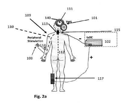
Figure 2a shows a further embodiment of the invention, including trans-spinal
stimulator
102, a sponge electrode 115 providing cathodal stimulation placed upon the
appropriate spinal
column segment. In practice of embodiments of the invention, for upper
extremity stimulation,
sponge electrode 115 is placed at cervical segment in the area of C6 to Ti,
and for lower
extremity is placed in the area of TIO and Li vertebral levels. These areas
have known
associations with distal nerves related to target muscles of interest, as will
be appreciated by
those skilled in the art. The return electrode is placed at a bony location
such as at the lower leg,
In an illustrative embodiment, during treatment, patients are seated
comfortably in
armchair. The cathodal tsDC electrode 115 is applied over the appropriate
spinal column
segment, e.g., segment 113. Ti\,4S coil is placed over the motor cortex
representation of upper
extremity for upper extremity group or over the representation of the lower
extremity for lower
extremity groups. The peripheral electrodes are placed over the nerve of
interest.
In an illustrative practice, tsDC and TMS stimulations are be applied at the
commencement of the session and remain on for the duration, simultaneously
with multi-pulse
9
CA 02924954 2016-03-21
WO 2014/138620
PCT/US2014/021889
peripheral stimulation. The conventional 10/20 system is used to locate the
appropriate
placement of the coil (or alternatively electrode) stimulation at the cortex,
as will he understood
by a person skilled in the art. Typical stimulation sessions run for 20
minutes and are repeated as
needed, several times per week, over a number of weeks, and as per level of
improvement of the
treated muscle(s) of interest.
An evoked potential or evoked response is an electrical potential of the
nervous system
generated following presentation of a stimulus, as distinct from spontaneous
potentials as
detected by electroencephalography (EEG), electromyography (EMG), or other
electrophysiological recording method. In a practice of the invention, during
stimulation, motor
evoked potential (MEP) is recorded conventionally via bipolar surface
electrodes of fixed inter-
electrode distance of 2.5 cm and EMG is recorded from ipsilateral and
contralateral (in relation
to stimulated motor cortex) muscles in the upper and lower extremities as
needed.
In an illustrative embodiment, stimulation intensity is adjusted to about 115%
of the
active threshold. Active motor threshold is defined as the minimum stimulus
intensity that
produces a consistent motor evoked response. In an illustrative embodiment,
magnetic
stimulation of stimulator 101 is applied at a frequency of 0.3 Hz, In an
illustrative "figure-eight"
magnetic stimulator coil 140 is used to apply such stimulation, shown in
Figure 2a and in Figure
3, the latter as part of an illustrative system 100 operating in conjunction
with a patient platform,
such as a medical chair 142, both of which would provide for the comfort of
the patient during
sessions. in one exemplary embodiment, motor cortex stimulation is carried out
using coil 140
positioned over the M1 region, shown in Figure 2a and 3. In other exemplary
embodiments,
motor cortex electrical stimulation is carried out using one or more
electrodes for effecting motor
cortex stimulation.
In an illustrative, but not limiting embodiment, TMS is done with Figure-of-
eight coil
140 positioned over the M1 region (as such region is known to those skilled in
the art). Subjects
are seated comfortably in an armchair. The head is strapped to a head-rest to
prevent movement
relative to coil 140. The coil is placed tangential to the skull. The coil is
held stably by a coil
stand or the like that allows easy adjustment.
In an illustrative embodiment, stimulator 102 delivers trans-spinal direct
current
stimulation (tsDC) to the spinal junction of interest 113 and is held at a
safe constant current,
ranging up to about 5mA, or higher, depending upon patient tolerance of felt
effect. Typical
CA 02924954 2016-03-21
WO 2014/138620
PCT/US2014/021889
session is twenty minutes. The active cathodal electrode of stimulator 102
(stimulator 102 "-") is
placed over the selected upper or lower area of spinal segment of the spinal
column. As shown
in the non-limiting illustration of Figure 11 a, for treatment of an upper
extremity motor
dysfunction issue, sponge electrode 115 is applied to the upper spine around
C6 to Ti and the
return sponge electrode 1.17 is placed over a non-critical, non-nerve,
location such as the bony
part of the leg, as shown. Sponge electrodes are used to deliver the constant
level trans-spinal
DC stimulation at the spinal junction without uncomfortable artifacts. A large
sponge electrode
117 is used on the positive return electrode at the leg.
As will be appreciated by those skilled in the art, the size of the active
cathodal sponge
electrode 115, i.e., the amount of surface area presented to the skin at the
spine, is selected
according to safety considerations in view of the level of energy being
applied, current density,
energy dissipation considerations, and known characteristic data of the
patient. Illustrative, non-
limiting examples of such patient characteristics may include size, weight,
age, diagnosis, prior
medical history, and special needs, for example. In an illustrative
embodiment, such data 118 is
loaded into system 100 at control center 105,
Embodiments of the present invention are derived from simultaneous
conditioning of the
spinal neurons at the spinal junction of interest by applying constant trans-
spinal DC stimulation
at the spinal junction combined with repetitive stimulation to affect the
cortex, applied to the
motor cortex or an extension thereof or proxy therefore, for evoking cortical
pulses, and pairs of
pulsed peripheral stimulation applied to the distal nerve at the target limb
and muscle(s) of
interest for evoking multiple peripheral evoked pulses one for cortical
stimulation and one for
spinal stimulation. The peripheral stimulation of the target limb is
synchronized with motor
cortex stimulation during continuous application of trans-spinal stimulation
at the spinal
junction,
95 Generally, the distal electrodes are placed on or about a nerve of the
upper extremity for
upper extremity treatment and of the lower extremity for the lower extremity
treatment. The
electrodes are placed across the nerve area so as to pass current
therethrough. in an illustrative
embodiment, a pulsed DC stimulation is applied to the limb muscles (leg, arm,
etc,).
Conventional stimulation electrodes are positioned at limb nerve(s) of
interest. In a large target,
one electrode may be placed close to the main nerve trunk to stimulate a large
group of muscles
and the other electrode is offset on such neural area to define the distal
neural stimulation path,
11
CA 02924954 2016-03-21
WO 2014/138620
PCT/US2014/021889
Exemplary and illustrative embodiments of simulators, stimulation, electrodes
and
magnetic field producing components are disclosed herein. It should be noted
that these
teachings are not limited to only these embodiments and that these embodiments
are presented to
further elucidate these teachings without limitation of the breadth and scope
of the disclosed
invention.
In practice of embodiments of the invention, the desired position of coil 140
is defined as
the location where TIVIS stimulation evokes the strongest contralateral
extremity MEP. Surface
electromyography (EMG) can be recorded from the muscles by use of adhesive
electrodes in a.
belly montage. Motor cortex excitability is measured by determining the
resting and active motor
thresholds of muscles of the upper extremity, such as anterior deltoid, biceps
brachii, triceps
braehii, flexor carpi ulnaris, extensor digitorum, and abductor pollicis
brevis. Assessing changes
in this group of muscles gives understanding of functional changes in the
whole upper extremity
following the treatment, as will be understood by a person skilled in the art.
In an illustrative, but non-limiting embodiment, threshold is defined as the
intensity of
stimulation required to elicit a detectable MEP during either rest or muscle
contraction. Rest is
determined by monitoring EMU not to exceed a low level blow 0.5mV, and not to
exceed 0.01
mV, Active threshold is measured for each muscle of interest while the subject
maintains
contraction against gravity. For example, subjects would maintain the wrist
joint in near full
range when testing the wrist extensor threshold, in this illustration.
The transcranial stimulation is performed in this embodiment using a MagStim
Rapid2
stimulator. Muscle motor evoked potential is recorded via bipolar surface
electrodes of fixed
inter-electrode distance of 2.5 cm. EMG is recorded from ipsilateral and
contralateral (in
relation to stimulated motor cortex) muscles in the upper or lower
extremities. The intensity is
adjusted to 115% of the active threshold. This is also equal to 95% of resting
motor threshold.
Active motor threshold is defined as the minimum stimulus intensity that
produces a consistent
motor evoked response.
In an illustrative embodiment, pulsed stimulation of the motor cortex in an
adult ranges at
100 400 mA, around 200, pulse width of 100 300 microseconds, around 200,
0.5 to 3 Hz
repetition rate, operating voltage 400-800. For a child, 70-100 miliiamps at
100 microseconds is
a target. Magnetic stimulation is applied similarly, as will be understood by
a person skilled in
12
CA 02924954 2016-03-21
WO 2014/138620
PCT/US2014/021889
the art, In an illustrative pulsed magnetic stimulation is delivered at a rate
of 0.5 to 3 Hz, 200
microsecond pulse width, reaching stimulation current levels equivalent to
electrical stimulation.
To calibrate the peripheral stimulation, we increase the pulsed DC to as high
level that
the patient can tolerate, adjusting the current intensity until the whole
target muscle groupis is
twitching, although this is adjusted based on patient tolerance, The two
criteria are to adjust the
peripheral pulse intensity for patient tolerance and muscle contraction. The
more contraction the
higher the enhancement. There must be a balance between the patient tolerance
of pain and the
amount of muscle contraction to produce desired results. These tolerance
levels are session
specific and must be detected at each session.
In practices of the present invention a magnetic stimulator coil that produces
a flux
density of 1,5 tesla has been used. Stimulation at about 75-85% of the maximum
has been
successful at a frequency of 0,3 Hz, i.,e., one pulse every 3 seconds,
continuous for the session.
A non-limiting maximum stimulation level is set at 1,5 testa for average
normal stimulations,
in one embodiment, peripheral stimulation is provided with a Digitimer
Stimulator
(model DS7AH) to stimulate peripheral muscles, Stimulation electrodes are
positioned close to
the main nerve trunks to stimulate a large group of muscles, or close to a
single nerve to more
narrowly focus this treatment to one target muscle. The electrodes are placed
on upper extremity
for upper extremity protocol and lower extremity for the lower extremity
protocol.
In one or more embodiments, the method of these teachings includes providing
pulsed
peripheral stimulation signals at the peripheral body part, providing a pulsed
motor cortex
stimulation signal to a motor cortex area, and providing a constant direct
current spinal
stimulation signal at a neural spinal junction, timing of the pulsed
peripheral stimulation signals
and the pulsed motor cortex stimulation signal being selected such that a
backward motor signal
from the stimulated peripheral body part and a pulsed motor signal from the
motor cortex area
are substantially simultaneously present at the neural spinal junction When
the neural spinal
junction is being stimulated by the constant direct current spinal stimulation
signal.
Referring to Figure lla in conjunction with Figure lib, an application of one
embodiment of the system and method of these teachings is shown, wherein a
neural pathway
110 is identified by dotted line running from motor cortex 111 down the spinal
cord 112 to the
location of a neural spinal junction 113 whereupon the neural pathway 110
branches out from
the spinal cord and extends down to the peripheral upper limbs, i.e., to arm
109 and the distal
13
CA 02924954 2016-03-21
WO 2014/138620
PCT/US2014/021889
nerves/muscles 114 of interest. Neural pathway 110 connects the motor cortex
111 to the distal
nerves/muscles 114 by way of the spinal junction 113. The present stimulation
invention
increases the motor cortex action potential arriving at the distal
nerves/muscles 114 by
stimulating spinal neuromotor excitability resulting in amplified motor
activity and improved
function and mobility.
In an illustrative practice of the invention, direct current spinal
stimulation signal is
provided at the neural spinal junction 113 to begin the protocol, and then the
pulsed peripheral
stimulation signals and the pulsed motor cortex stimulation signal are
applied. More specifically,
after spinal stimulation is applied, system controller/synchronizer 104A
applies a first
stimulation signal to stimulator 103 to apply a .first stimulation pulse to
the peripheral nerve
associated with an underperforming muscle of interest in a distal area. After
a first time delay
after providing the first pulse P1, a second pulse P2 is provided as the next
peripheral stimulation
signal to the nerve serving the muscle of interest in the distal area. After a
second time delay
following from providing the second pulse, the pulsed motor cortex stimulation
signal is applied.
The first and second time delays are selected such that a so-calted
"backward."-going motor
signal on the neural pathway from the nerve/muscle in the distal area flows
toward the neural
spinal junction while the pulsed motor signal from the motor cortex flows to
the neural junction,
all on the neural pathway of interest, and as a result they are substantially
simultaneously present
at the spinal junction when the neural spinal junction is being stimulated by
the continuous trans-
spinal direct current stimulation signal. The time delays are adjusted for
delay in signal travel
from start to end, e,g., from start of peripheral signal assent toward spinal
junction to arrival at
spinal junction. The actual time delay depends upon the distance to be
traveled and is adjusted
accordingly, further discussed below.
The pulsed motor cortex stimulation may be electrical or otherwise. In an
illustrative
instance, the pulsed motor cortex stimulation is provided by pulsed magnetic
field signal
generated by TMS. At times, the pulsed motor cortex stimulation is referred to
hereinafter for
convenience, and not as a limitation, as TMS. It will be understood that such
TMS stimulation
may be provided by non-TMS pulsed stimulation within the practice of
embodiments of the
invention.
In an illustrative embodiment, the direct current spinal stimulation signal,
provided as
trans-spinal direct current stimulation (tsDC), is applied first and remains
at a continuous and
14
CA 02924954 2016-03-21
WO 2014/138620
PCT/US2014/021889
fixed level of DC current (i.e., not substantially varying) while the other
stimulation signals are
being applied simultaneously the target spinal junction of interest A first
pulse P1 is provided as
a first peripheral stimulation to the nerve associated with the target needy
muscle in a distal area.
A first time delay after PI, a second pulse P2 is provided as a second
peripheral stimulation
signal to that nerve. After a second time delay after providing the second
peripheral stimulation
signal, the pulsed motor cortex stimulation signal (e.g., TIVIS) is provided
to the motor cortex
area. The first time delay and the second time delay are selected such that a
motor signal
traveling from the nerve at the target distal muscle to the spinal junction
and the pulsed motor
signal from the motor cortex to the spinal junction are substantially
simultaneously present at the
spinal junction when the spinal junction is simultaneously being stimulated by
the direct current
spinal stimulation signal. The motor signal traveling from the nerve at the
target distal muscle to
the spinal junction may be said to be reflecting or traveling backward, in the
sense that the
normal neural signal flow is from the spinal junction to the nerve of interest
It will therefore be understood that, in an illustrative embodiment, the
distal nerve is
doubly stimulated, with a first evoked response of the nerve providing a first
stimulation signal
that will travel to the brain and activate the somatosensory cortex to enhance
effect of the next
direct cortical stimulation from the controller/synchronizer 104, and then a
timing signal is
applied by controller/synchronizer 104 to evoke the next direct stimulation of
the motor cortex
to generate a pulse stimulation signal destined for the spinal junction, and
controller/synchronizer 104 applied a timing signal to the distal nerve to
evoke a pulsed signal
which will travel on the neural pathway backward toward the spinal junction,
both timed to
impact the spinal junction simultaneously with the tsDC stimulation at the
spinal junction.
In a further embodiment demonstrating motor improvement, such as after spinal
cord
injury, in practice of embodiments of the invention cathodal tsDC is combined
within a cortico-
sciatic associative (CSA) stimulation protocol, i.e,, during tsDC: there is a
evoked pulsed
stimulus from a distal nerve related to a neuromuscular dysfunction and
another evoked pulsed
stimulus from the motor cortex, both of which traverse the connecting neural
pathway and are
present simultaneous during tsDC stimulation at the spinal junction, in
practice of the invention.
In an illustrative embodiment, the nerve is doubly stimulated, with first
evoked response of the
nerve providing a first stimulation signal and then a timing signal is applied
by
controller/synchronizer 104 to evoke the next direct stimulation of the motor
cortex to generate a
CA 02924954 2016-03-21
WO 2014/138620
PCT/US2014/021889
pulse stimulation signal destined for the spinal junction, and a timing signal
is applied to the
distal nerve to evoke a pulsed signal which will travel on the neural pathway
backward toward
the spinal junction, both timed to impact the spinal junction simultaneously
with the tsDC
stimulation at the spinal junction.
In another embodiment, demonstrating motor improvement, such as after spinal
cord
injury, in practice of embodiments of the invention, cathodal tsDC is combined
with a spino-
sciatic associative (SSA) stimulation protocol, i.e., during tsDC there is
evoked a pulsed stimulus
at the target distal nerve and a pulsed cortical stimulus evoked at a spinal
cord location as local
proxy for direct stimulation at the motor cortex. Otherwise, the protocol
proceeds similar to the
CSA, but the cortical stimulation is achieve without direct stimulation on the
motor cortex.
Applying SSA or CSA with tsDC stimulation markedly enhances their immediate
and
long-term effects as opposed to SSA or CSA only. In each protocol, stimulation
produces
immediate enhancement of the induced spinal and cortical outputs,
respectively, depending on
the duration of the interstimulus interval, in which repetitive SSA or CSA.
stimulation produces
long-term potentiation of spinal and cortical outputs, respectively. Applying
SSA or CSA during
tsDC stimulation markedly enhances their immediate and long-term effects.
In one embodiment, behaving mice with unilateral SCI, four consecutive 20 min
sessions
of CSA plus tsDC markedly reduced error rate in a horizontal ladder-walking
test. This form of
artificially enhanced associative connection translates into a form of motor
relearning that does
not depend on practice or experience, Remarkably, favorable results were seen
near-term. In
another embodiment, repetitive SSA plus tsDC.," induced a significant
improvement compared
with baseline data during application and a significant increase of posttest
performance
compared with pretest,
For direct electrostimulation of the motor cortex, the cathode is placed at
the motor
cortex location and then the reference electrode is placed nearby. EEG
electrodes can be used
for the cortical stimulation along with conductive gel.
For SSA stimulation, an extension, or proxy, of the motor cortex is used, In
an illustrative
embodiment, for upper limb treatment, the electrodes are placed on the mastoid
location on the
head, in another illustrative embodiment, for lower extremity, the thoracic
spine can be used. In
either case the spinal junction lies in between the cortical and peripheral
stimulation sites on the
pathway of interest.
16
CA 02924954 2016-03-21
WO 2014/138620
PCT/US2014/021889
As shown in Figures 5-6, additional uses are also within the practice of the
present
invention, wherein the triple stimulation was successfully combined with
either SSA or CSA in
treatment of subjects. 'Reference is made to the text of that Figures 5-6.
Further exemplary embodiments are disclosed herein below. These teachings are
not
limited only to the exemplary embodiment and that the exemplary embodiment is
provided to
elucidate these teachings.
In an illustrative embodiment of the synchronization timing protocol
illustrated in Figure
2b, a stimulation cycle is initiated with a first distal stimulation pulse
(P1) being applied by
stimulator 103 to a peripheral nerve serving the distal muscle of interest.
After some time delay,
a motor cortex pulse (TMS) is applied to the motor cortex by stimulator 101.
Prior to applying
that TMS pulse, a second pulse (P2) is applied to the same peripheral. nerve
by stimulator 103.
The P2 pulse at the nerve is applied earlier because the peripheral nerve
signal that is generated
will take longer to arrive at the spinal junction than the TMS-generated
cortical pulse will take to
arrive there. The tsDC is applied to the area of the spinal junction
continuously for the session.
The first pulse applied to the peripheral nerve initiates a sensory response
at the site of
stimulation. This sensory response will travel to the brain and activate the
somatosensory cortex
having an effect during the time when the cortical pulse will be applied to
the motor cortex. In
embodiments of the invention, the timing is thus set to achieve the desired
simultaneous triple
stimulation of the spinal junction as part of this invention.
in one illustrative embodiment, treating a dysfunction muscle of the arm, the
two
peripheral pulses are applied before the cortical pulse. One peripheral pulse
is delivered at
approximately 30 ms before the cortical pulse and a second peripheral pulse is
delivered with a
delay ranging between 3 to 12 ms before the TMS pulse. Now the motor cortex-
issued pulse will
arrive at the neural junction in approximately 4-6ms to meet the peripheral
pulse from the arm.
One impact of this paradigm is to strengthen the connection between the
primary motor cortex
and the spinal cord.
17
CA 02924954 2016-03-21
WO 2014/138620
PCT/US2014/021889
Table 1, Peripheral delay: Estimating the inter-stimulus intervals (ISI):
Stimulated site F-wave delay Final estimated
peripheral
..(average) ........................................ delay
..
Wrist (median or ulnar nerve) F-wave=23 -2 5 (11-13 ms)
Elbow (median or ulnar nerve) F-wave-15-16 (7,5-8 ms)
Ankle (peroneal nerve) F-wave=45 (22,5 ms)
Knee (peroneal nerve) F-wave=27 (13.5 ins)
Ankle (tibial nerve) F-waye=44 (22 ms)
Knee (tibial nerve) F-wave=27.6 ________ (14 ms)
_____________
Illustrative embodiment of estimation of peripheral delay (the time of the
anti dromic
action potential) from the site of stimulation to reach the motoneurons' cell
bodies residing in the
spinal cord, reliance is placed on known F-wave literature. F-wave represents
the time of the
following processes: 1) action potential generation at the site of
stimulation; 2) action potential
backward propagation (toward the spinal cord or antidromic); 3) the time of
initiation at the
initial segment at the origin of the axon; 4) the time of forward propagation
(orthodromie) to the
peripheral site. Out of all these processes, the two with the significant
delay are the antidromic
1.0 and the orthodromic. After considering all these processes, the final
estimated peripheral delays
are shown in table 1.
Estimating the central delay (corticospinal pathway): This was obtained from
known
literature in which spinal potential was directly recorded from the surface of
the spinal cord in
response to cortical motor stimulation From these reports, the delay of the
Corticospinal Volley
recorded at the cervical region is 4.17 ms with electrical stimulation and 4.0
with TMS. The
delay of corticospinal volleys recorded from the lumbar cord extend from 8 to
14 ms.
The above data ¨peripheral and corticospinal delays ¨ used to estimate the
that
should be used to make the associative event to occur at the level of the
spinal cord. As seen in
the above data, the peripheral delay is always longer than the central one,
Therefore, the
peripheral electrical pulse would always start before the cortical pulse, and
the ISI would equal
peripheral delay minus cortieospinal delay.
In review of motor evoked potential (MEP) for each subject, the total delay
(peripheral
plus corticospinal) will equal the MEP delay for that subject. A chart of the
initial ISI for
18
CA 02924954 2016-03-21
WO 2014/138620
PCT/US2014/021889
different body location is shown in Table 1, This ISE is programmed in the
computer used to
generate the stimulating protocol.
Pulsed DC or magnetic stimulation has been used on the cortex. DC can be
applied
directly to the brain, without negative effect, but magnetic stimulation is
beneficial because there
is no artifact at the skin surface, Pulsed DC is used for peripheral
stimulation. In one
embodiment of the method of these teachings, the method includes the spinal
tsDC, and with
motor cortex and distal peripheral stimulation (augmented with electric-
induced somatosensory
stimulation from the distal muscle to the motor cortex), to treat subjects
with stroke, cerebral
palsy, and the like, as well as healthy subjects, for improved motor function,
In order to further elucidate these teachings, results from an exemplary human
study
embodiment are presented below, wherein eleven (11) subjects (N=11) were
treated as part of a
CP/Stroke ("CPIS") study. CPIS study consists of six healthy subjects who were
treated for two
sessions (one sham and one real) over two weeks, plus four CP patients and one
stroke patient
treated over six weeks.
Analyzed data from C:PIS study is consistent with the pre-clinical data
gathered from our
animal studies demonstrating that behavioral recovery can be induced by the
combined and
timed cortical, spinal cord and associated muscle stimulations of the Present
protocol and that
this type of artificially-induced associative connection translates into a
form of motor learning
that does not depend on practice or experience.
The most obvious mechanism of action by which behavioral improvements have
occurred with the present treatment is based on direct strengthening of the
neuroraotor pathway
by tsDC plus PAS applied to spared or newly sprouted descending motor
connections
contralateral to the injury. This stands as a positive expression of
neuroplasticity and
transference, where plasticity in one circuit promotes concurrent or
subsequent plasticity in
another.
A practice of the invention is discussed in the publication, S. Neurosei. 2013
Mar
13;33(11)4935-46, which is incorporated herein by reference in its entirety
for all purposes, and
is part of referenced provisional application.
Further Treatments in Humans
1. Normal Subjects: N=6
Eiectrophysiological assessment:
19
CA 02924954 2016-03-21
WO 2014/138620
PCT/US2014/021889
Six normals ("healthy") participated in two sessions (one per week). One
session was a
sham. In the active session of Present protocol, the six normal subjects were
treated and
demonstrated cortically evoked muscle contractions and amplified potentials,
see composite
graph of Figure 7a-d, The sham treated-subjects showed no amplification. In
the sham
experiments subjects were prepared in the same way as in the real treatment
except that tsDC+(-)
was quickly turned on and off (Sham not shown.)
Review of results of dexterity tests before and after Present protocol
demonstrates a
shortening of time in both peg-test and eight-position test. Figure 8 shows
shortened peg-board
time done under three levels of difficulty after the treatment. The peg test
levels of difficulty:
variable-reverse (V-R), constant-reverse (C-R), and no-order (N-0).
Additionally, muscle
strength (grip and pinch) was increased (not shown).
Figure 9 shows acceleration of the movement at the wrist joint increased
significantly
after full treatment. Longer bars after treatment show greater acceleration
and better grip
function. As shown in Figure 9, the speed of joint movements (acceleration)
was significantly
improved following the one session of the Present protocol . Acceleration of
movement at the
wrist joint increased significantly after the treatment. Longer bars after
treatment show greater
acceleration as a measure of improved motor control and function.
11. Subjects with Cerebral Palsy: N=4
Although all 4 patients showed significant improvements in functional
recovery,
analyzed results are currently available for one of the CP subjects as shown
below in Figures 10-
12 and 'before/after pictures are available for another CP subject (Figure
13).
Figure 10 shows three weeks assessment of one subject with cerebral palsy. On
the
treated side (right side), the combined treatment has significantly improved
the strength of the
hand.grip capability, while the handgrip ability on the untreated side was not
changed. Using a
hand grip tool, the force at five gripping positions was measured. Force is in
pounds.
improvement is major and enabling.
In Figure 11, the same subject from Figure 10 was unable to lift right arm or
to articulate
thumb before treatment (before). After three weeks, subject could partially
lift arm but with
limited rotation at shoulder; thumb not articulated (3 weeks). After six weeks
of treatment,
subject was able to raise and hold arm high and to usefully articulate thumb
(after).
CA 02924954 2016-03-21
WO 2014/138620
PCT/US2014/021889
Figure 12 shows three week assessment of same CP subject of Figures 10-11, In
A, the
EMG brain trace shows the voluntary contractions of the treated abductor poll
ci brevis muscle of
the right hand (treated side), Before the treatment the participant was not
able to contract the
right thumb into abduction (outward movement) as indicated by no EMG activity
(A, before),
However, after three weeks of treatment the patient was able to generate
movement as shown by
the increase in EMG activity (A, after),
As shown in B, the motor evoked potential recorded from the abductor polici
brevis
muscle was significantly improved. MEP was minimal before the treatment
(before). Good
improvement in MEP was detected after three weeks of treatment protocol and
was recorded
both during rest (after-rest) and during activity (after-active) indicating
restoration, of contraction
ability and thus making thumb useful.
Figure 13 shows longitudinal electrophysiological changes recorded from the
anterior
deltoid muscle in the same Cr' subject of Figures 10-12. The deltoid muscle is
the principle
flexor muscle of the shoulder joints. The data shows the underlying mechanism
of improvement
in shoulder movement for this subject. The subject was evaluated 7 times (6
times during
intervention and once four weeks after intervention. The strength of TMS pulse
and the location
of the stimulation was kept constant cross the evaluations,
Panel A shows examples of motor evoked potentials (MEPs) recorded before
commencing the 6 week intervention (upper signal, blue) and during evaluation
7 (lower signal,
red) four weeks after intervention ended. Note that these were recorded during
an active
condition in which the subject was holding the shoulder joint in flexion
position against gravity.
A silent period (SP) is the flat portion of an MEP trace following the
stimulus artifact in
which muscle activity was absent (silent), in Panel A, a silent period is
shown as the flat portion
leading up to vertical line (Hue) as obtained before the intervention
commenced. The relatively
longer flat trace leading up to vertical line (red) was recorded during
evaluation 7, four weeks
after the 6 week intervention had concluded. Substantially increased silent
period is seen in the
latter.
The increased duration of the silent period (red) indicates strengthening of
cortical or/and
spinal inhibitory mechanisms. The silent period is mediated by the
neurotransmitter GABA at the
cortical level, which is apparently enhanced here. Increased silent period
might be the underlying
mechanism leading to the reduction in spasticity and the better motor control
that was
21
CA 02924954 2016-03-21
WO 2014/138620
PCT/US2014/021889
demonstrated as a result of intervention for this CP subject. Panel D shows
averages for silent
periods for the 7 evaluations.
Figure 13 panel B shows averages of NIEPs during an active state in each
evaluation for
this CP subject The bar graph shows the average and standard error of mean.
The filled circles
are individual data points (7 to 11 points from each recording session).
Figure 13 panel C shows
averages of MEPs during rest state in which the subject was resting the
shoulder.
The data indicates improved performance over course of intervention.
Electrophysiological enhancements in motor activity evident at close of
intervention were
sustained at evaluation 7, four weeks after cessation of intervention.
Figures 14 shows before and after treatment for a different CP subject. The
dysfunctional
right hand grip is demonstrated during peg-test task before treatment
commenced. Significantly
improved grasping capability is demonstrated after completion of 6 week
intervention.
111. Subject with Stroke: N-1
Motor Skills:
Peg-board time for this stroke subject was reduced from 103 to 77 seconds (25
%).
Muscle strength:
The table below shows changes in muscle strength from before to after 6 weeks
treatment
for a stroke subject. (Note that numbers are in pounds and changes therefore
signify an enabling
outcome.)
MMT ................................................ Left jj ... eft After
Shoulder _______________________________________________ 14 23 ..
=
____________________________ Shoulder 28 ___ 33.5
Shoulder 5 17.5
=
____________________________ Shoulder IR 0 27,5
Shoulder ER ____________________________________________ 0 20
............................ Elbow Flexion 26 45
Elbow 15 ... 28.5
Wrist Flexors 10 15.5
Wrist ................................................ 5 25 ...
Grip 1 11.8 ____________ 19.7
_____________________________________________ Grip 2 17.$
................................................................ 29,7
............................ Gri: 3 ......... 18.5 31.8
Grip 4 15.7 27.8
.õ
Grip 5 ______________________________________ 14.1 24.3 ........
Pinch (key 14 16
97
CA 02924954 2016-03-21
WO 2014/138620
PCT/US2014/021889
Pad-to-pad HA 14.4-
_______________________________________________________ 6 9
Figure 15 shows, for this stroke subject, an improvement of 3D motion of the
left elbow
joint. Panel A, Before shows acceleration of the joint movement prior to
Treatment, and After
shows acceleration after treatment, where dexterity has returned. Panel B.
:Before The range of
movement before treatment is compared to After showing substantially improved
range of
motion.
Systems
The present invention incorporates electrical and magnetic stimulator
technology
currently known in the art into novel and non-obvious commercially viable and
meaningful
embodiments. It will be appreciated that elements and components described
herein may be
further divided into additional components or joined together to form fewer
components for
performing the same functions in various practices of the invention. The
following information is
provided by way of illustration and not limitation:
Referring to Figure 1., in the embodiment shown therein, stimulator 101
provides motor
cortex stimulation. :In embodiments of the invention: the first simulator 101
can be, for example,
but is not limited to, a source of pulsed magnetic stimulation consistent with
the disclosed
practices herein, and which may be a private label stimulator with
characteristics similar to a
commercially available stimulator, such as a known Magstim Rapid2 magnetic
stimulator which
is a transcranial magnetic stimulation unit, for providing the desired pulsed
magnetic stimulation,
or alternatively a commercially available pulsed DC electric stimulator such
as Digitimer [)1 85
Multipulse stimulator, which is used for commonly -transcranial stimulation,
and may be used
herein for pulsed motor cortex stimulation with standard commercially
available Hydrogel
electrodes from Axelgaard Manufacturing,
Stimulator 102 provides constant level continuous spinal stimulation at the
spinal neural
junction., which can be but is not limited to, trans-spinal direct current
stimulation (tsDC), Which
can be provided by, for example, but is not limited to, a private label
stimulator with
characteristics similar to a commercially available stimulator, such as a
Neuroconn DC-
Stimulator, which can be used as a micro-processor-controlled constant current
source, which
provides a single channel, unipolar (DC) stimulation, with an adjustable range
of current to 5,500
23
CA 02924954 2016-03-21
WO 2014/138620
PCT/US2014/021889
A. Stimulator 102 applies constant current tsDC stimulation to the spine via a
cathodal sponge
electrode and the return electrode is also sponge, with conductive saline or
gel.
Stimulator 103 provides stimulation of the peripheral nerves/muscles, which
can be, for
example, a source of pulsed DC stimulation consistent with the disclosed
practices herein, and
which may be a private label stimulator or a commercially available
stimulator, such as a known
Digitimer D185 Multipulse Stimulator, In an exemplary embodiment, a Digitimer
Stimulator
DS7AH is used to stimulate either motor cortex or nerves at peripheral muscles
along with
standard commercially available Hydrogel electrodes from Axe'guard
Manufacturing.
A system controller/synchronizer 104 is configured to control and synchronize
the
1.0 stimulation and in one embodiment can include a non-transitory computer
usable medium (such
as, but not limited to, RAM), In some embodiments the system can include a
channel amplifier
106, a data recorder 107 and a computer 108, where the computer is part of the
system controller
for stimulation, synchronization and data acquisition. MEPs are detected
conventionally.
This disclosure includes description by way of example of a device configured
to execute
functions (hereinafter referred to as computing device) which may be used with
the presently
disclosed subject matter. The description of the various components of a
computing device is not
intended to represent any particular architecture or manner of interconnecting
the components.
Other systems that have fewer or more components may Aso be used with the
disclosed subject
matter. A communication device may constitute a form of a computing device and
may at least
include a computing device. The computing device may include an inter-connect
(e.g., bus and
system core logic), which can interconnect such components of a computing
device to a data
processing device, such as a processor(s) or microprocessor(s), or other form
of partly or
completely programmable or pre-programmed device, e,g,, hard wired and or
application
specific integrated circuit ("AS1C") customized logic circuitry, such as a
controller or
microcontroller, a digital signal processor, or any other form of device that
can fetch instructions,
operate on pre-loaded/pre-programmed instructions, and/or followed
instructions found in hard-
wired or customized circuitry to carry out logic operations that, together,
perform steps of and
whole processes and functionalities as described in the present disclosure.
Each computer program may be implemented in any programming language, such as
assembly language, machine language, a high-level procedural programming
language, or an
24
CA 02924954 2016-03-21
WO 2014/138620
PCT/US2014/021889
object-oriented programming language. The programming language may be a
compiled or
interpreted programming language.
Each computer program may be implemented in a computer program product
tangibly
embodied in a computer-readable storage device for execution by a computer
processor. Method
steps of the invention may be performed by a computer processor executing a
program tangibly
embodied on a computer-readable medium to perform functions of the invention
by operating on
input and generating output.
In this description, various functions, functionalities and/or operations may
be described
as being performed by or caused by software program code to simplify
description. However,
those skilled in the art will recognize what is meant by such expressions is
that the functions
result from execution of the program code/instructions by a computing device
as described
above, e.g., including a processor, such as a microprocessor, microcontroller,
logic circuit or the
like. Alternatively, or in combination, the functions and operations can be
implemented using
special purpose circuitry, with or without software instructions, such as
using Application-
Specific Integrated Circuit (ASIC) or Field-Programmable Gate Array (PGA),
which may be
programmable, partly programmable or hard wired. The application specific
integrated circuit
("ASIC") logic may be such as gate arrays or standard cells, or the like,
implementing
customized logic by inetalization(s) interconnects of the base gate array ASIC
architecture or
selecting and providing metalization(s) interconnects between standard cell
functional blocks
included in a manufacturer's library of functional blocks, etc. Embodiments
can thus be
implemented using hardwired circuitry without program software
code/instructions, or in
combination with circuitry using programmed software code/instructions.
Thus, the techniques are limited neither to any specific combination of
hardware circuitry
and software, nor to any particular tangible source for the instructions
executed by the data
processor(s) within the computing device. While some embodiments can be
implemented in fully
functioning computers and computer systems, various embodiments are capable of
being
distributed as a computing device including, e.g., a variety of forms and
capable of being applied.
regardless of the particular type of machine or tangible computer-readable
media used to actually
effect the performance of the functions and operations and/or the distribution
of the performance
of the functions, functionalities and/or operations,
CA 02924954 2016-03-21
WO 2014/138620
PCT/US2014/021889
The interconnect may connect the data processing device to define logic
circuitry
including memory. The interconnect may be internal to the data processing
device, such as
coupling a microprocessor to on-board cache memory or external (to the
microprocessor)
memory such as main memory, or a disk drive or external to the computing
device, such as a
remote memory, a disc farm or other mass storage device, etc. Commercially
available
microprocessors, one or more of which could be a computing device or part of a
computing
device, include a PA-RISC series microprocessor from Hewlett-Packard Company,
an 80x86 or
Pentium series microprocessor from Intel Corporation, a PowerPC microprocessor
from IBM, a
Sparc microprocessor from Sun Microsystems, Inc, or a 68xxx series
microprocessor from
Motorola Corporation as examples.
The inter-connect in addition to interconnecting such as microprocessor(s) and
memory
may also interconnect such elements to a display controller and display
device, and/or to other
peripheral devices such as input/output (i/O) devices, o,g., through an
input/output controller(s).
Typical I/O devices can include a mouse, a. keyboard(s), a modem(s), a network
interface(s),
printers, scanners, video cameras and other devices which are well known in
the art. The inter-
connect may include one or more buses connected to one another through various
bridges,
controllers and/or adapters. In one embodiment the .1/0 controller includes a
USB (Universal
Serial Bus) adapter for controlling .USB peripherals, and/or an IEEE- 1394 bus
adapter for
controlling IEEE- 1394 peripherals.
The memory may include any tangible computer-readable media, which may include
but
are not limited to recordable and non-recordable type media such as volatile
and non-volatile
memory devices, such as volatile RAM (Random Access Memory), typically in
as
dynamic RAM (DRAM) which requires power continually in order to refresh or
maintain the
data in the memory, and non-volatile RAM (Read Only Memory), and other types
of non-volatile
memory, such as a hard drive, flash memory, detachable memory stick, etc. Non-
volatile
memory typically may include a magnetic hard drive, a magnetic optical drive,
or an optical
drive (e.g., a DVD RAM, a CD RAM, a MID or a CD), or 'other type of memory
system which
maintains data even after power is removed from the system.
For the purposes of describing and defining the present teachings, it is noted
that the term
"substantially" is utilized herein to represent the inherent degree of
uncertainty that may be
attributed to any quantitative comparison, value, measurement, or other
representation. The term
26
CA 02924954 2016-03-21
WO 2014/138620
PCT/US2014/021889
"substantially" is also utilized herein to represent the degree by which a
quantitative
representation may vary from a stated reference without resulting in a change
in the basic
function of the subject matter at issue.
While the invention has been described in terms of specific embodiments, it is
evident in
view of the foregoing description that numerous alternatives, modifications
and variations will
be apparent to those skilled in the art, Accordingly, the invention is
intended to encompass all
such alternatives, modifications and variations which fall within the scope
and spirit of the
invention and the following claims.
27