Note: Descriptions are shown in the official language in which they were submitted.
CA 02925451 2016-03-24
WO 2015/047811 PCT/US2014/056009
ULTRASONIC INSTRUMENT AND METHOD USING SAME
BACKGROUND OF THE INVENTION
This invention relates to an ultrasonic tool or instrument particularly, but
not
exclusively, for use in medical surgical procedures. This invention also
relates to an
associated process using the ultrasonic instrument or tool
Ultrasonic tools have become increasingly used in surgical procedures.
Ultrasonic
ablation tools are recognized for their accuracy, reliability and ease of use.
Ultrasonic bone
cutting blades may be designed to facilitate the cutting of bone without
damage to adjacent
soft tissues. See U.S. Patent No. 8,343,178. Ultrasonic debriders remove
necrotic or
otherwise damaged tissue without harming underlying healthy tissue. Ultrasonic
instruments
such as debriders can have integrated tissue treatment modalities such as high-
energy
electrical current transmission for cauterization (See U.S. Patent No.
6,648,839) and low-
energy electrical energy transmission for pain suppression (U.S. Patent
Application
Publication No. 2008/0146921) or stimulating tissue repair (U.S. Patent No.
8,025,672).
In the field of orthopedics, the cutting of living bone is a prerequisite for
many
procedures. Such procedures include the reconstruction of damaged tissue
structures due to
accidents, the grafting of healthy bone into areas damaged by disease, or the
correction of
congenital facial abnormalities like a receding chin line.
Ultrasonic tools like all other tools are subject to fatigue stressing and
wear. An
ultrasonic medical instrument may be in use so long as to degrade or exhibit
irregularities in a
cutting edge. Fatigue stress may make the instrument prone to failure at an
inopportune
moment. An uneven cutting edge may result in undesirable damage to target
tissues.
SUMMARY OF THE INVENTION
The present invention aims to provide an ultrasonic instrument with means for
preventing or reducing the chances of sudden failure and/or with means for
preventing or
reducing damage to organic tissues arising from instrument wear or fatigue.
Preferably it is
CA 02925451 2016-03-24
WO 2015/047811 PCT/US2014/056009
2
easy for medical personnel to monitor the instrument and to detect undue
instrument wear or
fatigue. The present invention thus provides a method for instrument use
wherein potential
damage to organic tissues of a patient is obviated or avoided.
An ultrasonic medical instrument in accordance with the present invention
comprises
a probe shaft having a connector at a proximal end and a head at a distal end.
The connector
(generally a threaded coupling) is configured for operative attachment to a
source of
ultrasonic mechanical vibratory energy, while the head has an operative
surface configured
for engagement with organic tissues of a patient. The probe shaft, the
connector, and the
head are made of a rigid first material, typically a metal or metal alloy but
possibly a ceramic.
At least one end-of-life indicator element is affixed to the probe shaft or
possibly the head,
the indicator element being made of a second material that gradually
disintegrates or
degrades with use of the instrument so as to provide a visible indicator of
degree of use and
remaining useful life of the instrument.
The indicator element material may be a ceramic or a polymeric material. The
indicator element material is biocompatible and preferably biodegradable.
Accordingly,
where material is shed from the indicator element during use thereof in
contact with organic
tissues of a patient, the shed material is accommodated by the body.
The end-of-life indicator element may be fastened (e.g., adhesively) to an
exterior
surface of the one of the probe shaft and the head. Alternatively, the end-of-
life indicator
element may be inserted or disposed in a recess formed in an exterior surface
of the one of
the probe shaft and the head.
The end-of-life indicator element may be a primary indicator element, with a
secondary or auxiliary indicator being provided that is initially hidden by
the primary end-of-
life indicator element and that becomes visible once the primary indicator
element degrades
or disintegrates sufficiently to reveal the auxiliary indicator.
CA 02925451 2016-03-24
WO 2015/047811 PCT/US2014/056009
3
The ultrasonic medical instrument may take any form, including, but not
limited to,
bone cutting blades, wound debriders, liposuction probes, etc.
The end-of-life indicator element and the location thereof on the ultrasonic
instrument
may be coordinated so that a visible disintegration or degradation of the
indicator element
effectively marks (coincides with) the end of the useful life of the
ultrasonic instrument. In
addition, the size of the indicator element may be varied within limits to
adjust the rate of
disintegration or degradation of the indicator element. Typically, the end-of-
life indicator
element is affixed to the one of the probe shaft and the head at a preselected
location within
one-half an operating wavelength of a distal end of the instrument.
A medical method in accordance with the present invention utilizes an
ultrasonic
medical instrument having an end-of-life indicator element affixed to one of
the probe shaft
and the head, the indicator element being made of a material that gradually
disintegrates with
use of the instrument. The method comprises applying an active or operative
surface of the
instrument to organic tissues of different patients (in successive surgical
operations),
activating the instrument with ultrasonic mechanical vibratory energy during
the applying of
the active or operative surface to the respective organic tissues,
periodically monitoring the
end-of-life indicator element over multiple uses of the instrument, each use
on organic tissues
of a respective one of the patients, and retiring the instrument from medical
use upon
detecting a predetermined amount of disintegration of the end-of-life
indicator element.
Where the end-of-life indicator element is made of a biocompatible and
biodegradable
material, the disintegration of the end-of-life indicator element includes
dissolving material of
the end-of-life indicator element into patients' organic tissues.
BRIEF DESCRIPTION OF THE DRAWING
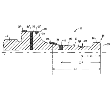
The sole Figure of the drawing is a schematic longitudinal quarter cross-
sectional
view of an ultrasonic instrument in accordance with the present invention,
showing a plurality
of end-of-life elements.
CA 02925451 2016-03-24
WO 2015/047811 PCT/US2014/056009
4
DETAILED DESCRIPTION
As illustrated in the drawing, an ultrasonic medical instrument 10 comprises a
probe
shaft 12 having a connector 14 at a proximal end and a head 16 at a distal
end. Connector 14
takes the form of an externally threaded coupling configured for operative
attachment to a
source of ultrasonic mechanical vibratory energy. Typically, the vibration
source is an
electromechanical transducer device including a stack of piezoelectric crystal
wafers. Head
16 has an operative surface 18 and/or 19 configured for engagement with
organic tissues of a
patient. Probe shaft 12, connector 14, and head 16 are made of a rigid
material, typically a
metal or metal alloy but possibly a ceramic. Instrument 10 is provided with
one or more end-
of-life indicator elements 20, 20', 22, 22', 24, 24' affixed to probe shaft 12
or possibly head
16. Indicator elements 20, 20', 22, 22', 24, 24' are each made of a material
that gradually
disintegrates or degrades with use of instrument 10 -- at a rate faster than a
rate of
disintegration of the material of probe shaft 12) -- so as to provide a
visible indicator of
degree of use and remaining useful life of the instrument.
Indicator elements 20 and 20' are fastened (e.g., adhesively) to an exterior
surface 26
or 28 of probe shaft 12. Indicator elements 22 and 22' are disposed in
respective shallow
recesses 30 and 32 formed in exterior surfaces 26 and 28 probe shaft 12.
Indicator elements
24 and 24' are disposed in respective deep recesses or bores 34 and 36 formed
in exterior
surfaces 26 and 28 probe shaft 12.
Any particular ultrasonic instrument 10 typically includes only one or two of
the end-
of-life indicator elements 20, 20', 22, 22', 24, 24'. Multiple indicator
elements 20, 20', 22, 22',
24, 24' ensure that users detect the erosion of at least one indicator
element. Multiple end-of-
life indicator elements 20, 20', 22, 22', 24, 24' may be used that are
designed to provide visual
indicators after different degrees of accumulated use. Thus, a first indicator
element may
signal that the expected life of the instrument 10 is 75% used, while another
indicates that
90% of the expected life has been used.
CA 02925451 2016-03-24
WO 2015/047811 PCT/US2014/056009
End-of-life indicator elements 20, 20', 22, 22', 24, 24' may be made of a
ceramic or a
polymeric material. In any case, indicator elements 20, 20', 22, 22', 24, 24'
are biocompatible
and preferably biodegradable. Accordingly, where material is shed from
indicator elements
20, 20', 22, 22', 24, 24' during use thereof in contact with organic tissues
of a patient, the shed
5 material is absorbed and metabolized by the body.
End-of-life indicator elements 20, 20', 22, 22', 24, 24' may each be a primary
indicator
element, with a secondary or auxiliary indicator being provided underneath, on
probe shaft
surface 26 or 28, that is initially hidden by the primary end-of-life
indicator element and that
becomes visible once the primary indicator element degrades or disintegrates
sufficiently to
reveal the auxiliary indicator. Examples of auxiliary indicators are symbols
such as a stop
sign or a do-not-enter sign, a word such as "recycle" or "discard," a graphic
such as a skull
and bones representation, and a color tab, such as a red dot, stripe or circle
(e.g., when inside
a cylindrical recess).
Instrument 10 exemplarily takes the form of a bone cutting blade, a wound
debrider, a
liposuction probe, etc.
End-of-life indicator elements 20, 20', 22, 22', 24, 24' and the locations
thereof on
instrument 10 may be coordinated so that a visible disintegration or
degradation of the
indicator element effectively coincides with the end of the useful life of the
ultrasonic
instrument. For instance, where an end-of-life indicator element 20, 20', 22,
22', 24, 24' is
made of a material that erodes or disintegrates slowly, the indicator is
preferably positioned at
or near a vibration node as determined by the instrument geometry and the
characteristic
operating frequency. Alternatively, where the end-of-life indicator is made of
a material that
erodes or disintegrates relatively quickly, the indicator is preferably
positioned at or near a
vibration anti-node as determined by the instrument geometry and the
characteristic operating
frequency. In addition, the size of the indicator element may be varied within
limits to adjust
the rate of disintegration or degradation of the indicator element. Typically,
an end-of-life
CA 02925451 2016-03-24
WO 2015/047811 PCT/US2014/056009
6
indicator element 20, 22, 24 is affixed to probe shaft 12 at a preselected
location within one-
half an operating wavelength of a distal end of the instrument. The drawing
figure shows
indicator element 22 disposed at a distance L211ess than a one-quarter
wavelength from the
distal tip (surface 19) of instrument 10, indicator element 24 disposed at a
distance 1,2 less
than a one-half wavelength from the distal tip (19), and indicator element 20
disposed at a
distance L1 about one-half wavelength from the distal tip (19).
A method of use of instrument 10 comprises applying an active or operative
surface
18 or 19 of the instrument to organic tissues of different patients in
successive surgical
operations over an extended period of time, and activating the instrument with
ultrasonic
mechanical vibratory energy or a pre-established ultrasonic frequency during
contact of the
active or operative surface 18, 19 with the respective organic tissues. One
periodically
monitors the end-of-life indicator element 20, 20', 22, 22', 24, and/or 24'
over multiple uses of
the instrument, each use on organic tissues of a respective one of the
patients. The instrument
is retired from medical use upon one's detecting a predetermined amount of
disintegration of
one or more of the end-of-life indicator elements 20, 20', 22, 22', 24, and
24'.
Where the end-of-life indicator element is made of a biocompatible and
biodegradable
material, the disintegration of the end-of-life indicator element includes
dissolving material of
the end-of-life indicator element into patients' organic tissues.
