Note: Descriptions are shown in the official language in which they were submitted.
CA 2,925,584
CPST Ref: 76029/00014
COMPRESSED BONE COMPOSITION AND METHODS OF USE THEREOF
Background of the Invention
[0001] The invention relates to methods of preparing compressed bone
compositions, bone implants,
and variants thereof. The invention also relates to methods of using the bone
implants and variants
thereof.
[0002] The invention relates to compressed bone compositions, particles,
fibers, implants, and
variants thereof, and the methods of preparing and making the same. The
invention also relates to
methods of using the bone compositions, particles, fibers, implants, and
variants thereof.
[0003] Demineralized cortical and cancellous bone compositions have been
widely used in the
induction of new bone formation for the treatment of a variety of clinical
pathologies. Typically,
bone materials are obtained from human or animal sources, processed,
demineralized, and made into
bone implants. Such bone implants may comprise bone compositions which may
include for example
compressed bone fibers and/or bone fibers. The bone implants may also comprise
growth factors,
proteins, cells, and other bioactive materials that may facilitate
osteoinduction and bone healing. In
general, it is desirable to develop new bone materials that have superior wet
and dry handling
characteristics for processing, and to provide an environment for the
attachment and functioning of
bioactive molecules.
Summary
[0004] The invention relates to methods of preparing compressed bone
compositions comprising
loading bone particles and/or fibers into a mold with a predetermined shape,
applying pressure to the
particles and/or fibers, and freeze drying the compressed bone particles
and/or fibers. In one aspect,
the pressure may be from 0.1 to 30 MPa. In another aspect, the predetermined
shape comprises
grooves. In another aspect, the compressed bone compositions retain their
integrity in liquid for at
least 5-30 minutes after being introduced into liquids. In another aspect,
pressure is applied to the
bone particles and/or fibers at room temperature. In another aspect, the
compressed bone
compositions do not comprise a binder or a chemical cross-linker.
[0005] The invention also relates to bone implants prepared by the methods
described herein. In one
aspect, the bone implants comprise grooves.
[0006] The invention relates to a bone composition comprising bone fibers,
wherein the bone fibers
comprise microfibers having an average width (W) of less than about 5 gm and
an average length
(L) : W ratio of greater than about 2.
1
Date Recue/Date Received 2021-03-19
CA 2,925,584
CPST Ref: 76029/00014
[0007] The invention also related to a method for preparing an individualized
bone implant,
comprising: loading bone composition into a mold that is based upon three
dimensional (3D) medical
imaging measurements taken from a bone structure of the individual for the
implant or prosthesis,
wherein the bone composition comprises microfibers having an average length
(L) : average width
(W) ratio greater than about 2; applying pressure of from 0.1 to 30 MPa to the
bone composition to fit
the mold; and freeze drying the compressed bone composition to make the bone
implant. In some
embodiments, the measurements are converted to computer aided designs to
generate custom molds
for compressing the bone fibers.
[0008] The invention also relates to bone implants prepared by the methods
described herein and the
method to use such bone implants.
Brief Description of the Drawings
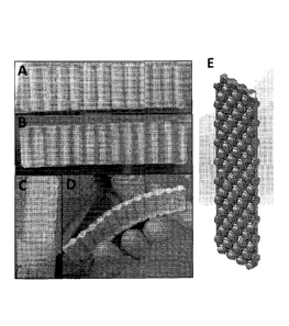
[0009] FIGURE 1 depicts a mirrored wave (A, D) and single wave (B) structures
of compressed
bone fibers after lyophilization. A bi-axial wave structure as frozen at -80
C before the
lyophilization is also shown (C). The inherent flexibility of the compressed
bone fibers is also
depicted (D). A design for the bubble-based graft for increased flexibility is
also shown (E).
[0010] FIGURE 2 illustrates samples of bone fibers wetted and thus expanded by
hydration in saline
for 30 minutes with scanning electron microscope (SEM) according to some
embodiments of the
present invention.
[0011] FIGURES 3A and 3B show the average length and width of bone fibers cut
by Computer
Numerical Control (CNC) between each donor according to some embodiments of
the present
invention.
[0012] FIGURE 4 shows the pore distribution of wetted bone fiber samples made
from bone fibers
cut with CNC via mercury porosimetry according to some embodiments of the
present invention.
[0013] FIGURE 5 shows the SEM image of a dry bone fiber sample according to
some embodiments
of the present invention, illustrating the bone fiber main bodies and
microfibers.
[0014] FIGURE 6 illustrates SEM images of samples of dry bone fibers in
different magnifications
(mag.) according to some embodiments of the present invention.
[0015] FIGURE 7 shows the amounts of BMP-2 growth factor in bone composition
samples
prepared by CNC 0.003 and 0.009.
[0016] FIGURE 8 shows sample SEM images of bone marrow-derived mesenchymal
stem cells
(BMSCs) growing on bone implants from demineralized bone matrix (DBM) fibers
according to some
embodiments of the present invention.
2
Date Recue/Date Received 2021-03-19
CA 2,925,584
CPST Ref: 76029/00014
[0017] FIGURE 9 illustrates BMSC growth on implants from bone fibers according
to some
embodiments of the present invention.
[0018] FIGURE 10 illustrates in vivo bone fiber spacing, cellularity, and
osteoblastogenic
differentiation for a bone composition implant according to some embodiments
of the present
invention.
[0019] FIGURE 11 demonstrates the percentage of implants passing
osteoinductivity (OD assays in
vivo for bone implants and the relationship with fiber packing density (loose
vs compressed)
according to some embodiments of the present invention.
[0020] FIGURE 12 shows the process of designing and molding a bone implant
according to some
embodiments of the present invention.
[0021] FIGURE 13A shows three point bend mechanical testing data, and FIGURE
13B shows 10
mm ball burst mechanical testing data.
Detailed Description of the Invention
[0022] The invention relates to methods of preparing compressed bone
compositions comprising
loading bone particles and/or fibers into a mold with a predetermined shape,
applying pressure to the
bone particles and/or fibers, and freeze drying the compressed bone particles
and/or fiber.
[0023] The invention also relates to a compressed bone composition comprising
bone fibers, wherein
the bone fibers comprise microfibers having an average width (W) of less than
about 5 gm and an
average length (L) : W ratio of greater than about 2.
[0024] The bone particles described herein include but are not limited to bone
fibers and/or powders.
The bone fibers described herein include but are not limited to bone fibers
and/or powders. Bone
particles and/or fibers may be prepared from cleaned and disinfected bone
fragments that have or have
not been freeze-dried, grounded/fractured, and cut into bone particles and/or
fibers. In some
embodiments, the bone particles and/or fibers are wetted and pre-freeze dried.
Bone particles and/or
fibers may be selected by, for example, using sieving devices (e.g. mesh
sieves) commercially
available to obtain particles and/or fibers within a desired size range. In
some embodiments, the
fibers are not sieved or sorted in obtaining fibers within a desired size
range.
[0025] In some embodiments, the bone particles may have an average diameter,
for example,
between about 125 microns and about 4 mm; between about 710 microns and about
2 mm; between
about 125 microns and about 500 microns; between about 125 microns and about
850 microns; or
between about 250 microns and about 710 microns. In some embodiments, the bone
particles have a
3
Date Recue/Date Received 2021-03-19
CA 2,925,584
CPST Ref: 76029/00014
median diameter of about from 10 to 1,000 microns, a median length of about
from 0.5 to 100 mm,
and a median thickness of about from 10 to 1000 microns. Certain embodiments
of the present
invention may include bone powder that is commercially available. For example,
a suitable bone
powder that is widely and reliably available is produced by LifeNet Health,
Virginia Beach, VA (e.g.
ground demineralized bone powder, and demineralized bone fiber). In some
embodiments, the bone
particles may be prepared by grinding, skiving, or Computer Numerical Control
(CNC) machining of
bone tissues. In some embodiments, the bone particles may be prepared by the
methods described in
U.S. Patent No. 7,744,597.
[0026] In some embodiments, the bone fibers may have an elongate main body. In
the present
application, the dimensions of the main body are referred to as the dimensions
of the bone fiber. For
example, the length of the bone fiber may be between about 100 microns and
about 50 mm; between
about 200 microns and about 20 mm; between about 500 microns and about 15 mm;
between about
600 microns and about 12 mm; between about 700 microns and about 11 mm;
between about 700
microns and about 10 mm; between about 700 microns and about 9 mm; between
about 700 microns
and about 8 mm; between about 700 microns and about 7 mm; between about 700
microns and about
6 mm; between about 900 microns and about 15 mm; between about 900 microns and
about 10 mm;
or between about 900 microns and about 9 mm. In addition, for example, the
width of the bone fibers
may be between about 5 microns and about 5 mm; between about 10 microns and
about 4 mm;
between about 20 microns and about 3 mm; between about 20 microns and about 2
mm; between
about 20 microns and about 1.5 mm; between about 20 microns and about 1 mm;
between about 20
microns and about 800 microns; between about 20 microns and about 700 microns;
between about 20
microns and about 600 microns; between about 70 microns and about 2 mm;
between about 70
microns and about 1.5 mm; between about 70 microns and about 1.4 mm; or
between about 70
microns and about 1.3 mm.
[0027] In some embodiments, the bone fibers in a bone composition may have an
average length and
an average width. For example, the average length of the bone fibers may be
between about 1 mm and
about 10 mm; between about 1.5 mm and about 5 mm; between about 2 mm and about
4 mm;
between about 2.5 mm and about 3 mm; between about 3 mm and about 5 mm;
between about 3 mm
and about 4 mm; or between about 3.5 mm and about 4 mm. The average width of
the bone fibers
may be between about 50 microns and about 1 mm, between about 100 microns and
about 800
microns, between about 150 microns and about 700 microns, between about 200
microns and about
500 microns, between about 200 microns and about 400 microns, between about
200 microns and
about 300 microns, between about 200 microns and about 250 microns, between
about 300 microns
and about 500 microns, between about 300 microns and about 400 microns,
between about 400
microns and about 500 microns, or between about 400 microns and about 450
microns. The average
4
Date Recue/Date Received 2021-03-19
CA 2,925,584
CPST Ref: 76029/00014
length of the bone fibers may be less than 20 mm, 10 mm, 9 mm, 8 mm, 7 mm, 6
mm, 5 mm, 4 mm,
or 3 mm. The average length of the bone fibers may be more than 500 microns, 1
mm, 1.5 mm, 2
mm, 2.5 mm, 3 mm, 3.5 mm, 4 mm, 4.5 mm, or 5 mm. The average width of the bone
fibers may be
less than 1 mm, 900 microns, 800 microns, 700 microns, 600 microns, 500
microns, 400 microns, 300
microns, or 250 microns. The average width of the bone fibers may be more than
10 microns, 20
microns, 30 microns, 40 microns, 50 microns, 75 microns, 100 microns, 150
microns, 200 microns,
300 microns, 400 microns, or 500 microns.
[0028] Certain embodiments of the present invention may include bone powder
that is commercially
available. For example, a suitable bone product that is widely and reliably
available is produced by
LifeNet Health, Inc., Virginia Beach, VA (e.g. demineralized bone fiber). In
some embodiments, the
bone particles and/or fibers may be prepared by grinding, skiving, and/or
Computer Numerical
Control (CNC) machining of bone tissues. In some embodiments, the bone
particles and/or fibers
may be prepared by the methods described in U.S. Patent No. 7,744,597.
[0029] In some embodiments, the bone fibers may comprise microfibers. The
microfibers may
comprise projections or spikes extending from the main body of the bone
fibers, but have a
significantly less width or diameter compared with the main diameter of the
bone fibers. A microfiber
may have a length, which is the measurement of the tip of the microfiber to
where the microfiber
connects to the main body of the bone fiber. The average length (L) of the
microfibers represents the
average of lengths for a representative number of microfibers from a sample. A
microfiber may have a
width, which is an average measurement of the microfiber's diameter. The
average width (W) of the
microfibers represents the average of widths for a representative number of
microfibers from a
sample.
[0030] In some embodiments, the width of the microfibers may range from about
0.5 to about 100
microns; in some embodiments, the width of the microfibers may range from
about 0.1 to about 30
microns; in some embodiments, the width of the microfibers may range from
about 0.2 to about 20
microns; in some embodiments, the width of the microfibers may range from
about 0.2 to about 10
microns; in some embodiments, the width of the microfibers may range from
about 0.2 to about 3
microns. In some embodiments, the average width (W) of the microfibers may
range from about 0.5
to about 20 microns, in some embodiments, the average width (W) of the
microfibers may range from
about 0.8 to about 10 microns; in some embodiments, the average width (W) of
the microfibers may
range from about 0.8 to about 6 microns; in some embodiments, the average
width (W) of the
microfibers may range from about 0.8 to about 3 microns; in some embodiments,
the average width
(W) of the microfibers may range from about 0.8 to about 2 microns; in some
embodiments, the
average width (W) of the microfibers may range from about 0.8 to about 1.5
microns; in some
Date Recue/Date Received 2021-03-19
CA 2,925,584
CPST Ref: 76029/00014
embodiments, the average width (W) of the microfibers may range from about 0.8
to about 1.4
microns. In some embodiments, the average width (W) may be less than about 6
microns, 5 microns,
4 microns, 3 microns, 2 microns, 1.6 microns, 1.5 microns, 1.4 microns, 1.3
microns, 1.2 microns, 1.1
microns, 1 micron, or 0.9 micron. In some embodiments, the average width (W)
may be more than
about 0.2 micron, 0.3 micron, 0.4 micron, 0.5 micron, 0.6 micron, 0.7 micron,
0.8 micron, 0.9 micron,
1 micron, 1.1 microns, 1.2 microns, 1.3 microns, or 1.35 microns.
[0031] In some embodiments, the length of the microfibers may range from about
0.5 to about 100
microns; in some embodiments, the length of the microfibers may range from
about 1 to about 50
microns; in some embodiments, the length of the microfibers may range from
about 2 to about 50
microns; in some embodiments, the length of the microfibers may range from
about 3 to about 20
microns; in some embodiments, the length of the microfibers may range from
about 3 to about 16
microns; in some embodiments, the length of the microfibers may range from
about 3 to about 11
microns. In some embodiments, the average length (L) of the microfibers may
range from about 3 to
about 20 microns, in some embodiments, the average length (L) of the
microfibers may range from
about 4 to about 15 microns; in some embodiments, the average length (L) of
the microfibers may
range from about 5 to about 13 microns; in some embodiments, the average
length (L) of the
microfibers may range from about 6 to about 10 microns; in some embodiments,
the average length
(L) of the microfibers may range from about 6 to about 8 microns; in some
embodiments, the average
length (L) of the microfibers may range from about 6 to about 7 microns. In
some embodiments, the
average length (L) of the microfibers may be less than about 20 microns, 15
microns, 12 microns, 10
microns, 9 microns, 8 microns, 7 microns, or 6.5 microns. In some embodiments,
the average length
(L) of the microfibers may be more than about 0.5 micron, 0.6 micron, 0.7
micron, 0.8 micron, 0.9
micron, 1 micron, 2 microns, 3 microns, 4 microns, 5 microns, 6 microns, 7
microns, 8 microns, 9
microns, or 10 microns.
[0032] In some embodiments, the average length (L): average width (W) ratio of
the microfibers may
range from about 0.5 to 50; in some embodiments, the L:W ratio of the
microfibers may range from
about 0.8 to 20; in some embodiments, the L:W ratio of the microfibers may
range from about 1 to 10;
in some embodiments, the L:W ratio of the microfibers may range from about 2
to 8; in some
embodiments, the L:W ratio of the microfibers may range from about 2 to 6; in
some embodiments,
the L:W ratio of the microfibers may range from about 2 to 5; in some
embodiments, the L:W ratio of
the microfibers may range from about 3 to 20; in some embodiments, the L:W
ratio of the microfibers
may range from about 3 to 10; in some embodiments, the L:W ratio of the
microfibers may range
from about 3 to 8; in some embodiments, the L:W ratio of the microfibers may
range from about 3 to
6; in some embodiments, the L:W ratio of the microfibers may range from about
4 to 20; in some
embodiments, the L:W ratio of the microfibers may range from about 4 to 10; in
some embodiments,
6
Date Recue/Date Received 2021-03-19
CA 2,925,584
CPST Ref: 76029/00014
the L:W ratio of the microfibers may range from about 4 to 8; in some
embodiments, the L:W ratio of
the microfibers may range from about 4 to 6; in some embodiments, the L:W
ratio of the microfibers
may range from about 5 to 20; in some embodiments, the L:W ratio of the
microfibers may range
from about 5 to 10; in some embodiments, the L:W ratio of the microfibers may
range from about 5 to
8; in some embodiments, the L:W ratio of the microfibers may range from about
6 to 20; in some
embodiments, the L:W ratio of the microfibers may range from about 6 to 10; in
some embodiments,
the L:W ratio of the microfibers may range from about 7 to 20; in some
embodiments, the L:W ratio
of the microfibers may range from about 7 to 10; in some embodiments, the L:W
ratio of the
microfibers may range from about 8 to 20; in some embodiments, the L:W ratio
of the microfibers
may range from about 9 to 20; and in some embodiments, the L:W ratio of the
microfibers may range
from about 10 to 20.
[0033] In some embodiments, the microfibers may have a L:W ratio that is more
than about 0.5, 0.8,
1, 1.5, 2.0, 2.1, 2.2, 2.3, 2.4, 2.5, 3.0, 3.5, 4.0,4.5, 5.0, 5.5, 6.0, 6.5,
7.0, or 7.5. In some
embodiments, the microfibers may have a L:W ratio that is less than about
20.0, 15.0, 14.0, 13.0,
12.0, 11.0, 10.0, 9.5, 9.0, 8.5, 8.0, 7.5, 7.0, 6.5, 6.0, 5.5 or 5Ø
[0034] In some embodiments, the mode for the pore size of a dry bone
composition may range from
to 30 microns, from 5 to 20 microns, from 10 to 500 microns, from 10 to 300
microns, from 10 to
100 microns, from 10 to 50 microns, from 10 to 30 microns, from 10 to 25
microns, from 15 to 25
microns, from 15 to 400 microns, from 20 to 300 microns, from 30 to 200
microns, from 30 to 100
microns, from 35 to 100 microns, from 40 to 100 microns, from 40 to 80
microns, from 40 to 70
microns, from 40 to 60 microns, from 50 to 100 microns, from 50 to 80 microns,
from 50 to 70
microns, from 50 to 60 microns, or from 54 to 57 microns. The mode for the
pore size of a dry bone
composition may be more than about 5, 10, 11, 12, 13, 14, 15, 16, 17, 18, 19,
20, 25, 30, 35, 40, 45,
50, 55, 60, 65, 70, 75, 80, 85, 90, 95 or 100 microns. The mode for the pore
size of a dry bone
composition may be less than about 500, 400, 300, 200, 150, 100, 90, 80, 70,
60, 50, 40, 30, 29, 28,
27, 26, 25, 24, 23, 22, 21, 20, 19 or 18 microns.
[0035] In some embodiments, the mean for the pore size of a dry bone
composition may range from
0.05 to 25 microns, from 0.05 to 20 microns, from 10 to 500 microns, from 15
to 400 microns, from
20 to 300 microns, from 30 to 200 microns, from 30 to 100 microns, from 35 to
100 microns, from 35
to 80 microns, from 35 to 70 microns, from 35 to 60 microns, from 35 to 50
microns, from 35 to 45
microns, from 40 to 100 microns, from 40 to 90 microns, from 40 to 80 microns,
from 40 to 70
microns, from 40 to 60 microns, or from 40 to 50 microns. The mean for the
pore size of a dry bone
composition may be more than about 5, 10, 15, 20, 25, 30, 35, 40, 45, 50, 55,
60, 65, 70, 75, 80, 85,
7
Date Recue/Date Received 2021-03-19
CA 2,925,584
CPST Ref: 76029/00014
90, 95 or 100 microns. The mean for the pore size of a dry bone composition
may be less than about
500, 400, 300, 200, 150, 100, 90, 80, 70, 60, 50, 40, or 30 microns.
[0036] The equilibrium state of a wet bone composition is reached when the
bone composition is
immersed in a solution (e.g. a saline solution or blood or cell suspension)
and refuses to accept any
more of the liquid. The equilibrium model of the bone composition may mimic
the state of the bone
composition in vivo. In some embodiments, when the bone composition is
immersed in a solution,
liquid or fluid, the bone composition swells, resulting in a hydrogel or a
moldable putty. In additional
embodiments, the bone composition reaches the equilibrium when it is immersed
in a solution at least
for 5 minutes, 15 minutes, 20 minutes, 30 minutes, 40 minutes, or 50 minutes.
[0037] In some embodiments, the mode for the pore size of a wet bone
composition at equilibrium
may range from 10 to 500 microns, from 15 to 400 microns, from 20 to 300
microns, from 20 to 250
microns, from 20 to 210 microns, from 25 to 250 microns, from 25 to 225
microns, from 25 to 210
microns, from 25 to 200 microns, from 25 to 190 microns, from 25 to 180
microns, from 25 to 170
microns, from 25 to 160 microns, from 25 to 100 microns, from 25 to 50
microns, from 25 to 45
microns, from 25 to 40 microns, from 25 to 35 microns, from 50 to 500 microns,
from 50 to 250
microns, from 50 to 200 microns, from 50 to 150 microns, from 50 to 130
microns, from 50 to 120
microns, from 50 to 115 microns, from 50 to 110 microns, from 50 to
1005microns, from 250 to 500
microns, from 250 to 400 microns, from 250 to 350 microns, from 250 to 300
microns, from 150 to
500 microns, from 150 to 400 microns, from 150 to 350 microns, from 150 to 300
microns, from 150
to 250 microns, from 150 to 240 microns, from 150 to 230 microns, from 150 to
220 microns, or from
150 to 210 microns. The mode for the pore size of a wet bone composition at
equilibrium may be
more than 5, 10, 15, 20, 30, 40, 45, 46, 47, 48, 49, 50, 51, 52, 53, 54, 55,
60, 70, 80, 90, 100, 110, 120,
130, 140, 150, 160, 170, 180, 190 or 200 microns. The mode for the pore size
of a wet bone
composition at equilibrium may be less than 500, 400, 300, 250, 240, 230, 220,
210, 200, 190, 180,
170, 160, 150, 140, 130, 120, 110, 109, 108, 107, 106, 105, 104, 100, 90, 80,
70, 60, 50, 45, 40, 35,
30, 25, or 20 microns.
[0038] In some embodiments, the mean for the pore size of a wet bone
composition at equilibrium
may range from 0.1 to 10 microns, 0.1 to 5 microns, 0.1 to 3 microns, from 10
to 300 microns, from
15 to 200 microns, from 20 to 100 microns, from 20 to 90 microns, from 20 to
80 microns, from 20 to
75 microns, from 20 to 70 microns, from 25 to 100 microns, from 25 to 90
microns, from 25 to 80
microns, from 25 to 75 microns, from 25 to 70 microns, from 25 to 65 microns,
from 25 to 60
microns, from 25 to 55 microns, from 25 to 51 microns, from 25 to 50 microns,
from 50 to 100
microns, from 50 to 90 microns, from 50 to 80 microns, from 50 to 75 microns,
or from 50 to 70
microns. The mean for the pore size at equilibrium may be more than 0.1, 0.2,
0.3, 0.4, 0.5, 0.7, 1, 5,
8
Date Recue/Date Received 2021-03-19
CA 2,925,584
CPST Ref: 76029/00014
10, 15, 20, 25,30, 35, 40,45, 50, 55, 60, 65, 70, 75, 80, 85, 90, 95, 100,
110, 120, 130, 140, or 150
microns. The mean for the pore size at equilibrium may be less than 200, 190,
180, 170, 160, 150,
140, 130, 120, 110, 100, 95, 90, 85, 80, 75, 70, 65, 60, 55, 50, 45, 40, 35,
30, 25, 20, 15, 10, 5, 4, 3, 2,
1, or 0.1 microns.
[0039] In some embodiments, the pore size of a wet bone composition at
equilibrium has a unimodal
distribution. In some embodiments, the pore size of a wet bone composition at
equilibrium does not
have a notable bimodal distribution. A bimodal distribution may be defined as
a pore size distribution
having two distinct peaks. A notable bimodal distribution may be defined as a
bimodal distribution
with a bimodal ratio (the ratio of one peaks versus the other peak) within the
range of 0.99 to 1.01,
0.95 to 1.05, 0.9 to 1.1, 0.85 to 1.18, 0.8 to 1.25, 0.7 to 1.43, 0.6 to 1.67,
0.5 to 2, 0.4 to 2.5, 0.3 to
3.33, 0.2 to 5, 0.15 to 6.67, 0.05 to 20, 0.04 to 25, 0.03 to 33, 0.02 to 50,
0.01 to 100, or 0.005 to 200.
[0040] As used herein, the term "about" modifying, for example, length, width,
distance, the quantity
of an ingredient in a composition, concentrations, volumes, process
temperature, process time, yields,
flow rates, pressures, and like values, and ranges thereof, refers to
variation in the numerical quantity
that can occur, for example, through typical measuring and handling procedures
used for making
compounds, compositions, concentrates or use formulations; through inadvertent
variation in these
procedures; through differences in the manufacture, source, or purity of
starting materials or
ingredients used to carry out the methods; and like considerations. The term
"about" also
encompasses amounts that differ due to aging of, for example, a composition,
formulation, or cell
culture with a particular initial concentration or mixture, and amounts that
differ due to mixing or
processing a composition or formulation with a particular initial
concentration or mixture. The term
"about" further may refer to a range of values that are similar to the stated
reference value. In certain
embodiments, the term "about" refers to a range of values that fall within 20,
19, 18, 17, 16, 15, 14,
13, 12, 11, 10,9, 8,7, 6, 5, 4, 3, 2, 1 percent or less of the stated
reference value.
[0041] In one aspect, the compressed bone compositions do not comprise a
binder or a chemical
cross-linker. In another aspect, a binder and/or a chemical cross-linker may
be included in the
compressed bone compositions. In some embodiments, such binders include, but
are not limited to,
glycerol/Preservont, acidic solutions (e.g. Lactic and trifluoroacitic acid),
buffering solutions (e.g.
phosphate), and adhesive binders (e.g. fibrin glues, bone cements, or
liquified bone). In another
aspect, crosslinking may be performed for the bone particles and/or fibers
before or after applying the
pressure by any conventional chemical crosslinking method (e.g. chemical
reagent-promoted,
chemically reactive linker-promoted and/or enzyme-promoted) and/or
dehydrothermal crosslinking
method (e.g. heat-promoted condensation), forming a covalently crosslinked
bone matrix. In
additional embodiments, the crosslinking comprises applying a cross-linking
agent to the bone matrix
9
Date Recue/Date Received 2021-03-19
CA 2,925,584
CPST Ref: 76029/00014
solution. For example, the cross-linking agent may be selected from the group
consisting of 1-Ethyl-
3-(3-dimethylaminopropyl)carbodiimide (EDC), EDC/hyaluronic acid, genipin, and
glutaraldehyde.
[0042] The bone from which the bone particles and/or fibers are derived
includes, but is not limited
to, autograft bone, allograft bone, and xenograft bone. Such bone includes any
bone from any source,
including, but not limited to, bone from a living human donor, bone from a
human cadaveric donor,
and bone from a living or non-living animal. The bone from which the fibers
are derived may include
cortical bone and/or cancellous bone and/or cortico-cancellous bone. The bone
from which the fibers
are derived may be obtained from any mammal, including but not limited to a
human, a cow, a pig, a
dog, a cat, a non-human primate, a rodent such as a rat or mouse, a horse, a
goat, a sheep, or a deer.
[0043] The bone from which the bone particles and/or fibers are derived may be
demineralized bone,
partially demineralized bone or non-demineralized bone. In one aspect, the
bone particles and/or
fibers may be demineralized prior to applying the pressure according to the
methods of the present
invention. -Demineralized bone" as used herein refers to bone having less than
about 8 wt % residual
calcium. In some embodiments, the demineralized bone has an average residual
calcium content of
less than 7, 6, 5, 4, 3, 2 or 1 wt%. In some embodiments, the demineralized
bone has an average
residual calcium content of from 0 to 4, from 0.5 to 4, from 1 to 4, from 2 to
4, from 3 to 4, from 0 to
3, from 0.5 to 3, from 1 to 3, from 2 to 3, from 0 to 2, from 1 to 2, or from
0 to 1 wt%.
Demineralization involves treating a bone tissue to remove its inorganic
mineral hydroxyapatite
material. The level of demineralization of a bone tissue is defined by the
amount (wt %) of residual
calcium found in the demineralized bone. In some embodiments, the
demineralized bone may still
contain physiologically active levels of growth and differentiation factors
(e.g., osteogenic growth
factors, such as bone morphogenetic proteins (BMPs) and insulin like growth
factor (IGF)) remaining
from the initial bone even after the demineralization treatment. In further
embodiments, the
demineralized bone may contain collagen, osteocalcin, osteonectin, bone sialo
protein, osteopontin,
and mixtures thereof. In one embodiment, the bone particles and/or fibers are
prepared from
demineralized bone. In other embodiment, the bone particles and/or fibers are
prepared from non-
demineralized bone tissue and the fibers are demineralized after bone fiber
formation. "Non-
demineralized bone" as used in the present application refers to bone that has
not been treated to
remove minerals present such as, for example, hydroxyapatite.
[0044] The bone particles and/or fibers of the present invention may be
demineralized or non-
demineralized. In some embodiments, the bone particles and/or fibers may be
combined with other
bone materials, such as bone powders and/or bone particulates, which may be
demineralized or non-
demineralized or synthetic. In some embodiments, the bone compositions and/or
bone implants of the
present invention may comprise bone particles and/or fibers and/or other bone
materials, such as bone
Date Recue/Date Received 2021-03-19
CA 2,925,584
CPST Ref: 76029/00014
powders and bone particulates, which may be demineralized or non-demineralized
or synthetic. In
additional embodiments, the bone implant may be combined with prosthesis with
or without adhesion.
[0045] In some embodiments, the bone particles and/or fibers, bone
compositions, and/or bone
implants may be cleaned after or before applying the pressure. In some
embodiments, the cleaning
comprising incubating the bone particles and/or fibers with an antibiotic, a
detergent, an alcohol,
and/or a H202. In some embodiments, the cleaning comprises ALLOWASH process.
In some
embodiments, the cleaning includes methods described in U.S. Patent Nos.
5,556,379, 5,797,871,
5,820,581, 5,977,034, and 5,977,432. In additional embodiments, the cleaning
excludes use of
alcohol.
[0046] In one aspect, the bone particles and/or fibers, bone compositions,
and/or bone implants may
be sterilized after or before applying the pressure. In some embodiments, the
bone particles and/or
fibers, compressed bone compositions, and/or bone implants may be sterilized
by gamma or e-beam
irradiation, ethylene oxide, or critical CO2.
[0047] In one aspect, the bone particles and/or fibers, bone compositions,
and/or bone implants may
be treated with a plasticizer composition. In some embodiments, the
plasticizer composition
comprises one or more plasticizers selected from the group consisting of
glycerol, adonitol, sorbitol,
ribitol, galactitol, D-galactose, 1,3-dihydroxypropanol, ethylene glycol,
triethylene glycol, propylene
glycol, glucose, sucrose, mannitol, xylitol, meso-erythritol, adipic acid,
proline, hydroxyproline,
polyol, and a fatty acid. For example, the plasticizer composition may include
those described in U.S.
Patent Nos. 6,293,970 and 7,063,726, and U.S. Patent Application Publication
Nos. 2010/0030340
and 2010/0185284.
[0048] In some embodiments, the bone particles and/or fibers, bone
compositions, and/or bone
implants may be cleaned before or after being sterilized, before or after
applying the pressure, and
before or after being treated with a plasticizer composition. In some
embodiments, the bone particles
and/or fibers, bone compositions, and/or bone implants may be sterilized
before or after being
cleaned, before or after applying the pressure, and before or after being
treated with a plasticizer
composition. In some embodiments, the bone particles and/or fibers, bone
compositions, and/or bone
implants may be compressed before or after being cleaned, before or after
being sterilized, and before
or after being treated with a plasticizer composition. In some embodiments,
the bone particles and/or
fibers, bone compositions, and/or bone implants may be treated with a
plasticizer composition before
or after being cleaned, before or after being sterilized, and before or after
applying the pressure.
[0049] The invention relates to methods of preparing compressed bone
compositions comprising
loading bone particles and/or fibers into a mold with a predetermined shape,
applying pressure to the
bone particles and/or fibers, and freeze drying the compressed bone particles
and/or fibers.
11
Date Recue/Date Received 2021-03-19
CA 2,925,584
CPST Ref: 76029/00014
[0050] In some embodiments, the pressure applied to the bone particles and/or
fibers may range from
about 1 Pa to about 300 MPa; in some embodiments, the pressure applied to the
bone particles and/or
fibers may range from about 100 Pa to about 100 MPa, from about 1 KPa to about
100 MPa, from
about 10 KPa to about 100 MPa, from about 100 KPa to about 100 MPa, from about
1 MPa to about
100 MPa, from about 100 KPa to about 1 MPa, from about 100 KPa to about 2 MPa,
from about 100
KPa to about 3 MPa, 100 KPa to about 4 MPa, 100 KPa to about 5 MPa, 100 KPa to
about 6 MPa,
about 1 MPa to about 2 MPa, from about 1 MPa to about 3 MPa, from about 1 MPa
to about 4 MPa,
from about 1 MPa to about 5 MPa, from about 1 MPa to about 6 MPa, from about 1
MPa to about 7
MPa, from about 2 MPa to about 3 MPa, or from about 2 MPa to about 6 MPa; and
in some
embodiments, the pressure applied to the bone particles and/or fibers may
range between 100 psi and
5000 psi, between 100 psi and 4000 psi, between 100 psi and 3000 psi, between
100 psi and 2000 psi,
between 100 psi and 1000 psi, between 100 psi and 990 psi, between 100 psi and
950 psi, between
100 psi and 905 psi, between 200 psi and 5000 psi, between 200 psi and 4000
psi, between 200 psi
and 3000 psi, between 200 psi and 2000 psi, between 200 psi and 1000 psi,
between 200 psi and 990
psi, between 200 psi and 950 psi, or between 200 psi and 905 psi (not
including the end values).
[0051] In some embodiments, the pressure applied to the bone particles and/or
fibers may be about 1,
2, 3, 4, 5, 6, 6.5, 7, 8, 9, 10, 11, 12, 13, 14, 15, 20, 21, 22, 23, 24, 25,
26, 27, 28, 29, 30 or 40 MPa or
less. In other embodiments, the pressure applied to the bone particles and/or
fibers may be about 0.1,
0.5, 1, 2, 3, 4, 5, 6, 6.5, 7, 8, 9, 10, 11, 12, 13, 14, 15, 20, 21, 22, 23,
24,25, 26, 27, 28, 29,30 or 35
MPa or more. In some embodiments, the pressure applied to the bone particles
and/or fibers may be
about 6000, 5000, 4000, 3000, 2000, 1000, 900, 800, 700, 600, 500, 400, 300,
or 200 psi or less. In
other embodiments, the pressure applied to the bone particles and/or fibers
may be about 100, 200,
300, 400, 500, 600, 700, 800, or 900 psi or more. In one aspect, the pressure
may be applied using a
screw type press or a pneumatic press to generate the requisite pressure. In
another aspect, the
pressure may be applied using a hydraulic press, application of a heavy
weight, or a spring loaded
device, powder-actuated, clamped, or electric motor-based pressurizations,
either as constant pressure
or variable pressure device. In another aspect, the pressure may be applied
for at least about 1, 2, 3, 4,
5, 10, 15, 20, 30, 40, 50, 60, 120, 180, 240, 300, or 360 minutes.
[0052] The pressure may be applied with any kind of mechanical force and with
or without any
device. In some embodiments, the pressure may be applied by a human, e.g.
pressing with hands
and/or fingers. In some embodiments, the pressure may be applied by a device
customized to produce
compressed bone compositions.
[0053] In some embodiments, the invention also related to a method for
preparing an individualized
bone implant, comprising: loading bone composition into a mold that is based
upon three dimensional
(3D) measurements taken from a bone structure of the individual for the
implant or prosthesis,
wherein the bone composition comprises microfibers having an average length
(L) : average width
12
Date Recue/Date Received 2021-03-19
CA 2,925,584
CPST Ref: 76029/00014
(W) ratio greater than about 2; applying pressure of from 0.1 to 30MPa to the
bone composition to fit
the mold; and freeze drying the compressed bone composition to make the bone
implant. In
additional embodiments, the method may include adding prosthesis to bone
implant with or without
adhesion. The prosthesis is a synthetic implant comprising a material(s) that
does not occur naturally
in bone. The prosthesis may include, for example, a metal(s) and a polymer(s),
such as titanium, gold,
platinum and steel with or without teflon.
[0054] The predetermined shape of the mold and the resulting compressed bone
compositions
include various sizes and structures, which may be reflected by the 3D
measurements of a bone
structure of a subject. The 3D measurements are a set of dimensions and values
that represent the size
and shape of the bone structure and the possibly relative positioning and
interactions of the various
components of the bone structure, or between the patient bone structure and
any concurrently
implanted prosthetic devices to which the custom cast fibers are also designed
to fit and integrate
with. The predetermined shape of the mold may be constructed by 3D printing of
the mold based on
the 3D medical imaging measurements. The custom shaped molds may also be
constructed by CNC
and other common modes of machining by subtractive manufacturing. Any suitable
3D printing
technology and device or any additive manufacturing technology and apparatus
may be used for the
current method.
[0055] The bone structure herein described may be any bone structure,
including one or more
components, from any part of the skeleton of a human or an animal. For
example, the bone structure
may be any segmental defect without load bearing. In some embodiments, the
bone structure may be
a vertebrate or a portion thereof, a femoral head or a femur trochanter, a
humeral head from a
humerus, or a segment or part of the fibula, humeral shaft, femoral shaft,
pelvis, cranial bones, facial
bones, hip, skull flap, mandible or tibia.
[0056] An "implant" refers to any object that is designed to be placed
partially or wholly within a
subject's body for one or more therapeutic or prophylactic purposes such as
for tissue augmentation,
contouring, restoring physiological function, repairing or restoring tissues
damaged by disease or
trauma, and/or delivering therapeutic agents to normal, damaged or diseased
organs and tissues. In
one aspect, the bone implant comprises grooves as described herein. The
subject may be any human
or non-human mammal, such as dog, horse, or primate in need of a bone implant.
An "individualized
implant" refers to an implant that is specifically designed and produced based
on the 3D
measurements of an individual subject or the average of a number of individual
subjects. The
individualized implant may provide a better structural fit for the subject's
bones that are to be repaired
or restored.
[0057] In one aspect, the predetermined shape of the mold and the resulting
compressed bone
compositions comprise grooves and/or undulations, where the compositions or
implants are thinner in
13
Date Recue/Date Received 2021-03-19
CA 2,925,584
CPST Ref: 76029/00014
some portions than in other portions of the compositions or implants,
respectively. The "groove" is
an area that is designed to have a thickness that is thinner than the
thickness of the surrounding areas
permitting a point, line, or area of bending, pivoting, and/or shaping. In one
embodiment, this
variation in thickness of the compositions and implants allows the
compositions and implants to be
more flexible than compositions and implants of uniform thickness, such that
the compositions and
implants may be bent, pivoted, shaped or twisted. In select embodiments, the
grooves or undulations
in the compressed bone compositions and/or the bone implants may have a
periodicity at least of 0.5,
2, 5 or 10 grooves/cm2. In additional embodiments, the groove(s) in the
compressed bone
compositions and/or the bone implants may have a periodicity at most of 2, 5,
10 or 15 grooves/cm2.
In additional embodiments, the predetermined shape of the compressed bone
compositions may
include pre-existing holes to allow for fixation with screws, sutures, or
other types of traction. Such
holes may be formed as a part of the mold and/or fiber product design.
[0058] In one aspect of the present invention, the compositions and implants
maintain their integrity
in liquids for at least about 5, 15, 30, 100, or 200 minutes. Thus, the
compressed bone compositions
described herein may retain the structural integrity prior to and during
surgical implantation after
rehydration. As used herein, the phrase "maintain integrity" when used in
conjunction with the
compositions and implants of the present invention is used to indicate that
all of part of the fibers of
the compositions and implants do not dissociate from one another, and the
compositions and implants
maintain their overall shape in the presence of liquids, such as buffers and
body fluids. For example,
at least 50, 60, 70, 80, 85, 90, 95, or 98 wt% of the original fibers of the
compositions remain in the
implant after 0.1, 1, 10, 100, 150, 200, or 300 hours.
[0059] In some embodiments, various methods may be applied to alter the
wettability of bone
particles and/or fibers and resulting compressed bone fiber strip described
herein. Some examples
include both physical and chemical means, including surface chemistry
modifications (e.g. with
plasma or changing the static charge, or by making large passages or channels
for water to enter),
chemical etching (e.g. acid etching). and addition of a hydrophilic molecule
(e.g. PreservonTm).
[0060] In some embodiments, the average thickness of the predetermined shape
of the mold or the
resulting compressed bone composition at the groove(s) may be thinner than the
average thickness of
the entire predetermined shape or the compressed bone composition, for
example, by about 1%, 2%,
3%, 5 %, 10%, 20%, 30%, 40%, 50%, 60%, 70%, 80%, or 90%. In some embodiments
of the present
disclosure, the compressed bone composition has a higher density at the groove
area(s) compared to
rest of the structure, for example, by about 1%, 2%, 3%, 5 %, 10%, 20%, 30%,
40%, 50%, 60%, 70%,
80%, or 90%.
[0061] In another aspect, the predetermined shape of the mold and the
resulting compressed bone
compositions have shapes that include a strip, a disc, with a concave facet,
with a convex facet, with
14
Date Recue/Date Received 2021-03-19
CA 2,925,584
CPST Ref: 76029/00014
either flat features, with steps, or with curves, comprising, but not limited
to, waves, with bubbles, or
with a bubble-wrap patterns. In some embodiments, other shapes may be used,
including, but not
limited to, cylinder, tubes (e.g. for filling with BMA, autograft, PRP, and
other bioactive agents), thin
sheets for rolling in autograft or wrapping around defects, mating with
synthetic implants (e.g. plugs
for intervertebral fusion device cavities, hollow part for fitting onto screws
or other devices, such as a
spinous process fusion plate, etc.), ring type designs (e.g. for segmental
defects), acetabular cup, ball
or sphere shaped (e.g. ball and socket revisions and resurfacing, and ball
shaped implants in the
thoracic spine and other areas) oral/cranial/maxillofacial applications such
as strips or alveolar ridge
reconstruction, and wedges (e.g. for Evans, Cotton, high tibial osteotomy),
and irregular shapes and
custom shapes that are patient defect specific, potentially as based on
computed tomography (CT) or
X-ray scans. The compressed bone particles and/or fibers may be used to fill a
load bearing material
(e.g. mineralized cortical or conricocancellous bone, metal, PEEK, synthetic
polymers, and cages) to
supply a source of osteoinductive and/or osteoconductive fibers in a non-
inductive graft. For
example, the shapes may include a conical or frustum shaped dowel to fill a
hole such as a surgical
screw hole. The compressed bone particles and/or fibers may also be non-load
bearing.
[0062] In one aspect of the present invention, the compressed bone composition
may be used to
prepare a combination product with a synthetic or metallic structure, e.g. a
framework, where the
compressed bone composition and the synthetic or metallic structure are
tethered or bound together.
In some embodiments, the synthetic or metallic structure may facilitate
surgical fixation or
stabilization of the combination product to the defect site, while the new
tissue can form on and
within the compressed bone composition, and remodel the composition partially
or completely. The
surgeon may then remove the synthetic or metallic structure. A traditional
composition cannot be
used in this approach since it is likely to dissemble during the healing and
tissue forming processes.
[0063] In another aspect, the predetermined shape of the mold and the
resulting compressed bone
compositions have at least one dimension of about 0.5 mm, 1 mm, 5 mm, 10 mm,
20 mm, 30 mm, 40
mm, 50 mm, 60 mm, 70 mm, 80 mm, 90 mm, 100 mm, 200 mm, 300 mm. In another
aspect, the
predetermined shape of the mold and the resulting compressed bone compositions
have variable sizes
from about 1 x 1 x 1 mm to about 100>< 100>< 10 mm, or about 100 mm x 25 mm
with 1 to 8 mm
thickness.
[0064] In one aspect, the bone particles and/or fibers may be directly
compressed, with or without an
additional mold, into or onto a load bearing material (e.g. mineralized
cortical bone, including, but not
limited to, femur sectioned as a hollow disc) or other hard materials (e.g.
ceramics, metals).
[0065] The bone particles and/or fibers may be freeze-dried to produce the
compressed bone
compositions. In additional embodiments, the compressed bone particles and/or
fibers may be
vacuum dried, heated (e.g. at a temperature from 37 to 41 C), and/or
dehydrothermal treated. In some
Date Recue/Date Received 2021-03-19
CA 2,925,584
CPST Ref: 76029/00014
embodiments, the bone particles and/or fibers, bone compositions, and/or bone
implants may be
freeze-dried before or after applying the pressure.
[0066] In one aspect, the pressure is applied to the bone particles and/or
fibers at room temperature,
which is defined as about 25 C. In another aspect, the pressure is applied to
the bone particles and/or
fibers at other temperatures, including, but not limited to, at least about
100 C, 90 C, 80 C, 70 C,
60 C 50 C 45 C 40 C 35 C 34 C 33 C 32 C 31 C 30 C 29 C 28 C 27 C 26 C
25 C, 24 C, 23 C, 22 C, 21 C, 20 C, 19 C, 18 C, 17 C, 16 C, 15 C, 14
C, 13 C, 12 C,
11 C, 10 C, 5 C or higher. In another aspect, the pressure is applied to
the bone particles and/or
fibers at other temperatures, including, but not limited to, less than about
100 C, 90 C, 80 C, 70 C,
60 C 50 C 45 C 40 C 35 C 34 C 33 C 32 C 31 C 30 C 29 C 28 C 27 C 26 C
25 C, 24 C, 23 C, 22 C, 21 C, 20 C, 19 C, 18 C, 17 C, 16 C, 15 C, 14
C, 13 C, 12 C,
11 C, 10 C, or 5 C.
[0067] The invention also relates to bone implants prepared by the methods of
the present invention.
In one aspect, the bone implant may not be a load bearing implant. The bone
implant describe herein
may have a wet compressive strength of less than 3 MPa, 2 MPa, 1 MPa, 0.5 MPa,
or 0.1 MPa.
[0068] In one aspect, the bone compositions comprising the bone implant and/or
bone implants
further comprise at least one cell and/or at least one bioactive factor. The
term "bioactive factor"
refers to a protein, carbohydrate, or mineral that has any effect on a
cellular activity. Examples of
bioactive factors include, but are not limited to, an osteogenic growth
factor, collagen,
glycosaminoglycans, osteonectin, bone sialo protein, an osteoinductive factor,
a chondrogenic factor,
a cytokine, a mitogenic factor, a chemotactic factor, a transforming growth
factor(TGF), a fibroblast
growth factor (FGF), an angiogenic factor, an insulin-like growth factor
(IGF), a platelet-derived
growth factor (PDGF), an epidermal growth factor (EGF), a vascular endothelial
growth factor
(VEGF), a nerve growth factor (NGF), a neurotrophin, a bone morphogenetic
protein (BMP),
osteogenin, osteopontin, osteocalcin, cementum attachment protein,
erythropoietin, thrombopoietin,
tumor necrosis factor (TNF), an interferon, a colony stimulating factor (CSF),
or an interleukin,
among others. The bioactive factor may be a BMP, PDGF, FGF, VEGF, TGF,
insulin, among others.
Examples of BMPs include but are not limited to BMP-2, BMP-3, BMP-4, BMP-5,
BMP-6, BMP-7,
BMP-8, BMP-9, BMP-10, BMP-11, BMP-12, BMP-13, BMP-14, BMP-15, any truncated or
modified
forms of BMP-2, BMP-3, BMP-4, BMP-5, BMP-6, BMP-7, BMP-8, BMP-9, BMP-10, BMP-
11,
BMP-12, BMP-13, BMP-14, or BMP-15, and a mixture thereof.
[0069] In some embodiments, the bioactive factor may include chemokines.
Chemokines refers to a
family of small proteins secreted from cells that promote the movement or
chemotaxis of nearby cells.
Some chemokines are considered pro-inflammatory and may be induced during an
immune response
16
Date Recue/Date Received 2021-03-19
CA 2,925,584
CPST Ref: 76029/00014
while others are considered homeostatic. Typically, chemokines exert their
chemoattractant function
and other functions by binding to one or more chemokine receptors. Chemokines
include proteins
isolated from natural sources as well as those made synthetically, by
recombinant means or by
chemical synthesis. Exemplary chemokines include, but are not limited to, MCP-
1, Eotaxin, SDF-113,
GRO-a, MIP-113, IL-8, IP-10, MCP-3, MIP-3a, MDC, MIP-la, BCA-1, GCP-2, ENA-78,
PBP, MIG,
PF-4, PF-4-varl, SDF-2, MCP-2, MCP-4, MIP-4, MIP-313, MIP-2a, MIP-213, MIP-5,
HCC-1,
RANTES, Eotaxin-2, TARC, 1-309, Lymphotactin, Lungkine, C10, MIP-ly, MCP-5,
LEC, Exodus-2,
MIP-3, TECK, Eotaxin-3, CTACK, MEC, SCM-113, I-TAC, BRAK, SR-PSOX,
Fractalkine, LD78-13,
MIP-1b2, and others known to those of skill in the art. References to
chemokines typically include
monomeric forms of such chemokines. Chemokines also include dimeric or other
multimeric forms.
[0070] In additional embodiments, the bioactive factor may also include small
molecules. Small
molecules include molecules, whether naturally-occurring or artificially
created (e.g., via chemical
synthesis) that has a relatively low molecular weight and that is not a
protein, a nucleic acid, or a
carbohydrate. In one aspect, the small molecule is one that has already been
deemed safe and
effective for use by the appropriate governmental agency or body. For example,
drugs for human use
listed by the FDA under 21 C.F.R. 330.5, 331 through 361 and 440 through
460; drugs for
veterinary use listed by the FDA under 21 C.F.R. 500 through 589, are all
considered acceptable
for use as the small molecules in the present disclosure. In another aspect,
the small molecules may
include agonists of a Sphingosine-1 -phosphate (S1P) agonist, such as
fingolimod (FTY720), which is
a synthetic compound that acts as an agonist of the S1P1, S1P3, S1P4, and SIPS
receptors when
phosphorylated into FTY720P. For example, the small molecule drugs may include
the following
molecules:
FTY720 (fingolimod)
11110
NH2
OH
OH
2-amino-2-(4-octylphenethyl)propane-1,3-diol,
17
Date Recue/Date Received 2021-03-19
CA 2,925,584
CPST Ref: 76029/00014
NH oil
(5)-2-amino-2-methy1-4-(4-octylphenyObutan-1-ol,
lek * NH2
HO
(1 -amino-3-(4-octylphenyl)cyclobutyl)methanol,
H2
HO
(1 -amino-3-(4-octylphenyl)cyclopentyl)methanol,
NH2
s*ss,s. OH
(1 -amino-2-(4-octylbenzyl)cyclopentyl)methanol,
H2N1
OH
18
Date Recue/Date Received 2021-03-19
CA 2,925,584
CPST Ref: 76029/00014
2-amino-2-(6-octyl- 1 ,2,3 ,4-tetrahydronaphthalen-2-y0propane-1,3-diol,
H2
OH
Fe"
2-amino-2-methy1-3-(5-octy1-2,3-dihydro-li/-inden-1-y0propan-1-ol, and
H2N
OH
\ NH
2-amino-2-(4-(4-octylpheny1)-1H-imidazol-2-y0propan-l-ol.
[0071] In one aspect, the bone compositions and/or bone implants further
comprise an accessory
polymer. An "accessory polymer" refers to a polymer that may be added to the
compressed bone
compositions and/or bone implants described herein and have any effect on
their physical, chemical,
and/or biological properties (e.g. tensile strength, hydrophillicity,
biocompatibility). For example, the
accessory polymer may be selected from the group consisting of
polycaprolactone, poly(glycolic
acid), poly(lactic acid), polydioxanone, poly (lactide-co-glycolide)
copolymers, polyesters
polysaccharides, polyhydroxyalkanoates , starch, polylactic acid, cellulose,
proteins, agar, silks,
alginate, collagen/gelatin, carrageenan, elastin, pectin, resilin, konjac,
adhesives, gums, polyamino
acids, polysaccharides, soy, zein, wheat gluten, casein, chitin/chitosan,
serum albumin, hyaluronic
acid, lipids/s urfactants, xanthan, acetoglycerides, waxes, surfactants,
dextran, emulsan, gelian,
polyphenols, levan, lignin, curd, ian, tannin, polygalactosamine, humic acid,
shellac, pullulan , poly-
gamma-glutamic acid, elsinan, natural rubber, yeast glucans, and synthetic
polymers from natural fats
and oils.
[0072] In another aspect, the bone compositions and/or bone implants further
comprise one or more
biocompatible fillers. The biocompatible fillers may include, but are not
limited to, tricalcium
phosphate, hydroxyl apatite, and other bioceramics, and bone pieces of various
sizes (e.g. as
particulate or other sized fibers or other geometries, and either cortical
and/or cancellous) mixed into
the shaped DBM fibers. The biocompatible fillers may serve as osteoinductive
or osteoconductive
matrices. For example, these fillers may be added to the CNC fibers during
pressing to result in a
19
Date Recue/Date Received 2021-03-19
CA 2,925,584
CPST Ref: 76029/00014
strip (or other shape) with the mixed materials.
[0073] In another aspect, the bone compositions and/or bone implants further
comprise one or more
biodegradable, biocompatible polymers. The biodegradable, biocompatible
polymers may include,
but are not limited to, ethylene vinyl acetate, polyanhydricles, polyglycolic
acid, collagen,
polyorthoesters, and polylactic acid. The biodegradable, biocompatible
polymers may further include
a number of synthetic biodegradable polymers that may serve as osteoconductive
or
chondroconductive biocompatible matrices with sustained release
characteristics. Descriptions of
these polymers can be found in Behravesh (1999) Clinical Orthopaedics 367,
S118 and Lu (2000)
Polymeric Delivery Vehicles for Bone Growth Factors in Controlled Drug
Delivery: Designing
Technologies for the Future, Park and Mrsny eds., American Chemical Society.
Examples of these
polymers include polya -hydroxy esters such as polylactic acid/polyglycolic
acid homopolymers and
copolymers, polyphosphazenes (PPHOS), polyanhydrides and poly(propylene
fumarates).
[0074] Polylactic acid/polyglycolic acid (PLGA) homo and copolymers are well
known in the art as
sustained release vehicles. The rate of release of the bioactive factors
described herein may be
adjusted by the skilled artisan by variation of polylactic acid to
polyglycolic acid ratio and the
molecular weight of the polymer (see Anderson (1997) Adv. Drug Deliv. Rev.
28:5. The
incorporation of PEG into the polymer as a blend to form microparticle
matrices allows further
alteration of the release profile of the active ingredient (see Cleek (1997)
J. Control Release 48, 259).
Ceramics such as calcium phosphate and hydroxyapatite may also be incorporated
into the sustained
release vehicles to improve mechanical qualities.
[0075] In another aspect, the bone compositions and/or bone implants further
comprise an
extracellular matrix component. For example, the extracellular matrix
component may include, but is
not limited to, collagen, glycosaminoglycans, osteocalcin, osteonectin, bone
sialo protein,
osteopontin, or mixtures thereof.
[0076] In one aspect, the density of the bone implants and/or bone
compositions is about 0.23, 0.24,
0.25, 0.26, 0.28, 0.30, 0.32, 0.33, 0.34, 0.35, 0.40, 0.50, 0.60 or 0.70 g/cm3
or smaller. In another
aspect, the density of the bone implants and/or bone compositions is about
0.22, 0.23, 0.24, 0.26, 0.28,
0.30, 0.32, 0.33, 0.34, or 0.35 g/cm3 or more. In another aspect, the density
of the bone implants
and/or bone compositions is from about 0.1 to about 0.7, from about 0.1 to
about 0.6, from about 0.2
to about 0.7, from about 0.2 to about 0.6, from about 0.2 to about 0.5, from
about 0.2 to about 0.4,
from about 0.2 to about 0.3 g/cm3.
[0077] The invention also relates to methods of promoting osteoinductivity,
with the methods
comprising culturing cells on a bone composition described herein. The
invention further relates to
Date Recue/Date Received 2021-03-19
CA 2,925,584
CPST Ref: 76029/00014
methods of promoting osteoconductivity, with the methods comprising culturing
cells on a bone
composition described herein. As used herein, "osteoinductivity" may refer to
causing cells to
differentiate into cells that are more osteoblast-like (e.g. in phenotype or
in gene and protein
expressions), or the term may refer to increasing the proliferation of
osteoblasts, or both.
"Osteoconductivity" may refer to accelerating the deposition of new bone or
the rate of bone growth.
The cells, prior to culture on the bone composition and/or bone implant of the
present invention, may
be undifferentiated or partially differentiated cells. The cells may be
present in culture or in a tissue,
organ or portion thereof or in an organism. The osteoinductive and/or
osteoconductive activity of the
bone composition may or may not be altered, including but not limited to,
enhanced activity, relative
to other compositions without the properties, e.g. the dimensions and L:W
rations of the microfibers,
described herein.
[0078] The invention also relates to methods of promoting chondroinductivity,
with the methods
comprising culturing cells on a bone composition described herein. The
invention further relates to
methods of promoting chondroconductivity, with the methods comprising
culturing cells on a bone
composition described herein. As used herein, "chondroinductivity" may refer
to causing cells to
differentiate into cells that are more chondrocyte-like (e.g. in phenotype or
in gene and protein
expressions), or the term may refer to increasing the proliferation of
chondrocytes, or both.
"Chondroconductivity" may refer to accelerating the deposition of new
cartilage or the rate of
cartilage growth. The cells, prior to culture on the bone composition of the
present invention, may be
undifferentiated or partially differentiated cells. The cells may be present
in culture or in a tissue,
organ or portion thereof or in an organism. The chondroinductive and/or
chondroconductive activity
of the bone composition may or may not be altered, including but not limited
to, enhanced activity,
relative to other compositions without the properties, e.g. the dimensions and
L:W ratios of the
microfibers, described herein.
[0079] Thus, the osteoconductive or chondroconductive activity of the bone
composition of the
present invention may be enhanced compared to other bone compositions. Of
course, the bone
compositions are considered to be osteoconductive or chondroconductive if
cells within the
biocompatible matrix begin to differentiate into more osteoblast-like or
chondrocyte-like appearing or
functional cells, respectively.
[0080] The invention also relates to methods of promoting ligament/tendon
differentiation and/or
growth, with the methods comprising culturing cells on a bone composition
described herein. As used
herein, "ligament/tendon differentiation" may refer to causing cells to
differentiate into cells that are
more ligament and/or tendon-like (e.g. in phenotype or in gene and protein
expressions), or the term
may refer to increasing the proliferation of ligament and/or tendon, or both.
"ligament/tendon
21
Date Recue/Date Received 2021-03-19
CA 2,925,584
CPST Ref: 76029/00014
differentiation growth" may refer to accelerating the deposition of new
ligament/tendon or the rate of
ligament/tendon growth. The cells, prior to culture on the bone composition of
the present invention,
may be undifferentiated or partially differentiated cells. The cells may be
present in culture or in a
tissue, organ or portion thereof or in an organism. The ligament/tendon
differentiation activity of the
bone composition may or may not be altered, including but not limited to,
enhanced activity, relative
to other compositions without the properties, e.g. the dimensions and L:W
rations of the microfibers,
described herein.
[0081] There are a variety of osteoblast, chondrocyte, ligament/tendon
differentiation markers that
may be measured to assess osteoinductivity, chondroinductivity, or
ligament/tendon differentiation,
respectively. For example, cells express alkaline phosphatases during the
early stages of
differentiation toward osteoblast lineages. Therefore, in vitro alkaline
phosphatase assays may be
used to evaluate osteoinductivity in cells cultured on the bone composition
described herein. The
ability of the bone composition of the present invention to stimulate or
induce the alkaline
phosphatase expression in an otherwise non-bone forming cells, such as
myoblast (C2C12 cells),
would indicate that the bone composition of the present invention has
osteoinductive activity. In
these assays, cells cultured on other bone composition without the properties
described herein are
used as negative controls to show that the baseline alkaline phosphatase
expression on non-bone
forming cells. The baseline of the osteoblastic markers in the negative
control need not be zero,
meaning that the cells in the negative control group may have at least some
level of phenotypic
marker(s). Accordingly, an "osteoinductive" bone composition of the present
invention would simply
cause an increase in the osteoblastic markers in experimental cells over
control grown on the other
compositions. Similarly, chondrocyte markers, including but not limited to
type X collagen, type II
collagen, Sox 9, Aggrecan. Matrilin-1 and CEP-68, to name a few, may be used
to assess
chondroinductive potential. Moreover, ligament/tendon markers, including but
not limited to
scleraxis, may be used to assess ligament/tendon differentiation potential.
[0082] Moreover, osteoinductivity, chondroinductivity, and ligament/tendon
differentiation may be
determined in tissue culture by investigating the ability of the bone
composition of the present
invention to differentiate or induce osteoblast phenotype, chondrocyte
phenotype, ligament/tendon
cell phenotype in cultured cells, such as primary cells, cell lines, or
explants. For example, the cells
may display increased production of a marker characteristic of osteoblasts
and/or chondrocytes, such
as alkaline phosphatase, etc. For example, the osteoinductive,
chondroinductive, ligament/tendon
differentiation potentials of the bone composition described herein may be
more than 0.2, 0.4, 0.6,
0.8, 1, 2, 3, 4, 5, 6, 7, 8, 9 or 10 times greater than the control
compositions and/or implants. In
another example, the osteoinductive, chondroinductive, ligament/tendon
differentiation potentials of
the culture on the composition and/or implant described herein may be more
than 10, 20, 30, 40, 50,
22
Date Recue/Date Received 2021-03-19
CA 2,925,584
CPST Ref: 76029/00014
60, 70, 80, 90, 100, 500 or even 1000 times greater than those of the control
composition and/or
implant.
[0083] Osteoinductivity, chondroinductivity, ligament/tendon differentiation,
for assessing the bone,
cartilage, ligament or tendon forming potential induced by the bone
composition and/or implant of the
present invention in a location such as muscle, may also be evaluated using a
suitable animal model.
For example, intramuscular implantation into a rodent biceps femoris has been
used as a model to
assess osteoinductive activity of bioactive factors.
[0084] The invention also relates to methods of promoting cell attachment,
proliferation or
maintaining the differentiated state or preventing de-differentiation of
osteoblasts, chondrocytes,
ligament cells, tendon cells and/or any cell type disclosed herein with the
methods comprising
culturing the cells on a bone composition described herein. The proliferative
activity of the bone
composition may or may not be altered, including but not limited to, enhanced
activity, relative to
other compositions without the properties, e.g. the dimensions and L:W rations
of the microfibers,
described herein.
[0085] Mitogenicity may be assessed by investigating cell proliferation
induced by the bone
composition and/or implant of the present invention using various in vitro
assays that measure
metabolic activity, such as MTT [3-(4,5-dimethylthiazol-2-y1)-2,5-
diphenyltetrazolium bromide]
assay, alamarBluet assay, and others. The alamarBluet assay uses a non-
cytotoxic reduction-
oxidation indicator to measure cell metabolic activity, making it a
nondestructive assay for assessing
the mitogenic activity of the bone composition and/or implant described
herein. Proliferation may
also be assessed by measuring DNA quantification, such as by using a
PicoGreenTM DNA assay,
radioactive labeling of DNA synthesis, such as [3H]thymidine labeling or BrdU
incorporation.
Proliferation may also be assessed via manual cell counting, such as using a
trypan blue
hemacytometer.
[0086] The invention also relates to methods of increasing or promoting
osteogenesis,
chondrogenesis, or ligament/tendon genesis in cells. The methods may comprise
culturing the cells
on a bone composition described herein. As used herein, "osteogenesis" is the
deposition new bone
material or formation of new bone, including, but not limited to,
intramembranous osteogenesis and
endochondral osteogenesis. As used herein, "chondrogenesis" is the deposition
new cartilage material
or formation of new cartilage. As used herein, "ligament/tendon genesis" is
the deposition new
ligament and/or tendon material or formation of new ligament and/or tendon.
The osteogenic,
chondrogenic, ligament, or tendon inducing activity of the bone composition
may or may not be
altered, including but not limited to, enhanced activity, relative to other
compositions without the
properties, e.g. the dimensions and L:W rations of the microfibers, described
herein. The cells may
23
Date Recue/Date Received 2021-03-19
CA 2,925,584
CPST Ref: 76029/00014
include cells in any tissue in which bone, cartilage, ligament, or tendon
formation is desired, such as,
but not limited to, bone, cartilage, ligament, muscle, tendon, etc.
[0087] The invention also relates to methods of treating a tissue or organ
defect or injury, for
example, a musculoskeletal, dental or soft-tissue defect or injury, in an
animal comprising
administering (1) cells cultured on the bone composition described herein
and/or (2) the bone implant
described herein to the tissue or organ defect (e.g. osseous defects, defects
in cartilage, ligament,
tendon, spinal disk, and tendon insertion site to bone).
[0088] The invention further relates to methods of treating a tissue or an
organ defect or injury, for
example a musculoskeletal, dental or soft-tissue defect, in an animal by
applying a bone composition
and/or implant described herein to the defect, and application to the defect
may be accomplished by
injecting the bone composition and/or implant into the defect, inserting the
composition and/or
implant between tissue or organ, or placing the bone composition and/or
implant on top of the defect.
The present invention is also directed to treating a defect or injury in an
organ by applying a bone
composition and/or implant to the defect.
[0089] In some embodiments, the cells described herein are progenitor cells or
adult (or somatic)
stem cells. In additional embodiments, the progenitor cells or the adult stem
cells are derived from
placenta, bone marrow, adipose tissue, blood vessel, amniotic fluid, synovial
fluid, synovial
membrane, pericardium, periosteum, dura, peripheral blood, umbilical blood,
menstrual blood, baby
teeth, nucleus pulposus, brain, skin, hair follicle, intestinal crypt, neural
tissue, or muscle, or
differentiated from a pluripotent cell type (embryonic stem cell, induced
pluripotent stem cell) into a
somatic stem cell type such as those from the aforementioned sources, or with
cells coursed from
transdifferentiated or directly differentiated cells, such as by way of
converting a fibroblast directly to
a mesenchymal stem cell or to a somatic cell such as an osteoblast.
EXAMPLES
[0090] Example 1
[0091] Debrided cortical bone was cut into fibers of 1.5 cm x 2 mm x 1 mm
dimensions using
Computer Numerical Control (CNC) machining. The fibers were treated by
Allowasht to remove
cellular components, fats, oils, and other soft tissues according to the
manufacturer's suggested
protocol. Then, the fibers were treated by PADTM processing from Lifenet
Health, Inc., Virginia
Beach, VA, wherein a series of pulsatile hydrochloric acid treatments are used
to remove the minerals
from the cortical bone fibers, leaving behind collagen matrix and the
endogenous proteins. The fibers
were rinsed in buffer and water to remove the residual acid, and were buffered
to a neutral pH range
24
Date Recue/Date Received 2021-03-19
CA 2,925,584
CPST Ref: 76029/00014
(around pH 7). The fibers were loaded into a mold of a predetermined shape.
Pressure of 6.227MPa
(around 903 psi) was applied to the fibers in the mold for 30 minutes. Pressed
fibers were frozen, and
the frozen pressed fibers were lyophilized and retained the predetermined
shape after lyophilization.
Changes in the dimension, weight and volume after 30 minute incubation in a
fluid are shown in
Table 1 below. The pressed fibers were also incubated in 37 C saline, in
which swelling and
dissociation were observed after 5 minutes. After 18 hours, the pressed fibers
in 37 C saline were
dissociated completely and no mechanical integrity could be observed.
Table 1
Parameters Fluid Length Width Change Fluid Weight: Volume
of
Change (+%) (+%) Fiber Weight Fluid
(cm')
(g:g)
CNC 0.009 PBS 1X 16.82 18.10 7.54:1 8.22
1 3/4 T g100 psi Blood
20 min press 8.61 18.33 4.93:1 4.84
CNC 0.003 PBS 1X 29.57 23.48 4.39:1 4.83
1 3/4 T g100 psi Blood
20 min press 14.84 14.08 3.08:1 3.26
Shaver PBS 1X 12.23 13.62 3.46:1 4.29
1 3/4 T g100 psi Blood
20 min press 19.35 14.41 6.33:1 7.13
CNC 0.009 PBS 1X 10.46 16.73 13.46:1 1.51
20 T g55 psi Blood
min press 45.25 49.63 13.07:1 0.58
CNC 0.009 PBS 1X 29.77 22.60 7.35:1 3.75
1 3/4 T g100 psi Blood
5 min press 13.34 16.12 6.07:1 3.27
CNC 0.003 PBS 1X 16.54 N/A 1.44:1 0.36
20 T g55 psi Blood
5 min press 15.48 N/A 2.37:1 1.16
CNC 0.003 PBS 1X 17.24 23.32 5.68:1 5.68
1 3/4 T g100 psi Blood
5 min press 10.38 10.05 4.16 2.04
Shaver PBS 1X 2.32 48.09 4.52:1 2.69
1 3/4 T g100 psi Blood
5 min press -1.36 9.24 4.23:1 3.81
Shaver PBS 1X 21.40 24.05 4.14:1 2.98
20 T g55 psi Blood
5 min press 4.86 20.84 5.91:1 2.64
Date Recue/Date Received 2021-03-19
CA 2,925,584
CPST Ref: 76029/00014
[0092] Example 2
[0093] Samples of demineralized bone fibers were made from CNC cutting from
two different
cutting programs (0.003" and 0.009" chiploads) and from a bone shaver machine
("shaver"). These
fibers were placed in either a 1.75 Ton press (about 6.5 MPa, operated at full
pressure, or about 900
psi on a 25mm x 100mm surface), or a 20 Ton press used at half pressure (-10
Ton, about 37 MPa) to
compressed fibers into the 25 mm x 100 mm "bubblewrap" mold for either 5 or 20
minutes of
pressure to produce the shapes shown. The samples created with the variable
fiber sizes and variable
pressure inputs were placed in either PBS or clotting cow blood for up to 30
minutes to determine the
amount of swelling (length and width change) and the weight change in samples
to determine the
volume of liquid absorbed. The amount of swelling and the weight change were
used to extrapolate
the void volume of the sample (porosity or pore size). A trend of increased
PBS vs blood absorption
was seen in low pressure (e.g. about 900 psi) samples, while the converse is
true for high pressure
(e.g. about 5160 psi) samples. Samples generated at high pressure could not be
handled (picked up as
a solid piece with tweezers) when they are kept in either blood or saline as
the product was pressed
too thin at many places and thus fragile both dry and more so when they are
wet. Nonetheless, the
low pressure (e.g. about 900 psi) samples could be handled readily as wetted
with blood, and could be
handled with care in saline samples. All samples matched the starting sample
weights.
[0094] Blood wetting for samples made with different pressures, about 900 psi,
450 psi, and 225 psi,
were compared. Moreover, grafts made at 900 psi but with 1/16" holes drilled
through each of the
"domes/bubbles" in the strip were compared to grafts made with preservon added
while filling the
mold (1m1/g tissue). While no visible difference was observed for the blood
wetting on the samples
made with different pressures, the drilled graft and the preservon graft
showed improved (i.e. reduced)
contact angles for the applied blood (i.e. about 2cc) as placed on the 25 x
25mm pressed fiber strip
sections. Moreover, a strip made with preservon had apparent higher
flexibility in the dry state.
[0095] Three point bend mechanical testing (ASTM D790) was performed on the
above bubblewrap-
shaped 0.009" samples pressed in the 1.75 Ton press in a dry state prior to
the incubation in any fluid.
The results of this testing is shown in Table 2 below and Figure 13A.
Table 2
Maximum Maximum Flexure Extension at
Flexure stress Flexure Maximum Flexure Energy at Maximum
(MPa) Strain (%) Stress (mm) Flexure Stress (J)
Mean 0.17 28.24 15.14772 0.00992
26
Date Recue/Date Received 2021-03-19
CA 2,925,584
CPST Ref: 76029/00014
Standard
Deviation 0.04157 9.04581 3.16062 0.00262
Minimum 0.12 17.08 12.35646 0.00691
Maximum 0.21 36.74 19.66009 0.01314
Range 0.09 19.66 7.30362 0.00623
[0096] lOmm Ball Burst Mechanical Testing was performed on the above
bubblewrap-shaped 0.009"
samples pressed in the 1.75 Ton press in a dry state prior to the incubation
in any fluid and in a wet
state after the incubation in blood. The experimental protocol according to
ASTM D3787
(astm.org/Standards/D3787.htm) modified with 10 mm ball, instead of 245 mm
ball was used. The
results of this testing is shown in Tables 3 and 4 below and Figure 13B.
Table 3: Ball Burst Mechanical Testing of Dry Samples
Maximum Load Normalized Maximum Slope Extension at Max
(N) Load (Automatic) (N/mm) Load (mm)
Mean 36.81 41.82682 21.21321 -4.28
Standard
Deviation 2.84769 3.23601 5.49289 1.4922
Coefficient of
Variation 7.73669 7.73669 25.89373 -34.8424
Minimum 34.42 39.11613 14.89845 -5.61
Maximum 39.96 45.40948 24.8851 -2.67
Range 5.54 6.29334 9.98665 2.94
Table 4: Ball Burst Mechanical Testing of Wet Samples
Maximum Load Normalized Maximum Slope Extension at Max
(N) Load (Automatic) (N/mm) Load (mm)
Mean 16.69 18.97064 5.18065 -5.71
Standard
Deviation 4.67489 5.31237 1.24254 0.32213
Coefficient of
Variation 28.00313 28.00313 23.98425 -5.64173
27
Date Recue/Date Received 2021-03-19
CA 2,925,584
CPST Ref: 76029/00014
Minimum 12.08 13.7269 4.02093 -6.12
Maximum 21.29 24.19875 6.92772 -5.33
Range 9.22 10.47185 2.90679 0.78
[0097] Example 3
[0098] Bone fibers from CNC and shaver cuts pressed into binder-free shapes
were placed into a
lumbar spine in a cadaver and hydrated with saline for 10 minutes. The
surrounding soft tissue was
replaced, and the tissue was massaged to simulate normal closure. The result
showed that the fibers
shapes maintained their general shapes and surface topology in this
implantation model. The fibers
were further found to conform to the defect space and migrate into the
interverterbral spaces which
increase native bone-to-implant contact as to enhance bone fusion.
[0099] Example 4
[00100] The polymer (Polycaprolactone or Polydioxanone) was dissolved in
chloroform or
hexafluoroisopropanol at 0.1 mg/ml and added to bone fibers from Example 1 in
1:1 ratio (dry weight
bone:dry weight polymer). The volatile solvents were allowed to evaporate in a
fume hood to leave a
polymer coating upon the bone fibers. Polymer coated bone fibers were then
compressed at 6.5 MPa
for a period from 30 min to 1 hour.
[00101]Example 5
[00102] Scanning electron microscope (SEM) provides a tool for the study and
characterization of
bone compositions. FIGURE 5 illustrates SEM images of sample bone fibers cut
with CNC (CNC
0.003 and CNC 0.009 with a 0.003" and 0.009" chipload on the cutter,
respectively) of the present
invention. The images were acquired after dry bone fibers were wetted in
saline for 30 minutes. SEM
images of different magnifications may be used for the measurement of bone
fiber dimensions, e.g.
width and length.
[00103] FIGURES 3A and 3B show the average length and width of bone fibers cut
with CNC from
three different donors as measured by SEM with a reference scale bar using
ImageJ64 (NIH
Shareware) according to some embodiments of the present invention.
[001041DBM fibers made by CNC cutting (with a 0.003" and 0.009" chipload on
the cutter) were
processed by Allowash and demineralization (PAD processing) and lyophilized.
The fibers were
mounted dry on carbon tape and sputter coated with gold by plasma deposition.
The coated fibers
were imaged by scanning electron microscopy at Jefferson Labs (Newport News,
VA) using a JOEL
JSM-6060LV. The fiber images with scale bar for reference were measured with
ImageJ64 (NIH
28
Date Recue/Date Received 2021-03-19
CA 2,925,584
CPST Ref: 76029/00014
shareware) to determine the average length and width of the fibers using 30
unique fibers and
statistically averaged in ImageJ64.
[00105] Table 5 shows the dimensions (average length, average width, length
range, and width range)
of the bone fibers cut with CNC derived from SEM images with the fibers
measured and dime snions
averaged by ImageJ64. The bone fibers were from three (3) donors and n=30 from
each donor for the
bone fiber samples.
Table 5
Sample Average Length Length Range Average Width
Range
SD Width SD
CNC 0.003 2854 1146 gm 743-5716gm 230 124 gm 24-601 gm
CNC 0.009 3818 1753 gm 987-8250gm 418 217 gm 75-
1258gm
[00106]Example 6
[00107] The bubblewrap-shaped 0.009" samples pressed in the 1.75 Ton press in
a wet state after the
incubation in PBS according to Example 2 above was prepared and pore size
distribution of the
sample was measured. FIGURE 4 shows the pore size distribution of wet CNC
0.009 bone fiber
sample via mercury porosimetry according to some embodiments of the present
invention where the
graft is represented at equilibrium after wetting and expanded for 30 minutes.
[00108] Compressed bone fibers (both CNC and Shaver) were allowashed,
demineralized (by PAD)
and compressed into bubble wrap molds. Compressed fibers in molds were
lyophilized to produce the
DBM shaped fiber strips. The strips were cut into 1 cm x lcm sections and
analyzed in triplicate for
each group by mercury intrusion and extrusion porosimetry to determine pore
volume and size
distribution, the total surface area, mean media and modal pore size,
cumulative and differential pore
volume and area distribution.
[00109] Table 6 shows a summary of pore sizes for dry and wet bone
compositions in the
bubblewrap-shaped samples.
Table 6
Sample Dry Avg. Dry Avg. Wet 5min Wet 5min Wet Wet Wet
Pore Size Pore Size Avg. Pore Avg. Pore 30min 30min 30min
Size Size Avg. Pore Avg. Pore Pore Size
(mode) (mean) (mode) (mean) Size Size Range
(mode) (mean)
29
Date Recue/Date Received 2021-03-19
CA 2,925,584
CPST Ref: 76029/00014
CNC 57 ILIM 50 ILIM 28 ILIM 27 ILIM 31 ILIM 26 ILIM
0.003-
0.003 1073 ILIM
CNC 54 ILIM 40 ILIM 205 [IM 70 ILIM 158 ILIM 70 ILIM
0.003-
0.009 1055 ILIM
Shaver 87 'LIM 45 ILIM 301 [IM 60 ILIM 295 ILIM 51 ILIM
0.003-
(BLX) & 0.9 ILIM 1059 ILIM
DBM
Fiber
Strip
*: notable bimodal distribution.
[00110] Table 7 shows a summary of pore sizes for additional dry bone
compositions in the
bubblewrap-shaped 0.009" samples prepared with different pressure.
Table 7
Mean Mode Median Pore diameter range
PSI
(micron) (micron) (micron) (micron)
225 38.23 31.79 50.16 0.003585-1064.391846
Sample A 450 37.7 31.4 47.22 0.003585-1064.391846
900 21.97 19.79 28.38 0.003587-1068.828857
225 32.72 33.71 39.66 0.003577-1059.996582
Sample B 450 1.251 29.83 40.58 0.003572-1064.391846
900 0.626 29.36 42.07 0.003581-1082.359253
[00111] The bone compositions made from bone fibers cut with CNC according to
some embodiments
of the present invention have a different average mode for pore size compared
with the shaver fibers.
In particular, the bone fibers cut with CNC do not show a notable bimodal
distribution of mode for
pore sizes after the dry fiber was wetted for 30 minutes.
[00112] Flat discs of the demineralized bone fibers (prepared by CNC 0.009"
chipload) having 10 mm
diameter were made by applying pressure from 225 psi to 14,000 psi (i.e. 225,
450, 900, 1800, 3600
and 14000 psi) were prepared, and the pore size of the dry discs were measured
by mercury
porosimetry. The porosity measured is shown in Table 8 below.
Table 8
Date Recue/Date Received 2021-03-19
CA 2,925,584
CPST Ref: 76029/00014
Mode
PSI Pore diameter range (microns)
(microns)
Sample A 225 18.43 0.003577-1068.828857
450 17.54 0.003588-1064.391846
900 18.09 0.003588-1073.302979
1800 23.53 0.007639-1051.309204
3600 21.54 0.003582-1059.996582
14000 1918 0.003587-1064.391846
Sample B 225 20.43 0.003583-1073.302979
450 16.88 0.003581-1068.828857
900 18.14 0.003574-1068.828857
1800 18.28 0.003584-1073.302979
3600 23.98 0.003587-1068.828857
14000 19.36 0.003589-1073.302979
[00113] Flat discs of the demineralized bone fibers (prepared by CNC 0.009"
chipload) having 10 mm
diameter were made by applying pressure from 225 psi to 14,000 psi (i.e. 225,
450, 900, 1800, 3600
and 14000 psi), were kept in PBS for 30 minutes, frozen and lyophilized. The
shapes of the discs
were maintained for the discs made with pressures at 900 psi or less, and the
pore sizes were
measured as shown in Table 9 below. On the other hand, the discs made with
pressures at 1800 or
above lost mechanical integrity after wetting and could not be tested for wet
compressive strength or
porosity by mercury porosimetry.
Table 9
Mode
PSI Pore diameter range (microns)
(microns)
Sample A 450 50.73 0.003577-1077.809448
900 102.3 0.003574-1064.391846
31
Date Recue/Date Received 2021-03-19
CA 2,925,584
CPST Ref: 76029/00014
Sample B 450 90.64 0.003578-1068.828857
900 104.6 0.003591-1068.828857
[00114]Example 7
[00115[FIGURE 5 shows an SEM image of a dry bone fiber sample, illustrating
the bone fiber main
body and microfibers. The length and width of a sample microfiber are
identified.
[00116] As shown in FIGURE 6, samples of dry bone fibers are visualized and
recorded in different
magnifications (mag.) with SEM.
[00117] Compressed DBM fibers (both CNC at 0.003" and 0.009" chipload and as
cut by a bone
Shaver) were allowashed, demineralized (PAD) and compressed into bubble wrap
molds.
Compressed fibers were lyophilized to produce the DBM shaped fiber strips.
Compressed DBM fibers
strips (¨lcm x lcm) were mounted dry with carbon tape on a stub and sputter
coated with gold by
plasma deposition. The coated fibers were imaged by SEM at Jefferson Labs
(Newport News, VA)
using a JOEL JSM-6060LV, with different magnifications.
[00118] The microfibers were more identifiable in the images with 1,000 and
3,000 times
magnification, as demonstrated in FIGURE 5 with 3000 times magnification.
00119 Using representative scanning electron microscope images, the average
length (L) and width
(W) of the microfibers seen projecting off of the approximated main fiber body
on the CNC cut (CNC
0.003 and CNC 0.009) and bone shaver cut demineralized bone fibers (Shaver)
were measured (n=20
points) by ImageJ64 (NIH shareware). The average length (L), width (W) and
respective ranges are
shown, with the length-to-width (L : W) ratio calculated. The resulting
average microfiber
dimensions are shown in Table 10.
Table 10
Sample Average Length Length Average Width
Average
Range Range
(L) Width (W) L:W
Ratio
CNC 0.003 6.853 3.015 gm 2.909- 1.346 0.582 gm 0.434-
5.092
10.717gm 2.330gm
CNC 0.009 6.414+5.016gm 2.976- 0.849 0.467 gm 0.239-
7.555
16.141gm 1.660gm
32
Date Recue/Date Received 2021-03-19
CA 2,925,584
CPST Ref: 76029/00014
Shaver 12.302 11.717 gm 4.310- 5.989 4.994 2.915- 2.054
38.570gm 18.790gm
[00120]R&D systems QuantakineTM kit for BMP-2 was used to measure the amount
of BMP in the
bone composition samples. The samples of the 0.003" and 0.009" cut fibers and
comparative DBM
particulate from 3 donors were digested overnight in collagenase and added to
the plate according to
the manufacturer's instructions. Results, as shown in FIGURE 7, demonstrated
that the 0.009" cut
fibers preserve more BMP-2 compared to the 0.003" cut fibers after
demineralization process.
[00121]Example 8
[00122[FIGURE 8 shows sample SEM images of bone marrow stem cells (BMSCs) on
bone implants
from DBM fibers (CNC 0.009) after a day or week of culture.
[00123[200,000 bone marrow derived mesenchymal stem cells were cultured on
compressed bone
fibers (CNC 0.009) in xeno-free, serum-free StemProTM (Invitrogen) for up to 7
days. The fibers with
the cells were fixed at day one and say seven in glutaraldehyde in cacodylate
buffer and then
dehydrated with osmium tetroxide. DBM fibers strips (-1 cm x 1 cm) were
mounted dry on a carbon
tape on a stub and sputter coated with gold by plasma deposition. The coated
fibers were imaged with
SEM at Jefferson Labs (Newport News, VA) using a JOEL JSM-6060LV. The image of
the culture
sample after 7 days of culture shows that the cells were spread across DBM
fibers (and apparently
undergoing mitosis).
[00124[FIGURE 9 illustrates BMSC growth on bone fiber where the growth is
shown with relative
florescence units (RFU) by an AlamarBlueTM assay. 100,000 bone marrow derived
mesenchymal stem
cells (BMSCs) were seeded upon a 48 well tissue culture plate coated on the
bottom with weight-
matched DBM fibers from 3 different donors (prepared from using 0.003" or
0.009" CNC chipload)
and compared to cells grown alone without bone fibers on tissue culture
plastic (BMSCs only group).
AlamarBlue dye reagent was added to the media daily and the media was
collected and measured on a
fluorescent plate reader to determine the relative fluorescent units from each
well daily, corresponding
to the cellular metabolic activity for each cell-substrate from 1 to 8 days of
culture.
[00125] As shown by FIGURE 9, the metabolic activity of the BMSCs was observed
to increase daily
on DBM fibers, suggesting lack of cytotoxicity from the fibers. BMSCs growing
on bone fibers cut
with CNC demonstrate enhanced cell growth.
[00126]Example 9
00127I FIGURE 10 illustrates an image of an exemplary bone composition
implant, showing in vivo
bone fiber spacing and cellularity. Compressed fibers from 3 unique donors
were processed into
DBM fibers with either a 0.003" or 0.009" CNC chipload. The two fiber groups
pressed into strips
33
Date Recue/Date Received 2021-03-19
CA 2,925,584
CPST Ref: 76029/00014
were sectioned to 25mg samples, terminally sterilized, and then implanted into
athymic mice (n=4
implants per donor for a total of 12 implants per CNC group) and compared to
non-pressed fibers
from different CNC cut fiber geometries. The implants were explanted after 4
weeks in life and
prepared for H&E histology to asses new bone element formation induced by the
implants. Cellularity
seen around all fibers suggested fiber spacing to be suitable for cell
infiltration. The image shows the
results of Hematoxylin and Eosin (H&E) staining of an explanted shaped DBM
fibers along with the
graft-induced new bone elements seen throughout the implants for the DBM fiber
strip and loose fiber
implants, with 12 separate sections scored by histopathology metrics per fiber
group to give a
percentage of each group showing new bone element formation.
[00128[FIGURE 11 demonstrates the percentage pass rate of osteoinductivity (OD
for bone implants
and the relationship with fiber packing density. As shown in FIGURE 11,
pressing the CNC 0.003
fibers significantly enhances OI pass rate.
[00129]Example 10
[00130[FIGURE 12 shows the process of designing and molding a bone implant
(bone fiber graft)
according to some embodiments of the present invention.
[00131] The mold comprises a 3D printed base plate with a concave facet and a
core block with a
convex facet. The mold is produced by 3D printing based on the 3D computer
imaging (computed
tomography) measures of an individual's bone structure adjacent to the
acetabulum. The computer
aided design (CAD) drawings of an implant anatomically match the patient bone
for generating the
molds required to product the custom cast shaped DBM fiber implant. This
approach is in contract to
the nonspecific compressed fiber strips. The process of the present invention
allows any patient-
specific hard tissue to be converted from a 3D medical image scan to a
computer rendering, whereby
the negative space around the rendering is used to design molds which may then
be manufactured
(additive or subtractive means) to rapidly produce a patient-matched DBM fiber
implant to reduce
surgical time. This approach is also proven for generating fiber shapes which
mate with prosthetics,
such as artificial hip implants.
34
Date Recue/Date Received 2021-03-19