Note: Descriptions are shown in the official language in which they were submitted.
CA 02925947 2016-03-31
WO 2015/048874
PCT/CA2013/050744
1
SCANNING SYSTEM, METHOD, AND CORRESPONDING BRACKET
Field of the invention:
The present invention relates to scanning. More particularly, the present
invention relates to a scanning system for scanning an object, as well as to a
corresponding method and bracket.
Background of the invention:
Scanning machines are often used to scan objects. These machines emit a
source radiation in the direction of the object being scanned. The reaction of
the
object with the source radiation produces a signature, which can be read by
detectors so as to identify or analyze the object.
The Applicant has filed the following patent applications related to scanning
systems and methods: CA 2,574,402 and US 2008/0063140 Al.
The Applicant is also aware of the following prior art: US 6,018,562 A, and
US 6,218,943 B1.
Hence, in light of the aforementioned, there is a need for a device which, by
virtue of its design and components, would be able to overcome or at least
minimize some of the drawbacks of the prior art.
Summary of the invention:
One object of the present invention is to provide a solution to at least one
of
the above-mentioned prior art drawbacks.
In accordance with an aspect of the present invention, there is provided a
scanning system for scanning an object comprising:
CA 02925947 2016-03-31
WO 2015/048874
PCT/CA2013/050744
2
a frame having a scanning chamber for receiving the object, the scanning
chamber having opposed side surfaces and a third surface extending between the
side surfaces;
a displacement assembly engageable with the scanning chamber, the
displacement assembly configured for receiving the object and displacing the
object into the scanning chamber;
a source mountable to the frame about the scanning chamber, the source
configured for emitting electromagnetic radiation against the object in the
scanning
chamber so that the electromagnetic radiation impacts and passes through the
object; and
a plurality of detectors mounted to the frame at least partially about the
scanning chamber, each detector being inclined at a detector angle, the
detector
angle of at least one of the detectors having a different value than the
detector
angle of an adjacent detector, each detector configured for detecting the
electromagnetic radiation passed through the object, thereby scanning the
object.
In some embodiments, the source is inclined relative to a horizontal, such
as about 20 relative to the horizontal. The source can be mounted to the
frame
below the displacement assembly, or above the displacement assembly. The
source may be configured for emitting electromagnetic radiation from a focal
point
as a beam having beam boundaries, the beam boundaries being separated by an
angular interval of about 80 .
In some embodiments, each detector has a detector card having a centre
point and edges. Each detector may be inclined at a corresponding detector
angle
such that the centre point of each detector card is substantially
perpendicular to
the focal point. Further optionally, each detector card is inclined at a
corresponding
detector angle such that the edges of each detector card engage at least one
of
the third surface and the side surfaces of the scanning chamber.
In some embodiments, the plurality of detectors can include two rows of
detectors mounted to the frame, the first row of detectors being disposed
adjacent
CA 02925947 2016-03-31
WO 2015/048874
PCT/CA2013/050744
3
to one of the side surfaces of the scanning chamber, and the second row of
detectors being disposed adjacent to the third surface of the scanning
chamber.
The first and second rows of detectors may form an angle with respect to a
horizontal between about 89 and about 45 , or betw een about 80 and about 70
.
In accordance with another general aspect of the invention, there is
provided a bracket for a scanning system for scanning an object, comprising:
a bracket frame mountable to the scanning system, the bracket frame being
inclined relative to a horizontal;
a source for emitting electromagnetic radiation through the object, the
source mountable to the bracket frame; and
a plurality of detectors mountable to the bracket frame, each detector being
inclined at a detector angle, the detector angle of at least one of the
detectors
having a different value than the detector angle of an adjacent detector, each
detector configured for detecting the electromagnetic radiation passed through
the
object, thereby scanning the object.
In accordance with yet another general aspect of the invention, there is
provided a method of generating a three-dimensional image of a scanned object,
comprising the steps of:
receiving, at an input port, image data of an object, having been captured
via a plurality of detectors disposed at least partially about the object, at
least two
adjacent ones of the detectors being angled one with respect to the other;
by means of a processor, generating from the image data, an image
representing a perspective view of the object; and
storing said image into a storage for presenting on a display, a three-
dimensional representation of the object.
In some embodiments, the generating comprises juxtaposing into a row, the
image data received from adjacent detectors. In some embodiments, the image
data received comprises image captures of segments of the object, and the
juxtaposing step is repeated for each one of the segments, the generating step
CA 02925947 2016-03-31
WO 2015/048874
PCT/CA2013/050744
4
further comprising concatenating the rows.
In some embodiments, the method further comprises normalizing the image
of the object, by means of a normalizing module integrated in the processor.
The
normalizing may comprises at least one of: correcting an offset for a given
pixel of
the image data in relation to an offset reference, and correcting a gain for a
given
pixel of the image data in relation to a gain reference.
In some embodiments, the image data received comprises low energy
absorption data and high energy absorption data for each of one or more pixel
of
the image data. The method may further comprise fusing the low energy
absorption data and high energy absorption data for one or more pixel of the
image of the object, by means of a fusion module integrated in the processor.
In some embodiments, the method further comprises calculating, by means
of a calculator integrated in the processor, an atomic number for one or more
pixel
of the image data. The calculating may comprise:
receiving said low energy absorption data and high energy absorption data;
receiving a signal level of a source emission detected by the detectors; and
referencing, via reference data stored in the storage, an atomic number to
the combination of the low energy absorption data, the high energy absorption
data and the signal level of the source emission.
In some embodiments, the method further comprises sharpening the image
of the object. The sharpening may include convoluting the image data to
enhance
portions of the image representing edges of the object.
In accordance with yet another general aspect of the invention, there is
provided a data storage (preferably non-transitional) comprising data and
instructions for execution by a processor to generate a three-dimensional
image of
a scanned object, said data and instructions comprising:
code means for receiving image data of an object, having been captured via
CA 02925947 2016-03-31
WO 2015/048874
PCT/CA2013/050744
a plurality of detectors disposed at least partially about the object, at
least two
adjacent ones of the detectors being angled one with respect to the other;
code means for generating from the image data, an image representing a
perspective view of the object; and
5 code means
for storing said image into a storage for presenting on a
display, a three-dimensional representation of the object.
In accordance with yet another general aspect of the invention, there is
provided a system for generating a three-dimensional image of a scanned
object,
comprising:
an input port for receiving image data of an object, having been captured
via a plurality of detectors disposed at least partially about the object and
in an
angled configuration, wherein at least two adjacent ones of the detectors are
angled one with respect to the other;
a processor for generating from the image data, an image representing a
perspective view of the object; and
a storage for presenting the image on a display as a three-dimensional
representation of the object.
In accordance with still another general aspect of the invention, there is
provided a scanning system for scanning an object, comprising:
a radiation source for emitting electromagnetic radiation toward an object to
be scanned; and
a plurality of detectors disposed at least partially about a scanning area,
each detector being mounted substantially perpendicularly in relation to the
radiation source for capturing the radiation traversing the object from
different
angles and thereby scanning the object according to a perspective view.
The components, advantages and other features of the system and
corresponding method and bracket will become more apparent upon reading of
the following non-restrictive description of some optional configurations,
given for
the purpose of exemplification only, with reference to the accompanying
drawings.
CA 02925947 2016-03-31
WO 2015/048874
PCT/CA2013/050744
6
Brief description of the drawings:
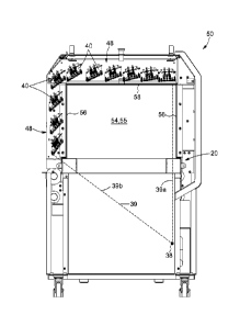
Figure 1 is a perspective view of a scanning system providing a partially
exposed view of a source, a displacement assembly, and a plurality of
detectors of
the scanning system, according to an optional embodiment of the present
invention.
Figure 2 is a front view of the scanning system of Figure 1.
Figure 3 is a side elevational view of the scanning system of Figure 1.
Figure 4 is a perspective view of a source, a plurality of detectors, and a
displacement assembly for a scanning system, according to an optional
embodiment of the present invention.
Figure 5 is a schematic view of a plurality of detectors disposed about a
scanning chamber, according to an optional embodiment of the present
invention.
Figure 6 is a perspective view of a bracket for a scanning system, according
to an optional embodiment of the present invention.
Figure 7 is a schematic representation of a portion of the scanning system,
according to an embodiment of the present invention.
Figures 8a to 8c are diagrams showing steps of a method executed by the
scanning system, according to an embodiment of the present invention.
Figure 9 is a diagram of a step of the method executed by the scanning
system, according to an embodiment of the present invention.
VT10001 CA
7
Figure 10 is an image of an object having been scanned by the scanning
system, according to an embodiment of the present invention, prior to a
normalizing step.
Figure 11 is an image of an object having been scanned by the scanning
system, according to an embodiment of the present invention, after a
normalizing
step.
Detailed description of preferred embodiments of the invention:
In the following description, the same numerical references refer to similar
elements. Furthermore, for the sake of simplicity and clarity, namely so as to
not
unduly burden the figures with several references numbers, not all figures
contain
references to all the components and features, and references to some
components and features may be found in only one figure, and components and
features of the present invention illustrated in other figures can be easily
inferred
therefrom. The embodiments, geometrical configurations, materials mentioned
and/or dimensions shown in the figures are optional, and are given for
exemplification purposes only.
Furthermore, although the present invention may be used for scanning
objects, such as for threat detection or imaging, and as a result, is
sometimes
described in the context of a possible use for detecting dangerous objects
and/or
producing 3D images, it is understood that it may be used for other purposes,
and
in other fields and/or activities. For this reason, expressions such as
"scan", "threat
detection", "dangerous object", "chemical", "imaging", "3D", etc. as used
herein
should not be taken as to limit the scope of the present invention to the
detection
of threats or the production of 3D images in particular. These expressions
Date Recue/Date Received 2020-04-16
CA 02925947 2016-03-31
WO 2015/048874 PCT/CA2013/050744
8
encompass all other kinds of materials, objects and/or purposes with which the
present invention could be used and may be useful, as can be easily
understood.
Broadly described, the present invention provides a system, method, and
bracket which can improve the process for scanning and detecting an object so
as
to generate a 30 image of said object, as but one possible example of a use of
the
invention.
According to a general aspect, there is provided a system 10 for scanning
an object 12, an example of which is shown in Figure 1. The system 10 can be
any
assembly or collection of components intended to form a machine or structure
capable of providing the functionality and advantages described in the present
disclosure. The object 12 can be any item, of any shape or configuration,
which
can be scanned. The object 12 can be made from living or non-living material.
The
term "scan" or "scanning" as used herein refers to the ability of the system
10 to
scrutinize, analyse and/or examine the object 12 for any suitable purpose. In
some
embodiments, "scanning" refers to the ability of the system 10 to project a
source
of radiation in a predetermined pattern towards the object 12 in order to
obtain
information about the object 12. One possible purpose for scanning the object
12
is to detect the composition or nature of the object 12 in order to determine
whether it may constitute a threat. Another possible purpose for scanning is
to
generate an image of the object and/or its interior. Yet another possible
purpose
for scanning the object 12 is to assess its interior contents or make-up. It
can thus
be appreciated that the system 10 can scan the object 12 for any number of
.. reasons, all of which are within the scope of the present disclosure.
The system 10 has a frame 50, an example of which is also shown in Figure
1. The frame 50 can be any partially, or fully, open or closed structure which
gives
shape to the system 10 and provides it with structural support. It can thus be
appreciated that the frame 50 can take many different configurations in order
to
achieve such functionality. In the embodiments shown in the figures, the frame
50
is shown as a substantially rectangular cuboid, but it is appreciated that it
is not
CA 02925947 2016-03-31
WO 2015/048874
PCT/CA2013/050744
9
limited to this shape. In some embodiments, the frame 50 can be supported by
wheels 52 or other suitable displacement devices, thereby advantageously
allowing the system 10 to be displaced as desired.
The frame 50 has a scanning chamber 54, an example of which is shown in
Figure 2, which receives the object 12 to be scanned. The scanning chamber 54
can be any enclosed, or partially enclosed, volume or space mounted in, near,
or
around the frame 50. The scanning chamber 54 has side surfaces 56, each one
being in opposed relation to the other, as well as a third surface 58 which
extends
between the side surfaces 56. The surfaces 56,58 can be walls or other planar
or
non-planar faces which define the boundaries of the scanning chamber 54. Such
a
configuration of surfaces 56,58 allows for many different types of scanning
chambers 54 to be used.
In one possible embodiment, the scanning chamber 54 can be a drawer into
which the object 12 can be placed so as to be scanned. The drawer scanning
chamber 54 can have two vertical side surfaces 56 supported and connected
together by a bottom horizontal third surface 58 upon which the object 12 can
rest.
In another possible embodiment, the scanning chamber 54 can consist of a
relatively large framework into which vehicles and other large devices can be
placed. This framework scanning chamber 54 can have two vertical side surfaces
56 connected together by a top horizontal third surface 58, thus defining a
passage through which the object 12 can be displaced. Such a framework
scanning chamber 54 can advantageously be used to scan cargo containers and
other similar large objects.
In yet another possible embodiment, an example of which is shown in the
figures, the scanning chamber 54 is a tunnel 55. The tunnel 55 can have an
opening at each of its ends, thus defining a passage or channel which extends
through some, or all, of the frame 50. The dimensions of the tunnel 55 can
vary
depending upon a number of factors, such as the following non-limitative list:
the
nature and shape of objects 12 to be scanned, the cost of implementation of
the
CA 02925947 2016-03-31
WO 2015/048874
PCT/CA2013/050744
system 10, and the space available for the system 10. Although shown as
substantially box-like, the tunnel 55 can be non-linear, winding, or take
other
configurations which permit it to receive the object 12 to be scanned.
5 The system 10
also has a displacement assembly 20, an example of which
is shown in Figure 1. The displacement assembly 20 engages with the scanning
chamber 54 so that it can receive the object 12 and displace it into the
scanning
chamber 54. In so doing, the displacement assembly 20 advantageously maintains
safety by helping to ensure that no effort is required by an operator to move
the
10 object 12
into the scanning chamber 54. The nature of the displacement assembly
20, and its relationship with the scanning chamber 54, can vary.
Indeed, in the optional embodiment where the scanning chamber 54 is a
drawer, the displacement assembly 20 can be a mechanism which engages the
drawer scanning chamber 54 from the exterior of the drawer scanning chamber 54
so as to open and close the drawer scanning chamber 54. In the optional
embodiment where the scanning chamber 54 is a framework, the displacement
assembly 20 can be a vehicle or other similar mover of the object 12 so as to
engage the framework scanning chamber 54 by moving the object 12 into the
passage defined by the framework scanning chamber 54. In the optional
embodiment where the scanning chamber 54 is a tunnel 55, the displacement
assembly 20 can be a conveyor 22 or conveyor belt. The conveyor 22 engages
the scanning chamber 54, which can be a tunnel 55, by extending through the
scanning chamber 54 so as to convey the object 12 through the scanning chamber
54.
In any of its configurations, the displacement assembly 20 can stop,
accelerate, decelerate, or otherwise control the displacement of the object
12, and
its direction, within or through the scanning chamber 54. Such functionality
advantageously allows for the object 12 to be rescanned or further analysed,
if
desired. Furthermore, and in light of the preceding description of some of its
optional embodiments, it can be appreciated that the displacement assembly 20
CA 02925947 2016-03-31
WO 2015/048874 PCT/CA2013/050744
11
can take many different configurations, and engage the scanning chamber 54 in
many different ways.
The system 10 also has a source 30, an example of which is also shown in
.. Figure 1. The source 30 emits electromagnetic (or EM) radiation against the
object
12 in the scanning chamber 54. In most embodiments, but not necessarily all,
the
EM radiation is X-rays, and the term "X-rays" will thus be used throughout the
present disclosure, such use being understood as not limiting the source 30 to
emitting only X-ray radiation. The emission of the EM radiation can be
performed
continuously, at discrete intervals, or only as the object 12 is displaced
into, or
passed through, the scanning chamber 54. The term "against" refers to the
ability
of the EM radiation to impact the object 12 and pass through the same. The
effect
of the object 12 on the EM radiation as it passes through can be detected by
the
detectors described below. It will be appreciated that not all of the EM
radiation
emitted by the source 30 must impact the object 12.
The source 30 is "mountable to the frame about the scanning chamber",
which means that it can fixedly or removably attached to the frame 50 in
proximity
to the scanning chamber 54. The source 30 can also mounted to the frame 30 so
that it can rotate, pivot, or be displaced around the object 12, which can
remain in
a fixed position. The actual location and position of the source 30 can depend
on
the configuration of the scanning chamber 54 and the displacement assembly 20,
among other possible factors. In some embodiments, the source 30 can be
mounted to the frame 50 at a location below the displacement assembly 20, as
shown in Figure 3. This configuration can be suitable where the displacement
assembly 20 is a conveyor 22, and it allows the source 30 to emit the EM
radiation
in an upward direction into the scanning chamber 54 so as to impact the object
12
being conveyed on the conveyor 22. In some embodiments, the source 30 can be
mounted to the frame 50 at a location that is above the displacement assembly
20.
This configuration can be suitable where the displacement assembly 20 is a
framework displacement assembly 20, for example, and it allows the source 30
to
emit the EM radiation in a downward direction into the scanning chamber 54 to
the
CA 02925947 2016-03-31
WO 2015/048874
PCT/CA2013/050744
12
object 12. The source 30 can also be mounted to the frame from the sides or
from
other orientations, as required.
The source 30 can have many different configurations. In one possible
embodiment, an example of which is shown in Figure 3, the source 30 is
inclined
relative to a horizontal plane at an angle A. The inclination of the source 30
at the
angle A allows for the generation of data which may be useful for certain
purposes. For example, where the purpose of scanning the object 12 is to
generate a 3D image of the object 12, the inclination of the source 30 (and
thus of
the X-rays emitted by the source 30 and impacting the object 12) permits
obtaining
data for the top and the sides of the object 12, thus advantageously allowing
for
the production of a perspective 3D view of the object. In contrast,
conventional
non-inclined sources may not be able to generate data regarding the top or
sides
of an object, and may thus be able to generate only 2-D views. The angle A can
vary depending on a number of factors, such as the size of the scanning
chamber
54, the position of the source 30 relative to the scanning chamber 54, the
coverage of the EM radiation in the scanning chamber 54, etc. In most
embodiments, but not necessarily all, the angle A is between about 10 and
about
.
In some embodiments, an example of which is shown in Figure 3, the
source 30 includes an X-ray emitter 32 which can be mounted to, and removed
from, a mounting surface 34 of an inclined support 36. The X-ray emitter 32
can
emit X-rays into the scanning chamber 54 and against the object 12 found
therein
or displaced therethrough. The mounting surface 34 can be any face or area
upon
which the X-ray emitter 32 can be attached, and the inclined support 36 can be
any structure of device which supports the X-ray emitter 32. The mounting
surface
34 may be inclined so as to form an angle B with respect to the horizontal. In
being
inclined, the mounting surface 34 also inclines the X-ray emitter 32 attached
thereto, thereby advantageously allowing the X-ray emitter 32 to emit X-rays
at an
angle to the horizontal. Optionally, the angle B can vary between about 1 and
about 45 , and further optionally between about 10 and about 20 Such a range
CA 02925947 2016-03-31
WO 2015/048874
PCT/CA2013/050744
13
of values for angle B can optimize the quantity of information in the
resulting image
produced of the object 12.
In some embodiments, the source 30 can emit EM radiation or X-rays from
a focal point 38 as a beam 39. The beam 39 can be any fan or cone beam having
an angular width as it is emitted from the focal point 38. The angular width
of the
beam 39 observed from one direction may be different from the angular width of
the same beam 39 observed from another direction. This can be better
appreciated by comparing the example of the beam 39 as shown in Figures 2 and
3. In Figure 3, the beam 39 has an angular width that is smaller (i.e. fewer
degrees
wide) than the angular width of the same beam 39 shown in Figure 2. The beam
39 can be defined by its beam boundaries 39a,39b. The angular width or
interval
between these beam boundaries 39a,39b can vary, and can optionally be about
80 . In some embodiments, and as shown in Figure 2, one of the beam
boundaries 39b can be substantially aligned with one of the side surface 56 of
the
scanning chamber 54. Such an alignment can advantageously allow the beam 39
to cover or span the entire useful width of the scanning chamber 54, and allow
for
generating an image that is more representative of the object 12.
The system 10 also has a plurality of detectors 40, examples of which are
shown in Figure 4. The detectors 40 receive the EM radiation which passes
through the object 12, thus scanning the object 12 by allowing for the EM
radiation
to be analysed. The detectors 40 are mounted or fixed to the frame 50 at least
partially about the scanning chamber 54. The expression "at least partially
about
the scanning chamber" refers to the orientation and positioning of the
detectors 40
in proximity to the scanning chamber 54 so that they can detect the EM
radiation
passed through the object. Such orientation and positioning can vary depending
on numerous factors such as the size of the scanning chamber 54, the desired
detector 40 coverage, and the type of detector 40 used. In the optional
embodiments shown in the figures, the detectors 40 are positioned in an "L"-
shape
on the frame 50 so that they cover the top and one of the sides of the
scanning
chamber 54. In other optional embodiments, the detectors 40 can be displaced
CA 02925947 2016-03-31
WO 2015/048874
PCT/CA2013/050744
14
around the object 12, which remains in a fixed position. Other configurations
are
within the scope of the present disclosure.
In some embodiments, each detector 40 includes a detector card 42 which
has a centre point 44 and edges 46. The detector card 42 can be any suitable
detector card 42 such as those manufactured by Detection Technologies Ltd.,
United Kingdom. Each of these detector cards 42 can have a centre point 44,
which corresponds to the geographical centre of the detector cards 42. The
edges
46 of each detector card 42 define its boundaries. The detectors 40 and/or the
system 10 can be linked to a central processing Unit (CPU) 100 (see Figure 7)
or
other processing device so that the data detected by the detectors 40 can be
analysed, processed, and used to output information, such as a 3D perspective
view of the object 12, as better explained further below with reference to
Figure 7.
According to an embodiment described and illustrated herein, each detector
40 comprises a first scintillator 80, a filter 82, and a second scintillator
84, all of
which are sandwiched together, as is schematically represented in Figure 7.
The
X-Ray emission impacts the first scintillator 80 which detects a lower portion
of the
X-Ray signal. Residual low energy signal is then stopped by the filter 82.
Finally
the remaining signal from the X-Ray emission reaches the second scintillator
84
which detects a higher portion of the X-Ray signal.
Each detector 40 is inclined at a detector angle D. In most embodiments,
but not necessarily all, the detector angle D is defined with respect to a
horizontal
plane. The detector angle D of one or more of the detectors 40 is different
than the
detector angle D of an adjacent detector 40. For example, this can mean that
the
detector angle D of at least one detector 40 is different than the detector
angle D
of all the other detectors 40. This can also mean that each detector 40 has a
detector angle D that is different from the detector angles D of its
neighbouring
detectors 40. The term "adjacent" in this context refers to neighbouring
detectors
40, whether they are located directly next to, or nearby, the at least one
detector
having a different detector angle D. The determination of the detector angle D
CA 02925947 2016-03-31
WO 2015/048874 PCT/CA2013/050744
for each detector 40 can depend upon numerous factors such as, but not limited
to: the angle of the source 30 relative to the horizontal, the position of the
source
30 relative to the scanning chamber 54, the position of the centre point 44 of
each
detector card 42 relative to the source 30, etc.
5
In some embodiments, the detector angle D of each detector card 42 may
optionally be determined by satisfying the following two requirements: 1) the
centre point 44 of each detector card 42 is substantially perpendicular to the
focal
point 38 of the beam 39, and 2) each detector card 43 is positioned as close
as
10 possible to the focal point 38 of the beam 39.
Reference is made to Figure 5. In order to satisfy the first requirement, a
circle of diameter r can be drawn from the focal point 38 of the beam 39 of
the
source 30. Each detector card 42 can be placed on this circle such that it is
15 tangent with the circle, and such that its centre point 44 is
perpendicular to the
focal point 38. In order to satisfy the second criterion, each detector card
42 can
be brought as close as possible to the surfaces 56,58 of the scanning chamber
54
and/or tunnel 55, thus bringing these detector cards 42 as close as possible
to the
focal point 38 without being inside the scanning chamber 54. This is shown
schematically in Figure 5 with the arrows directing the detector cards 42
towards
the scanning chamber 54. In practice, bring the detector cards 42 closer to
the
scanning chamber 54 can involve placing one of the edges 46 of the detector
cards 42i in contact with the either the side surfaces 56 or the third surface
58 of
the scanning chamber 54 and/or tunnel 55. Indeed, some edges 46, such as those
of the detector cards 42i located on the side of the scanning chamber 54, can
engage the side surfaces 56 whereas other edges 46 can engage the third
surface
58. Optionally, the edges 46 of neighbouring detector cards 42i can overlap
one
another. It can thus be appreciated from the schematic shown in Figure 5 how
the
optional positioning and configuration of the detectors 40 of Figure 4 can be
achieved.
VT10001 CA
16
Returning to Figure 2, the plurality of detectors 40 can have two rows 48 of
detectors 40 mounted to the frame 50. The first row 48 can be disposed
adjacent
to one or more of the side surfaces 56, while the second row 48 can be
disposed
adjacent to the third surface 58 of the scanning chamber 54. It can thus be
appreciated that the inclination of the source 30 and detectors 40
advantageously
allows for the generation of an accurate and representative 3D perspective
image
of the object 12, thus eliminating the need for numerous detectors surrounding
three or more sides of the scanning chamber as is known in the prior art.
Optionally, the first and second rows 48 of detectors 40 can form an angle C
with
respect to a horizontal plane, as shown in Figure 1. The angle C can vary, and
can
optionally have values ranging from about 89 to ab out 450, and further
optionally
from about 80 to about 70 .
According to another general aspect, there is a provided a bracket 60 for a
scanning system, such as the one described above. Referring now to Figure 6,
the
bracket 60 has a bracket frame 62 which can be mounted to, about, or within
the
scanning system and which is inclined relative to the horizontal. The angle of
inclination can be between about 1 and about 450, and optionally between
about
10 and about 20 . The bracket 60 also has a source 30, such as the one
described above, which emits EM radiation or X-rays against the object 12
being
scanned. Finally, the bracket 60 has multiple detectors 40, which can have
varying
detector angles as explained above, and which detect the EM radiation that
passes through the object 12.
The image processing of the above-described scanning system will now be
better explained, with reference to Figure 7, with further reference to Figure
1 and
to 4, as well as Figures 8 to 11. In general terms, an input port 103 receives
image data of an object, having been captured via a plurality of detectors 40
disposed at least partially about the object 12, at least two adjacent ones of
the
detectors being angled one with respect to the other, in that a given detector
40 is
angled differently with respect to an adjacent detector 40. The processor 100
generates from the image data, an image representing a perspective view of the
Date Recue/Date Received 2020-04-16
CA 02925947 2016-03-31
WO 2015/048874 PCT/CA2013/050744
17
object. The image may then be store in a storage 112 for further presenting
the
image on a display 110, as a three-dimensional representation of the object
12.
There is thus provided a corresponding system 101 which performs this process
in
order to generate the image.
In the context of the present description, the term "processor" refers to an
electronic circuitry that can execute computer instructions, such as a central
processing unit (CPU), a microprocessor, a controller, and/or the like. A
plurality of
such processors may be provided, according to embodiments of the present
invention, as can be understood by a person skilled in the art. The processor
may
be provided within one or more general purpose computer, for example, and/or
any other suitable computing device.
Still in the context of the present description, the term "storage" refers to
any computer data storage device or assembly of such devices including, for
example: a temporary storage unit such as a random-access memory (RAM) or
dynamic RAM; a permanent storage such as a hard disk; an optical storage
device, such as a CD or DVD (rewritable or write once/read only); a flash
memory;
and/or the like. A plurality of such storage devices may be provided, as can
be
understood by a person skilled in the art.
According to the present embodiment, the X-Ray source 30 emits a
continuous spectrum of X-Rays, ranging from a lower energy range such as 10 to
70 kV (+/-) up to higher energy ranges such as 60 to 250 kV (+/-).
It is to be understood that depending on particular embodiments of the
present invention, the lower energy range may be as low as 1kV and the higher
energy ranges may be greater that the values given above in relation to the
described embodiment.
As the object 12 is subjected to the X-Rays, the detectors 40 capture the X-
Ray energy that traverses the object 12. As previously mentioned, the first
CA 02925947 2016-03-31
WO 2015/048874
PCT/CA2013/050744
18
scintillator 80 detects a lower portion of an X-Ray signal, while the second
scintillator 84 detects a higher portion of the X-Ray signal. The high energy
range
penetrates more easily through denser materials, while the low energy range
provides better contrast for image portions corresponding to lighter
materials.
Broadly, each of the scintillators 80, 82 converts the X-Ray energy to light.
A photo-diode 86 then captures the light and generates a corresponding
electric
signal. The electric signal is further digitized by a converter 88. The
digitized value
is associated to a pixel of the image which represents the object.
In the present embodiment, the detectors' physical arrangement in the
scanner system determines the arrangement of the raw data extracted therefrom.
More particularly, according to the embodiment illustrated herein, detectors
40 are
aligned in a row, positioned as previously mentioned, along an "L"-shaped
configuration, as schematically represented in Figure 8(a). For a given scan
capture, image data is received from each detector and organized based on the
positioning of the detectors 40, as schematically represented at Figure 8(b),
where
each subdivision 90 represents data captured by a corresponding detector at a
given scan capture. Each subdivision 90 corresponds to 64 pixels of image
data,
in the present embodiment. The resulting row 91 provide a segment of the
resulting image.
As the object 12 moves through the scanning chamber 54, the detectors
perform scan captures sequentially at a given rate, which may depend on their
integration time, i.e. exposure time. Thus several of said "rows" of data are
acquired in a given scanning process. The rows are juxtaposed as schematically
represented in Figure 8(c), to represent all the image captures of the
detectors in a
given scanning process, thereby producing the resulting image 96, 99 of the
object
12 (see Figures 10 and 11). This image provides a perspective view of the
image,
as can be seen in Figures 10 and 11, by virtue of the detectors being
positioned as
different angles depending on their location in relation to the source
emission.
VT10001 CA
19
In Figure 9, the rows "LOW1" and "HIGH1" correspond to the image capture
of each of the low energy absorption scintillator 80 and the low energy
absorption
scintillator 84, respectively. The rows "LOVV2" and "HIGH2" represent data
collected from a second set of detectors, which may be located for example
across
from the first row of detectors 40, to detect an emission from another source,
in
accordance with an alternative embodiment. Thus each of the subsection 92 of
the
row "LOW1" corresponds to a subsection 94 of the row "HIGH1". For example, the
first boxes 92a and 94a correspond together and represent a capture of a same
portion of the object 12 being scanned.
As previously mentioned, the X-Ray energy is translated into a digitized
value, for each pixel, via the scintillators 80, 82, the photo-diodes 86 and
the
converter 88. In the conversion by the photo-diodes 86 of the light into an
electric
signal, some error may occur, in that a given light source may result in
different
electrical signals due to the fact that every detector card behave slightly
differently
to the presence or absence of X-Ray signal.
Thus, in order to correct these variations and for the final image to appear
more homogeneously, a normalization module 102, by means of the CPU 100,
normalizes (or "calibrates") each pixel of the low and high energy captures,
by
correcting an offset and a gain in the light conversion. Figure 10 shows a raw
image 96 of the scanned object 12, prior to normalization. Figure 11 shows the
image 99 after normalization.
The offset is determined based on the signal perceived by the scintillators of
the detectors when no source is emitted. When no source is emitted, as
represented by the dark band 97 appearing in FIG. 10, it is expected that the
scintillators would transmit a signal corresponding to 0, the theoretic value.
However, due to some imperfections in the hardware, a signal is still
generated,
and each pixel may correspond to a different signal. The offset for each pixel
corresponds to the difference between the value perceived (for example, 130,
160,
110) based on the signal generated when there is no source emission, and the
Date Recue/Date Received 2020-04-16
CA 02925947 2016-03-31
WO 2015/048874 PCT/CA2013/050744
value 0, the theoretic value. Thus, to correct the offset for each pixel, the
corresponding offset value is subtracted from the pixel's value.
After removing the offset, there are still variations in the capture of each
5 detector when fully exposed to the source emission, as represented by the
light
band 98 appearing in FIG. 10. Thus, for every pixel, the mean maximum value
representing the gain is determined. To correct the inhomogeneous response of
each pixel, the measured value is divided by this maximum signal at full
exposure.
These values are finally multiplied by a fixed identical scaling factor.
The high and low energy information is then fused, at an image fusion
module 104, by means of the CPU 100. More particularly, each pixel of the
image
results from a combination of high energy data in some proportion and low
energy
data in some proportion. Depending on the density of the material detected, it
may
be desirable to emphasize the low energy information or the high energy
information in suitable proportion. Indeed, as previously mentioned, the high
energy range penetrates more easily through denser materials, while the low
energy range provides better contrast for image portions corresponding to
lighter
materials. The high and low energy data is thus combined accordingly to better
illustrate particular regions of the image. For example, a pixel may be the
result of
25% of the high energy data and 75% of the low energy data because it is
determined by the X-Ray signal is relatively high, meaning that it is more
desirable
to see contrast. The proportion of high and low energy is determined based on
ranges of low energy data value and/or high energy data value for a particular
pixel.
An atomic number is then associated to each pixel of the image, via an
atomic number calculation module 106, by means of the CPU 100. More
particularly, the atomic number is determined based on the low energy
absorption
data and high energy absorption data, as well as a signal level of a source
emission 30.
CA 02925947 2016-03-31
WO 2015/048874
PCT/CA2013/050744
21
In a calibration step, materials having a known atomic number are scanned,
in order to correlate each of their particular combination of low and high
energy, for
a given source signal level, with their atomic number. Based on the
correlations
made based on the known materials, a set of reference data is generated. The
reference data includes combinations of low and high energy (at a given source
signal level) and their corresponding atomic number. Thus, for each pixel, the
combination of the corresponding low and high energy data, is correlated with
a
corresponding atomic number.
The image is then sharpened via a filtering module 108, by means of the
CPU 100, in order to reduce blurriness when the image is displayed for viewing
on
a display 110. More particularly, the image data is convoluted to enhance
portions
of the image representing edges of the object 12.
The resulting image is then store in a database 112, from the basis of which
a three-dimensional or perspective representation of the object 12 may be
presented on the display 110.
Embodiments of the present invention thus provide the advantage of
generating a three-dimensional or perspective representation of the object 12,
by
virtue of the detectors being positioned at different angles depending on
their
location in relation to the source emission, and of enhancing detection
capabilities,
thereby allowing for an operator to better analyze the object 12.
Further advantageously, such a three-dimensional or perspective
representation may provide more a revealing image of the object 12 when
compared to two-dimensional images generated by traditional scanners. More
specifically, such a representation may allow a user to visualize more walls
or
boundaries of the object 12, and may have fewer "dark spots" corresponding to
parts of the object 12 which have planes aligned with the plane of the EM
radiation
emitted by the source.
CA 02925947 2016-03-31
WO 2015/048874
PCT/CA2013/050744
22
It is to be understood that, in accordance with alternate embodiments, the
above-described system and method may be adapted to operate with a single
energy level of X-Ray signal captured at the detectors, as well as a
plurality, i.e.
two or more of such energy levels of X-Ray signal (instead of only low energy
and
high energy, as in the context of the above-described embodiments). Indeed,
any
suitable ranges of energy levels may be defined and captured by the detectors
and further processed, for example to obtain more information on the
composition
of the object being scanned.
Of course, numerous other modifications could be made to the above-
described embodiments without departing from the scope of the invention, as
defined in the appended claims.