Note: Descriptions are shown in the official language in which they were submitted.
CA 02926142 2016-04-01
WO 2014/067021
PCT/CH2013/000189
1
CLOSURE DEVICE
CROSS-REFERENCE TO OTHER APPLICATIONS
[01] This application claims priority under Title 35, U.S.C. Sec. 119, from
Swiss Patent
Application No. 02168/12, which was filed on October 29, 2012, and is hereby
incorporated by
reference herein in its entirety.
BACKGROUND
[02] A closure device is typically used to occlude an access hole that is
surgically opened to
facilitate a therapeutic cardiac or vascular procedure. The closure device is
used to seal off the
access hole in order to re-establish the integrity of an accessed organ wall
or vessel. A closure
CA 02926142 2016-04-01
WO 2014/067021 PCT/CH2013/000189
2
device can also be used to close anatomical defects such as a septal defect in
an atrial septum.
Typical closure of an access hole involves the use of sutures or large closure
devices. Use of
sutures or large closure devices requires disturbance of skin and other tissue
along the access path
to the access hole. The disturbance may be greater than the disturbance
required to perform the
therapeutic procedure.
[03] It would be desirable, therefore, to provide apparatus and methods for
closing an access
hole that do not require disturbance greater than that required to perform the
therapeutic
procedure.
BRIEF DESCRIPTION OF THE DRAWINGS
[04] The objects and advantages of the invention will be apparent upon
consideration of the
following detailed description, taken in conjunction with the accompanying
drawings, in which like
reference characters refer to like parts throughout, and in which:
[05] FIG. 1 is an illustrative cross-sectional view of apparatus in
accordance with the principles of
the invention along with illustrative anatomy in connection with which the
invention may be
practiced;
[06] FIG. 2 is an illustrative cross-sectional view of the apparatus shown
in FIG. 1 along with the
anatomy shown in FIG. 1 when the apparatus is in a different state from that
shown in FIG. 1;
[07] FIG. 3 is an illustrative cross-sectional view of the apparatus shown
in FIG. 2 along with the
anatomy shown in FIG. 2 when the apparatus is in a different state from that
shown in FIG. 2;
[08] FIG. 4 is an illustrative cross-sectional view of the apparatus shown
in FIG. 3 along with the
anatomy shown in FIG. 3 when the apparatus is in a different state from that
shown in FIG. 3;
[09] FIG. 5 is an illustrative cross-sectional view of the apparatus shown
in FIG. 4 along with the
anatomy shown in FIG. 4 when the apparatus is in a different state from that
shown in FIG. 4;
[010] FIG. 6 is an illustrative cross-sectional view of the apparatus shown in
FIG. 5 along with the
anatomy shown in FIG. 5 when the apparatus is in a different state from that
shown in FIG. 5;
[011] FIG. 7 is an illustrative cross-sectional view of the apparatus shown in
FIG. 6 along with the
anatomy shown in FIG. 6 when the apparatus is in a different state from that
shown in FIG. 6;
CA 02926142 2016-04-01
WO 2014/067021
PCT/CH2013/000189
3
[012] FIG. 8 is an illustrative perspective view of other apparatus in
accordance with the principles
of the invention;
[013] FIG. 9 is an illustrative perspective view of parts of yet other
apparatus in accordance with
the principles of the invention along with other anatomy, in partial cross
section, in connection with
which the invention may be practiced;
[014] FIG. 10 is an illustrative partial cross-sectional view of still other
apparatus, in accordance
with the principles of the invention, along with the anatomy shown in FIG. 5,
taken from a direction
like that shown by lines 10-10 (shown in FIG. 5) of the apparatus shown in
FIG. 5;
[0151 FIG. 11 is an illustrative cross-sectional view of still other apparatus
in accordance with the
principles of the invention;
[016] FIG. 12 is an illustrative perspective view of still other apparatus in
accordance with the
principles of the invention;
[017] FIG. 13 is an illustrative perspective view of still other apparatus in
accordance with the
principles of the invention;
[018] FIG. 14 is an illustrative perspective view of still other apparatus in
accordance with the
principles of the invention;
[019] FIG. 15 is an illustrative partial cross-sectional, partial perspective
view of still other
apparatus in accordance with the principles of the invention along with the
anatomy shown in FIG.
1; and
[020] FIG. 16 is an illustrative perspective view of still other apparatus in
accordance with the
principles of the invention.
DETAILED DESCRIPTION
[021] Apparatus and methods for closing an access hole in a heart, blood
vessel or other anatomy
are provided. The apparatus may include a heart plug. The heart plug may
include an absorbent
mass that is configured to apply pressure to myocardial tissue in the access
hole by absorbing a
fluid. The pressure may create friction-based anchoring of the plug in the
access hole.
CA 02926142 2016-04-01
WO 2014/067021
PCT/CH2013/000189
4
[022] The mass may have a longitudinal axis. The mass may have a central axis.
The longitudinal
axis may coincide with the central axis. The mass may have a radial direction.
The radial direction
may be perpendicular or substantially perpendicular to the axis.
[023] Wetting of the plug by the fluid may cause the plug to expand. The plug
may expand radially.
The plug may expand longitudinally. Liquid-swollen plug material may expand
until the expansion
force of the plug equals the resistance of the surrounding tissue. When the
expansion force equals
the resistance of the surrounding, a friction-based anchoring may be created.
[024] Stored elastic energy in material in the plug may contribute to
expansion when the plug
interacts with fluid. Material in the plug may absorb the fluid by wicking.
The wicking may
contribute to the expansion. The expansion may continue until the plug is
constrained by
surrounding anatomy. The expansion may continue until expansion forces
generated by the wicking
are balanced by external forces from the anatomy on the plug. The plug may be
a plug that does
not include a non-expandable cover.
[025] If the plug is compressed, for example by heart contraction, when the
fluid is present in the
plug, the plug may resist the compression. Elastic properties of material in
the plug may produce
some or all of the resistance. Some or all of the resistance may be generated
by wetting of the plug
material by the fluid. Some or all of the resistance may be generated by
viscous resistance to
expulsion of a fraction of the fluid through the material. Some or all of the
resistance may be
generated by frictional forces acting between fibers or other elements inside
the plug. Some or all
of the resistance may be generated by contact forces acting between fibers or
other elements
inside the plug.
[026] When the compression is reversed, for example by heart relaxation, the
plug may rebound.
The plug may rebound based on one or more of the aforementioned mechanisms
that provide
expansion, resistance to compression or by any other suitable mechanism. For
example, wicking
may contribute to the rebound. Restoration of equilibrium between surface
forces at the contact
between the fluid and the plug material may contribute to the rebound. A
pressure drop in the plug
as the plug is expanded from reversal of the compression may contribute to the
rebound.
[027] When saturated or partially saturated with the fluid, the plug may be
resistant to
compression and retain its seal against the heart wall. When saturated or
partially saturated with
the fluid, the plug may be compliant and avoid causing stress concentrations
that may injure the
CA 02926142 2016-04-01
WO 2014/067021
PCT/CH2013/000189
heart wall. When saturated or partially saturated with the fluid, the plug may
be both resistant to
compression and compliant.
[028] The plug may efficiently close the access hole. The plug may provide a
rapid seal. The plug
may provide a permanent seal. The plug may provide an atraumatic seal. The
plug may restore
anatomical integrity to the organ in which the access hole is disposed. The
plug may restore
functional integrity to the organ in which the access hole is disposed.
[029] The plug may be self-sealing. The plug may be self-anchoring. The plug
may be self-
centering in a plane transverse or perpendicular to the access hole.
[030] When the hole is in the heart, the hole may be at an apex of the heart.
For example, the
hole may be at a ventricular apex of the heart.
[031] The plug may include any suitable biocompatible material. The
biocompatible material may
be self-expanding. The biocompatible material may be self-anchoring to heart
tissue.
[032] The plug may include any suitable bioabsorbable material. The
bioabsorbable material may
be self-expanding. The bioabsorbable material may be self-anchoring to heart
tissue.
[033] The plug may include any suitable biodegradable material. The
biodegradable material may
be self-expanding. The biodegradable material may be self-anchoring to heart
tissue.
[034] The mass may have an unexpanded state. The mass may have an expanded
state. The
expanded state may be a state in which the plug is partially saturated with a
fluid. The expanded
state may be a state in which the plug is saturated with the fluid. The fluid
may be blood, water,
saline solution, plasma or any other suitable fluid.
[035] The mass may have an unexpanded state diameter that is selected for
rapid occlusion of the
hole. The mass may have an unexpanded state diameter that is selected for
sliding clearance within
a delivery catheter having an outside diameter sized for traversal of the
hole. The mass may have
an unexpanded state diameter that is selected for both rapid occlusion of the
hole and sliding
clearance within a delivery catheter having an outside diameter sized for
traversal of the hole.
[036] The mass may have a length that is selected to match the length of the
hole. The selected
length may be a length of the mass when the mass is unexpanded. The selected
length may be an
expected length of the mass when the mass is subsequently expanded. The
selected length may be
selected to match a length that is less than the length of the hole. The mass
may have a length
selected to match a length that is greater than the length of the hole. The
mass may be positioned
CA 02926142 2016-04-01
WO 2014/067021 PCT/CH2013/000189
6
so that the mass extends out of a distal end of the hole. The mass may be
positioned so that the
mass extends out of a proximal end of the hole. When the mass is positioned to
extend out of the
distal end of the hole, the mass may extend radially outward to a radius
greater than the radius of
the hole. The mass may form a shape like that of a champagne bottle cork. The
greater radius may
provide anchoring by interference with an annular region of heart tissue
adjacent a distal end of the
hole.
[037] The mass may include a growth factor treatment. The treatment may be
applied to a
surface of the mass.
[038] The mass may include any suitable biocompatible material. The mass may
include any
suitable bioabsorbable material. The mass may include any suitable
biodegradable material.
[039] The mass may include a matrix. The matrix may define interstitial space
for absorption of
the fluid.
[040] The plug may include an antibiotic pharmacological agent. The antibiotic
agent may be
supported by the matrix. The heart plug may include a polymerizing agent. The
polymerizing agent
may be configured to polymerize a blood constituent. The polymerizing agent
may be supported by
the matrix. The polymerizing agent may be configured to provide a polymerized
blood constituent in
the interstitial space to give the plug elastic properties.
[041] The heart plug may include a photoactivated compound. The compound may
include
powder. The compound may be provided in a coating on the mass. The compound
may be
activated by applying light through a catheter. When activated in the presence
of the fluid, the
compound may provide the plug with elastic properties.
[042] The matrix may include nonwoven material. The matrix may include fibrous
matter. The
fibrous matter may include cellulose. The fibrous matter may include cotton.
The fibrous matter
may include rayon. The fibrous matter may include polyester. The fibrous
matter may include
polyethlyne. The fibrous matter may include any other suitable material.
[043] The fibrous matter may be prepared in a manner that provides for
expansion shortly after
initial contact with the fluid. Apparatus and methods for such preparation are
set forth, for
example, in U.S. Patent Nos. 6,310,269 and 6,748,634, which are hereby
incorporated herein in
their entireties.
[044] The matrix may include a gelatinous material.
CA 02926142 2016-04-01
WO 2014/067021 PCT/CH2013/000189
7
[045] The matrix may define a porous polymer network.
[046] The heart plug may include a cannula. The cannula may be a permanent
part of the heart
plug. The permanent cannula may be non-removable in the sense that it is
allowed to remain in the
plug when the plug is permanently deployed in the heart. The cannula may be
removable from the
heart plug. The cannula may extend through the mass. The cannula may extend
along the entire
longitudinal axis of the mass, thereby passing through the mass. The cannula
may extend partially
through the mass. The cannula may be configured to receive a guide wire. The
cannula may extend
partially through the mass.
[047] The heart plug may include a carriage loop. The loop may extend away
from an outer
surface of the mass. The loop may be configured to receive a guide wire. The
guide wire may be
used as a "tandem wire," along which the plug may slide, with the tandem wire
offset from the
central axis of the plug. The tandem wire may be at a greater radial distance
from the central axis
than is the outer surface of the plug.
[048] The heart plug may include a porous veiling about the mass. The mass may
be configured to
press the veiling against the myocardial tissue when the mass expands. The
mass may be
configured to press the veiling against the myocardial tissue in response to
absorption of the fluid
by the mass.
[049] The veiling may include any suitable biocompatible material. The veiling
may include any
suitable bioabsorbable material. The veiling may include any suitable
biodegradable material. The
veiling may include nonwoven material. The veiling may include cellulose. The
veiling may include
polyester. The veiling may include polyethylene.
[050] The mass may have a dry diameter. The mass may have a wet diameter. The
wet diameter
may be a saturated diameter. The wet diameter may be a partially saturated
diameter. The wet
diameter may be greater than the dry diameter. The veiling may be
nonexpendable relative to the
mass. The veiling may be expandable. When the veiling is nonexpandable
relative to the mass, the
veiling may have a maximum diameter.
[051] The access hole may have a diastolic diameter when the heart is relaxed.
The saturated
diameter of the mass may be greater than the diastolic diameter of the hole.
The maximum
diameter of the veiling may be greater than the wet diameter of the mass. The
maximum diameter
CA 02926142 2016-04-01
WO 2014/067021 PCT/CH2013/000189
8
of the veiling may be greater than the wet diameter of the mass when the mass
is fully expanded by
absorption.
[052] The plug may be provided in different compressed diameters corresponding
to different
access hole diameters. The plug may be provided in different compressed
lengths corresponding to
different access hole lengths. One or more plugs of different diameters or
lengths may be provided
in a kit.
[053] The plug may include an anchor. The anchor may be one of a plurality of
anchors. Each of
the plurality of anchors may have one or more features in common with the
other anchors. The
anchor may have a base. The base may be affixed to the mass. The anchor may be
affixed to an
outer radial surface of the mass. The anchor may be affixed to the distal end
of the mass. The
anchor may be affixed to the proximal end of the mass. The anchor may be
affixed to the veiling.
[054] The anchor may have an engagement end. The engagement end may be
configured to
engage the heart. The engagement end may be configured to atraumatically
engage heart muscle.
The anchor may be any suitable anchor. The engagement end may include a
piercing tip that is
supported by a stem that points radially outward and proximal from the base.
The engagement end
may extend, for example, up to about 0.5 mm, along the stem, from the base.
The engagement end
may extend, for example, up to about 1 mm, along the stem, from the base. The
engagement end
may extend, for example, up to about 2 mm, along the stem, from the base. The
piercing tip may
thus engage the myocardium upon deployment of the plug. The piercing tip may
be pin-like. The
piercing tip may thus pierce the myocardium by shifting the plug in the
proximal direction.
[055] The anchor may have the form of a link of a chain. Alternating
interlinked links may present
a radially outward protrusion to the myocardium. The links may provide
traction on the myocardium
in a manner similar to the way traction chains for vehicle tires provide
traction on travel surfaces.
[056] The anchor may be one of a plurality of anchors. The plurality of
anchors may be arranged
about the surface of the plug to engage the myocardial tissue when the plug is
deployed in the
access hole. The anchors may be linked to each other by a girdle that
encircles or partially
encircles the plug. The plug may include one or more girdles of anchors.
[057] The plug may include an imaging marker. The marker may extend from a
distal portion of
the mass. The marker may be configured to signal registration of the distal
portion with an orifice at
a distal end of the hole. The marker may be radiopaque. The marker may be
selected for acoustic
CA 02926142 2016-04-01
WO 2014/067021 PCT/CH2013/000189
9
reflection contrast. The marker may be selected for magnetic resonance imaging
contrast. The
marker may provide a visual aid for positioning the plug in the access hole.
The marker may provide
a visual aid for delivering the plug to the access hole. The marker may be
distributed about the
surface, or within the volume, of the plug in a patterned fashion such that an
expanded portion of
the plug may be visually distinguished from an unexpanded portion of the plug.
For example, the
marker may include a lobed thread that encircles or partially encircles the
plug and expands with
the plug. A marker thread may be formed from a girdle of anchors.
[058] The heart plug may include a cap. The heart plug may include a non-
thrombogenic cap. The
cap may include an umbrella form. The cap may include a disc form. The cap may
extend, at a
distal end of the mass, radially away from a central axis of the mass. The cap
may include an
elastomeric material, an alloy such as that available from Nitinol Devices &
Components, Inc.,
Fremont California, under the trade name "Nitinol," a surgical steel, a
surgical steel with non-
thrombogenic coating, a polymeric material, a film, a membrane or any other
suitable material. The
cap may be attached to the distal end of the mass. The cap may be pinned to
the distal end of the
mass. The cap may be glued to the distal end of the mass. The cap may be
sutured to the distal
end of the mass. The cap may include biocompatible material. The cap may
include bioabsorbable
material. The cap may include biodegradable material. The cap may include
material that is similar
to or the same as material that is included in the veiling.
[059] The mass may have a first diameter when the mass is in a compressed
state. The mass may
have a second diameter when the mass is in an expanded state. The cap may have
a cap diameter
that is greater than the first diameter. The cap may be directly affixed to a
cannula. The cap may
extend away from the cannula. The cannula may extend substantially along a
central axis of the
plug from a proximal end of the plug to a distal end of the plug. The cannula
may be configured to
receive a guidewire.
[060] The cap diameter may be about the same size as the second diameter.
[061] The plug may include a proximal cap. The proximal cap may extend, at a
proximal end of the
mass, radially away from the central axis.
[062] The proximal cap may be directly affixed to the cannula. The proximal
cap may extend away
from the cannula.
CA 02926142 2016-04-01
WO 2014/067021 PCT/CH2013/000189
[063] The distal cap may be used to anchor the plug against heart tissue near
the distal end of the
access hole. The proximal cap may be used to anchor the plug against heart
tissue near the
proximal end of the access hole.
[064] The distal cap may prevent the fluid from contacting the mass. The
distal cap may prevent
the fluid from contacting the mass when the mass is in the delivery catheter.
The distal cap may
prevent the fluid from contacting the mass when the mass or a portion of the
mass is positioned
inside a chamber of the heart. The distal cap may prevent the fluid from
contacting the mass until
the mass is positioned in the access hole. The distal cap may thus prevent the
mass from
expanding before the mass is desirably positioned in the access hole.
[065] The plug may include an electrically conductive member. The plug may
include an
electrode. The plug may include a conductive lead. The veiling may support the
electrode. The
electrode may be configured to electrically engage the tissue. The lead may
have a first terminal.
The first terminal may be connected to the electrode. The first terminal may
be connected to a
second terminal. The second terminal may provide communication to a device
external to the
heart.
[066] The electrically conductive member may be supported by the elongated
member. The
electrically conductive member may be configured to deliver to the heart wall
a current that
modifies a contraction frequency of the heart. The apparatus may include one,
two, three, four, 10
or more, or any suitable number of electrically conductive members. The
electrically conductive
member may be an electrode.
[067] The electrically conductive member may be used to provide current to the
heart in
conjunction with another electrically conductive member that is placed
elsewhere in the heart, on
the heart, or on the patient's skin and also provides pacing current to the
patient's tissue.
[068] The elongated member may include any suitable biocompatible material
such as polymer,
stainless steel, nickel titanium alloy or any other suitable material.
[069] The apparatus may include, for each electrically conductive member, a
current supply lead.
The current supply lead may receive one or more cardiac pacing signals from a
cardiac pacing signal
generator. A connector may be provided for placing the current supply lead in
electrical
communication with the cardiac pacing signal generator. The cardiac pacing
signal generator may
include any suitable pacing device.
CA 02926142 2016-04-01
WO 2014/067021 PCT/CH2013/000189
11
[070] In some procedures, more than one of the apparatus may be used together.
For example, a
first instrument having electrically conductive members for transferring
pulses to the heart and a
second instrument having electrically conductive members for transferring
pulses to the heart may
be coaxially arranged, the first inside the second. The first instrument may
be extend from the
distal end of the second instrument and be advanced into the myocardium to
perform a first
procedure. During the first procedure, pulses may be transferred to the heart
from the first
instrument.
[071] After the first procedure, the second instrument may be advanced along
the first instrument
into the myocardium. When the second instrument advances into the myocardium,
pulses may be
transferred to the heart from the second instrument. A current switch may be
provided to transfer
electrical energy from the first instrument to the second instrument. The
current switch may
analyze an electrical characteristic of one or both of the first and second
instruments to detect the
succession of the second instrument in the access opening. The current switch
may deactivate the
first instrument and activate the second instrument upon or about the time of
the succession. The
electrical characteristic may include a continuity. The electrical
characteristic may include an
impedance.
[072] The electrically conductive member may be configured to provide to the
heart wall a series
of pulses. The pulses may be quantified by pacing parameters. The pacing
parameters may include
voltage, current, energy, duration, pulse frequency, maxima and minima
thereof, and any other
suitable pacing parameters.
[073] Table 1 shows illustrative ranges of some pacing parameters.
Table 1. Illustrative ranges of pulse current.
Current Pulse Pulse
duration frequency
From To From about To about From about To about
about (mA) about (ms) (ms) (Hz) (Hz)
(mA)
0.01 0.05 0.01 0.05 1 3
0.05 0.1 0.05 0.1 3 10
CA 02926142 2016-04-01
WO 2014/067021
PCT/C112013/000189
12
Current Pulse Pulse
duration frequency
From To From about To about From about To about
about (mA) about (ms) (ms) (Hz) (Hz)
(mA)
0.1 1 0.1 1 10 30
1 3 1 3 30 60
3 10 3 10 60 100
20 10 20 100 130
30 20 40 130 160
40 40 60 160 200
50 60 80 200 230
60 80 100 230 260
100 100 200 260 300
[074] Each pulse may carry from about 0.1 to about 40 milliamp ("mA"). Each
pulse may have a
duration that is in the range of about 0.1 to about 100 millisecond ("ms").
The pulses may be
delivered with a frequency of about 10 to about 300 pulses per second.
[075] The electrically conductive member may include copper, silver, gold,
platinum, polymer or
any other suitable conductive material. The electrically conductive member may
include conductive
wire, tape, foil, sheet, rod, bar, tube, shot or any other suitable form.
[076] The electrically conductive member may be configured to be in indirect
contact with the
heart wall.
[077] The electrically conductive member may be configured as an antenna. The
antenna may
sense a native cardiac electric field in a chamber on the interior side of the
heart wall. The antenna
may communicate a signal that corresponds to the field to a receiver exterior
the heart wall. The
receiver may be part of an electrocardiograph device. The antenna may
communicate the cardiac
signal via a transmission line. The antenna may communicate the cardiac signal
wirelessly.
CA 02926142 2016-04-01
WO 2014/067021
PCT/CH2013/000189
13
[078] The apparatus may include a pressure sensor. The pressure sensor may be
supported by
the mass. The pressure senor may be supported by the electrically conductive
member. The
pressure sensor may be configured to sense a pressure in the chamber. The
pressure sensor may
be configured to sense a pressure in the heart wall. The pressure sensor may
be configured to
sense a pressure in the access hole. The pressure sensor may be configured to
transmit a
corresponding pressure signal to a receiver exterior the heart wall. The
antenna may communicate
the pressure signal via a transmission line. The antenna may communicate the
pressure signal
wirelessly.
[079] The apparatus may include a chemical sensor. The chemical sensor may be
supported by
the mass. The chemical sensor may be supported by the electrically conductive
member. The
chemical sensor may be configured to measure chemical values such as, for
example, pH, lactate,
cardiac enzymes, electrolytes. The chemical sensor may be configured to
transmit a corresponding
signal to a receiver exterior the heart wall. The chemical sensor may transmit
the chemical signal
via a transmission line. The chemical sensor may transmit the chemical signal
wirelessly.
[080] The chemical sensor may be calibrated to sense a chemical species. The
species may be
present in a chamber interior the heart wall. The species may be present at a
myocardial tissue
surface that is exposed in a heart wall access opening and transmit a
corresponding chemical signal
to a receiver exterior the heart wall.
[081] The chemical sensor may detect the chemical value based on conductivity
of the heart wall.
The chemical sensor may detect the chemical value based on capacitance of the
heart wall. The
chemical sensor may detect the chemical value based on an electrical potential
of the heart wall.
The chemical sensor may include a porous layer. The chemical sensor may detect
the chemical
value based on conductivity of the porous layer. The chemical sensor may
detect the chemical
value based on capacitance of the porous layer. The chemical sensor may detect
the chemical
value based on an electrical potential of the porous layer.
[082] The apparatus may include a processor that is configured to change a
pacing parameter, for
example, a frequency of the current, based on the native cardiac signal. The
processor may be
configured to change the pacing parameter based on one or more pressure
signals. The processor
may be configured to change the pacing parameter based on one or more chemical
signals.
CA 02926142 2016-04-01
WO 2014/067021
PCT/CH2013/0010189
14
[083] The electrically conductive member may be configured to be released from
the mass and
inserted in the heart wall. The electrically conductive member may be inserted
into the
endocardium. The electrically conductive member may be inserted into the
myocardium. The
electrically conductive member may be inserted into the pericardium. The
electrically conductive
member may be fixed onto the endocardium. The electrically conductive member
may be fixed onto
the myocardium. The electrically conductive member may be fixed onto the
pericardium.
[084] The electrically conductive member may be anchored in the heart wall.
The electrically
conductive member may be anchored by a barb, a coil or any other suitable
anchor. The electrically
conductive member may be a wire. The wire may have a distal end that is driven
into the heart wall.
The electrically conductive member may be configured to be released from the
elongated member
and placed on the heart wall. The electrically conductive member may be left
in place in the heart
wall after removal of the elongated member from the access opening. The
electrically conductive
member may later be removed from the heart.
[085] The apparatus may include a conductor that is attached to the
electrically conductive
member and runs proximally from the electrically conductive member through a
lumen of a delivery
device.
[086] The plug may include, for each electrode, a current supply lead. The
current supply lead
may receive one or more cardiac pacing signals from a cardiac pacing signal
generator. A
connector may be provided for placing the current supply lead in electrical
communication with the
cardiac pacing signal generator. The cardiac pacing signal generator may
include any suitable
pacing device.
[087] The mass may include a stem that extends between the distal end and the
proximal end.
The mass may include a shaft that extends between the distal end and the
proximal end. The shaft
may have one or more features in common with the stem. The stem may have a
first diameter. The
distal end may have a second diameter. The second diameter may be greater than
the first
diameter. The proximal end may have a third diameter. The third diameter may
be greater than the
first diameter.
[088] The electrode may discharge from the distal end. The electrode may
discharge from the
proximal end. The electrode may discharge from the stem. The electrode may
discharge from the
shaft.
CA 02926142 2016-04-01
WO 2014/067021
PCT/CH2013/000189
[089] The mass may include an electrical energy storage source such as a
battery. The mass may
include a pacing signal generator. The battery may supply electrical current
to the electrodes. The
signal generator may control the current so that the current is provided in a
therapeutic form.
[090] The battery may be separate from the mass. The battery may be separately
implantable in
the patient. When the battery is implanted separately from the mass, the
battery may be in wired
electrical communication with the mass.
[091] The battery may be inductively recharged from a source exterior the
patient.
[092] The apparatus may include a processor that is configured to change a
pacing parameter, for
example, a frequency of the current based on the native cardiac signal. The
processor may be
configured to change the pacing parameter based on one or more pressure
signals. The processor
may be configured to change the pacing parameter based on one or more chemical
signals.
[093] The plug may include a closure device for percutaneous insertion. The
plug may include a
closure device for surgical implantation. The closure device may expand from a
smaller diameter to
a larger diameter. The closure device may be embodied as a shaft that expands
from a smaller
diameter to a larger diameter. The shaft may include an inner lumen. The shaft
may have a
proximal end and a distal end. One or both of the ends may include an umbrella
or anchor like
structure. The shaft may include radiopaque markers. The radiopaque markers
may be on the
outer surface of the shaft. The radiopaque markers may be in the inside of the
shaft. The
radiopaque markers may be both on the outer surface of and inside the shaft.
The radiopaque
markers may be the umbrella or anchor like ends. The shaft expansion may
initiate automatically by
interaction with the fluid.
(094] The shaft may be made from biocompatible cellulose or cellulose-like
material having
haemostatic properties. The shaft may be made from any biocompatible material
able to be swollen
when wetted and having hemostatic properties. The shaft may be self-anchoring
to surrounding
tissue.
[095] The shaft may have an outer lining. The outer lining may improve
anchoring. The outer
lining may improve healing of the surrounding tissue. The outer lining may
facilitate ingrowth of the
surrounding tissue.
[096] The shaft may work like a plug to immediately seal off the opening in
the heart wall. The
shaft may be bio-absorbable. The shaft may be biodegradable.
CA 02926142 2016-04-01
WO 2014/067021
PCT/CH2013/000189
16
[097] Wire-guided delivery of the closure device may be achieved
percutaneously.
[098] The closure device may include a pacing electrode. The pacing electrode
may be temporary.
The pacing electrode may be removed from the in-situ closure device.
[099] Delivery of the closure device may be accomplished using an access
device.
[0100] The methods may include a method for occluding the access hole. The
methods may
include introducing the plug into the access hole; positioning the plug
adjacent myocardial tissue
that is exposed in the hole; and releasing the plug so that the plug, by
exerting a traction on the
tissue, resists being dislodged from the hole by systolic blood pressure.
[0101] The positioning may include placing the plug in direct contact with the
tissue such that the
traction is transmitted through the contact and not through an anchor. The
placing may include
distributing contact between the plug and the tissue so that substantially all
of the tissue is
contacted by the plug. The placing may include distributing contact between
the plug and the tissue
so that substantially all of the tissue in a length of the hole is contacted
by the plug.
[0102] The length may define a distal portion of the hole. The length may
define a proximal portion
of the hole.
[0103] The length may range from about 5% to about 10% of the length of the
hole, from about 5%
to about 10% of the length of the hole, from about 11% to about 20% of the
length of the hole, from
about 21% to about 30% of the length of the hole, from about 31% to about 40%
of the length of the
hole, from about 41% to about 50% of the length of the hole, from about 51% to
about 60% of the
length of the hole, from about 61% to about 70% of the length of the hole,
from about 71% to about
80% of the length of the hole, from about 81% to about 90% of the length of
the hole, from about
91% to about 100% of the length of the hole. The plug may have a length that
is from about 100% to
about 105% of the length of the hole. The plug may have a length that is from
about 106% to about
110% of the length of the hole. The plug may have any other suitable length.
[0104] The introducing, the positioning and the releasing may be completed in
a period that has a
duration that is less than a duration of time required for a clotting cascade
to cause clotting
material to sufficiently engage the plug with the tissue to resist the
systolic pressure.
[0105] The method may include expanding the plug by injecting the fluid into
the plug.
[0106] The plug may be formed substantially in situ by injecting foam into a
space defined by the
distal cap, the proximal cap and the myocardial tissue.
CA 02926142 2016-04-01
WO 2014/067021
PCT/CH2013/000189
17
[0107] The method may include injecting the polymerizing agent into the plug.
The method
may include injecting the photoactive compound into the plug.
[0108] The injecting may include transforming blood, absorbed in a matrix of
the plug, into a solid.
[0109] The systolic pressure may be in the range from about 60 to about 80 mm
Hg. The systolic
pressure may be in the range from about 81 to about 100 mm Hg. The systolic
pressure may be in
the range from about 101 to about 120 mm Hg. The systolic pressure may be in
the range from
about 121 to about 140 mm Hg. The systolic pressure may be in the range from
about 141 to about
160 mm Hg. The systolic pressure may be in the range from about 161 to about
180 mm Hg. The
systolic pressure may be in the range from about 181 to about 200 mm Hg. The
systolic pressure
may be in the range from about 201 to about 225 mm Hg. The systolic pressure
may be in the
range from about 226 to about 250 mm Hg.
[0110] The systolic pressure may be greater than 100 mm Hg. The systolic
pressure may be
greater than 120 mm Hg. The systolic pressure may be greater than 160 mm Hg.
The systolic
pressure may be greater than 180 mm Hg. The systolic pressure may be greater
than 250 mm Hg.
[0111] The introducing may include delivering the plug percutaneously. The
delivering may include
passing the plug through an epidermal incision having a length no greater than
about one
centimeter.
[0112] The method may include, after the releasing, permanently closing the
epidermal incision.
[0113] The absorbent mass may be an absorbent core. The releasing may include
deploying the
absorbent core and the porous veiling, disposed about the core and configured
to be displaced
against the myocardial tissue by expansion of the core.
[0114] The introducing may include providing at a distal end of the plug a
cap. The cap may extend
radially away from a cylindrical axis of the plug. The cylindrical axis may be
the central axis. The
cap may be configured to retain fibers of the plug when the plug is in contact
with the fluid. The cap
may thus reduce the likelihood of the fluid entraining fibers as the fluid
flows near the cap.
[0115] The introducing may include advancing the plug along a guidewire that
passes through the
access hole. The advancing may involve an over-the-wire arrangement. The
advancing may involve
a tandem wire arrangement.
[0116] The plug may be configured to seal, by absorption of the fluid, a lumen
that is configured to
translate along the guide wire.
CA 02926142 2016-04-01
WO 2014/067021
PCT/CH2013/000189
18
[0117] The releasing may include disengaging a coupling between a proximal end
of the plug and a
distal end of a delivery wire. The delivery wire may be a wire that is not a
guide wire.
[0118] The positioning may include advancing the plug distally in the hole,
along a catheter lumen,
using a pusher.
[0119] The pusher may be a pusher that is not coupled to the plug. For
example, the pusher may
be a pusher that is not configured to pull the plug proximally.
[0120] The positioning may include moving the plug proximally in the hole by
moving a delivery
catheter proximally in the hole.
[0121] The introducing may include providing on a distal end of the plug an
articulating radiopaque
marker that mechanically signals detection of an orifice of the hole at the
distal end of the hole,
interior to the heart. For example, the marker may signal detection of the
orifice by deforming,
being displaced or being rotated by an edge of the orifice as the plug is
drawn proximally through
the hole.
[0122] The releasing may include aligning a distal end of the plug with the
internal orifice.
[0123] The positioning may include deploying a distal cap that extends, at a
distal end of the plug,
radially away from a central axis of the plug. The distal cap may include
fluoroscopically or
acoustically detectable material.
[0124] The deploying may include sliding the distal cap along a guide wire.
The releasing may
include deploying a proximal cap that extends, at a proximal end of the plug,
radially away from a
central axis of the plug. The proximal cap may include fluoroscopically or
acoustically detectable
material.
[0125] The deploying may include sliding the distal cap along the guide wire.
[0126] The method may include engaging a distal cap of the plug against
endocardial tissue
adjacent the hole interior the heart.
[0127] The method may include delivering electrical current to a conductor
held against the tissue
by expansion of the plug. The delivering may include controlling a heart
rhythm. The method may
include receiving from the conductor current corresponding to a heart rhythm.
The method may
include receiving from the plug an electrical signal indicative of a position
of the plug in the hole.
The signal may indicate the position based on a resistance measurement. The
signal may indicate
the position based on an impedance measurement. The signal may indicate the
position based on a
CA 02926142 2016-04-01
WO 2014/067021
PCT/CH2013/000189
19
capacitance measurement. The method may include providing an electrical
excitation to the plug
before receiving the electrical signal. The signal may indicate the position
based on time-domain
reflection of an excitation signal.
[0128] The releasing may include deploying a distal end of the plug distal a
distal orifice of the hole
so that the distal end expands to a diameter greater than a diameter of the
orifice.
[0129] Apparatus and methods in accordance with the invention will now be
described in
connection with the Figures. The features are illustrated in the context of
selected embodiments. It
will be understood that features shown in connection with one of the
embodiments may be
practiced in accordance with the principles of the invention along with
features shown in connection
with others of the embodiments.
[0130] Apparatus and methods described herein are illustrative. Apparatus and
methods of the
invention may involve some or all of the features of the illustrative
apparatus and/or some or all of
the steps of the illustrative methods. The steps of the methods may be
performed in an order other
than the order shown and described herein. Some embodiments may omit steps
shown and
described in connection with the illustrative methods. Some embodiments may
include steps that
are not shown and described in connection with the illustrative methods.
[0131] The apparatus and methods of the invention will be described in
connection with
embodiments and features of illustrative heart treatment devices and
associated hardware and
instrumentation. The device and associated hardware and instruments will be
described now with
reference to the FIGS. It is to be understood that other embodiments may be
utilized and
structural, functional and procedural modifications may be made without
departing from the scope
and spirit of the present invention.
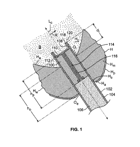
[0132] FIG. 1 schematically shows illustrative plug 100 for occlusion of an
access hole such as Hp
in a heart such as heart H. The occlusion may be performed on anatomy in or
about heart H. The
occlusion may be performed on anatomy in or about a chamber of heart H such as
chamber Fic.
Chamber H0 may be a left ventricle, a right ventricle, a left atrium or a
right atrium. The occlusion
may be performed on vasculature in or about heart H or on any other structure
in or about heart H.
Heart H may contract at a frequency.
[0133] Heart H may include pericardium Hp, myocardium Hp, and endocardium He.
Heart H may
include apex Hp. Heart H may include heart wall H. Heart wall H may include
one or more of
CA 02926142 2016-04-01
WO 2014/067021
PCT/CH2013/000189
pericardium Ho, myocardium Fin., and endocardium He. Heart wall Hw may include
a septum between
two cardiac atria. Heart wall Hw may include a septum between two cardiac
ventricles.
[0134] Access hole Ho may extend through heart wall H. Access hole Ho may
extend from exterior
orifice 0, at a exterior side of heart wall Hw to interior orifice Of at an
interior side of heart wall 1-1,4.
Exterior orifice 0, may be separated from interior orifice 0, by access hole
length ho.
[0135] Plug 100 may define central axis Lo. Central axis 1_, may define radial
direction R.
[0136] Plug 100 is shown inside delivery device 102. Delivery device 102 may
include catheter
104. Delivery device 102 may include pusher member 106. Plug 100 may be loaded
into delivery
device 102 prior to insertion of delivery device 102 into heart H.
[0137] Plug 100 may include cap 108. Cap 108 may have a configuration, such as
that shown in
FIG. 1, in which cap 108 blocks or partially blocks a fluid such as blood B
from contacting plug 100.
Cap 108 may obstruct or partially obstruct the flow of blood B toward plug
100. Cap 108 may
prevent or partially prevent plug 100 from expanding by absorption of blood B.
Cap 108 may
include material that may be viewed by non-visible-spectrum imaging
instruments.
[0138] Cap 108 may be disposed at or near distal end 110 of plug 100. Cap 108
may be supported
by longitudinal member 114. Longitudinal member 114 may be cannulated to
accommodate a
guidewire (not shown). Longitudinal member 114 may be present within cannula
116. Longitudinal
member 114 may be drawn proximally to retract cap 108 through cannula 116
after placement of
plug 100. When cap 108 is retracted, plug 100 may absorb blood B and expand in
hole Ho.
[0139] Cap 108 may be biased to extend radially outward in radial direction R.
Outer perimeter
118 of cap 108 may be deflected longitudinally against inner wall 112 of
catheter 104 when plug
100 is loaded into delivery device 102 prior to insertion into heart H. Distal
end 108 of delivery
device 102 may be delivered into access hole Ho.
[0140] Cap 108 may have any suitable flexible structure. For example, cap 108
may include a
mesh, a membrane, a membrane overlain mesh, a iris-like petals, a thin film or
any other suitable
structure. Cap 108 may be fixed to longitudinal member 114. Cap 108 may be
rigidly fixed to
longitudinal member 114. Cap 108 may be pivotably fixed to longitudinal member
114. Cap 108
may be fixed to longitudinal member 114 by an umbrella expansion/retraction
mechanism.
Cannula 116 may have a fluted distal end to receive cap 108 during retraction.
CA 02926142 2016-04-01
WO 2014/067021
PCT/CH2013/000189
21
[0141] One or both of longitudinal member 114 and cannula 116 may be withdrawn
through a
cannula or cannulae (neither shown) of pusher member 106.
[0142] Plug 100 may have length hp. Length hp may be a length that is selected
to be in proportion
to access hole length ho. Plug 100 is illustrated as being about 100% of
length ho.
[0143] Distal end 120 of catheter 104 is positioned a distance di distal from
interior orifice Oi of
access hole Ho. This places cap 108 distal interior orifice Oi.
[0144] FIG. 2 shows that catheter 104 may been drawn proximally to deploy cap
108 in chamber
Hc distal distal end 120 of catheter 104. Pusher member 106 may be held
relative to heart H so
that plug 100 remains in place during the drawing of catheter 104. Perimeter
118 of cap 108 may
extend away from axis Lc in radial direction R. Distal end 110 of plug 100 may
be maintained at a
position distal of interior orifice Oi.
[0145] FIG. 3 shows that delivery device 102 may be drawn proximally to bring
perimeter 118 of
cap 108 in contact with heart wall Hw adjacent interior orifice Oi. Heart wall
Hw adjacent interior
orifice Oi may include tissue of endocardium He. As catheter 104 is drawn
proximally, perimeter
118 of cap 108 may deflect to an angle a (from radial direction R) from
contact with heart wall Hw.
The deflection of perimeter 118 may signal that distal end 110 of plug 100 is
positioned at or near
interior orifice Oi. When perimeter 118 is at angle a, blood B may contact
distal end 110 of plug
100.
[0146] FIG. 4 shows that pusher member 106 may hold plug 100 in position in
access hole Ho
while catheter 104 is drawn proximally. Plug 100 may wick blood B into the
interior and more
proximal regions of plug 100. As distal end 120 of catheter 104 is drawn
proximally, increasing
region 122 of plug 100 expands to fill access hole Ho.
[0147] FIG. 5 shows plug 100 in access hole Ho. Cap 118 may be withdrawn
through cannula 116.
Longitudinal member 114 and cannula 116 may be withdrawn through pusher member
106 through
cannula or cannulae (neither shown).
[0148] FIG. 6 shows that catheter 104 may be completely withdrawn from access
hole Ho. Pusher
member 106 may hold plug 110 in place while pusher member 106 is retracted
from hole Ho. Plug
110 may hold itself in place by outward radial force on heart wall Hw while
pusher member is
retracted.
CA 02926142 2016-04-01
WO 2014/067021
PCT/CH2013/000189
22
[0149] FIG. 7 delivery device separated from plug 100. Plug 100 is retained in
access hole Ho.
Plug 100 may be retained entirely by outward radial forces against heart wall
Hw from absorption of
blood B in plug 100. Plug 100 may be retained partially by outward radial
forces against heart wall
Hw from absorption of blood B in plug 100. Plug 100 may be retained partially
by bonding to
against heart wall Hw. The bonding may involve glue. The bonding may involve
blood clotting. Plug
100 may be retained partially by engagement with heart H by one or more
anchors such as barbs or
tines.
[0150] FIG. 8 shows illustrative plug 800. Illustrative plug 800 may have one
or more features in
common with plug 100 (shown in FIG. 1). Plug 800 may include absorptive mass
802. Mass 802
may absorb fluid such as blood B (shown in FIG. 1). Absorption of the fluid
may cause mass 802 to
expand radially in direction R. Plug 800 may include veiling 804. Veiling 804
may surround mass
802. Veiling 804 may retain elements, such as fibers, of mass 802. Veiling 804
may provide
traction on Hw (shown in FIG. 1) when mass 802 exerts pressure against heart
wall Hw. The
traction may retain or help retain plug 802 in heart H (shown in FIG. 1) when
mass 802 exerts
pressure against heart wall Hw.
[0151] Veiling 804 may be expandable. Veiling 804 may have a fixed diameter.
When veiling 804
has a fixed diameter, veiling 804 may be folded into mass 802 during
compression of mass 802 into
the compressed state.
[0152] Veiling 804 or a separate veiling may cover distal face 810 of plug
800. Veiling 804 or a
separate veiling may cover proximal face 812 of plug 800. A veiling on one or
both of the terminal
faces may retain elements of mass 802. Such a veiling may attenuate a rate of
absorption of blood
B by mass 802.
[0153] A veiling on proximal face 812 may be removable attached.
[0154] A veiling on distal face 810 may be removably attached. The veiling may
attenuate the rate
of absorption of blood B at distal face 810 of mass 80 when plug 800 is in
heart chamber Hc.
[0155] Such a veiling may be retracted through a lumen (not shown) in plug
802. The lumen may
be the lumen of a cannula. The lumen may be formed directly in mass 802. The
lumen may be
formed by use of a die in pressing mass 802 into the compressed state.
[0156] FIG. 9 shows plug 900 in heart Plug 900
may have one or more features in common with
one or both of plugs 100 and 800. Plug 900 is lodged in access hole Ho. Plug
900 may include
CA 02926142 2016-04-01
WO 2014/067021
PCT/CH2013/000189
23
mass 902. Plug 900 may include veiling 904. Mass 902 may exert radially
outward pressure
against heart wall H'w to hold plug 900 in access hole H'o against pressure
from blood in chamber
H'c while heart H' is beating. Plug 900 extends longitudinally beyond heart
wall H'w in both the
distal and proximal directions.
[0157] FIG. 10 shows illustrative plug 1000 in a view similar to that taken
along lines 10-10 (shown
in FIG. 5). Plug 1000 may have one or more features in common with one or more
of plugs 100
(shown in FIG. 1), 800 (shown in FIG. 8) and 900 (shown in FIG. 9). Plug 1000
is shown in a state of
deployment similar to the state of deployment shown in FIG. 5.
[0158] Plug 1000 may include a mass. Plug 1000 may include a veil. Plug 1000
may include
marker 1002 that may be radiographically or acoustically viewable using
medical imaging
instrumentation. Marker 1002 may define a pattern. The pattern may be
distributed about plug
1000 so that during deployment of plug 1000, expansion of section 1004 of plug
1000 to radius re,
relative to restriction of section 1006 by catheter 1008 to radius rõ may be
observed.
[0159] Marker 1002 may be disposed circumferentially about plug 1000. Marker
1002 may
include one or more lobes such as lobe 1008 to allow marker 1002 to expand
with plug 1000.
Marker 1002 may include one or more "threads" such as thread 1010. Thread 1002
may be
disposed around the circumference of plug 1000.
[0160] When section 1004 expands to radius rõ the lobes in that section may
open. The lobes may
open to render a thread partially or wholly circumferential or circular. This
may provide a visual
distinction relative to the lobes in section 1006.
[0161] One or more threads may be disposed helically around plug 1000. Marker
1002 may
include discrete particles or objects in addition to or instead of threads.
[0162] Marker 1002 may be disposed about the mass of plug 1000. Marker 1002
may be disposed
about a veiling of plug 1000. Marker 1002 may be printed on the mass. Marker
1002 may be
woven into the mass. Marker 1002 may be printed on the veiling. Marker 1002
may be woven into
the veiling.
[0163] FIG. 11 shows illustrative plug 1100. Plug 1100 may have one or more
features in common
with one or more of plugs 100 (shown in FIG. 1), 800 (shown in FIG. 8), 900
(shown in FIG. 9) and
1000 (shown in FIG. 10). Plug 1100 may include longitudinal member 1104.
Longitudinal member
1104 may support distal cap 1106. Longitudinal member 1104 and distal cap 1106
may form a
CA 02926142 2016-04-01
WO 2014/067021
PCT/CH2013/000189
24
unitary member. Longitudinal member 1104 may support proximal cap 1108.
Longitudinal
member 1104 and proximal cap 1108 may form a unitary member. Longitudinal
member 1104 and
proximal cap 1108 may be joined by a releasable connection such as 1110.
Connection 1110 may
be a push-twist connection or any other suitable type of connection.
[0164] One or both of caps 1106 and 1108 may be biased in the longitudinal
direction so that a
perimeter of the caps presses against a terminal face of the corresponding
cap.
[0165] FIG. 12 shows illustrative apparatus 1200 for closing an access hole in
a heart. Apparatus
1200 may include delivery catheter 1202. Apparatus 1200 may include plug 1204.
Apparatus
1200 may include pusher member 1206.
[0166] Delivery catheter 1202 may have one or more features in common with one
or both of
delivery catheter 104 (shown in FIG. 1) and delivery catheter 1008 (shown in
FIG. 10).
[0167] Plug 1200 may have one or more features in common with one or more of
plugs 100 (shown
in FIG. 1), 800 (shown in FIG. 8), 900 (shown in FIG. 9), 1000 (shown in FIG.
10), and 1100 (shown
in FIG. 11).
[0168] Pusher member 1206 may have one or more features in common with pusher
member 106
(shown in FIG. 1).
[0169] FIG. 13 shows illustrative apparatus 1300 for closing an access hole in
a heart. Apparatus
1300 may include plug 1304. Apparatus 1300 may include pusher member 1306.
[0170] Plug 1304 may have one or more features in common with one or more of
plugs 100 (shown
in FIG. 1), 800 (shown in FIG. 8), 900 (shown in FIG. 9), 1000 (shown in FIG.
10), 1100 (shown in
FIG. 11) and 1204 (shown in FIG. 12).
[0171] Pusher member 1306 may have one or more features in common with one or
both of pusher
member 106 (shown in FIG. 1) and pusher member 1206 (shown in FIG. 12).
[0172] Plug 1304 may include distal cap 1308. Plug 1304 may include proximal
cap 1310. One or
both of distal cap 1308 and proximal cap 1310 may have one or more features in
common with one
or more of cap 108 (shown in FIG. 1) and caps 1106 and 1108 (shown in FIG.
11).
[0173] Plug 1304 may be releasable engaged to pusher member at coupling 1312.
[0174] FIG. 14 shows illustrative plug 1400. Plug 1400 may have one or more
features in common
with one or more of plugs 100 (shown in FIG. 1), 800 (shown in FIG. 8), 900
(shown in FIG. 9), 1000
(shown in FIG. 10), 1100 (shown in FIG. 11), 1204 (shown in FIG. 12) and 1304
(shown in FIG. 13).
CA 02926142 2016-04-01
WO 2014/067021
PCT/CH2013/000189
[0175] Plug 1400 may include one or more carriage loops such as carriage loop
1402. Carriage
loop 1402 may receive guidewire 1406 for delivery into an access hole such as
Ho in heart wall Hw.
Plug 1400 may be delivered to the access hole using a delivery catheter such
as 104. Plug 1400
may be delivered to the access hole without a delivery catheter.
[0176] FIG. 15 shows illustrative plug 1500 in heart H (shown in FIG. 1). Plug
1500 may have one
or more features in common with one or more of plugs 100 (shown in FIG. 1),
800 (shown in FIG. 8),
900 (shown in FIG. 9), 1000 (shown in FIG. 10), 1100 (shown in FIG. 11), 1204
(shown in FIG. 12),
1304 (shown in FIG. 13) and 1400 (shown in FIG. 14).
[0177] Plug 1500 may include distal cap 1502. Plug 1500 may include proximal
cap 1504. One or
both of caps 1502 and 1504 may have one or more features in common with one or
more of caps
108 (shown in FIG. 1), 1106 and 1108 (shown in FIG. 11), and 1308 and 1310
(shown in FIG. 13).
[0178] One or both of the caps may include a septum. The septum may be self-
sealing after
penetration by a needle.
[0179] Plug 1500 may include shaft 1506. Shaft 1506 may include a mass. Plug
1506 may include
a veiling. Shaft 1506 may be expandable by injection of a fluid through the
needle. Shaft 1506 may
expand to press against heart H by absorption of the fluid.
[0180] Plug 1500 may be delivered to access hole using one more of guidewire
1508, delivery
catheter 1510 and access device 1512. Access device 1512 may have an internal
lumen that has
an internal valve for obstructing blood flow from heart H. The valve may
permit passage of
instrumentation, prostheses, closure devices and other suitable objects
through access device
1512. Access device 1512 may be anchored to heart H. Access device 1512 may be
configured to
remain in access hole Ho without being anchored to heart H. Access device 1512
may be held in
access hole Ho manually by a practitioner.
[0181] FIG. 16 shows illustrative plug 1600. Plug 1600 may have one or more
features in common
with one or more of plugs 100 (shown in FIG. 1), 800 (shown in FIG. 8), 900
(shown in FIG. 9), 1000
(shown in FIG. 10), 1100 (shown in FIG. 11), 1204 (shown in FIG. 12), 1304
(shown in FIG. 13),
1400 (shown in FIG. 14) and 1504 (shown in FIG. 15).
[0182] Plug 1600 may include one or more electrically conductive members such
as 1602.
[0183] Electrically conductive member 1602 may be used to provide electrical
pulses to heart wall
Hw to change the contraction frequency. An electrically conductive member such
as 1602 may be
CA 02926142 2016-04-01
WO 2014/067021
PCT/CH2013/000189
26
placed in direct contact with heart wall Hw to provide the electrical pulses.
The energy may be
supplied via a lead such as 1604 from a source (not shown). The energy may be
supplied wirelessly
from the source. The source may be programmable via a control panel (not
shown). The source
110 may be incorporated into plug 1600. The source 110 may be implanted in a
patient near plug
1600. Source 110 may be or include a pacing device.
[0184] An electrically conductive member may have a distal end that is placed
in electrical
communication with epidermal tissue on the body in which the heart is
disposed.
[0185] An electrically conductive member may sense a native cardiac electric
field. A signal
corresponding to the field may be transmitted to the source. The signal may be
transmitted via
cable (not shown). The signal may be transmitted wirelessly. Plug 1600 may
include one or more
electrically conductive members that are wired to provide pulses to heart wall
Hõ, and one or more
electrically conductive members that are wired to transmit a native cardiac
electric field signal to an
electrocardiograph device.
[0186] Conductive member 1602 may be disposed about a mass of plug 1600.
Conductive
member 1602 may be disposed about a veiling of plug 1600. Conductive member
1602 may be
printed on the mass. Conductive member 1602 may be woven into the mass.
Conductive member
1602 may be printed on the veiling. Conductive member 1602 may be woven into
the veiling.
[0187] Thus, apparatus and methods for closing an access hole have been
provided. Persons
skilled in the art will appreciate that the present invention can be practiced
by other than the
described embodiments, which are presented for purposes of illustration rather
than of limitation.
[0188] The present invention is limited only by the claims that follow.