Note: Descriptions are shown in the official language in which they were submitted.
CA 02926382 2016-04-05
WO 2015/055825 PCT/EP2014/072339
1
METHOD FOR DIAGNOSIS OF PRIMARY HYPERALDOSTERONISM
FIELD OF THE INVENTION
The present invention relates to methods and kits for the diagnosis of primary
hyperaldosteron-
ism (PHA). In particular, the present invention relates to the use of a new
diagnostic parameter
that is composed of the ratio between the angiotensin II (Ang II or Ang 1-8)
level, in particular
the steady state equilibrium Ang II level, and the aldosterone level in a
biological sample, such
as e.g. plasma. The ratio of the two measured parameters is used to diagnose
PHA in patients
and has clear advantages over currently used diagnostic methods.
BACKGROUND OF THE INVENTION
PHA, also known as primary aldosteronism, is characterized by the
overproduction of the min-
eralocorticoid hormone aldosterone being not a result of excessive renin
secretion. Aldosterone
causes increase in sodium and water retention and potassium excretion in the
kidneys, leading
to arterial hypertension. The diagnosis of PHA in patients with arterial
hypertension is a signifi-
cant analytical challenge due to the interference of currently available tests
with anti-
hypertensive treatments and the insufficient diagnostic power of the employed
assays. PHA has
many causes, including adrenal hyperplasia and adrenal carcinoma. When it
occurs due to a
solitary aldosterone-secreting adrenal adenoma, which is a type of benign
tumor and is the
most frequent cause of PHA (66% of cases), it is known as Conn's syndrome.
Other causes of
PHA include bilateral idiopathic adrenal hyperplasia (30% of cases), primary
(unilateral) adrenal
hyperplasia (2% of cases), aldosterone-producing adrenocortical carcinoma (<1%
of cases),
familial hyperaldosteronism (FH), glucocorticoid-remediable aldosteronism (FH
type I, <1% of
cases), FH type II (<2% of cases) and ectopic aldosterone-producing adenoma or
carcinoma (<
0.1% of cases) (Williams textbook of endocrinology. (11th ed.). Philadelphia:
Saunders/Elsevier.
2008. ISBN 978-1-4160-2911-3.). However, due to the limited diagnostic
capabilities, data
about the prevalence of subforms of PHA are divergent. Recent studies indicate
that the preva-
lence of aldosteronism due to bilateral idiopathic adrenal hyperplasia (IAN)
is higher than had
previously been believed, for as many as 75% of PHA cases. Once diagnosed, PHA
can be
usually cured by a surgical intervention.
Measuring aldosterone alone is not considered adequate to diagnose primary
hyperaldosteron-
ism. It is known that in contrast to measuring the aldosterone levels alone,
the diagnostic speci-
ficity and sensitivity for detecting PHA can be improved by measuring renin
activity or concen-
tration and aldosterone and combining the two parameters to a arithmetic
ratio, the aldosterone-
to-renin ratio (ARR), which is currently used for diagnosis of PHA (Tiu S,
Choi C, Shek C, Ng Y,
Chan F, Ng C, Kong A (2005). "The use of aldosterone-renin ratio as a
diagnostic test for prima-
CA 02926382 2016-04-05
WO 2015/055825 PCT/EP2014/072339
2
ry hyperaldosteronism and its test characteristics under different conditions
of blood sampling".
J Clin Endocrinol Metab 90(1): 8. doi:10.1210/jc.2004-1149. PMID 15483077).
The Aldoste-
rone-to-renin ratio (ARR) is the mass concentration of aldosterone divided by
the renin activi-
ty and/or renin concentration in blood plasma. The Aldosterone-to-renin ratio
can be given in
ng/dL per ng/(mLih), that is, nanogram per decilitre of aldosterone per
nanogram per (millilitre x
hour) of renin. Also, it can be given in pmol/liter per pg/(literih), where
aldosterone is given in
molar concentration. The former can be converted to the latter by multiplying
with 27.6. Also, the
inverse value is occasionally given, that is, the renin-to-aldosterone ratio,
the value of which is
the multiplicative inverse of the aldosterone-to-renin ratio. Ratios between
aldosterone and ren-
in might also be calculated using other concentration units (mass unit per ml
and/or amount unit
per ml) for any of the two parameters resulting in different absolute values
for the ratio while
containing the similar information. The concentration of renin used for
calculation of the ARR
might also be given in pg Ul E/ml, which is a unit frequently used in clinical
diagnostics that also
reflects the renin concentration.
The cutoff (or threshold) of normal individuals from those with primary
hyperaldosteronism
based on the ARR is significantly affected by the conditions of testing, such
as body position
and time of day. On average, an ARR cutoff of 23.6 ng/dL per ng/(mLih),
expressed in alterna-
tive units as 650 pmol/liter per pg/(literih), has been estimated to have a
sensitivity of 97%
and specificity of 94% (Tiu et al, cited above). An ARR value in an individual
that is higher than
the cutoff is used in the prior art to indicate primary hyperaldosteronism.
If the inverse ratio (i.e. renin-to-aldosterone) ratio is used, a value lower
than the cutoff is con-
sidered to indicate primary hyperaldosteronism.
However, the broad range of ARR displayed by patients suffering from PHA
allows no clear-cut
and reliable discrimination between essential hypertension and PHA, thus
leading to false-
positive and/or false-negative diagnostic results and treatment decisions.
Special medication
and dietary requirements together with a sophisticated testing protocol
involving saline infusion
are required to improve the diagnostic power of the ARR in a confirmatory
testing procedure
subsequent to ARR screening.
It is suggested by endocrine societies to screen for PHA in patient groups at
risk including pa-
tients with Joint National Commission stage 2 (>160 ¨179/100 ¨109 mm Hg),
stage 3 (>180/110
mm Hg), or drug-resistant hypertension; hypertension and spontaneous or
diuretic-induced
hypokalemia; hypertension with adrenal incidentaloma; or hypertension and a
family history of
early-onset hypertension or cerebrovascular accident at a young age (<40 yr).
Hypertensive
CA 02926382 2016-04-05
WO 2015/055825 PCT/EP2014/072339
3
first-degree relatives of patients with PHA show also an increased risk for
PHA. According to
clinical guidelines, the standard way to the diagnosis of PHA till the
decision of the curing sur-
gery is considered to be laborious and represents several risks for the
patients.
The rational behind the measurement of the ARR to diagnose PHA lies behind the
physiological
pathways responsible for aldosterone secretion in the adrenal cortex. Renin is
a key enzyme of
the renin-angiotensin-system (RAS) producing angiotensin I from
angiotensinogen, which is
converted to Ang II via other peptidases. Ang II is known to bind to AT1-
Receptors (AT1R) lead-
ing to the secretion of aldosterone, which results in it's physiologic effects
in the kidney and oth-
er tissues. Under healthy conditions, the RAS regulates plasma aldosterone
levels. Under the
condition of PHA, aldosterone production becomes partially independent of the
RAS, meaning
that renin is not further necessary to maintain aldosterone production. The
measurement of the
ARR tries to make use of this deregulation of renin and aldosterone. PHA
patients usually have
increased plasma aldosterone levels. Therefore, some investigators require
elevated aldoste-
rone levels in addition to elevated ARR for a positive screening test for PHA
(usually aldoste-
rone >15 ng/dI).
The diagnostic process for PHA is started with an ARR case detection test in
patient groups
specified above (John W. Funder et al.; J Clin Endocrinol Metab. September
2008, 93(9):3266 ¨
3281). In case this first measured ARR value exceeds a certain threshold, the
patient is sub-
jected to further testing in order to assure the validity of the obtained
results. Of note, the exact
value for the ARR threshold is still discussed in the literature, due to the
frequent occurrence of
false negatives and positives.
While there are few anti-hypertensive drugs that are thought to have only
limited effects on the
measured ARR value, many anti-hypertensive drugs are known to interfere
strongly with ARR
testing. The main cause of interference is represented by strong impact of
these drugs on renin
concentration and renin activity, leading to altered ARR results. As a
consequence, a wash out
phase of anti-hypertensive drugs is usually necessary before confirmation
testing, which is a
considerable risk for the hypertensive patients. Confirmation testing itself
consists of a time con-
suming and cost intensive clinical procedure that is intended to reduce the
renin levels of pa-
tients in response to osmotic or drug challenges in combination with ARR
testing before and
after the procedure.
A very common PHA confirmation test is a saline infusion test, where two
liters of 0.9% saline is
administered to the patient in the course of 4 hours. The volume increase
should result in a de-
crease in renin activity and concentration. Post test aldosterone levels below
50 pg/ml are
CA 02926382 2016-04-05
WO 2015/055825 PCT/EP2014/072339
4
thought to indicate the absence of PHA, while post test aldosterone levels
above 100 pg/m1 are
interpreted as a probable sign of PHA. Values between 50 pg/ml and 100 pg/ml
are regarded to
be indeterminate (John W. Funder et al.; J Olin Endocrinol Metab. September
2008, 93(9):3266
¨3281).
Positive confirmation testing triggers further clinical tests including
adrenal imaging techniques,
such as e.g. computed tomography (CT) and adrenal vein sampling (AVS) to
determine the
source of excessive and renin independent aldosterone production. Once the
subtype is classi-
fied, unilateral adrenalectomy or treatment with mineralocorticoid receptor
antagonists can be
performed.
The key step in the diagnostic process is case detection in high-risk
hypertensive patients. The
ARR as a case detection test could easily show false positive and negative
outcomes as among
hypertensive patients the ARR value distribution in PHA patients was shown to
overlap with the
ARR value distribution of in non-PHA patients (Gary L. Schwartz and Stephen T.
Turner; Clini-
cal Chemistry 51, No. 2, 2005), which could easily lead to unnecessary
mistreatment of pa-
tients. In case of false positives, these mistreatments can result in severe
complications as a
drug wash out phase of several weeks in a patient being hypertensive despite
taking at least
three anti-hypertensive drugs pose a significant risk of fatal cardiovascular
events during this
period of uncontrolled blood pressure. In addition to that, confirmation
testing usually requires
hospitalization and constant monitoring by physicians being time and money
consuming for the
patient and the healthcare system. In case of a false negative result, the PHA
patient will con-
tinue to live with resistant hypertension, which has a fatal prognosis due to
a strongly increased
risk for life threatening cardiovascular events like strokes or heart attacks.
The present invention provides a method for the diagnosis of primary
hyperaldosteronism in a
subject, comprising obtaining a biological sample from the subject, measuring
the aldosterone
level and the Ang II level, and calculating the ratio thereof (aldosterone-to-
angiotensin II ratio,
AA2R). Said method has substantial advantages over the above described
currently used ARR-
based diagnostic methods.
BRIEF DESCIPTION OF THE INVENTION
In one aspect, the present invention relates to a method for the diagnosis of
primary hyperal-
dosteronism in a subject, comprising obtaining a biological sample from the
subject, measuring
the aldosterone level and the Ang II level, and calculating the ratio thereof
(aldosterone-to-
angiotensin II ratio, AA2R). In a second aspect, the present invention relates
to a kit for diag-
nosing PHA, comprising an Ang II standard, an aldosterone standard, and
optionally further
CA 02926382 2016-04-05
WO 2015/055825 PCT/EP2014/072339
comprising a manual and/or further components.
BRIEF DESCRIPTION OF THE FIGURES
Figure 1: Left panel: Ang II to PRA-Ratio in 14 healty volunteers. Right
panel: Ang II to PRC-
Ratio in plasma of healthy untreated and ACE-Inhibitor treated healthy
volunteers.
Figure 2: Left panel: Ang II to PRA-Ratio in untreated and ACE-Inhibitor
treated healthy volun-
teers (n=14). Middle panel: PRA in untreated and ACE-Inhibitor treated healthy
volunteers
(n=14). Right panel: Ang II to PRC-Ratio in untreated and ACE-Inhibitor
treated healthy volun-
teers (n=4).
Figure 3: Comparison of ARR and AA2R for one non-PHA patient and one confirmed
PHA pa-
tient pre and post 4h saline infusion confirmation test. Left panel: Values
are given as percent-
age of pre infusion signal for non-PHA patient. Right panel: Comparison of ARR
and AA2R spe-
cific discrimination factors between non-PHA and PHA patient are shown for
each time point.
Figure 4: Impact of anti-hypertensive treatments on active renin
concentration. Plasma samples
were collected from healthy volunteers before (Predose) and 4h post
administration of a single
dose of an ACE inhibitor (left), renin inhibitor (middle) or angiotensin
receptor blocker (ARB,
right). Mean values of 5 healthy volunteers +/- SEM are shown in the graphs.
Figure 5: Comparison of the impact of RAS blocker administration on the ARR
(upper panel)
and the AA2R (lower panel). Plasma samples were collected from healthy
volunteers before
(Predose) and 4h post administration of a single dose of an ACE inhibitor
(left), renin inhibitor
(middle) or angiotensin receptor blocker (ARB, right). Plasma aldosterone
concentration, equi-
librium angiotensin II concentration and active renin concentration were
measured and the ARR
and the AA2R was calculated for each sample. For calculation of the ARR,
aldosterone concen-
trations in ng/L were divided by the plasma active renin concentration in
ng/L. For calculation of
the AA2R, aldosterone concentrations in pmol/L were divided by Ang ll
concentrations in
pmol/L. Mean values of 5 healthy volunteers +/- SEM are shown in the graphs.
DETAILED DESCRIPTION OF THE INVENTION
Currently available methods for the diagnosis of PHA in patients make use of
the correlation
between plasma renin activity or plasma renin concentration and the plasma
aldosterone con-
centration. Calculation of the aldosterone to renin ratio (ARR) was shown to
allow a partial dis-
crimination between non-PHA and PHA patients. However, false positive as well
as false nega-
tive results are frequent. Renin is known to be responsible for the production
of Angiotensin I
CA 02926382 2016-04-05
WO 2015/055825 PCT/EP2014/072339
6
(Ang 1), which serves as a substrate for Ang 11 formation by other proteolytic
enzymes like chy-
mase or angiotensin-converting enzyme (ACE). Ang 11 is the main effector
hormone of the RAS
and is mainly responsible for RAS mediated physiologic functions including the
regulation of
fluid balance and blood pressure. It is widely accepted that renin activity
and concentration
serve as surrogate markers for the activity of the RAS (Swales JD and Thurston
HJ; Clin Endo-
crinol Metab. 1977 Jul; 45(1):159-63), which is used to support the use of the
ARR as a diag-
nostic marker for PHA.
Surprisingly it turned out that Ang 11 levels measured in equilibrated plasma
samples (i.e. Ang 11
levels measured according to the steady state equilibrium (SSE) method as
described below,
also called equilibrium Ang 11 levels) show a poor correlation with plasma
renin activity (PRA)
and plasma renin concentration (PRC), indicated by a huge variability in the
Ang II to PRA and
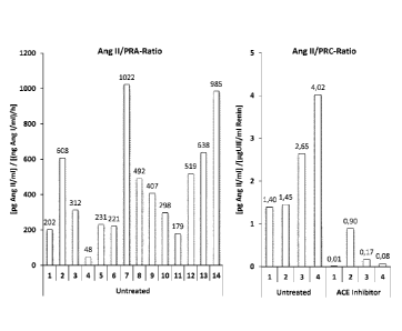
Ang 11 to PRC ratios when individual subjects are compared (Figure 1, left and
right panel).
Blood was collected from 14 healthy volunteers without anti-hypertensive
treatment. Plasma
was isolated by centrifugation and equilibrium Ang 11 levels were measured in
stabilized sam-
ples following 60 min of plasma equilibration at 37 C. The methods of
measuring RAS levels in
steady state equilibrium are further described in WO 2013/182237. For
measurement of PRA,
similar samples were subjected to an Angiotensin I formation assay as
described (Bystrom et
al., Clin. Chem. 56(2010), 1561-1569]). Angiotensin I was quantified by mass
spectrometry and
the plasma renin activity was calculated in (ng Ang 1/m1)/h. The graph shows
the equilibrium Ang
11 to renin activity ratio. The ratios of the 14 donors were in a range
between 48 and 1022 [pg
Ang 11/m1]/[(ng Ang 1/m1)/h], which is 11% and 232% of the mean of all 14
donors.
In a second approach, plasma renin concentration (PRC) was determined in 4
untreated and 4
ACE inhibitor treated volunteers using a commercially available and clinically
applied antibody
based renin assay (Diasorin). Equilibrium Ang II levels were measured by mass
spectrometry
following 60 min of plasma equilibration at 37 C and sample stabilization. The
equilibrium Ang II
to PRC ratio was calculated and shown in the graph for untreated and ACE
inhibitor treated
patients. The unit of the shown ratio is [pg Ang 11/ml]/[pgUIE/m1 Renin]. In
all patient cohorts
investigated, the Ang 11 to PRA and the Ang II to PRC ratio were found to be
highly variable.
Moreover, ACE-Inhibitor treatment resulted in a significant reduction of both
the Ang II to PRC
(Figure 2, right panel) and Ang II to PRA ratios (Figure 2, left panel).
In conclusion, a similar renin concentration and/or activity result(s) in
different Ang II concentra-
tions in individual donors indicating that renin activity and/or concentration
is a poor marker for
physiologic activity of the RAS. As a consequence, the ARR insufficiently
displays the RAS ac-
tivity related aldosterone level, putting a question mark over the suitability
of the ARR as a
screening tool for PHA. These considerations further explain the limitations
in PHA case detec-
tion via ARR measurements, which is prone to false positive and false negative
results and
highly dependent on the therapeutic background of patients (John W. Funder et
al.; J Clin En-
CA 02926382 2016-04-05
WO 2015/055825 PCT/EP2014/072339
7
docrinol Metab. September 2008, 93(9):3266-3281;, and Gary L. Schwartz and
Stephen T.
Turner; Clinical Chemistry 51, No. 2, 2005).
Moreover Ang ll to PRA and Ang II to PRC ratios are significantly affected by
ACE-Inhibitor
treatment (Figure 1, right panel; Figure 2, left and right panel).
Surprisingly, while ACE-Inhibitor
treatment increases renin activity and concentration, the Ang II to renin
ratio was significantly
reduced (Figure 2, middle panel).
Therefore we concluded that the poor correlation of renin concentration and
activity with Ang II
levels in the absence and presence of an anti-hypertensive drug like an ACE-
Inhibitor might
cause the limited predictive power of the ARR for the diagnosis of PHA.
In contrast, the present invention relates to a method for the diagnosis of
primary hyperaldoste-
ronism in a subject, comprising measuring the aldosterone level and the
angiotensin II (Ang II)
level in a biological sample from the subject, and calculating the ratio
between the aldosterone
level and the Ang II level (aldosterone-to-angiotensin II ratio, AA2R). In one
embodiment, the
present invention relates to a method for the diagnosis of primary
hyperaldosteronism in a sub-
ject, comprising obtaining a biological sample from the subject, measuring the
aldosterone level
and the angiotensin II level, and combining them to an arithmetic ratio
(aldosterone-to-
angiotensin ll ratio, AA2R).
The term "level" as used herein refers to the concentration of a substance
(e.g. a component of
the RAS, such as renin, Ang II, aldosterone etc.) in a biological sample, such
as e.g. blood,
plasma or serum. Said concentration may be given in mol/L, mmol/ml, pg UIE/ml,
ng/ml, pg/ml
or any other concentration unit.
In an embodiment, a high AA2R indicates primary hyperaldosteronism and a low
AA2R indi-
cates no primary hyperaldosteronism. In an embodiment, a high AA2R as compared
to the
AA2R of one or more confirmed non-PHA subjects indicates primary
hyperaldosteronism and/or
a low AA2R as compared to the AA2R of one or more confirmed PHA subjects
indicates no pri-
mary hyperaldosteronism. In an embodiment, an AA2R similar to the AA2R of one
or more con-
firmed non-PHA patients indicates no primary hyperaldosteronism and/or an AA2R
similar to the
AA2R of one or more confirmed PHA patients indicates primary
hyperaldosteronism. In an em-
bodiment, the term "similar" as used above shall mean that the difference
between the respec-
tive ratios (i.e. the AA2Rs) is less than 100%, 90%, 80%, 70%, 60%, 50%, 40%,
30%, 20%,
10%, or 5%.
The AA2R turned out to show an improved positive to negative ratio as shown by
the compari-
CA 02926382 2016-04-05
WO 2015/055825 PCT/EP2014/072339
8
son of one non-PHA hypertensive patient with a hypertensive PHA patient pre
and post a saline
infusion test (SIT) (Example 1, Figure 3). In the left panel, for each
individual test (ARR and
AA2R), test results were related to the pre-SIT value and expressed in
percent. The PHA pa-
tient was clearly positive according to ARR test criteria, with pre and post
saline infusion test
(SIT) plasma aldosterone levels of 471 pg/ml and 548 pg/ml respectively, and a
resulting pre
and post ARR (Aldosterone to PRO ratio) of 588.8 and 421.5 respectively (PRO
pre SIT: 0.8 pg
UIE/m1; PRO post SIT: 1.3 pg UIE/m1). The non-PHA patient showed pre and post
SIT plasma
aldosterone concentration of 190 pg/ml and 64 pg/ml respectively with a pre
and post SIT ARR
of 19.2 and 16.4 respectively, which is clearly negative according to test
criteria (John W. Fun-
der et al.; J Olin Endocrinol Metab. September 2008, 93(9):3266 ¨3281).
Moreover, assay spe-
cific discrimination factors were calculated as a measure of diagnostic
performance (or diagnos-
tic power) and compared for ARR and AA2R (Figure 3, right panel). The
discrimination factor is
useful to compare different tests by measuring two identical samples with both
tests, of which
one is a true negative and one is a true positive sample. The discrimination
factor represents
the ratio between the true positive signal and the true negative signal. A
high discrimination fac-
tor means that the difference between a true negative and a true positive
sample is high, which
implies a better diagnostic performance compared with a test with a lower
discrimination factor
obtained for similar samples. When relating the pre-SIT ARR value of the
confirmed PHA pa-
tient to the pre-SIT ARR value of the confirmed non-PHA patient, a
discrimination factor of 30.7
between the confirmed non-PHA patient (true negative) and the confirmed PHA
patient (true
positive) is obtained. The post-SIT discrimination factor between non-PHA and
the PHA patient
was 25.5 when analyzed by ARR.
Surprisingly, the analysis of the identical samples from the same two patients
by AA2R revealed
a discrimination factor of 150.8 for pre-SIT samples and a discrimination
factor between the
non-PHA and the PHA patient of 325.0 for post-SIT samples (Figure 3, right
panel). We con-
clude that using AA2R instead of ARR strongly increases the factor between
negative and posi-
tive signals, leading to a markedly increased diagnostic performance.
In one embodiment of the present invention, the ratio of values between one or
more confirmed
PHA positive subjects and one or more confirmed PHA negative subjects (i.e.
the discrimination
factor) based on the AA2R is higher than between the same data sets based on
the ARR. Ac-
cordingly, in an embodiment, the ratio between the AA2R of one or more
confirmed PHA posi-
tive subjects and the AA2R of one or more confirmed PHA negative subjects is
higher than the
ratio of the same data set based on the ARR. In other words, the ratio between
the AA2R of one
or more confirmed PHA positive subjects and the AA2R one or more confirmed PHA
negative
subjects is higher than the ratio between the ARR of the same one or more
confirmed PHA
CA 02926382 2016-04-05
WO 2015/055825 PCT/EP2014/072339
9
positive subjects and the ARR the same one or more confirmed PHA negative
subjects.
The term "discrimination factor" as used herein may refer to the ratio of the
diagnostic parame-
ter (e.g. ARR or AA2R) of one or more confirmed PHA subjects (or confirmed PHA
positive sub-
jects) to the diagnostic parameter (e.g. ARR or AA2R) of one or more confirmed
non-PHA sub-
jects (or confirmed PHA negative subjects), or to one or more mean values of
such parameters,
e.g. a mean value of the ARR or AA2R of a cohort of confirmed PHA subjects and
a mean value
of the ARR or AA2R of a cohort of confirmed non-PHA subjects. Accordingly, the
discrimination
factor is the ratio of the ARR of one or more confirmed PHA subjects to the
ARR of one or more
confirmed non-PHA subjects, or the AA2R of one or more confirmed PHA subjects
to the AA2R
of one or more confirmed non-PHA subjects. The discrimination factor is a
measure for the di-
agnostic performance (or diagnostic power) of the respective diagnostic
parameter or test. The
higher the ratio, the higher is the diagnostic power, and the lower is the
risk of false positive
and/or false negative results. The term "confirmed PHA subject" refers to a
subject suffering
from PHA and having been diagnosed as positive either by the conventional
methods (e.g. a
first screening test measuring the ARR, and at least one confirmation test
measuring the ARR a
second or third or more times, optionally prior to and after a SIT), or having
been diagnosed as
positive by the methods according to the present invention (e.g. the AA2R
measurement not
requiring any confirmation testing), and/or may even have been confirmed by
surgery and/or
imaging techniques (e.g. CT and/or adrenal vein sampling.) The term "confirmed
non-PHA sub-
ject" refers to a subject not suffering from PHA and having been diagnosed as
negative either
by the conventional methods (e.g. a first screening test measuring the ARR,
and optionally one
or more confirmation tests measuring the ARR a second or third or more times,
optionally prior
to and after a SIT), or having been diagnosed as negative by the methods
according to the pre-
sent invention (e.g. the AA2R measurement not requiring any confirmation
testing or other con-
firmation measures, such as e.g. imaging techniques). One or more samples from
one or more
"confirmed non-PHA subjects" or from one or more "confirmed PHA subjects" can
be used as
normal controls in the methods of the invention for comparison with the sample
under investiga-
tion, i.e. one or more samples from one or more "confirmed non-PHA subjects"
can be used as
negative control, and/or one or more samples from one or more "confirmed PHA
subjects" can
be used as positive control. For example, a high AA2R as compared to the AA2R
of one or
more confirmed non-PHA subjects indicates primary hyperaldosteronism, and/or a
low AA2R as
compared to the AA2R of one or more confirmed PHA subjects indicates no
primary hyperal-
dosteronism. If two or more samples are used as negative and/or positive
control, the mean
value (i.e. the arithmetic mean) or the median of the corresponding samples
may be deter-
mined. Based on such control samples or values thereof, a discrimination
threshold (or cutoff)
may be determined, above which the subject is diagnosed to be PHA positive,
and below which
CA 02926382 2016-04-05
WO 2015/055825 PCT/EP2014/072339
the subject is diagnosed to be PHA negative. Such threshold may also be
determined based on
the AA2R value distribution in a patient cohort comprising PHA positive and
PHA negative sub-
jects. The threshold may be determined separately for different patient
cohorts (e.g. different
thresholds may be determined for patient groups treated with different anti-
hypertensive drugs).
Any of the parameters described above that may be used to determine a
threshold (or compari-
son level) can be used either alone or in combination with one or more of the
other parameters
in order to result in a final threshold.
Although the methods of the present invention may not require any confirmation
testing, as al-
ready stated above, confirmation testing may nevertheless be done (e.g. if
desired by a physi-
cian or patient). Furthermore, the methods of the invention itself may be used
as confirmation
testing, i.e. applied after a first screening test has been done with
classical methods based on
the ARR.
In an embodiment, the discrimination factor for one or more given data pairs
or data sets (e.g.
one or more suspected or confirmed PHA subjects compared to one or more
suspected or con-
firmed non-PHA subjects, or the mean value of a cohort of suspected or
confirmed PHA sub-
jects compared to the mean value of a cohort of suspected or confirmed non-PHA
subjects) as
determined based on the AA2R is higher than the discrimination factor for the
same data pairs
or data sets as determined based on the ARR, in particular based on the ARR of
a screening
test (i.e. the first measurement of the ARR, and/or the ARR prior to any
confirmation testing),
and/or based on the ARR of a confirmation test (i.e. the second or further
measurement of the
ARR, and/or the ARR following any screening testing). In one embodiment, the
discrimination
factor for one or more given data pairs or data sets (e.g. one or more
suspected or confirmed
PHA subjects compared to one or more suspected or confirmed non-PHA subjects,
or the mean
value of a cohort of suspected or confirmed PHA subjects compared to the mean
value of a
cohort of suspected or confirmed non-PHA subjects) as determined based on the
AA2R is high-
er than, in particular significantly higher than, the discrimination factor
for the same data pairs or
data sets as determined based on the ARR, in particular based on the ARR of a
screening test
(i.e. the first measurement of the ARR, and/or the ARR prior to any
confirmation testing), and/or
based on the ARR of a confirmation test (i.e. the second or further
measurement of the ARR,
and/or the ARR following any screening testing). In one embodiment, the
discrimination factor
for one or more given data pairs or data sets (e.g. one or more suspected or
confirmed PHA
subjects compared to one or more suspected or confirmed non-PHA subjects, or
the mean val-
ue of a cohort of suspected or confirmed PHA subjects compared to the mean
value of a cohort
of suspected or confirmed non-PHA subjects) as determined based on the AA2R is
at least
20%, 30%, 40%, 50%, 60%, 70%, 80%, 90%, 100%, 150%, 200%, 250%, 300%, 350%,
400%,
CA 02926382 2016-04-05
WO 2015/055825 PCT/EP2014/072339
11
450%, or 500% higher than the discrimination factor for the same data pairs or
data sets as de-
termined based on the ARR, in particular based on the ARR of a screening test
(i.e. the first
measurement of the ARR, and/or the ARR prior to any confirmation testing),
and/or based on
the ARR of a confirmation test (i.e. the second or further measurement of the
ARR, and/or the
ARR following any screening testing). In one embodiment, the discrimination
factor for one or
more given data pairs or data sets (e.g. one or more suspected or confirmed
PHA subjects
compared to one or more suspected or confirmed non-PHA subjects, or the mean
value of a
cohort of suspected or confirmed PHA subjects compared to the mean value of a
cohort of sus-
pected or confirmed non-PHA subjects) as determined based on the AA2R is at
least 50 per-
cent, two-fold, three-fold, four-fold, five-fold, six-fold, seven-fold, eight-
fold, nine-fold, or ten-fold
higher than the discrimination factor for the same data pairs or data sets as
determined based
on the ARR, in particular based on the ARR of a screening test (i.e. the first
measurement of the
ARR, and/or the ARR prior to any confirmation testing), and/or based on the
ARR of a confirma-
tion test (i.e. the second or further measurement of the ARR, and/or the ARR
following any
screening testing).
The term "suspected PHA subject" refers to a subject suspected to suffering
from PHA and hav-
ing not yet been diagnosed as positive either by the conventional methods
(e.g. a first screening
test measuring the ARR, and at least one confirmation test measuring the ARR a
second or
third or more times, optionally prior to and after a SIT), or not having been
diagnosed as positive
by the methods according to the present invention (e.g. the AA2R measurement
not requiring
any confirmation testing), or having been diagnosed previously but with no
clear outcome,
and/or the previous diagnosis requiring further confirmation. The term
"suspected non-PHA sub-
ject" refers to a subject suspected to not suffering from PHA and having not
yet been diagnosed
as negative either by the conventional methods (e.g. a first screening test
measuring the ARR,
and optionally one or more confirmation tests measuring the ARR a second or
third or more
times, optionally prior to and after a SIT), or not yet having been diagnosed
as negative by the
methods according to the present invention (e.g. the AA2R measurement not
requiring any con-
firmation testing), or having been diagnosed previously but with no clear
outcome, and/or the
previous diagnosis requiring further confirmation.
A patient cohort might be subjected to pre-selection criteria (e.g.: minimal
blood pressure, mini-
mal aldosterone level, certain drug treatment) prior comparison of selectivity
and/or sensitivity
between AA2R and ARR. For example, some investigators conducting ARR based
screening
tests require a minimal aldosterone level of 15 ng/dI for a positive screening
test result, which
might result in an altered sensitivity and/or specificity compared to a non
pre-selected patient
cohort.
CA 02926382 2016-04-05
WO 2015/055825 PCT/EP2014/072339
12
In one embodiment, the specificity of the method of the invention in a defined
patient cohort is
equal or higher than the specificity of the classical ARR methods in the same
patient cohort, in
particular based on the ARR of a screening test (i.e. the first measurement of
the ARR, and/or
the ARR prior to any confirmation testing). The specificity may be given in
percent, wherein
number of true-negatives
specificity [%] = = 100
(number of true-negatives + number of false-positives)
In one embodiment, the specificity of the method of the invention is higher
than the specificity of
the classical ARR methods, in particular significantly higher than the
classical ARR methods. In
one embodiment, the specificity is higher than 93%, 94%, 95%, 96%, 97%, 98%,
or 99%. In one
embodiment, the specificity of the method is at least 94%, 95%, 96%, 97%, 98%,
or 99%. In
one embodiment, the specificity of the method is 100%.
In one embodiment, the sensitivity of the method of the invention in a defined
patient cohort is
equal or higher than the sensitivity of the classical ARR methods in the same
patient cohort, in
particular based on the ARR of a screening test (i.e. the first measurement of
the ARR, and/or
the ARR prior to any confirmation testing).
The sensitivity may be given in percent, wherein
number of true-positives
sensitivity [%] - = 100
(number of true-positives + number of false-negatives)
In one embodiment, the sensitivity of the method of the invention is higher
than the sensitivity of
the classical ARR methods, in particular significantly higher than the
classical ARR methods. In
one embodiment, the sensitivity of the method is at least 93%, 94%, 95%, 96%,
97%, 98%, or
99%. In one embodiment, the sensitivity of the method is higher than 93%, 94%,
95%, 96%,
97%, 98%, or 99%. In one embodiment, the sensitivity of the method is 100%.
In an embodiment, at least 90%, 91%, 92%, 93%, 94%, 95%, 96%, 97%, 98%, or 99%
of all
confirmed PHA subjects have a higher AA2R than at least 90%, 91%, 92%, 93%,
94%, 95%,
CA 02926382 2016-04-05
WO 2015/055825 PCT/EP2014/072339
13
96%, 97%, 98%, or 99% of all confirmed non-PHA subjects. With the methods and
kits of the
present invention, it is possible to clearly differentiate between PHA and non-
PHA subjects,
since the degree of overlap of the AA2R value distribution of PHA and non-PHA
subjects is
substantially lower than the overlap of the ARR values of PHA and non-PHA
subjects. In an
embodiment, the overlap is 10% or less, 9% or less, 8% or less, 7% or less, 6%
or less, 5% or
less, 4% or less, 3% or less, 2% or less, 1% or less.
Subjects that are suspected to suffer from PHA are usually under anti-
hypertensive treatment.
For example, the subject has received and/or receives one or more
pharmaceutical composi-
tions (herein also referred to as drugs) or treatments, in particular anti-
hypertensive pharmaceu-
tical compositions and/or treatments, at the time the diagnosis of PHA is
made. The interference
of the ARR test with anti-hypertensive treatments represents a well-known
obstacle for the di-
agnostics of PHA in hypertensive patients. Resistant hypertensive patients
represent a high-risk
group for PHA and are by definition treated with at least three anti-
hypertensive drugs simulta-
neously, while still suffering from pathologically elevated blood pressure.
Many of the clinically
used anti-hypertensive drugs are known to have an impact on renin and
aldosterone levels,
while renin is usually stronger affected than aldosterone, which results in
unpredictable shifts in
ARR test results leading to false negative and false positive diagnostic
decisions (John W. Fun-
der et al.; J Olin Endocrinol Metab. September 2008, 93(9):3266 ¨3281).
A widely used group of anti-hypertensive agents in clinical use is the group
of RAS blockers.
RAS blockers interfere with the RAS in order to either reduce the level of Ang
II (e.g. Renin-
Inhibitors, ACE-Inhibitors) or block the action of Ang II at the ATi-Receptor
(e.g. Angiotensin-
Receptor-Blockers, ARBs). In addition to RAS blockers, RAS activators may be
used as anti-
hypertensive agents. For example, ACE2 can be administered to treat
hypertension, as de-
scribed e.g. in W02004/000367 and W02008/151347.
Drugs and/or treatments that affect, especially increase, renin activity
and/or renin concentration
affect, especially decrease, the ARR. The diagnostic power of the ARR is
decreased by such
drugs and/or treatments. Treatment of 5 healthy volunteers with single doses
of different anti-
hypertensive agents (Example 2) resulted in a highly significant, 3 to 10-fold
increase in the
concentration of active plasma renin (Figure 4). These drug-induced changes in
active renin
concentration profoundly affected the ARR in these individuals.
The administration of a single dose of an ACE inhibitor (10 mg Enalapril), a
renin inhibitor (150
mg Aliskiren) or an ARB (50mg Losartan) resulted in a highly significant and
profound decrease
in the ARR (Figure 5, upper panel), which results in a strongly reduced
diagnostic power of the
ARR in the presence of these anti-hypertensive drugs. Low ARR values that are
caused by anti-
hypertensive treatments are lead to false negative outcomes. In contrast to
the ARR, most anti-
hypertensive drugs did not significantly affect the AA2R, except for ARBs
(Figure 5, lower pan-
CA 02926382 2016-04-05
WO 2015/055825 PCT/EP2014/072339
14
el). The comparison of the AA2R before and after drug administration revealed
that neither the
administration of an ACE inhibitor, nor the administration of a renin
inhibitor resulted in signifi-
cant changes in the AA2R, while the ARR was significantly decreased in
response to drug
treatment. The ARB, that prevents the binding of Ang II to AT1 receptors
therefore resulting in
Ang II accumulation together with an increase in active renin concentration,
was the only drug
resulting in a significant decrease of the AA2R, while the ARR is
significantly affected by every
anti-hypertensive drug tested.
Renin activity and/or renin concentration is controlled via multiple
physiologic regulatory mecha-
nisms. Any drug that interferes with such regulatory mechanism might affect
renin activity and/or
concentration. For example, diuretics are a common class of anti-hypertensive
drugs that re-
duce blood pressure by enhancing diuresis. Enhanced diuresis results in
increased in renin ac-
tivity and/or concentration. Therefore, the treatment of subjects with
diuretics decreases the
ARR. Examples for diuretics in clinical use are furosemide, torsemide,
hydrochlorothiazide,
azetazolamide, methazolamide, eplerenone, spironolactone, amiloride, and
triamterene.
The effects on renin activity and/or concentration might also be mediated
indirectly by a block-
ade of one or more steps in the RAS cascade that are downstream of renin.
For example, the blockade of the conversion of Ang I to Ang II by ACE
inhibitors is relevant to
the ARR as it results in increased renin activity and/or concentration via a
physiologic compen-
sation mechanism. This effect on renin and thus, the ARR can be also the case
for other drugs
interfering with enzymatic reactions of the RAS, such as the conversion of Ang
I to Ang 2-10 by
aminopeptidase, and/or the conversion of Ang Ito Ang 1-9 by ACE2. Accordingly,
one or more
drugs or treatments affecting one or more of these RAS steps impair(s) the ARR-
based diagno-
sis and lead(s) to false positive and/or false negative results. Thus, a
subject to be diagnosed
with ARR-based methods may not be treated with one or more such drugs or
treatments.
In contrast to the ARR-based methods, the methods of the invention can also be
applied to sub-
jects that are treated with one or more of such RAS affecting drugs and/or
treatments as de-
scribed above, e.g. with one or more drugs or treatments that result in a
decreased diagnostic
power of the ARR (such as e.g. ACE inhibitors, ACE2, Renin inhibitors etc.).
In particular, the
methods of the invention can be applied also on subjects that are treated with
agents affecting
(especially increasing) the renin activity and/or concentration, i.e. the
methods of the invention
are independent of such treatments that affect (especially increase) the renin
activity and/or
concentration. In one embodiment, the subject is treated with one or more
pharmaceutical com-
positions that decrease the diagnostic power of the ARR. In an embodiment, the
subject is
treated with one or more pharmaceutical compositions that decrease the
diagnostic power of
the ARR, and said treatment does either not decrease the diagnostic power of
the AA2R or de-
creases the diagnostic power of the AA2R to a lesser extent.
CA 02926382 2016-04-05
WO 2015/055825 PCT/EP2014/072339
In one embodiment, the subject is under treatment, e.g. has received and/or
receives one or
more pharmaceutical compositions or treatments. In one embodiment, the subject
is under said
treatment at the time of diagnosis, e.g. at the time the AA2R is measured
and/or the sample is
taken from the subject. In one embodiment, said treatment is a RAS interfering
treatment, e.g.
the administration of one or more RAS interfering or RAS affecting
pharmaceutical composi-
tions. In an embodiment, the subject is under anti-hypertensive treatment.
However, in one embodiment, the subject is not treated with one or more
pharmaceutical com-
positions and/or treatments that affect the physiologic link between Ang II
and aldosterone se-
cretion (in particular the signaling via AT1 receptors), such as e.g. ARBs.
Accordingly, in an
embodiment of the invention, the subject is not treated with angiotensin
receptor blockers
(ARBs). In one embodiment, the method is independent of one or more anti-
hypertensive treat-
ments of the subject, except for treatment with angiotensin receptor blockers
(ARBs).
In an embodiment, the subject is under anti-hypertensive treatment, except for
ARBs. In an em-
bodiment, the subject is under anti-hypertensive treatment, except for
pharmaceutical composi-
tions affecting the aldosterone level, but not excluding Ang ll and/or renin
mediated effects on
the aldosterone level. In an embodiment, the subject is treated with one or
more pharmaceutical
compositions that increase renin concentration and/or activity. In an
embodiment, the subject is
under anti-hypertensive treatment, except for ARBs and except for
pharmaceutical compositions
affecting the aldosterone level, but not excluding compositions causing Ang II
mediated effects
on the aldosterone level.
In an embodiment, the terms "is/are treated with" (or "is/are not treated
with") or "is under treat-
ment with" as used herein refer to subjects (or patients) that are treated
with (or are not treated
with) the respective one or more drugs and/or treatments at the time of
diagnosis, e.g. at the
time the AA2R is measured and/or the sample is taken from the subject. Said
diagnosis may be
a one-step diagnosis, such as e.g. the measurement of the AA2R at one point in
time, or the
first diagnosis, or any further diagnosis, including confirmation testing. In
a further embodiment,
the subject is treated (or is not treated) with said drugs or treatments at
the time of diagnosis
and for a certain time period prior to diagnosis. In an embodiment, said time
period is at least 1,
2, 3, 4, 5, 6, 7, 8, 9, 10,11, 12,13, and/or 14 days.
The term "affect" as used herein shall mean that a parameter, such as e.g. an
activity, level, or
ratio, is (or is not) affected, altered or changed, e.g. increased or
decreased. In particular, the
parameter is (or is not) substantially affected. In one embodiment, the
parameter is (or is not)
affected more than 10%, 20%, 30% 40%, 50%, 60%, 70%, 80%, 90%, or 100%. In one
embod-
iment, if one parameter is affected (or not affected) more or less than
another, the parameter is
(or is not) at least 10%, 20%, 30%, 40% or 50%, 60%, 70%, 80%, 90%, or 100%
more affected
CA 02926382 2016-04-05
WO 2015/055825 PCT/EP2014/072339
16
than the other. In one embodiment, the parameter is (or is not) increased more
than 10%, 20%,
30% 40%, 50%, 60%, 70%, 80%, 90%, or 100%. In one embodiment, the parameter is
(or is
not) decreased more than 10%, 20%, 30% 40%, 50%, 60%, 70%, 80%, 90%, or 100%.
Another class of anti-hypertensive drugs or treatments that affect the ARR
and/or the AA2R is
represented by pharmaceutical compositions or treatments that affect (in
particular substantially
and/or directly affect) the aldosterone level, e.g. pharmaceutical
compositions that affect the
biosynthesis, half-life, and/or degradation of aldosterone, leading to altered
plasma aldosterone
levels. Thus, such drugs or treatments that alter the ARR and/or the AA2R via
affecting the
plasma aldosterone level may also be excluded from the treatment of the
subject to be diag-
nosed with the methods of the invention. In particular, drugs or treatments
that affect the AA2R
in a way that the diagnostic power of the AA2R under such treatment is lower
compared to the
diagnostic power of the ARR under the same treatment are to be excluded from
the methods
according to the invention. Accordingly, in one embodiment, the subject may be
treated with
one or more anti-hypertensive drugs or treatments as described above, with the
exception of
ARBs and/or drugs that affect the aldosterone biosynthesis, half-life, and/or
degradation.
Since drugs or treatments affecting Ang II (e.g. ACE inhibitors) can affect
the aldosterone level,
for the avoidance of doubt, it should be clarified that such drugs or
treatments decreasing aldos-
terone via decreasing the level of Ang II and therefore, decreasing its action
on AT1 receptors
and resulting in diminished aldosterone secretion, need not to be excluded.
For example, therapeutic administration of the ACE inhibitor Captopril leads
to a decrease in
Ang II levels, while renin levels increase. Thus, ACE inhibitor treatment
results in decreased
aldosterone levels and increased renin activity and/or concentration. Such
treatments would not
substantially decrease or would even increase the diagnostic power of the
AA2R, but would
decrease the diagnostic power of the ARR, and thus, need not to be excluded
from the methods
of the invention and are actually preferred embodiments of the invention.
Only those pharmaceutical compositions and/or treatments may be excluded from
the methods
according to the invention, that affect or alter the AA2R in a way that the
diagnostic power of the
AA2R is lower compared to the diagnostic power of the ARR under similar
treatment conditions
(or in a way that the discrimination factor for a given data pair tested by
AA2R is lower than the
discrimination factor for the similar data pair tested by ARR).
Accordingly, in one embodiment, the subject is treated with one or more
pharmaceutical com-
positions that lower the diagnostic power of the ARR, but not the diagnostic
power of the AA2R,
CA 02926382 2016-04-05
WO 2015/055825 PCT/EP2014/072339
17
or the subject is treated with one or more pharmaceutical compositions that
lower the diagnostic
power of the ARR and/or of the AA2R, but in a way that the diagnostic power of
the AA2R is still
higher than the diagnostic power of the ARR, which may be indicated by a
higher discrimination
factor, specificity and/or selectivity.
The subject may or may not undergo one or more treatments, including anti-
hypertensive treat-
ments (i.e. the subject may be under treatment, e.g. anti-hypertensive
treatment) at the time of
diagnosis and/or prior to diagnosis. In case the diagnostic power under such a
treatment is low-
er for the AA2R compared to the ARR, the subject might not undergo one or more
such treat-
ments, or such treatments need to be discontinued and followed by a washout
phase of such
treatment prior to the diagnosis based on the AA2R, as further described
herein. In one embod-
iment, the subject to be diagnosed with the methods of the invention may be
treated with one or
more pharmaceutical compositions and/or treatments, except for those
treatments that de-
crease the diagnostic power of the AA2R so that it is lower compared to the
diagnostic power of
the ARR under the similar treatment. In one embodiment, the method is
independent of one or
more treatments (especially independent of one or more anti-hypertensive
treatments) of the
subject, except for treatment with angiotensin receptor blockers (ARBs) and/or
drugs affecting
biosynthesis, half-life, and/or degradation of aldosterone.
In an embodiment, the subject to be diagnosed with the methods of the
invention may be treat-
ed with one or more pharmaceutical compositions and/or treatments that do not
affect aldoste-
rone and/or Ang II. In an embodiment, the subject may be treated with one or
more pharmaceu-
tical compositions and/or treatments that do not significantly affect
aldosterone and/or Ang II. In
an embodiment, the subject may be treated with one or more pharmaceutical
compositions
and/or treatments that do not affect the AA2R. In an embodiment, the subject
may be treated
with one or more pharmaceutical compositions and/or treatments that do not
significantly affect
the AA2R. In an embodiment, the subject may be treated with one or more
pharmaceutical
compositions and/or treatments that affect one or more of the given
parameter(s) of the AA2R
(i.e. the Ang II and/or aldosterone level) and/or the ratio thereof (i.e. the
AA2R) not more than
5%, 10%, 15%, or 20%. For example, the subject to be diagnosed with the
methods of the in-
vention may be treated with one or more anti-hypertensive treatments (e.g.
with one or more
RAS inhibitors) that alone and/or in combination result in an AA2R based
discrimination factor
that is (still) higher than the discrimination factor based on the ARR.
Treatment with anti-hypertensive drugs might even increase the AA2R, which
could result in a
shift in the discrimination threshold. In another embodiment of the present
invention, the subject
is treated with one or more drugs or treatments increasing the AA2R.
CA 02926382 2016-04-05
WO 2015/055825 PCT/EP2014/072339
18
As described herein, one important advantage of the method of the invention is
that the AA2R is
much less prone to interference by anti-hypertensive treatment than the ARR.
In other words,
the ARR is affected (and even significantly altered) by many more anti-
hypertensive drugs
and/or treatments than the AA2R. Even if the AA2R might be affected by an anti-
hypertensive
treatment, the methods of the invention provide for an improved diagnostic
power of the AA2R
over the ARR under anti-hypertensive treatment, as indicated by a higher
discrimination factor.
Only those defined pharmaceutical compositions and/or treatments that may be
excluded from
an AA2R-based diagnosis (as described in the embodiments specified above) may
be washed
out of the blood system of the subject prior to diagnosis. Accordingly, the
subject could either
discontinue such medication and/or treatments, or the one or more
pharmaceutical composi-
tions and/or treatments may be replaced by other suitable pharmaceutical
compositions and/or
treatments, in particular other anti-hypertensive pharmaceutical cornpositions
and/or treatments
that do not (or do not significantly) affect the AA2R.
For the avoidance of doubt, if it is referred to an effect of one or more
pharmaceutical composi-
tions and/or treatments, or to one or more pharmaceutical compositions and/or
treatments that
affect (or do not affect) a parameter and/or ratio, it means that the effect
is caused (or is not
caused) either by one pharmaceutical composition or treatment alone, or by the
combination of
two or more pharmaceutical compositions and/or treatments, i.e. the one or
more pharmaceuti-
cal compositions and/or treatments cause (or do not cause) said effect either
alone, or in com-
bination.
In one embodiment, the subject is treated with one or more drugs or treatments
that alter the
ARR. In an embodiment, the subject is treated with one or more pharmaceutical
compositions
that decrease the diagnostic power of the ARR. In an embodiment, the subject
is treated with
one or more pharmaceutical compositions that decrease the diagnostic power of
the ARR, but
do not decrease the diagnostic power of the AA2R, or decrease the diagnostic
power of the
AA2R to a lesser extent than the ARR. In an embodiment, the subject is treated
with one or
more pharmaceutical compositions that increase the diagnostic power of the
ARR, but increase
the diagnostic power of the AA2R to a larger extent. In an embodiment, the
subject is treated
with one or more pharmaceutical compositions that increase the diagnostic
power of the AA2R.
In an embodiment, the subject is treated with one or more pharmaceutical
compositions that
results in a higher diagnostic power of the AA2R compared to the diagnostic
power of the ARR.
In an embodiment, the subject is treated with one or more pharmaceutical
compositions select-
ed from renin inhibitors, ACE inhibitors, ACE2, diuretics and/or calcium
channel blockers, or
combinations thereof. In an embodiment, the subject is treated with one or
more ACE inhibitors.
CA 02926382 2016-04-05
WO 2015/055825 PCT/EP2014/072339
19
In another embodiment, the subject is treated with one ore more renin
inhibitors.
In an embodiment, the treatments and/or pharmaceutical compositions affect one
or more pa-
rameter(s) of the ARR (i.e. the renin concentration and/or activity and/or the
aldosterone level)
and/or the ratio thereof (i.e. the ARR) more than 5%, 10%, 15%, or 20%. In an
embodiment, the
treatments and/or pharmaceutical compositions affect one or more parameter(s)
of the ARR (i.e.
the renin concentration and/or activity and/or the aldosterone level) and/or
the ratio thereof (i.e.
the ARR) more than 5%, 10%, 15%, or 20%, but affect the given parameter(s) of
the AA2R (i.e.
the Ang ll and/or aldosterone level) and/or the ratio thereof (i.e. the AA2R)
not more than 5%,
10%, 15%, or 20%. In one embodiment, the subject is not treated with one or
more drugs or
treatments that lower the discrimination factor of the AA2R as compared to the
ARR. In one
embodiment, the subject is treated with one or more drugs or treatments that
alter the ARR, but
not the AA2R.
A person skilled in the art can easily determine, whether or not a treatment
affects one or both
of the parameters used for diagnosis (e.g. renin level and/or renin activity,
and/or aldosterone
level for the ARR-based diagnosis, and Ang II and/or aldosterone level for the
AA2R-based di-
agnosis according to the invention). For example, for many antihypertensive
treatments on the
market the effect(s) on the RAS and/or one or more of its components is
described in the litera-
ture (see e.g. Table 4 in Funder et al., cited above). Furthermore, the
effect(s) may be deter-
mined with standard methods, e.g. measuring the levels of one or more
parameters in biological
samples prior to and during or after treatment or comparing groups of
differently treated patients
using statistical methods. Alternatively or in addition, such effect(s) of a
treatment may be de-
termined by a RAS fingerprint analysis, i.e. the measurement of level(s) of
one or more RAS
components, especially in a steady state equilibrium, as described above and
in WO
2013/182237.
The diagnostic power can be determined as described herein or in the prior
art. Thus, a skilled
person can easily determine the diagnostic power of the ARR and the AA2R, and
compare
both.
In one embodiment, the treatment comprises the administration of at least one
pharmaceutical
composition affecting the renin-angiotensin system (RAS), except for ARBs. In
one embodi-
ment, the treatment comprises the administration of at least one
pharmaceutical composition
affecting the renin-angiotensin system (RAS), except for pharmaceutical
compositions affecting
the aldosterone level, but not excluding Ang II mediated effects on the
aldosterone level. In an
embodiment, the treatment comprises the administration of at least one
pharmaceutical compo-
CA 02926382 2016-04-05
WO 2015/055825 PCT/EP2014/072339
sition affecting the renin-angiotensin system (RAS), except for ARBs and
except for pharmaceu-
tical compositions affecting the aldosterone level, but not excluding Ang II
mediated effects on
the aldosterone level. In an embodiment, the pharmaceutical composition
affecting the renin-
angiotensin system (RAS) does directly or indirectly affect (or interfere
with) the RAS. In one
embodiment, the pharmaceutical composition affecting (or interfering with) the
renin-angiotensin
system (RAS) is a composition that affects (or interfere with) the renin
expression and/or activi-
ty, either directly of indirectly. Such RAS interfering drugs may comprise one
or more N- or C-
terminal ACE inhibitors, renin inhibitors, aminopeptidase inhibitors, and/or
other compounds
affecting the expression and/or secretion endogenous of RAS enzymes into the
circulation. In
an embodiment, the RAS affecting drugs may comprise lisinopril, capropril,
aliskiren, amastatin,
angiotensin converting enzyme 2 (ACE2), neutral endopeptidase, also called
neprilysin (NEP),
and/or other compounds affecting the expression and/or secretion of endogenous
RAS en-
zymes, and/or combinations thereof. In an embodiment, the treatment may also
comprise a
specific diet, e.g. a salt-reduced diet, and/or a DASH diet (Dietary
Approaches to Stop Hyper-
tension). However, at least one of the parameters used for the current ARR-
based diagnosis of
PHA is often influenced by one or more of such treatments.
In contrast to the ARR, the AA2R is not affected by most of the anti-
hypertensive treatments,
especially RAS blocker treatment. During the treatment with RAS blockers,
renin increases be-
cause of a well-known regulatory feedback mechanism induced by a lack of Ang
II and leading
to renin secretion. Ang II suppression further leads to decreased angiotensin
dependent aldos-
terone secretion, which causes the AA2R to remain stable while the ARR drops
due to the renin
increase.
For example, ACE inhibitor treatment is associated with an increase in renin
activity and con-
centration, which we could confirm when comparing PRA in between untreated
patients and
patients under ACE inhibitor (Figure 2, middle panel). Due to an increase of
renin activity and
concentration in response to ACE-Inhibitor treatment, ARR is not suitable to
screen patients
undergoing such treatment, as the ARR is strongly decreased while renin is
increased, leading
to a higher number of false negative test results and therefore a decrease in
assay sensitivity.
As PHA screens are primarily performed in hypertensive patients, the
interference with anti-
hypertensive drugs is very frequent and well known ( John W. Funder et al.; J
Clin Endocrinol
Metab. September 2008, 93(9):3266 ¨3281). Previously explained drug
interferences raise the
need for confirmation tests that are designed to increase the test performance
but obviously
impose a significant cardiovascular risks for patients as anti-hypertensive
medication has to be
stopped for several weeks before the confirmation test, which leads to severe
hypertension that
could cause fatal cardiovascular events like stroke and heart failure.
CA 02926382 2016-04-05
WO 2015/055825 PCT/EP2014/072339
21
A very common PHA confirmation test is the above described saline infusion
test. The ARR test
is used to confirm PHA under these defined conditions, which are intended to
increase ARR
assay performance and diagnostic power (John W. Funder et al.; J Olin
Endocrinol Metab. Sep-
tember 2008, 93(9):3266 ¨3281).
For example, usual treatments of subjects and factors or treatments that
affect the ARR, i.e. the
current diagnostic parameter, are described in the clinical practice guideline
in Funder et al.
2008, cited above. Moreover, both the aldosterone level as well as the renin
level may be af-
fected by such treatment(s). Thus, the ARR test leads to false positive and/or
false negative
results resulting in inappropriate treatments. In many cases, the two
parameters, i.e. the renin
level and the aldosterone level, are even affected conversely, which would
extremely falsify the
ratio and thus, the diagnostic outcome. However, the method of the present
invention is inde-
pendent of such treatments, except for pharmaceutical compositions affecting
the aldosterone
level, but not excluding Ang II mediated effects on the aldosterone level, and
except for the
treatment with angiotensin receptor blockers (ARBs). This class of anti-
hypertensive drugs
(ARBs) directly binds to the AT1-Receptor, therefore preventing Ang II
signaling. The previously
explained feedback mechanism leads to increased renin concentration and
activity, which re-
sults in Ang ll accumulation. This would artificially lower the AA2R, which
has to be considered
in future PHA testing using the AA2R. However, a patient on ARB might be
easily switched to
another RAS blocker like an ACE-Inhibitor before measuring the AA2R.
Alternatively, the one or
more ARBs and/or one or more pharmaceutical compositions affecting the
aldosterone level,
but not excluding Ang II mediated effects on the aldosterone level (as further
described above),
may be washed out prior to applying the diagnostic method of the invention.
In one embodiment, the steady state equilibrium level of angiotensin ll is
measured. The term
"steady state equilibrium" (SSE) or "SSE method" or "equilibrium level" or
"equilibrium concen-
tration" as used herein means the measurement of at least one peptidic
degradation product
(e.g. Ang II) of a proteolytic cascade (e.g. the RAS) in a biological sample,
especially a blood
sample or a blood derived sample, wherein the sample is incubated until a
steady state equilib-
rium is reached for said at least one peptidic degradation product involved in
said proteolytic
cascade and wherein said at least one peptidic degradation product in a steady
state equilibri-
um concentration (or equilibrium concentration) is quantified in the sample.
In particular, the
term "steady state equilibrium" (SSE) as used herein means that the actual
overall degradation
rate of at least one peptidic degradation product involved in the proteolytic
cascade is equal to
the actual overall formation rate of said peptidic degradation product,
thereby leading to a stable
concentration of said peptidic degradation product, i.e. a steady state
equilibrium peptide con-
CA 02926382 2016-04-05
WO 2015/055825 PCT/EP2014/072339
22
centration which does not substantially vary over a certain time period, as
further specified be-
low. In said steady state equilibrium, the actual overall formation rate of a
peptidic degradation
product is defined by the sum of the actual turnover rates of all enzymes
involved in the for-
mation of said peptidic degradation product, i.e. said peptidic degradation
product is a direct
product of said enzyme(s). The actual overall degradation rate of a peptidic
degradation product
is defined by the sum of the actual turnover rates of all enzymes involved in
the degradation of
said peptidic degradation product, i.e. said peptidic degradation product is a
direct substrate of
said enzyme(s). The steady state equilibrium is further described in WO
2013/182237.
The term "actual" as used herein means the actual (or effective) formation or
degradation rate of
a peptide, such as Ang II, or the actual or effective turnover rate of an
enzyme, such as ACE,
ACE2, and/or aminoepeptidase, under the conditions as present in the sample.
The term "equal to" as used herein means that the peptide concentration
resulting from any
such equal formation or degradation rate(s) of said peptide (the "steady state
equilibrium pep-
tide concentration" or "steady state equilibrium peptide level"), or resulting
from any such equal
turnover rates of at least two enzymes involved in the formation or
degradation of said peptide
(the "steady state equilibrium enzyme turnover rate"), does not vary more than
15%, 14%, 13%,
12%, 11%, 10%, 9%, 8%, 7%, 6%, 5%, 4%, 3%, 2%, or 1% over a time period of at
least 30
minutes (min), 60 minutes, 90 minutes, 120 minutes, 150 minutes, 180 minutes,
210 minutes,
240 minutes, 270 minutes, or 300 minutes. Accordingly, the actual overall
turnover rates of the
enzymes involved in degradation of said peptide are determined by the actual
overall formation
rates of their substrate peptide(s), so that any newly or additionally formed
substrate is degrad-
ed. However, this does not necessarily mean that the net concentration of said
peptide is zero,
but the net concentration as present in the sample in the steady state
equilibrium does not sig-
nificantly vary as further described above.
Accordingly, in an embodiment of the invention, the concentration of said at
least one peptidic
degradation product (e.g. Ang II) of the proteolytic cascade (e.g. the RAS)
remains within a con-
stant range over the time period of the steady state equilibrium, despite a
continuous flow of
formation and degradation. In one embodiment of the invention, the
concentration of said at
least one peptidic degradation product in steady state equilibrium does not
vary more than 15%,
14%, 13%, 12%, 11%, 10%, 9%, 8%, 7%, 6%, 5%, 4%, 3%, 2%, or 1% over a time
period of at
least 30 minutes, 60 minutes, 90 minutes, 120 minutes, 150 minutes, 180
minutes, 210 minutes,
240 minutes, 270 minutes, or 300 minutes. In one embodiment, the concentration
of said at
least one peptidic degradation product in steady state equilibrium does not
vary more than 15%,
or not more than 10%, within 60 minutes. Accordingly, said peptide does
neither significantly
CA 02926382 2016-04-05
WO 2015/055825 PCT/EP2014/072339
23
accumulate nor significantly diminish during the above specified time periods.
In an embodiment, the sample is incubated for up to 15 minutes, 20 minutes, 25
minutes, 30
minutes, 60 minutes, 90 minutes, 120 minutes, 150 minutes, 180 minutes, 210
minutes, 240
minutes, 270 minutes, or up to 300 minutes, before the at least one peptidic
degradation prod-
uct (in particular Ang II) in steady state equilibrium concentration is
quantified in the sample. In
another embodiment, the sample may be incubated for more than 6 hours (h),
especially for up
to 8 h, 12 h, 18 h, 24 h or up to 48 h. Suitable incubation time periods
mainly dependent on the
given proteolytic cascade, on the peptidic analytes to be quantified, on the
nature of the sample
and on the incubation parameters. Such incubation time periods can easily be
determined by a
person skilled in the art. In one embodiment, the steady state equilibrium is
conserved (or stabi-
lised or frozen or quenched) after incubation. The terms "conserved",
"stabilised", "frozen", and
"quenched" as used herein shall mean the conservation of a biochemical status,
e.g. the con-
servation of peptide levels, e.g. by inhibition of proteolytic degradation.
The stabilisation of the
steady state equilibrium peptide levels (or the in vivo peptide levels) can be
done by addition of
one more protease inhibitors, especially by addition of a protease inhibitor
cocktail. Accordingly,
one or more protease inhibitors may be added after the incubation until a
steady state equilibri-
um is reached for at least one peptidic degradation product (in particular Ang
II). Suitable prote-
ase inhibitors or combinations thereof can be selected by a person skilled in
the art and may
e.g. comprise a combination of specific or non-specific enzyme inhibitors, or
a combination
thereof. The one or more protease inhibitors or the protease inhibitor
cocktail ensure that espe-
cially the proteolytic steps of the cascade which are of interest (i.e. the
enzymes forming and
degrading the peptide to be measured, i.e. Ang II), or each enzyme of the
proteolytic cascade is
completely inhibited.
In one embodiment, each step of the proteolytic cascade is inhibited, i.e.
each enzyme involved
in the proteolytic cascade is inhibited by at least one component of the
protease inhibitor cock-
tail. In another embodiment, the protease inhibitor cocktail comprises at
least one specific or
non-specific inhibitor of each class of proteases involved in the proteolytic
cascade. The prote-
ase inhibitor cocktail may comprise one or more inhibitors inhibiting one or
more enzymes in-
volved in the proteolytic cascade. Examples for such inhibitors of the RAS are
lisinopril (ACE
inhibitor) and aliskiren (renin inhibitor). The protease inhibitor cocktail
may also comprise one or
more inhibitors inhibiting one or more groups of enzymes involved in the
proteolytic cascade,
such as e.g. Ethylenediaminetetraacetic acid (EDTA, inhibits
metalloproteases). Furthermore,
the protease inhibitor cocktail may comprise one or more non-specific
inhibitors. In one embod-
iment, the protease inhibitor cocktail comprises a combination of at least two
of the aforemen-
tioned classes of inhibitors. In another embodiment, the protease inhibitor
cocktail comprises
CA 02926382 2016-04-05
WO 2015/055825 PCT/EP2014/072339
24
one or more inhibitors of the feeding enzyme, especially specific inhibitors
of the feeding en-
zyme.
For example, at least two, at least three, or at least four protease
inhibitors are added to the
sample. In one embodiment, the protease inhibitor cocktail comprises Pepstatin
A, 1,10-
Phenanthroline, EDTA, p-Hydroxymercuri-benzoic acid and the renin inhibitor
peptide Z-Arg-
Arg-P ro-P he-H is-Sta-I le-H is-Lys (Z-Arg).
Alternatively, or in addition to the use of one or more protease inhibitors or
a protease inhibitor
cocktail, the steady state equilibrium may be conserved by the addition of one
or more cha-
otropic agents, such as sodium iodide, sodium perchlorate, lithium
perchlorate, magnesium
chloride, guanidine thiocyanate (GTC), guanidinium chloride, phenol, propanol,
butanol, etha-
nol, sodium dodecyl sulfate, thiourea, urea or others.
Alternatively, or in addition to the use of one or more protease inhibitors or
a protease inhibitor
cocktail, the steady state equilibrium may be conserved by other means of
physical inactivation
of the enzymes in the sample, for example, denaturation of the enzyme induced
by heat, salt,
pH, or detergent; or by cooling, e.g. placing the samples on ice directly
after incubation. For one
or more of the further processing steps of the samples, e.g. the plasma or
serum separation and
the separation by solid phase extraction (SPE; e.g. for matrix depletion
and/or peptide enrich-
ment), an according ambient temperature can be selected as well to ensure that
all enzymes in
the sample are inactive. For example, any sample pre-treatment or sample
processing prior to
sample analysis may be done at 4 C ambient temperature (or lower), at least up
to the com-
plete denaturation or inactivation of the enzymes involved in the proteolytic
cascade (e.g. until
eluation from the SPE column or cartridge).
In contrast, classical plasma renin assays (PRA) are aimed on complete
inhibition of the degra-
dation pathways of angiotensin I (Ang I) immediately after the sample has been
taken, and the
enzyme activity is calculated based on the accumulation rate of the peptide
formed by said en-
zyme. Even if the inhibition of the Ang I degradation pathways may be
incomplete in such PRA
assays, they are still far from any steady state equilibrium according to the
invention, since the
net concentration of Ang I significantly changes over time. In the PRA assay
as described e.g.
in Bystrom et al. (Clin. Chem. 56(2010), 1561-1569), the Ang I concentration
significantly in-
creases over time. Moreover, state-of-the-art assays are seeking to assess the
overall activity of
the RAS by measuring the conformational activated form of renin in plasma
samples by en-
zyme-linked immunosorbent assay (ELISA) and radioimmunoassay (RIA) based
methods (DRG
Diagnostics, http://www.drg-diagnostics.de/49-1-DRG+Renin+active+ELISA.html).
In contrast to
CA 02926382 2016-04-05
WO 2015/055825 PCT/EP2014/072339
the SSE measurements as described above, these assays critically depend on the
specificity of
the used antibody and allow no conclusions about the concentration of the
effector peptides in
the samples, which are responsible for the physiologic effects of the RAS. The
reason for that is
that there exist multiple enzymes affecting the level of effector peptides.
All these peptides may
be simultaneously analysed by SSE measurements while state-of-the-art assays
focus on just
one enzyme activity or concentration per sample.
In an embodiment of the invention, in the steady state equilibrium, the actual
turnover rate of the
feeding enzyme is maximal, i.e. the feeding substrate is present in vast molar
excess compared
to the feeding enzyme, and any addition of external feeding substrate would
not further increase
the actual turnover rate of the feeding enzyme. Accordingly, the feeding
enzyme of a proteolytic
cascade is the enzyme, which is responsible for the feeding conversion
reaction, i.e. the rate-
limiting step of the subsequent proteolytic cascade (or the bottleneck of the
proteolytic cas-
cade). For the RAS proteolytic cascade under physiologic conditions, the
feeding enzyme is
renin, which is responsible for the conversion of angiotensinogen to
angiotensin I. In physiologi-
cal systems (e.g. in the body, in blood, plasma or serum samples),
angiotensinogen as the sub-
strate for renin is present in vast molar excess of renin. However, in one
embodiment of the
invention, one or more, or all other enzymes of the RAS proteolytic cascade,
such as e.g. ami-
nopeptidases (AP), especially aminopeptidase A (APA) and/or aminopeptidase N
(APN), dipep-
tidyl aminopeptidases (DAP), carboxypeptidases (especially ACE2), dipeptidyl
carboxypepti-
dases (especially ACE), and/or endopeptidases (especially neutral
endopeptidase, also called
neprilysin), are present in the sample at concentrations sufficient to degrade
any newly or addi-
tionally formed substrate and thus, allow the establishment of a steady state
equilibrium for said
enzymes and peptide(s) during incubation, i.e. their actual overall turnover
rates are determined
by the actual overall formation rates of their substrate peptide(s).
According to the present invention, the term "feeding enzyme" shall mean an
enzyme with a
maximal actual turnover rate, i.e. with an actual turnover rate that is the
maximal achievable
turnover rate for said enzyme in the sample. The term "maximal achievable
turnover rate" shall
mean the turnover rate of an enzyme contained in the sample, which can be
achieved under the
given conditions in the sample, if the substrate peptide is (or would be)
present in vast molar
excess compared to the enzyme (or a virtually inexhaustible amount) at least
until the steady
state equilibrium is reached. Accordingly, the actual turnover rate of the
feeding enzyme cannot
be further increased by the addition of external substrate, since the feeding
substrate is already
present in vast molar excess compared to the feeding enzyme. If, for example,
any external
substrate peptide (i.e. a peptide involved in the proteolytic cascade) or an
analogue of such
substrate is added to a sample before or during the incubation until a steady
state equilibrium is
CA 02926382 2016-04-05
WO 2015/055825 PCT/EP2014/072339
26
reached, this may -according to the present definition- result in a change of
the rate-limiting
step(s) for the proteolytic cascade, and thus, also of the feeding enzyme(s)
of the proteolytic
cascade, if the amount of added substrate peptide is sufficient to result in a
maximal achievable
turnover rate of at least one enzyme involved in the degradation of said
substrate peptide (i.e.
an enzyme other than the feeding enzyme under physiologic conditions). For
example, if the
proteolytic cascade under investigation is the RAS, and if a vast molar excess
of an Ang II (or
an analogue thereof, e.g. a Ang ll carrying a mass label or a any covalent
modification including
amino acid exchanges) compared to one or more or all enzymes involved in Ang
II degradation
is added to the sample before or during incubation until a steady state
equilibrium is reached, at
least one or even all enzymes involved in the degradation of Ang Ang II (e.g.
ACE2, AP, and/or
DAP) would reach maximal achievable turnover rates and thus, would become the
rate-limiting
feeding step(s) for the subsequent proteolytic reaction(s) of the cascade.
In an embodiment of the invention, feeding enzyme may be added to the sample.
The addition
of feeding enzyme increases the flow-through of the enzyme cascade and thus,
leads to in-
creased absolute levels of peptides, while the relative levels (peptide
ratios) remain unchanged.
Accordingly, the steady state equilibrium levels of the one or more peptides
still reflect the phys-
iological situation, i.e. the enzyme activities, however, the overall peptide
levels are increased
proportionally. This would be useful, for example, if the peptide levels
measured without the
addition of feeding enzyme would be below the detection limit of the method
used to quantify
the peptide(s). Optionally, feeding substrate may be added as well. This may
ensure that feed-
ing substrate is and remains in molar excess compared to the feeding enzyme
and that feeding
substrate is still present in virtually inexhaustible amounts leading to a
turnover rate of the feed-
ing enzyme, which is stable for a certain time defining the steady state
equilibrium, even if feed-
ing enzyme is added.
In one embodiment, the proteolytic cascade is the RAS, and the peptide to be
analysed is Ang
II. In said embodiment, the actual turnover rate of said Ang II degrading
enzyme in the steady
state equilibrium is equal to the actual turnover rate of ACE, which is the
Ang II forming enzyme,
if only one enzymatic Ang II degradation pathway is "open" in the sample, i.e.
only one Ang II
degrading enzyme is active. If more than one Ang II degradation enzymes are
active in the
sample, the sum of the actual turnover rates of said active Ang II degrading
enzymes (i.e. the
actual overall degradation rate of Ang II) is equal to the actual turnover
rate of ACE (i.e. the ac-
tual overall Ang II formation rate) in said embodiment.
Accordingly, a steady state equilibrium is reached for the peptide, e.g. Ang
II, and the related
enzyme(s), i.e. the enzyme(s) forming or degrading said peptide (e.g. Ang II).
As described
CA 02926382 2016-04-05
WO 2015/055825 PCT/EP2014/072339
27
above, the steady state equilibrium is reached, if the net concentration of
the at least one pep-
tide (in particular Ang II), two or more peptides, or all peptides involved in
the proteolytic cas-
cade, does not vary more than 15%, 14%, 13%, 12%, 11%, 10%, 9%, 8%, 7%, 6%,
5%, 4%,
3%, 2%, or 1% over a time period of at least 30 minutes, 60 minutes, 90
minutes, 120 minutes,
150 minutes, 180 minutes, 210 minutes, 240 minutes, 270 minutes, or 300
minutes. Said steady
state equilibrium is reached after incubation of the sample for 15 minutes, 20
minutes, 25
minutes, 30 minutes, 60 minutes, 90 minutes, 120 minutes, 150 minutes, 180
minutes, 210
minutes, 240 minutes, 270 minutes, or 300 minutes, or for 8, 12, 18, 24, or 48
h. The steady
state equilibrium continues for at least 30 minutes, 60 minutes, 90 minutes,
120 minutes, 150
minutes, 180 minutes, 210 minutes, 240 minutes, 270 minutes, or 300 minutes.
In an embodiment of the invention, in the steady state equilibrium the overall
maximal achieva-
ble degradation rate of at least one peptide involved in the proteolytic
cascade (e.g. Ang II) is
equal to or higher than its actual overall formation rate. According to the
invention, the maximal
achievable degradation rate of a peptide is the overall degradation rate which
could be achieved
under the given conditions in the presence of a vast molar excess of said
peptide compared to
each enzyme degrading said pep-tide (or a virtually inexhaustible amount of
said peptide), i.e.
by addition of external peptide. Accordingly, the maximal achievable
degradation rate of a pep-
tide is the sum of maximal achievable turnover rates of all enzymes involved
in the degradation
of said peptide.
In one embodiment, at the start of the incubation time the amount of all
enzymes involved in the
proteolytic cascade is in excess of the amount of their respective
substrate(s), except for the
feeding enzyme of the proteolytic cascade. In another embodiment, the amount
of said one or
more enzymes or of all enzymes involved in the proteolytic cascade is in
excess of the amount
of its/their respective substrate(s) during the entire time period of
incubation, except for the
feeding enzyme of the proteolytic cascade.
In another embodiment, the conditions as specified above (i.e. that the amount
of enzyme is in
excess of the amount of the respective substrate, and/or that the rate of
formation of a peptide
is equal to the rate of degradation in steady state equilibrium) apply at
least to the one or more
peptides to be analysed (e.g. Ang II), and/or the respective enzyme(s) which
form or degrade
said peptide(s) to be analysed (e,g, Ang II).
For example, if the SSE method of the present invention is used to examine a
component of the
RAS proteolytic cascade, and the peptide to be analysed is Ang II, this means
that until a
steady state equilibrium is reached, the actual rate of formation of Ang II by
ACE is higher than
CA 02926382 2016-04-05
WO 2015/055825 PCT/EP2014/072339
28
the sum of the actual degradation rates of all enzymes involved in Ang II
degradation, including
but not limited to ACE2, AP, and/or DAP. Accordingly, the Ang II concentration
increases, i.e.
Ang II accumulates, until a steady state equilibrium is reached. When the
steady state equilibri-
um is reached, the actual rate of formation of Ang II is equal to the sum of
the actual turnover
rates of all enzymes involved in Ang II degradation (the actual overall
degradation rate of Ang
II).
Accordingly, in an embodiment of the steady state equilibrium of the present
invention, at least
one proteolytic degradation reaction has to be active or "open" for the
peptide to be analysed,
e.g. Ang II, to an extent which allows that the actual overall degradation
rate is equal to the ac-
tual overall formation rate of said peptide, i.e. is not inhibited or
"closed", e.g. by use of one or
more protease inhibitors, to an extent that the actual overall formation rate
exceeds the actual
or maximum achievable overall degradation rate of the said peptide.
In one embodiment, chelating agents, such as e.g. EDTA, ethylene glycol
tetraacetic acid (EG-
TA), 8-hydroxyquinoline, phenanthroline and dimercaprol (also called British-
anti-Lewisite or
BAL), are not added before and/or during the incubation until a steady state
equilibrium is
reached, especially not for the RAS cascade or other proteolytic cascades
where metalloprote-
ases are involved, since chelating agents have an inhibitory effect on
metalloproteases through
chelating of bivalent ions.
In one embodiment, chaotropic agents, such as e.g. sodium iodide, sodium
perchlorate, lithium
perchlorate, magnesium chloride, guanidine thiocyanate (GTC), guanidinium
chloride, phenol,
propanol, butanol, ethanol, sodium dodecyl sulfate, thiourea, urea, and/or
others, are not added
before and/or during the incubation until a steady state equilibrium is
reached.
In one embodiment, the steady state equilibrium is reached under physiological
conditions for
said proteolytic cascade, which means that the components of the proteolytic
cascade (en-
zymes and substrate or product peptides), their total and/or relative amounts
as present in the
biological sample as taken from the body, as well as the matrix in respect to
composition and/or
pH of the sample are not or not substantially modified before and/or during
the incubation until a
steady state equilibrium is reached. In one embodiment, the concentrations of
the enzymes
and/or peptides involved in the proteolytic cascade present in the biological
sample as taken
from the body are not modified before and/or during the incubation until a
steady state equilibri-
um is reached.
In another embodiment, substrates or substrate analogues of any enzyme(s)
involved in the
CA 02926382 2016-04-05
WO 2015/055825 PCT/EP2014/072339
29
proteolytic cascade, such as e.g. internal standards or degradation standards,
whether in their
native form or modified by labeling (e.g. isotopic and/or fluorescent
labeling, and/or amino acid
modifications or exchanges of at least one amino acid), are not added before
and/or during the
incubation until a steady state equilibrium is reached. In one embodiment of
the present inven-
tion, the proteolytic cascade is the RAS, and neither angiotensinogen,
angiotensin I and/or Ang
II, nor any analogues thereof, are added before and/or during the incubation
until a steady state
equilibrium is reached.
In another embodiment, further substances or reagents, such as e.g. buffer
substances (Tris,
PBS, MES, HEPES, citrate, borate, carbonate or hydrogen carbonate (or
bicarbonate) and/or
other buffer substances or respective buffer solutions are not added before
and/or during the
incubation until a steady state equilibrium is reached.
In still another embodiment, substances or reagents, such as e.g. EDTA, EGTA,
PMSF,
AEBSF, BSA, maleic acid, maleic anhydride, formic acid, and/or water (in any
form, e.g. de-
ionised and/or distilled etc.) are not added before and/or during the
incubation until a steady
state equilibrium is reached.
However, and not withstanding the foregoing, one or more such afore mentioned
protease in-
hibitors, chelating agents, chaotropic agents, substrates, standards, BSA,
buffers, and/or other
substances or reagents may be added once the steady state equilibrium is
reached and option-
ally quenched.
Especially, one or more standards, e.g. internal standards and/or degradation
standards, may
be added once a steady state equilibrium is reached and frozen. Standards are,
for example,
peptides of the proteolytic cascade, which are modified by mass labeling
and/or chemical label-
ing (e.g. isotopic and/or fluorescent labeling, and/or amino acid
modifications, and ore the use
of mass tags, and/or exchanges of at least one amino acid). Accordingly,
internal standards are
stable isotope labeled internal standards, e.g. disclosed in WO 03/016861 A.
In one embodi-
ment, the biological sample is incubated as taken from the subject (ex vivo),
i.e. the matrix of
the sample and/or the concentrations of the components of the proteolytic
cascade to be ana-
lysed are not modified, but optionally further processed (e.g. to obtain
plasma or serum), either
before or after a steady state equilibrium is reached and stabilised.
Optionally, anti-coagulants,
i.e. substances, which prevent coagulation (stopping blood from clotting), may
be added to the
biological sample before and/or during incubation until a steady state
equilibrium is reached.
However, such anti-coagulants should not substantially affect the proteases of
the proteolytic
cascade to be analysed. A suitable anti-coagulant for use in the SSE method
according to the
CA 02926382 2016-04-05
WO 2015/055825 PCT/EP2014/072339
present invention is heparin.
Prior to analysis, the samples may be pre-treated or further processed, e.g.
by plasma or serum
separation (e.g. by centrifugation or activation of coagulation followed by
centrifugation), and/or
purification by solid phase extraction (SPE), e.g. for matrix depletion and/or
peptide enrichment.
Accordingly, the solid phase extraction may be carried out with a reversed
phase chromatog-
raphy material, a hydrophobic interaction chromatography material, an ion
exchange material,
affinity chromatography material, e.g. a reversed phase chromatography
material, especially a
018, 08 or 06H5 (Phenyl) material.
In one embodiment, the one or more analyte(s) (e.g. Ang II) is/are
concentrated to dryness after
eluting from the solid surface and may be reconstituted in a high pressure
liquid chromatog-
raphy (also called high performance liquid chromatography, HPLC) compatible
solvent, meaning
that the composition of the solvent does not interfere with binding of the one
or more analytes to
the MS coupled HPLC column. The reconstitution solvent is e.g. an aqueous
solvent which
might be supplemented with additives including propanol, butanol, 2-butanol,
pentanol, 2-
propanol, acetone, methyl ethyl ketone, acetonitrile, methanol, ethanol, acids
or bases in order
to enhance solubility of analytes and/or facilitate binding of analytes to the
HPLC column.
In another embodiment, the SSE methods according to the invention comprise the
steps:
providing a sample treated with an anti-coagulant; optionally, further
processing the sample to
obtain a plasma or serum sample; incubating the sample until a steady state
equilibrium is
reached for at least one peptidic degradation product involved in the
proteolytic cascade; con-
serving said steady state equilibrium; optionally, adding one or more internal
standard(s) once
the steady state equilibrium is conserved; conducting a solid phase extraction
with the sample;
and analysing the sample. The plasma or serum separation may be done either
prior to or after
the step of incubation until a steady state equilibrium is reached (and
optionally stabilised), de-
pending on whether the steady state equilibrium is to be investigated in
plasma, serum, or
whole blood.
The analysis of the at least one peptidic degradation product, optionally in
steady state equilib-
rium concentration, or the aldosterone level may be done e.g. by mass
spectrometry (MS); by
liquid chromatography, such as high pressure liquid chromatography (also
called high perfor-
mance liquid chromatography, HPLC); especially by liquid chromatography-
electrospray ionisa-
tion-mass spectrometry (LC-MS), and/or liquid chromatography-tandem mass
spectrometry
(LC-MS/MS). For example, Cui et al. (Anal Biochem. 369 (2007), 27-33) disclose
liquid chroma-
tography-electrospray ionisation-mass spectrometry and liquid chromatography
tandem mass
CA 02926382 2016-04-05
WO 2015/055825 PCT/EP2014/072339
31
spectrometry methods for quantifying angiotensin peptides. For each peptide or
analyte and
corresponding internal standards, different mass transitions can be measured.
The performance
of the method may be monitored using quality control samples.
Such quality control samples may include, for example, biological samples with
pre-defined ana-
lyte concentrations, as well as synthetic samples comprising a mixture of pre-
defined concentra-
tions of synthetic peptides. For example, the quality control sample may be a
pooled blood,
plasma, or serum sample or pooled tissue homogenate sample with pre-defined
concentrations
of one or more peptides. Angiotensin peptide concentrations (e.g. the Ang II
concentration) may
be calculated by relating endogenous peptide signals to internal standard
signals provided that
integrated signals achieved a signal-to-noise ratio above 10.
Furthermore, the analysis may be done by radio immune assay (RIA) or enzyme
linked im-
munosorbent assay (ELISA). Optionally, a HPLC purification step may be done
prior to the RIA
or ELISA based quantification of peptidic degradation products of the
proteolytic cascade.
In one embodiment, the sample pre-treatment, sample processing, and/or the
analysis of the
samples may be done in a multiwell format, e.g. on 96 well plates.
A steady state equilibrium concentration of a peptide (e.g. Ang II) in that
context means that its
rate of formation is equal to its rate of degradation leading to a peptide
(e.g. Ang II) concentra-
tion, which does not substantially or significantly change over time for a
certain period of time,
and which is strongly dependent on affinities of the enzymes to their
substrates under the given
conditions rather than maximal enzyme conversion rates, as further described
above. Since the
present invention deals with biological systems, it is clear that the term
"steady state equilibri-
um" cannot be regarded as a single point to be reached, but more as a kinetic
target region of
peptide concentrations, which do not significantly change over time for a
certain period of time.
More accurate time periods until such steady state equilibrium concentrations
are reached are
mainly dependent on the given proteolytic cascade, on the peptidic analytes to
be quantified, on
the nature of the sample and on the incubation parameters. This can easily be
determined for
each cascade. In general, the "steady state equilibrium window" wherein the
quantification ac-
cording to the present invention can be performed is rather large, at least
for some of the prote-
olytic cascades, especially those in blood. Usually, the steady state
equilibrium is reached after
a certain incubation time, which is empirically determined (e.g. 30 minutes
for the RAS system)
and then stays stable for an extended period of time (e.g. 6 hours (h) for the
RAS system).
Then, the steady state equilibrium is disturbed by effects such as degradation
and inactivation
of the involved en-zymes or a lack of feeding substrate in the sample. The
feeding substrate
concentration based time of stability (ts) for a given cascade can be
calculated by dividing the
CA 02926382 2016-04-05
WO 2015/055825 PCT/EP2014/072339
32
concentration of the feeding substrate (or feeding precursor peptide) (cf)
reduced by an en-
zyme- and sample-specific constant defining the minimal substrate
concentration to achieve the
maximal turnover rate of the feeding enzyme in the sample (cmin), by the
turnover rate of the
feeding enzyme of the cascade, thereby defining the feed rate (Vf) of the
cascade.
ts = (cf ¨ Cmin)Aif
ts feeding substrate concentration based time of stability [h]
Cf feeding substrate concentration [mol/L]
cmin sample specific minimal substrate concentration to achieve the maximal
turnover rate of
the feeding enzyme in the sample [mol/L]
Vf feed rate of the cascade [[mol/L]/[h]]
and
Cmin = f 'CE
excess factor
cE feeding enzyme concentration
For example, the application of these formulas above on the RAS, where the
feeding conver-
sion is carried out by renin, yields a calculated feeding substrate
concentration based time of
stability of the RAS steady state equilibrium of about 60 to 200 hours based
on different pub-
lished values for PRA (plasma renin activity, PRC (plasma renin
concentration), see e.g.
[Nishiyama et al. 2010, and Bystrom et al., Olin. Chem. 56(2010), 1561-1569],
and applying a
AGT concentration of e.g. 70pg/m1 plasma and an excess factor of e.g. 1000. Of
course, said
calculated feeding substrate concentration based time of stability should
serve as a rough and
theoretic reference point only, since the actual time of stability of the
steady state equilibrium
may differ significantly in the samples.
In contrast to state-of-the-art methods, where inhibitors are used to
immediately stabilise pep-
tides produced by certain enzymes with limited success [Bystrom et al., Olin.
Chem. 56(2010),
1561-1569], according to the present invention the sample is allowed to reach
an enzyme activi-
ty defined steady state equilibrium for at least one peptide involved in the
proteolytic cascade,
e.g. Ang II. This innovative approach allows a highly re-producible assessment
of Ang II in the
physiological sample matrix while integrating enzyme activities involved in
the metabolism of
Ang II. Another advantage over state-of-the-art technologies is that substrate
concentrations in
the assay according to the present invention generally remain below the
concentration of me-
CA 02926382 2016-04-05
WO 2015/055825 PCT/EP2014/072339
33
tabolising enzymes (except for the feeding enzyme), taking into consideration
the affinity of the
enzyme for each single substrate under the given conditions in the sample
(e.g. physiologic
conditions) in contrast to in vitro enzyme activity assays, where this
important feature is ne-
glected for means of simplification by using excess amounts of substrate.
Specifically for blood samples, it is important for reaching the steady state
equilibrium for at
least one peptide involved in the proteolytic cascade (e.g. Ang II), that the
proteases of the pro-
teolytic cascade to be observed with the present SSE method are not inhibited
by addition of
protease inhibitors to the sample, at least not to an extend which does not
allow at least one
enzyme involved in the degradation of said at least one peptide to work until
the steady state is
reached for said at least one peptide, i.e. at least one degradation enzyme of
said peptide(s)
has to be active to an extend to allow steady state equilibrium for said
peptide(s). Therefore, in
one embodiment, protease inhibitors are not added to the sample to an extent,
that the activities
of the proteases involved in the formation and degradation of the at least one
peptide to be ana-
lysed (e.g. Ang II) are significantly inhibited, before and/or during the
incubation until a steady
state equilibrium is reached. According to said embodiment, the samples are
not combined with
such protease inhibitors or, if such inhibitors have already been added, such
inhibitors are inhib-
ited (in their protease inhibiting function) or removed before and/or during
the incubation until a
steady state equilibrium is reached. Of course, inhibitors which do not affect
the proteases of
the relevant proteolytic cascade which should be studied by the SSE method
according to the
present invention, but which inhibit other proteolytic activities (e.g.
inhibitors of blood coagula-
tion if the RAS is studied), can be added to the sample, because this would
not affect the ability
of the relevant proteolytic cascade (i.e. the cascade to be analysed, e.g. the
RAS) to reach a
steady state equilibrium for at least one peptide of the cascade.
It is advantageous to quantify the one or more proteolytic degradation
products (e.g. Ang II) in
the steady state equilibrium. This is essentially different from prior art
analyses, which usually
apply quantification of analytes in a status of the proteolytic cascade
immediately stabilised after
the samples (i.e. blood samples) are taken from the subjects, i.e. not in a
steady state equilibri-
um. Usually, such prior art samples have been treated with protease inhibitors
immediately after
taking of the samples in order to inhibit un-wanted enzyme dependent changes
in the cascade.
The SSE method, however, uses such enzyme dependent changes in analysing the
physiologic
or biochemical status of the subject concerning the proteolytic cascade by
specifically allowing
the proteases of said cascade under investigation to perform their proteolytic
activity until a
steady state equilibrium is reached. This will usually lead to a change in the
amount and com-
position of the peptidic degradation products in the proteolytic cascade under
investigation
compared to the sample immediately stabilised after the taking of the sample
from the subject.
CA 02926382 2016-04-05
WO 2015/055825 PCT/EP2014/072339
34
The sample specific proteolytic activity leads to a steady state equilibrium
which is much more
indicative of the biochemical status of the subject concerning this cascade
than the immediately
stabilised sample (without the incubation step until a steady state
equilibrium is reached).
As already indicated above, the steady state equilibrium according to the
present invention is
not a single, quantitatively exactly determined and isolated point, but a
status where changes in
the relative ratios have been substantially reduced in the sample. Usually,
such a steady state
equilibrium can be reached by applying usual incubation conditions for the
given samples and
the cascade under investigation. As specified above, the sample may be
incubated for up to 15
minutes, 20 minutes, 25 minutes, 30 minutes, 60 minutes, 90 minutes, 120
minutes, 150
minutes, 180 minutes, 210 minutes, 240 minutes, 270 minutes, or 300 minutes.
For the RAS
and/or the bradykinin system, the samples may be incubated for at least 30 min
to up to 300
min, or for at least 30 min to up to 180 min, or for at least 30 min to up to
120 min, or for at least
30 min to up to 90 min, or for at least 30 min to up to 60 min. Suitable
incubation temperatures
are those present in the physiologic system or those, wherein the proteases of
the proteolytic
cascade under investigation have their optimal temperature of action, e.g. at
a temperature of
30 to 50 C, 35 to 40 C, or especially of about 37 C (specifically for human
blood or blood de-
rived samples).
Since the SSE method according to the present invention applies the
proteolytic activities con-
tained in the sample, the one or more samples, especially blood samples,
should be free of
added protease inhibitors for the proteolytic cascade before the steady state
equilibrium is
reached.
Such protease inhibitors may be added after the incubation until a steady
state equilibrium is
reached and stabilised. This safeguards that the peptide concentration(s) (in
particular the Ang
ll concentration) reflecting the steady state equilibrium is/are still present
during the quantifica-
tion step (although the steady state equilibrium is usually stable over a
certain period of time,
this provides additional quality assurance for the SSE method).
The SSE method is dependent on an exact and accurate quantification of the
peptidic degrada-
tion products. Since many samples, especially blood samples contain proteins,
salts, acids, ba-
ses, lipids, phospholipids or other components, which can disturb peptide
quantification; meth-
ods for pre-treatment of the samples before quantification may be applied.
In one embodiment, the subject is a subject suffering from hypertension,
and/or is resistant to
antihypertensive treatment, and/or is suspected to suffer from PHA, and/or is
in need for a PHA
CA 02926382 2016-04-05
WO 2015/055825 PCT/EP2014/072339
positive or negative diagnosis. In one embodiment, the subject is a human. In
an embodiment,
the subject is an animal, e.g. a cat, dog, horse, rat, mouse, rabbit, pig
and/or cattle.
As described above, the methods of the prior art usually comprise one
screening phase, in
which the ARR is determined once, followed by confirmation testing, in which
the ARR or aldos-
terone alone is determined at least a second time until PHA can be diagnosed
with sufficient
sensitivity and specificity. The reason for this strategy is the frequent
occurrence of false posi-
tive results. The above described saline infusion confirmation test would even
comprise the de-
termination of the ARR three times (once at screening, a second time in
confirmation testing
prior to saline infusion, and a third time after saline infusion). In
contrast, the methods of the
present invention allow a diagnosis based on one test or step, e.g. the
measurement of the
AA2R only once.
Accordingly, in one embodiment, the method is a one-step diagnosis. In an
embodiment, the
diagnosis comprises only one measurement of the AA2R (at one point in time).
In one embodi-
ment, the angiotensin II level and/or the aldosterone level is measured only
once.
In an embodiment, said one-step diagnosis leads to a confirmed diagnosis. In
an embodiment,
said one-step diagnosis does not require confirmation testing, such as e.g.
any second or fur-
ther measurement of the AA2R or other parameters. In one embodiment, no saline
infusion test
is required.
In an embodiment, the biological sample is a blood sample or a blood derived
sample. For ex-
ample, the blood sample or blood derived sample is whole blood, serum, EDTA
plasma, heparin
plasma, citrate plasma, heparin blood, EDTA blood, or citrate blood. In one
embodiment, at
least one of the levels is measured by mass spectrometry. In one embodiment,
at least one of
the levels is measured by antibody based quantification methods, such as e.g.
ELISA. In one
embodiment, both levels are measured by mass spectrometry. In one embodiment,
both levels
are measured by antibody based quantification methods. This can be easily
done, as kits for
both individual analytes are commercially available. However, mass
spectrometry has signifi-
cant advantages over antibody based quantification methods regarding
selectivity and repro-
ducibility.
In another aspect, the present invention relates to a kit for diagnosing PHA,
comprising an angi-
otensin II standard, and an aldosterone standard.
In an embodiment, the kit may further comprise a manual, one or more solvents,
one or more
WO 2015/055825 PCT/EP2014/072339
36
detergents, and/or one or more solid phase extraction cartridges.
In one embodiment, the manual comprises a description of the method according
to the invention, in
particular, a description of any of the above described embodiments of the
method. In an
embodiment, the kit comprises an isotope labeled angiotensin II standard
and/or an isotope labeled
aldosterone standard. In an embodiment, the kit comprises an angiotensin II
antibody and/or an
aldosterone antibody.
The invention is further exemplified by the following embodiments, which can
be readily combined
with any one of items Ito 15 recited below:
1. A method for the diagnosis of primary hyperaldosteronism in a subject,
comprising measuring
the aldosterone level and the Ang II level, and combining them to an
arithmetic ratio
(aldosterone-to-ang II-ratio, AA2R) or a method as defined in the claims,
especially claim 1.
2. The method of embodiment 1, wherein a high AA2R indicates primary
hyperaldosteronism and
a low AA2R indicates no primary hyperaldosteronism.
3. The method of embodiment 1 or 2, wherein the discrimination factor based on
the AA2R of a
given data pair or data set is higher than the discrimination factor based on
the ARR of the
same data pair or data set.
4. The method of any of embodiments 1 to 3, wherein the specificity of the
method is higher than
93%, 94%, 95%, 96%, 97%, 98% or 99%.
5. The method of any of the preceding embodiments, wherein the sensitivity of
the method is at
least 93%, 94%, 95%, 96%, 97%, 98%, or 99%.
6. The method of any of the preceding embodiments, wherein at least 95% of all
confirmed PHA
subjects have a higher AA2R than at least 95% of all confirmed non-PHA
subjects.
7. The method of any of the preceding embodiments, wherein said method is
independent of
any treatment of the subject, except for ARBs and except for pharmaceutical
compositions
affecting the aldosterone level, but not excluding Ang II mediated effects on
the aldosterone
level.
8. The method of embodiment 7, wherein said treatment is a RAS interfering
treatment.
9. The method of any of the preceding embodiments, wherein the steady state
equilibrium level of
angiotensin II is measured.
10. The method of any of the preceding embodiments, wherein the subject is a
human.
11. The method of any of the preceding embodiments, wherein the subject is
under anti-
hypertensive treatment, except for ARBs and except for pharmaceutical
compositions
affecting the aldosterone level, but not excluding Ang II mediated effects on
the aldosterone
level.
CA 2926382 2019-09-06
CA 02926382 2016-04-05
WO 2015/055825 PCT/EP2014/072339
37
12. The method of embodiment 11, wherein said treatment comprises the
administration of at
least one pharmaceutical composition affecting the renin-angiotensin system
(RAS).
13. The method of any of the preceding embodiments, wherein the method is a
one-step diag-
nosis and does not require any confirmation testing.
14. The method of any of the preceding embodiments, wherein the levels are
measured only
once.
15. The method of any of the preceding embodiments, wherein no saline infusion
test is re-
quired.
16. The method of any of the preceding embodiments, wherein the biological
sample is a blood
sample or a blood derived sample.
17. The method of any of the preceding embodiments, wherein the blood sample
or blood de-
rived sample is whole blood, serum, plasma, heparin blood, EDTA blood, or
citrate blood.
18. The method of any of the preceding embodiments, wherein at least one of
the levels is
measured by mass spectrometry.
19. The method of any of the preceding embodiments, wherein at least one of
the levels is
measured by antibody based quantification methods.
20. A kit for diagnosing PHA, comprising an angiotensin ll standard, an
aldosterone standard,
and a manual.
21. The kit of embodiment 20, further comprising one or more solvents,
detergents, and/or solid
phase extraction cartridges.
22. The kit of embodiment 20 or 21, wherein the manual comprises a description
of the method
according to any of embodiments Ito 19.
23. The kit of any of embodiments 20 to 22, wherein the kit is for a mass
spectrometry quantifi-
cation method of at least one level and comprises an isotope labeled
angiotensin II stand-
ard and/or an isotope labeled aldosterone standard.
24. The kit of any of embodiments 20 to 23, wherein the kit is for an antibody
based quantifica-
tion method of at least one level and comprises an angiotensin II antibody
and/or an aldos-
terone antibody.
EXAMPLES
Example 1: Measurement of AA2R in plasma of hypertensive patients.
Saline Infusion Test (SIT) and Sample Collection
One non-PHA patient and one PHA patient were subjected to SIT. Patients
underwent SIT ac-
cording to endocrine society guidelines ( John W. Funder et al.; J Clin
Endocrinol Metab. Sep-
tember 2008, 93(9):3266 ¨3281). Briefly, two liters of 0.9% saline was
administered to the pa-
tient in the course of 4 hours (Saline infusion test, SIT). EDTA blood and
heparin blood samples
CA 02926382 2016-04-05
WO 2015/055825 PCT/EP2014/072339
38
were taken before (pre-SIT) and after (post-SIT) the 4h saline infusion. Blood
samples were
centrifuged in a cooled centrifuge at 3000 g for 10 min and frozen at -80 C
until analysis by
mass spectrometry.
Measurement of equilibrium Ang II levels
Equilibrium Ang II levels were determined by quantification of the steady-
state angiotensin pep-
tide levels in equilibrated heparin plasma samples. Therefore, thawed heparin
plasma samples
were incubated for 30 min at 37 C in a water bath. Following equilibration,
plasma angiotensin
peptide levels were stabilized by the addition of protease inhibitors and
equilibrium peptide lev-
els were subsequently quantified by mass spectrometry. Therefore, stable
isotope-labeled Ang
ll was added to the stabilized plasma samples at a concentration of 200 pg/ml
for internal
standardization. Following C18-based solid-phase-extraction, samples were
subjected to mass
spectrometry analysis using a reversed-phase analytical column (Acquity UPLCO
C18, Waters)
operating in line with a XEVO TQ-S triple quadrupole mass spectrometer
(Waters) in MRM
mode. Two different mass transitions were measured per analyte and standard
and the concen-
trations were calculated by relating endogenous signals to internal standard
signals under con-
sideration of the corresponding response factors determined by calibration
curves in human
blank plasma.
Measurement of plasma aldosterone levels
Stable isotope-labeled aldosterone was added to the stabilized plasma samples
at a concentra-
tion of 500 pg/ml for internal standardization. Following C18-based solid-
phase-extraction, sam-
ples were subjected to mass spectrometry analysis using a reversed-phase
analytical column
(Acquity UPLCO C18, Waters) operating in line with a XEVO TQ-S triple
quadrupole mass
spectrometer (Waters) in MRM mode. Two different mass transitions were
measured per ana-
lyte and standard and the concentrations were calculated by relating
endogenous signals to
internal standard signals under consideration of the corresponding response
factors determined
by calibration curves in human blank plasma.
Calculation
Obtained concentrations of aldosterone were divided by the obtained
concentrations for Ang II
in each plasma sample.
Example 2: Measurement of the AA2R in healthy volunteers receiving different
anti-
hypertensive treatments.
Treatments and Sample Collection
Single doses of three different anti-hypertensive drugs were administered to 5
healthy volun-
teers on 3 different treatment days separated by one week. 4h following
administration of a sin-
gle dose of 10 mg Enalapril (ACE-Inhibito), 50 mg Losartan (ARB) or 150 mg
Aliskiren (Renin-
CA 02926382 2016-04-05
WO 2015/055825 PCT/EP2014/072339
39
Inhibitor), heparin blood was collected by venous puncture and plasma was
separated by cen-
trifugation. Samples were frozen at -80 C till analysis.
Measurement of equilibrium Ang II levels
Equilibrium Ang II levels were determined by quantification of the steady-
state angiotensin pep-
tide levels in equilibrated heparin plasma samples. Therefore, thawed heparin
plasma samples
were incubated for 60 min at 37 C in a water bath. Following equilibration,
plasma angiotensin
peptide levels were stabilized by the addition of protease inhibitors and
equilibrium peptide lev-
els were subsequently quantified by mass spectrometry. Therefore, stable
isotope-labeled Ang
ll was added to the stabilized plasma samples at a concentration of 200 pg/ml
for internal
standardization. Following C18-based solid-phase-extraction, samples were
subjected to mass
spectrometry analysis using a reversed-phase analytical column (Acquity UPLCO
C18, Waters)
operating in line with a XEVO TQ-S triple quadrupole mass spectrometer
(Waters) in MRM
mode. Two different mass transitions were measured per analyte and standard
and the concen-
trations were calculated by relating endogenous signals to internal standard
signals under con-
sideration of the corresponding response factors determined by calibration
curves in human
blank plasma.
Measurement of plasma aldosterone levels
Stable isotope-labeled aldosterone was added to the stabilized plasma samples
at a concentra-
tion of 500 pg/ml for internal standardization. Following C18-based solid-
phase-extraction, sam-
ples were subjected to mass spectrometry analysis using a reversed-phase
analytical column
(Acquity UPLCO C18, Waters) operating in line with a XEVO TQ-S triple
quadrupole mass
spectrometer (Waters) in MRM mode. Two different mass transitions were
measured per ana-
lyte and standard and the concentrations were calculated by relating
endogenous signals to
internal standard signals under consideration of the corresponding response
factors determined
by calibration curves in human blank plasma.