Note: Descriptions are shown in the official language in which they were submitted.
CA 02926531 2016-04-05
WO 2015/052663
PCT/1B2014/065147
CARDIAC PROSTHESES AND THEIR DEPLOYMENT
RELATED APPLICATIONS
[0001] The present application claims the benefit under 35 U.S.C. 119(e) of
U.S. Provisional
Application 61/888,048 filed on October 8, 2013, the disclosure of which is
incorporated herein
by reference.
TECHNICAL FIELD
[0002] Embodiments of the invention relate to cardiac prosthesis and
delivery systems for
cardiac prostheses.
BACKGROUND
[0003] The human heart, and generally all mammalian hearts, comprises two
blood pumps that
operate in synchrony to oxygenate and deliver oxygenated blood to the body. A
first pump
receives deoxygenated blood after it has coursed through blood vessels in the
circulatory system
to deliver oxygen and nutrients to the various parts the body, and pumps the
deoxygenated blood
through the lungs to be oxygenated. The second pump receives the oxygenated
blood from the
lungs and pumps it to flow through the blood vessels of the circulatory system
and deliver
oxygen and nutrients to the body parts. The two pumps are located adjacent
each other in the
heart and each pump comprises two chambers, an atrium that receives blood and
a ventricle that
pumps blood.
[0004] The first pump, which receives deoxygenated blood to be pumped to
the lungs, is located
on the right side of the heart and its atrium and ventricle are accordingly
referred to as the right
atrium and right ventricle. The second pump, which receives oxygenated blood
to be pumped to
the body, is located on the left side of the heart and its atrium and
ventricle are referred to as the
left atrium and left ventricle of the heart. The right and left atria are
separated by a wall in the
heart referred to as the interatrial septum and the right and left ventricles
are separated by a wall
in the heart referred to as the interventricular septum.
[0005] Deoxygenated blood enters the right atrium via blood vessels
referred to as the superior
vena cava and inferior vena cava. During a part of the heart cycle referred to
as diastole the right
ventricle is relaxed and the deoxygenated blood in the right atrium flows from
the right atrium
into the right ventricle via a valve, referred to as a tricuspid valve, which
connects the right
atrium to the right ventricle. The right ventricle contracts during a part of
the heart cycle referred
to as systole, to pump the deoxygenated blood that it receives from the right
atrium out of the
1
CA 02926531 2016-04-05
WO 2015/052663
PCT/1B2014/065147
ventricle and into the pulmonary artery via a valve referred to as the
pulmonary valve. The
pulmonary valve interfaces the pulmonary artery with the right ventricle. The
pulmonary artery
delivers the deoxygenated blood to the lungs for oxygenation. The tricuspid
and pulmonary
valves control direction of blood flow in the right side of the heart. The
tricuspid valve opens to
let deoxygenated blood flow from the right atrium into the right ventricle and
closes to prevent
deoxygenated blood from regurgitating into the right atrium when the right
ventricle contracts.
The pulmonary valve opens to let blood enter the pulmonary artery when the
right ventricle
contracts and closes to prevent blood regurgitating into the right ventricle
when the right
ventricle relaxes to receive blood from the right atrium.
[0006] The left atrium receives oxygenated blood from the lungs via
pulmonary veins.
Oxygenated blood flows from the left atrium into the left ventricle during
diastole via a bicuspid
valve referred to as the mitral valve, which opens during diastole to allow
blood flow from the
left atrium to the left ventricle. The left ventricle contracts during systole
to pump the
oxygenated blood that it receives from the left atrium out of the heart
through the aortic valve
and into the aorta, for delivery to the body. The mitral valve operates to
prevent regurgitation of
oxygenated blood from the left ventricle to the left atrium when the left
ventricle contracts to
pump oxygenated blood into the aorta. The aortic valve closes to prevent blood
from
regurgitating into the left ventricle when the left ventricle relaxes to
receive blood from the left
atrium.
[0007] Each valve comprises a set of matching "flaps", also referred to as
"leaflets" or "cusps".
that are mounted to and extend from a supporting structure of fibrous tissue.
The supporting
structure has a shape reminiscent of an annulus and is often conventionally
referred to as the
annulus of the valve. The leaflets are configured to align and overlap each
other, or coapt, along
free edges of the leaflets to close the valve. The valve opens when the
leaflets are pushed away
from each other and their free edges part. The aortic, pulmonary, and
tricuspid valves comprise
three leaflets. The mitral valve comprises two leaflets.
[0008] The leaflets in a valve open and close in response to a gradient in
blood pressure across
the valve generated by a difference between blood pressure on opposite sides
of the valve. When
the gradient is negative in a "downstream flow" or antegrade direction, in
which the valve is
intended to enable blood flow, the leaflets are pushed apart in the
downstream, antegrade
direction by the pressure gradient and the valve opens. When the gradient is
positive in the
2
CA 02926531 2016-04-05
WO 2015/052663
PCT/1B2014/065147
downstream direction, the leaflets are pushed together in the upstream or
retrograde direction so
that their respective edges meet to align and coapt, and the valve closes.
[0009] For example, the leaflets in the mitral valve are pushed apart
during diastole to open the
mitral valve and allow blood flow from the left atrium into the left ventricle
when pressure in the
left atrium is greater than pressure in the left ventricle. The leaflets in
the mitral valve are pushed
together so that their edges coapt to close the valve during systole when
pressure in the left
ventricle is greater than pressure in the left atrium to prevent regurgitation
of blood into the left
atrium.
[0010] Each valve is configured to prevent misalignment or prolapse of its
leaflets as a result of
positive pressure gradients pushing the leaflets upstream past a region in
which the leaflets
properly align and coapt to close the valve. A construction of fibrous tissue
in the leaflets of the
pulmonary and aortic valves operates to prevent prolapse of the leaflets in
the pulmonary and
aortic valves. A configuration of cord-like tendons, referred to as chordae
tendineae, connected
to muscular protrusions, referred to as papillary muscles, that project from
the left ventricle wall
tie the leaflets of the mitral valve to the walls of the left ventricle. The
chordate tendinea provide
dynamic anchoring of the mitral valve leaflets to the left ventricle wall that
operate to limit
upstream motion of the leaflets and prevent their prolapse into the left
atrium during systole.
Similarly, a configuration of chordae tendineae and papillary muscles
cooperate to prevent
prolapse of the tricuspid valve leaflets into the right atrium.
[0011] Efficient cardiac valve function can be complex and a cardiac valve
may become
compromised by disease or injury to an extent that warrants surgical
intervention to effect its
repair or replacement. For example, normal mitral valve opening and closing
and prevention of
regurgitation of blood from the left ventricle into the left atrium is
dependent on coordinated
temporal cooperation of the mitral leaflets, the mitral annulus, the chordae,
papillary muscles,
left ventricle, and left atrium. Malfunction of any of these components of a
person's heart may
lead to mitral valve dysfunction and regurgitation that warrants surgical
intervention to provide
the person with an acceptable state of health and quality of life.
SUMMARY
[0012] An aspect of an embodiment of the invention relates to providing a
prosthetic heart valve
and transcatheter method of deploying the prosthetic heart valve to replace a
native heart valve.
Optionally, the prosthetic heart valve is a prosthetic mitral valve (PMV) for
controlling blood
flow between a patient's left atrium and left ventricle.
3
CA 02926531 2016-04-05
WO 2015/052663
PCT/1B2014/065147
[0013] In an embodiment of the invention the prosthetic mitral valve
comprises a wire mesh
configured to self expand, or be expanded by balloon, from a cylindrical
collapsed state to a
"cinch-girdle" expanded state having upper and lower cup-like structures,
optionally referred to
as "cups", joined at a relatively narrow waist. The PMV is positioned between
the leaflets of the
native valve it is intended to replace with the wire mesh constrained in the
collapsed state, and
released to expand to the expanded state, hereinafter also referred to as a
deployed state, to push
aside the native leaflets and replace the native valve. The narrow waist of
the PMV is shaped to
seat on the annulus of the native valve, with the upper and lower cups located
respectively in the
left atrium and left ventricle embracing the annulus.
[0014] The lower cup optionally comprises a plurality hooks which are
shaped to puncture and
anchor in the wall of the ventricle, optionally in a sub-annular tissue region
of the left ventricle,
upon expansion of the PMV to the deployed state. Optionally, the hooks are
"shoulder hooks"
located on the lower cup in the vicinity of the narrow waist. In an embodiment
of the invention
the lower cup comprises a plurality of tails each having at least one hook
shaped to puncture and
anchor in the wall of the ventricle. The tails are configured to splay out and
drive the hooks into
the ventricle wall when the PMV expands to its deployed state.
[0015] The narrow waist embracing the native mitral valve annulus and the
hooks anchored to
the ventricle wall operate to stabilize the position of the PMV in the heart
and reduce a
probability of the PMV dislodging as the heart pumps and pressure gradients
across the PMV
change. Prosthetic leaflets that are mounted to the wire mesh conform to the
cinch-girdle form of
the deployed PMV and respond to blood pressure gradients between the left
atrium and left
ventricle to open and close the PMV. The leaflets contoured to the hourglass
shape aid in
reducing paravalvular leakage of blood.
[0016] A transcatheter delivery system (TDS) for deploying a PMV in
accordance with an
embodiment of the invention to replace a native mitral valve of a patient's
heart comprises a
delivery tube having mounted to a distal end of the delivery tube a wire
scaffolding configured to
self expand, or be expanded by balloon from a cylindrical collapsed state to
an expanded state.
The expanded state of the scaffolding is designed so that the scaffolding may
be positioned in the
left atrium of the heart to contact walls of the atrium and atrial tissue in
the region of the native
valve. The PMV is mounted in its collapsed cylindrical state on the delivery
tube so that,
optionally, a portion of the PMV that self expands to form the upper cup of
the deployed PMV is
concentric with and lies over the wire scaffolding. The PMV is held fixed to
the delivery tube,
4
CA 02926531 2016-04-05
WO 2015/052663
PCT/1B2014/065147
optionally by at least one spur comprised on the mounting tube and a PMV
control tube
concentric with the delivery tube. The at least one spur mates with a tail of
the PMV, and the
PMV control tube presses on the tail to hold the spur and tail mated and
thereby the PMV fixed to
the delivery tube. The PMV control tube may be translatable in a proximal
direction along the
delivery tube to release the PMV from the delivery tube. A control sheath
concentric with the
delivery tube and the PMV control tube constrains the scaffolding and the
portion of the PMV
overlying the scaffolding in their respective collapsed states. The control
sheath may be
translatable in a proximal direction to release the scaffolding and the
overlying portion of the
PMV so that they expand to their respective expanded states.
[0017] To deploy the PMV in accordance with an embodiment of the invention,
the delivery
tube is optionally apically inserted into the heart and through the native
mitral valve to position
the scaffolding and upper cup portion of the PMV overlying the scaffolding in
their collapsed
states in the atrium. The control sheath is then translated to release the
scaffolding and overlying
upper cup portion of the PMV so that the scaffolding expands to contact the
left atrium wall and
the overlying portion of the PMV expands to form the upper cup of the PMV and
cup the
scaffolding. The PMV control tube on the other hand remains positioned to lock
the tails of the
lower cup to the delivery tube and constrain the portion of the PMV that
expands to form the
lower cup in the collapsed state and maintain the PMV in a partially expanded
state. In the
partially expanded state, with the lower cup of the PMV collapsed and locked
to the delivery
tube, the delivery tube may be maneuvered to adjust position the PMV so that
it is
advantageously located before being opened and fully deployed to replace the
native mitral
valve. Adjustment of the position of the PMV is facilitated by the expanded
scaffolding, which
by contacting the atrium wall and atrial tissue in the vicinity of the mitral
valve moderates
motion of the atrium and the native mitral valve relative to the PMV.
[0018] Upon properly positioning the half opened PMV at the native mitral
valve, the PMV
control tube is translated to release the collapsed lower cup of the PMV to
assume its expanded
cup shape and enable the PMV tails splay open and drive and anchor their hooks
into the
ventricle wall. Optionally, the hook of a PMV tail is driven into the
ventricle wall at the
submitral annular position or in the mid or apical part of the ventricle
walls. With expansion of
the lower cup and the hooks anchored in the ventricle wall the PMV is fully
deployed,
anchored to the left ventricle with its cinch-girdle form seated on and
embracing the native mitral
valve annulus. Following deployment, the control sheath is translated along
the delivery tube
CA 02926531 2016-04-05
WO 2015/052663
PCT/1B2014/065147
toward the distal end of the tube to collapse the scaffolding. The delivery
tube and collapsed
scaffolding are then withdrawn and removed from the heart.
[0019] According to an embodiment of the invention a TDS, hereinafter also
referred to as an
independent action TDS (IA-TDS) may comprise a scaffolding in a collapsed
state and a PMV in
a collapsed state that may not overlie the scaffolding. The scaffolding may be
constrained
between inner and outer scaffolding control tubes and the PMV may be
constrained between
inner and outer PMV control tubes. The scaffolding and PMV control tubes are
controllable to
position and release the scaffolding and PMV from their collapsed states to
their respective
expanded states independent of each other. In an embodiment of the invention
the scaffolding
may be housed in an outer control tube, hereinafter also referred to as a
scaffolding deployment
tube, and a push control rod mounted inside the outer control tube may be used
to push the
scaffolding out of the outer control tube to deploy the scaffolding.
[0020] In an embodiment of the invention, a PMV, hereinafter referred to as
a "crown PMV",
deployed by an IA-TDS in accordance with an embodiment of the invention may
comprise a
wire mesh having a shape reminiscent of a crown. The crown PMV may be formed
having tails
that splay out to drive anchor hooks into the ventricle wall of a heart into
which the PMV is
deployed. Optionally, the crown PMV comprises leaflet support struts to which
portions of
leaflets of the PMV are mounted. Whereas a crown PMV in accordance with an
embodiment of
the invention is described as deployed using an IA-TDS, a crown PMV may be
deployed by any
suitable TDS, such as, by way of example, the TDS described with reference to
the "hourglass
PMV".
[0021] In the discussion, unless otherwise stated, adjectives such as
"substantially" and "about"
modifying a condition or relationship characteristic of a feature or features
of an embodiment of
the invention, are understood to mean that the condition or characteristic is
defined to within
tolerances that are acceptable for operation of the embodiment for an
application for which it is
intended. Unless otherwise indicated, the word "or" in the description and
claims is considered
to be the inclusive "or" rather than the exclusive or, and indicates at least
one of, or any
combination of items it conjoins.
[0022] This Summary is provided to introduce a selection of concepts in a
simplified form that
are further described below in the Detailed Description. This Summary is not
intended to identify
key features or essential features of the claimed subject matter, nor is it
intended to be used to
limit the scope of the claimed subject matter.
6
CA 02926531 2016-04-05
WO 2015/052663
PCT/1B2014/065147
BRIEF DESCRIPTION OF FIGURES
[0023] Non-limiting examples of embodiments of the invention are described
below with
reference to figures attached hereto that are listed following this paragraph.
Identical features
that appear in more than one figure are generally labeled with a same label in
all the figures in
which they appear. A label labeling an icon representing a given feature of an
embodiment of the
invention in a figure may be used to reference the given feature. Dimensions
of features shown in
the figures are chosen for convenience and clarity of presentation and are not
necessarily shown
to scale.
[0024] Fig. 1 schematically shows a cross section of a human heart that
displays the heart
chambers and cardiac valves;
[0025] Fig. 2A schematically shows a PMV in accordance with an embodiment
of the invention;
[0026] Figs. 2B-2C schematically show variations of the PMV shown in Fig.
2A;
[0027] Fig. 2D schematically shows the PMV shown in Fig. 2A deployed in a
heart, in
accordance with an embodiment of the invention;
[0028] Figs. 3A-3F schematically show components of a TDS and relationships
of the
components, in accordance with an embodiment of the invention;
[0029] Figs. 4A-4E schematically show operating states of the TDS shown in
Figs. 2A-2F, in
accordance with an embodiment of the invention;
[0030] Figs. 5A-5F schematically illustrate use of the TDS to deploy a PMV
in a heart, in
accordance with an embodiment of the invention;
[0031] Fig. 6 schematically shows another TDS for deploying a PMV, in
accordance with an
embodiment of the invention;
[0032] Figs. 7A schematically show components of an IA-TDS and
relationships of the
components, in accordance with an embodiment of the invention;
[0033] Figs. 7B-7H schematically show different operating states of the IA-
TDS shown in Fig
7A, in accordance with an embodiment of the invention;
7
CA 02926531 2016-04-05
WO 2015/052663
PCT/1B2014/065147
[0034] Figs. 7I-7L, schematically show PMVs, in accordance with embodiments
of the
invention; and
[0035] Figs. 8A-8G schematically illustrate use of the IA- TDS shown in
Figs. 7A-7H to deploy
a PMV in a heart, in accordance with an embodiment of the invention.
DETAILED DESCRIPTION
[0036] Fig. 1 shows a schematic, stylized cross section of a human heart 20
having a right atrium
31 and a right ventricle 32 that communicate via a tricuspid valve 33 and a
left atrium 41 and left
ventricle 42 that communicate via a mitral valve 43. Tricuspid valve 33 has
three leaflets 34,
only two of which are shown in Fig. 1, that are tied by chordae tendineae 35
and papillary
muscles 36 to the wall 37 of the right ventricle. Right ventricle 32
communicates with the
pulmonary artery 38 via the pulmonary valve 39. Mitral valve 43 has two
leaflets, anterior and
posterior leaflets 44 (anterior leaflet 44 is in continuity with the wall of
the aorta) and 45
respectively that are supported and extend from the mitral annulus 46. Mitral
valve leaflets 44
and 45 are respectively tied by chordae tendineae 47 and papillary muscles 48
to the ventricle
wall 49. The left ventricle communicates with the aorta 50 via the aortic
valve 51.
[0037] Deoxygenated blood returning from parts of the body enters right
atrium 31 and passes
through tricuspid valve 33 to enter right ventricle 32 during diastole when
leaflets 34 of the
tricuspid valve 33 are separated (as schematically shown n Fig. 1 to open the
tricuspid valve and
the right ventricle relaxed. Flow of deoxygenated blood into the right atrium
and through
tricuspid valve 33 into the right ventricle is schematically indicated by
dashed line block arrows
61. During systole right ventricle 32 contracts to pump the deoxygenated blood
through
pulmonary valve 38 and into the pulmonary artery 39 for delivery to the lungs.
During systole
leaflets 34 of tricuspid valve 33 coapt and the tricuspid valve 33 closes to
prevent deoxygenated
blood pumped by the right ventricle from regurgitating into the right atrium.
Flow of
deoxygenated blood pumped by right ventricle 32 into pulmonary artery 39 is
schematically
indicated by solid line block arrows 62.
[0038] Oxygenated blood from the lungs enters left atrium 41 and passes
through mitral valve 43
to enter left ventricle 42 during diastole when leaflets 44 and 45 are
separated (as shown in Fig.
1) to open the mitral valve and the left ventricle is relaxed. Flow of
oxygenated blood into the left
atrium and through mitral valve 33 into the left ventricle is schematically
indicated by dashed
block arrows 71. During systole left ventricle 32 contracts to pump the
oxygenated blood
through the aortic valve 51 and into the aorta 50 for delivery to the body.
During systole leaflets
8
CA 02926531 2016-04-05
WO 2015/052663
PCT/1B2014/065147
44 and 45 coapt to close mitral valve 43 and prevent oxygenated blood pumped
by the left
ventricle from regurgitating into the left atrium.
[0039] Valves 33, 39, 43, and 51 operate to direct flow of blood in the
heart and out from the
heart and their proper and efficient function are required to maintain a
person's health and
quality of life. Various different disease processes may result in damage to a
heart valve and
compromise valve functioning. For example, functioning of the mitral valve may
be
compromised by various degrees of stenosis, calcification, distortion of the
mitral valve annulus,
torn chordae tendineae, and faulty left ventricle functioning. Valve
dysfunction and concomitant
regurgitation may become so severe as to warrant surgical intervention to
provide a person with
an acceptable state of health and quality of life.
[0040] Fig. 2A schematically shows a PMV 100 that self expands when not
constrained from a
cylindrical collapsed shape to a cinch-waist expanded shape, which may be used
to replace a
native mitral valve, in accordance with an embodiment of the invention. PMV
100, which is
shown in its expanded state in Fig. 2A, is delivered to a site of a native
mitral valve that it is to
replace in the collapsed state. Delivery and deployment of PMV 100 to the
location of the native
mitral valve it replaces and a transcatheter delivery system for effecting the
delivery are
discussed below with reference to Figs. 3A-3F.
[0041] PMV100 comprises a cinch-girdle wire mesh 102 having an upper cup
104 and a lower
cup 106 connected by a narrow waist region 108. Upper cup 104 is configured to
be positioned in
the left atrium. Lower cup 106 is configured to be positioned in the left
ventricle and comprises,
optionally, a plurality of three tails 110 each having optionally two hooks
112 for anchoring
PMV to the wall of the left ventricle. An inset 113 shows a portion of a tail
110 and hooks 112
that it comprises greatly enlarged for convenience of viewing. Narrow waist
108 is configured to
seat on the annulus of the native mitral valve that the PMV replaces with
upper and lower cups
embracing the annulus. A plurality of optionally three artificial leaflets 120
that operate to open
and close PMV 100 are sewn to wire mesh 102 and optionally form part of a
skirt 122 that
follows the cinch-waist contour of wire mesh 102.
[0042] Figs. 2B-2C schematically show variations of the PMV 100, in
accordance with an
embodiment of the invention. Fig. 2B schematically shows a PMV 130, which
optionally is
identical or similar to PMV 100 except for comprising "shoulder" hooks 131
located on lower
cup 106. Shoulder hooks 131 fold out from wire mesh 102 when PMV 130 expands
from a
collapsed state to an expanded state and are configured to penetrate and
anchor to sub-annular
9
CA 02926531 2016-04-05
WO 2015/052663
PCT/1B2014/065147
tissue in left ventricle 42 (Figs. 1, 2D), on the underside of annulus 46 or
just below mitral
annulus 46 (Figs. 1, 2D) along wall 49 of the ventricle. Fig. 2C schematically
show a PMV 140
that is optionally identical or similar to PMV 100 but comprises "everting
hooks" 142 which fold
back from or fold out from wire mesh 102 when PMV 140 expands from a collapsed
state to an
expanded state. Everting hooks 142 are configured to anchor PMV 140 to a sub-
annular tissue
region in ventricle 42, on the underside of annulus 46 or just below the
annulus along wall 49 of
the ventricle.
[0043] Fig. 2D schematically shows PMV 100 deployed to replace native
mitral valve 43 of
schematic heart 20 shown in Fig. 2A. When deployed, PMV 100 pushes native
leaflets 44 and 45
aside towards wall 49 of ventricle 42, and waist region 108 of the PMV seats
on annulus 46 of
native mitral valve 43. Upper and lower cups 104 and 106 of PMV embrace
annulus 46 from the
atrial side of annulus 46 and the ventricle side of annulus 46 respectively,
and hooks 112
puncture and anchor into the wall 49 of ventricle 42. The anchoring of hooks
112 in wall 49 of
ventricle 42 and the embrace of annulus 46 by upper and lower cups 104 and 106
provide a
robust anchor of PMV 100 in place of native mitral valve 43 between atrium 41
and ventricle 42.
Leaflet skirt 122 conforming to the cinch-waist contour of PMV 100 operates to
seal PMV 100 to
annulus 46 and reduce probability of paravalvular leakage of blood around the
PMV.
[0044] It is noted that whereas when properly deployed, all hooks 112
comprised in PMV 100
are anchored in ventricle wall 49, Fig. 2D, shows only one tail 110 and hook
112 belonging to the
tail in contact with ventricle wall 49 because the cross section view provided
by the figure does
not readily provide the three dimensional image required to show all hooks 112
properly
anchored in ventricle wall 49.
[0045] Fig. 3A schematically shows a transcatheter delivery system (TDS)
200 for delivering
and deploying a prosthetic cardiac valve, such as PMV 100 shown in Figs. 2A
and 2D, in
accordance with an embodiment of the invention. Figs. 3B - 3F schematically
show enlarged
images of features and components of PMV 100 and TDS 200 comprised in a distal
portion 201
of TDS 200. Distal portion 201 is positioned in the heart at the site of a
native mitral valve being
replaced by PMV 100 when using TDS 200 to deploy PMV 100 to replace the native
mitral
valve. Operation of TDS 200 is discussed below with reference to Figs. 4A-4E
and a transapical
mitral valve replacement (TAMVR) procedure for deploying PMV 200 using TDS 200
is
schematically illustrated in Figs. 5A-5D and discussed with reference to the
figures.
CA 02926531 2016-04-05
WO 2015/052663
PCT/1B2014/065147
[0046] Referring to Fig. 3A, TDS 200 optionally comprises a delivery tube
202 and, a PMV
control tube 204 and a control sheath 206 concentrically mounted to the
delivery tube. PMV
control tube 204 is translatable along delivery tube 202 and may be locked to
the delivery tube at
locations along the length of the delivery tube by rotating a knob 208 to
which the PMV control
tube is connected. Control sheath 206 is coupled to a draw handle 210 and may
be moved back
and forth along PMV control tube 204 by translating draw handle 210 along the
PMV control
tube. Optionally, draw handle 210 "hugs" PMV control tube 204 so that whereas
control sheath
206 may be relatively easily moved along PMV control tube 204, friction
between the draw
handle and the PMV control tube is generally sufficient to prevent its
displacement along the
PMV control tube without manual operation of the draw handle.
[0047] An optionally self-expanding wire scaffolding 250 in a cylindrical
collapsed state is
connected at a distal end 212 (Fig. 3B) of delivery tube 202. Optionally, a
PMV 100 is mounted
in its collapsed cylindrical state to delivery tube 202 so that a portion of
the PMV that expands to
form upper cup 104 (Fig. 2A) overlaps a portion of scaffolding 250. Control
sheath 206, when it
surrounds wire scaffolding 250 and PMV 100, as shown in Fig. 3A, and Fig. 3F
discussed below,
prevents their release and expansion away from their respective collapsed
states.
[0048] Scaffolding 250 is optionally rotatably secured to distal end 212 of
delivery tube 202 by
an end ring 214 and a tube collar 216 displaced from the end ring at the
distal end of delivery
tube 202. End ring 214 and tube collar 216 are schematically shown in Fig. 3B.
Displacement of
tube collar 216 from end ring 214 forms an annular recess 218 surrounding
delivery tube 202
between the end ring and the tube collar. In an embodiment of the invention,
as schematically
shown in Fig. 3C scaffolding 250 comprises a scaffolding collar 252, which is
captured in
annular recess 218 between end ring 214 and tube collar 216 to mount
scaffolding 250 to distal
end 212 of delivery tube 202. Scaffolding collar 250 has an inner diameter
smaller than an outer
diameter of either end ring 214 or tube collar 216 but sufficiently larger
than an outer diameter of
delivery tube 202 to allow scaffolding 250 to rotate about an axis (not shown)
of the delivery
tube.
[0049] Tube ring 216 comprises at least one spur 220 that is used to secure
PMV 100 to delivery
tube 202. In an embodiment of the invention, PMV 100 is mounted to delivery
tube 202 so that a
portion of the PMV, as shown in Fig. 3D, overlays scaffolding 250, and each
tail 110 comprising
hooks 112 sits on a spur 220. PMV control tube 204 overlays and presses
together tails 110 and
the spurs 220 that they respectively lie on so that as long as the PMV control
tube 204 lies and
11
CA 02926531 2016-04-05
WO 2015/052663
PCT/1B2014/065147
presses on the tails, PMV 100 is held to delivery tube 202 so that it does not
translate or rotate
with respect to the delivery tube. When "locking" tails 110 to spurs 220,
control tube 204 also
prevents the lower portion of PMV 100 from expanding to an expanded state when
control
sheath 206 is translated towards the proximal end of delivery tube 202 to
release scaffolding 250
and the portion of PMV overlying the scaffolding to expand to their respective
expanded states.
Fig. 3E schematically shows PMV control tube 204 lying and pressing on tails
110 of PMV 100.
Fig. 3F shows an enlarged schematic image of the distal portion 201 of TDS 200
in which control
sheath 206 constrains scaffolding 250 and PMV 100 to their collapsed states.
[0050] Hereinafter, positions of control sheath 206 and PMV control tube
204 in which they are
constraining scaffolding 250 and/or PMV 100 or portions thereof may be
referred to as
"constraining positions". Positions to which the control shaft and/or the PMV
control tube are
moved to release scaffolding 250 and/or PMV 100 or portions thereof may be
referred to as
"releasing positions".
[0051] Figs. 4A-4E schematically show operation of TDS 200's control sheath
206 and PMV
control tube 204 to release scaffolding 250 and PMV 100 to expand from their
collapsed to their
expanded states and to return the scaffolding and PMV to their collapsed
states, in accordance
with an embodiment of the invention. In Figs. 4A-4E and figures that follow,
for convenience of
presentation, leaflets 120 shown in Fig. 2A that operate to open and close PMV
100 are not
shown.
[0052] Fig. 4A is identical to Fig. 3A and schematically shows TDS 200 in
which PMV control
tube 204 is locked in a restraining position along delivery tube 202 by
locking knob 208, and
control sheath 206 is in a restraining position along PMV control tube 204. As
a result,
scaffolding 250 and PMV 100 comprised in distal portion 201 of TDS 200 are in
collapsed
states. In a transapical mitral valve replacement, TAMVR, procedure in
accordance with an
embodiment of the invention discussed below with reference to figures 5A-5D,
distal portion
201 of TDS 200 is introduced into the heart and positioned at the site of the
mitral valve being
replaced in a state, hereinafter a "delivery state", similar to that shown in
Fig. 4A.
[0053] In Fig. 4B, PMV control tube 204 remains locked in the constraining
position shown in
Fig. 4A and draw handle 210 is manually translated in a proximal direction
indicated by a block
arrow 260 to move control sheath 206 along PMV control tube 204 to a releasing
position.
Translation of control sheath 206 to the releasing position releases
scaffolding 250 to expand
from its collapsed state in Fig. 4A to its expanded state, in which it assumes
a relatively large
12
CA 02926531 2016-04-05
WO 2015/052663
PCT/1B2014/065147
ball-like volume. The restraining position of PMV control tube 204 prevents
PMV 100 from
expanding completely to an expanded state as a result of translation of
control sheath 206, and
the PMV is partially expanded in Fig. 4B. When partially expanded, portions of
PMV 100 that
form upper cup 104 and waist 106 (see for example, Fig. 4C) expand while a
portion that
expands to form lower cup 108 (Fig. 4C) remains collapsed.
[0054] In the TAMVR procedure discussed below, fully expanded scaffolding
250 is used to fill
and contact the wall of the left atrium and stabilize location of TDS 200
relative to the native
mitral valve being replaced to facilitate proper positioning of PMV 100 before
the PMV is
deployed in its fully expanded state.
[0055] In Fig. 4C locking knob 208 is rotated to unlock PMV control tube
204 from delivery
tube 202 and the locking knob and PMV control tube, and with them control
sheath 206, are
translated in a proximal direction indicated by block arrow 262 to a releasing
position.
Translation of PMV control tube 204 to a releasing position completely
releases PMV 100 to
fully expand to its expansion state. In the fully expanded state, PMV 100
takes on its fully
deployed cinch-waist form, with upper and lower cups 104 and 108 joined by a
relatively narrow
waist 106 (Figs. 2A and 2D) and tails 110 splayed out and hooks 112 raised for
anchoring into
the wall of a ventricle, as shown in Figs 2A and 2D and discussed below. Inset
264 shows an
enlarged image of hooks 112.
[0056] Once PMV 100 is in a fully deployed state, draw handle 210 may be
translated in a distal
direction indicated by a block arrow as shown in Fig. 4D, to encompass and
collapse scaffolding
250 back to its collapsed state. Once returned to its collapsed state TDS 200
may be removed
from PMV 100 and the heart to leave only deployed PMV 100 replacing the native
mitral valve.
Fig. 4E schematically shows PMV 100 after TDS 200 has been removed.
[0057] Fig. 5A schematically shows cutaway image of a heart 500 having a
native mitral valve
43 between the left atrium 41 and left ventricle 42 that is to be replaced by
PMV 100 in a
TAMVR procedure in accordance with an embodiment of the invention. Left atrium
has a wall
141 and left ventricle 42 has wall 49. Mitral valve 43 comprises an annulus 46
that support and
from which anterior and posterior leaflets 44 and 45 extend. The leaflets are
anchored to
ventricle wall 49 by papillary muscles 48 and chordae tendineae 47.
[0058] Fig. 5B schematically shows TDS 200 at the beginning of the TAMVR
procedure after it
has been introduced into the heart by puncturing the apex of the heart and
been positioned so that
a distal portion 201 of TDS 200 has passed through native mitral valve 43 to
be positioned in left
13
CA 02926531 2016-04-05
WO 2015/052663
PCT/1B2014/065147
atrium 41. The heart may be punctured and TDS 200 positioned in the left
atrium using any of
well established procedures known in the art.
[0059] In
Fig. 5C scaffolding 250 is expanded and PMV 100 is partially expanded by
operating
TDS 200 as discussed above with reference to Fig. 4B. With 250 expanded and
PMV 100
partially expanded, PMV 100 may be oriented by rotating and/or tilting the TDS
to a desired
position advantageous for expanding PMV 100 to a deployed state replacing
native mitral valve
43. Expanded 250 facilitates orienting PMV 100 during the TAMVR procedure, in
accordance
with an embodiment of the invention, by stabilizing operation and motion of
native mitral valve
43 relative to TDS 200 and, optionally contributing to improved functioning of
the mitral valve.
[0060] In an
embodiment of the invention, scaffolding 250 is designed so that in its
expanded
state, as schematically shown in Fig. 5C, it contacts tissue in the vicinity
of native mitral valve 43
and walls 141 of atrium 41, including a top wall 141(Figs. 5A, 5B) of the
atrium also
distinguished for clarity by the label 141'. Scaffolding 250 may be designed
to assume in its
expanded state any of various shapes advantageous for adapting the scaffolding
to the particular
shape of atrium 41 or performing a desired stabilizing or function. For
example, the expanded
state of scaffolding 250 may be substantially spherical, mushroom shaped, or
elliptically shaped
or assume a shape that is not rotationally symmetric. The scaffolding may
contribute to improved
functioning of the mitral valve, for example by limiting motion of native
leaflets 44 and 45 and
as a result their possible prolapse into left atrium 41, or by applying force
to annulus 46 of the
mitral valve that alters shape or functioning of the annulus that improves
leaflet coaptation.
[0061]
Scaffolding 250 may be made from any suitable material that may be self
expanding or
conveniently expanded using any of various balloon expansion technologies
known in the art.
For example, scaffolding 250 may be formed from a shape memory alloy such as
nitinol.
Scaffolding 250 may be flexible when expanded and/or sufficiently rigid to
generate changes in
the shape of left atrium 41 or annulus 46 of native mitral valve 43 when
deployed in atrium 41.
[0062] In
Fig. 5D after PMV 100 is partially expanded as shown in Fig. 5C, TDS 200 is
operated
as discussed above with reference to Fig. 4C to fully expand PMV 100 to its
cinch-waist
deployed shape. In the fully expanded state upper and lower cups 104 and 106
of PMV 100
embrace annulus 46 of native mitral valve 43, and hooks 112 are anchored in
wall 49 of ventricle
42. Optionally PMV 100 comprises at least one shoulder hook 131 and/or at
least one everting
hook 141, as schematically shown in Figs. 2D and 2C for PMV 130 and 140
respectively, which
14
CA 02926531 2016-04-05
WO 2015/052663
PCT/1B2014/065147
anchor PMV 100 to a sub-annular tissue region in ventricle 42, on the
underside of annulus 46, or
just below the annulus along wall 49 of the ventricle.
[0063] In
Fig. 5E TDS 200 is operated to collapse scaffolding 250 back to its collapsed
state as
discussed with reference to Fig. 4D in preparation for removing TDS 200 from
heart 500 while
leaving PMV fully deployed. Fig. 5F schematically shows heart 500 after TDS
200 has been
removed from heart 500.
[0064] Fig.
6 schematically shows an independent action TDS IA-TDS 300, for delivering and
deploying a PMV, such as PMV 100 to replace a native mitral valve in
accordance with an
embodiment of the invention. IA-TDS 300 comprises inner and outer scaffolding
control tubes
301 and 302 respectively for positioning and controlling release of
scaffolding 250 from its
collapsed state to an expanded state and inner and outer PMV control tubes 311
and 312
respectively for positioning and controlling release of PMV 100 from its
collapsed state to its
expanded state.
[0065] In
its collapsed state as shown in Fig. 6, scaffolding 250 is concentric with and
constrained in its collapsed state between inner and outer scaffolding control
tubes 301 and 302.
Scaffolding 250 is fixed to inner scaffolding control tube 301 optionally by
fixing scaffold collar
252 to the inner scaffolding control tube. Outer scaffolding control tube 302
is controllable to be
translated along inner scaffolding control tube 301. Translating outer
scaffolding control tube in
a proximal direction indicated by dashed arrow 320 releases scaffolding 250 to
its expanded
state, for example the expanded state schematically shown in Fig. 5C.
Translating outer
scaffolding control tube 302 so that it covers a portion but not all of
scaffolding 250 distal to
scaffolding collar 252 allows the scaffolding to partially expand. Scaffolding
250 expands to its
fully expanded state when outer scaffolding control tube 302 is translated
proximally so that the
outer scaffolding control tube does not overlie any portion of scaffolding 250
distal of
scaffolding collar 252.
[0066] PMV
100 is similarly constrained in its collapsed state between inner and outer
PMV
control tubes 311 and 312. Outer PMV control tube 312 is controllable to be
translated along
inner PMV control tube 311, and inner PMV control tube is translatable along
outer scaffolding
control tube 302. PMV 100 is held fixed to inner PMV control tube 311 by a
configuration of
small teeth (not shown) that releasably mesh with holes 114 in PMV 100 between
struts 115
optionally in tails 110 of the PMV. As long as outer PMV control tube 312 is
positioned to cover
CA 02926531 2016-04-05
WO 2015/052663
PCT/1B2014/065147
struts 115 and the small teeth, the teeth are constrained to mesh with the
holes between the struts
and PMV 100 is held fixed to inner PMV control tube 311.
[0067] Translating outer PMV control tube 312 in a proximal direction
indicated by a dashed
arrow 330 releases PMV 100 from its collapsed state to its expanded state.
Translating outer
PMV control tube 312 proximally so that it covers a portion, but not all of
PMV 100, releases
PMV 100 to partially expand, for example to partially expand to a partially
expanded state
shown in Fig. 5C in which upper cup 104 is expanded and lower cup 106 is not
expanded. PMV
100 expands to its fully expanded state, such as shown in Fig. 5E, when outer
PMV control tube
312 is translated proximally so that the outer PMV control tube 312 does not
overlie any portion
of PMV 100. Translating outer PMV control tube 312 proximally so that it no
longer covers tails
110, releases the tails from the small teeth that mesh with holes 114 and
struts 115 and
completely releases PMV 100 from inner and outer PMV control tubes 311 and
312.
[0068] IA-TDS 300 enables positioning and deploying a PMV similar to PMV
100 independent
of positioning and deploying a scaffolding, such as scaffolding 250, in a
procedure to replace a
native mitral valve with the PMV, in accordance with an embodiment of the
invention.
[0069] Fig. 7A schematically shows, partially cutaway, another IA-TDS, IA-
TDS 400, for
delivering and deploying an optionally self expanding PMV 150, in accordance
with an
embodiment of the invention. Optionally PMV 150 is a crown PMV.
[0070] IA-TDS 400 optionally comprises a scaffolding control handle 410,
connected to a
scaffolding housing tube 412 and a PMV deployment handle 440 that may be
locked and sealed
to scaffolding housing tube 412 by rotating a handle 442 of, optionally, a
Touhy valve 444,
coupled to the PMV handle. Scaffolding control handle 410 and PMV deployment
handle 440
are shown cutaway, and in insets 411 and 441 respectively, enlarged for
convenience of
presentation. PMV 150 is mounted at a distal end 402 of IA-TDS 400 to a PMV
delivery tube
446 and is shown enlarged in an inset 401. An end of IA-TDS 400 opposite
distal end 402 may
be referred to as a proximal end 403 of the IA-TDS.
[0071] PMV delivery tube 446 surrounds scaffolding housing tube 412 and is
fixed to PMV
deployment handle 440, optionally by fixing the PMV delivery tube to an o-ring
housing 448,
which seats in PMV deployment handle 440 and is optionally press fit to Touhy
valve 444. A
control sheath 450 and a PMV release tube 452 having a capture cup 453 are
respectively
coupled to PMV deployment handle 440 by slide carriages 454 and 470.
16
CA 02926531 2016-04-05
WO 2015/052663
PCT/1B2014/065147
[0072] Slide carriage 454 to which control sheath 450 is coupled, is
optionally a ratchet slide
carriage that is translatable back and forth in a slide channel 455 formed in
PMV deployment
handle 440. Ratchet slide carriage 454 has a toothed lever arm 456 that is
attached to a finger
button 457 and engages a toothed rack 459 in slide channel 455. Finger button
457 protrudes out
of PMV deployment handle 440 through a slot (not shown) in the handle. Ratchet
slide carriage
454 may be translated in slide channel 455 by pressing down on finger button
457 to disengage
ratchet slide carriage 454 from rack 459, moving finger button 457 to a
desired location along
slide channel 455, and releasing the finger button to reengage the ratchet
slide carriage to rack
459 and lock the ratchet slide carriage in place. Control sheath 450 moves
with ratchet slide
carriage 454, and translation of carriage 454 back and forth in slide channel
455 translates
control sheath 450 back and forth along PMV delivery tube 446 and PMV release
tube 452.
[0073] PMV release tube 452 is coupled to a slide carriage 470 housed and
translatable back and
forth, in slide channel 455. Translation of slide carriage 470 in slide
channel 455 translates PMV
release tube 452 and its capture cup 453 along PMV delivery tube 412. Slide
carriage 470, shown
in cross section in inset 441, may be locked in slide channel 455 by a slide
bolt 472 slidably
mounted to PMV deployment handle 440, which couples to a slot in slide
carriage 470.
[0074] PMV 150, which is shown in a collapsed state and mounted to PMV
delivery tube 446 in
Fig. 7A, is optionally a crown PMV comprising a crown wire mesh 151,
hereinafter also a
"crown mesh", tails 152 having anchor hooks 153 (shown in Fig. 7H but not in
Fig. 7A), for
anchoring the PMV to the left ventricle wall of a heart in which the PMV is
deployed, and leaflet
mounting struts 154 to which leaflets of the PMV are attached. PMV 150 is also
shown in its
collapsed state in Fig. 7C and in a deployed state in Fig. 7H. Fig. 71
schematically shows PMV
150 in the collapsed state greatly enlarged for convenience of viewing, and
Fig. 7J shows PMV
in its deployed state. Fig. 7K schematically shows a PMV 160 which is a
variation of PMV 150.
[0075] PMV 150 is not released from its collapsed state as long as mesh
crown 151 remains
inside control sheath 150 and capture cup 453 remains cupping tails 152 and
struts 154, as
schematically shown in Fig. 7A. PMV 150 may be partially expanded by
translating ratchet slide
carriage 454 in PMV deployment handle 440 towards proximal end 403 to move
control sheath
450 in proximal direction 403 sufficiently to uncover mesh crown 151 of PMV150
and allow the
mesh crown to partially self expand. Moving slide carriage 470 in a proximal
direction by
operating figure button 457 to translate PMV release tube 452 and capture cup
453 proximally,
17
CA 02926531 2016-04-05
WO 2015/052663
PCT/1B2014/065147
releases tails 152 and struts 154 from capture cup 454 and allows PMV 150 to
fully expand to a
deployed state.
[0076] Distal end of scaffolding housing tube 412, located at distal end
402 of IA-TDS 400, is
formed having scaffolding wire exit holes 413 through which, optionally shape
memory,
scaffolding wires 700 protrude to form a scaffolding for the left atrium 41
and leaflets 44 and 45
of mitral valve 43 (Fig. 1) during deployment of PMV 150. The scaffolding
wires are not shown
in Fig. 7A to facilitate discussion of details of distal end 402 OF IA-TDS
400. The scaffolding
wires, their deployment and a scaffolding that they form in accordance with an
embodiment of
the invention are shown in Figs 7B-7H that follow, are referenced by reference
number 700, and
are discussed below with reference to the figures.
[0077] An amount by which the scaffolding wires protrude out of scaffolding
housing tube 412
through exit holes 413 is controlled by a push rod 420 connected to a ratchet
slide carriage 422
that is located in and translatable along a slide chamber 424 formed in
scaffolding control handle
410. Ratchet slide carriage 422 has a finger button 425 having teeth 426, one
of which is shown
in inset 411, that engages a toothed rack 430 in scaffolding control handle
410. Ratchet slide
carriage 422 may be translated in slide channel 424 by pressing down on finger
button 425 to
disengage ratchet slide carriage 422 from rack 430, moving the finger button
to a desired
location along slide channel 424, and releasing the finger button to reengage
the ratchet slide
carriage to rack 430 to lock the carriage in place. Push rod 420 moves with
ratchet slide carriage
422 and translation of the slide carriage back and forth in slide channel 424
translates push rod
420 back and forth in scaffolding housing tube 412. Translation of ratchet
slide carriage 422, and
with it push rod 420, towards distal end 402 of IA-TDS 400 pushes scaffolding
wires out of
scaffolding housing tube 412 through exit holes 413 to form a scaffolding.
Translation of ratchet
slide carriage 422 and with it push rod 420 away from distal end 402 and
towards proximal end
403 of IA-TDS 400 retracts the scaffolding wires through exit holes 413 into
scaffolding housing
tube 412 to collapse the scaffold.
[0078] Stages in operation of IA-DTS 400 and functioning of its components
during deployment
of PMV 150 are illustrated in Figs. 7C-7H and discussed with reference to the
figures. Direction
of motion of components of IA-TDS 400 during a procedure to deploy PMV 100, is
indicated by
referencing the motion as being in the direction of distal end 402 of IA-TDS
400 or in the
direction of the proximal end 403 of the IA-TDS. Deployment of PMV 150 in a
heart is
schematically illustrated in Figs. 8A-8G
18
CA 02926531 2016-04-05
WO 2015/052663
PCT/1B2014/065147
[0079] IA-
TDS 400 is introduced into a patient's body and apically into the patient's
heart in a
state schematically shown in Fig. 7A, in which finger button 457 is maximally
distal in PMV
control handle 440, finger button 425 is maximally proximal in scaffolding
control handle 410,
and PMV 150 is in its collapsed state mounted to PMV delivery tube 446.
[0080] In
Fig. 7B scaffolding handle 410 is moved distally towards PMV deployment handle
440 to push scaffolding housing tube 412 out of control sheath 450. After
extending the
scaffolding housing tube out of control sheath 450, the scaffolding control
handle may be locked
in place relative to PMV deployment handle 440 by rotating Touhy handle 442 to
seal the PMV
deployment handle to scaffolding housing tube 412.
[0081] In
Fig. 7C finger button 425 of scaffolding control handle 410 and ratchet
carriage 422
are displaced distally to move push rod 420 in scaffolding housing tube 412
toward distal end
402. Motion of push rod 420 in scaffolding housing tube 412 push scaffolding
wires 700 to
increase an amount by which they protrude out from the scaffolding housing via
exit holes 413
shown in Fig. 7A, and optionally to form a small "umbrella" shape 701. The
umbrella shape
tends to prevent scaffolding wires 700 from getting caught and deformed on a
region of the wall
of the left atrium of a heart in which PMV 150 is being deployed during
extension of the
scaffolding wires to form a scaffolding that stabilizes the heart's left
atrium and/or mitral valve.
Features of distal end 402 of IA-TDS 400 and scaffolding wires 700 are shown
enlarged in an
inset 451.
[0082] Fig.
7D schematically shows finger button 425 translated distally beyond its
position
shown in Fig. 7C to increase an amount by which scaffolding wires 700 protrude
out from
scaffolding housing tube 412 and to form a scaffolding, hereinafter also
referred to as a discus
scaffolding 702, having an imaginary envelope in a shape reminiscent,
optionally, of a thick
discus like shape. A size of discus scaffolding 702 is matched to a size of a
heart chamber in
which it is to be used to advantageously stabilize the chamber and/or the TDS
to facilitate
performing a procedure in or adjacent to the chamber. Optionally the heart
chamber is the left
atrium. Optionally the procedure is a procedure to repair or replace the
mitral valve.
[0083] Fig.
7E schematically shows finger button 425 translated to its maximum distal
displacement toward distal end 402 to move push rod 420 in scaffolding housing
tube 412
maximally distal and thereby extend scaffolding wires 700 to their maximum
extension outside
of scaffolding housing tube 412. When maximally extended, scaffolding wires
700 optionally
form a scaffolding, hereinafter also referred to as a lampshade scaffolding
704, having an
19
CA 02926531 2016-04-05
WO 2015/052663
PCT/1B2014/065147
imaginary envelope in a shape reminiscent a lampshade. A size of lampshade
scaffolding 704 is
matched to a size of a heart chamber in which it is to be used to
advantageously stabilize the
chamber and/or the TDS to facilitate performing a procedure in or adjacent to
the chamber.
Optionally, the heart chamber is the left atrium. Optionally the procedure is
a procedure to repair
or replace the mitral valve. In 7E control sheath 450 and PMV release tube 452
have not been
moved relative to PMV 150 and the PMV is in its collapsed state seated on PMV
delivery tube
446 (Fig. 7C).
[0084] In
Fig. 7F, finger button 457 is moved towards proximal end 403 to displace
control
sheath proximally and expose crown 151 of PMV 150 so that the PMV can
partially expand. In
Fig. 7F crown 151 is outside of lampshade scaffolding 704. A position of PMV
150 relative to a
scaffolding, such as discus scaffolding 702 or lampshade scaffolding 704,
provided by,
optionally shape memory, scaffolding wires 700 may be determined by a relative
position of
scaffolding control handle 410 and PMV deployment handle 440. By changing a
distance
between them by moving PMV deployment handle 440 along scaffolding housing
tube 412 and
locking the PMV deployment handle to the scaffolding housing tube, the
position of PMV 150
may be adapted to particular features of a patient's heart advantageous for
deploying PMV 150.
[0085] For
example, as schematically shown in Fig. 7G, PMV 150 may be positioned inside
landscape scaffolding 704 to be advantageously positioned for deployment in a
patient's heart,
by determining distance between PMV deployment handle 440 and scaffolding
control handle
410. PMV 150 may be freed to fully self expand while inside lampshade
scaffolding 704. During
self expansion, PMV 150 pushes aside elements of lampshade scaffolding 704
that might
interfere with proper expansion.
[0086] In
Fig. 7H PMV 150 is schematically shown in a fully expanded deployed state, in
accordance with an embodiment of the invention. Crown 151 is fully expanded
and tails 152
splayed out to drive anchor hooks 153 into the wall 49 of ventricle 42 (shown
in Fig. 8G, not in
Fig. 7H) to anchor PMV 150 to a heart in which it is deployed. Expansion of
PMV 150 is
provided by moving finger button 457 to its maximal proximal position in PMV
deployment
handle 440. In moving finger button 457, ratchet slide carriage 454 contacts
slide carriage 470
and both ratchet slide carriage 457 and slide carriage 470 move to their
respective maximal
proximal displacement, pulling with them control sheath 450 and release tube
452 respectively
to their respective maximal proximal displacements. When maximally displaced
proximally,
control sheath 450 and release tube 452 do not constrain PMV 150 and the PMV
self expands to
CA 02926531 2016-04-05
WO 2015/052663
PCT/1B2014/065147
its deployed state as shown in Fig. 7H. Following deployment of PMV 150 finger
button 425 of
scaffolding control handle 410 is moved proximally to retract push rod 20 and
scaffolding wires
700 to collapse lampshade scaffolding 704 and prepare IA-TDS 400 for removal
from a heart
into which it has been introduced.
[0087] It is noted that positioning PMV 150 may be performed not only by
determining its
distance from a scaffolding provided by scaffolding wires 700, but also by
rotating the PMV
about an axis (not shown) of scaffolding housing tube 412. As schematically
shown in Fig. 71,
PMV 150 is mounted to PMV delivery tube and held in place on the delivery tube
optionally by
nubs that prevent the PMV from rotating relative to the PMV delivery tube,
which enables the
PMV to be rotated to a rotational position in a patient's heart advantageous
for deployment of the
PMV.
[0088] Figs. 71 and 7J schematically show enlarged images of PMV 150, in a
collapsed state and
fully deployed state respectively in accordance with an embodiment of the
invention. As noted
above and schematically shown in Fig. 71 PMV 150 is mounted to PMV delivery
tube 446 and
rotationally registered to the delivery tube optionally by nubs 447. Crown
151, tails 152 and their
anchor hooks 153 are shown before being splayed out into their deployed state.
Leaflet mounting
struts 154 are nested in tails 152. When PMV 150 is in its expanded deployed
state as
schematically shown in Fig. 7J tails 152 and struts 154 are located at
substantially same angular
positions on crown 151. Optionally the tails and struts are symmetrically
spaced around the
circumference of crown 151 at angular intervals substantially equal to 1200.
Fig. 7K
schematically shows PMV 150 in its expanded state with leaflets 157 sewn to
struts 154 and a
portion of crown 151
[0089] Whereas tails 152 and struts 154 comprised in PMV 150 are nested and
symmetrically
and evenly spaced around crown 151, PMVs in accordance with embodiments of the
invention
are not limited to nested, symmetric or evenly spaced tails and/or leaflet
support struts. By way
of example, Fig. 7L schematically shows a PMV 160 in accordance with an
embodiment of the
invention, comprising a crown 151, leaflet support struts 164 and tails 162.
While leaflet support
struts 164 are symmetrically and evenly spaced around crown 151, two support
struts 164 are not
nested in tails 162, and tails 162 are not evenly spaced around crown 151.
[0090] Figs 8A-8G schematically illustrate, as noted above, use of IA-TDS
400 in performance
of a TAMVR procedure to replace a native mitral valve 43 of a heart 500 with
PMV 150, in
accordance with an embodiment of the invention.
21
CA 02926531 2016-04-05
WO 2015/052663
PCT/1B2014/065147
[0091] Fig. 8A schematically shows IA-TDS 400 at the beginning of the TAMVR
procedure
after a medical professional (not shown) such as a cardiac surgeon, has
introduced IA-TDS 400
into a patient's heart 500 by puncturing the apex of heart 500 and navigating
control sheath 450
through native mitral valve 43 so that it is positioned in left atrium 41. In
Fig. 8A IA-TDS 400 is
in a state as shown in Fig. 7A.
[0092] In Fig. 8B, scaffolding control handle 410 has been moved distally
towards PMV
deployment handle 440 to extend scaffolding housing tube 412 out from control
sheath 450.
IA-TDS 400 is in a state as shown in Fig. 7B. In Figs. 8C and 8D scaffolding
control handle 410
has been operated to deploy scaffolding wires 700 out from scaffolding housing
tube 412 to
respectively form umbrella 710 as shown in Fig. 7C and then discus scaffolding
702 as shown in
Fig. 7D. Whereas discus scaffolding 702 may be advantageous for deploying PMV
150, the
medical professional has determined that it is preferable to deploy a
lampshade scaffolding 704
such as that discussed above with reference to Figs. 7E-7G. Fig. 8E
schematically shows
scaffolding control handle having been operated to deploy lampshade
scaffolding 704 in atrium
41 to stabilize the atrium and leaflets of mitral valve 43.
[0093] In Fig. 8F the medical professional has positioned PMV 150 inside
lampshade
scaffolding 704, a position similar to that discussed with respect to Fig. 7G,
and operated PMV
deployment handle 440 to partially expand PMV 150. While partially expanded,
the medical
professional is able to maneuver IA-TDS 450 to translate and/or rotate PMV 150
to determine a
position of the PMV advantageous for replacing mitral valve 43 with PMV 150.
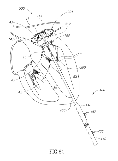
[0094] Fig. 8G schematically shows IA-TDS 400 after it has been operated to
fully expand and
deploy PMV 150 as a replacement for native mitral valve 43 and begin the
procedure of
collapsing scaffolding 704 in preparation of removing IA-TDS 400 from heart
500. The state of
fully deployed PMV 150 and retraction of scaffolding wires 700 from lampshade
scaffolding
704 shown in Fig. 8F to discus scaffolding 702 in Fig. 8G is similar to that
discussed with
reference to Fig. 7H.
[0095] It is noted that whereas IA-TDS 400 is described illustrated in
Figs. 8A-8g as being used
to deploy PMV 150, use of IA-TDS 400 in accordance with an embodiment of the
invention, is
not limited to deployment of PMV 150. IA-TDS 400 may be used for example to
deploy PMV
100, 130, 140 (Figs 2A, 2B, 2C) or PMV 160.
[0096] In the description and claims of the present application, each of
the verbs, "comprise"
"include" and "have", and conjugates thereof, are used to indicate that the
object or objects of the
22
CA 02926531 2016-04-05
WO 2015/052663
PCT/1B2014/065147
verb are not necessarily a complete listing of components, elements or parts
of the subject or
subjects of the verb.
[0097] Descriptions of embodiments of the invention in the present
application are provided by
way of example and are not intended to limit the scope of the invention. The
described
embodiments comprise different features, not all of which are required in all
embodiments of the
invention. Some embodiments utilize only some of the features or possible
combinations of the
features. Variations of embodiments of the invention that are described, and
embodiments of the
invention comprising different combinations of features noted in the described
embodiments,
will occur to persons of the art. The scope of the invention is limited only
by the claims.
23