Note: Descriptions are shown in the official language in which they were submitted.
LIQUID TO LIQUID BIOLOGICAL PARTICLE FRACTIONATION
AND CONCENTRATION
[0ool] This Patent Application claims priority to U.S. Provisional
Patent
Application Serial No. 61/715,451, filed October 18, 2012.
GOVERNMENT INTERESTS
[0002] This subject disclosure was made with U.S. Government support
under
Department of Homeland Security (DHS) Grant No. D12PC00287. The
government has certain rights in this subject disclosure.
BACKGROUND OF THE SUBJECT DISCLOSURE
Field of the Subject Disclosure
[0003] The subject disclosure relates generally to the field of sample
preparation. More particularly, the subject disclosure relates to devices,
systems and methods for fractionating and concentrating substances within a
fluid sample.
Background of the Subject Disclosure
[0004] The difficulties of detecting and quantifying particles in air
and liquids
are well known. Existing systems all begin to fail as concentration falls away
until eventually, with diminished concentrations of analyte, there is an
inability
to detect at all. This poses a significant problem for national security
where,
for example, the postal anthrax attacks of 2001 and the subsequent war on
terrorism have revealed shortcomings in the sampling and detection of
biothreats. The
1
Date Recue/Date Received 2020-08-31
CA 02927117 2016-04-12
WO 2014/063125
PCT/US2013/065800
medical arts are similarly affected by the existing limits on detection, as
are the
environmental sciences.
[0005] In the fields of biodefense and aerosol research it is common to
collect
aerosols into a liquid sample using a wet cyclone or similar device. The
aerosol
is collected into an aqueous sample so that subsequent analysis of biological
particles can be performed using standard techniques that primarily require
that
the sample be contained in liquid. These "wet" collectors have many failings,
including: difficulty in maintaining a set fluid volume, difficulties with
buildup of
particle matter in the device, and requirements for storage of the fluid in
varying
environmental conditions.
[0006] Dry filters have long been used for collection of aerosols, as
well as for
collection of particles from liquids. However, dry filters fail primarily for
the use of
identifying biological particles because detectors generally require a liquid
sample and it is extremely difficult to remove the particles into a liquid.
Methods
for removing particles from flat filters are common but are tedious,
inefficient, and
require large liquid volumes.
[0007] Concentration of particles from a liquid is traditionally
performed using
centrifugation. Centrifugal force is used for the separation of mixtures
according
to differences in the density of the individual components present in the
mixture.
This force separates a mixture forming a pellet of relatively dense material
at the
bottom of the tube. The remaining solution, referred to as the supernate or
supernatant liquid, may then be carefully decanted from the tube without
disturbing the pellet, or withdrawn using a Pasteur pipette. The rate of
2
CA 02927117 2016-04-12
WO 2014/063125
PCT/US2013/065800
centrifugation is specified by the acceleration applied to the sample, and is
typically measured in revolutions per minute (RPM) or g-forces. The particle
settling velocity in centrifugation is a function of the particle's size and
shape,
centrifugal acceleration, the volume fraction of solids present, the density
difference between the particle and the liquid, and viscosity of the liquid.
[0008] Problems with the centrifugation technique limit its
applicability. The
settling velocity of particles in the micron size range is quite low.
Consequently,
centrifugal concentration of these particles takes several minutes to several
hours. The actual time varies depending on the volume of the sample, the
equipment used, and the skill of the operator.
[0009] Centrifugation techniques are tedious in that they are normally
made up of
multiple steps each requiring a high level of concentration from the operator.
Most microbiology laboratories process large numbers of samples by
centrifugation on a daily basis. The potential for human error is high due to
the
tedious nature and automation of these techniques is difficult and costly.
Centrifugation also generally requires powered equipment. Thus, many
situations, such as emergency response, prevent their use.
[0010] Other concentration techniques have been explored and primarily
fall into
three technology groups ¨ microfluidic/electrophoretic based, filtration
based, and
capture based. However, each of these techniques has disadvantages that
prevent their use in certain situations.
3
CA 02927117 2016-04-12
WO 2014/063125
PCT/US2013/065800
SUMMARY OF THE SUBJECT DISCLOSURE
[0011] In light of the limitations of conventional techniques, what is
needed is a
single device for fractionating and concentrating a fluid sample into several
component concentrations.
[0012] In so doing, the present subject disclosure presents novel,
rapid, efficient
one-pass membrane filter based fractionation and concentration devices,
systems and methods that fractionate and concentrate particles, and especially
biological particles suspended in liquid from a dilute feed suspension
("feed") into
size fractioned and concentrated sample suspensions (retentate), eliminating
the
separated fluid (permeate) in a separate stream. The subject disclosure is
particularly useful for the fractionation and concentration of suspended
biological
particles, such as proteins/toxins, viruses, DNA, and bacteria in the size
range of
approximately 0.001 micron to 20 microns diameter. Concentration of these
particles is advantageous for detection of target particles in a dilute
suspension,
because concentrating them into a small volume makes them easier to detect.
Fractionation is performed in "cascade" fashion, in order to concentrate
particles
below the size cut of each preceding stage remaining in the separated fluid in
a
concentrated sample suspension. This process can also be used to create a
"band-pass" concentration for concentration of a particular target size
particle
within a narrow range. The device uses pressure on the feed side, vacuum on
the permeate side, and/or mechanical shear to accelerate the separation
process, and may include an added surfactant to increase efficiency.
Integrated
pneumatic, hydraulic, or mechanical valving and a novel vacuum startup
4
CA 02927117 2016-04-12
WO 2014/063125
PCT/US2013/065800
procedure allow for startup of wet membranes while reducing liquid hold-up
volume in the device. The cascade filter stack is unique in that the sample
flow is
perpendicular to the surface of a stack of filters, in series, enclosed in a
housing
with only a small open interstitial space between each filter with elution of
the
filters performed by a simultaneous wet foam elution performed parallel, or
tangential, to the retentate filter surface through the small interstitial
space.
Foam elution is performed simultaneously one each of the filter stages, so
that
transmembrane pressure across each membrane during elution remains
essentially zero or near to it. In this way, flow of elution fluid through the
membranes is eliminated or significantly reduced, so that the tangential flow
velocity and elution efficiency are maximized. The extraction foam can be
prepared from pressurized gas and a surfactant dissolved in the collection
fluid.
[0013] In one exemplary embodiment, the present subject disclosure is a
device
for fractionation and concentration of particles from a fluid sample. The
device
includes a cartridge containing staged filters having porous surface in series
of
decreasing pore size for capture of particles from a fluid sample; and a
permeate
pressure source in fluid communication with the cartridge; wherein the
particles
are eluted from the porous surfaces and dispensed in a reduced fluid volume.
[0014] In another exemplary embodiment, the present subject disclosure
is a
system for fractionation and concentration of particles from a fluid sample.
The
method includes a reservoir holding a fluid sample; a fractionation and
concentration cartridge including two or more staged filters; a permeate
pressure
device in fluid communication with the cartridge; a concentrating unit
including an
actuating integral valving to move sample through the cartridge; and a fluid
dispenser source for collecting concentrated samples from the cartridge
staged filters; wherein the fluid sample is moved through the concentrating
unit, then the concentrated samples are eluted from the filters and dispensed.
[0015] In yet another exemplary embodiment, the present subject
disclosure is
a system for rapid fractionation and concentration of particles from a fluid
sample. The system includes introducing a sample into the sample reservoir;
initiating a fractionation and concentration cycle; passing the fluid sample
through a series of filters; eluting a plurality of particles of decreasing
particle
size from each filter stage; and extracting a concentrated sample from each
filter stage.
6
Date Recue/Date Received 2020-08-31
[0015a] Accordingly, in one aspect of the present invention there is
provided a device for fractionation and concentration of particles from a
fluid
sample, the device comprising:
a cartridge containing an inlet port and an outlet port and having
alternating layers of plastic spacers and filters positioned serially between
the
inlet port and outlet port, wherein the inlet port, filters, and outlet port
are
positioned along a longitudinal axis, the longitudinal axis being
perpendicular
to the filters, each of the filters having a porous surface with all pores on
a
given filter being of a constant size, wherein the filters are arranged in
series
of decreasing pore size for capture of particles from a fluid sample, the
alternating layers of plastic spacers and filters creating a plurality of
internal
volume chambers adjacent each porous surface, wherein the cartridge is
adapted to direct a fluid sample flow in a substantially unidirectional manner
along the longitudinal axis in each chamber, into the inlet port,
perpendicularly
through the decreasing pore size filters, and out of the outlet port, in a
single
pass, such that all of the fluid sample that entered into the inlet port
passes
through the outlet port while the particles are retained at a given filter
according to particle size and do not pass to a subsequent filter if they are
larger than the pore size of the given filter;
a permeate pressure source in fluid communication with the cartridge;
and
a foam injection port at each end of the plurality of internal volume
chambers and a retentate port at each opposite end of the plurality of
internal
volume chambers,
wherein the particles are eluted tangentially from said each porous
surface by foam injected by the foam injecting port and collected through the
retentate port, and dispensed in a reduced fluid volume.
6a
Date Recue/Date Received 2020-08-31
[0015b] According to another aspect of the present invention there is
provided a system for fractionation and concentration of particles from a
fluid
sample, the system comprising:
a reservoir holding a fluid sample;
a fractionation and concentration device as described herein;
a concentrating unit including an actuating integral valving to move
sample through the fractionation and concentration device; and
a fluid dispenser source for collecting concentrated samples from the
fractionation and concentration device filters;
wherein the fluid sample is moved through the concentrating unit, then
the concentrated samples are eluted from the filters and
dispensed.
[0015c] According to yet another aspect of the present invention there is
provided a method for rapid fractionation and concentration of particles from
a
fluid sample, the method comprising:
introducing a sample into the sample reservoir;
initiating a fractionation and concentration cycle using a device as
described herein;
passing the fluid sample through the filters in the device;
eluting a plurality of particles of decreasing particle size from each
filter; and
extracting a concentrated sample from each filter.
6b
Date Recue/Date Received 2020-08-31
BRIEF DESCRIPTION OF THE DRAWINGS
[0016] FIG. 1A shows a manifold portion of a five stage fluidics device,
according to an exemplary embodiment of the present subject disclosure.
[0017] FIG. 1B shows a clamp portion of a five stage fluidics device,
according to an exemplary embodiment of the present subject disclosure.
[0018] FIG. 1C shows an exploded view of a five stage fluidics device,
according to an exemplary embodiment of the present subject disclosure.
[0019] FIG. 2 shows an internal fluid volume view of a five stage
fluidics
device, according to an exemplary embodiment of the present subject
disclosure.
[0020] FIG. 3 shows an internal fluid volume view of a two stage
fluidics
device, according to an exemplary embodiment of the present subject
disclosure.
[0021] FIG. 4 shows an internal fluid volume view of a three stage
fluidics
device,
6c
Date Recue/Date Received 2020-08-31
CA 02927117 2016-04-12
WO 2014/063125
PCT/US2013/065800
according to an exemplary embodiment of the present subject disclosure.
[0022] FIG. 5 shows a flow chart for fractionation and concentration,
according to
an exemplary embodiment of the present subject disclosure.
[0023] FIG. 6 shows a two cartridge system, according to an exemplary
embodiment of the present subject disclosure.
[0024] FIG. 7A shows a cross sectional view of a two stage filter stack
with
integral filter supports, according to an exemplary embodiment of the present
subject disclosure.
[0025] FIG. 7B shows a cross sectional view of a two stage filter stack
with no
filter support, according to an exemplary embodiment of the present subject
disclosure.
[0026] FIGS. 8A-8X show a flow chart with detailed steps of a process
for
fractionation and concentration, according to an exemplary embodiment of the
present subject disclosure.
DETAILED DESCRIPTION OF THE SUBJECT DISCLOSURE
[0027] The present subject disclosure relates generally to the fields of
bioterrorism security, medicine, and environmental science. Rapid, reliable
detection of airborne biothreats is a significant need for the protection of
civilians
and military personal from pandemic outbreaks and bioterrorist events. Best in
class biothreat detection systems use aerosol collectors to capture particles
into
a liquid volume in the range of 2 to 12 mL. Samples are then processed using a
number of sample preparation techniques and analyzed by rapid microbiological
7
CA 02927117 2016-04-12
WO 2014/063125
PCT/US2013/065800
methods, including real-time quantitative polymerase chain reaction (qPCR) and
ultra-high throughput sequencing (UHTS) and/or gold-standard culture based
methods. While the state of the art for rapid detectors, collectors, and
identifiers
has advanced dramatically in recent years, advancement of sample preparation
techniques has lagged significantly and considerable improvements are needed
in these techniques.
[0028] Detect/collect/identify systems for airborne biothreats must
operate
correctly in all types of indoor and outdoor environments. Urban, industrial,
and
rural outdoor environments as well indoor environments range from very low to
very high particle concentrations. Detection of threats in these varied
environments often hinges on the ability of the system to capture and identify
rare threat particles in what can be a highly varied, complex mixture of
organic
and inorganic debris particles, innocuous microbes, pollen, fungal spores, and
mammalian cells.
[0029] Better automated sample preparation techniques are needed so
problems
currently associated with detection of rare particles in complex environmental
samples can be overcome. Inhibition of identification techniques due to
environmental debris is a common problem with these systems due to the varied,
high-level complex mixtures of particle and chemical inhibitors. UHTS, qPCR,
and other rapid detection techniques can also fail due to high levels of
background clutter. Breakdown of bioinfornnatic systems used for UHTS data
analysis due to high background clutter levels is one of the biggest hurdles
that
must be overcome before cutting-edge sequencing can be adapted to
8
CA 02927117 2016-04-12
WO 2014/063125
PCT/US2013/065800
autonomous biothreat detection applications. There is also a significant
requirement to be able to differentiate between target agents coming from
whole,
viable cells and those present as free DNA or free proteins. The inability to
rapidly determine if the target particle is a whole viable cell or is only
present as
free DNA or protein signature, as is the norm in today's biothreat detection
systems, does not allow organizations to differentiate between what may be an
actual terrorist event from potentially catastrophic false alarms associated
with
hoaxes or natural events.
[0030] Aerosol samples and other samples of importance (e.g., surface,
liquid,
clinical, food, etc.), often contain a significant amount and wide range of
non-
target debris including organic and inorganic matter and biological materials.
As
described above, these non-target materials can significantly affect the
performance of sample preparation and agent identification techniques with a
common side effect of inhibition. Conventional sample preparation techniques
exist for removing these inhibitors, but they are slow and perform best when
volumes of only a few hundred microliters are processed ¨ demonstrating the
mismatch between collected sample size and the volume that can be processed
and analyzed by available technologies. This mismatch raises the true system
detection limit to levels significantly higher than the desired detection
limit and
creates a significant likelihood of false negative results when, as would
typically
be the case, only trace levels of signature are present.
[0031] A wide range of existing, and developing, rapid analysis
platforms are
potentially useful technologies for detection and identification needs.
Detection
9
CA 02927117 2016-04-12
WO 2014/063125
PCT/US2013/065800
and identification may key on whole organisms, nucleic acids, or proteins.
Culture based analysis, antibiotic susceptibility testing, and functional
assays all
require live organism samples. Common nucleic acid techniques include qPCR,
UHTS, and hybridization arrays. ELISA and other immunoassay techniques,
mass spectrometry, chromatography techniques, and other techniques may be
used for protein analysis. There are significant reasons in some cases to
choose
one of these techniques over the other or in some cases to analyze with more
than one technique. Additionally some techniques lend themselves to use in
autonomous detection platforms and some are used only in laboratory settings.
Further, it is difficult to determine what techniques may receive precedence
in the
near future as costs fall or new improved methods are developed. This
difficulty
in determining what detection and identification system may be used warrants
the need for a plug-and-play type of sample preparation system that is capable
of
delivering the needed sample fractions in a concentrated form for each
potential
type of analysis.
[0032] Robust, fast, and sensitive detection systems are needed, but
currently
most systems fail to meet these needs due to deficiencies in sample
preparation.
The sample preparation system must be capable of autonomous operation for a
month or more without maintenance. The same environmental particles and
inhibitors that commonly cause issues with the identifier can also lead to
failure
of the sample preparation system, especially after repeated use over extended
periods of time. The time required for the sample preparation methods used for
these complex samples is a large portion of the total time needed for
CA 02927117 2016-04-12
WO 2014/063125
PCT/US2013/065800
identification and, even so, the methods are only capable of processing a very
small portion of the available sample.
[0033] The present subject disclosure presents a novel technique of
fractionating
multiple components simultaneously. It may be used in numerous fields,
including, but not limited to, bioterrorism detection. For example, exemplary
and
specific fields of use include, but are not limited to:
1. Aerosol sampling for bioterrorism threat agents
a. Where the sample results in a liquid sample for analysis
b. Where the sample can contain target agent(s) that are thought to
be a substantial threat to the health of humans
i. Where a list of the potential threat (target) agent(s) can be
taken from the U.S. Food and Drug Administration's Centers
for Disease Control and Prevention (CDC) Select Agents A,
B, or C list (See List 1, below)
c. Where the sample can contain target agent(s) that are thought to
be a threat to the health of humans, animals or plants, causing
societal disruption and economic harm
i. Where a list of the potential threat (target) agents can be
taken from the CDC agent list
(httpliwww.bt.cdc.goviagentlagentlistasp), or List 2, below
d. Where the resulting sample can contain test particles, target
agent(s) or surrogate(s) in a concentration too small for detection
by the chosen method
i. Where concentration of the sample into a smaller volume
can result in detection of the threat agent(s) of interest by
one or a combination of the following methods:
1. Where detection of the threat agent(s) is performed
by polymerase chain reaction (PCR) or PCR-like
methods
2. Where detection of the threat agent(s) of interest is
performed by immunoassay methods
3. Where detection of the threat agent(s) of interest is
performed by ultraviolet light fluorescence methods
ii. Or where concentration and analysis resulting in a non-
detect result can provide assurance that if the target agent is
11
CA 02927117 2016-04-12
WO 2014/063125
PCT/US2013/065800
present, it is present in such a low quantity that the resulting
risk to the affected population is minimal
iii. Where separation of the sample into desirable size fractions
can concentrate the target particles into separate but equally
concentrated size fractions for analysis by different detection
methods listed in 1.c.i. above, such as:
1. Separating and concentrating particles larger than 0.2
microns to separate and concentrate bacteria
iv. Where a small size range or "band-pass" can be separated
out and concentrated for interrogation for a particular threat
agent or surrogate, such as:
1. separating and concentrating particles from 0.2
microns diameter to 2 microns diameter to separate
bacterial spores and concentrate them separately
from smaller and larger particles present in the initial
sample
2. separating and concentrating particles from 0.005
microns to 0.2 microns diameter to separate most
viruses and concentrate them separately from smaller
and larger particles present in the initial sample
(examples include viral equine encephalitis, or VEE;
0.06 microns diameter).
3. separating and concentrating particles from 0.001
microns (approximately 5 kiloDaltons) to 0.01 microns
(approximately 100 kiloDaltons) to separate toxins
and proteins and concentrate them separately from
smaller and larger particles present in the initial
sample
2. The above types of sampling and analysis are performed for the fields of
homeland security, corporate security, and military force protection:
a. Automated sampling and analysis systems such as those
developed for government programs Portal Shield, Joint Programs
Biological Detection System (JPBDS), US Postal Service Biological
Detection System (BDS), and systems under development, such as
the Biological Aerosol Networked Detection (BAND) system and
Rapid Aerosol Biological Identification System (RABIS)
b. Manual systems such as bioaerosol collection using air/liquid
impingers, including the All Glass Impinger (AGI-30, Ace Glass,
Inc., Vineland, NJ), Greenburg-Smith impingers, and SKC
Biosamplers provide samples that are in the 20-100 mL size range,
and can be concentrated down to the 4-400 uL volume range using
the Innova Prep device and process described here
12
CA 02927117 2016-04-12
WO 2014/063125
PCT/US2013/065800
c. Samples resulting from manual swabbing of surfaces onto wetted
swabs, pads, or pieces of filter material are often taken for
bioterrorism security monitoring and are typically extracted into a
volume of liquid resulting in a 2 to 20 mL volume initial sample.
Samples like these can be quickly concentrated to much smaller
volumes in the range of 4-400 uL using the InnovaPrep
3. Water sampling for bioterrorism threat agents
a. Where the sample can contain target agent(s) that are thought to
be a substantial threat to the health of humans by ingestion or
contact
i. Where a list of the potential threat (target) agent(s) can be
taken from the U.S. Food and Drug Administration's Centers
for Disease Control and Prevention (CDC) Select Agents A,
B, or C list (See List 1, below)
b. Where the sample can contain target agent(s) that are thought to
be a threat to the health of humans, animals or plants, causing
societal disruption and economic harm
i. Where a list of the potential threat (target) agents can be
taken from the CDC agent list
(httpliwww.bt.cdc.goviagentiagentlistasp), or List 2, below
c. Where the resulting sample can contain test particles, target
agent(s) or surrogate(s) in a concentration too small for detection
by the chosen method
i. Where concentration of the sample into a smaller volume
can result in detection of the threat agent(s) of interest by
one or a combination of the following methods:
1. Where detection of the threat agent(s) is performed
by polymerase chain reaction (PCR) or PCR-like
methods
2. Where detection of the threat agent(s) of interest is
performed by immunoassay methods
3. Where detection of the threat agent(s) of interest is
performed by ultraviolet light fluorescence methods
ii. Or where concentration and analysis resulting in a non-
detect result can provide assurance that if the target agent is
present, it is present in such a low quantity that the resulting
risk to the affected population is minimal
iii. Where separation of the sample into desirable size fractions
can concentrate the target particles into separate but equally
13
CA 02927117 2016-04-12
WO 2014/063125
PCT/US2013/065800
concentrated size fractions for analysis by different detection
methods listed in 1.c.i. above, such as:
1. Separating and concentrating particles larger than 0.2
microns to separate and concentrate bacteria
iv. Where a small size range or "band-pass" can be separated
out and concentrated for interrogation for a particular threat
agent or surrogate, such as:
1. separating and concentrating particles from 0.2
microns diameter to 2 microns diameter to separate
bacterial spores and concentrate them separately
from smaller and larger particles present in the initial
sample
2. separating and concentrating particles from 0.005
microns to 0.2 microns diameter to separate most
viruses and concentrate them separately from smaller
and larger particles present in the initial sample
(examples include viral equine encephalitis, or VEE;
0.06 microns diameter).
3. separating and concentrating particles from 0.001
microns (approximately 5 kiloDaltons) to 0.01 microns
(approximately 100 kiloDaltons) to separate toxins
and proteins and concentrate them separately from
smaller and larger particles present in the initial
sample
4. The above types of sampling and analysis are performed for the fields of
homeland security, corporate security, and military force protection:
a. Water samples taken from water sources used to produce potable
water for consumption by the public or government use
b. Water samples taken to determine a source of production of
bioterrorism agents
c. Water samples taken to determine whether biological
decontamination has been effective
5. Agricultural samples for bioterrorism threat agents
a. Where the sample can contain target agent(s) that are thought to
be a substantial threat to the health of plants or animals, or
indirectly to humans after ingestion of contaminated agricultural
products
b. Where the sample is liquid or can be extracted into a liquid for
analysis
14
CA 02927117 2016-04-12
WO 2014/063125
PCT/US2013/065800
i. Where a list of the potential threat (target) agent(s) can be
taken from the U.S. Food and Drug Administration's Centers
for Disease Control and Prevention (CDC) Select Agents A,
B, or C list (See List 1, below)
c. Where the sample can contain target agent(s) that are thought to
be a threat to the health of humans, animals or plants, causing
societal disruption and economic harm
i. Where a list of the potential threat (target) agents can be
taken from the CDC agent list
(http://wwwbt.cdc.goviagentiagentlistasp), or List 2, below
d. Where the resulting sample can contain test particles, target
agent(s) or surrogate(s) in a concentration too small for detection
by the chosen method
i. Where concentration of the sample into a smaller volume
can result in detection of the threat agent(s) of interest by
one or a combination of the following methods:
1. Where detection of the threat agent(s) is performed
by polymerase chain reaction (PCR) or PCR-like
methods
2. Where detection of the threat agent(s) of interest is
performed by immunoassay methods
3. Where detection of the threat agent(s) of interest is
performed by ultraviolet light fluorescence methods
ii. Or where concentration and analysis resulting in a non-
detect result can provide assurance that if the target agent is
present, it is present in such a low quantity that the resulting
risk to the affected population is minimal
iii. Where separation of the sample into desirable size fractions
can concentrate the target particles into separate but equally
concentrated size fractions for analysis by different detection
methods listed in 1.c.i. above, such as:
1. Separating and concentrating particles larger than 0.2
microns to separate and concentrate bacteria
iv. Where a small size range or "band-pass" can be separated
out and concentrated for interrogation for a particular threat
agent or surrogate, such as:
1. separating and concentrating particles from 0.2
microns diameter to 2 microns diameter to separate
bacterial spores and concentrate them separately
from smaller and larger particles present in the initial
sample
CA 02927117 2016-04-12
WO 2014/063125
PCT/US2013/065800
2. separating and concentrating particles from 0.005
microns to 0.2 microns diameter to separate most
viruses and concentrate them separately from smaller
and larger particles present in the initial sample
(examples include viral equine encephalitis, or VEE;
0.06 microns diameter).
3. separating and concentrating particles from 0.001
microns (approximately 5 kiloDaltons) to 0.01 microns
(approximately 100 kiloDaltons) to separate toxins
and proteins and concentrate them separately from
smaller and larger particles present in the initial
sample
v. Where exclusion of interferent particles such as diesel soot
is desirable to improve the performance of the analysis
method [minimization of interference or improvement of
"contrast" may be desirable for all fields]
6. The above types of sampling and analysis are performed for the fields of
homeland security, corporate security, and military force protection:
a. Where foodstuffs such as milk is monitored for toxin contamination
such as by ricin
b. Where meatpacking plants are monitored for biological
contamination by E. coli, Listeria spp. Such monitoring is also
conducted for quality assurance,such as hazard assessment and
critical control point (HACCP) programs
c. For bottled water production
[0034] The present subject disclosure may be used to assist in
identifying agents
from the following lists:
List 1: CDC Category A and B Bioterrorism Agents List
Category A (definition below)
Anthrax (Bacillus anthracis)
Botulism (Clostridium botulinum toxin)
Plague (Yersinia pestis)
Smallpox (variola major)
Tularemia (Francisella tularensis)
Viral hemorrhagic fevers (filoviruses [e.g., Ebola, Marburg] and arenaviruses
[e.g., Lassa, Machupo])
Category B (definition belowl
Brucellosis (BruceIla species)
Epsilon toxin of Clostridium perfringens
Food safety threats (e.g., Salmonella species, Escherichia coli 0157:H7,
Shigella)
16
CA 02927117 2016-04-12
WO 2014/063125
PCT/US2013/065800
Glanders (Burkholderia mallei)
Melioidosis (Burkholderia pseudomallei)
Psittacosis (Chlamydia psittaci)
Q fever (Coxiella bumetii)
Ricin toxin from Ricinus comnnunis (castor beans)
Staphylococcal enterotoxin B
Typhus fever (Rickettsia prowazekii)
Viral encephalitis (alphaviruses [e.g., Venezuelan equine encephalitis,
eastern
equine encephalitis, western equine encephalitis])
Water safety threats (e.g., Vibrio cholerae, Cryptosporidium parvum)
List 2: Secondary Potential Biological Threat Agents
Viri/prions
Flaviviruses (Yellow fever virus, West Nile virus, Dengue, Japanese
Encephalitis,
TBE, etc.)
Hep A, B, C
Prions (CJD, BSE, CWD)
Alphaviruses (VEE, EEE, WEE)
Nipah virus
Rabies virus
Rhinovirus (could be modified?)
Polioviruses
Hantaviruses
Filoviruses (Ebola, Marburg, Lassa)
Bacilli
Mycobacterium tuberculosis, drug resistant
Mycobacteria other than TB, like C. leprae
Streptococcus pneumoniae
S. pyogenes
S. aureus
Clostridium tetani
C. difficile
Bacillus cereus
Coxiella brunette (Q fever)
Francisella tularensis
Borrelia recurrentis
Rickettsia rickettsii
R. prowazekii
Shigella sonnei
Bartonella henselae
Yersinia enterolitica
Y. pseudotuberculosis
Neisseria meningitidis
Legionella pneumophila
Burkholderia pseudomallei
17
CA 02927117 2016-04-12
WO 2014/063125
PCT/US2013/065800
PastureIla multocida
Other Pathogenic Microorganisms
Cryptosporidium parvunn
Histoplasma capsulatunn
Cryptococcus neoformans
Aspergillus niger
Pathogenic fungi
Acremomium spp.
Alternaria alternate
Apophysonnyces elegans
Aspergillus terreus
Bipolaris spp.
Bipolaris spicifera
Blastoschizomyces capitatus
Candida krusei
Candida lusitaniae
Cladophialophora bantiana
Cunnihamella berholletiae
Curvularia lunata
Exserohilum rostratum
Fusarium moniliforme
Fusarium solani
Hansenula anomala
Lasiodilodia theobromae
Malassezia furfur
Paecilomyces lilacinus
Paecilomyces bariotii
Penicillium mameffei
Phialemonium curvatum
Philophora parasitica
P. richardsiae
Ramichloridium spp.
Rhizomucor pusillus
Rhizopus rhizopodiformus
Rhodotorula rubra
Sacchromyces cerevisiae
Scedosporium prolificans
Trichosporon beigelii (T. asahii)
Wangiella dermatitidis
[0035] The
present subject disclosure may be used to assist in identifying various
agents of varying sizes:
18
CA 02927117 2016-04-12
WO 2014/063125
PCT/US2013/065800
Definition of Category A Diseases/Agents
The U.S. public health system and primary healthcare providers must be
prepared to address various biological agents, including pathogens that are
rarely seen in the United States. High-priority agents include organisms that
pose
a risk to national security because they
= can be easily disseminated or transmitted from person to person;
= result in high mortality rates and have the potential for major public
health
impact;
= might cause public panic and social disruption; and
= require special action for public health preparedness.
Definition of Category B Diseases/Agents
Second highest priority agents include those that
= are moderately easy to disseminate;
= result in moderate morbidity rates and low mortality rates; and
= require specific enhancements of CDC's diagnostic capacity and
enhanced disease surveillance.
Definition of Category C Diseases/Agents
Third highest priority agents include emerging pathogens that could be
engineered for mass dissemination in the future because of
= availability;
= ease of production and dissemination; and
= potential for high morbidity and mortality rates and major health impact
Physical Sizes of some Agents and Surrogates:
TARGET:
= Bacillus thuringiensis endospore ¨
approximately 1 pm
= Bacillus anthracis endospore -
approximately 1pm
= Yersinia pestis - Gram negative rod-ovoid 0.5-0.8 pm
in width and 1-
3 pm in length
= Yersinia rohdei ¨ approximately 1pm
4, Venezuelan Equine Encephalitis ¨ 70 nm (0.07 pm)
= Gamma-killed MS2 ¨ 2 mD or about 25 nm (0.025 pm) (but will pass
through a 300 kD pore size but is retained by a 100 kD pore size Wick and
McCubbin - ECBC)
= Ovalbumin ¨ 45 kD or 6 nm (0.006 pm)
19
CA 02927117 2016-04-12
WO 2014/063125
PCT/US2013/065800
. Botulinum Toxoid A ¨ 150 to 900 kD or 10 nm to 70 nm (0.01 pm to
0.07 pm)(Normally published as 150 kD however some publications state
that toxoid A can be released as complexes comprised of the 150 kD toxin
protein along with associated non-toxin proteins and can therefore be
released in 900 kD, 500 kD, and 300 kD forms.
= DNA - 1000 Bp or 600 kD up to 15,000 Bp or 9 mD
[0036] Specific fields of use in the medical field include, but are not
limited to:
1. The above types of sampling and analysis are performed for the fields of
medical research and diagnostics:
a. In cancer research where very low concentrations of experimental
drugs in body fluids or urine are the targets of analysis
b. In allergy diagnosis where low quantities of specific antigens are
the targets of analysis in body fluids
c. In health effects research regarding the determination of health
effects known to be caused by various materials in inhaled
particulate matter with aerodynamic diameter below 2.5 microns
(PM 2.5). this area overlaps with environmental studies (see
below).
d. In forensic medicine where low concentrations of toxins or venoms
are the targets of analysis in body fluids
e. In operating rooms [surface extraction and air monitoring, add]
f. In pharmaceutical manufacturing where the biological aerosol
particulate matter concentration is regulated by the US Food and
Drug Administration
[0037] Specific fields of use in the environmental studies field
include, but are not
limited to:
[similar to outline above, modified to fit the environmental applications]
2. The above types of sampling and analysis are performed for the field of
environmental study:
a. In health effects research regarding the determination of health
effects known to be caused by various materials in inhaled
particulate matter with aerodynamic diameter below 2.5 microns
(PM 2.5)
b. High altitude aerosol research where low quantities of particulate
are collected and must be concentrated for study
c. In cleanrooms where very low aerosol concentrations of aerosol
particles are collected for monitoring aimed at source control
CA 02927117 2016-04-12
WO 2014/063125
PCT/US2013/065800
d. For separation of populations of particles collected at different
heights above the ground (profiling studies)
[0038] The present subject disclosure has been developed as a unique
membrane filter based fractionation and concentration system that is capable
of
separating particles by size and concentrating those particles into small
(<100
pL) sample volumes. A novel approach was developed in which the membrane
filters are stacked, in order of decreasing pore size, inside a single
cartridge with
a small interstitial space, or in some cases a solid filter support and
further
reduced interstitial space, between each membrane filter. Sample flow is
introduced perpendicular to the first filter surface and is pushed or pulled,
in
series, directly through each of the membrane in the cartridge. Because the
cartridge can be designed for reuse, and because wet hydrophilic membrane
filters will not allow air to flow through at pressures below the bubble
point, a
novel vacuum startup method is used to allow air to be removed from the
interstitial space and other internal volume, so that the sample process can
be
initiated. A series of channels and associated valves, integral to the
cartridge,
are used to link each stage back to a pump to allow for negative pressure to
be
pulled on the system.
[0039] After negative pressure has been pulled on the system, the sample
flow is
introduced as described above. The entire sample is flowed through the
cartridge, until air reaches the first membrane filter and the system locks
up. The
vacuum startup valves are then actuated one by one to allow the remaining
fluid
to be pushed through the remaining membrane filters. When then entire sample
volume has been processed then the cartridge inlet and outlet valves are
closed
21
CA 02927117 2016-04-12
WO 2014/063125
PCT/US2013/065800
and a retentate valve is opened on each stage. A wet carbonated foam is then
introduced into one end of the cartridge, which subsequently travels the
length of
the cartridge, tangential to the retentate surface of each membrane. Finally
the
foam is dispensed out of the retentate port into a separate sample container
for
each membrane filter. The foam then breaks down into a liquid leaving a small
concentrate fraction associated with each membrane filter stage.
[0040] The subject disclosure of the present application, which
describes liquid-
to-liquid fractionation and concentration devices, systems and methods,
provides
a novel means of rapidly and efficiently separating and then concentrating
biological samples. Significant advantages are offered over current methods
including, but not limited to: improved separation efficiency, improved
concentration efficiency, shorter process times, automation, and integration
into
automated systems. Like centrifugation, filtration, and the other conventional
methods, this present technique concentrates the collected sample prior to
analysis, but with many further advantages, including but not limited to: 1)
the
liquid volume of the sample is quickly reduced. Unlike centrifugation, which
typically takes 10 to 30 minutes to concentrate micron-sized particles, this
process can be accomplished in 5 to 60 seconds for a 10 mL initial volume.
Unlike conventional hollow fiber filter concentration, in which the initial
sample is
recycled many times through the filter taking from several minutes to hours in
order to concentrate a particle such as a protein or enzyme into a volume of
several milliliters, the sample is passed straight through in one pass. This
results
in a much smaller volume of liquid on the order of 100 to 400 microliters, or
22
CA 02927117 2016-04-12
WO 2014/063125
PCT/US2013/065800
passed straight through in dead-end fashion and then extracted in a volume of
liquid or foam in the range of 4 to 400 microliters. Unlike typical single-
pass flat
filtration, the sample remains in liquid form for transport and analysis. The
detection limit for the target agent is lowered, with respect to the media
originally
sampled. 2) The final sample volume is reduced much further than in previously
known methods, while kept in liquid form, allowing detection in devices such
as
multi-well plate readers that utilize small input samples. 3) The reduced-size
samples can be more efficiently stored and transported by microfluidic
handling
methods. 4) The device can be constructed to separate particles in one pass
into different size fractions for analysis for certain agents. For example,
cells and
spores can be concentrated separately from viruses and biological toxins.
Further, the size range that is concentrated can be narrow, or "band-pass" to
concentrate a small size range fraction from a complex matrix, such as an
environmental sample 5) The device can be used to reduce the onboard fluid
storage capacity of aerosol samplers, by recycling the cleaned liquid back to
the
collection cycle after the sampled particles are removed into a small volume
for
analysis. 6) This device is much more readily adapted to automated systems
than other technologies including centrifugation, flat filtration, and other
methods.
The flow-through nature of the device allows for straightforward configuration
into
an automated detection system. 7) This device is significantly more robust in
nature than new microfluidic concentration systems such as dielectrophoresis
concentration systems. Dielectrophoresis systems developed by Sandia have
internal flow paths of small diameters that can create significant clogging
during
23
CA 02927117 2016-04-12
WO 2014/063125
PCT/US2013/065800
processing of fluids with high particle concentrations. Commercially available
hollow fiber filters, while possessing pores of up to a maximum of
approximately
0.5 pm diameter, will take significantly longer to clog, due to the high
number of
pores and the tangential flow cleaning with the preferred surfactant foam. 8)
The
InnovaPrep system is much smaller than any commercially available liquid to
liquid concentrator. Necessary components can be arranged in such a way as to
take advantage of any empty space in the system being integrated. 9) The
device it made almost entirely of low cost, readily available components. This
significantly lowers the cost of integration and makes it more practical than
other
methods concentration.
[0041] An exemplary embodiment of a device according to the present
subject
disclosure is presented in Figs. 1A-1C. In these figures, an exemplary device
100, which can be used for fractionation and concentration of components
within
a fluid, is presented. The device 100 includes a manifold portion 101, with a
manifold mounting flange 111 used to connect or secure the device, and a clamp
plate 102 which together serve as end pieces to the device 100, and the
fluidic
stack 108 are held within those end pieces. Clamping bolts 103 maintain a
sealed condition for the device 100 when it is operated. Alignment pins 104
serve to maintain the structural integrity of the device and provide an easier
method to put all components together. Fluid connections 105 are the ports
where fluid is introduced into the device 100. Pneumatic control line 106
regulates the pressure within the device 100.
[0042] Figure 1C shows an exploded view of a laminated multi-stage
24
CA 02927117 2016-04-12
WO 2014/063125
PCT/US2013/065800
concentration cell device 100 with bolts 103 and alignment pins 104 removed
for
sake of clarity. The fluidic stack 108 comprises of numerous layers of hard
plastic layers 121 with holes cut out in specific shapes and geometries and
fluid
paths etched in the surface of, and gaskets 122 enclosing filter portions 123.
The device 100 is constructed by compressing alternating layers of plastic 121
and filter media 123 between two fluidic blocks 122 which allow the cell to
interface with the rest of the fluidic system. This type of cartridge may be
constructed using the method shown or may be constructed using bonding and
construction techniques that are commonly used in microfluidic device
construction. An exemplary cartridge is shown having 5 filters. In this case,
the
membrane filters could be made up with filter types, or similar filter types
to that
shown below.
= Filter 1 ¨ 6 pm track-etched polycarbonate membrane filter for large
particle removal
= Filter 2 ¨ Affinity based filter for removal of humics
= Filter 3 ¨ 0.4 pm track-etched polycarbonate membrane filter for bacteria
capture
= Filter 4 ¨ 0.02 pm block copolymer membrane filter for virus and nucleic
acid capture
= Filter 5 ¨ 10 kD block copolymer membrane filter for protein capture
In this way the system would produce fractions of target particles containing
the
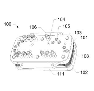
following particle types, with reduced numbers or concentrations of
interfering
particles or humics.
= Whole Bacteria
= Viruses and free nucleic acids
= Proteins
[0043] Fig. 7A shows a cross sectional view of a two stage filter stack
with
CA 02927117 2016-04-12
WO 2014/063125
PCT/US2013/065800
integral filter supports. This cell is constructed by laminating several
layers of
different material together to create fluid channels. The components are (1)
ridged plastic substrate in which fluid paths are etched, (2) soft plastic
substrate
which functions as a gasket creating a gas and liquid tight seal between
layers,
(3) filtration media, (4) filter support ridges, and (5) fluid paths
connecting one
filter stage to the next such that the permeate of the first stage filter
becomes the
sample of the second stage filter.
[0044] Fig. 7B shows a cross sectional view of a five stage filter
stack. This cell is
also constructed by laminating layers of material together; the filter media
is
arranged such that the sample travels through all five filter stages in a
single
step, starting with the largest pore diameter filter and ending with the
smallest.
The components are (6) the sample inlet port, (7) soft or ridged substrate
sealing
the layers together and creating fluid channels, (8) filtration media, (9) the
permeate port. (10) shows the direction of the sample flow.
[0045] It should be noted that although the exemplary embodiment shown
in
Figures 1A-1C includes five layers of filters, any number is possible, and the
technique to make and use the device will be similar, and understood by one
having ordinary skill in the art when considering the present disclosure.
[0046] Once the device 100 is properly aligned with alignment pins 104
and
securely fastened with bolts 103, a fluidic internal volume 200 is created
with
numerous chambers, passageways and connections. Such internal fluid volume
200 is shown in Fig. 2. It should be noted that this internal fluidic volume
is
created as a result of the laser cut passageways of the various hard plastic
26
CA 02927117 2016-04-12
WO 2014/063125
PCT/US2013/065800
layers 121, gaskets 122, and filters 123 used in the fluidic stack 108.
[0047] Internal fluidic volume 200 shows various paths for the fluidic
stack 108
assembly for a five stage concentrator. 202, 204 and 206 are pneumatic control
lines, and used to control filter stage 1 bypass valve (humic acid removal)
202,
Filter stage 2 bypass valve (prefilter) 204, and decontamination isolation
valves
206.
[0048] Various fluid lines include the decontamination fluid outlet port
208, the
filter stage 3 retentate port (concentration stage 1) 210, the filter stage 1
retentate port (Humic acid removal) 212, the filter stage 4 retentate port
(concentration stage 2) 214, the filter stage 2 retentate port (Prefilter)
216, and
the filter stage 5 retentate port (concentration stage 3) 218.
[0049] Further pneumatic control lines include the filter stage 5 bypass
valve
(Concentration stage 3) 220, filter stage 4 bypass valve (concentration stage
2)
222, filter stage 3 bypass valve (concentration stage 1) 224, and the master
filter
isolation valve 226.
[0050] Further fluid lines include the gas flush port 228, the foam
injection port
230, the sample inlet port 232, the sample outlet port (permeate) 234, and the
decontamination fluid inlet port 236. Part of the pneumatic control line is
the
feed/permeate isolation valve 238. Finally, the various filter stages include
filter
stage 1 250, filter stage 2 252, filter stage 3 254, filter stage 4 256, and
filter
stage 5 258.
[0051] All of the components and internal fluid channels for the five
stage fluidic
stack shown in Fig. 2 work together in the manner as described in further
detail
27
CA 02927117 2016-04-12
WO 2014/063125
PCT/US2013/065800
below. It is noted that the five stage fluidic stack shown in Fig. 2 is merely
exemplary, and that the present disclosure is not limited to such an exemplary
embodiment. For example, a two stage fluidic internal volume is shown in Fig.
3
and a three stage fluidic internal volume is shown in Fig. 4. Other numbers
are
also possible and within the purview of one having ordinary skill in the art.
[0052] For sake of completeness, the components of the two stage
fluidics
internal volume 300 include:
301 Microvalve control pneumatic control diaphragm (7 shown)
302 Micro fluidic valve (7 shown)
303 (pneumatic control line) Filter stage 1 bypass valve
304 (Fluid line) Sample outlet (Permeate)
305 (Fluid line) Sample inlet (Feed)
306 (Fluid line) Filter stage 1 retentate
307 (Fluid line) Filter stage 2 retentate
308 (pneumatic control line) Master filter isolation valve
309 (pneumatic control line) Filter stage 2 bypass valve
310 (Fluid line) Foam injection port
311 Filter stage 1
312 Filter stage 2
[0053] For sake of completeness, the components of the three stage
fluidics
internal volume 400 include:
401 Stage 1 foam inlet
402 Stage 1 foam microvalve
403 Stage 2 foam inlet
404 Stage 2 foam microvalve
405 Stage 3 foam inlet
406 Stage 3 foam microvalve
407 Filter stage 1 bypass valve
408 Filter stage 2 bypass valve
409 Sample inlet (feed)
410 Stage 1 retentate outlet
28
CA 02927117 2016-04-12
WO 2014/063125
PCT/US2013/065800
411 Stage 1 retentate valve
412 Stage 2 retentate outlet
413 Stage 2 retentate valve
414 Stage 3 retentate outlet
415 Stage 3 retentate valve
416 Filter stage 1
417 Filter stage 2
418 Filter stage 3
[0054] A
process flow diagram for an exemplary system according to the present
subject disclosure is presented in Fig. 5. The orange boxes 522, 525, 542,
545,
547, 551 contain the final six concentrated fractions that will be recovered
from
the input sample shown in the red box 501. Two fractionation/concentration
fluidics cartridges 510 and 540 will be used to produce the six fractions 522,
525,
542, 545, 547, 551. Cartridge A 510 will separate the input sample into
fractions
containing whole cells 514, free nucleic acids 522, and free proteins 525. A
portion of the whole cell fraction 514 from Cartridge A 510 will be lysed and
then
Cartridge B 540 will be used to separate the whole cell lysate 543 into
fractions
containing cell debris 545, nucleic acids 547, and proteins 551. Cartridge A
510
separations will be performed with stages A.1 511, A.2 513, A.3 515, A.4 521,
and A.5 524. Cartridge B 540 will house stages B.1 544, B.2 548, and B.3 550.
Prior to Cartridge A 510, a novel, replenishable media column loaded with
Polyvinylpolypyrrolidone (PVPP) media will be used to remove humic substances
while allowing target materials to pass. Stage A.1 511 will use a large pore
membrane to remove environmental debris and inhibitors 530, including large
particulate matter, from the input sample. Stage A.2 513 will be a novel,
replenishable Polyvinylpolypyrrolidone (PVPP) Sol-gel membrane used to
remove humic substances 531 while allowing target materials to pass. Stage
29
CA 02927117 2016-04-12
WO 2014/063125
PCT/US2013/065800
A.3 515 is used to capture whole viable organisms. A portion of this fraction
is
then archived for later analysis and a portion is lysed for rapid detection.
The
permeate fluid from this stage will contain free solution nucleic acids and
free
solution proteins which are subsequently separated into a nucleic acid
fraction
and a protein fraction with Stages A.4 521 and A.5 524, respectively. The
lysed
fraction of whole viable cells, to be used for rapid detection, is separated
into
three fractions containing cellular debris, nucleic acids and proteins with
Stages
B.1 544, B.2 548 and B3 550, respectively. The various permeates 512, 514,
516, 523, 549 are shown to indicate the remaining substances of the process.
Permeate waste 526 and 552 indicate the end result of the processes of
cartridge A 510 and cartridge B 540, respectively.
[0055] Fig. 6, in conjunction with Fig. 5, provides a flow schematic of
a layout of
an exemplary version of a two cartridge system 600. Figs. 5 and 6 should be
considered jointly for the proceeding discussion. During operation, a sample
with
a volume of 1 mL to 50 mL is fed into the sample input reservoir. A processing
cycle is then initiated. The first step is preparation of the cartridges 601,
603 for
processing using a novel vacuum startup method. Because the membranes 606
are hydrophilic in nature and are wet for every sample processed after the
first,
startup requires that air be evacuated from the system 600 so that the liquid
samples can be brought into contact with the membranes. It should be noted
that only one membrane 606 is pointed out in the figure for sake of clarity,
but
multiple membranes are shown. Pulling negative pressure on each stage within
the cartridges 601, 603 performs this action. Pneumatic valves within the
CA 02927117 2016-04-12
WO 2014/063125
PCT/US2013/065800
cartridges 601, 603 are activated to allow a vacuum to be applied through a
three-way valve 602, 604 at the top of each cartridge, respectively. When a
sufficient vacuum has been achieved, the valves are closed so that negative
pressure is captured within the cartridge. The entire vacuum startup process
is
anticipated to take less than 20 seconds to perform.
[0056] When the vacuum startup is complete the sample is processed
through a
PVPP column for humic removal followed by Cartridge A 520, 601. Fluid then
flows through all four stages of Cartridge A 520, 601 in a single pass.
Because
the interstitial space between membranes 606 is small (less than 300 pL ) and
because the membranes are arranged in series the total hold-up volume in
Cartridge A 520, 601 will be less than 1.5 mL with a processing rate that is
limited primarily only by the slowest membrane in the cartridge. Total time to
process a 10 mL sample through the humic removal column and Cartridge A
510, 601 is approximately 10 minutes. When the all of the liquid sample passes
through the Stage A.1 511 membrane the system will lock up since air will not
pass through a wet hydrophilic membrane. A Liquid Flow Switch is then used to
determine when the system has locked up and air pressure is applied to the
next
stage so that liquid can be pushed through the next membrane filter. This
process is continued until all the liquid has been evacuated from the system.
[0057] When the entire sample has been processed each Stage is extracted
simultaneously. By performing the extraction process simultaneously, pressures
across each membrane are balanced and flow through the membranes does not
occur since the pressure is equal on both sides. This process provides for the
31
CA 02927117 2016-04-12
WO 2014/063125
PCT/US2013/065800
best possible concentration efficiencies with the smallest resulting
extraction
volume. The extraction process takes place by opening and closing a single
extraction fluid valve connected, through internal cartridge fluidics, to each
stage.
The valve is opened for a short period of time (15 to 50 msec) to allow
extraction
fluid to be dispensed rapidly into the interstitial space between each
membrane.
Once dispensed the extraction fluid quickly forms wet, viscous foam that
travels
the length of the membrane and is dispensed into separate capture reservoirs
for
each stage.
[0058] Concentrates released from Cartridge A 510 will include fractions
containing environmental waste debris for disposal, whole cells, free nucleic
acids, and free proteins. The whole cell concentrate from Cartridge A 510 will
be
split into an archived sample and a sample available for secondary processing.
The sample available for secondary processing is then processed using a flow-
though mechanical cell lysis system. A wet foam elution flush is performed
post-
lysis to ensure highly efficient and rapid removal of lysed material from the
lysis
system. The subsequent volume of approximately 1 mL of lysed material is then
be processed in Cartridge B 540.
[0059] Cartridge B 540 operation will essentially be identical to that
of Cartridge A
510 with the exception that it will only have three membrane stages. In Stage
1
544 the cellular debris created during the lysis process will be removed.
Stage 2
548 will capture nucleic acids. Stage 3 will capture proteins 550.
[0060] A detailed 24-step process diagram for a single cartridge
fractionation/concentration instrument operation is provided in Figs. 8A-8X.
The
32
CA 02927117 2016-04-12
WO 2014/063125
PCT/US2013/065800
figures clearly demonstrate the action at each step. They will be summarized
here.
[0061] The initial state is shown in Fig. 8X as the conclusive step, and
indicates
that:
= All valves are closed
= Syringe is homed
= Rotary valve is at position 1 (waste)
= The cell is filled with NaOH storage solution
= The sample has been placed in the feed reservoir
[0062] Step 1 is shown in Fig. 8A and indicates that:
= The user is prompted to: "Place a waste container under the retentate
ports" and press "OK'
= The rotary valve rotates OW to position 6 (NaOH reservoir)
[0063] Step 2 is shown in Fig. 8B and indicates that:
= The syringe draws 3mL of NaOH
[0064] Step 3 is shown in Fig. 8C and indicates that:
= The Rotary valve rotates CCW to position 2 (NaOH inlet)
= The Humic Stage !so. valves open
= The syringe slowly pushes all 3mL of NaOH through the cell (-6mUmin)
[0065] Step 4 is shown in Fig. 8D and indicates that:
= The syringe completes its stroke
= The following valves change state simultaneously:
o Bypass Valves 1-5 open
o The isolation valves open
o The Humic Stage Iso. valves close
[0066] Step 5 is shown in Fig. 8E and indicates that:
33
CA 02927117 2016-04-12
WO 2014/063125
PCT/US2013/065800
= The Gas Valve pulses to force the NaOH out the retentate ports
[0067] Step 6 is shown in Fig. 8F and indicates that:
= The foam valve pulses several times to rinse the cell
[0068] Step 7 is shown in Fig. 8G and indicates that:
= The Gas Valve pulses to push out the rest of the foam
[0069] Step 8 is shown in Fig. 8H and indicates that:
= The user is prompted to: "Place a sample container under the retentate
ports" and press "Ok"
= The following valves change position simultaneously:
o The Isolation Valves close
o The Feed/Perm !so. Valves open
o The Humic Stage !so. Valves open
= The syringe draws its full volume
[0070] Step 9 is shown in Fig. 81 and indicates that:
= Diagnostic: The cell should now be at a full vacuum, from now until Step
12, the pressure should not increase by a significant amount. The user
should be prompted if it is beyond the limit.
= The following valves change position simultaneously:
o Filter Bypasses 1-5 close
o Hunnic Stage !so. Valves close
o Rotary valve rotates CCW to position 1 (waste)
= The syringe expels its full volume
[0071] Step 10 is shown in Fig. 8J and indicates that:
= Rotary valve rotates CW to position 5 (Feed Reservoir)
= The syringe draws in the feed sample
[0072] Step 11 is shown in Fig. 8K and indicates that:
34
CA 02927117 2016-04-12
WO 2014/063125
PCT/US2013/065800
= The Feed Fluid Sensor sees no fluid
= The syringe draws and additional 10mL of air
[0073] Step 12 is shown in Fig. 8L and indicates that:
= The rotary valve rotates CCW to position 4 (blocked)
= The syringe draws full volume
[0074] Step 13 is shown in Fig. 8M and indicates that:
= The rotary valve rotates CCW to position 3 (Cell inlet)
= The syringe starts driving the feed sample at the pressure setpoint
[0075] Step 14 is shown in Fig. 8N and indicates that:
= The Inlet Fluid Sensor sees no fluid
= The first stage locks up and the syringe must stop to prevent exceeding
the
pressure setpoint
[0076] Step 15 is shown in Fig. 80 and indicates that:
= As each stage locks up, the Bypass valve for that stage is opened
allowing air to
pass around the filter
[0077] Step 16 is shown in Fig. 8P and indicates that:
= After the final Filter Bypass valve has been opened, the pressure will
drop rapidly
to ambient
= The syringe continues its stroke to expel its full volume
[0078] Step 17 is shown in Fig. 8Q and indicates that:
= The syringe completes its stroke
= All of the Bypass valves close
= The Feed/Perm Iso. valves close
= The Isolation valves open
CA 02927117 2016-04-12
WO 2014/063125
PCT/US2013/065800
[0079] Step 18 is shown in Fig. 8R and indicates that:
= The Foam Valve pulses to elute the cell
[0080] Step 19 is shown in Fig. 8S and indicates that:
= The Gas Valve pulses to push out the remaining foam
= The user is prompted: "Elute again" or "Complete run"
o If "Elute again"; repeat steps 18 and 19
o If "Complete run"; continue to step 20
= Step 1 is shown in Fig. 8A and indicates that:
[0081] Step 20 is shown in Fig. 8T and indicates that:
= The following valves change position simultaneously:
o The Isolation Valves close
o The Filter Bypass Valves 1-5 open
o The Feed/Perm Iso. Valves open
o The Humic Stage Iso. Valves open
= After a short pause, the syringe draws its full volume
[0082] Step 21 is shown in Fig. 8U and indicates that:
= The rotary valve rotates CCW to position I (waste)
= The Feed/Perm Iso. Valves close
= The Filter Bypass Valves 1-5 close
= The syringe expels it's full volume
[0083] Step 22 is shown in Fig. 8V and indicates that:
= The rotary valve rotates CW to position 6 (NaOH reservoir)
= The syringe draws 4mL
[0084] Step 23 is shown in Fig. 8W and indicates that:
= The rotary valve rotates CCW to position 2 (NaOH inlet)
= The syringe slowly pushes the NaOH into the cell (-6mL/min)
36
CA 02927117 2016-04-12
WO 2014/063125
PCT/US2013/065800
= 3mL of the fluid fills the inside of the cell, while the additional lmL
back flushes
the humic stage and goes to waste
[0085] Step 24 is shown in Fig. 8X and indicates that:
= The Humic Stage Iso. Valves close
= The rotary valve rotates CCW to position I (waste)
= The system resets
[0086] The foam extraction process is summarized below. Sample
extraction can
be performed into a small volume using foam made from the extraction
surfactant. This procedure cleans the concentrator, while simultaneously
enhancing extraction efficiency and allowing for greatly reduced retentate
volumes. A small volume of liquid can be used to create a large volume of
foam.
Since the boundaries of the bubbles present in the foam must remain intact to
remain a foam, the boundaries of the bubbles at the interface of the filter
and the
extraction foam must always be touching. As the foam sweeps tangentially
across the surface of the filters, it sweeps the concentrate through the
device.
When the foam is extracted from the device and collapses, the remaining
product
is a small volume of liquid. This volume can be in a range of less than 5
microliters to 1 milliliter. In its simplest form, the foam may be made in a
separate container, and then injected to sweep the sample from the
concentrator
into the sample collection port. However, the use of a sample loop to measure
the amount of liquid used to make the foam is preferred in order to generate
samples of consistent size. In addition to surfactant foams that are generated
by
mixing air and a surfactant solution the foam may also be generated with a
carbonated surfactant solution. Following carbonation, the solution is
agitated by
37
CA 02927117 2016-04-12
WO 2014/063125
PCT/US2013/065800
dispensing through an orifice, frit, filter, or capillary tube. The surfactant
foam
extraction methods described here can also be used for extraction and cleaning
of other collection surfaces in aerosol samplers and collectors. The use of
foam
to extract these surfaces can provide a significant increase in extraction
efficiency and significant decrease in final sample volume. Foam made using
pressurized carbon dioxide has been shown in our experiments to be compatible
with collection of viable Bacillus atrophaeus spores. A US Army Natick
Research and Development Engineering Center report, Natick/TR-94/019, also
indicates that Bacillus stereothermophilus spore suspensions in buffered
carbonated solutions were not harmed, but that germination was inhibited. This
inhibition was reversed upon plating for enumeration. It is also known that
carbon dioxide inhibits the growth of many microorganisms. This fact has been
exploited in preventing bacterial food spoilage in food by using modified
atmosphere packing (MAP, e.g., Baker, R.C., et. al., 1986, Effect of an
elevated
level of carbon dioxide containing atmosphere on the growth of spoilage and
pathogenic bacteria at 2, 5, and 13 C. Poult. Sci. 65: 729-737). The inventors
believe, based on data contained in the referenced report, that storage of the
extraction buffer under carbon dioxide pressure will preserve the extraction
fluid
from growth of contaminants. Further, since the foam generation method is
driven by the evolution of gas from the dissolved state in the surfactant
extraction
fluid, it continues to generate new bubbles as old bubbles burst during
passage
though the fiber. The energy of the bursting bubbles assists in extracting
particles from the fiber filter into the reduced-volume sample. The majority
of the
38
bubbles in the extraction foam burst soon after release from the extraction
cell,
resulting in a much smaller volume sample, which is essentially liquid in
nature.
[0087] The following U.S. Patent Application Publication Numbers
disclose
various techniques of foam elution, as discussed in the present disclosure:
U.S. 2014-0186819; U.S. 2010-0313686; U.S. 2011-0061474;
U.S. 2011-0067505; and U.S. 2011-0197685.
[0088] The foregoing disclosure of the exemplary embodiments of the
present
subject disclosure has been presented for purposes of illustration and
description. It is not intended to be exhaustive or to limit the subject
disclosure to
the precise forms disclosed. Many variations and modifications of the
embodiments described herein will be apparent to one of ordinary skill in the
art
in light of the above disclosure. The scope of the subject disclosure is to be
defined only by the claims appended hereto, and by their equivalents.
[0089] Further, in describing representative embodiments of the present
subject
disclosure, the specification may have presented the method and/or process of
the present subject disclosure as a particular sequence of steps. However, to
the
extent that the method or process does not rely on the particular order of
steps
set forth herein, the method or process should not be limited to the
particular
sequence of steps described. As one of ordinary skill in the art would
appreciate,
other sequences of steps may be possible. Therefore, the particular order of
the
steps set forth in the specification should not be construed as limitations on
the
claims. In addition, the claims directed to the method and/or
39
Date Recue/Date Received 2020-08-31
CA 02927117 2016-04-12
WO 2014/063125
PCT/US2013/065800
process of the present subject disclosure should not be limited to the
performance of their steps in the order written, and one skilled in the art
can
readily appreciate that the sequences may be varied and still remain within
the
spirit and scope of the present subject disclosure.