Note: Descriptions are shown in the official language in which they were submitted.
1
SYSTEM AND METHOD FOR PREDICTING SCOLIOSIS PROGRESSION
FIELD OF THE INVENTION
[0001] The present invention relates to methods for evaluating scoliosis
prognosis.
[0002] In particular, the present invention relates to methods and systems
for
predicting the progression of scoliosis, stratifying a subject having a
scoliosis and
assessing the efficacy of a brace on a subject having a scoliosis.
BACKGROUND
[0003] Spinal deformities and scoliosis in particular, represent the most
prevalent
type of orthopedic deformities in children and adolescents. Adolescent
idiopathic
scoliosis (AIS) is a three-dimensional spinal deformity with a prevalence of
1.34% in
children between 6 and 17 years old for a Cobb angle of 10 or more.
[0004] Classical risk factors such as skeletal maturity, initial Cobb angle
and type
of curvature were found to predict final Cobb angle but to a certain extent
only. There
is still no reliable method to predict whether an individual's curve will
progress and
how severe the progression will be. Current treatments are only available to
patients
with a curvature > 25 .
[0005] The only treatment available today for patients with a moderate
curvature
(<40 but >25 ) is external bracing. Bracing never corrects a curve but rather
stabilizes the curve during the time an adolescent is growing, although its
effectiveness is questionable (50% of those wearing a brace simply do not
benefit). It
has also been shown that bracing typically proves ineffective on two (2)
patients out
of three (3). For patients with a curvature >40 , the current option is the
surgical
correction.
[0006] Unfortunately, there is no proven method available to identify which
affected children or adolescents may require treatment based on the risk of
progression. Consequently, the application of current treatments is delayed
until a
significant deformity is detected or until a significant progression is
clearly
demonstrated, resulting in a delayed and less optimal treatment. Also, the
uncertainty
CA 2927955 2017-08-09
CA 02927955 2016-03-31
WO 2014/056098
PCT/CA2013/000884
2
related to curve progression and outcome creates anxiety for families and
patients
with scoliosis as well as unnecessary psychosocial stresses associated with
brace
treatment. The failure to accurately predict the risk of progression can also
lead to
inadequate treatment, as well as unnecessary medical visits and radiographs.
[0007] There is thus a need for a method of predicting the scoliosis curve
progression, particularly in treatment decisions for individuals who are
diagnosed
with scoliosis.
SUMMARY
[0008] There is described herein a method and system for predicting
scoliosis
curve progression based on measuring a combination of predictive factors. A
predictive model is created based on type of curvature, skeletal maturity and
three-
dimensional (3D) spine parameters. The predictive model may thus enable early
prognosis of scoliosis, stratifying of subjects having a scoliosis as well as
early
clinical intervention to mitigate progression of the disease. It may also
allow selection
of subjects for clinical trials involving less invasive treatment methods.
[0009] The 3D spine parameters are selected from one or more of the six
categories of 3D measurements or parameters: angle of plane of maximum
curvature, initial Cobb angles (kyphosis, lordosis), 3D wedging (apical
vertebra,
apical disks), rotation (upper and lower junctional vertebra, apical vertebra,
thoracolumbar junction and mean pen-apical intervertebral) rotation, torsion
(geometrical and/or mechanical torsion) and slenderness (height/width ratio).
[00010] In accordance with a broad aspect, there is provided a system for
generating a final Cobb angle prediction for idiopathic scoliosis, the system
comprising a memory having stored thereon a predictive model based on 3D
morphological spine parameters, curve type, and skeletal maturity; a
processor; and
at least one application stored in the memory and executable by the processor
for
receiving patient-specific 3D morphological spine parameters, a selected curve
type,
and a selected skeletal maturity, retrieving the predictive model, and
modeling a
progression curve of the idiopathic scoliosis to generate the final Cobb angle
prediction.
[00011] In some embodiments, the at least one application is further
configured to
receive two-dimensional spine data, reconstruct a three-dimensional spine
morphology, and extract the patient-specific 3D morphological spine parameters
therefrom.
CA 02927955 2016-03-31
WO 2014/056098
PCT/CA2013/000884
3
[00012] In some embodiments, the patient-specific 3D morphological spine
parameters comprise at least one of an initial Cobb angle, a plane of maximal
deformation, a three-dimensional wedging of vertebral body and disk, an axial
intervertebral rotation of an apex, upper and lower junctional level and
thoracolumbar
level, slenderness, and torsion.
[00013] In some embodiments, the at least one application is executable by the
processor for computing the initial Cobb angle in at least one of a frontal
plane of the
reconstructed three-dimensional spine morphology, a sagittal plane of the
reconstructed three-dimensional spine morphology, and the plane of maximal
deformation.
[00014] In some embodiments, the at least one application is executable by the
processor for applying the patient-specific 3D morphological spine parameters,
the
selected curve type, and the selected skeletal maturity to the retrieved
predictive
model for modeling the progression curve from the initial Cobb angle to a
predicted
final Cobb angle, the predicted final Cobb angle indicative of a forecasted
evolution
of the idiopathic scoliosis at the selected skeletal maturity.
[00015] In some embodiments, the at least one application is executable by the
processor for computing the plane of maximal deformation as a plane in the
reconstructed three-dimensional spine morphology having an axial angle that
extends around a direction in which the initial Cobb angle is maximal.
[00016] In some embodiments, the at least one application is executable by the
processor for computing three-dimensional wedging of junctional and pen-apical
disk
levels of the reconstructed three-dimensional spine morphology, and a sum of
three-
dimensional wedging of all thoracic and lumbar disks of the reconstructed
three-
dimensional spine morphology.
[00017] In some embodiments, the at least one application is executable by the
processor for computing the axial intervertebral rotation of a superior
vertebra of the
reconstructed three-dimensional spine morphology relative to an inferior
vertebra of
the reconstructed three-dimensional spine morphology, the inferior vertebra
adjacent
the superior vertebra and the superior and inferior vertebrae each having
defined
therefor in the reconstructed three-dimensional spine morphology a local axis
plane
comprising a first axis, the rotation computed by projecting the first axis of
the
superior vertebra onto the local axis plane of the inferior vertebra.
[00018] In some embodiments, the at least one application is executable by the
processor for computing the slenderness as a ratio of a height to a width of a
body of
each one of thoracic and lumbar vertebrae of the reconstructed three-
dimensional
spine morphology.
CA 02927955 2016-03-31
WO 2014/056098
PCT/CA2013/000884
4
[00019] In some embodiments, the at least one application is executable by the
processor for receiving the patient-specific 3D morphological spine parameters
comprising at least one of a mechanical torsion and a geometrical torsion.
[00020] In some embodiments, the at least one application is executable by the
processor for calculating the mechanical torsion by computing a first sum of
the axial
intervertebral rotation for all vertebrae in a first hemicurvature of a main
idiopathic
scoliosis curve in the reconstructed three-dimensional spine morphology, a
second
sum of the axial intervertebral rotation for all vertebrae in a second
hemicurvature of
the main curve, and a mean of the first sum and the second sum, the first
hemicurvature defined between an upper end vertebra and an apex of the main
curve and the second hemicurvature defined between a lower end vertebra of the
main curve and the apex.
[00021] In some embodiments, the at least one application is executable by the
processor for receiving the selected curve type comprising one of single right
thoracic, double with main thoracic, double with main lumbar, triple, single
left
thoracolumbar, single left lumbar, and left thoracic-right lumbar.
[00022] In some embodiments, the at least one application is executable by the
processor for receiving the selected skeletal maturity comprising skeletal
maturity
data indicative of one of a first stage skeletal maturity and a second stage
skeletal
maturity, the first stage skeletal maturity characterized by an open
triradiate cartilage
with a Risser grade equal to zero and the second stage skeletal maturity
characterized by one of a Risser grade equal to one and a closed triradiate
cartilage
with a Risser grade equal to zero.
[00023] In some embodiments, the memory has stored therein a plurality of
treatment options each suitable for treating the idiopathic scoliosis and
having
associated therewith at least one of a range of final Cobb angles and a rate
of
change of idiopathic scoliosis curve progression, and further wherein the at
least one
application is executable by the processor for querying the memory with at
least one
of the final Cobb angle prediction and the modelled progression curve to
retrieve a
selected one of the plurality of treatment options and for outputting the
final Cobb
angle prediction and the selected treatment option.
[00024] In some embodiments, the memory has stored thereon the predictive
model comprising a general linear statistical model associating the final Cobb
angle
prediction with selected predictors, the selected predictors comprising the 3D
morphological spine parameters, curve type, and skeletal maturity and
determined by
a backward selection procedure.
CA 02927955 2016-03-31
WO 2014/056098
PCT/CA2013/000884
[00025] In accordance with another broad aspect, there is provided a computer-
implemented method for generating a final Cobb angle prediction for idiopathic
scoliosis , the method comprising receiving patient-specific 3D morphological
spine
parameters, a selected curve type, and a selected skeletal maturity; applying
the
patient-specific 3D morphological spine parameters, a selected curve type, and
a
selected skeletal maturity to a predictive model based on 3D morphological
spine
parameters, curve type, and skeletal maturity, and generating the final Cobb
angle
prediction by modeling a progression curve of the idiopathic scoliosis.
[00026] In some embodiments, the method further comprises receiving two-
dimensional spine data, reconstructing a three-dimensional spine morphology,
and
extracting the patient-specific 3D morphological spine parameters therefrom.
[00027] In some embodiments, receiving the patient-specific 3D morphological
spine parameters comprises receiving at least one of an initial Cobb angle, a
plane of
maximal deformation, a three-dimensional wedging of vertebral body and disk,
an
axial intervertebral rotation of an apex, upper and lower junctional level and
thoracolumbar level, slenderness, and torsion.
[00028] In some embodiments, receiving the patient-specific 3D morphological
spine parameters comprises receiving the initial Cobb angle computed in at
least one
of a frontal plane of the reconstructed three-dimensional spine morphology, a
sag ittal
plane of the reconstructed three-dimensional spine morphology, and the plane
of
maximal deformation.
[00029] In some embodiments, receiving the patient-specific 3D morphological
spine parameters comprises receiving the plane of maximal deformation as a
plane
in the reconstructed three-dimensional spine morphology having an axial angle
that
extends around a direction in which the initial Cobb angle is maximal.
[00030] In some embodiments, receiving the patient-specific 3D spine
parameters
comprises receiving three-dimensional wedging of junctional and pen-apical
disk
levels of the reconstructed three-dimensional spine morphology and a sum of
three-
dimensional wedging of all thoracic and lumbar disks of the reconstructed
three-
dimensional spine morphology.
[00031] In some embodiments, receiving the patient-specific 3D morphological
spine parameters comprises receiving the axial intervertebral rotation
computed for a
superior vertebra of the reconstructed three-dimensional spine morphology
relative to
an inferior vertebra of the reconstructed three-dimensional spine morphology,
the
inferior vertebra adjacent the superior vertebra and the superior and inferior
vertebrae each having defined therefor in the reconstructed three-dimensional
spine
morphology a local axis plane comprising a first axis, the rotation computed
by
CA 02927955 2016-03-31
WO 2014/056098
PCT/CA2013/000884
6
projecting the first axis of the superior vertebra onto the local axis plane
of the inferior
vertebra.
[00032] In some embodiments, receiving the patient-specific 3D morphological
spine parameters comprises receiving the slenderness computed as a ratio of a
height to a width of a body of each one of thoracic and lumbar vertebrae of
the
reconstructed three-dimensional spine morphology.
[00033] In some embodiments, receiving the patient-specific 3D morphological
spine parameters comprises receiving the torsion obtained by computing a first
sum
of the axial intervertebral rotation for all vertebrae in a first
hemicurvature of a main
idiopathic scoliosis curve in the reconstructed three-dimensional spine
morphology, a
second sum of the axial intervertebral rotation for all vertebrae in a second
hemicurvature of the main curve , and a mean of the first sum and the second
sum,
the first hemicurvature defined between an upper end vertebra and an apex of
the
main curve and the second hemicurvature defined between a lower end vertebra
of
the main curve and the apex.
[00034] In some embodiments, the method further comprises querying a memory
with at least one of the generated final Cobb angle prediction and the
modelled
progression curve to retrieve a selected one of a plurality of treatment
options stored
in the memory, each of the plurality of treatment options suitable for
treating the
idiopathic scoliosis and having associated therewith at least one of a range
of final
Cobb angles and a rate of change of idiopathic scoliosis curve progression,
and
outputting the final Cobb angle prediction and the selected treatment option.
[00035] In accordance with yet another broad aspect, there is provided a
computer
readable medium having stored thereon program code executable by a processor
generating a final Cobb angle prediction for idiopathic scoliosis, the program
code
executable for receiving patient-specific 3D morphological spine parameters, a
selected curve type, and a selected skeletal maturity; applying the patient-
specific 3D
morphological spine parameters, a selected curve type, and a selected skeletal
maturity to a predictive model based on 3D morphological spine parameters,
curve
type, and skeletal maturity, and generating the final Cobb angle prediction by
modeling a progression curve of the idiopathic scoliosis.
[00036] This technique of predicting scoliosis curve progression may help
monitor
patients with AIS and help tailor their treatment plan accordingly.
[00037] For the present specification, "Cobb angle" refers to a measure of the
curvature of the spine, determined from measurements made on X-ray
photographs.
Specifically, scoliosis is defined by the Cobb angle. The Cobb angle is
illustratively
computed as the angle formed between a line drawn parallel (or perpendicular)
to the
CA 02927955 2016-03-31
WO 2014/056098
PCT/CA2013/000884
7
superior endplate of the uppermost vertebra involved in the AIS deformity a
line
drawn parallel (or perpendicular) to the inferior endplate of of the lowermost
vertebra
involved. A lateral and rotational spinal curvature of the spine with a Cobb
angle of
>100 is defined as scoliosis. "Risser sign" refers to a measurement of
skeletal
maturity. Skeletal maturity can be divided into three sequential stages: 1)
Risser 0
with open triradiate cartilage, 2) Risser 0 with closed triradiate cartilage
or Risser 1,
and 3) Risser 2 or greater. The second stage correlates with the rapid
acceleration
phase. More precisely, a Risser sign is defined by the amount of calcification
present
in the iliac apophysis, divided into quartiles, and measures the progressive
ossification from anterolaterally to posteronnedially. A Risser grade of 1
signifies up to
25 percent ossification of the iliac apophysis, proceeding to grade 4, which
signifies
100 percent ossification. A Risser grade of 5 means the iliac apophysis has
fused to
the iliac crest after 100 percent ossification. Children usually progress from
a Risser
grade 1 to a grade 5 over a two-year period during the most rapid skeletal
growth.
[00038] Many other uses and advantages of the present invention will be
apparent
to those skilled in the art upon review of the detailed description herein.
Solely for
clarity of discussion, the invention is described in the sections below by way
of non-
limiting examples.
BRIEF DESCRIPTION OF THE DRAWINGS
[00039] Figure 1 illustrates a 3D reconstruction of a scoliotic spine with
plane of
maximal deformity represented by a triangle for each curvature (thoracic
proximal
curve, main thoracic and lumbar) in the axis system with x axis anterior, y
axis
left and z axis cephalad;
[00040] Figure 2A illustrates the Vertebral body 3D wedging;
[00041] Figure 2B is an illustration of the mean of the two apical 3D disks
wedging;
[00042] Figure 3A is an illustration of intervertebral rotation;
[00043] Figure 3B in an illustration of slenderness with height/width (h/w)
ratio of a
single vertebral body;
[00044] Figure 4 is an illustration of torsion. x (mean) (sum) 0 (angle);
[00045] Figure 5 is a flowchart of an exemplary method for creating a
predictive
model for AIS;
CA 02927955 2016-03-31
WO 2014/056098
PCT/CA2013/000884
8
[00046] Figure 6 represents the frequency histogram with final Cobb angle on
x
axis and frequency on y axis with a normal curve illustrated;
[00047] Figure 7 is a block diagram of an exemplary system for predictive
modeling
of AIS;
[00048] Figure 8 is a block diagram of an exemplary system for the predictive
model system of figure 7; and
[00049] Figure 9 is a block diagram of an exemplary application running on the
predictive model system of figure 8.
DETAILED DESCRIPTION
[00050] There is described a method and system for predicting final Cobb angle
in
idiopathic scoliosis based on information available at a first visit. In one
embodiment,
the method and system apply to AIS, as described herein. It should however be
understood that other types of scoliosis, such as early onset idiopathic
scoliosis, may
also apply. A plane of maximal curvature is provided as a risk factor of
progression.
One or more of the following predictive factors are combined in order to
obtain the
predictive model: type of curvature, skeletal maturity, initial Cobb angle,
angle of
plane of maximal curvature, 3D wedging of junctional and pen-apical disks
(e.g. T3-
T4, T8-T9, T11-T12 disks) and sum of thoracic and lumbar 3D disks wedging.
[00051] Classical risk factors such as skeletal maturity, initial Cobb angle
and type
of curvature are found to predict final Cobb angle to a certain extent. The
addition of
the plane of maximal curvature as well as the sum of the disk wedging of the
thoracic
and lumbar levels and three specific 3D junctional and pen-apical disks
wedging
levels (e.g. T3-T4, 18-T9, T11-T12) improves the overall prediction of the
final Cobb
angle.
[00052] A study was performed with the objective of developing a predictive
model
of the final Cobb angle in adolescent idiopathic scoliosis based on 3D spine
parameters. A prospective cohort was recruited in a single center from January
2006
to May 2010. The inclusion criteria were (1) first visit with an orthopedic
surgeon with
a diagnosis of AIS, (2) Cobb angle between 11 and 40 degrees, and (3) Risser
sign
of 0 or 1. The exclusion criteria were (1) congenital, neuromuscular or
syndromic
scoliosis. Patients with a Risser sign of 2 or greater were also excluded.
Curves
greater than 40 degrees were also excluded because they fall into a category
in
which some surgeons will consider a fusion surgery.
CA 02927955 2016-03-31
WO 2014/056098
PCT/CA2013/000884
9
[00053] At the first and for all subsequent visits, each patient had a lateral
and PA
spine radiographs. Patients were followed by one of four (4) spine surgeons
with
intervals of follow up chosen by treating surgeon. The endpoint for the study
occurred
when patients reached skeletal maturity (at least Risser 4) or when a fusion
surgery
was performed. Brace treatment was allowed according to the treating
physician, but
brace had to be removed the night before appointment.
[00054] For all patients, the curve type was defined either as a single right
thoracic,
double with main thoracic, double with main lumbar, triple, single left
thoracolumbar,
single left lumbar or other (left thoracic and right lumbar). The Risser sign
and
triradiate cartilage status (open or closed) was evaluated at the first visit.
The skeletal
maturity status was set as either stage 0 (open triradiate cartilage and
Risser 0) or
stage 1 (Risser 0 with closed triradiate cartilage or Risser 1).
[00055] All patients had a 3D spinal reconstruction of the spine at the first
visit from
the PA and lateral radiographs. Reconstructions were done with two softwares:
Spine
3D (LIS3D, Montreal, Canada) and IdefX (LIO, Montreal, Canada), by one
research
assistant expert in the technique. Two different softwares were used in order
to
conform with the specifications proper to each of the two radiographic imaging
systems used in the current study: Spine 3D was used with the Fuji system (58
first
patients of the cohort) and IdefX was used with the EOSTM system (75 last
patients of
the cohort). The Spine 3D software uses algorithms based on direct linear
transformation combined with the Non Stereo Corresponding Points algorithm
(NSCP); this is based on identification of corresponding anatomical landmarks
on
vertebrae from stereo-radiographs. IdefX software uses a semi-automated (SA)
method based on a priori knowledge. Both softwares generated 3D
reconstructions
of comparable accuracy. There is no difference in terms of mean errors between
3D
vertebral models obtained from stereo-radiography (NSCP and SA) and CT-scan
reconstructions. The precision of these reconstructions has been shown to be
very
satisfactory with mean point-to-surface errors of less than 1.5 mm and less
than two
degrees for angular measurements when compared to conventional CT-Scan
reconstructions.
[00056] All measurements were computerized 3D radiologic measurements done
with the same custom software IdefX (1_10, Montreal, Canada) for all
reconstructions.
[00057] The calculated 3D parameters were illustratively divided in six (6)
categories consisting of global (whole spine), regional (scoliotic segment) or
local
(vertebra) descriptors. The centroid of each vertebra is defined as the point
half way
CA 02927955 2016-03-31
WO 2014/056098
PCT/CA2013/000884
between the center of the upper and lower endplates of the vertebra. The
global axis
system is defined by the SRS 3D terminology group as follows: the origin is at
the
center of the upper endplate of Si, the z axis is vertical (gravity line)
and the y
axis is between the anterior superior iliac spine and pointing to the left.
The local
vertebra axis system is defined by the SRS 3D terminology group as follows:
the
origin is at the centroid of the vertebral body, the local 'z axis passes
through the
centers of the upper and lower endplates and pointing in a cephalad direction,
and 'y'
axis is parallel to a line joining similar landmarks on the bases of the right
and left
pedicles pointing to the left. An exemplary set of the 3D parameters for each
parameter category is as follows. It should be understood that each parameter
category may comprise several 3D parameters.
[00058] 1 ¨ Cobb Angles: Cobb angles were defined as the angle between the
upper and lower end plate of the respective end vertebrae of a curve. Cobb
angle
was measured in the frontal plane, in the plane of maximal deformation in 3D
and in
the sagittal plane for thoracic kyphosis (T4-T12) and lumbar lordosis (L1-L5).
[00059] 2 ¨ Plane of maximal deformation: Referring now to figure 1, there is
illustrated a plane 102 of maximal deformation. The axial angle (not shown) of
the
plane 102 is around a direction, e.g. a global z-axis, in which the Cobb angle
is
maximal. The plane 102 of maximal deformation is illustratively represented by
a
triangle 1041, 1042, 1043 for each curvature in the spine 106, e.g. for the
thoracic
proximal curve, main thoracic curvature, and lumbar curvature, respectively.
[00060] 3 ¨ Three-dimensional wedging of vertebral body and disk: Figures 2a
and
2b illustrate three-dimensional wedging e3D of vertebral body and disk.
Wedging of
the apical vertebral body 202 in the plane 102 of maximal deformation (3D
plane)
and mean maximal 3D wedging of the two apical intervertebral disks as in 2041,
2042
are shown. Maximal 3D wedging represents the wedging measured in the plane,
wherein the wedging value is maximal around the vertical axis. If the apex was
a disk
(see figure 2b), then the mean of the 3D wedging 013D, bo ....23D of both
apical vertebral
bodies was calculated and only the 3D wedging of the apical disk was reported
instead of the mean of two apical disks. 3D disk wedging was analyzed for all
levels
of the thoracic and lumbar spine (from T1-T2 to L4-L5).
[00061] 4 ¨ Axial intervertebral rotation of the apex, upper and lower
junctional
level and thoracolumbar level: This is shown in figure 3a. In particular,
rotation
between two adjacent vertebrae 3021, 3022 at upper, apical and lower curve
level
and thoracolumbar junction (T12-L1) with reference to the local axis system of
the
CA 02927955 2016-03-31
WO 2014/056098
PCT/CA2013/000884
11
inferior vertebra 3022 are illustrated. The rotation e
-AXIAL of the superior vertebra 3021
with respect to the inferior vertebra 3022 was calculated after projecting the
local x-
axis of the superior vertebra 3021 into the x-y plane of the local axis system
of the
inferior vertebra 3022. The definition of the SRS 3D terminology group for the
intervertebral rotation is the projected angles between the local axis of two
adjacent
vertebrae.
[00062] 5 ¨ Slenderness: Figure 3b illustrates slenderness (local T6, 112 and
L4
and regional T1-L5), or the ratio between the height h (distance between the
superior
and inferior end plates at the center of the vertebrae) and the width w
(measured at
the center of the vertebrae using a line perpendicular to the height line in
medio-
lateral direction) of the vertebral body for T6, T12 and L4 vertebrae. Ratio
may also
be found between the length of the spine between Ti and L5 and the mean of the
width of vertebral bodies of T6, T12 and L4. The same calculations were made
with
the width being replaced by the depth (a line perpendicular to the height line
at the
center of the vertebra in the anteroposterior direction). The length between
T1-L5 is
the length of a line starting at the center of the upper endplate of Ti,
passing through
the centroid of all vertebrae down to the center of the lower endplate of L5.
The line
was smoothed using a cubic spline function. T6 and L4 were selected and T12
was
added as a thoracolumbar landmark. It should however be understood that
slenderness calculation is not limited to T6, 112, and L4 vertebrae and may
apply to
any thoracic or lumbar vertebra.
[00063] 6 ¨ Torsion: Figure 4 illustrates mechanical torsion, or the mean of
the sum
of intervertebral axial rotation (measured according to the local referential
of the
inferior vertebrae) for all vertebrae included in the two hemicurvatures
(between
upper end vertebra and apex and between lower end vertebra and apex, not
shown)
of the main scoliotic curve 402 of the spine 106. For this purpose, a first
sume
- -AXIAL1
of intervertebral axial rotation for all vertebrae in the first hemicurvature
(not shown)
is computed. A second sum Y
- 9AXIAL2 of intervertebral axial rotation for all vertebrae in
the second hemicurvature (not shown) is further computed. The mean of the
first and
second sumse Y
- -AXIAL1, leAXIAL2 is then computed to obtain the value of the torsion. As
discussed above, geometrical torsion may also apply.
[00064] In a specific embodiment, the output of the prediction method was
defined
as the main Cobb angle measured on a posteroanterior (PA) radiograph at the
earliest visit where skeletal maturity (minimum Risser 4) was reached or just
before
fusion surgery.
CA 02927955 2016-03-31
WO 2014/056098
PCT/CA2013/000884
12
[00065] Figure 5 is a flowchart of an exemplary method for generating the
predictive model 500. The first step 502 was to assess the normality of the
output
data from a frequency histogram as well as from subjective analysis of the
normal
distribution.
[00066] Due to the large number of variables, the second step 504 was to do
univariate analyses to select the most relevant predictors to be included in
the
multivariate analysis. Initially, the correlations between final Cobb angle at
skeletal
maturity and local, regional and global parameters of the spine can be
performed in
order to identify parameters associated with a p value of 0.1 or less.
[00067] The third step 506 was done to reduce the number of categories for the
curve type. A one-way analysis of variance (ANOVA) can be done to compare the
six
different curve types in terms of final Cobb angle at skeletal maturity with a
level of
significance of 0.05, in order to merge curve types resulting in similar final
Cobb
angle at skeletal maturity. The objective of this step was to reduce the
number of
different categories for the type of curve input in the model.
[00068] The final step 508 consisted in creating the predictive model based on
a
General Linear Model (GLM). A backward selection procedure approach was
performed to select predictors. P-values were first obtained for each
predictor
included in the full model (curve type and skeletal maturity stage were
included as
fixed factors and all retained spinal parameters were included as covariates).
Interaction was added between categorical variable to test if a change in the
simple
main effect of one variable over the level of the second was significant.
[00069] The predictor with the larger p-value was then eliminated and the
model
was refitted. This was done until all remaining predictors were associated
with a p-
value smaller than the stopping criterion set at 0.05. In the GLM, association
between
the final Cobb angle at skeletal maturity and selected predictors was assessed
and
expressed as beta coefficient (8 coefficient) and 95% confidence interval
(Cl).
[00070] All statistical analyses were done with SPSS 20.0 software package
(SPSS, inc., Chicago, Illinois, USA).
[00071] In one exemplary embodiment, a prospective cohort of 133 AIS was
followed from skeletal immaturity to maturity (mean 37 months). A total of 172
AIS
patients were entered in the cohort. At the time of the analysis, 133 patients
could be
included (77.3%). Overall, 17 were lost to follow up, 13 were still skeletally
immature
and 3D reconstruction was impossible for 9 patients due to calibration errors.
Descriptive characteristics of the cohort are presented in table 1, using the
following
CA 02927955 2016-03-31
WO 2014/056098
PCT/CA2013/000884
13
acronyms: n (sample size), TR (triradiate cartilage), RT (right thoracic), RT-
LL (right
thoracic- left lumbar), LL-RT (left lumbar-right thoracic), LTL (left
thoracolumbar),
other (left thoracic, right lumbar).
Cohort
133
Age (years) 12,6 1,2
Sex Male 16
Female 117
Risser 0 and TR open 48
0 and TR 47
closed
1 38
Cobb angle (degrees) 22,1 8,4
Follow up (month) 36,7 13,6
Type RT 35
RT-LL 22
LL-RT 26
Triple 7
LTL 36
Other 7
Treatment Observation 51
Brace 66
Fusion surgery 16
TABLE 1
[00072] Computerized measurements were done on reconstructed 3D spines
radiographs of the first visit. There were six (6) categories of measurements
or
parameters, each category comprising several measurements or parameters: angle
of plane of maximum curvature, Cobb angles (kyphosis, lordosis), 3D wedging
(apical vertebra, apical disks), rotation (upper and lower junctional
vertebra, apical
vertebra, thoracolumbar junction), mean pen-apical intervertebral rotation
(geometrical and/or mechanical torsion) and slenderness (height/width ratio).
A
general linear model analysis with backward procedure was done with final Cobb
angle (either just before surgery or at skeletal maturity) as outcome and 3D
spine
parameters as predictors. Skeletal maturity stage and type of curvature were
also
included in the model.
[00073] In a specific embodiment, the predictive model was obtained with a
determination coefficient of 0,715. Included predictors were a three (3)
stages
skeletal maturity system and type of curvature. The initial frontal Cobb angle
was
also included as well as the angle of the plane of maximal curvature. The four
(4)
other predictive factors of final Cobb angle were the 3D wedging of T3-T4, T8-
T9 and
T11-T12 disks, and the sum of 3D wedging of all thoracic and lumbar disks. As
CA 02927955 2016-03-31
WO 2014/056098
PCT/CA2013/000884
14
discussed above, it should be understood that, in other embodiments, 3D
wedging of
junctional and pen-apical disk levels other than T3-T4, T8-T9, and T11-T12 may
apply.
[00074] The final Cobb angle distribution followed a normal distribution, as
shown
by the histogram presented in figure 6.
[00075] Pearson's correlations with the final Cobb angle were done for a total
of
forty-one (41) spinal parameters. There were thirty (30) parameters resulting
in a
correlation associated with a p-value under 0.1. The results of the
correlation
analysis are illustrated in table 2.
Parameters Pearson coefficient P-value
3D kyphosis (T4-T12) -0,285 0,001
Mean apical disks 3D wedging 0,364 0,000
Proximal disk 3D wedging 0,23 0,007
Distal disk 3D wedging -0,174 0,043
Distal IV rotation -0,16 0,063
Thoracolumbar IV rotation (T12-L1) -0,159 0,071
Apical IV rotation -0,164 0,057
Cobb angle in the plane of maximal 0,287 0,001
deformation
Angle of the plane of maximal deformation 0,501 0,000
Torsion 0,412 0,000
Cobb angle frontal plane 0,659 0,000
T6 Slenderness (depth) -0,169 0,050
T6 Slenderness (width) -0,183 0,034
L4 Slenderness (depth) -0,203 0,018
L4 Slenderness (width) -0,165 0,055
T1-L5 Slenderness (width) -0,226 0,008
T1-L5 Slenderness (depth) -0,198 0,021
T1-T2 3D disk wedging 0,379 0,000
T2-T3 3D disk wedging 0,268 0,002
T3-T4 3D disk wedging 0,386 0,000
T5-T6 3D disk wedging 0,182 0,034
T6-T7 3D disk wedging 0,192 0,025
T7-T8 3D disk wedging 0,33 0,000 _
T8-T9 3D disk wedging 0,466 0,000
T9-T10 3D disk wedging 0,314 0,000
T10-T11 3D disk wedging 0,341 0,000
T11-T12 3D disk wedging 0,249 0,004
T12-L1 3D disk wedging 0,305 0,000
L1-L2 3D disk wedging 0,184 0,033
Sum of 3D disks wedging (Thoracic and 0,412 0,000
lurnbar)
TABLE 2
[00076] For the type of curvature, the ANOVA analysis reduced the six (6)
categories into four (4) types which are (1) right thoracic, double with main
left lumbar
CA 02927955 2016-03-31
WO 2014/056098 PCT/CA2013/000884
and other type (left thoracic, right lumbar), (2) triple, (3) left
thoracolumbar, and (4)
double with main right thoracic.
[00077] With regards to the GLM analysis, skeletal maturity, type of curve, 2D
initial
Cobb angle, angle of the plane of maximal deformation, disk wedging of T3-T4,
T8-
T9, T11-T12 and sum of lumbar and thoracic wedging were found to be predictors
of
the final Cobb angle. Table 3 illustrates the GLM (R2=0,715, F=22,956,
p<0,000) to
determine predictors of final Cobb angle.
Parameters n Estimated 95% Cl P-
coefficient Upper Lower value
Intercept 133 0,288 -7,788 8,364 0,944
Angle of plane of maximal 133 0,177 0,097 0,256 0,000
curvature
2D Cobb angle 133 0,714 0,479 0,949 0,000
T3-T4 disk wedging 133 1,185 0,456 1,914 0,002
T8-T9 disk wedging 133 0,992 0,24 1,745 0,010
T11-T12 disk wedging 133 0.868 0,133 1,603 0,021
Sum of all thoracic and lumbar 133 -0,134 -0,251 -0,016 0,026
disk wedging
Maturity 0 48 8,7 1,041 16,359 0,026
1 85 Ob
Type of 1 68 -4,566 -9,599 0,466 0,075
curvature 2 7 3,959 -8,637 16,556 0,535
3 36 -3,201 -8,728 2,326 0,254
4 22 Ob
Interaction Type Maturity
1 0 26 -2,868 -11,454 5,718 0,510
1 1 42 0b
2 0 5 8,969 -6,854 24,793 0,264
2 1 2 Ob
3 0 10 -14,56 -24,276 -4,843 0,004
3 1 26 Ob
4 0 7 Ob
4 1 15 Ob
TABLE 3
[00078] All continuous predictors increased the final value of Cobb angle
except
the sum of disk wedging for which the 13 coefficient is negative (-0,134). The
initial
Cobb angle has a coefficient of 0.714. If the patient has a skeletal maturity
stage of 0,
8.7 are added to the final Cobb angle prediction when compared to a similar
patient
with skeletal maturity stage 1. For the type of curvature, 4.6 (type 1) or
3.2 (type 3)
are subtracted to the final Cobb angle, or 4.0 is added for type 2, when
compared to
a similar patient with a type 4 curve. This is adjusted with the interaction
contribution.
A type 1 with 0 as maturity stage will have 2.9 subtracted, a type 2 with 0
as maturity
stage will have 9.0 added and type 3 with 0 as maturity stage will have 14.6
CA 02927955 2016-03-31
WO 2014/056098
PCT/CA2013/000884
16
subtracted to the final Cobb angle prediction. R2 of this predictive model is
0,715,
which means that it explains 71,5% of variance.
[00079] Some p-values for the categorical predictors are over 0.05 when
evaluating
their main effect in the GLM. However, these categorical predictors were kept
in the
model because their contribution was significant when considered in
interaction
between each other.
[00080] Predictors of progression were identified for immature patients with
AIS
that will facilitate the prediction of progression until skeletal maturity in
mild and
moderate curves with a Cobb angle between 11 and 40 . The prediction model
can
explain 71,5% of the variance in the final Cobb angle at skeletal maturity
using only
information taken from the initial visit.
[00081] Basics predictors included in the model are the Cobb angle, type of
curvature and skeletal maturity at the initial visit.
[00082] One 3D parameter comprised in the model is the angle of the plane of
maximal deformation. This parameter is associated with the rotation of the
curve and
may be more sensitive to detect progressive AIS than traditional Cobb angle.
[00083] The four (4) other predictors comprised in the model are disc wedging
(at
junctional and pen-apical disk levels, e.g.T3-T4, T8-T9, T11-T12, and sum of
all). T3-
T4 and T11-T12 levels that were identified usually represent junctional level
and T8-
T9 either junctional or apical level depending on the type of curvature (for a
thoracic
curve it will represent apical level and for thoracolumbar curve, junctional
level).
Wedging of T3-T4 disks has the largest effect on final Cobb angle prediction.
[00084] The statistical model chosen was a GLM with a backward procedure to
select the predictors. A stepwise selection variant is widely used in medical
application and it was chosen because it represents a good strategy to find
the best
fitting model. It is accepted that a sample size of more than a hundred (100)
is
required for linear modeling. Another way to determine the sample size of
linear
modeling is to have at least ten (10) times the degree of freedom included in
model.
This model has thirteen (13) degrees of freedom (six (6) continuous
predictors, one
(1) for maturity stage, three (3) for curve type and three (3) for the
combination of
maturity stage and type of curvature), so the sample size of one hundred and
thirty
three (133) is suitable.
[00085] Referring to Figure 7, a communication system 700 for providing health
care providers with support in predicting a curve of progression for AIS will
now be
CA 02927955 2016-03-31
WO 2014/056098
PCT/CA2013/000884
17
described. The system 700 comprises a plurality of devices as in 702 adapted
to
communicate with a predictive model system 704 over a network 706. The devices
702 comprise any device, such as a personal computer, a personal digital
assistant,
a smart phone, or the like, which is configured to communicate over the
network 706,
such as the Internet, the Public Switch Telephone Network (PSTN), a cellular
network, or others known to those skilled in the art. Although illustrated as
being
separate and remote from the devices 702, it should be understood that the
predictive model system 704 may also be integrated with the devices 702,
either as a
downloaded software application, a firmware application, or a combination
thereof.
[00086] One or more databases 708 may be integrated directly into the
predictive
model system 704 or may be provided separately and/or remotely therefrom, as
illustrated. In the case of a remote access to the databases 708, access may
occur
via any type of network 706, as indicated above. The databases 708 may be
provided as collections of data or information organized for rapid search and
retrieval
by a computer. The databases 708 may be structured to facilitate storage,
retrieval,
modification, and deletion of data in conjunction with various data-processing
operations. The databases 708 may consist of a file or sets of files that can
be
broken down into records, each of which consists of one or more fields.
Database
information may be retrieved through queries using keywords and sorting
commands,
in order to rapidly search, rearrange, group, and select the field. The
databases 708
may be any organization of data on a data storage medium, such as one or more
servers.
[00087] In one embodiment, the databases 708 are secure web servers and
Hypertext Transport Protocol Secure (HTTPS) capable of supporting Transport
Layer
Security (TLS), which is a protocol used for access to the data.
Communications to
and from the secure web servers may be secured using Secure Sockets Layer
(S6L). Identity verification of a user may be performed using usernames and
passwords for all users. Various levels of access rights may be provided to
multiple
levels of users.
[00088] Alternatively, any known communication protocols that enable devices
within a computer network to exchange information may be used. Examples of
protocols are as follows: IP (Internet Protocol), UDP (User Datagram
Protocol), TCP
(Transmission Control Protocol), DHCP (Dynamic Host Configuration Protocol),
HTTP (Hypertext Transfer Protocol), FTP (File Transfer Protocol), Telnet
(Telnet
Remote Protocol), SSH (Secure Shell Remote Protocol).
CA 02927955 2016-03-31
WO 2014/056098
PCT/CA2013/000884
18
[00089] Referring now to Figure 8, the predictive model system 704
illustratively
comprises a user interface 802 through which the user may interact with the
predictive model system 704. In particular and as will be discussed in further
detail
herein below, the user (e.g. a physician) may use the user interface 802 to
submit
information to the predictive model system 704. As indicated above, the
information
may be obtained during the first visit, and comprise basis predictors, such as
Cobb
angle, type of curvature, and skeletal maturity, as well s 3D morphologic
parameters.
The user interface 802 may be used to access the information from a memory 806
located locally or remotely from the predictive model system 704.
[00090] The predictive model system 704 further comprises a processor 804,
which
may be any device that can perform operations on data. Examples are a central
processing unit (CPU), a front-end processor, a microprocessor, a graphics
processing unit (GPUNPU), a physics processing unit (PPU), a digital signal
processor, and a network processor. A plurality of applications 808a 808n
are
illustratively running on the processor 804 for performing operations required
at the
processor 804 in order to output a predicted final Cobb angle based on the
information entered via the user interface 802. It should be understood that
while the
applications 808a ... 808n presented herein are illustrated and described as
separate
entities, they may be combined or separated in a variety of ways.
[00091] The processor 804 is in communication with memory 806 which may
receive and store data. The memory 806 may be a main memory, such as a high
speed Random Access Memory (RAM), or an auxiliary storage unit, such as a hard
disk or flash memory. The memory 806 may be any other type of memory, such as
a
Read-Only Memory (ROM), Erasable Programmable Read-Only Memory (EPROM),
or optical storage media such as a videodisc and a compact disc.
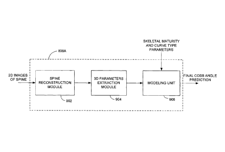
[00092] Figure 9 illustratively represents application 808a for generating a
final
Cobb angle prediction. Two-dimensional images of the spine, such as those
obtained
from radiographic imaging systems or other imaging systems, are provided to a
spine
reconstruction module 902. Three-dimensional morphology of the spine is thus
provided and a 3D parameters extraction module 904 is configured to receive
the 3D
data and extract therefrom parameters such as the initial Cobb angle, the
plane of
maximal deformation, the three-dimensional wedging of vertebral body and disk,
the
axial intervertebral rotation of the apex, upper and lower junctional level
and
thoracolumbar level, slenderness, and torsion. These parameters are provided
to a
modeling unit 906 and combined with the skeletal maturity and curve type
parameters to model the progression curve of AIS and output a final Cobb angle
prediction value. The output of the predictive model system 704 is an aid to
the
19
treating physician to determine if the risk of progression warrants additional
treatment.
[00093] In some embodiments, the predictive model system 704 is further
adapted
to sketch the curve of progression using the initial Cobb angle and the final
Cobb
angle. This curve may be output to the user via the user interface 802 or
another
output device, such as a printer. In some embodiments, the predictive model
system
704 is also adapted to select from a series of recommended treatment options
as a
function of the final Cobb angle and/or the curve of progression generated
using the
initial and final Cobb angles. The treatment options may be categorized as a
function
of ranges of final Cobb angles and/or rates of change of the curve of
progression
such that selection is made of a most appropriate recommended treatment. The
selected treatment(s) may then be output to the devices 702 for rendering
thereon
via the user interface 802 or other output device. Other embodiments for
assisting
the treating physician with treatment options once the final Cobb angle
prediction has
been generated will be readily understood by those skilled in the art.
[00094] While illustrated in the block diagrams as groups of discrete
components
communicating with each other via distinct data signal connections, it will be
understood by those skilled in the art that the present embodiments are
provided by
a combination of hardware and software components, with some components being
implemented by a given function or operation of a hardware or software system,
and
many of the data paths illustrated being implemented by data communication
within a
computer application or operating system. The structure illustrated is thus
provided
for efficiency of teaching the present embodiment. It should be noted that the
present
invention can be carried out as a method, can be embodied in a system, or on a
computer readable medium. The embodiments of the invention described above are
intended to be exemplary only.
CA 2927955 2017-08-09