Note: Descriptions are shown in the official language in which they were submitted.
W02015/061593
PCT/11S2014/062008
SENSOR WITH OPTICAL INTERFACE
10
FIELD OF INVENTION
The present invention relates to devices for detection
and measurement of carbohydrate analytes e.g. glucose. Further
aspects of the invention relate to components of such devices;
to systems including such devices including closed-loop
insulin-infusion systems; and to methods of making and using
such devices, components and systems.
BACKGROUND
The pancreas of a normal healthy person produces and
releases insulin into the blood stream in response to elevated
blood plasma glucose levels. Beta cells (13-cells), which
reside in the pancreas, produce and secrete the insulin into
the blood stream, as it is needed. If become
incapacitated or produce insufficient quantities of insulin,
then insulin must be provided to the body from another source.
Traditionally, since insulin cannot be taken orally,
insulin has been injected with a syringe. More recently, the
use of infusion pump therapy has been increasing, especially
for delivering insulin for diabetics. For example, external
infusion pumps are worn on a belt, in a pocket, or the like,
1
CA 2928082 2017-08-25
CA 02928082 2016-04-19
WO 2015/061593
PCT/US2014/062008
and deliver insulin into the body via an infusion tube with a
percutaneous needle or a cannula placed in the subcutaneous
tissue. Physicians have recognized that continuous infusion
provides greater control of a diabetic's condition, and are
increasingly prescribing it for patients.
Infusion pump devices and systems are relatively well-
known in the medical arts for use in delivering or dispensing
a prescribed medication, such as insulin, to a patient. In one
form, such devices comprise a relatively compact pump housing
adapted to receive a syringe or reservoir carrying a
prescribed medication for administration to the patient
through infusion tubing and an associated catheter or infusion
set. Programmable controls can operate the infusion pump
continuously or at periodic intervals to obtain a closely
controlled and accurate delivery of the medication over an
extended period of time. Such infusion pumps are used to
administer insulin and other medications.
There is a baseline insulin need for each body which, in
diabetic individuals, may generally be maintained by
administration of a basal amount of insulin to the patient on
a continual, or continuous, basis using infusion pumps.
However, when additional glucose (i.e., beyond the basal
level) appears in a diabetic individual's body, such as, for
example, when the individual consumes a meal, the amount and
timing of the insulin to be administered must be determined so
as to adequately account for the additional glucose while, at
the same time, avoiding infusion of too much insulin.
Typically, a bolus amount of insulin is administered to
compensate for meals (i.e., meal bolus). It is common for
diabetics to determine the amount of insulin that they may
need to cover an anticipated meal based on carbohydrate
content of the meal.
2
CA 02928082 2016-04-19
WO 2015/061593
PCT/US2014/062008
Over the years, a variety of glucose sensors have been
developed for use in obtaining an indication of blood glucose
levels in a diabetic patient. Such readings are useful in
monitoring and/or adjusting a treatment regimen which
typically includes the regular administration of Insulin to
the patient.
It has been observed that the concentration of analytes
in subcutaneous or interstitial fluid correlates with the
concentration of said analytes in the blood, and consequently
there have been several reports of the use of glucose sensors
which are sited in a subcutaneous location. Such sensors may
pass through the skin or may be remotely interrogated.
Sensors which pass through the skin may include a base
component which remains attached to the user's body, and a
removable reader component used to obtain a reading from the
sensor.
Several types of technology are available, with two of
the most common and developed being electrochemical sensing
and optical sensing. These types of sensor may be combined in
an orthogonally redundant system as described in
W02013/036943.
Small and flexible electrochemical sensors, for example
those constructed in accordance with thin film mask
techniques, can be used to obtain periodic readings over an
extended period of time.
Mansouri and Schultz (Biotechnology 1984; 2: pp. 885-
890), Meadows and Schultz (Anal. Chim. Acta. 1993 280: pp. 21-
30) and U.S. Pat. No. 4,344,438 all describe devices for the
in situ monitoring of low molecular weight compounds in the
blood by optical means. These devices are designed to be
inserted into a blood vessel or placed subcutaneously with
3
CA 02928082 2016-04-19
WO 2015/061593
PCT/US2014/062008
optical fiber connections to an external light source and an
external detector.
One form of optical sensing makes use of a proximity-
based signal generating/modulating moiety pair (discussed in
U.S. Pat. No. 6232120), which is typically an energy transfer
donor-acceptor pair (comprising an energy donor moiety and an
energy acceptor moiety). The energy donor moiety is
photoluminescent (usually fluorescent).
In such methods, an energy transfer donor-acceptor pair is
brought into contact with the sample (such as subcutaneous
fluid) to be analyzed. The sample is then illuminated and
the resultant emission detected. One moiety of the donor-
acceptor pair is bound to a receptor carrier (for example a
carbohydrate binding molecule), while the other moiety of the
donor-acceptor pair (bound to a ligand carrier, for example a
carbohydrate analog) and any analyte (for example
carbohydrate) present compete for binding sites on the
receptor carrier. Energy transfer occurs between the donors
and the acceptors when they are brought together.
An example of such donor-acceptor energy transfer is
fluorescence resonance energy transfer (Forster resonance
energy transfer, FRET), which is non-radiative transfer of the
excited-state energy from the initially excited donor (D) to
an acceptor (A).
An important characteristic of FRET is that it occurs over
distances comparable to the dimensions of biological
macromolecules. The distance at which FRET is 50% efficient,
called the Forster distance, is typically in the range of 20-
60 A. Forster distances ranging from 20 to 90 A are convenient
for competitive binding studies.
Energy transfer produces a detectable lifetime change
(reduction) of the fluorescence of the energy donor moiety.
4
CA 02928082 2016-04-19
WO 2015/061593
PCT/US2014/062008
Also, a proportion of the fluorescent signal emitted by the
energy donor moiety is quenched.
The lifetime change is reduced or even eliminated by the
competitive binding of the analyte. Thus, by measuring the
apparent luminescence lifetime, for example, by phase-
modulation fluorometry or time-resolved fluorometry (see
Lakowicz, Principles of Fluorescence Spectroscopy, Plenum
Press, 1983, Chapter 3), the amount of analyte in the sample
can be determined. The intensity decay time and phase angles
of the donor are expected to increase with increasing analyte
concentration. Thus, the FRET mechanism permits interrogation
of the equilibrium state optically by illuminating the assay
and measuring either the lifetime of the excited state
("lifetime interrogation"), and/or the intensity of the
emitted fluorescence from the donor fluorophore ("intensity
interrogation"). The latter approach is preferred, as it
exposes the assay to 25 times less light than with the
lifetime interrogation.
The FRET mechanism offers several advantages. First, FRET
fluorescence lifetime measurements are generally insensitive
to the relative position of the sensor and the reader unit as
long as they are within optical reach of each other, and are
also insensitive to changes in the environment, which helps
make the system virtually calibration free. Second, FRET is
considered very sensitive if the appropriate donor-acceptor
ratio and suitable donor-acceptor geometry are obtained. These
principles have been used in glucose sensing by energy
transfer. W091/09312 describes a subcutaneous method and
device that employs an affinity assay based on glucose
(incorporating an energy transfer donor-acceptor pair) that is
interrogated remotely by optical means. Commonly-assigned
W097/19188, W000/02048, W002/30275, W003/006992, W003/072172,
5
CA 02928082 2016-04-19
WO 2015/061593
PCT/US2014/062008
W005/059037, W005/064318, W005/110207, W006/010604,
W006/061207, W006/061208, W007/065653, W009/024521 and
W009/024522 each describe developments of such methods and
devices.
The above-described optical sensor technology offers several
advantages over other available technologies. Optical sensors
perform well in both the dermis and the subcutaneous region,
which allows the optical sensor to maintain functionality even
as the sensor is partially explanted, providing the patient
with a measurement until the patient is able to replace the
sensor. Due to the non-consuming and stable nature of the
assay, the measurement technique is insensitive to bio-
fouling. As such, It offers the possibility of one single
point calibration throughout the entire lifetime of the
sensor. Furthermore, the assay typically contains a reference
dye, which remains stable with changing glucose
concentrations, but is affected by many non-glucose induced
changes. Therefore, it serves as a sensor diagnostic tool for
the optical sensor, indicating when the integrity of the
membrane has been compromised or the optical connection is
misaligned.
Electrochemical sensors as described above have been
applied in a telemetered characteristic monitor system as
described, e.g., in commonly-assigned U.S. Pat. No. 6,809,653.
A characteristic monitoring system of the type described
above is of practical use only after it has been calibrated
based on the unique characteristics of the individual user.
Accordingly, the user is required to calibrate the sensor
externally. More specifically, a diabetic patient is required
to utilize a finger-stick blood glucose meter reading an
average of two to four times per day for the duration that the
characteristic monitor system is used. Each time, blood is
6
CA 02928082 2016-04-19
WO 2015/061593
PCT/US2014/062008
drawn from the user's finger and analyzed by the blood glucose
meter to provide a real-time blood sugar level for the user.
The user then inputs this data into the glucose monitor as the
user's current blood sugar level which is used to calibrate
the glucose monitoring system.
Such external calibrations, however, are disadvantageous
for various reasons. For example, blood glucose meters include
inherent margins of error and only provide discrete readings
at one point in time per use. Moreover, even if completely
accurate, blood glucose meters are cumbersome to use (e.g.,
one should not operate an automobile and take a finger stick
meter reading at the same time) and are also susceptible to
improper use. Furthermore, there is a cost, not to mention
pain and discomfort, associated with each application of the
finger stick. Thus, finger stick replacement remains a goal
for the next generation of glucose monitoring systems.
As sensor technology improves, there is greater desire to
use the sensor values to control the infusion of insulin in a
closed-loop system (i.e., an artificial pancreas system).
Specifically, a closed-loop system for diabetes includes a
glucose sensor and an insulin infusion pump attached to the
patient, wherein the delivery of insulin is automatically
administered by the controller of the infusion pump-rather
than by the user/patient-based on the sensor's glucose value
readings. The benefits of a closed-loop system are several-
fold, including tighter glycemic control during the night when
the majority of hypoglycemic events occur.
An accurate and reliable sensor has long been identified
as a necessity for closed-loop realization. Glucose sensor
technology has been evolving in an effort to meet the accuracy
required for finger stick replacement and the reliability
needed for consistent closed-loop functionality.
7
CA 02928082 2016-04-19
WO 2015/061593
PCT/US2014/062008
The inventors have found that performance of optical sensors
which include base and reader components is very sensitive to
the alignment of optical components in these components.
SUMMARY OF INVENTION
In a first aspect, the invention provides a device for the
detection or measurement of a carbohydrate analyte in fluid
comprising:
- an optical sensor comprising components of an assay
for carbohydrate analyte, the readout of which is a
detectable or measurable optical signal, and a light
guide having a distal portion optically coupled to
the assay components and a proximal portion; and
- a reader for interrogating the optical sensor, the
reader comprising an assay interrogating system
including a lens; and
- an interface portion forming part of at least one of
the optical sensor and the reader, the interface
portion being capable of removably constraining the
proximal portion of the light guide and the lens of
the assay interrogating system in an optically
coupled arrangement.
Preferably, the device is suitable for use in vivo. In
preferred embodiments, the proximal portion of the light guide
is disposed externally to a body of a user and the distal
portion of the light guide and the assay components are placed
internally in the user's body.
Preferably, the proximal portion of the light guide includes
a proximal end of the light guide.
Preferably, when the proximal portion of the light guide and
the lens are constrained in the optically coupled arrangement,
the proximal end of the light guide is within the optical axis
8
CA 02928082 2016-04-19
WO 2015/061593
PCT/US2014/062008
tolerance and/or the focal plane tolerance of the lens. The
optical axis tolerance and focal plane tolerance acceptable
for good performance are dependent on the fiber diameter, lens
parameters and light source intensity.
Typically, the lens optical axis is parallel to the light
guide optical axis as measured at the proximal portion of the
light guide. Optical simulations show that a deviation of 8
from parallel allows light transmission of approximately 90%
of the maximum light transmission which occurs when the
optical axes are parallel. Thus, a deviation of the angles of
the optical axes of up to 8 from parallel is acceptable.
The focal plane tolerance is discussed herein in terms of
axes x and y, parallel to the focal plane of the lens and
centered on the optical axis of the lens. During use of
preferred embodiments of the device, axis x is preferably
generally parallel to the skin of the user, and axis y is
preferably generally perpendicular to the skin of the user.
The optical axis tolerance is discussed herein in terms of
axis z, normal to the focal plane of the lens and centered on
the focal plane of the lens. During use of preferred
embodiments of the device, axis z is preferably generally
parallel to the skin of the user.
Axes x, y and z, and the focal plane, focal point and
optical axis of the lens are indicated in Figs. 7 and 8. The
point where x, y and z are 0 corresponds to the intersection
between the optical axis of the light guide and the proximal
end of the light guide being exactly at the focal point of the
lens i.e. optimal optical coupling.
Preferably, the relative position of the light guide and
lens is constrained within a tolerance range of 10 to 200 pm
in each of the x, y and z directions.
9
CA 02928082 2016-04-19
WO 2015/061593
PCT/US2014/062008
More preferably, the relative position of the light guide
and lens is constrained within a tolerance range of less than
or equal to 125 pm, preferably less than or equal to 50
pm, in the x and y directions (that is, in preferred
embodiments, the optical axis of the light guide is aligned so
that it is displaced by a maximum of at most 50 um in any
transverse direction from the optical axis of the lens).
Preferably, the relative position of the light guide and
lens is constrained within a tolerance range of less than or
equal to 125 pm, preferably less than or equal to 70 pm,
in the z direction (that is, in preferred embodiments, the
position of the proximal portion of the light guide is
constrained so that it is displaced by a maximum of at most 70
pm from the focal point along the optical axis of the lens).
Experiments indicate however that a tolerance of 250 pm may be
acceptable in the z direction.
Preferably, the device further comprises a detachable
connection between the optical sensor and the reader, the
detachable connection being capable of further constraining
the proximal portion of the light guide and the lens of the
assay interrogating system within the optically coupled
arrangement. Thus, the interface portion may constrain the
relative position of the light guide and lens in the x, y and
z directions, but more preferably the interface portion
constrains the relative position of the light guide and lens
in the x and y directions, with the relative position in the z
direction being constrained by a separate connection of the
optical sensor and reader as discussed in more detail below.
Preferably, the interface portion is a light guide
interface portion forming part of the reader as discussed in
more detail below. Generally, it is preferred for cost
reasons to include constructional features on the reader
CA 02928082 2016-04-19
WO 2015/061593
PCT/US2014/062008
rather than the optical sensor where possible, as the optical
sensor has a shorter useful lifetime.
However, some or all of the interface portion may form
part of the optical sensor. Thus, the optical sensor may
include a female portion (for example a collar around the
light guide) capable of interacting with a male portion on the
reader. For example, a collar around the light guide may
interact with a protrusion on the reader. This has the
potential advantage of protecting the light guide.
Components of Device
Components of the device include some or all of:
- optical sensor
- base including connectors
- light guide
assay compartment
- assay components
- reader
- light guide interface portion
- assay interrogating system
lens
- light detector
- other optical components including beam
splitters, filters and mirrors
- optical system housing
lens-retaining insert
- transmitter and further components
- housing including connectors
These components are discussed in more detail below.
Optical Sensor
In preferred embodiments, the optical sensor remains in
position on/in the user's body over its lifetime which may for
11
CA 02928082 2016-04-19
WO 2015/061593
PCT/US2014/062008
example be up to 7 days. The reader typically remains
connected to the sensor throughout the sensor's lifetime.
Base
Preferably, the proximal portion of the optical sensor
light guide (disposed externally to the user's body) is
mounted to a base.
The base is preferably mounted in use to the user's skin
e.g. using adhesive on its lower surface or overtaping. Taping
arrangements used in the ENLITE"" sensor may be applied.
The base may be formed partially or completely of
plastics.
Preferably, the optical sensor and reader include at
least one connection arrangement for mechanical connection, so
that the optical sensor and reader can be connected, detached
and re-connected.
More preferably, the optical sensor is adapted to connect
to the reader by means of several connection arrangements as
explained below. It will be appreciated that where one
component of a connection arrangement is described as being
part of the optical sensor and another component as being part
of the reader, the opposite is also possible. A separate
connector component may also be used, for example the further
locking component mentioned below. Connection arrangements
used in the SOFT" sensor may be applied.
As part of such a connection arrangement, the optical
sensor base preferably Includes a projecting portion for
engaging a bore of the reader. The projecting portion may
include an engaging surface, e.g. one or more 0-rings, to
facilitate engagement. This may provide a general location
for the proximal portion of the light guide, with its specific
location being determined by the light guide interface portion
discussed below. Preferably the projecting portion locates
12
CA 02928082 2016-04-19
WO 2015/061593
PCT/US2014/062008
the proximal end of the light guide within the light guide
interface portion of the reader.
As an additional or alternative part of such a connection
arrangement, the optical sensor preferably includes connectors
for detachable connection to complementary connectors of the
reader. The connectors suitably form part of the base. The
connectors of the optical sensor preferably include one or
more fasteners or latches, preferably moveable latches, for
example flexible clips (e.g. resiliently biased clips), which
may interact with fixed connectors of the reader. Preferably,
the connectors control the relative position of the light
guide and lens in the z direction.
As an additional or alternative part of such a connection
arrangement, the optical sensor and reader may comprise an
anti-rotation arrangement (e.g. a keying arrangement) to
prevent relative rotation of the optical sensor and reader
when mounted to one another. In a preferred embodiment, one
or more lugs on the reader engage complementary recesses on
the optical sensor base. The lugs/recesses may for example be
above and below the projecting portion and/or to each side of
the projecting portion.
Optionally, a further locking component (also referred to
herein as a "clip") inhibits or prevents relative movement of
the light guide and lens in the z direction, preferably by
occupying space between the optical sensor and reader. The
locking component is suitably a separate component and is
typically of plastics.
Relative movement of the lens guide and lens is
optionally limited further still by connection of the reader
to the optical sensor's overtaping e.g. via hook and loop
fastening, and/or by overtaping of the reader.
13
GA 02928082 2016-04-19
WO 2015/061593
PCT/US2014/062008
Preferably, the base at least initially includes an
injector (also referred to herein as an "insertion device" or
"serter") for positioning the distal end of the light guide in
the user's body. This may be a needle which partially or
fully encases the distal portion of the sensor e.g. a needle
of C-shaped cross-section as disclosed in W02013/036493. The
insertion device is preferably designed to minimize trauma and
maximize patient comfort and consistency of sensor delivery.
Insertion devices used in the ENLIIETm sensor may be applied.
Optionally, the base includes components of an
electrochemical sensor.
Light Guide
Preferably, the light guide comprises one or more optical
fibers. Preferably, the outer diameter of the light guide (or
alternatively of each optical fiber) is in the range of 50 to
600 pm, more preferably in the range of 200 to 300 pm (e.g.
235 to 275 pm). Optical fibers of a low diameter may not
capture sufficient light to transmit a good optical signal,
whereas optical fibers of a high diameter are potentially
painful to the user.
Preferably, the angle between the longitudinal axes of
the proximal and distal ends of the light guide is in the
range of 0 to 90 . An angle approaching 90 is preferred
for needle insertion and to keep the device height to a
minimum (as this allows the distal portion of the light guide
to be perpendicular to the user's skin while the proximal
portion is parallel to the user's skin), but a high angle may
cause light guide cladding to crack. An angle of 45 may be
used. Preferably, the light guide is flexible.
Preferably, the proximal end of the light guide includes
an end face which is preferably planar and is preferably
perpendicular to a longitudinal axis of the light guide.
14
CA 02928082 2016-04-19
WO 2015/061593
PCT/US2014/062008
The light guide is preferably of plastics. It preferably
has a cladding e.g. a cladding of thickness around 10 pm. The
light guide should have very low or no absorption of water and
other liquids.
The light guide should transmit light of the wavelengths
used for excitation, assay signal and reference with little to
no attenuation.
Assay Compartment
Preferably, the assay components are retained in one or
more assay compartments. In preferred embodiments an assay
compartment is defined by the distal part of the light guide
and a material that permits diffusion of the analyte but not
the assay components (e.g. an analyte-permeable membrane).
Preferred materials are co-polymers having hydrophobic units
and hydrophilic units, the hydrophilic units each comprising
an ester of polyethylene glycol and a diacid, as disclosed in
W005/1102007. Particularly preferred materials are
1000PEGT8OPBT20 disclosed therein and 1000PEGT7OPBT30.
Suitably, the assay compartment is at or close to the
distal end of the light guide. The assay compartment may lie
wholly or partly within a recess or through hole of the light
guide. Examples of such designs include a laser-drilled hole,
or a rectangular cavity in the side wall of the light guide.
Assay Components
Preferred assay components are discussed in
W02013/036943.
In preferred embodiments, the analyte is glucose.
The assay is preferably a competitive assay.
Preferably, the assay components include an analyte
binding molecule labelled with one of a proximity based signal
generating/modulating moiety pair; and an analyte analog
capable of competing with the analyte for binding to the
CA 02928082 2016-04-19
WO 2015/061593
PCT/US2014/062008
analyte binding molecule labelled with the other of the
proximity based signal generating/modulating moiety pair. The
assay may further include a reference fluorophore which
serves, inter alia, as a sensor diagnostic tool.
Preferably, the assay components include a carbohydrate
binding molecule and a carbohydrate analog, and energy donor
and acceptor moieties (also referred to as "the FRET pair")
which provide an optical signal. The energy acceptor moiety
has an absorption spectrum overlapping the energy donor
moiety's emission spectrum. More preferably, the energy
acceptor moiety is non-fluorescent.
In preferred embodiments, the assay is a competitive
glucose binding affinity assay that includes a glucose
receptor (the carbohydrate binding molecule), a glucose
.. analog, a first fluorophore (the energy donor moiety) labeled
onto the glucose receptor, and an acceptor dye (the energy
acceptor moiety) labeled onto the glucose analog.
A preferred carbohydrate binding molecule is labelled MBL
(mannose binding lectin). Concanavalin A is another
carbohydrate binding molecule of interest.
Preferred carbohydrate analogs are labelled macromolecules
bearing carbohydrate or carbohydrate mimetic moieties.
Examples include optionally derivatized labelled dextran e.g.
110 kDa dextran; labelled synthetic polymers bearing pendant
carbohydrate or carbohydrate mimetic moieties; labelled
proteins bearing pendant carbohydrate or carbohydrate mimetic
moieties. Such carbohydrate analogs are disclosed for example
in W007/065653 and W006/061208.
Preferred energy donor moieties are Alexa Fluor
fluorophores, Texas Red, and Cy5. Alexa Fluor 594 (AF594) is
particularly preferred. AF647, QSY 21, and AF750 are
16
CA 02928082 2016-04-19
WO 2015/061593
PCT/US2014/062008
appropriate for use in conjunction with a laser diode source
at 645 nm.
A preferred energy acceptor moiety is hexamethoxy
crystalviolet-1 (HMCV1, a proprietary crystal violet
.. derivative manufactured by Medtronic, Inc.), disclosed in
W005/059037. This is particularly suitable where the
carbohydrate analog molecule is dextran.
A preferred reference fluorophore is Alexa Fluor 700
(AF700). The reference fluorophore is preferably labeled onto
Human Serum Albumin (HSA) or another macromolecule which does
not bind significantly to the carbohydrate binding molecule.
In preferred embodiments of the invention, it has been
found that a degree of labeling (DOL) with AF594 of about 0.8-
1 AF594/CRD and 5 HMCV1 molecules per dextran molecule gives
optimal dose-response.
The assay components may also comprise a protective
formulation for radiation sterilization.
Reader
The reader is also referred to as the "recording device"
or "Glucose sensor transmitter or recorder" (GST/GSR).
The reader is used to interrogate the optical sensor, and
can be removably physically and optically coupled thereto.
The reader preferably includes a housing, e.g. of
plastics. The reader may be wearable on the body of the user,
and may be sized so as to have a volume of no more than 15 cm3
(e.g. about 11 cm3) and a weight of about 10 g. Preferably, the
reader has a life of 2 years or more. The reader may need to
be charged periodically e.g. every 15 days.
The reader preferably includes components of connection
.. arrangements for detachable connection to the optical sensor.
This is discussed in more detail above. In preferred
embodiments the reader Includes: a bore for engaging a
17
CA 02928082 2016-04-19
WO 2015/061593
PCT/US2014/062008
projection portion of the optical sensor; fixed connectors for
connection to moveable connectors of the optical sensor; anti-
rotation lugs for engaging recesses of the optical sensor.
Some or all of these features are suitably formed as part of
reader housing.
Assay Interrogating System
The reader includes an assay interrogating system which
receives an optical signal from the optical sensor via the
lens referred to above. The assay interrogating system is
preferably an optoelectronic interrogating system. The assay
interrogation system may operate via lifetime and/or intensity
Interrogation of the assay as discussed above.
The lens is preferably a focusing/converging lens e.g. a
biconvex lens. The lens is preferably of plastics.
The assay interrogating system suitably includes one or
more light detectors e.g. photodiodes. In use, an optical
signal in the form of light reaches the proximal end of the
light guide, and is focussed via the lens and then transmitted
to the light detectors.
Preferably, the assay interrogating system includes an
illumination source e.g. an LED or a red laser diode, with the
latter enabling a substantial reduction in the size and volume
of the reader. AF647, QSY 21, and AF750 may be used in
conjunction with a laser diode source at 645 nm. Typically,
the illumination source is used to interrogate the assay via
the light guide.
The assay interrogating system may include filters for
example in the form of a filter substrate having one or more
coatings to effect, e.g., an excitation filter and/or one or
more emission filters.
18
CA 02928082 2016-04-19
WO 2015/061593
PCT/US2014/062008
The assay interrogating system may include further
components e.g. beam splitters and/or mirrors. Typically beam
splitters are present, but mirrors may not be present.
Components of the assay interrogation system are
preferably included in an optical system housing sub-unit of
the reader, which is preferably mounted to the reader housing.
Components of the assay interrogation system are
preferably electrically connected to a printed circuit board
assembly (PCBA). The components of the assay interrogating
system may be mounted to the PCBA via alignment pins and/or
screws, or the components may be electrically connected to the
PCBA via flex connectors.
As an alternative to separate optical components, the
assay interrogating system may be manufactured as a wafer-
scale stacked planar integrated optical system (SPIOS) and
diced into smaller units. A Stacked Planar Integrated Optical
System (SPIOS) may be created by fixing one multi-functional
filter layer between two injection molded layers of optical
components to forms a solid block, which is self-supporting.
Light Guide Interface Portion and Lens
The reader preferably includes a light guide interface
portion (also referred to herein as an "interface portion") of
the reader. This is suitably a block of tightly controlled
dimensions containing a blind or through opening. The light
guide interface portion of the reader is preferably mounted to
the optical box e.g. via an interference fit.
Preferably, the light guide interface portion comprises a
female part, e.g. a flared opening, adapted to receive the
proximal portion of the light guide.
The term "flared opening" includes arrangements with a
narrow portion proximal to the lens and a wider portion distal
to the lens. The proximal portion of the flared opening
19
CA 02928082 2016-04-19
WO 2015/061593
PCT/US2014/062008
preferably ends in a light guide alignment channel adapted to
accommodate the proximal end of the light guide. Suitably,
the alignment channel is a circular cross-section channel of
diameter slightly larger (e.g. up to 50 pm larger, more
preferably up to 30 pm larger) than the light guide. The
flared opening guides the light guide proximal end face into
its optical coupling position with the lens, without damaging
its surface.
The flared opening preferably comprises one or more
chamfers i.e. continuous curved and/or planar surfaces which
are not interrupted by edges, projections or other abrupt
discontinuities in shape.
The flared opening of the light guide interface portion
of the reader is preferably partially or completely conical or
frusto-conical in shape. The cone half angle (or effective
half angle) is preferably in the range of 25 to 43 e.g.
around 30 . A half angle which is too high may prevent the
light guide from being guided into position correctly.
However, the opening need not be conical, so that the
half angle may vary from the proximal to the distal portion of
the flared opening, or around the flared opening. For
example, an arrangement with two or more chamfers, e.g. a
double chamfer arrangement, may be used, as shown in Fig. 10.
In the light guide interface portion on the left, a double
frusto-conical arrangement is shown with an outer half angle
of 45 and an inner half angle of 20 . In the light guide
interface portion on the right, a single frpstoconical
arrangement is shown with a half angle of 30 . However, a
single chamfer arrangement may be preferred, because of the
lack of an edge which could cause wear as discussed below.
Preferably, the flared opening of the light guide
interface portion has a very smooth surface. This is to avoid
CA 02928082 2016-04-19
WO 2015/061593
PCT/US2014/062008
wear of the light guide on contact with the surface. Such
wear could damage the light guide cladding, resulting in
debris which blocks light transmission.
Smoothness may be measured as Roughness Average.
Preferably, the flared opening has a surface of Roughness
Average 16 to 32 microinch over a 0.004 inch long roughness
cut-off (0.8 to 1.6 pm over a distance of 100 pm).
The flared opening of the light guide interface portion
may be built to a particular smoothness specification or
polished to achieve this, as discussed below.
The assay interrogating system of the reader has optical
components including a lens, as discussed above.
Accurate positioning and angling of the lens are
important in ensuring that the relative positioning of the
light guide and lens is within the desired tolerance ranges
set out above.
The lens is preferably mounted to the light guide
interface portion of the reader. Suitably the light guide
interface portion includes a recess in which the lens is
positioned such that it can be optically coupled to the light
guide. Suitably the recess comprises a retaining lip or other
similar arrangement to control the position of the face of the
lens adjacent to the optical sensor.
The lens is preferably held in place within the light
guide interface portion using a lens-retaining insert.
In alternative arrangements, however, the lens and/or
lens-retaining insert may be mounted directly to the reader
housing, or to the optical system housing, e.g. by means of
adhesive or a set screw, or the lens may be integrally formed
with one of these components.
The lens-retaining insert may be held in position by a
screw thread arrangement (preferably with at least a count of
21
CA 02928082 2016-04-19
WO 2015/061593
PCT/US2014/062008
3 to 4), by a press fit/interference fit arrangement (e.g.
using crushed ribs), by a resilient arrangement (e.g. using a
spring) and/or using adhesive. A combination of a screw thread
arrangement and adhesive is envisaged. The lens-retaining
insert must not interfere with the optical signal. The
adhesive should transmit light in the near IR and visible
regions, and should not fluoresce, particularly in those
regions.
Preferably, the internal surfaces of the light guide
interface portion and/or the optical system housing or reader
housing in which the lens is mounted are optically black so as
to reduce surface reflections (from signal or stray light)
from reaching the light detectors. Suitably, these surfaces
absorb wavelengths in the visible range (e.g. 300-800 nm).
This may be achieved using coatings, e.g. having surface color
and/or associated fine texture. The use of such coatings is
particularly important if reflective metals are used in
forming the device. Preferably, the internal surfaces are
black in color. Black sealing tape may be used.
Suitable materials for the light guide interface portion
include metals, plastics and ceramics. In a preferred
embodiment the material used is steel e.g. stainless steel.
Alternative materials include aluminum, titanium, KOVARTM,
INVART' and other alloys used in optical applications, plastics
such as polyoxymethylene (POM, DELRINTI, PVC, cyclic olefinic
copolymers (COP/COC, TOPAS", ZEONEXT', ZEONORTm etc.). KOVART'
and INVART' are preferred choices because of their low
coefficients of thermal expansion.
Preferred plastics are optically black as discussed
above. Metals can be oxidized (e.g. aluminum to aluminum hard
or soft oxide coating, steel to black oxide and so on) or may
22
CA 02928082 2016-04-19
WO 2015/061593
PCT/US2014/062008
have black material deposited or impregnated on the surface to
absorb light in the visible or near IR range.
The lens-retaining insert is formed of steel or stainless
steel in preferred embodiments. The materials used for the
light guide interface portion and lens-retaining insert should
ideally be of the same type to match coefficients of thermal
expansion.
Further Components of Reader
The reader is preferably capable of reading, filtering,
processing and/or transmitting optical signal values
representing analyte concentration values. Preferably, the
reader includes instrumentation to convert an optical signal
from the optical sensor to an analyte concentration value.
The reader may further comprise a transmitter for transmitting
detected or measured analyte data.
Formation of Components
As explained above, the dimensions of the components, in
particular the interface portion and the lens-retaining insert
must be tightly controlled, and a smooth surface on the
interface portion is desirable.
These components are preferably formed by molding or
machining.
Multi-axis machining methods are appropriate to provide
the desired tightly controlled dimensional tolerances. Swiss
screw turning/machining is the preferred method. Deterministic
machining and laser machining may be used.
For molded parts micro-molding is the preferred method.
Preferably, the molding tools are precisely machined tools
formed using multi-axis machining methods.
Preferably, the reader components are cleaned e.g. to
remove particulates, debris, dirt, machine oils and/or low
23
CA 02928082 2016-04-19
WO 2015/061593
PCT/US2014/062008
surface tension agents. Suitable solvents for cleaning include
IPA, hexane, acetone and THE.
As mentioned above, the flared opening of the light guide
interface portion may be built to a particular smoothness
specification or polished to achieve this. It is important to
use polishing materials which cannot impregnate the light
guide interface portion and thereby abrade the light guide.
For this reason, diamond polishing is not preferred. Hard
wood is a preferred polishing material.
Implantation of Sensor
The distal portion of the sensor is preferably introduced
subcutaneously or within the skin of a user e.g. Into the
dermis or epidermis. Alternatively it may be implanted in
and/or through inter-peritoneal or peritoneal tissue. The user
is preferably a human.
Preferably, the distal portion of the sensor is implanted
or injected e.g. using a needle which may at least initially
form part of the optical sensor as mentioned above.
Preferably, the needle does not remain in the skin.
Systems Including Device
Optionally, the device also includes one or more non-
optical sensors for the carbohydrate analyte e.g.
electrochemical sensors as mentioned above. The
electrochemical sensor may include a plurality of electrodes.
Respective distal portions of the optical sensor and the non-
optical sensor may be co-located within the user's body, and
may be implanted together.
The device may form part of a system further comprising a
hand-held monitor haying a display and/or an insulin pump. The
system may be a closed-loop system, with predictive
diagnostics and minimal requirements for external calibration.
24
CA 02928082 2016-04-19
WO 2015/061593 PCT/US2014/062008
The reader may wirelessly transmit analyte values to the hand-
held monitor and/or insulin pump.
Further Aspects of Invention
In a second aspect, the invention relates to a reader for
use in a device as described above, comprising an assay
interrogating system including a lens; and a light guide
interface portion comprising a flared opening adapted to
receive a proximal portion of a light guide.
In a third aspect, the invention relates to a reader for
use in a device as described above, comprising a housing; an
assay interrogating system including a lens, and a lens-
retaining insert holding the lens in position within the
housing.
In a fourth aspect, the invention relates to a method of
detecting or measuring a carbohydrate analyte using a device
as described above, comprising detecting or measuring the
optical signal readout of the assay components via the light
guide using the assay interrogating system of the reader. The
method may further comprise initial implantation of the assay
components and distal portion of the light guide into the body
of a user.
Preferably, and suitably between implantation and
detection or measurement, the method further comprises a step
of optically coupling the proximal portion of the light guide
to the lens of the assay interrogating system via the
interface portion. Preferably, the optical sensor and the
reader are also connected via the connection arrangement.
The method may also include a step of separating the
optical sensor and reader such that the proximal portion of
the light guide and the lens of the assay interrogating system
are no longer coupled. Optionally, the optical sensor is then
replaced with a further optical sensor.
CA 02928082 2016-04-19
WO 2015/061593
PCT/US2014/062008
In a fifth aspect, the invention relates to an interface
portion as described above.
All features described in connection with any aspect of
the invention can be used with any other aspect of the
invention.
DRAWINGS
The invention will be further described with reference to
a preferred embodiment, as shown in the drawings in which:
Fig. 1 shows schematically a preferred embodiment of the
device of the invention, including the assay interrogating
system.
Fig. 2 shows the distal portion of the light guide of the
optical sensor of Fig. 1 in more detail.
Fig. 3a is a side view of a needle for housing and
deploying the distal portion of the light guide of the optical
sensor of Fig. 2. Fig. 3b is a perspective view of the
connected optical sensor (left) and reader (right) of the
preferred embodiment of the invention, showing the position of
the needle of Fig. 3a before injection.
Fig. 4 is a perspective view of the optical sensor (left)
and reader (right) of Fig. 3b before connection.
Fig. 5 is a plan view of the connected optical sensor and
reader of Fig. 3b, with the upper shell of the reader housing
removed.
Fig. 6 is a cross-sectional view of the connection
between the optical sensor and reader of Fig. 5.
Fig. 7 is a cross-sectional view of the lens fiber
interface portion of Fig. 6, also showing the proximal portion
of the optical fiber, the lens and the lens-retaining insert.
Fig. 8 is a perspective view of the optical sensor of
Fig. 4, showing an enlarged view of the projecting portion.
26
CA 02928082 2016-04-19
WO 2015/061593
PCT/US2014/062008
Fig. 9 is a perspective view of the reader of Fig. 4.
Fig. 10 is a cross-sectional view of two lens fiber
interface portions alternative to that of Fig. 7.
Fig. 11 is a perspective view of an alternative
embodiment of the Invention, including a locking component.
The locking component is shown before and after engagement
with the sensor and reader (top and bottom respectively).
DETAILED DESCRIPTION
In the following description, reference is made to the
accompanying drawings which form a part hereof and which
illustrate several embodiments of the present invention. It is
understood that other embodiments may be utilized and
structural and operational changes may be made without
departing from the scope of the present invention.
DEFINITIONS
The term "optical axis" in relation to a lens refers to an
imaginary line that defines the path along which light
propagates through the system. Often but not necessarily this
coincides with the mechanical axis and axis of rotational
symmetry of the lens. The lens optical axis is shown as 14 in
Fig. 7.
The term "optical axis" in relation to a light guide refers
to an imaginary line that defines the path along which light
propagates through the system. Often but not necessarily this
coincides with the longitudinal axis and axis of rotational
symmetry of the light guide. Where the light guide is not
straight, the optical axis may be defined in terms of a cross-
section. If the cross-section is cut such that the shape of
the section is the same as the shape of the proximal end,
typically the optical axis is a line that travels
27
CA 02928082 2016-04-19
WO 2015/061593
PCT/US2014/062008
perpendicular to the cross-sectional face and through its
centroid. The light guide optical axis is shown as 16 in Fig.
7.
The term "focal point" in relation to a lens refers to a
point on the lens optical axis at which initially collimated
rays are brought to a focus, e.g. in air; it is separated from
the lens by the focal distance. A lens will typically have
front and rear focal points. The focal point referred to
herein is generally that on the same side as the light guide.
The focal point is shown as 10 in Fig. 7.
The term "focal plane" in relation to a lens refers to a
plane perpendicular to the lens optical axis and containing
the focal point. This may also be referred to as the "back
(or rear) focal plane". The focal plane is shown as 12 in
Fig. 7.
The term "focal plane tolerance" (or transverse tolerance)
as used herein refers to the range of positions of the light
guide optical axis within the focal plane wherein coupling
between the light guide and lens is such that light
transmission is at least 80% of the maximum light transmission
which occurs when the light guide optical axis is at the focal
point of the lens.
The term "optical axis tolerance" (or axial tolerance) as
used herein refers to the range of positions of the proximal
end of the light guide along the lens optical axis wherein
coupling between the light guide and lens is such that light
transmission is at least 80% of the maximum light transmission
which occurs when the proximal end of the light guide is at
the focal point of the lens.
In the discussion herein, preferred embodiments of the
devices, systems, and methods of the invention are described
with reference to glucose as the analyte whose
28
W02015/061593
PCT/US2014/062008
level/concentration in the blood and/or bodily fluids of the
user is to be determined. However, this is by way of
illustration and not limitation, as the principles, devices,
systems, and methods of the present invention may be used for
sensing and/or determining the level of a variety of other
physiological parameters, agents, characteristics, and/or
compositions.
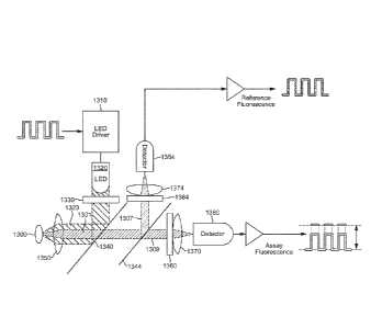
The preferred embodiment of the device is shown
schematically in Fig. 1. The device includes an optical sensor
1300, shown in more detail in the following figures.
Optical Sensor of Preferred Embodiment
The optical sensor 100 includes a base 130 (Figs. 4, 8)
and an optical fiber 110 (also referred to herein as a "light
guide") having a proximal portion 116 (Fig. 2) mounted to the
base 130 as explained in more detail below.
The optical fiber 110 is formed of plastics having
tensile and fatigue properties that ensure robustness.
The proximal portion 116 of the optical fiber 110
terminates in a proximal end 117 (in the form of a planar face
perpendicular to the mechanical/optical axis 16 of the optical
fiber 110 (Figs. 2, 7).
The distal portion 112 of the optical fiber 110 (Fig. 2)
is designed for insertion into a user's body as described in
more detail below. A glucose-permeable membrane of
PolyActive(TM) (a biocompatible, biodegradable polymer
1000PEGT7OPBT30 from Integra Orthobiologics, Irvine, CA) is
heat sealed to the fiber's distal end 115 to form an assay
compartment 120 housing assay components 125.
The optical sensor base 130 is of polycarbonate. It is
generally wide and flat in form, with a planar lower plate 135
(Fig. 5). The lower plate 135 is provided on its lower
surface with a contact adhesive sheet (not shown) for mounting
29
CA 2928082 2017-08-25
CA 02928082 2016-04-19
WO 2015/061593
PCT/US2014/062008
to a user's skin, the adhesive being initially protected by a
cover sheet (not shown). The adhesive is capable of strong
adhesion for 7 days.
The base has a front end for connection to the reader 200
as described in more detail below, and a back end (Fig. 4). A
projecting portion 140 for insertion into the reader 200
extends from the front end of the optical sensor base 130
parallel to the lower plate 135; the projecting portion 140 is
generally cylindrical in shape. The projecting portion 140
has two annular recesses, each being provided with an 0-ring
145 (Figs. 4, 6, 8).
A connector in the form of a resiliently biased flexible
arm 150 extends along each side of the optical sensor base 130
from its back end to beyond its front end, separated from the
body of the optical sensor base 150 by a space (Figs. 4, 5,
8). The front end of each arm 150 terminates in a latch 155,
such that there is a latch 155 on either side of the
projecting portion 140. The arms 150 can be flexed inwards
towards the projecting portion 140 but will return to their
initial positions when released. Grips 160 in the form of
ribs are provided on the outer sides of the arms to assist in
flexing.
Above and below the projecting portion 140 of the optical
sensor base 130, the optical sensor base 130 contains recesses
165 (Fig. 8) which form part of an anti-rotation arrangement
as described below.
The optical sensor base 130 contains a through channel
170 (Fig. 6) extending from the back end to the projecting
portion 140. The optical fiber 110 passes through the channel
170 and its proximal end 117 protrudes from a front face of
the projecting portion 140 (Figs. 4, 8). The optical fiber 110
is held in place within the optical sensor base 130 by UV-
CA 02928082 2016-04-19
WO 2015/061593
PCT/US2014/062008
curing adhesive which is back-filled through the channel 170.
The proximal end 117 of the optical fiber 110 is initially
covered by a protective cap of vinyl plastics (not shown)
which is held in place by the 0-rings 145.
An insertion device 500 passes through a channel in the
optical sensor base 130 at an angle of approximately 45 to
the lower plate 135 (Figs. 3a and 3b). The insertion device
500 relies on a disposable, automatically retracting needle
510 of C-shaped cross-section which is designed with the
optical sensor base 130 to deliver the distal portion 112 of
the optical sensor 100 through the user's skin at an angle of
approximately 45 .
Reader of Preferred Embodiment
The reader 200 (Figs. 4, 5, 6, 9) comprises a two-part
housing of polycarbonate/acrylonitrile butadiene styrene
blend (BAYBLENDT1 having a base and an upper shell. The lower
surface of the reader base is provided with one element of
hook and loop fastening (not shown).
The reader 200 houses the optical system used to
interrogate the assay (also referred to herein as the -assay
interrogating system").
The optical system (Figs. 1, 5) is essentially a modified
epi-fluorescence set-up with one light source to excite (i.e.
illuminate) the assay and two detectors to detect the
fluorescence emitted from the assay and the internal
reference, respectively. As noted, the intensity of the
emitted fluorescence correlates to the glucose concentration.
Here, the measured intensity of the emitted fluorescence is
affected by the intensity of the light source and the coupling
between the assay and the optical system.
A driver circuit 1310 modulates a LED 1320 at a low
frequency (solely with the purpose of eliminating the 1/f
31
W02015/061593
PCT/US2014/062008
noise and canceling out ambient light) with a wavelength range
capable of simultaneously exciting the assay and reference
fluorophores. The LED output is filtered using a multilayer
dielectrical filter 1330 to select a distinct wavelength
region. The filtered LED output is reflected by a first
dichroic beam splitter 1340 and focused onto the optical
sensor 1300, which includes the assay and the reference
(also referred to herein as the "reference fluorophore"), by a
lens 1350. The interface between the optical sensor and the
lens 1350 is described in more detail below.
The assay and the reference emit fluorescence. The
emitted fluorescence 1301 and the reflected excitation light
1323 are picked up and collimated by the lens 1350. The first
dichroic beam splitter 1340 transmits the fluorescence 1301.
However, it reflects the majority of the back reflected
excitation light 1323. A second beam splitter 1344 reflects
the reference fluorescence 1307 at a 90 angle, but it
transmits the assay fluorescence 1309. A first emission filter
1360 with a distinct wavelength region red shifted with
respect to, and not overlapping, the pass band of the
excitation filter and matching the desired part of the assay
fluorescence spectrum then blocks the remaining part of the
excitation light and transmits the assay fluorescence.
Similarly, a second emission filter 1364 with a distinct
wavelength region red shifted with respect to, and not
overlapping, the pass band of the excitation filter and
matching the desired part of the assay fluorescence blocks the
remaining part of the excitation light and transmits the
reference fluorescence 1307. Thus, in effect, only the
fluorescence from the assay and the fluorescence from the
reference are focused onto their respective photo detectors
(also referred to herein as "light detectors") 1380, 1384
32
CA 2928082 2017-08-25
CA 02928082 2016-04-19
WO 2015/061593
PCT/US2014/062008
using respective lenses 1370, 1374. The ratio between the
detected assay fluorescence and the detected reference
fluorescence correlates with the glucose concentration in the
assay.
The optical system is mounted within the reader housing
in an optically black optical system housing 202 (Fig. 5).
Components of the optical system are electrically connected to
a printed circuit board assembly 204.
The light guide interface portion 220 of the reader 200
is now described.
The reader housing includes a generally cylindrical bore
205 into which the projecting portion 140 of the optical
sensor base 130 is to be inserted (Figs. 6, 9). The inside
surface of the bore 205 engages the 0-rings 145 of the optical
sensor base 130 (Fig. 6). Two fixed connectors in the form of
projections 210 complementary to the latches 155 on the
optical sensor base 130 are located at the mouth of the reader
housing bore 205 (Fig. 5). Above and below the bore 205, the
reader housing is provided with lugs 215 as part of the anti-
rotation arrangement (Fig. 9).
The light guide interface portion 220 of the reader 200
is mounted within the optical system housing 202 at the
internal end of the reader housing bore (Figs. 5, 6). This
light guide interface portion 220 is shown in Fig. 7.
The light guide interface portion 220 is generally in the
form of a stepped cylindrical block having a through channel
as described below. The block is formed by machining. At the
inner end of the block as defined below, the block wall is
notched such that it forms three fingers (not shown).
From the outer end to the inner end (i.e. moving away
from the optical sensor 100 when connected to the reader 200
in use) the channel has a smooth flared opening in the form of
33
WO 2015/06093
PCMS2014/062008
a frustoconical portion 225 of half angle 30 with a wide
mouth 232; a narrow light guide alignment channel 230Aof
constant diameter extending from the narrow end 234 of the
frustoconical part 230B and a wide recess 235 in which the
lens 1350 is mounted, the wide recess 235 being defined by the
three fingers of the block wall, which are provided with an
internal screw thread.
The lens 1350 is a biconvex convergent lens of focal
length approximately 2 mm. The lens 1350 is mounted with its
optical axis 14 parallel to and aligned with the light guide
alignment channel 230A. The lens 1350 is held in place by a
generally tubular lens-retaining insert 240 of complementary
shape to the lens 1350. The lens-retaining insert 240 has an
external screw thread which engages the internal screw thread
of the light guide interface portion 220. The lens-retaining
insert 240 is also fixed in position by means of adhesive.
The assay photodetector 1380 is positioned within the lens-
retaining insert 240 such that light is focused onto it by the
lens 1350.
The reader 200 houses further components (not all shown)
including diagnostics, one or more microprocessors and/or
digital signal processors (DSPs), memory, a RF communication
chip (using, e.g., 2.4 GHz TelD protocol), and a rechargeable
battery 250. The reader is capable of the conversion of
signals received from the sensors to glucose values and of
wireless communication, including transmission of the glucose
values (or an averaged, weighted, or otherwise modified
version thereof) to a monitor, an infusion pump or a display
device (not shown).
The reader includes electrical contacts 245 for interface
with a charger (not shown) and with the optical sensor base
130.
34
CA 2928082 2020-03-20
WO 2015/061593
PCT/US2014/062008
Use of Device
In use, the optical sensor base 130 is mounted to the
skin of a user by removal of the cover sheet and application
of the adhesive to the skin. The distal portion 112 of the
optical fiber 110 of the optical sensor 100, including the
assay compartment 120, is implanted subcutaneously using
insertion device 500. The needle 510 automatically retracts.
The optical sensor base 130 is further secured to the skin by
overtaping (not shown) to reduce the risk of the optical
sensor 100 being pulled out of the skin accidentally. The
overtaping has an element of hook and loop fastening (not
shown) complementary to that on the reader base.
The reader 200 is connected to the optical sensor 100 as
follows.
The projecting portion 140 of the optical sensor base 130
is pushed into the bore 205 of the reader 200. The positional
tolerance of the projecting portion 140 within the bore 205 is
approximately 0.02 inches ( 500 pm) in the x and y
directions. This locates the proximal end 117 of the optical
fiber 115 within the frustoconical portion 225 of the light
guide interface portion 220 of the reader 200.
The frustoconical portion 225 serves to direct the
proximal end 117 of the optical fiber 115 into the light guide
alignment channel 230A so that it is constrained in the x and y
directions. The positional tolerance of the optical fiber 115
inside the light guide alignment channel 230A is approximately
10 pm.
The arms 150 of the optical sensor base 130 are flexed
inwards using the grips 160. When the optical sensor
projecting portion 140 is fully inserted into the reader 200,
the arms 150 are released and return to their initial
positions, wherein the latches 155 engage the projections 210
CA 2928082 2020-03-20
CA 02928082 2016-04-19
WO 2015/061593
PCT/US2014/062008
of the reader 200 such that the optical sensor 100 and reader
200 cannot be separated by pulling apart. The 0-
rings 145 of
the optical sensor 100 engage the bore 205 of the reader 200
to prevent relative movement of the optical fiber 110 and the
lens 1350 of the reader in the z direction. The 0-rings 145
provide a radial seal between the optical sensor 100 and the
reader 200 to water-resistance standard IPX8.
When the optical sensor 100 and reader 200 are connected,
lugs 215 of the reader 200 engage recesses 165 of the optical
sensor 100 in an anti-rotation arrangement, preventing
relative rotation of the optical sensor 100 and reader 200.
Thus, the proximal end 117 of the optical fiber 110 is
constrained at or very close to the focal point 10 of the lens
1350, with the lens optical axis 14 and optical fiber optical
axis 16 aligned.
Relative motion between the reader 200 and sensor 100 is
further prevented by engagement of the complementary hook and
loop fastening elements of the reader base and the sensor
overtaping.
The assay interrogating system is interrogated as
explained above. As shown in Fig. 2, excitation light travels
from the proximal end 117 of the optical fiber 115 to the
assay components 125, and the fluorescence response travels
back up the optical fiber 110 to the assay interrogating
system. Light from the proximal end 117 of the optical fiber
115 is focused by the lens 1350 onto the light detector 1380.
Accurate relative positioning of the proximal end 117 of the
optical fiber 110 and the lens 1350 ensures that light is
efficiently coupled from the optical fiber 110 into the assay
interrogating system.
To separate the optical sensor 100 from the reader 200,
the optical sensor connector arms 150 are flexed inwards to
36
CA 02928082 2016-04-19
WO 2015/061593
PCT/US2014/062008
release the latches 155 from the reader connector projections
210, after which the reader 200 slides easily away from the
optical sensor 100.
In an alternative embodiment shown in Fig. 11, a further
separate locking component 520 is provided to inhibit relative
motion between the reader 200 and optical sensor 100. The
locking component 520 is of plastics, and is adapted to engage
the upper surface of the optical sensor base 130 and adjacent
parts of the shell of the reader 200 when the optical sensor
100 and reader 200 are connected. Thus, the locking component
520 is generally in the form of a plate complementary to the
upper surface of the optical sensor base 130, but with a space
550 at the forward central part. Descending parts 530 of the
locking component 520 correspond to the spaces between the
sensor body and the flexible arms 150. Forward wings 540 of
the locking component 520 are complementary to the upper
surface of adjacent parts of the shell of the reader 200. A
tab 560 extends from each wing 540 into the space 550.
The locking component 520 is connected to and
disconnected from the connected optical sensor 100 and reader
200 vertically (i.e. in the y direction). The locking
component 520 lies generally above the optical sensor base
130, with forward wings 540 engaging the shell of the reader
200. Parts 530 engage the spaces defined by the optical sensor
base 130, flexible arms 150 and reader body, preventing the
optical sensor base 130 from moving further towards the reader
200. Tabs 560 occupy positions between the optical sensor 100
and reader 200 to either side of the lugs 215/ recesses 165 of
the anti-rotation arrangement described above, preventing the
optical sensor base 130 from moving away from the reader 200.
In this way, the locking component 520 prevents relative
37
CA 02928082 2016-04-19
WO 2015/061593
PCT/US2014/062008
movement between the optical sensor 100 and reader 200,
especially in the z direction.
EXAMPLES
Comparative Example
In an optical sensor with the optical fiber optical axis
parallel to the lens optical axis, it was found that if the
optical fiber optical axis was not within 50 pm of the lens
optical axis in the x and y directions, the optical signal was
reduced by 20 to 50 %. Theory suggests that if the optical
fiber optical axis was not within 100 pm of the lens optical
axis, the optical signal would be reduced by 80 %.
It was also found that if the proximal end of the optical
fiber was not within 200 pm of the lens focal point in the z
direction, the optical signal was reduced by 20 '8
(experimental results) to 30 t (theoretical results). The
relationship between distance and performance was not linear,
and performance dropped off at an increasing rate as the
proximal end of the optical fiber was moved away from the
focal point.
Advantages of the preferred embodiments of the invention
include:
- the tolerance in the x and y directions permitted by the
light guide alignment channel of the light guide
interface portion of the reader is very low, whereas the
tolerance in the z direction permitted by the connection
between the reader and the optical sensor is slightly
higher. It was a contribution of the present inventors
to realize that higher tolerance in the z direction is
acceptable. It was a further contribution of the present
Inventors to appreciate that different arrangements can
38
W02015/061593
PCT/US2014/062008
be used to constrain movement in the x and y directions
and the z direction.
- The smooth flared opening of the light guide interface
portion assists in insertion of the optical fiber into
the very narrow light guide alignment channel. Without
this feature it would be difficult to insert the delicate
optical fiber into the light guide channel without
damaging the optical fiber.
- The light guide interface portion is included on the
reader rather than the optical sensor. The reader has a
longer life than the optical sensor, and so this type of
arrangement is more cost-effective.
While the description above refers to particular
embodiments of the present invention, it will be understood
that many modifications may be made without departing from the
spirit thereof. The accompanying claims are intended to cover
such modifications as would fall within the true scope and
spirit of the present invention.
The presently disclosed embodiments are therefore to be
considered in all respects as illustrative and not
restrictive, the scope of the invention being indicated by the
appended claims, and all changes which come within the meaning
and range of equivalency of the claims are therefore intended
to be embraced therein.
In this specification, unless expressly otherwise
indicated, the word 'or' is used in the sense of an operator
that returns a true value when either or both of the stated
conditions is met, as opposed to the operator 'exclusive or'
which requires that only one of the conditions is met. The
word 'comprising' is used in the sense of 'including' rather
than to mean 'consisting of'.
39
CA 2928082 2017-08-25
WO 2015/061593
PCT/US2014/062008
No
acknowledgement of any prior published document herein should
be taken to be an admission or representation that the
teaching thereof was common general knowledge at the date
hereof.
CA 2928082 2017-08-25