Note: Descriptions are shown in the official language in which they were submitted.
CA 02928201 2016-04-20
WO 2015/066617
PCT/US2014/063697
FOLEY CATHETER WITH RING ELECTRODES
Background
[01] Conventionally, a radical prostatectomy is performed by cutting an
incision at the base of the pelvic bone to gain access to the prostate. Once
visible, the prostate is cut from the surrounding tissue and removed. Because
the
area around the prostate is rich in nerves and muscles that support sexual and
urinary functions, a radical prostatectomy can cause severe side effects,
including
sexual dysfunction and incontinence. Although laparoscopic and robotic surgery
have shown promising potential in reducing erectile dysfunction as a side
effect
of prostate gland removal, erectile dysfunction does still occur.
[02] In many invasive medical procedures, such as a radical prostatectomy,
steps can be taken to preserve healthy surrounding tissues while performing
the
procedure on a target tissue. A surgeon attempts to guard against
unintentional
damage to surrounding nerves while excising tissue. This damage may result
from direct trauma (e.g. an incision) or "blind" trauma, such as stretching,
torsion, compression, ischemia, thermal damage, electrical damage, or other
surgical manipulations. Blind damage is of particular concern because the
damage may be cumulative over the course of the surgery but may not be
recognizable by the surgeon during the surgery.
[03] One conventional technique of preserving the nerve includes the
surgeon
periodically applying a stimulation probe at the nerve and simultaneously
measuring the neurogenic response from an associated innervated muscle via
electromyography or other techniques. Accordingly, each time the surgeon
desires to check the health or integrity of the nerve, the surgeon will
maneuver
the probe to contact the nerve, and apply the stimulation signal. After
measuring
and observing the response to the stimulus, the surgeon removes the probe from
contact with the nerve.
[04] Unfortunately, this conventional technique can lead to many
inconsistencies. For example, it is difficult to establish accurate
information
- 1 -
CA 02928201 2016-04-20
WO 2015/066617
PCT/US2014/063697
about the response of an unimpaired nerve because the stimulation probe is
placed in a slightly different location each time it is applied, resulting in
a slightly
different stimulus to the nerve. This contact variability in applying the
stimulus
leads to a slightly different response pattern. Accordingly, the slightly
different
locations of stimulation tend to cloud ascertainment of a normal or typical
response of the innervated muscle (when the nerve is not impaired) and also
cloud identification of a response signal that corresponds to an impairment or
disturbance of the nerve. Moreover, because the stimulation probe is applied
intermittently, there is no assurance whether the response signal is being
measured at the time that the nerve is being impaired or being measured at the
time the nerve is not being impaired.
[05] In addition to radical prostatectomy surgeries, similar issues arise
in other
pelvic surgeries (e.g., hysterectomy, bladder cancer, colorectal surgery,
ureter,
vasectomy, etc.). Accordingly, the conventional techniques used during medical
procedures to monitor the health of a nerve fall short of the consistency and
accuracy that would be desirable to reliably ascertain the integrity of the
nerve
during surgery.
Summary
[06] One embodiment is directed to an apparatus for monitoring a nerve. The
apparatus includes a Foley type catheter having an exterior surface, and first
and
second pairs of ring electrodes formed on the exterior surface of the Foley
catheter. Each of the first and second pairs of ring electrodes is configured
to
perform at least one of stimulating the nerve and recording nerve activity.
Brief Description of the Drawings
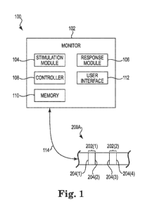
[07] Figure 1 is a block diagram illustrating a nerve stimulation and
monitoring system according to one embodiment.
- 2 -
CA 02928201 2016-04-20
WO 2015/066617
PCT/US2014/063697
[08] Figure 2 is a diagram illustrating a Foley catheter with three
electrode
pairs that is suitable for use with the system shown in Figure 1 according to
one
embodiment.
[09] Figure 3 is a diagram illustrating a Foley catheter with two electrode
pairs
that is suitable for use with the system shown in Figure 1 according to one
embodiment.
[10] Figure 4 is a diagram illustrating a Foley catheter with two electrode
pairs
that is suitable for use with the system shown in Figure 1 according to
another
embodiment.
[11] Figure 5 is a diagram illustrating a Foley catheter with three
electrode
pairs that is suitable for use with the system shown in Figure 1 according to
another embodiment.
[12] Figure 6 is a diagram illustrating a Foley catheter with two electrode
pairs
that is positioned within the urethra of a human male subject according to one
embodiment.
Detailed Description
[13] As used herein, the terms "distal" and "proximal" define a position or
direction with respect to the treating clinician or clinician's control device
(e.g., a
handle assembly). "Distal" or "distally" can refer to a position distant from
or in
a direction away from the clinician or clinician's control device. "Proximal"
and
"proximally" can refer to a position near or in a direction toward the
clinician or
clinician's control device.
[14] Some embodiments of the present disclosure are directed to
electrically
monitoring a nerve during a surgical procedure on a target tissue that is in
the
vicinity of the nerve. In general terms, the method includes applying
stimulation
signals to a nerve adjacent to the target tissue via electrodes on a Foley
type
-3 -
CA 02928201 2016-04-20
WO 2015/066617
PCT/US2014/063697
catheter. In some embodiments, a neurogenic response is recorded (e.g.,
measured) at the nerve as a direct nerve potential.
[15] In some embodiments, the term neurogenic refers to a neural-related
response or activity initiated by natural neural processes (i.e., spontaneous
nerve
activity, or nerve activity evoked by natural activation of its axonal
membrane or
receptors), while in other embodiments, the term neurogenic refers to a neural-
related response or activity initiated by an external stimulus, such as, but
not
limited to an evoked potential stimulus. In yet other embodiments, the term
neurogenic refers to a neural-related response or activity caused by both a
naturally neural process and an external stimulus. In some embodiments, the
term nerve refers to neuro structures in general or some specific neuro
structures,
including (but not limited to) one or more of an entire nerve, a nerve fiber,
multiple nerve fibers, an axon, a spatial grouping of axons, or a functional
grouping of axons within the nerve.
[16] By positioning a Foley catheter with nerve electrodes (of one of the
embodiments of the present disclosure) relative to a target nerve and
monitoring
the ensuing neurogenic response, a surgeon can achieve and maintain a hands-
free, automatic continuous (or substantially continuous) monitoring of the
health
and integrity of a nerve in a reliably consistent manner during a surgical
procedure.
[17] One embodiment is directed to a Foley catheter with electrodes for
intraoperative neurophysiological monitoring (IONM) and post-operative and
intraoperative recovery stimulation. In one embodiment, the Foley catheter
includes a plurality of pairs of electrodes for recording nerve action
potentials
during a pelvic surgery, such as a radical prostatectomy, hysterectomy,
bladder
cancer, colorectal surgery, ureter, vasectomy, or other pelvic surgery.
[18] Figure 1 is a block diagram illustrating a nerve stimulation and
monitoring system 100 according to one embodiment. System 100 includes
monitor 102 and Foley type catheter 200A. Monitor 102 includes stimulation
- 4 -
CA 02928201 2016-04-20
WO 2015/066617
PCT/US2014/063697
module 104, response module 106, controller 108, memory 110, and user
interface 112. Foley catheter 200A includes a plurality of pairs 202(1)-202(2)
of
electrodes (collectively referred to as electrode pairs 202). Electrode pair
202(1)
includes electrodes 204(1) and 204(2), and electrode pair 202(2) includes
electrodes 204(3) and 204(4). Electrodes 204(1)-204(4) are collectively
referred
to as electrodes 204. In one embodiment, electrode pair 202(2) comprises
bipolar
stimulation electrodes, and electrode pair 202(1) comprises bipolar response
electrodes. In other embodiments, electrode pairs 202 can comprise any
combination of stimulation electrodes and response electrodes, including all
stimulation electrodes or all response electrodes. Stimulation may also be
provided via a probe.
[19] In operation according to one embodiment, the stimulation module 104
of
the monitor 102 applies a stimulation signal to a nerve via stimulation
electrode
pair 202(2), while response module 106 of monitor 102 measures a neurogenic
response signal at the nerve via measuring a direct action potential with the
response electrode pair 202(1). The response is communicated to a surgeon via
user interface 112 of the monitor 102. Accordingly, by using monitor 102, a
surgeon can determine the relative health and function of a nerve by
stimulating
that nerve and measuring a corresponding neurogenic response. In some
embodiments, one or more of electrodes pairs 202 record nerve activity evoked
by a separate stimulating bipolar probe. In other embodiments, one or more of
the electrode pairs 202 stimulates nerves, and a separate bipolar probe is
used to
record the evoked nerve activity.
[20] In one embodiment, controller 108 of monitor 102 comprises one or more
processing units and associated memories configured to generate control
signals
directing the operation of monitor 102. In particular, in response to or based
upon commands received via user interface 112 and/or instructions contained in
the memory 110 associated with controller 108, controller 108 generates
control
signals directing operation of stimulation module 104 and/or response module
106.
-5 -
CA 02928201 2016-04-20
WO 2015/066617
PCT/US2014/063697
[21] The term "processing unit", as used herein, means a presently
developed
or future developed processing unit that executes sequences of instructions
contained in a memory. Execution of the sequences of instructions causes the
processing unit to perform steps such as generating control signals. The
instructions may be loaded in a random access memory (RAM) for execution by
the processing unit from a read only memory (ROM), a mass storage device, or
some other persistent storage, as represented by memory 110. In other
embodiments, hard wired circuitry may be used in place of or in combination
with software instructions to implement the functions described. For example,
controller 108 may be embodied as part of one or more application-specific
integrated circuits (ASICs). Unless otherwise specifically noted, the
controller
108 is not limited to any specific combination of hardware circuitry and
software,
nor limited to any particular source for the instructions executed by the
processing unit.
[22] In one embodiment, each of the electrodes 204 is a ring electrode that
completely surrounds a circumference of the Foley catheter 200A. In one
embodiment, each electrode pair 202 is a bipolar electrode pair, with one of
the
electrodes 204 in the pair being an anode, and the other electrode 204 in the
pair
being a cathode. When the electrodes 204(1) and 204(2) are configured as an
anode and a cathode, this electrode pair 202(1) can deliver bipolar
stimulation to
nerves proximate a target site, or provide bipolar recording of nerve activity
proximate the target site. When the electrodes 204(3) and 204(4) are
configured
as an anode and a cathode, this electrode pair 202(2) can deliver bipolar
stimulation to nerves proximate a target site, or provide bipolar recording of
nerve activity proximate the target site. In one embodiment, a second one of
the
electrode pairs 202 (e.g., electrode pair 202(2)) is configured to stimulate
nerves,
and a first one of the electrode pairs 202 (e.g., electrode pair 202(1)) is
spaced
apart from the second electrode pair along the Foley catheter 200A and is
configured to measure the action potential of the nerves resulting from the
stimuli
of the second electrode pair. Action potential is the electrical activity
developed
- 6 -
CA 02928201 2016-04-20
WO 2015/066617
PCT/US2014/063697
in a nerve cell during activity (e.g., induced by a stimulus from the second
electrode pair).
[23] Electrodes 204 are communicatively coupled to monitor 102 via
communication link 114. In one embodiment, communication link 114 includes
signal wires or conductors that are operatively coupled to the electrodes 204
to
drive nerve stimulation, record nerve activity, and/or otherwise provide a
signal
between the electrodes 204 and monitor 102. In one embodiment, the signal
wires extend through the catheter 200A to a proximal end of the catheter 200A
where the signal wires can be operatively connected to the monitor 102. In one
embodiment, monitor 102 comprises a Nerve Integrity Monitor ("NIM") made
available by Medtronic Xomed of Jacksonville, Fla., which provides
intraoperative nerve monitoring capabilities using visual and/or audible
indications of nerve activity.
[24] The electrode pairs 202 according to one embodiment are positioned to
accommodate most prostate sizes, by placing one of the pairs 202 close to the
membranous urethra. The membranous urethra is the level where the nerves
innervating the external genitalia of both sexes exit the pelvis, and thus are
an
optimal location for recording or stimulating these nerves. In addition,
having
more than one pair of electrodes allows for stimulating and recording the
nerves.
Also, the electrode pairs 202 allow for recording spontaneous nerve activity
during the surgery. They also allow for stimulating the nerves during surgical
recovery for therapeutic facilitation of nerve improvement and recovery in the
event of iatrogenic nerve injury or irritation. In some embodiments, electrode
pairs 202 are used for spontaneous recording during surgical dissection.
[25] Figure 2 is a diagram illustrating a Foley catheter 200B with three
electrode pairs that is suitable for use with the system 100 shown in Figure 1
according to one embodiment. A catheter, such as catheter 200B, may be
understood to have various sections according to their disposition when the
catheter is inserted into a human subject. Foley catheter 200B has a proximal
portion 210 that remains outside of the human subject, a central portion 212
that
- 7 -
CA 02928201 2016-04-20
WO 2015/066617
PCT/US2014/063697
traverses the urethra, and a distal portion 214 that resides in the urinary
bladder.
Foley catheter 200B comprises a flexible tube 222 that is passed through the
urethra and into the bladder. The Foley catheter 200B is held in place by an
inflatable balloon 216 that stabilizes the device in place, and prevents
inadvertent
withdrawal from the bladder. The Foley catheter 200B includes at least two
separated lumens 218 and 220 along its length. Lumen 220 is open at both ends
and serves as a conduit that drains urine from the bladder, and lumen 218
serves
as an air or fluid conduit that allows the balloon 216 to be controllably
inflated
when it lies inside the bladder, in order to stop it from slipping out. The
signal
wires 221 of communication link 114 (Figure 1) may be disposed in a lumen of
catheter 200B that allows communication of sensing signals between distally
disposed electrodes 204 and the proximal portion 210 of the catheter 200B.
[26] Foley catheter 200B includes three pairs 202(1)-202(3) of electrodes
(collectively referred to as electrode pairs 202). Electrode pair 202(1)
includes
electrodes 204(1) and 204(2), and electrode pair 202(2) includes electrodes
204(3) and 204(4), and electrode pair 202(3) includes electrodes 204(5) and
204(6). Electrodes 204(1)-204(6) are collectively referred to as electrodes
204.
In one embodiment, the electrode pair closest to the membranous urethra is
used
to either stimulate or record nerve activity. In another embodiment, at least
one
of the electrode pairs 202 comprises bipolar stimulation electrodes, and at
least
one of the electrode pairs 202 comprises bipolar recording electrodes. In
other
embodiments, electrode pairs 202 are used in combination with a separate
bipolar
probe.
[27] The individual electrodes 204 in each electrode pair 202 are
longitudinally spaced apart from each other along the length of the catheter
200B
by a distance, D1, which is about 5-10 mm in one embodiment. This inter-
electrode distance for each electrode pair 202 optimizes the recorded signal-
to-
noise for periprostatic nerves based on their known conduction velocity (i.e.,
0.5-
30 m/sec). In other embodiments, the electrodes 204 can be spaced closer
together or further apart. In various embodiments, the separation between the
electrodes 204 in each electrode pair 202 is selected to enhance the signal to
- 8 -
CA 02928201 2016-04-20
WO 2015/066617
PCT/US2014/063697
noise ratio for recording nerve activity for a particular type of nerve fiber
(e.g.,
A-delta fibers, B-fibers, and/or C-fibers).
[28] In one embodiment, the Foley catheter 200B has an overall length, D5,
of
about 425 mm. In one embodiment, the distance, D4, from the proximal end of
the balloon 216 to the most distally disposed electrode 204(6) is about 35-
40mm.
In one implementation, the distance, D4, is about 37.5 mm. In one embodiment,
the pitch of the electrode pairs 202 is about 18-22 mm (e.g., the distance
measured from the center point of one electrode pair 202 to the center point
of an
adjacent electrode pair 202). In one implementation, the pitch of the
electrode
pairs 202 is about 20 mm, so the distance, D3, is about 20 mm, and the
distance,
D2, is about 40 mm. The distance, D6, represents the lateral width of
electrodes
204, which is about 1 mm in one embodiment.
[29] Figure 3 is a diagram illustrating a Foley catheter 200C with two
electrode pairs that is suitable for use with the system 100 shown in Figure 1
according to one embodiment. Foley catheter 200C according to one
embodiment is configured to be used for a prostate up to 5.5 cm. Foley
catheter
200C has a proximal portion 210 that remains outside of the human subject, a
central portion 212 that traverses the urethra, and a distal portion 214 that
resides
in the urinary bladder. Foley catheter 200C comprises a flexible tube 222 that
is
passed through the urethra and into the bladder. The Foley catheter 200C is
held
in place by an inflatable balloon 216 that stabilizes the device in place, and
prevents inadvertent withdrawal from the bladder. The Foley catheter 200C
includes at least two separated lumens 218 and 220 along its length. Lumen 220
is open at both ends and serves as a conduit that drains urine from the
bladder,
and lumen 218 serves as an air or fluid conduit that allows the balloon 216 to
be
controllably inflated when it lies inside the bladder, in order to stop it
from
slipping out. The signal wires of communication link 114 (Figure 1) may be
disposed in a lumen of catheter 200C that allows communication of sensing
signals between distally disposed electrodes 204 and the proximal portion 210
of
the catheter 200C.
- 9 -
CA 02928201 2016-04-20
WO 2015/066617
PCT/US2014/063697
[30] Foley catheter 200C includes two pairs 202(1)-202(2) of electrodes
(collectively referred to as electrode pairs 202). Electrode pair 202(1)
includes
electrodes 204(1) and 204(2), and electrode pair 202(2) includes electrodes
204(3) and 204(4). Electrodes 204(1)-204(4) are collectively referred to as
electrodes 204. In one embodiment, at least one of the electrode pairs 202
comprises bipolar stimulation electrodes, and at least one of the electrode
pairs
202 comprises bipolar recording electrodes. In other embodiments, electrode
pairs 202 are used in combination with a separate bipolar probe.
[31] The individual electrodes 204 in each electrode pair 202 are
longitudinally spaced apart from each other along the length of the catheter
200C
by a distance, D10, which is about 5-10 mm in one embodiment. This inter-
electrode distance for each electrode pair 202 optimizes the recorded signal-
to-
noise for periprostatic nerves based on their known conduction velocity (i.e.,
0.5-
30 m/sec). In other embodiments, the electrodes 204 can be spaced closer
together or further apart.
[32] In one embodiment, the distance, D11, from the proximal end of the
balloon 216 to the center of the most distally disposed electrode pair 202(2)
is
about 47-57 mm. In one specific implementation, the distance, D11, is about 52
mm. In one embodiment, the pitch of the electrode pairs 202 is about 8-12 mm
(e.g., the distance measured from the center point of one electrode pair 202
to the
center point of an adjacent electrode pair 202), so the distance, D12, is
about 8-12
mm. In one specific implementation, the distance D12 is about 10 mm.
[33] Figure 4 is a diagram illustrating a Foley catheter 200D with two
electrode pairs that is suitable for use with the system 100 shown in Figure 1
according to another embodiment. Foley catheter 200D according to one
embodiment is configured to be used for a prostate larger than 5.5 cm. Foley
catheter 200D has a proximal portion 210 that remains outside of the human
subject, a central portion 212 that traverses the urethra, and a distal
portion 214
that resides in the urinary bladder. Foley catheter 200D comprises a flexible
tube
222 that is passed through the urethra and into the bladder. The Foley
catheter
- 10 -
CA 02928201 2016-04-20
WO 2015/066617
PCT/US2014/063697
200D is held in place by an inflatable balloon 216 that stabilizes the device
in
place, and prevents inadvertent withdrawal from the bladder. The Foley
catheter
200D includes at least two separated lumens 218 and 220 along its length.
Lumen 220 is open at both ends and serves as a conduit that drains urine from
the
bladder, and lumen 218 serves as an air or fluid conduit that allows the
balloon
216 to be controllably inflated when it lies inside the bladder, in order to
stop it
from slipping out. The signal wires of communication link 114 (Figure 1) may
be disposed in a lumen of catheter 200D that allows communication of sensing
signals between distally disposed electrodes 204 and the proximal portion 210
of
the catheter 200D.
[34] Foley catheter 200D includes two pairs 202(1)-202(2) of electrodes
(collectively referred to as electrode pairs 202). Electrode pair 202(1)
includes
electrodes 204(1) and 204(2), and electrode pair 202(2) includes electrodes
204(3) and 204(4). Electrodes 204(1)-204(4) are collectively referred to as
electrodes 204. In one embodiment, at least one of the electrode pairs 202
comprises bipolar stimulation electrodes, and at least one of the electrode
pairs
202 comprises bipolar recording electrodes. In other embodiments, electrode
pairs 202 are used in combination with a separate bipolar probe.
[35] The individual electrodes 204 in each electrode pair 202 are
longitudinally spaced apart from each other along the length of the catheter
200D
by a distance, D20, which is about 5-10 mm in one embodiment. This inter-
electrode distance for each electrode pair 202 optimizes the recorded signal-
to-
noise for periprostatic nerves based on their known conduction velocity (i.e.,
0.5-
30 m/sec). In other embodiments, the electrodes 204 can be spaced closer
together or further apart.
[36] In one embodiment, the distance, D21, from the proximal end of the
balloon 216 to the center of the most distally disposed electrode pair 202(2)
is
about 68-78 mm. In one specific implementation, the distance, D21, is about 73
mm. In one embodiment, the pitch of the electrode pairs 202 is about 10.5-14.5
mm (e.g., the distance measured from the center point of one electrode pair
202
- 11 -
CA 02928201 2016-04-20
WO 2015/066617
PCT/US2014/063697
to the center point of an adjacent electrode pair 202), so the distance, D22,
is
about 10.5-14.5 mm. In one specific implementation, the distance D22 is about
12.5 mm.
[37] Figure 5 is a diagram illustrating a Foley catheter 200E with three
electrode pairs that is suitable for use with the system 100 shown in Figure 1
according to another embodiment. Foley catheter 200E has a proximal portion
210 that remains outside of the human subject, a central portion 212 that
traverses
the urethra, and a distal portion 214 that resides in the urinary bladder.
Foley
catheter 200E comprises a flexible tube 222 that is passed through the urethra
and
into the bladder. The Foley catheter 200E is held in place by an inflatable
balloon 216 that stabilizes the device in place, and prevents inadvertent
withdrawal from the bladder. The Foley catheter 200E includes at least two
separated lumens 218 and 220 along its length. Lumen 220 is open at both ends
and serves as a conduit that drains urine from the bladder, and lumen 218
serves
as an air or fluid conduit that allows the balloon 216 to be controllably
inflated
when it lies inside the bladder, in order to stop it from slipping out. The
signal
wires of communication liffl( 114 (Figure 1) may be disposed in a lumen of
catheter 200E that allows communication of sensing signals between distally
disposed electrodes 204 and the proximal portion 210 of the catheter 200E.
[38] Foley catheter 200D includes three pairs 202(1)-202(3) of electrodes
(collectively referred to as electrode pairs 202). Electrode pair 202(1)
includes
electrodes 204(1) and 204(2), and electrode pair 202(2) includes electrodes
204(3) and 204(4), and electrode pair 202(3) includes electrodes 204(5) and
205(6). Electrodes 204(1)-204(6) are collectively referred to as electrodes
204.
In one embodiment, at least one of the electrode pairs 202 comprises bipolar
stimulation electrodes, and at least one of the electrode pairs 202 comprises
bipolar recording electrodes. In other embodiments, electrode pairs 202 are
used
in combination with a separate bipolar probe.
[39] The individual electrodes 204 in each electrode pair 202 are
longitudinally spaced apart from each other along the length of the catheter
200E
- 12 -
CA 02928201 2016-04-20
WO 2015/066617
PCT/US2014/063697
by a distance, D30, which is about 5-10 mm in one embodiment. This inter-
electrode distance for each electrode pair 202 optimizes the recorded signal-
to-
noise for periprostatic nerves based on their known conduction velocity (i.e.,
0.5-
30 m/sec). In other embodiments, the electrodes 204 can be spaced closer
together or further apart.
[40] In one embodiment, the distance, D31, from the proximal end of the
balloon 216 to the center of the most distally disposed electrode pair 202(3)
is
about 5-15 mm. In one specific implementation, the distance, D31, is about 10
mm.
[41] Figure 6 is a diagram illustrating a Foley catheter 200F with two
electrode
pairs that is positioned within the urethra of a human male subject according
to
one embodiment. Foley catheter 200F is suitable for use with the system 100
shown in Figure 1. It is noted that any of the Foley catheters 200A-200E
described herein can also be positioned and function as described below with
respect to Foley catheter 200F. Portions of the male subject shown in Figure 6
include urinary bladder 602, symphysis pubis 604, prostate 606, rectum 608,
urinary sphincter 609, penis 610, scrotum 612, testis 614, bulb 616, and
urethra
618. There are three main sections of the male urethra 618: (1) Prostatic
urethra
620 (i.e., the portion of the urethra 618 within prostate 606; (2) Membranous
urethra 622 (i.e., the portion of the urethra 618 within the urinary sphincter
609;
and (3) Penile urethra 624 (i.e., the portion of the urethra 618 within the
penis
610).
[42] Foley catheter 200F has a proximal portion that remains outside of the
human subject, a central portion that traverses the urethra 618, and a distal
portion that resides in the urinary bladder 602. Foley catheter 200F comprises
a
flexible tube that is passed through the urethra 618 and into the bladder 602.
The
Foley catheter 200F is held in place by an inflatable balloon 216 that
stabilizes
the device in place, and prevents inadvertent withdrawal from the bladder 602.
The Foley catheter 200F includes at least two separated lumens and along its
length. A first one of the lumens is open at both ends and serves as a conduit
that
- 13 -
CA 02928201 2016-04-20
WO 2015/066617
PCT/US2014/063697
drains urine from the bladder 602, and a second one of the lumens serves as an
air
or fluid conduit that allows the balloon 216 to be controllably inflated when
it lies
inside the bladder 602, in order to stop it from slipping out. The signal
wires of
communication liffl( 114 (Figure 1) may be disposed in a lumen of catheter
200F
that allows communication of sensing signals between distally disposed
electrodes and the proximal portion of the catheter 200F.
[43] Foley catheter 200F includes two pairs 202(1)-202(2) of electrodes
(collectively referred to as electrode pairs 202). In one embodiment, at least
one
of the electrode pairs 202 comprises bipolar stimulation electrodes, and at
least
one of the electrode pairs 202 comprises bipolar recording electrodes. In
other
embodiments, electrode pairs 202 are used in combination with a separate
bipolar
probe.
[44] The individual electrodes in each electrode pair 202 are
longitudinally
spaced apart from each other along the length of the catheter 200F by a
distance
of about 5-10 mm in one embodiment. This inter-electrode distance for each
electrode pair 202 optimizes the recorded signal-to-noise for periprostatic
nerves
based on their known conduction velocity (i.e., 0.5-30 m/sec). In other
embodiments, the electrodes can be spaced closer together or further apart.
[45] The Foley catheter 200F is shown in the urethra 618 with the balloon
216
inflated. The distal electrode pair 202(2) is positioned at the membranous
urethra
622, and the proximal electrode pair 202(1) is positioned below the membranous
urethra at the bulb 616. A large component of periprostatic nerves are
autonomic
nerves responsible for erection, ejaculation and continence. The membranous
urethra 622 is where all the nerves innervating the external genitalia of
either
gender converge prior to exiting the pelvis, so it is an optimum location for
nerve
access from within the urethra. Note that, in Figure 6, the distance between
the
proximal end of the balloon 216 and the distal electrode pair 202(2) equals
the
length of the prostate 606.
- 14 -
CA 02928201 2016-04-20
WO 2015/066617
PCT/US2014/063697
[46] In one embodiment, the Foley catheter 200F is configured to stimulate
the
periprostatic nerves with a second electrode pair (e.g., electrode pair
202(2)) and
record nerve activity with a first electrode pair (e.g., electrode pair
202(1)). In
further embodiments, the first electrode pair 202(1) can be configured to
provide
stimulation and the second electrode pair 202(2) can be configured to record
the
resultant nerve activity.
[47] The first and second electrode pairs 202(1) and 202(2) can be spaced
far
enough apart from one another such that the signal artifact associated with
the
bipolar stimulation from the second electrode pair 202(2), which is less than
that
which would be produced by monopolar stimulation, does not substantially
engulf or otherwise interfere with the signal being recorded at the first
electrode
pair 202(1). The magnitude of the signal artifact at the first electrode pair
202(1)
depends at least in part on the conduction velocity of the nerve fibers and
the
spacing between the stimulus and recording electrodes. C-fibers, B-fibers, and
A-delta-fibers, such as those found in nerves, have relatively low conduction
velocities (e.g., no more than 3 m/s for C-fibers, about 3-14 m/s for B-
fibers, and
about 12-30 m/s for A-delta fibers). As such, when the first electrode pair
202(1)
is configured to record periprostatic nerve activity, the first electrode pair
202(1)
can be positioned at least 10 mm spaced apart from the second electrode pair
202(2) along the longitudinal axis of the catheter 200F to reduce the signal
artifact recorded by the first electrode pair 202(1). In other embodiments,
the
first and second electrode pairs 202(1) and 202(2) can be spaced different
distances apart from one another along the longitudinal axis of the catheter
200F.
[48] In one embodiment, system 100 (Figure 1) is configured to
automatically
measure the distance between the stimulating and recording electrodes in real-
time in order to calculate the nerve conduction velocity (NCV) of compound
nerve action potentials (CNAPs). Knowing the nerve conduction velocity of the
recorded nerves helps to identify the function of the nerves. Inter-electrode
distances can be determined using a number of different technologies such as
measuring tissue impedances between two sites, and time-of-flight of
electromagnetic fields. Knowing the distance between the two electrodes and
the
- 15 -
CA 02928201 2016-04-20
WO 2015/066617
PCT/US2014/063697
latency of the evoked CNAP allows system 100 to calculate the NCV, and list
the
fiber types being recorded in real-time with user interface 112. This gives
the
surgeon information of the type of nerves being recorded (e.g., autonomic vs.
somatic), which in turn is important for identifying the nerves innervating
the
penis.
[49] One embodiment is directed to an apparatus for monitoring a nerve. The
apparatus includes a Foley type catheter having an exterior surface, and first
and
second pairs of ring electrodes formed on the exterior surface of the Foley
catheter. Each of the first and second pairs of ring electrodes is configured
to
perform at least one of stimulating the nerve and recording nerve activity.
[50] The apparatus according to one embodiment includes at least one
conductor coupled to the first and second pairs of ring electrodes that is
configured to carry signals between the first and second pairs of ring
electrodes
and a processing apparatus. In one embodiment, the first and second pairs of
ring
electrodes each comprise an anode ring electrode and a cathode ring electrode.
The second pair of ring electrodes is configured to deliver bipolar nerve
stimulation, and the first pair of ring electrodes is configured to provide
bipolar
recording of nerve activity.
[51] The apparatus according to one embodiment includes a third pair of
ring
electrodes formed on the exterior surface of the Foley catheter, wherein the
third
pair of ring electrodes is configured to perform at least one of stimulating
the
nerve and recording nerve activity. In one embodiment, the first pair of ring
electrodes includes first and second ring electrodes that are separated by a
distance of about 5 mm to 10 mm, and the second pair of ring electrodes
includes
third and fourth ring electrodes that are separated by a distance of about 5
mm to
mm, and wherein the third pair of ring electrodes includes fifth and sixth
ring
electrodes that are separated by a distance of about 5 mm to 10 mm. In one
embodiment, a center of the first pair of ring electrodes is separated from a
center
of the second pair of ring electrodes by a distance of about 18 mm to about 22
mm, and a center of the second pair of ring electrodes is separated from a
center
- 16 -
CA 02928201 2016-04-20
WO 2015/066617
PCT/US2014/063697
of the third pair of ring electrodes by a distance of about 18 mm to about 22
mm.
In one embodiment, the third pair of ring electrodes is most distally
positioned on
the Foley catheter, and the third pair of ring electrodes is positioned
proximally
from a proximal end of a balloon of the Foley catheter by a distance of about
35-
40 mm.
[52] In one embodiment, the first pair of ring electrodes includes first
and
second ring electrodes that are separated by a distance of about 5 mm to 10
mm,
and the second pair of ring electrodes includes third and fourth ring
electrodes
that are separated by a distance of about 5 mm to 10 mm. In one embodiment, a
center of the first pair of ring electrodes is separated from a center of the
second
pair of ring electrodes by a distance of about 8 mm to about 12 mm. In one
embodiment, the second pair of ring electrodes is most distally positioned on
the
Foley catheter, and the second pair of ring electrodes is positioned
proximally
from a proximal end of a balloon of the Foley catheter by a distance of about
47-
57 mm.
[53] In one embodiment, a center of the first pair of ring electrodes is
separated from a center of the second pair of ring electrodes by a distance of
about 10.5 mm to about 14.5 mm. In one embodiment, the second pair of ring
electrodes is most distally positioned on the Foley catheter, and wherein the
second pair of ring electrodes is positioned proximally from a proximal end of
a
balloon of the Foley catheter by a distance of about 68-78 mm.
[54] In one embodiment, a third pair of ring electrodes is formed on the
exterior surface of the Foley catheter, wherein the third pair of ring
electrodes is
configured to perform at least one of stimulating the nerve and recording
nerve
activity, wherein the third pair of ring electrodes is most distally
positioned on
the Foley catheter, and wherein the third pair of ring electrodes is
positioned
proximally from a proximal end of a balloon of the Foley catheter by a
distance
of about 5-15 mm.
- 17 -
CA 02928201 2016-04-20
WO 2015/066617
PCT/US2014/063697
[55] In one embodiment, at least one of the first and second pairs of ring
electrodes is configured to be used for spontaneous recording during surgical
dissection. In one embodiment, at least one of the first and second pairs of
ring
electrodes is configured to provide therapeutic stimulation.
[56] Another embodiment is directed to a method of monitoring nerve
activity.
The method includes deploying a Foley type catheter in a urethra of a human
patient, wherein the Foley type catheter comprises a first pair of ring
electrodes
and a second pair of ring electrodes. The method includes stimulating a nerve,
and recording nerve activity resulting from the stimulation with at least one
of the
first and second pairs of ring electrodes.
[57] In one embodiment, the method further includes positioning the second
pair of ring electrodes at a membranous urethra of the human patient for the
stimulation. The method according to one embodiment further includes
calculating a nerve conduction velocity based on the recorded nerve activity,
identifying a nerve fiber type corresponding to the calculated nerve
conduction
velocity, and displaying the identified nerve fiber type on a monitor coupled
to
the Foley type catheter. In one embodiment, the Foley type catheter includes a
third pair of ring electrodes, and the method further includes performing at
least
one of stimulating the nerve and recording nerve activity with the third pair
of
ring electrodes.
[58] Yet another embodiment is directed to a system for monitoring a nerve
from a urethra of a patient. The system includes a Foley type catheter
configured
to be deployed in the urethra and comprising a first pair of ring electrodes
and a
second pair of ring electrodes. Each of the first and second pairs of ring
electrodes is configured to perform at least one of stimulating the nerve and
recording nerve activity. The
system includes a monitor apparatus in
communication with the first and second pairs of ring electrodes to process
the
recorded nerve activity. In one embodiment, the Foley type catheter further
includes a third pair of ring electrodes configured to perform at least one of
stimulating the nerve and recording nerve activity.
- 18 -
CA 02928201 2016-04-20
WO 2015/066617
PCT/US2014/063697
[59] While the incidence of erectile dysfunction has decreased
significantly in
the last 10 years, there is still an unmet need. The periprostatic nerves are
too
small to see, even with 20x endoscopic magnification, so the surgeons are
literally blind to the nerve location, and the risk of erectile dysfunction is
10% -
46% in robotic, and 14% - 79% in open procedures. Embodiments disclosed
herein can be used during nerve sparing radical prostatectomy surgery or other
pelvic surgeries to help surgeons locate the nerves, reducing the risk of
nerve
injury and erectile dysfunction. Embodiments disclosed herein can be used for
open, laparoscopic, and robotic pelvic procedures. For post operation
recovery,
one pair of the electrodes is used to stimulate nerves, which can help the
recovery
of nerves that were stretched during the surgery.
[60] In one embodiment, a Foley catheter with two pairs of ring electrodes
is
used in female pelvic surgical procedures. The first pair is 4.5 cm from the
pair
center to the proximal side of the balloon, and the second pair is located 2.5
cm
from the pair center to the proximal side of the balloon. The pair closest to
the
membranous urethra is used to either stimulate nerves or record nerve action
potentials. This design allows for locating (mapping) nerves within the
pelvis,
recording spontaneous nerve action potentials, or therapeutic stimulation of
the
nerves intraoperatively or postoperatively to facilitate nerve recovery or
functional improvement in the event of iatrogenic nerve injury or irritation.
[61] Although the present disclosure has been described with reference to
preferred embodiments, workers skilled in the art will recognize that changes
can
be made in form and detail without departing from the spirit and scope of the
present disclosure.
- 19 -