Note: Descriptions are shown in the official language in which they were submitted.
SYSTEM FOR INJECTION TRAINING
BACKGROUND
[0002] A
variety of medical injection procedures are often performed in
prophylactic, curative, therapeutic, or cosmetic treatments.
Injections may be
administered in various locations on the body, such as under the conjunctiva,
into
arteries, bone marrow, the spine, the sternum, the pleural space of the chest
region,
the peritoneal cavity, joint spaces, and internal organs. Injections can also
be helpful
in administering medication directly into anatomic locations that are
generating pain.
These injections may be administered intravenously (through the vein),
intramuscularly (into the muscle), intradermally (beneath the skin),
subcutaneously
(into the fatty layer of skin) or intraperitoneal injections (into the body
cavity).
Injections can be performed on humans as well as animals. The methods of
administering injections typically range for different procedures and may
depend on
the substance being injected, needle size, or area of injection.
[0003]
Injections are not limited to treating medical conditions, but may be
expanded to treating aesthetic imperfections or restorative cosmetic
procedures.
Many of these procedures are performed through injections of various products
into
different parts of the body. The aesthetics and therapeutic industry consists
of two
main categories of injectable products: neuromodulators and dermal fillers.
The
neuromodulator industry commonly utilizes nerve-inhibiting products such as
Botox ,
Dysport , and Xeomin . The dermal filler industry utilizes products
administered by
providers to patients for both cosmetic and therapeutic reasons, such as, for
example,
Juvederm , Restylane , Belotero , Sculptra , Artefill , and others. These
providers
or injectors may include plastic surgeons, facial plastic surgeons,
oculoplastic
surgeons, dermatologists, nurse practitioners, dentists and nurses.
CA 2928460 2928460 2018-10-23
CA 02928460 2016-04-21
WO 2014/070799
PCT/US2013/067352
SUMMARY
OM] One of the major problems in the administration of injections is
that
there is no official certification or training process. Anyone with a minimal
medical
related license may inject a patient. These "injectors" may include primary
care
physicians, dentists, veterinarians, nurse practitioners, nurses, physician's
assistants, or aesthetic spa physicians. However, the qualifications and
training
requirements for injectors vary by country, state, and county. For example, in
most
states in the United States, the only requirement to inject patients with
neuromodulators and/or fillers is a nursing degree or medical degree. This
causes
major problems with uniformity and expertise in administering injections. The
drawbacks with lack of uniformity in training and expertise are widespread
throughout the medical industry. Doctors and practitioners often are not well
trained
in administering injections for diagnostic, therapeutic, and cosmetic chemical
substances. This lack of training often leads to instances of chronic pain,
headaches, bruising, swelling, or bleeding in patients.
[0005] Current injection training options are classroom based, with
hands-
on training performed on live models. The availability of models is limited.
Moreover, even when available, live models are limited in the number and type
of
injections that may be performed on them. The need for live models is
restrictive
because injectors are unable to be exposed to a wide and diverse range of
situations
in which to practice. For example, it may be difficult to find live models
with different
skin tones or densities. This makes the training process less effective
because
patients often have diverse anatomical features as well as varying
prophylactic,
curative, therapeutic, or cosmetic needs. Live models are also restrictive
because
injectors are unable to practice injection methods on internal organs due to
health
considerations. As a result of these limited training scenarios, individuals
seeking
treatments involving injections have a much higher risk of being treated by an
inexperienced injector. This may result in low patient satisfaction with the
results or
failed procedures. In many instances, patients have experienced lumpiness from
incorrect dermal filler injections. Some failed procedures may result in
irreversible
problems and permanent damage to a patient's body. For example, patients have
experienced vision loss, direct injury to the globe of the eye, and brain
infarctions
where injectors have incorrectly performed dermal filler procedures. Other
examples
of side effects include inflammatory granuloma, skin necrosis,
endophthalmitis,
-2-
CA 02928460 2016-04-21
WO 2014/070799
PCT/US2013/067352
injectable-related vascular compromise, cellulitis, biofilm, subcutaneous
nodules,
fibrotic nodules, and other infections.
[0006] As a result of the varying qualifications and training
requirements
for injectors, there is currently no standard to train, educate, and certify
providers on
the proper and accurate process of various injection techniques. Patients
seeking
injections also have few resources for determining the qualifications or
experience of
a care practitioner.
100071 The present disclosure generally relates to an injection
apparatus
and training system for prophylactic, curative, therapeutic, acupuncture, or
cosmetic
injection training and certification. The training system eliminates the need
to find
live models for hands-on training sessions. The training system provides
feedback
on trainees and the accuracy of injection procedures performed. In an
embodiment,
feedback is provided in real time. The training system can be used as a
measurement on how the "trainee" is doing prior to receiving actual product by
the
manufacturing company as a measure of qualification. The training system
reduces
the risks associated with inexperienced and uncertified medical personnel
performing
injection procedures.
[0008] The training system can be used to educate, train, and certify
medical personnel for injection procedures. It can also be utilized as a
testing
program for certifying medical personnel. The system will enable users to
practice a
variety of injections, ranging from on label to off label product injections.
In some
embodiments, the system may allow users to train for therapeutic treatments.
In
other embodiments, the system may allow users to train for injections into
arteries,
bone marrow, the spine, the sternum, the pleural space of the chest region,
the
peritoneal cavity, joint spaces, internal organs, or any other injection
sites. The
system may be used for any type of injection, including, but not limited to
those
involving prophylactic, curative, therapeutic, or cosmetic treatments in both
humans
and animals. In other applications, the systems disclosed herein can be used
for
dental application and training for dental procedures.
[0009] In one embodiment, there are three main components of the
training system: (1) a training apparatus (also referred to interchangeable as
an
injection apparatus throughout the present disclosure) which features an
anatomically accurate model of a human or human body part necessary for
injection
-3-
CA 02928460 2016-04-21
WO 2014/070799
PCT/US2013/067352
training, (2) a camera associated with the training apparatus, and (3) a
testing tool
with light emitting capabilities. In an embodiment, a fourth component of the
training
system can include a output device that can run an application which receives
communications from the training apparatus or camera and generates information
regarding injection parameters based on the communications from the injection
apparatus or camera. In an embodiment, the images captured by the camera are
processed by a processor included either in the injection apparatus or in the
camera
before being communicated to the output device. This processing can include,
for
example, determining an indication of one or more injection parameters. In an
embodiment, the anatomical model can include various injection conditions,
such as,
for example, layered skin, available in multiple tones and textures to mimic a
diverse
span of age, race, and skin texture. In an embodiment, the layered skin can be
removable and/or replaceable. The apparatus can simulate any human or animal
part, such as, for example, the face, head, brain, neck, back, chest, spine,
torso,
arms, legs, hands, feet, mouth, or any other body part or portion of the body
of
interest. In an embodiment, the testing tool can be, for example a syringe or
hypodermic needle. In an embodiment, the injection apparatus is reusable. In
an
embodiment, the injection apparatus is disposable.
[0010] Although the present disclosure specifically describes the use of
a
camera, it is to be understood that the principles disclosed throughout the
present
disclosure can apply to any light detector or light detection device.
Moreover, by
referring to a camera, the present disclosure is not limited to a visible
light detection
device, rather, any visible or non-visible light detector or detection device
can be
used as would be understood by a person of skill in the art with any
embodiment
disclosed herein.
[0011] In one embodiment, the injection apparatus can feature an
anatomically correct model of an animal or animal body part. The animal or
animal
body part can have a base layer that can be covered in removable skin, animal
hair,
or scales to replicate the look and feel of a real animal. The skin, animal
hair, or
scales can be in different colors, coarseness, thickness, density, or
stiffness.
[0012] In some embodiments, the base layer of the apparatus may be a
clear plastic shell simulating a human or animal body part, such as, for
example, a
human or animal head. The plastic shell can be covered with layers of
elastomer
-4-
CA 02928460 2016-04-21
WO 2014/070799
PCT/US2013/067352
membranes simulating human or animal muscle or skin. In an embodiment, one or
more of these layers can be removable and/or replaceable. In some embodiments,
the top layer of injectable skin consists of separate layers simulating
mammalian
skin: the epidermis, dermis, and hypodermis. The layers of injectable muscle
and
skin may be of uniform density. In other embodiments, the layers of skin may
be
thicker or thinner to simulate the skin of humans or animals with uneven skin
layers
or damaged skin. The separate layers of injectable skin may consist of
elastomers
simulating the look and feel of human or animal skin and muscle. The
injectable
muscle and skin layers may have a different transparency. For example, the
different layers may be opaque, tinted, or clear.
[0013] In one embodiment, the injection apparatus may be used for
injections in different areas of the human or animal body. For example, the
injection
apparatus may simulate the rear torso and buttocks of a human for epidural
injections. The injection apparatus can also simulate the back of the neck of
an
animal for subcutaneous injections. The injection apparatus may also simulate
different organs of the human or body, such as the heart, brain, or liver. In
some
embodiments, the injection apparatus can simulate different bones in human or
animal bodies that require injections or extractions. The simulated bones can
contain extractable material that simulates bone marrow. The bones can be
separately used as a training apparatus or be placed within an anatomically
correct
model of a simulated human or animal body part. For example, bone marrow
extractions can be performed by inserting the testing tool through skin,
muscle, and
bone layers of a training apparatus. In one embodiment, the injection
apparatus may
be covered in different removable layers of material for detecting an
injection. The
different layers may be opaque, tinted, marked or clear. In some embodiments,
the
different removable layers of the injection apparatus may be embedded with
sensors
that can be pierced by a testing tool. In an embodiment, the apparatus is a
human
or animal mouth that can be used to perform dental or periodontic procedures.
[0014] In one embodiment, a testing tool is provided. In an embodiment,
the testing tool is in the form of a hypodermic needle. The hypodermic needle
can
be part of a syringe. The hypodermic needle can be of any gauge. The testing
tool
can be of any size or shape and designed to simulate the size and shape of an
injection tool, such as a syringe, used for any particular type of injection
being
practiced. In an embodiment, the testing tool has a light source that emits
light at the
-5-
CA 02928460 2016-04-21
WO 2014/070799
PCT/US2013/067352
head of the needle. In an embodiment, a fiber optic is in the needle. For
example,
the fiber optic can be inserted into or threaded through the needle and
configured to
emit light from a light source through the tip or head of the needle. The
light source
may be one or more an LEDs, laser diodes, or any other light emitting device
or
combination of devices. In an embodiment, the light source can emit light
along a
spectrum of visible. In other embodiments, the light source can emit light of
non-
visible light, such as infrared light. In some embodiments, the light emitted
from the
light source is attenuated by each layer of simulated skin or muscle. The
testing tool
can have a barrel. The testing tool can also have a plunger associated with
the
barrel.
[0015] The testing
tool can be used to practice injections on the injection
apparatus. In an embodiment, the light emitted through the tip or head of the
needle
of the testing tool is attenuated by the injection apparatus. The attenuated
light is
detected by the camera. As the needle portion of the testing tool penetrates
through
each layer of the injection apparatus material, different colors, intensities,
fluorescence, textures, graph lines, polarization, or other visual effects of
light will be
detected by the camera (or any other visible or non-visible light detector).
The
resulting visual effects of the attenuated light are detected or viewed by the
camera.
The visual effects can represent the differences in location of the injection,
depth of
the injection, pressure of an injection exerted by the user and/or angle of
injection.
This information, detected by the camera, can be communicated to an output
device
for data collection, testing or certification purposes. Although the
disclosure
discloses the use of a camera, the disclosure and claims are not limited to
the use of
a visible light camera, or typical consumer photography cameras. Rather, the
term
camera, as used herein, can, in some embodiments, extend to the use of any
light
detectors or light detection devices, including, for example, photodiodes,
infrared,
polarization, fluorescent or ultraviolet light or thermal imaging cameras or
other
devices used to detect the presence or absence of visible or non-visible
light.
[0016] In some
embodiments, a camera is placed within or proximate to
the injection apparatus. The camera can send the information detected to a
processing unit. The processing unit communicates with an output device which
can
display the results received from an injection. The output
device, also
interchangeably referred to herein as an interface device, user device or
display
device, can include any type of display useful to a user, such as, for
example, a
-6-
CA 02928460 2016-04-21
WO 2014/070799
PCT/US2013/067352
tablet, phone, laptop or desktop computer, television, projector or any other
electronic or paper based display technology. The processing unit can also
collect
the information for use in data gathering or informatics. Information about
the
injection can also be gathered from the testing tool. The output device may
include
lights, graphical displays, audio devices, or user controls. The output device
can be
an electronic, computer, or mobile device. This can include, for example, a
smart
phone or tablet. The output device can run a dedicated application configured
to
receive wireless communication directly from the camera and/or testing tool
and
analyze this information for feedback and display to a user. Alternatively, a
separate
processor in the injection apparatus and/or testing tool can process the
information
before sending the processed information to the output device for display.
[0017] In some embodiments, the injection apparatus can be configured to
mimic certain muscle contraction conditions common with a particular type of
injection. For example, this can include contractions of facial features, such
as
furrowing of an eyebrow, squinting of the eyes, or pursing of the lips. The
removable
skin can also include blemishes, such as scars or wrinkles.
[0018] In an embodiment, the layers of material surrounding the
injection
apparatus have different pigmentations. For example, in an embodiment where
the
injection apparatus has three layers of pigmentation, the first layer may be
opaque,
the second layer may be tinted, and the third layer may be clear. The
pigmentation
of the layers selectively alters the testing tool light to display a different
color or
intensity of light as it passes through each layer. This resulting attenuated
light from
the testing tool is then detected or viewed by the camera enclosed in the
injection
apparatus. The output device is configured with software to recognize the
color,
direction, and intensity of light detected by the camera. Based on the color,
direction, and intensity detected by the camera, a software program may
determine
the depth, pressure, or angle of injection. Similarly, markings or other
identification
options can be used to identify the depth, pressure, or angle of injection as
described
below.
[0019] In an embodiment, a system for cosmetic or therapeutic training
configured to aid in training a care provider to provide cosmetic or
therapeutic
injections is disclosed. The system includes a testing tool that has an
injection
needle head, the testing tool configured to emit light. The system also
includes an
-7-
CA 02928460 2016-04-21
WO 2014/070799
PCT/US2013/067352
apparatus configured to receive an injection and attenuate the light emitted
by the
testing tool, the attenuation representative of an injection parameter; and a
light
detector, the light detector positioned to detect the light attenuated by the
apparatus.
In an embodiment, the system includes a processor configured to receive and
process an indication of the detected light from the light detector. In an
embodiment,
the system includes a display device. In an embodiment, the display device is
configured to display the indication of the detected light from the light
detector. In an
embodiment, the indication of the detected light is an image. In an
embodiment, the
display device is configured to display injection measurement data. In an
embodiment, the injection parameter is a depth of the injection. In an
embodiment,
the injection parameter is an angle of injection. In an embodiment, the
injection
parameter is pressure. In an embodiment, the processor is configured to
determine
an accuracy of the injection. In an embodiment, the apparatus includes a
plurality of
nesting layers, wherein each layer provides a different attenuation of light.
In an
embodiment, each of the plurality of nesting layers are colored. In an
embodiment,
at least some of the plurality of nesting layers are translucent. In an
embodiment,
the plurality of nesting layers include a removable skin layer, a muscle
layer, and a
nerve layer. In an embodiment, the removable skin layer is configured to
represent
one or more of different ages, ethnicities, races, textures, or thicknesses of
human
skin. In an embodiment, the removable skin layer is transparent and the muscle
layer is visible through the skin layer. In an embodiment, the removable skin
layer
simulates cosmetic conditions. In an embodiment, one or more injection sites
are
positioned at locations on the injection apparatus which correspond to
injection
locations for cosmetic conditions and therapeutic treatment. In an embodiment,
the
testing tool comprises a light source configured to emit visible light. In an
embodiment, the light detector comprises a camera.
100201 In an embodiment, method of injection training is disclosed. The
method can include providing a testing tool simulating a syringe and
configured to
emit light; providing an injection apparatus, the injection apparatus
configured to
provide a simulation of a testing site and configured to attenuate the light
emitted by
the testing tool according to a desired parameter of an injection; providing a
light
detector configured to detect the attenuated light; using the testing tool to
inject the
injection apparatus; and detecting, using the light detector, the light
attenuated by
the injection apparatus during the injection. In an embodiment, the method
also
-8-
CA 02928460 2016-04-21
WO 2014/070799
PCT/US2013/067352
includes analyzing the detected attenuated light from the camera to determine
an
accuracy of the injection. In an embodiment, the parameter under test is the
depth
and location of the injection. In an embodiment, the parameter under test is
one or
more of speed, pressure, angle, depth or location. In an embodiment, the
injection
apparatus is configured to attenuate the light by providing a plurality of
nesting layers
each including a different level of light attenuation. In an embodiment, each
of the
plurality of nesting layers is a different color.
[0021] In an embodiment, a testing tool is disclosed. The testing tool
can
include a needle; a barrel; and a light source configured to emit light from
the needle.
In an embodiment, the testing tool also includes a plunger. As will be
understood by
those of skill in the art, the testing tool described throughout this
disclosure and in
every embodiment of the disclosure can be configured to simulate all or
various
combinations of parts of a typical syringe or hypodermic needle. For example,
this
can include a needle, a barrel, and a plunger or any combination thereof.
Also, any
light source or combination of elements described herein can also be included
in the
testing tool. In any of the embodiments disclosed herein, unneeded or
unnecessary
parts of the testing tool can be left off. For example, in some embodiments, a
plunger is unnecessary and left off the device.
[0022] In an embodiment, the testing tool can include a sensor
configured
to determine a relative position of the plunger with respect to the barrel. In
an
embodiment, the sensor is potentiometer. In an embodiment, the sensor is
housed
proximate the barrel. In an embodiment, the sensor is housed away from the
barrel.
In an embodiment, the testing tool includes a friction system configured to
simulate
an injection. In an embodiment, the needle is hollow. In an embodiment, the
testing
tool includes an optical fiber configured to transmit the emitted light to the
tip of the
needle. In an embodiment, the emitted light is visible light. In an
embodiment, the
emitted light is one or more of visible light, non-visible light, ultraviolet
light, polarized
light, infrared light or fluorescent light.
[0023] In an embodiment, an injection apparatus configured to simulate
at
least a portion of a patient under test is disclosed. The injection apparatus
includes
a first structure configured to simulate a portion of a patient under test;
and at least
one injection layer configured to simulate an injection condition, the
injection layer
configured to attenuate emitted light from a testing tool such that at least
one testing
-9-
CA 02928460 2016-04-21
WO 2014/070799
PCT/US2013/067352
parameter of a desired injection can be determined. In an embodiment, the
injection
apparatus includes at least two injection layers, wherein each injection layer
is
configured to attenuate the emitted light. In an embodiment, each of the two
or more
injection layers attenuates the emitted light differently. In an embodiment,
each of
the two or more injection layers is tinted with a different color and the
emitted light is
visible light. In an embodiment, the patient is a human. In an embodiment, the
patient is an animal. In an embodiment, the first structure is a head. In an
embodiment, the first structure is a back. In an embodiment, the first
structure is a
chest.
[0024] In an embodiment, a testing tool is disclosed. The testing tool
includes a needle; and an optical fiber configured to receive emitted light
from a light
source through a proximate end of the optical fiber, the optical fiber further
configured to emit light out of a distal end of the optical fiber, the optical
fiber
positioned in the needle so that light is emitted at a head of the needle. In
an
embodiment, the needle is a hypodermic needle. In an embodiment, the distal
end
of the optical fiber is located at a tip of the needle. In an embodiment, the
emitted
light is visible light. In an embodiment, the emitted light is one or more of
visible
light, non-visible light, ultraviolet light, infrared light or fluorescent
light. In an
embodiment, the testing tool includes a syringe. In an embodiment, the testing
tool
includes a barrel and plunger. In an embodiment, the testing tool includes a
sensor
configured to determine a relative position of the plunger with respect to the
barrel.
In an embodiment, the sensor is potentiometer. In an embodiment, the sensor is
housed proximate the barrel. In an embodiment, the sensor is housed away from
the barrel. In an embodiment, the testing tool includes a friction system
configured
to simulate an injection. In an embodiment, the testing tool includes a
friction system
configured to simulate an injection. In an embodiment, the needle is hollow.
[0025] In an embodiment, a method of using a testing tool for injection
training is disclosed. The method includes providing a testing tool, the
testing tool
including a needle; an optical fiber configured to emit light out of a distal
end of the
optical fiber, the optical fiber positioned in the needle so that light is
emitted at a
head of the needle; and a light source configured to emit light through a
proximate
end of the optical fiber. The method also includes using the testing tool to
inject an
injection apparatus; and detecting the emitted light after attenuation by the
injection
apparatus to determine an injection parameter. In an embodiment, the needle is
a
-10-
hypodermic needle. In an embodiment, the distal end of the optical fiber is
located at
a tip of the needle. In an embodiment, the emitted light is visible light. In
an
embodiment, the emitted light is one or more of visible light, non-visible
light,
ultraviolet light, infrared light or fluorescent light. In an embodiment, the
method
further includes providing a syringe. In an embodiment, the method further
includes
providing a barrel and plunger. In an embodiment, the method further includes
providing a sensor configured to determine a relative position of the plunger
with
respect to the barrel. In an embodiment, the sensor is potentiometer. In an
embodiment, the sensor is housed proximate the barrel. In an embodiment, the
sensor is housed away from the barrel. In an embodiment, the method further
includes providing a friction system configured to simulate an injection. In
an
embodiment, the method further includes providing a friction system configured
to
simulate an injection. In an embodiment, the needle is hollow. In an
embodiment, the
method further includes storing the injection parameter in an electronic
storage
device. In an embodiment, the method further includes compiling a plurality of
injection parameters from an injector and determining an accuracy rating of
injection.
In an embodiment, the method further includes publically publishing the
accuracy
rating.
[0026] In an
embodiment, a method of rating an injector is disclosed. The
method includes using an injection apparatus to detect injection parameters
about an
injection by an injector using a testing tool; and determining a rating of the
injector
from the injection parameters. In an embodiment, the injector is a primary
care
physician, dentist, veterinarian, nurse practitioner, nurse, physician's
assistant,
aesthetic spa physician, plastic surgeon, facial plastic surgeon, oculoplastic
surgeon,
or dermatologist. In an embodiment, the rating is an accuracy of injections.
In an
embodiment, the rating is an experience of the injector. In an embodiment, the
rating
indicates a quality of the injector. In an embodiment, the rating is
publically published.
In an embodiment, the rating is one or more of education, years of experience,
performance results with the injection apparatus, or patient reviews.
-11-
CA 2928460 2018-10-23
[0026a] In another embodiment, there is provided a system for cosmetic or
therapeutic training configured to aid in training a care provider to provide
cosmetic or
therapeutic injections, the system comprising: a testing tool having an
injection needle
head, the testing tool configured to emit light; a synthetic apparatus
configured to
receive an injection and attenuate the light emitted by the testing tool, the
attenuation
representative of an injection parameter; and a light detector, the light
detector
positioned to detect the light attenuated by the apparatus.
[0026b] In another embodiment, there is provided a method of injection
training, the method comprising providing a testing tool simulating a syringe
and
configured to emit light, providing an injection apparatus having an
anatomical
structure, the injection apparatus configured to provide a simulation of a
testing site
and configured to attenuate the light emitted by the testing tool according to
a desired
parameter of an injection. The method further involves providing a light
detector
configured to detect the attenuated light, using the testing tool to inject
the injection
apparatus and detecting, using the light detector, the light attenuated by the
injection
apparatus during the injection.
[0026c] In another embodiment, there is provided a testing tool comprising a
needle, a barrel cooperating with a proximal portion of the needle, a light
source
configured to emit a plurality of different intensities of light, and an
optical fiber located
inside a central portion of the needle and configured to receive the light
emitted from
the light source and transmit the light through the needle from the proximal
portion of
the needle near the barrel to a distal portion of the needle near a tip of the
needle so
that the light is emitted from the tip of the needle.
[0026d] In another embodiment, there is provided an injection apparatus
configured to simulate at least a portion of a patient under test. The
apparatus
includes a first structure configured to simulate a portion of a patient under
test, the
first structure comprising a first synthetic anatomical layer configured to
simulate a
first muscle layer or a first skin layer and attenuate emitted light from a
testing tool
and a second synthetic anatomical layer different from the first synthetic
anatomical
layer, the second synthetic anatomical layer configured to simulate a second
muscle
layer or a second skin layer and attenuate the emitted light from the testing
tool, the
-11a-
Date Recue/Date Received 2020-12-03
first and second synthetic anatomical layers being configured to attenuate
emitted
light from the testing tool differently. The injection apparatus further
comprises a light
detector positioned inside of the first structure to detect the light
attenuated by the
synthetic anatomical structure, the detected light being used to determine at
least one
testing parameter of a desired injection.
[0026e] In another embodiment, there is provided a method of rating an
injector, the method involving using an injection apparatus to detect
injection
parameters about an injection by an injector using a testing tool configured
to emit
light. The injection apparatus includes a first synthetic anatomical layer
configured to
simulate a first muscle layer or a first skin layer and attenuate the emitted
light from a
testing tool and a second synthetic anatomical layer different from the first
synthetic
anatomical layer, the second synthetic anatomical layer configured to simulate
a
second muscle layer or a second skin layer and attenuate the emitted light
from the
testing tool, the first and second synthetic anatomical layers being
configured to
attenuate emitted light from the testing tool differently. The method further
involves
detecting light emitted from the testing tool using a light detector
positioned inside of
the injection apparatus and determining a rating of the injector from the
injection
parameters based on the detected light.
[0026f] In another embodiment, there is provided a testing tool including a
hypodermic needle comprising a hollow opening extending through a central
portion
thereof, from a proximal end of the hypodermic needle to a distal end of the
hypodermic needle, the distal end being near a head of the hypodermic needle;
and
an optical fiber extending through the hollow portion of the hypodermic
needle, the
optical fiber configured to receive emitted light from a light source through
a
proximate end of the optical fiber. The optical fiber is further configured to
emit light
out of a distal end of the optical fiber and is positioned in the needle so
that light is
emitted from a central point at the head of the hypodermic needle.
[0026g] In another embodiment, there is provided a system for cosmetic or
therapeutic training. The system includes a testing tool comprising an
injection
syringe including a needle, barrel and plunger configured to be used by a user
to
simulate an injection and an injection apparatus configured to simulate an
anatomical
-11b-
Date Recue/Date Received 2020-12-03
structure of a human face including the appearance and feel of human skin and
muscle. The injection apparatus is configured to remain stationary and to
receive an
injection by the testing tool. The injection apparatus includes a first
synthetic
anatomical layer configured to simulate a first muscle layer or a first skin
layer and a
second synthetic anatomical layer different from the first synthetic
anatomical layer,
the second synthetic anatomical layer configured to simulate a second muscle
layer
or a second skin layer. The first and second synthetic anatomical layers have
different densities to simulate a feeling of injection into the anatomical
structure. The
system further includes a detection system including a first portion and a
second
portion, the first portion connected to the testing tool and the second
portion
configured to remain stationary and to detect a position of the first portion
connected
to the testing tool as the testing tool penetrates through each of the first
and second
synthetic anatomical layers of the injection apparatus. A processor is
configured to
receive an indication of the detected testing tool and determine an injection
parameter. A display device is configured to display different layers of the
anatomical
structure.
[0026h] In another embodiment, there is provided a method of assessing an
injection. The method involves using a testing tool on an injection apparatus,
the
testing tool comprising an injection needle head and a sensor. The injection
apparatus comprises a first synthetic anatomical layer configured to simulate
a first
muscle layer or a first skin layer and a second synthetic anatomical layer
different
from the first synthetic anatomical layer, the second synthetic anatomical
layer
configured to simulate a second muscle layer or a second skin layer The first
and
second synthetic anatomical layers have different densities to simulate a
feeling of
the anatomical structure. The method further involves transmitting a signal
from the
sensor in the testing tool to a processor, determining an injection parameter
about
the injection and displaying the skin layer and a portion of the muscle layer
on a
display device, the portion of the muscle layer being in a targeted zone for
the
injection.
[0026i] In another embodiment, there is provided a system for cosmetic or
therapeutic training. The system includes a testing tool comprising an
injection
-11c-
Date Recue/Date Received 2020-12-03
syringe including a needle, barrel and plunger configured to be used by a user
to
simulate an injection. The system further includes an injection apparatus
configured
to simulate an anatomical structure of a human face and configured to receive
the
injection by the testing tool. The system further includes a detection system
configured to detect a position of the testing tool as the testing tool
penetrates
through the injection apparatus, and a processor configured to receive an
indication
of a position of the testing tool and determine one or more injection
parameters
including a location of the injection. The system further includes a display
device
configured to display the anatomical structure and the location of the
simulated
.. injection on the displayed anatomical structure. The display device is
configured to
display a plurality of indicators on the displayed anatomical structure
corresponding to
locations of a plurality of injections.
[0026j] In another embodiment, there is provided a system for cosmetic or
therapeutic training. The system includes a testing tool comprising an
injection
syringe including a needle, barrel and plunger configured to be used by a user
to
simulate an injection, and an injection apparatus configured to simulate an
anatomical
structure of a human face. The injection apparatus is configured to receive an
injection by the testing tool and includes a plurality of synthetic anatomical
layers.
The system further includes a detection system separate from the injection
apparatus.
.. The detection system is configured to remain stationary and detect a
position of the
testing tool as the testing tool penetrates through the injection apparatus. A
processor is configured to receive an indication of a position of the first
portion and
determine an injection parameter. The system further includes a display device
configured to display different layers of the anatomical structure, and a
stand
configured to support the detection system.
[0026k] In another embodiment, there is provided a system for cosmetic or
therapeutic training. The system includes a testing tool including an
injection syringe
including a needle, barrel and plunger configured to be used by a user to
simulate an
injection, and an injection apparatus configured to simulate an anatomical
structure of
a human face. The injection apparatus is configured to receive the injection
by the
testing tool. The system further includes a detection system configured to
detect a
-11d-
Date Recue/Date Received 2020-12-03
position of the testing tool as the testing tool penetrates through the
injection
apparatus, and a processor configured to receive an indication of a position
of the
testing tool and to determine one or more injection parameters including a
location of
the injection. The system further includes a display device configured to
display the
anatomical structure and the location of the simulated injection on the
displayed
anatomical structure. The display device is configured to allow a user to
select a
therapeutic treatment.
BRIEF DESCRIPTION OF THE DRAWINGS
[0027] Figure 1 depicts an embodiment of the injection apparatus, testing
tool and output device.
[0028] Figure 2A depicts an embodiment of the testing tool.
-lie-
Date Recue/Date Received 2020-12-03
CA 02928460 2016-04-21
WO 2014/070799
PCT/US2013/067352
[0029] Figure 2B depicts an embodiment of the testing tool with a linear
potentiometer fixed to the testing tool.
[0030] Figure 2C depicts an embodiment of the testing tool with a linear
potentiometer remotely connected to the testing tool.
[0031] Figure 2D depicts a cross section of the multi-lumen sheath.
[0032] Figure 3 depicts a schematic diagram of a testing tool for
injection
training.
[0033] Figure 4A depicts the side view of one embodiment of the
injection
apparatus with a surrounding testing layer of simulated skin and muscle
covering a
portion of the injection apparatus.
[0034] Figure 4B depicts the side view of one embodiment of the
injection
apparatus with a surrounding testing layer of simulated skin and muscle
covering the
entire injection apparatus.
[0035] Figure 5 depicts the side view of one embodiment of the injection
apparatus with a grid displayed with injection sites.
[0036] Figure 6 a schematic diagram of the camera system.
[0037] Figure 7 illustrates a view of an embodiment of the injection
apparatus with a removable band.
[0038] Figure 8 depicts an exploded perspective view of an embodiment of
a removable band.
[0039] Figure 9 depicts a cross sectional view of simulated human skin
and
muscle layers at an injection site.
[0040] Figure 10 illustrates the progression of a testing tool being
injected
into an injection apparatus.
[0041] Figure 11 illustrates a front view of an injection apparatus for
cosmetic training with cosmetic conditions labeled to corresponding injection
sites on
a muscle layer.
[0042] Figure 12 illustrates an injection apparatus and output display
depicting the corresponding image of the injection apparatus.
[0043] Figure 13 is a flowchart illustrating an embodiment of a method
for
utilizing an injection apparatus.
[0044] Figure 14 is a flowchart illustrating an embodiment of a method
for
utilizing an injection apparatus in a simulated injection test.
-12-
CA 02928460 2016-04-21
WO 2014/070799
PCT/US2013/067352
[0045] Figure 15 illustrates a user interface for injection training
through an
injection apparatus.
[0046] Figure 16 illustrates an injection apparatus for prophylactic,
curative, therapeutic, or cosmetic training with a skin layer displayed.
[0047] Figure 17 illustrates an injection apparatus for therapeutic
neuromodulation training with the muscle layer displayed.
[0048] Figure 18 illustrates an injection apparatus for injection
training with
injection sites displayed on a muscle layer.
[0049] Figure 19 illustrates an injection apparatus for injection
training with
a muscle layer displayed and labeled.
[0050] Figure 20 illustrates an injection apparatus for injection
training with
a muscle layer display and labeled with cosmetic flaws.
[0051] Figure 21 illustrates the back view of an injection apparatus for
therapeutic neuromodulation training with a human face, head, neck and upper
torso.
[0052] Figure 22A illustrates the display of a mapped dermal filler
injection
and one embodiment of scoring the injection.
[0053] Figure 22B illustrates the expanded view of a mapped dermal
filler
injection.
[0054] Figure 23 depicts a resulting output of an injection test on an
output
device.
[0055] Figure 24 illustrates an injection apparatus for injection or
open
brain surgery training with a human brain displayed and labeled.
[0056] Figure 25 illustrates an injection apparatus for injection or eye
surgery training with a human eye displayed.
[0057] Figure 26 illustrates an injection apparatus for injection or
spinal
surgery training with a human spine displayed.
[0058] Figure 27 illustrates an injection apparatus for injection
training with
the anatomy of a dog displayed.
[0059] Figure 28 illustrates an injection apparatus for injection
training with
the anatomy of a rat displayed.
-13-
CA 02928460 2016-04-21
WO 2014/070799
PCT/US2013/067352
DETAILED DESCRIPTION
(0060] Embodiments will now be described with reference to the
accompanying figures, wherein like numerals refer to like elements throughout.
The
terminology used in the description presented herein is not intended to be
interpreted
in any limited or restrictive manner, simply because it is being utilized in
conjunction
with a detailed description of certain specific embodiments of the disclosure.
Furthermore, embodiments of the disclosure may include several novel features,
no
single one of which is solely responsible for its desirable attributes or
which is
essential to practicing the present disclosure.
[0061] Figure 1 depicts the use of an injection apparatus 100 used for
injection training. The injection apparatus 100 can be used for any type of
injection
training involved with administering diagnostic and therapeutic chemical
substances.
For example, injection training can be provided for epidural techniques and
intracardiac injections. In one embodiment, the injection apparatus 100 can
anatomically model the face, neck, and head of a human or animal. Although not
shown in the accompanying drawings, the injection apparatus can model other
injection sites including the chest, arms, mouth, back, buttocks, etc. The
injection
apparatus 100 may also represent any body part of a human or animal, including
internal organs. In some embodiments, the injection apparatus 100 may consist
of a
simulated skull and layers of muscle and skin.
0062] A testing tool 110 is also illustrated which can be used with the
injection apparatus 100 and in conjunction with a camera 120 located within
the
injection apparatus 100. The testing tool 110 may simulate any type of
equipment
used in connection with an injection or minimally invasive procedure, such as
a
needle, catheter or cannula. As described in further detail below, the camera
120
can capture visual indications of the user's injection using the testing tool
110. The
visual indications provide an operator or user with information regarding the
location,
depth, pressure, or angle of the injection. In an embodiment, the testing tool
110
contains a light source that emits light through the needle portion of the
testing tool
which is used to aid in obtaining the visual indications detectable by a
camera 120.
The light source can emit visible light. In an embodiment, a gradient of white
light is
emitted through the needle portion of the testing tool. Other colors of
visible light can
also be used, such as green, blue, red, yellow or any combination of those
colors. In
-14-
CA 02928460 2016-04-21
WO 2014/070799
PCT/US2013/067352
an alternative embodiment, the light source may emit light along a spectrum of
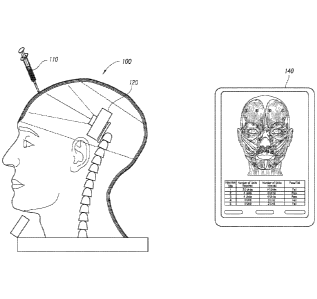
visible or non-visible light, such as fluorescent or ultraviolet light. In
some
embodiments, the light emitted from the light source is attenuated differently
depending on which layer of simulated skin or muscle of the injection
apparatus 100
is penetrated. Different colors, directions, graph lines, visual patterns,
polarization,
fluorescence, or intensities of light can be captured by the camera 120 as the
testing
tool 110 is injected through the different layers of material surrounding the
injection
apparatus 100. The resulting light detected by the camera 120 can be used to
determine the location of the injection, the pressure exerted by the user, the
angle of
injection, or the depth of the injection. This information can be detected,
for example
by a camera 120, and communicated to a user interface device 140 for testing
or
certification purposes.
[0063] The camera 120
within the simulated skull of the injection apparatus
captures the attenuated light of an injection through video recording and/or
photographic images. The camera 120 can include a processor and can
communicate the camera output to a user interface device 140. The information
gathered from the camera 120 and testing tool 110 may be communicated to a
user
interface 140 for data collection, testing or certification purposes. The
camera output
can be raw or processed video or images obtained from the camera 120. The
camera processor can include software configured to interpret the visual
indications
for testing purposes or can merely pass images to the user interface device
140 for
further processing. In an embodiment, the user interface device 140 can also
communicate instructions to the camera 120 and/or testing tool 110.
[0064] Figure 2
depicts an embodiment of the testing tool 110. In one
embodiment, the testing tool 110 includes a needle 212, plunger 209, and
barrel
210. The testing tool 110 may be activated by pressing the switch button 200
which
moves switch contacts 201 and connects the battery 203. The battery 203 and
switch button 200 are within the housing 202 of the testing tool 110. The
battery 203
powers the LED 204 to emit a light through a lens 205. The emitted light then
travels
through an optical fiber 207 that captures the focused light. Alternatively,
the light
can travel through a hollow needle, without the use of optical fibers. The
optical fiber
207 is held within the plunger 209, barrel 210, and needle 212 of the testing
tool 110
through a slide slot 208. Alternatively, the fiber optic and other components
are all
fully incorporated into the testing tool to create a standalone device. The
light
-15-
CA 02928460 2016-04-21
WO 2014/070799
PCT/US2013/067352
emitted from the LED 204 travels through the optical fiber 207 and is emitted
at the
tip of the needle 212 as a focused light 213. In one embodiment, the needle
212 of
the testing tool 110 may be hollow and allow light to enter without a focused
tip. The
LED 204 may emit light of different spectrums through a lens 205. In some
embodiments, the light emitted from the LED 204 is attenuated once it
penetrates
each layer of simulated skin or muscle. Once the needle portion 212 of the
testing
tool 110 penetrates through each layer of tinted material, different colors or
intensities of light can be detected by the camera 120. In some embodiments, a
friction 0-ring 211 allows the plunger 209 to be pushed downward and causes
the
needle 212 to go forward for an injection.
[0065] In some embodiments, the light viewed by the camera 120 from the
needle 212 can change from a round to oval shape. This can occur when the
needle
moves out of alignment with the camera 120. The length and width of the oval
viewed by the camera 120 can indicate the angle of the injection while the
direction
of the oval along its longer axis can indicate the direction of the injection.
[0066] In some
embodiments, fluid can be stored within the testing tool
110 and injected into the injection apparatus 100. The fluid injected can
simulate the
consistency of the injection substance that would be injected for a real
treatment.
Once the injection apparatus 100 receives the injection, a camera 120 captures
the
location, speed, pressure, and angle of the injection. The camera 120 sends
this
information in video or photographic form to the processor 604 which analyzes
the
detailed information and determines desired injection parameters. In some
embodiments, the data associated with the injection can be determined by the
testing tool 110 sending information wirelessiy to the processor 604. For
example,
the testing tool may detect the friction experienced by the friction o-ring
211 to send
to the processor 604 information about the speed and pressure of an injection.
In
one embodiment, an accelerometer can be attached to the testing tool 110 to
provide information on an angle of injection. Another process for conveying
information to the processor 604 includes alternating frequencies or patterns
of light
emitted by the LED. Alternatively, the information from the camera 120 and
testing
tool 110 can be sent directly to the output device 140 for processing.
[0067] In one
embodiment, the plunger 209 of the testing tool 110 may be
able to detect the angle, speed, friction, and depth of an injection. This can
be
-16-
CA 02928460 2016-04-21
WO 2014/070799
PCT/US2013/067352
accomplished with a wired or wireless electrical signal being transmitted from
a
sensor placed in the testing tool 100. In some embodiments, a cable can be
placed
parallel to the light fiber that can read the injection parameters, such as
the pressure,
speed, or acceleration of the injection. For example, the electrical signal
transmitted
from the sensor can detect 0-5 volts of electricity, which can represent the
amount of
pressure being exerted by the user when utilizing the testing tool 110. In
other
embodiments, the electrical signal may emit a certain frequency that
represents the
pressure exerted. For example, a frequency of 100 Hz can represent low
pressure
while a frequency of 1,000 Hz can represent high pressure exerted by the user.
In
an embodiment, the LED can be modulated at a modulation rate corresponding to
an
angle, speed, friction or depth of an injection. This modulated light can be
detected
by the camera and used to determine the desired injection parameters without
the
need for a separate data communication path between the testing tool and the
rest
of the system. In some embodiments, a wireless transmitter can be placed in
the
testing tool that communicates directly to the user interface device 140 and
displays
the parameters of the injection.
[0068] In some embodiments, the testing tool 110 can inject a
fluorescent
fluid into the injection apparatus 100. The layers of simulated muscle and
skin may
be configured to have a reservoir that accepts these fluid injections. The
fluorescent
fluid may be visible through transparent, opaque, or lightly pigmented
material in the
simulated skin and muscle layers. In one embodiment, a UV lamp may be placed
within the injection apparatus 100 in order for a user to clearly see the
injection and
injected fluid going into the injection apparatus 100.
[0069] In some embodiments, the testing tool 110 may also be powered
with a plug in cable. The testing tool 110 can send information over a
wireless
network or portable computer to process information about an injection. The
signals
may send information related to the 3D location, depth, intensity, or pressure
exerted
by the user when practicing the injection.
[NM Figure 2B depicts an embodiment of the testing tool with a
position
transducer, such as, for example, a linear potentiometer fixed to the testing
tool. The
linear potentiometer can be used to track the depression of the plunger 209
relative
to the barrel 210. This information can then be used by the system to
determine a
volume of injection. This information, in connection with the location and
depth
-17-
CA 02928460 2016-04-21
WO 2014/070799
PCT/US2013/067352
information obtained by the injection apparatus can be used to determine fluid
volume distribution information about the injection.
[0071] In some embodiments, the transducer or potentiometer can be
connected to a slider 216. The linear potentiometer 214 measures the position
of
the plunger 209 of the testing tool relative to the barrel 210. In some
embodiments,
the linear potentiometer 214 may be fixed to the plunger 209 of the testing
tool. A
slider 216 may be attached through a slot 225 in the barrel 210 and mated with
a
pocket within the plunger 209. The slider 216 moves with the plunger 209 to
allow
the transducer to output the position of the plunger 209 through an output pin
215.
The transducer then electronically communicates through a connection with a
processor 604, which calculates the simulated volume and distribution of the
injection. This calculation may be completed by using the parameters of the
plunger
209 displacement and the diameter of the barrel 210.
[0072] In some embodiments, the testing tool determines the pressure
applied by the injector. This can be accomplished by measuring the force
applied to
the plunger 209 through a thin film force sensor 217 on the plunger 209
flange.
Electrical connections to the force sensor and linear potentiometer may be
placed
along with the optical fiber 207 in a multi-lumen sheath. The force sensor 217
electrically communicates with a processor 604 (Figure 6) and send the
parameters
associated with the applied force to the plunger 209. The injector can be used
for
various procedures with various needle sizes, medication viscosities, and skin
or
muscle properties.
[0073] Figure 2C depicts an embodiment of the testing tool with a linear
potentiometer 214 remotely connected to the testing tool. The motion of the
plunger
209 is measured using a pull wire 219 that is connected to the plunger 209 and
to
the slider 216 of a remote transducer. The pull wire 219 may be routed along a
multi
lumen sheath 218 that carries the optical fiber 207.
[0074] In an embodiment, the remote transducer is used in conjunction
with a force application system to simulate the viscosity encountered during
an
injection. In an embodiment, a needle drag can be designed to simulate a real
injection. The needle drag can be determined based on the elasticity of the
injection
apparatus layers (for example, as measured in durorneters), the coefficient of
friction
between the plunger and the barrel and the needle diameter and length. Figure
2C
-18-
CA 02928460 2016-04-21
WO 2014/070799
PCT/US2013/067352
illustrates a force application system including a tension spring 222, steel
tensile strip
221, steel friction block 223, and a controllable friction device, such as an
electromagnetic brake. The electromagnetic brake is activated when current
from a
power source is applied to a coil 220. Once current is removed from the coil
220, the
electromagnetic brake returns to its resting state. These elements of the
testing tool
provide the resistance necessary for a simulated injection. The
electromagnetic
brake can be controlled by the processor 604 to simulate the feel and
resistance of
an actual injection. Alternatively, the parameters of the brake force applied
can be
preset. A fixation post 224 may be used to lock the barrel 210 and multi lumen
sheath 218 together. In some embodiments, the electromagnetic brake may be
adjusted to simulate the resistance of skin, tissue, or muscle that the needle
212
would be penetrating. For example, an injector performing a lumbar nerve root
sleeve injection would be able to feel the resistance of a fluid containing
corticosteroid and long-acting local anesthetic. The electromagnetic brake
also
provides the resistance corresponding to a hypothetical patient's skin,
tissue, or
muscle. The injector applies the corresponding amount of force necessary to
correctly perform the injection.
[00751 Figure 2D depicts a cross section of the multi-lumen sheath 218.
The multi lumen sheath 218 holds the optical fiber 207 and the pull wire 219.
The
pull wire 219 may be attached to the plunger 209 and moves the slider 216. As
the
plunger 209 moves, the pull wire may move through the multi lumen sheath 218.
[00761 Figure 3 depicts a schematic diagram of a testing tool for
injection
training. In one embodiment, the testing tool 110 has a needle 212, plunger
209,
and barrel 210. The testing tool 110 can have a light fiber and/or light
emitting diode
(LED) 320 as a light source. The focus of the light source may be on the end
of the
fiber inside the needle portion of the testing tool 110. The testing tool 110
is
activated by a switch button 300 which connects the battery and activates the
LED
320. In some embodiments, the testing tool 110 is a portable battery operated,
fully
functional, stand-alone device. The battery 310 powers the LED 320 and
activates
the LED 320 once the switch button is turned to the on position. The LED 320
emits
light through a fiber optic cable 330 so that light 340 shines through the
needle
portion of the testing tool 110. In some embodiments, the testing tool 110 may
be
connected through a cable to a processor 604 which is able to communicate with
the
testing tool 110 in order to receive information on testing parameters and
provide
-19-
CA 02928460 2016-04-21
WO 2014/070799
PCT/US2013/067352
programing instructions to the testing tool 110. In other embodiments, the
testing
tool 110 may wirelessly communicate with the processor 604.
[0077] Figure 4A
depicts a side view of one embodiment of the injection
apparatus 100 with a surrounding removable layer divided into three separate
simulated human skin and muscle layers 410, 420, 430. In some embodiments, the
skin layers may be separated to represent the epidermis, dermis, and
hypodermis.
The layers of skin and muscle may be of uniform or different densities. In
other
embodiments, the layers of skin and muscle may be thicker or thinner to
simulate the
skin of patients with uneven skin, muscle layers, or damaged skin. In some
embodiments, each separate layer may be of a different density or color. For
example, in Figure 4, the first layer 410 may represent the epidermis as
opaque.
The second layer 420 may represent the dermis and as tinted. The third layer
430
may represent the muscle and as clear. More or fewer layers of simulated skin
and
muscle can also be used depending on the desired injection and the level of
detail
required.
[0078] In some
embodiments, each separate layer of skin or muscle 410,
420, 430 may be of a different transparency, density or color. In some
embodiments, the different intensity or colors can be viewed by the camera
after the
testing tool 110 is inserted into the simulated skin or muscle. This can allow
a
camera 120 to send information to a processor 604 related to the location,
pressure,
angle, or depth of an injection. In other embodiments, the injectable muscle
and skin
layers may be of uniform density, consistency, or color. In some embodiments,
the
injectable muscle and skin layers 410, 420, 430 may be made of an elastomer.
In an
embodiment, the elastomer may simulate the elasticity of human skin and range
from 5-35 on the durometer "A" scale. The simulated skin and muscle layers
410,
420, 430 may also consist of different angled fibers that deflect light
emitted from a
testing tool in different directions to allow for location, depth. angle and
pressure
analysis based on the optical properties observed. In an embodiment, the
fibers can
be a pattern printed on each skin or muscle layer 410, 420, 430 that
selectively block
light viewed by the camera. Depending on the angle of the fibers within each
layer
of the skin and muscle layers 410, 420, 430, the light emitted from a testing
tool may
be deflected at that angle. For example, the first layer 410 may have threaded
angled fibers directed at a 45 degree angle. The second layer 420 may have
threaded angled fibers directed at a 55 degree angle. The third layer 430 may
have
-20-
CA 02928460 2016-04-21
WO 2014/070799
PCT/US2013/067352
threaded angled fibers directed at a 65 degree angle. Depending on which layer
an
injector has penetrated, the light emitted from a testing tool 110 may be
deflected in
a different direction. If the injector has penetrated the second layer 420,
the light
should be deflected at a 55 degree angle. The deflection of the light emitted
from
the testing tool 110 is captured by a camera 120 and sent to a processor 604.
The
processor 604 analyzes the intensity, deflection, and clarity of the light
emitted from
the testing tool 110 to generate results about the injection.
[0079] In some embodiments, the layers of skin or muscle 410, 420, 430
may be dyed with carbon black particles or similar light-obscuring agents. The
density of the carbon black particles can be adjusted to substantially block
emitted
light from reaching the camera through all layers. As the needle portion of
the
testing tool 110 travels through each layer, more light is viewed by the
camera. The
carbon black particles obscure light so that an injection into each layer may
represent a different intensity of light. In some embodiments, this will allow
a camera
120 placed within the injection apparatus 100 to detect the layer of skin or
muscle
410, 420, 430 which is being penetrated by the light source. In one
embodiment, the
different layers of skin or muscle may be dyed with translucent color. These
translucent layers will attenuate the light emitted from a testing tool in
different ways.
The degree and color of attenuation of the light after it has traveled through
the
simulated muscle and skin layers can then be detected by the camera and used
to
analyze the injection.
[0080] In an embodiment, the system includes an injection apparatus 100
for injection procedures on different parts of the human body. In an
embodiment,
there are at least three nesting layers of the apparatus: the skeletal
structure layer,
muscle layer, and top layer of simulated skin. A nerve layer can also be
present
within the muscle layer. This allows trainees to visualize and study the
layers of
muscle and nerves underneath the skin layer to become familiar with human
facial
anatomy. Veins or arteries can also be included and embedded within the muscle
layer. The veins or arteries may be of a different color or density than the
muscle
and skin layers. The injectable muscle and skin layers 410, 420, 430
anatomically
match that of the human body. In some embodiments, the injection apparatus 100
may simulate the internal organs or other body parts of a human or animal. In
some
embodiments, injectable muscle and skin layers 410, 420, 430, may be color
coded
-21-
CA 02928460 2016-04-21
WO 2014/070799
PCT/US2013/067352
so that a trainee may be able to identify the different sections of the human
body or
muscles associated with each simulated condition.
[0081] The depicted layer on the injection apparatus 100 in Figure 4A
simulates human skin and muscle and has the same feel, permeability, and
appearance as human skin. The skin and muscle layers may be removable,
reusable, and replaceable to simulate a variety of patients having different
injection
conditions. For example, the skin may vary by the age, ethnicity, race,
texture, or
thickness of different test patients. In some embodiments, the skin may
simulate
certain cosmetic conditions. For example, the skin may have wrinkles, scars,
hyper-
pigmentation, lacerations, or other blemishes typically treated by injections.
The
various embodiments of skin types allow the trainee to gain a wide variety of
experience dealing with different skin types. The muscle layers may consist of
thicker or thinner layers to represent different densities in muscle tone. In
some
embodiments, the different density or color of the skin or muscle may allow a
testing
tool and camera to detect the depth and location of an injection.
[0082] In some embodiments, the injection apparatus 100 is configured to
represent human facial features, such as those features associated with
smiling or
frowning, as would be encountered during certain cosmetic or therapeutic
injections.
In some embodiments, the apparatus can model various cosmetic conditions or
damaged areas of the human body. For example, these cosmetic conditions may
include glabeliar frown lines, horizontal forehead lines, temporal brow lifts,
crow's
feet (lateral canthal lines), lower eyelids, nasalis bunny lines, vertical lip
lines,
gummy smiles, nasolabial folds (NLFs). marionette lines, pre-jowl sulcus,
labiomental
crease, and mictface, facial lipoatrophy, lip augmentation, mouth frowns
(depressor
anguli oils), apple dumpling chin, horizontal neck lines, vertical platysmal
bands,
acne blemishes, accident scars, or asymmetry. In some embodiments, the skin
can
be manipulated to mimic actual facial movement, such as furrowing of the brow,
squinting of the eyes, and pursing of the lips. Users of the injection
apparatus may
be able to pinch the skin, stretch the skin, or grab a portion of the muscle
in order to
simulate a real injection. The injection apparatus 100 may be programmed to
display various cosmetic conditions through a user interface device 140. There
may
also be buttons available on the injection apparatus 100 for programming
cosmetic
conditions. In some embodiments, the skin layer may be manufactured with pre-
determined cosmetic conditions.
-22-
CA 02928460 2016-04-21
WO 2014/070799
PCT/US2013/067352
[0083] In one embodiment, programs for individual injection sites may be
sold separately or in a package. The user interface device 140 may be updated
with
various injection tests for different parts of the human or animal body. For
example,
an injection test can be purchased for Botox injections. The injection sites
for parts
of the human face could be downloaded onto the user interface device 140 and
unlocked by a user. For example, the targeted injection sites for toxin
cosmetic
injections for a human face may include the frontalis (forehead lines),
glabellar
complex (procerus and corrugators) frown lines, orbicularis oculi-lateral
canthal area,
crow's feet lines, nasalis-bunny lines, orbicularis oris-vertical lip lines,
depressor
anguli oris, mentalis, masseter, platysma, depressor septi nasi, levator labii
superioris alaeque nasi, gland hypertrophy, or labial artery. The program can
communicate with the processor 604 to control the movement of the camera 120
to
record or measure the specific injection sites for injection testing. The
program can
also communicate with the processor 604 to change the pigmentation or color of
the
skin layers 410, 420, 430 of the injection apparatus 100. In some embodiments,
the
program can be set to simulate a specific type of injection scenario. For
example, a
user can set the user interface device 140 to simulate crow's feet on the
injection
apparatus 100. The skin layers 410, 420, 430 would be mechanically moved to
simulate the wrinkles at the edge of the injection apparatus 100 to form
crow's feet.
Once the user correctly injects the injection apparatus 100 at the injection
site for
crow's feet, the injection apparatus 100 would mechanically smooth out the
wrinkles
from the crow's feet.
[0084] In one embodiment, the program can inform the user of the type of
treatment performed on the injection apparatus 100 through the user interface
device
140. For example, the user interface device 140 may educate the user on the
type
of treatment, such as whether it is therapeutic, sub-therapeutic, or super-
therapeutic.
[0085] The injection apparatus 100 may also be used for therapeutic
treatment training. These treatments may include those related to
blepharospasm,
strabismus, or chronic migraines, and others. For example, Botox injections
can be
practiced to induce localized, partial paralysis on the apparatus for
treatment of
blepharospasm. In some embodiments, the injection apparatus may be manipulated
to display the different physical features associated with conditions
requiring
therapeutic treatment. For example, the injection apparatus 100 may display a
-23-
CA 02928460 2016-04-21
WO 2014/070799
PCT/US2013/067352
squinted eye or be cross-eyed when it is programmed as a patient with
strabismus.
Upon therapeutic treatment by a trainee, the injection apparatus 100
mechanically
readjusts to fix the condition.
[0086] In some embodiments, the base layer 400 allows the injection
apparatus 100 to keep its structure and holds the components of the injectable
muscle and skin layer in place. The base layer 400 may be mechanical and
moveable in response to an injection from the testing tool 110. The base layer
400
may be mapped with a grid of target zones. For example, the inside or outside
of the
base layer 400 may have imprinted lines that represent zones of injection. The
grid
of target zones may correspond to an image on a user interface device 140 that
is
able to show the accuracy of an injection. The grid can show the face from the
inside of the camera and what the muscles look like. This can occur, for
example, in
a training mode. In some embodiments the top skin layer 410 may have visual
targets which display the location for injection corresponding to a cosmetic
condition
or therapeutic treatment. These visual targets may be color coded so that a
user
may be different injection zones that should be targeted for administering
different
injections.
[0087] In some embodiments, the base layer 400 of the apparatus may be
a clear plastic shell. The plastic shell layer may be covered with removable
layers of
elastomer membranes simulating human muscle or skin. The plastic shell may
simulate the look of any human body part, including any internal organs. In
some
embodiments, the injection apparatus 100 simulates an entire human or animal
body
part with or without removable layers of elastomer membranes simulating human
skin or muscles.
[0088] In an embodiment, the injection apparatus may have a camera 120
attached to a pivotable stand 460 and placed within the injection apparatus
100. The
pivotable stand 460 may be attached to a removable base 470 that allows a user
to
physically change the direction of the pivotable stand 460. In some
embodiments,
the pivotable stand 460 may be mechanically movable upon detection of a signal
received through a processor 604. The processor 604 may receive a signal to
change the location of the pivotable stand 460 through a output device 140.
[0089] In some embodiments, the camera 120 may be positioned so it may
swing into different positions in response to a shift gate. This allows a user
to move
-24-
CA 02928460 2016-04-21
WO 2014/070799
PCT/US2013/067352
the camera 120 to focus on different target zones without having to manually
move
the camera within the injection apparatus 100. The camera 120 may include an
angular grid sensing filter that can detect its position and rotate itself
according to a
displayed grid within the injection apparatus 100. In an embodiment, the
camera
120 is set to focus on either color or line orientations within the injection
apparatus
100. The camera 120 may read a user's injection based on the information
received
from the light emitted from the testing tool 110 in conjunction with the
location
determined by a grid embedded in the base layer 400 of the injection apparatus
100.
[00901 In some embodiments the camera 120 may have a broad or
focused range 450. For example, a broad range camera may be used when there is
no specific target area that is being focused on for testing or certification
purposes.
A focused range camera can be positioned to aim at a zone for injection. In
some
embodiments, the camera 120 is configured to communicate with a user interface
device 140 to display the results of an injection. In an embodiment, the
results of the
injection may be determined by the intensity and color viewed by the camera
120
after the testing tool 110 has been injected into the different layers of skin
or muscle.
The range 450 of the camera 120 may be manually adjusted by setting the camera
to encompass a smaller or bigger range. The range 450 of the camera 120 may
also
be adjusted by inputting a grid location into the output device 140 and
communicated
to the camera 120. The camera 120 then adjusts its targeted location.
[0091] The camera 120 can output video to a user interface device 140
through a wired or wireless connection. In an embodiment, the output device
140 is
equipped with software to read and analyze the results obtained from the
video. In
an embodiment, the software is able to analyze the results and generate a
score or
evaluation of the user's injection. The software can also report and capture
data on
the trainee's experience. The software may also play back the user's injection
and
display errors or provide feedback acceptable injections. In an embodiment,
the
software includes a biometric sensor to identify each trainee.
[0092] Figure 4B depicts a side view of one embodiment of the injection
apparatus 100 with a surrounding removable layer including at least three
simulated
human skin and muscle layers 410, 420, 430. The surrounding removable layer
may
cover one section of the injection apparatus 100 or the entire injection
apparatus
100, for example, as illustrated in Figure 4B. In some embodiments, the human
skin
-25-
CA 02928460 2016-04-21
WO 2014/070799
PCT/US2013/067352
and muscle layers 410, 420, 430 can be removed separately. As discussed above,
more or fewer skin and muscle layers can be used and the present disclosure is
not
intended to be limited to three layers.
[0093] In an embodiment, the injector or administrator of injection
training
may choose to focus on a specific area of the injection apparatus and only
have the
removable layer surrounding that area. The injector may then observe the
injection
apparatus 100 to see how an injection penetrates through the different layers
of skin,
muscle, and nerves. This embodiment may be used, for example, for novice
injectors who require visual guidance for the depth of their injections.
[0094] Figure 5 depicts the side view of an embodiment of the injection
apparatus 100 with a grid displayed with injection sites. The grid lines 500
are used
by a user or a camera 120 to determine the location of an injection site 520.
The
grid lines 500 may represent locations on an x-y plane or a three dimensional
x-y-z
axis so that a camera 120 may easily transfer location data to a processor
604. The
grid lines 500 may be placed in an arrangement where each created section may
represent an injection site 520. The sections may also be targeted areas that
are
used by the camera 120 to indicate where to focus for a particular injection.
The grid
lines 500 may be placed in different directions long the injection apparatus
100 or
angled to accommodate any grooves or bumps on the injection apparatus 100.
[0095] In some embodiments, the injection apparatus 100 may have
sensors embedded within the different human skin and muscle layers 410, 420,
430.
The sensors may be located on an injection site 520, multiple injection sites
520, or
continuously throughout the entire human skin and muscle layers 410, 420, 430.
Once an area has been treated by an injection, the sensor may communicate with
the testing tool 110 or the injection apparatus 100 to provide the information
associated with the injection. For example, the sensor would be able to test
reads
the treatment from the sensors. The pressure applied to the area of injection
may be
detected by the training tool and the parameters of the injection may capture
the
depth, pressure, and angle of a user's injection. The parameters may then be
compared to a pre-determined treatment and provided to a user interface device
140, which displays the testing results.
[0096] In some embodiments, the injection apparatus 100 may have
inflatable pads embedded within the different human skin and muscle layers
410,
-26-
CA 02928460 2016-04-21
WO 2014/070799
PCT/US2013/067352
420, 430. The inflating and deflating of the pads may be initiated by an
attached
sensor detecting the penetration of an injection. The inflatable pads may be
located
on the injection site 520. The inflatable pad may independently inflate or
deflate
proportionally to the location, depth, pressure, and angle of a user's
injection. The
inflation and resulting size of the pads may differ at various injection sites
520,
depending on the natural human reaction to an injection in that area. Once a
user
has completed an administered test, the inflatable pad may deflate and return
the
human skin and muscle layers to their original condition. The inflation pads
allow the
asymmetries of an injection to be observed and addressed by the injector. In
an
embodiment, the testing tool injects air into the inflatable pads, skin and/or
muscle
layers so that a user can observe how the injection has affected the
apparatus. This
allows the trainee to see in real time the effect of the injection. For
example, the
effect can be watching the apparatus "age". In an embodiment, the trainee can
also
deflate the fat pads. This allows a trainee to practice determining how much
injection is required for a given patient.
[0097] In some embodiments, the injection apparatus 100 can be
configured to turn on the measurement and/or analysis of different injection
sites
520. A software program communicating through the user interface device 140
can
selectively enable certain procedures, for example, through separate software
purchases or upgrades related to particular injection sites 520. For example,
the
injection sites 520 for Botox procedures can be enabled. The injection sites
520 for
treating cosmetic conditions such as furrowed brows, crow's feet, or adding
volume
to lips can also be separately enabled. Once the testing tool 110 injects that
particular injection site 520 corresponding to the cosmetic condition, the
camera 120
views the injection and communicates the results to the processor 604. The
results
are generated and displayed through the user interface device 140.
[0098] Figure 6 depicts further detail of the camera 120. The processor
604 controls the various functions of the camera 120 and the camera interface
605.
The camera interface 605 can include, for example. an optional output display
606,
optional speaker driver 607, optional speaker 608, optional wired or wireless
communication transceiver 609, optional user input 610, and on/off switch 611.
The
processor 604 can transfer data to the output display 606, including display
updates
and visual alarms. The processor 604 can also be configured to interpret user
inputs
from the user input 610. The camera interface 605 may receive information from
the
-27-
CA 02928460 2016-04-21
WO 2014/070799
PCT/US2013/067352
user input 610 and send the user input data through the processor 604 so that
the
display data generated on the output display 606 can change according to a
user's
selection through the user interface device 140. The processor 604 may
generate
various alarm signals when the user is in the testing or training mode of the
injection
apparatus 100. The processor 604 controls a speaker driver 607 which then
actuates a speaker 608 to provide real-time information regarding a user's
progress
for injections. The speaker 608 may provide audible indications, allowing the
user to
know when an accurate or inaccurate injection has been made. For example, the
speaker 608 may emit a buzz sound when the injection is inaccurate and a beep
sound when the injection is accurate.
[0099] The camera 120 receives its power from batteries in the camera or
through a power supply 602. A power manager 603 monitors the on/off switch of
the
camera 120 and the output device 140 and turns each on or off accordingly. The
batteries in the camera 120 may either be alkaline rechargeable batteries or
another
renewable power source. The camera 120 may also be powered with a plug in
cable. In some embodiments, the camera can send information over a wireless
network or directly to portable computer to process information about an
injection
using the wireless communication transceiver 609. The wired or wireless
transceiver
609 can communicate over any known protocol including Bluetooth, Zigbee, WiFi,
Ethernet, USB, or any other wired or wireless communication protocols.
[0100] A non-volatile memory 600 is connected to the processor 604 via a
high-speed bus. In the present embodiment, the memory 600 is erasable and
allows
a user to store information including operating software, user configurable
command
options and information related to different types of injections, including
recorded
images or video. The memory 600 may also store user-specific data. For
example,
a user who has completed several injections on a certain day may store results
of
those several injections and access the results at a later time. In addition,
information obtained by the injection apparatus can be stored and sent to a
central
repository for analysis with testing information from other devices. The
central
repository, can be, for example, a server or cloud computing device.
[0101] Figure 7 illustrates a view of one embodiment of the injection
apparatus 100 with a removable band 700. The removable band 700 allows for
replacement of portions of the injection apparatus without the need to replace
larger
-28-
CA 02928460 2016-04-21
WO 2014/070799
PCT/US2013/067352
portions 720 of the simulated tissue. Multiple different removable bands can
also be
used. For example, a forehead band can be used in conjunction with a separate
cheek skin band. In some embodiments, a removable band 700 may be placed on
the base layer 400 of the injection apparatus 100. These embodiments allow a
user
to replace only the targeted injection areas. The removable band 700 may be
placed
on any portion of the injection apparatus 100. For example, testing for an
injection
involving cardiac treatment may require a removable band being placed over the
area of the simulated heart that needs treatment. The removable band 700 may
have layers of simulated human skin or muscle 700 attached. Each separate
layer
of skin or muscle 700 may be of a different transparency, density, or color.
In some
embodiments, the opaque layer obscures light from the needle portion of the
testing
tool 110 being inserted through shallower layers or muscle or skin 700 from
being
viewed by the camera. For example, in an embodiment with three layers of
muscle
or skin, the top layer may be opaque, the middle layer may be tinted red, and
the
bottom layer may be green and tinted. In some embodiments, the layers may be
composed of material with different angled threaded material. This angled
threading
will deflect the light emitted from a testing tool in different directions so
that the
camera may capture the depth, pressure, and angle of a user's injection.
[0102] Figure 8 depicts an exploded view of an embodiment of a
removable band 700 which can be used in conjunction with the injection
apparatus.
In this particular embodiment, there are three layers of the removable band
700
which are visually exploded for explanatory purposes. The layers 800, 810, 820
may
consist of different materials, colors, or angled threading. For example, in
Figure 8,
the first layer 800 may be made of a transparent elastomer layer with surface
lines
that are based in a right direction. The second layer 810 may consist of a
transparent elastomer layer with surface lines that are based in a left
direction. The
third layer 820 may be targeted with grid lines or windows that are outlined
or
colored. In some embodiments, grid lines or targeted windows 830 allow the
camera
to easily find a certain zone for injection. By sending keystroke data to the
processor
604, the camera 120 may be easily rotated and directed toward a target area of
injection. The surface lines on the different layers may represent the angled
threading of each layer. The angled threading allows light emitted from the
testing
tool 110 to be reflected in different directions so that the camera 120 may
gather
visual information about the injection and send it to a processor 604. The
processor
-29-
CA 02928460 2016-04-21
WO 2014/070799
PCT/US2013/067352
604 may then analyze the different angles projected from the testing tool 110
and
determine the accuracy of the user's injection.
[0103] Figure 9 depicts a cross sectional view of a top skin layer 900
divided into three separate simulated human skin or muscle layers 900, 910,
920
and being injected by a testing tool A, B, C. The camera 120 may be targeted
toward an injection zone of A, B, and C and capture the visual injection
through
video or photographs. In one embodiment, the testing tool 110 may emit an
intensity
of light that is attenuated differently as it penetrates different layers of
skin or muscle.
For example, no light is detected by the camera 120 while the testing tool 110
has
not made contact with the skin layer (shown in A). The light detected by the
camera
120 when the testing tool 110 reaches the second layer of skin may be a more
intense and of a different color in comparison to A (shown in B). The light
detected
by the camera 120 when the testing tool 110 reaches the third layer of muscle
may
be the most intense and of a different color in comparison to A and 13 (shown
in C).
In some embodiments, an unattenuated light may signal that the user has
penetrated
through all the layers of skin and muscle and hit the bone of the injection
apparatus
100.
[0104] In some embodiments, the separate simulated skin or muscle
layers may consist of different angled fibers. As a result of these angled
fibers, the
light emitted from the testing tool 110 may be deflected in different
directions. For
example, the fibers present in the lowest layer of simulated muscle or skin
may be at
a 45 degree angle, the second layer of simulated muscle or skin may be at a 60
degree angle, and the top layer of simulated muscle or skin may be at a 75
degree
angle. As the camera 120 views the emitted light from the testing tool, it is
able to
capture information about the injection into the layer of muscle or skin. The
output
device 140 may receive this information and generate a report determining the
depth, pressure, or angle of the users injection.
[0105] Figure 10 illustrates the progression of a testing tool 110 being
injected into an injection apparatus 100. In this embodiment, the testing tool
110 is
shown as being inserted into a removable band 700 with three layers of
simulated
skin or muscle. Although shown with respect to a removable band embodiment, it
is
to be understood that this process applies equally to all embodiments
disclosed
herein. The camera 120 within the injection apparatus 100 can focus on the
targeted
-30-
CA 02928460 2016-04-21
WO 2014/070799
PCT/US2013/067352
injection zone. In some embodiments, the camera 120 can record or take
pictures of
the actual injection and send this information to the processor 604 or output
device
140. As the injection is placed within the different layers of simulated skin
or muscle,
the intensity, angle, color or other visual indication of the light viewed by
the camera
120 from the testing tool 110 may change. In other embodiment, other non-
visible
light attenuation techniques can be used. In some embodiments that have angled
threading of the different layers, the camera 120 can capture fewer line
directions as
the testing tool passes through each layer of simulated skin or muscle. For
example,
there are multiple line directions displayed when the testing tool 110 is
injected into
the first layer 1010 of simulated skin or muscle (shown in 1010a). There are
fewer
lines displayed when the testing tool 110 is injected into the second layer
1020 of
simulated skin or muscle (shown in 1020a). When the testing tool 110 is
injected
into the deepest layer of simulated skin or muscle, there are no line
directions
present (shown in 1030a). The only visual display to the camera 120 is of the
light
emitted from the testing tool 110.
101061 Figure 11 illustrates a front view of an injection apparatus 100
for
cosmetic training with cosmetic conditions labeled to corresponding injection
sites on
a muscle layer 1100. The muscle layer 1100 is available for a trainee to view
and
study the nerves and muscles of the face, head and neck. In this particular
embodiment, common facial aesthetic conditions corresponding to certain
muscles
are labeled on the injection apparatus 100. In this particular embodiment, the
chart
1010 displays the injection units for Botox corresponding to each injection
site. For
example, the targeted site I corresponds with the targeted muscle, procerus.
The
trainee is required to inject 20 units of Botox to remove furrow lines from
this area.
It may show units or may just show the muscle targeted for injection. This
type of
visual display of the labeled injection apparatus and the chart 1010
displaying
required injection units is available through the user interface for each
different
condition treated. For example, the user interface may display a different
chart for
epidural or cardiac injections. The chart 1010 can be a separate paper or it
can be
displayed graphically on the output device 140 as part of the training system.
101071 Figure 12 illustrates an injection apparatus 100 and output
display
1220 depicting the corresponding image of the injection apparatus 100. The
skin
layer 1210 of the injection apparatus 100 is shown and targeted zones of
injection
1200 are labeled on the surface. In some embodiments, the targeted zones of
-31-
CA 02928460 2016-04-21
WO 2014/070799
PCT/US2013/067352
injection 1200 may be different shapes, be distinguished by grid lines, or by
color.
The injection apparatus 100 receives an injection from a testing tool 110 and
a
camera 120 captures the visual display of an injection and transfers the
information
to a processor 604 or the output device 140. The processor 604 or output
device
140 then analyzes the visual information and generates an output with detailed
information regarding the parameters of the injection. This information is
then
displayed on the output device 1220. In some embodiments, the output device
1220
may detect information in real time and display injection parameters to a
user.
[0108] Figure 13 is a flowchart illustrating one embodiment of a method
for
utilizing an injection apparatus 100. In block 1300, the output device 140
receives
input from the user regarding the action that the user would like to perform
on the
output device 140. The output device 140 may be programmed to facilitate
several
program modes. For example, the user may select to enter either the training
or
testing mode for the output device 140. In block 1310, the output device 140
may
communicate with the processor 604 to retrieve stored information from memory
600. This information may include a pre-set injection test for a specific
treatment or
a display of injection sites for learning purposes. The user may also access
information related to previous injection tests for a particular user. In
block 1320, the
processor 604 can generate data associated with a selected mode. For example,
when user selects the training mode, the processor 604 can retrieve
information from
memory 600 about different types of injections to educate the user. The
processor
604 may also receive directions to engage in testing mode. This prompts the
processor 604 to activate the camera 120 for injection testing purposes. In
block
1330, the output device 140 may display the output corresponding to the
selected
mode. For example, the output device 140 may display the results of a
simulated
injection or display feedback on the injection.
[0109] Figure 14 is a flowchart illustrating one embodiment of a method
for
utilizing an injection apparatus 100 in a simulated injection test. At block
1400, the
output device 140 receives user input to perform testing at a targeted
injection site.
At block 1410, this request is sent to the processor 604 which, in an optional
embodiment, sends signals to a camera 120 to rotate to a targeted injection
site. At
block 1420, an injection performed by a user through a testing tool 110 is
detected
by a camera 120. Information regarding the injection may be directly
communicated
by the testing tool 110 through either a cable or a wireless connection to a
processor
-32-
CA 02928460 2016-04-21
WO 2014/070799
PCT/US2013/067352
604. At block 1430, the camera 120 records either a video or captures a
photograph
of the injection at the targeted injection site. At block 1440, the video or
photograph
is analyzed by the processor 604. The processor 604 is able to determine one
or
more injection parameters. such as, for example, the angle, depth, pressure,
and
location of the injection. At block 1450, the injection parameters analyzed by
the
processor 604 is displayed on a output device 140.
[0110] Figure 15 illustrates a user interface for injection training
through an
injection apparatus 100. The main menu of the user interface allows a user to
select
different modes of operation (e.g., training mode, testing mode) and different
features (e.g., view previous reports. settings). The main menu also allows
the user
to calibrate the injection apparatus 100. Once the user has selected an option
from
the main menu, the user selects the submit button 1510 and proceeds to another
display of the user interface.
[0111] In order to maintain the overall performance of the injection
apparatus 100 in conjunction with the testing tool 110 and camera 120, a
calibration
device can be provided that will check the accuracy of the testing tool 110
with
respect to the camera output. This may be completed either automatically after
a set
number of injections or manually when requested by a user. In some
embodiments,
the accuracy of the testing tool 110 may be calibrated to have a better than
about 0.5
mm precision.
[0112] Figure 16 illustrates an injection apparatus with a skin layer
displayed. In some embodiments, the output device displays this information to
allow the trainee and trainer to view the ideal injections for each skin layer
site. In
some embodiments, the skin layer may be color coded and the different sections
of
the skin may be removable or replaceable. For example, the skin layer may be
sectioned into different target zones so that a user may remove the skin layer
of a
targeted zone and observe the injection results into the muscle layer of the
training
apparatus 100.
[0113] Figure 17 illustrates an injection apparatus 100 for therapeutic
neuromodulation training with the muscle layer 1400 displayed. Injection sites
are
marked on the front view of the injection apparatus 100. In some embodiments,
the
injection sites may be color coded to allow the user to visualize targeted
muscles.
-33-
CA 02928460 2016-04-21
WO 2014/070799
PCT/US2013/067352
[0114] The output device 140 may allow the user to rotate the display
presented between landscape and portrait views. The injection apparatus 100
may
also be physically rotated and this rotation may be detected by the processor
604,
which then sends a signal to the output device 140 to rotate the image
displayed.
The output device 140 may receive communications sent from a testing tool 110
to
the processor 604 regarding this change in direction of the injection
apparatus 100
and display the change accordingly. In an embodiment, the image displayed is a
three dimensional image. In another embodiment, two dimensional images are
displayed.
[0115] Figure 18 illustrates a output device 140 display of an injection
apparatus 100 for injection training with injection sites displayed on a
muscle layer.
The injection sites 1810 depicted on this embodiment of the injection
apparatus
include injection sites 1810 which correspond to those locations targeting
neuromodulation or cosmetic treatments, including, for example, fillers. Some
filler
injection sites may include the forehead, temples, brows, superior sulcus,
nose, tear
troughs, lateral cheeks, medial cheeks, submalar areafbucal fat pad, malar
groove,
nasal labial folds, pre-auricular fosse, lateral mandible, pre-jowl sulcus,
chin, lips,
ears, marionette lines, fine lines throughout the face, neckless lines or
vertical lip
lines. In some embodiments, these injection sites 1810 can be located at
different
areas on the body that target the brain or nervous system. The treatments may
include those related to gastric disorders, Parkinson's disease, or urologic
disorders.
These therapeutic conditions are not solely related to resulting
neuromodulation or
cosmetic conditions, but may be a combination of other conditions, such as
nerve
conditions or other physical problems.
[0116] Figure 19 illustrates a output device 140 display of an injection
apparatus 100 for injection training with a muscle layer 1900 displayed. The
injection sites 1910 are labeled according to their muscle name. In some
embodiments, the displayed injection apparatus 100 may have different muscles
illustrated with different colors. This allows the trainee to easily visualize
the
locations of each muscle as well as how big the muscle may be with respect to
the
rest of the human face. A user is able to access displays for different
treatments
with targeted muscles or areas of injections highlighted or marked for the
user's
reference. This display may be seen by a user at the end of a simulated
injection
-34-
CA 02928460 2016-04-21
WO 2014/070799
PCT/US2013/067352
testing so that the user may learn from any errors that were made during the
simulation.
[0117] Figure 20 illustrates a output device 140 display of an injection
apparatus 100 with a muscle layer 2000 displayed and labeled. The injection
sites
2010 are labeled according to the corresponding cosmetic flaws. In some
embodiments, the injection apparatus 100 displayed on the output device 140
may
show different conditions needed for treatment. For example, the output device
140
may display areas needing injections for treating cardiac conditions on a
heart. In an
embodiment, the output device 140 is equipped with multi-lingual capabilities
and a
user can select a language from menu settings to be used by the output device
140.
[0118] Figure 21 illustrates the back view of an injection apparatus 100
for
therapeutic neuromodulation training 2100 with a human head, neck and upper
torso. These injection sites 2110 correspond to locations targeting
neuromodulation
treatments. The injection sites 2110 may be physically marked on the injection
apparatus 100 or only shown on the output device 140. The injection sites 2110
may
be presented in different shapes and allow a camera placed within the
injection
apparatus 100 to focus in on a targeted zone for different treatments. For
example,
the injection apparatus 100 may simulate the back of a human for epidural
injections.
The training apparatus may also simulate different organs of the human body,
such
as the heart, brain, or liver. In one embodiment, the training apparatus may
be
covered in different layers of material for detecting an injection.
[0119] Figure 22A illustrates the display of a mapped dermal filler
injection
and one embodiment of scoring the injection. As an injector is performing a
simulated injection, the output device 140 can provide real-time display of
the
desired and actual injection paths and dispersion volumes. For example, the
output
device 140 can show the target path for a dermal filler 2210 and the actual
path of
the injection 2220 performed by the injector. The output device 140 can also
show
the target volume 2230 along a needle path and the actual volume 2240 of the
injection. In some embodiments, the output device 140 may display other
parameters associated with the injection, such as the location of the needle
212
when it is inserted into the injection apparatus 100, the force of the
injection, or the
angle of the injection.
-35-
CA 02928460 2016-04-21
WO 2014/070799
PCT/US2013/067352
[0120] Figure 22B illustrates the expanded view of a mapped dermal
filler
injection of Figure 22A. In son-le embodiments, the camera 120 detects the
actual
path of the injection 2220. The various paths and injection points may be
displayed
in various colors. The actual volume injected into the injection apparatus 100
may
be calculated by measuring the position of the testing tool plunger 209. The
position
and volume accuracy may be calculated and displayed by the output device 140.
[0121] Figure 23 depicts a resulting output of an injection test on the
output
device 140. The displayed apparatus 2200 has the muscle layer shown and
injection
sites 2210. The process for displaying the output of the testing results
begins with
the output device 140 receiving instructions to simulate a pre-set cosmetic
condition
or therapeutic treatment. In one embodiment, a user has the option to input
requirements for a specific injection. The individual being tested or trained
can then
prepare the testing tool 110 with the injection product that corresponds to
the
cosmetic condition or therapeutic treatment. By utilizing the camera 120
stored
within the injection apparatus 100, the 3D coordinates of an injection can be
determined based on position and intensity of the attenuated detected light
received
from the testing tool 110 and/or in conjunction with location markings as
described
above. The processor 604 or output device 140 may receive a wired or wireless
signal detection of the 30 location, pressure, and angle of the injection on
the
injection apparatus 100. The 3D coordinates of the testing tool can be used to
evaluate the accuracy of the injection.
[0122] The results are displayed in a chart 2220 that informs a user or
operator of an injector's performance. The output device 140 or software
application
reports the parameters of the injection collected from the testing tool 110 or
camera
120. In some embodiments, the output device 140 and/or software application
provides feedback on the results. For example, the feedback may include
whether
the injection was made in the correct location, the depth of the injection,
and areas in
which the injection could have been improved. In one embodiment, the feedback
may include whether a user passed or failed an injection test corresponding to
a
cosmetic condition or therapeutic treatment. The results may also be in the
form of a
score, accuracy rating, or an overall rating.
[0123] In this particular example of Figure 23, the user failed the
injection
standard at injection sites 1, 4, and 5 and passed the injection standards at
injection
-36-
CA 02928460 2016-04-21
WO 2014/070799
PCT/US2013/067352
sites 2 and 3. The user may choose to view the results in only the chart form
or with
indicators displayed on the injection apparatus shown on the output device
140. For
example, the indicators may be Xs or Os showing the accurate or inaccurate
injections.
[0124] After completing the injection test, the user may select a
different
view of the injection apparatus 100 or choose to enter a learning mode from
the main
menu 2250. The user has the option of starting over by pressing the new test
button
2240 or printing the report 2230. The user interface provides the user or
operator
with the option of saving the injectors results into the software program for
later
access.
[0125] The test data and other data collected by the devices and systems
of the present disclosure can also be analyzed using data analytics. For
example,
data analytics software can analyze data or information collected from and
associated with patients and injectors who use the injection apparatus 100.
This
data can be collected from a large number of patients and injectors and
compiled for
analysis, or data can be collected and analyzed separately for each patient or
injector. The data can be stored in an electronic storage device local to the
injector
or at a remote location. In an embodiment, the injection records can be
associated
with or collected from electronic medical records (EMR). In an embodiment, the
data
associated with a patient or injector may be accessible by linking the
individual's
information with a fingerprint or username and password. The fingerprint may
be
read by a biometric sensor. In some embodiments, an injector may access his or
her progress when performing injections on any injection apparatus and each
training or test result may be stored. In some embodiments, the patient will
have a
compilation of all their medical information stored in a database that can be
retrieved
once their profile is accessed on a output device 140. The information may
include
personal information, medical history, and types of procedures which have been
performed on the patient, which, for example, can be stored in the form of an
EMR.
Injectors who use the injection apparatus 100 may include those who are
certified,
are in the process of being certified, doctors, nurses, or other medical
practitioners.
[0126] The software may keep track of an injectors progress of injections
performed on the injection apparatus. Based on the injector's performance,
there
may be a score, rating or ranking calculated and presented to a user
requesting
-37-
CA 02928460 2016-04-21
WO 2014/070799
PCT/US2013/067352
information on the injector. The score, rating or ranking provides an
indication of an
accuracy of the injections performed, an estimated skill level of the
injector, an
indication of the experience of the injector or the number of injections
performed, or
any other measure indicative of the quality of the injector. A separate score
or
ranking may be available for different types of injections or injection
locations. For
example, a user searching for an injector experienced in treating crow's feet
may pull
up a list of injectors in a geographic area. The injectors may be listed by
ranking,
rating or score based on one or more of education, years of experience,
performance results with the injection apparatus, or patient reviews. The data
can
also be collected from multiple patients or injectors and analyzed to
determine a bulk
average. This can be used to determine the effectiveness of a treatment or the
risks
associated with treatment.
[0127] Figure 24 depicts an injection apparatus 100 for injection or
open
brain surgery training with a simulated human (or animal) brain 2410 displayed
and
labeled. In some embodiments, an injector is able to practice brain surgeries
requiring removal of skin and bone. For example, an injector may perform a
craniotomy, where the bone flap is temporarily removed and then replaced after
brain surgery. Craniotomy procedures may be performed for brain tumors,
aneurysms, blood clots, removing arteriovenous malformation, draining brain
abscess, repairing skull fractures, repairing tears in the brain lining,
relieving brain
pressure, or epilepsy. In performing simulated open brain surgery training,
the
injector may surgically remove a portion of the skin, muscle, or skeletal
layers 2400
of the injection apparatus 100. The different portions of the simulated human
brain
2410 may be color coded or have varying densities. Different colors,
directions, or
intensities of light can be captured by the camera 120 as the testing tool 110
is
injected through the different sections of the human brain 2410. The injector
also
may remove a portion of the human brain 2410 in order to simulate performing a
biopsy.
[0128] In some embodiments, the testing tool 110 may be a scalpel or
other equipment used for incisions. The resulting colors, directions,
intensities or
other visual effects of light detected by the camera 120 represent the
location,
differences in pressure exerted by the user, angle of injection, or the depth
of the
injection. This information can be detected, for example by a camera 120, and
communicated to a user interface device 140 for testing or certification
purposes.
-38-
CA 02928460 2016-04-21
WO 2014/070799
PCT/US2013/067352
The camera 120 may be an endoscope so it may fit within the simulated human
brain 2410 or other simulated organs which may not be capable of containing a
bigger camera 120. An endoscope may be used for training procedures on other
organs, such as the bladder, ureters, or kidneys. The camera 120 is able to
detect
the size and location of the portion which is removed from the injection
apparatus
110. Alternatively, only a portion of the body part is provided and an
opposing
portion is left open so that a camera can be positioned to detect the testing
tool. For
example, in Figure 23, the fact can be left open so that a camera can be
positioned
to view the opposite side of the brain from an injection or treatment site.
[0129] At the end of the simulated operation, the injector or therapist
may
return the removed portion of the skin, muscle, or skeletal layers 2400 into
the
simulated human brain 2410. This can be accomplished by attaching the skin
incision to the injection apparatus 110 with sutures or surgical staples.
[0130] In some embodiments, the injection apparatus 100 may be used in
connection with non-invasive or minimally invasive surgical techniques which
do not
require incisions or do not require large incisions. For example, an injector
may be
able to perform a simulated brain surgery with radiation, where a high dose of
radiation is applied to problematic nerves. This does not require the injector
to open
up the injection apparatus 100 to view the simulated human brain 2410, but
still
allows the injector to practice this technique by using the output device 140
to view
the simulated human brain 2410.
[0131] Figure 25 depicts an injection apparatus for injection or eye
surgery
training with a human eye 2500 displayed. The injection apparatus may be used
for
therapeutic eye treatment, such as age-related macular degeneration. Injectors
may
perform anti-vascular endothelial growth factor injection therapy on the
simulated
human eye 2500. In some embodiments, the injector may be required to numb the
simulated human eye 2500 and clean it with antiseptics as part of the training
procedure. Other procedures may include performing cataract surgery, glaucoma
surgery, intravitreal Kenalog injections, or canaloplasty.
[0132] In some embodiments, the camera 120 may be placed within the
injection apparatus and focused on the simulated human eye 2500. The camera
120
may also be an endoscope that captures the administered injection or surgical
procedure. In some embodiments, the coats or sections of the eye may be have a
-39-
CA 02928460 2016-04-21
WO 2014/070799
PCT/US2013/067352
different color or density. For example, fibrous tunic may be opaque, the
vascular
tunic or uvea may be tinted, and the retina may be clear. Once an injection is
placed
by the injector into the eye, the camera 120 may detect the parameters of the
injection.
[0133] Figure 26
illustrates an injection apparatus for injection or spinal
surgery training with a human spine 2600 displayed. The camera 120 may be
placed within the injection apparatus 100 so that it is may move and focus on
specific sections of the human spine 2600. The injection apparatus may be used
for
practicing spinal procedures, such as lumbar nerve root sleeve,
radiculography,
lumbar facet infiltrations, ligamentary infiltration on the sacroiliac joint,
lumbar
epidural pain therapy, epidural-sacral epidural-dorsal, or epidural perineural
injections. In some embodiments, the different segments of the spine may be of
a
different color or density. The camera 120 may detect the angle of the spinal
injections, which is particularly important in identifying successful spinal
procedures.
10134] Figure 27
illustrates an injection apparatus for injection training with
the anatomy of a dog 2700 displayed. The injection apparatus may simulate the
entire body of an animal or only a specific section of the body or any
internal organs.
Cameras 120 may be placed within the injection apparatus at different
locations of
the dog body 2700. The training apparatus may be used to practice inserting
intravenous, central line, or arterial catheters into the dog 2600. Injectors
may also
use the training apparatus for performing procedures on extracting fluids from
internal organs. For example,
the injector may practice cystocentesis,
abdominocentesis, pericardiocentesis, thoracentesis, or myelograms. collecting
data
analysis present disclosure, various doctors and practice procedures and can
[0135] In some embodiments, the veins of a dog 2700 may have a
different color or density than the other portions of the injection apparatus.
This is
particularly helpful for injectors who wish to practice intravenous
injections. For
example, injectors who want to practice euthanasia procedures may be given a
solution that has the same viscosity of pentobarbital or phenytoin, which are
commonly used by veterinarian in administering euthanasia procedures.
[0136] Figure 28
depicts an injection apparatus for injection training with
the anatomy of a rat 2800 displayed. For smaller injection apparatuses, an
endoscope may be used as the camera 120. The different organs or sections of
the
-40-
CA 02928460 2016-04-21
WO 2014/070799
PCT/US2013/067352
rat 2800 may be color coded or be of a different density. In some embodiments,
portions of the rat may be surgically removed for procedures to simulate a
biopsy.
[0137] The term "injection" as used herein includes it usual and
customary
meaning of an injection, but is also to be interpreted broad enough to
encompass, for
example, the insertion of a catheter device or the use of simple needles, such
as
would be used in an acupuncture therapy. The techniques involved, particularly
a
camera embedded in a model of a living subject and a tool with a light emitter
can be
applied to any therapeutic procedure. For example, the tool can be a catheter
and
the procedure can be a minimally invasive procedure requiring the catheter to
be
located in a particular location.
TERMINOLOGY / ADDITIONAL EMBODIMENTS
[0138] Conditional language used herein, such as, among others, "can,"
"could," "might," "may," "e.g.," and the like, unless specifically stated
otherwise, or
otherwise understood within the context as used, is generally intended to
convey that
certain embodiments include, while other embodiments do not include, certain
features, elements and/or states. Thus, such conditional language is not
generally
intended to imply that features, elements and/or states are in any way
required for
one or more embodiments or that one or more embodiments necessarily include
logic for deciding, with or without author input or prompting, whether these
features,
elements and/or states are included or are to be performed in any particular
embodiment.
[0139] Depending on the embodiment, certain acts, events, or functions
of
any of the methods described herein can be performed in a different sequence,
can
be added, merged, or left out altogether (e.g., not all described acts or
events are
necessary for the practice of the method). Moreover, in certain embodiments,
acts or
events can be performed concurrently, e.g., through multi-threaded processing,
interrupt processing, or multiple processors or processor cores, rather than
sequentially.
[0140] The various illustrative logical blocks, modules, circuits, and
algorithm steps described in connection with the embodiments disclosed herein
can
be implemented as electronic hardware, computer software, or combinations of
both.
To clearly illustrate this interchangeability of hardware and software,
various
-41-
CA 02928460 2016-04-21
WO 2014/070799
PCT/US2013/067352
illustrative components, blocks, modules, circuits, and steps have been
described
above generally in terms of their functionality. Whether such functionality is
implemented as hardware or software depends upon the particular application
and
design constraints imposed on the overall system. The described functionality
can be
implemented in varying ways for each particular application, but such
embodiment
decisions should not be interpreted as causing a departure from the scope of
the
disclosure.
[0141] The various illustrative logical blocks, modules, and circuits
described in connection with the embodiments disclosed herein can be
implemented
or performed with a general purpose processor, a digital signal processor
(DSP), an
application specific integrated circuit (ASIC), a field programmable gate
array
(FPGA) or other programmable logic device, discrete gate or transistor logic,
discrete
hardware components, or any combination thereof designed to perform the
functions
described herein. A general purpose processor can be a microprocessor, but in
the
alternative, the processor can be any conventional processor, controller,
microcontroller, or state machine. A processor can also be implemented as a
combination of computing devices, e.g., a combination of a DSP and a
microprocessor. a plurality of microprocessors, one or more microprocessors in
conjunction with a DSP core, or any other such configuration.
[0142] The blocks of the methods and algorithms described in connection
with the embodiments disclosed herein can be embodied directly in hardware, in
a
software module executed by a processor, or in a combination of the two. A
software module can reside in RAM memory, flash memory, ROM memory, EPROM
memory, EEPROM memory, registers, a hard disk, a removable disk, a CD-ROM, or
any other form of computer-readable storage medium known in the art. An
exemplary storage medium is coupled to a processor such that the processor can
read information from, and write information to, the storage medium. In the
alternative, the storage medium can be integral to the processor. The
processor and
the storage medium can reside in an ASIC. The ASIC can reside in a user
terminal.
In the alternative, the processor and the storage medium can reside as
discrete
components in a user terminal.
[0143] While the above detailed description has shown, described, and
pointed out novel features as applied to various embodiments, it will be
understood
-42-
CA 02928460 2016-04-21
WO 2014/070799
PCT/US2013/067352
that various omissions, substitutions, and changes in the form and details of
the
devices or algorithms illustrated can be made without departing from the
spirit of the
disclosure. As will be recognized, certain embodiments of the disclosures
described
herein can be embodied within a form that does not provide all of the features
and
benefits set forth herein, as some features can be used or practiced
separately from
others. The scope of certain disclosures disclosed herein is indicated by the
appended claims rather than by the foregoing description. All changes which
come
within the meaning and range of equivalency of the claims are to be embraced
within
their scope.