Note: Descriptions are shown in the official language in which they were submitted.
CA 02928639 2016-04-25
WO 2015/065524
PCT/US2014/038993
SUSPENSION AND CLUSTERING OF HUMAN PLURIPOTENT STEM
CELLS FOR DIFFERENTIATION INTO PANCREATIC ENDOCRINE CELLS
CROSS-REFERENCE TO RELATED APPLICATIONS
[01] This application is a continuation-in-part of U.S. Application No.
13/998,974 (filed
December 30, 2013), which claims priority to U.S. Provisional Application
61/747,799 (filed
on December 31, 2012) and U.S. Provisional Application 61/962,158 (filed on
November 1,
2013), which are incorporated by reference in their entireties.
FIELD OF THE INVENTION
[02] The present invention is in the field of cell differentiation
including preparing
embryonic stem cells and other pluripotent cells that maintain pluripotency in
aggregated cell
cluster for differentiation to endoderm progenitor cells, pancreatic endocrine
cells, mesoderm
cells or ectoderm cells. In one aspect, the invention discloses a method of
generating clusters
of pluripotent stem cells and maintaining them in suspension culture for
differentiation to
pancreatic endoderm, pancreatic endocrine precursor cells, and single-hormone
pancreatic
endocrine cells,
BACKGROUND
[03] Advances in cell-replacement therapy for Type 1 diabetes mellitus and
a shortage of
transplantable islets of Langerhans have focused interest on developing
sources of insulin-
producing cells, or p cells, appropriate for engraftment. One approach is the
generation of
functional p cells from pluripotent stem cells, such as, embryonic stem cells.
[04] In vertebrate embryonic development, a pluripotent cell gives rise to
a group of cells
comprising three germ layers (ectoderm, mesoderm, and endoderm) in a process
known as
gastrulation. Tissues such as, thyroid, thymus, pancreas, gut, and liver, will
develop from the
endoderm, via an intermediate stage. The intermediate stage in this process is
the formation
of definitive endoderm.
[05] By the end of gastrulation, the endoderm is partitioned into anterior-
posterior
domains that can be recognized by the expression of a panel of factors that
uniquely mark
anterior, mid, and posterior regions of the endoderm. For example, HHEX, and
SOX2
1
SUBSTITUTE SHEET (RULE 26)
CA 02928639 2016-04-25
WO 2015/065524
PCT/US2014/038993
identify the anterior region while CDX1, 2, and 4 identify the posterior
region of the
endoderm.
[06] Migration of endoderm tissue brings the endoderm into close proximity
with
different mesodermal tissues that help in regionalization of the gut tube.
This is
accomplished by a plethora of secreted factors, such as FGFs, Wnts, TGF-Bs,
retinoic acid
("RA"), and BMP ligands and their antagonists. For example, FGF4 and BMP are
reported
to promote CDX2 expression in the presumptive hindgut endoderm and repress
expression of
the anterior genes HHEX and SOX2 (2000 Development, 127:1563-1567). WNT
signaling
has also been shown to work in parallel to FGF signaling to promote hindgut
development
and inhibit foregut fate (2007 Development, 134:2207-2217). Lastly, secreted
retinoic acid
by mesenchyme regulates the foregut-hindgut boundary (2002 Curr Biol, 12:1215-
1220).
[07] The level of expression of specific transcription factors may be used
to designate the
identity of a tissue. During transformation of the definitive endoderm into a
primitive gut
tube, the gut tube becomes regionalized into broad domains that can be
observed at the
molecular level by restricted gene expression patterns. For example, the
regionalized
pancreas domain in the gut tube shows a very high expression of PDX1 and very
low
expression of CDX2 and SOX2. PDX1, NKX6.1, PTF1A, and NKX2.2 are highly
expressed
in pancreatic tissue; and expression of CDX2 is high in intestine tissue.
[08] Formation of the pancreas arises from the differentiation of
definitive endoderm into
pancreatic endoderm. Dorsal and ventral pancreatic domains arise from the
foregut
epithelium. Foregut also gives rise to the esophagus, trachea, lungs, thyroid,
stomach, liver,
pancreas, and bile duct system.
[09] Cells of the pancreatic endoderm express the pancreatic-duodenal
homeobox gene
PDX1. In the absence of PDX1, the pancreas fails to develop beyond the
formation of
ventral and dorsal buds. Thus, PDX1 expression marks a critical step in
pancreatic
organogenesis. The mature pancreas contains both, exocrine tissue and
endocrine tissue
arising from the differentiation of pancreatic endoderm.
[010] D'Amour et al. describes the production of enriched cultures of human
embryonic
stem cell-derived definitive endoderm in the presence of a high concentration
of activin and
low serum (Nature Biotechnol 2005, 23:1534-1541; U.S. Patent No. 7,704,738).
Transplanting these cells under the kidney capsule of mice reportedly resulted
in
differentiation into more mature cells with characteristics of endodermal
tissue (U.S. Patent
No. 7,704,738). Human embryonic stem cell derived definitive endoderm cells
can be further
differentiated into PDX1 positive cells after addition of FGF10 and retinoic
acid (U.S. Patent
2
CA 02928639 2016-04-25
WO 2015/065524
PCT/US2014/038993
App. Pub. No. 2005/0266554A1). Subsequent transplantation of these pancreatic
precursor
cells in the fat pad of immune deficient mice resulted in the formation of
functional
pancreatic endocrine cells following a 3-4 month maturation phase (U.S. Patent
No.
7,993,920 and U.S. Patent No. 7,534,608).
10111 Fisk et al. report a system for producing pancreatic islet cells from
human
embryonic stem cells (U.S. Patent No. 7,033,831). Small molecule inhibitors
have also been
used for induction of pancreatic endocrine precursor cells. For example, small
molecule
inhibitors of TGF-B receptor and BMP receptors (Development 2011, 138:861-871;
Diabetes
2011, 60:239-247) have been used to significantly enhance the number of
pancreatic
endocrine cells. In addition, small molecule activators have also been used to
generate
definitive endoderm cells or pancreatic precursor cells (Curr Opin Cell Biol
2009, 21:727-
732; Nature Chem Biol 2009, 5:258-265).
[012] Great strides have been made in improving protocols for culturing
progenitor cells
such as pluripotent stem cells. PCT Publication No. W02007/026353 (Amit et
al.) discloses
maintaining human embryonic stem cells in an undifferentiated state in a two-
dimensional
culture system. Ludwig et al., 2006 (Nature Biotechnology, 24: 185-7)
discloses a TeSR1
defined medium for culturing human embryonic stem cells on a matrix. U.S.
Patent App.
Pub. No. 2007/0155013 (Akaike et al.) discloses a method of growing
pluripotent stem cells
in suspension using a carrier that adheres to the pluripotent stem cells, and
U.S. Patent App.
Pub. No. 2009/0029462 (Beardsley et al.) discloses methods of expanding
pluripotent stem
cells in suspension using microcarriers or cell encapsulation. PCT Publication
No. WO
2008/015682 (Amit et al.) discloses a method of expanding and maintaining
human
embryonic stem cells in a suspension culture under culturing conditions devoid
of substrate
adherence.
[013] U.S. Patent App. Pub. No. 2008/0159994 (Mantalaris et al.) discloses a
method of
culturing human embryonic stem cells encapsulated within alginate beads in a
three-
dimensional culture system.
[014] Despite these advances, a need still remains for a method to
successfully culture
pluripotent stem cells in a three-dimensional culture system that may
differentiate to
functional endocrine cells.
3
CA 02928639 2016-04-25
WO 2015/065524
PCT/US2014/038993
BRIEF DESCRIPTION OF THE DRAWINGS
[015] The foregoing summary, as well as the following detailed description of
the
invention, will be better understood when read in conjunction with the
appended figures. For
the purpose of illustrating the invention, the figures demonstrate embodiments
of the present
invention. It should be understood, however, that the invention is not limited
to the precise
arrangements, examples, and instrumentalities shown.
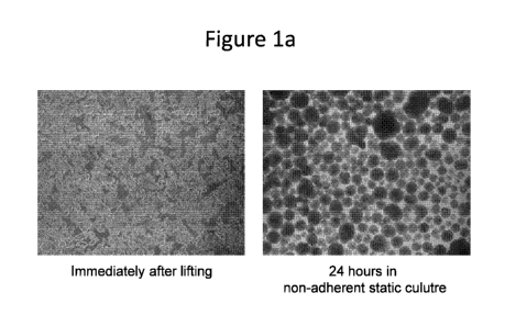
[016] Figure la shows micrographs of Dispase -treated cells of the human
embryonic
stem ("hES") cell line H1 immediately after lifting (left hand panel) and
after 24 hours in
non-adherent static culture (right hand panel) according to Example 1. The
cells after lifting
(left hand panel) resembled fragments of monolayer with an average fragment
diameter of
about 20-30 microns each fragment consisting of clumps of cells. After 24
hours in non-
adherent static culture, the cells assumed a cluster-like configuration.
[017] Figure lb shows the results of flow cytometry for CD9, SSEA4, CXCR4, TRA-
1-60
and TRA-1-81 for the Dispase -treated cells of the human embryonic stem cell
line H1 after
culturing for 4 days in a 125 mL spinner flask containing 25 mL mTeSR i media
according
to Example 1. The cells exhibited high expression for markers of pluripotency
(CD9,
SSEA4, TRA-1-60 and TRA-1-81) with almost no expression of CXCR4, a marker for
differentiation.
[018] Figure lc shows micrographs of the Dispase -treated cells of the human
embryonic
stem cell line H1 after 72 and 96 hours of differentiation at the end of stage
1. Visible in
Figure lc are loose cell aggregates after 72 hours at 4X magnification (left
hand panel), 96
hours at 4X magnification (center panel) and 96 hours at 10X magnification
(right hand
panel).
[019] Figure id shows flow cytometry results for the Dispase -treated cells of
the human
embryonic stem cell line H1 at the end of stage 1 differentiation for the
markers CD9, CD184
(CXCR4) and CD99 (see Example 1). As shown in Figure id, expression of CD9, a
marker
for pluripotency, was nearly eliminated, while the expression of markers of
definitive
endoderm differentiation CXCR4 (CD184) and CD99 were quite high.
[020] Figure le shows quantitative reverse transcription polymerase chain
reaction (qRT-
PCR) results for expression of selected genes associated with pluripotency and
genes
associated with definitive endoderm for the Dispase -treated cells of the
human embryonic
stem cell line H1 at the end of stage 1 compared to undifferentiated H1 (WA01)
hES cells
(see Example 1). The cells at the end of stage 1 showed a dramatic decrease in
the
4
CA 02928639 2016-04-25
WO 2015/065524
PCT/US2014/038993
expression of pluripotency genes (CD9, NANOG, and POU5F1/OCT4) and a large
increase
in genes associated with definitive endoderm (CXCR4, CERBERUS (CER1), GSC,
FOXA2,
GATA4, GATA6, MNX1, and SOX17) versus undifferentiated WA01 hES cells.
[021] Figure if shows micrographs of the Dispase -treated cells of the human
embryonic
stem cell line H1 as the cells further differentiated from definitive endoderm
toward the
pancreatic endoderm (see Example 1). Clear morphological changes to cells and
cell clusters
are visible as differentiation progresses from stage 2, day 1 (top left hand
panel) to stage 2,
day 3 (top right hand panel) to stage 3, day 4 (lower left hand panel) and
stage 4, day 1 (lower
right hand panel).
[022] Figure 2a shows flow cytometry data for EDTA-treated cells of the human
embryonic stem cell line H1 after 2 days of culture in stirred suspension
culture post-EDTA
treatment, and prior to transition to differentiation culture, for markers
associated with
pluripotency and differentiation according to Example 2. The data showed high
expression
for the markers of pluripotency (CD9, SSEA4, TRA-1-60, and TRA-1-81) with
almost no
expression of a marker for differentiation (CXCR4).
[023] Figure 2b shows micrographs of the EDTA-treated cells of the human
embryonic
stem cell line H1 differentiated into stage 1, day 3 cells grown in spinner
flask and stage 2
day 2, stage 4 day 1 and stage 4 day 3 cells grown in spinner flasks or
Erlenmeyer flasks
according to Example 2. Suspension differentiated cultures formed
substantially uniform and
homogenous populations of cells in spherical aggregates.
[024] Figure 2c shows flow cytometry data for the EDTA-treated cells of the
human
embryonic stem cell line H1 at the end of stage 1 for cell surface markers of
pluripotency and
endoderm differentiation. As visible in Figure 2c, expression of CD9, a marker
for
pluripotency, was nearly eliminated while expression for CXCR4 (CD184), a
marker for
definitive endoderm differentiation was quite high.
[025] Figure 2d shows qRT-PCR results for expression of selected genes
associated with
pluripotency and genes associated with definitive endoderm for the EDTA-
treated cells of the
human embryonic stem cell line H1 at the end of stage 1 compared to
undifferentiated H1
(WA01) hES cells (see Example 2). Figure 2d shows a decrease in the expression
of
pluripotency genes (CD9, Nanog, and POU5F1/OCT4) and a large increase in genes
associated with definitive endoderm (CXCR4, CERBERUS ("CER1"), FOXA2, GATA4,
GATA6, MNX1, and SOX17).
[026] Figure 2e shows flow cytometry data for markers indicative of
differentiation
(NKX6.1, CDX2, 50X2, and Chromagranin) for the EDTA-treated cells of the human
CA 02928639 2016-04-25
WO 2015/065524
PCT/US2014/038993
embryonic stem cell line H1 which were differentiated from stage 1 to
pancreatic endoderm
cells by suspension in spinner flasks or Erlenmeyer flasks according to
Example 2. The flow
cytometry data shows high levels of NKX6.1, a transcription factor required
for functional 13
cells, and high levels of endocrine pancreas markers such as synaptophysin
(data not shown)
and chromogranin with both suspension formats.
[027] Figure 2f shows qRT-PCR results for expression of selected genes
associated with
differentiation for the EDTA-treated cells of the human embryonic stem cell
line H1 which
were further differentiated from stage 1 to pancreatic endoderm cells by
suspension in
spinner flasks or Erlenmeyer flasks according to Example 2. The data is
compared to
expression in WA01 hES cells. The RT-PCR results show high levels of
expression of
pancreatic precursor genes.
[028] Figure 3a shows a micrograph of cells of the human embryonic stem cell
line H1,
which had been lifted from a static culture following treatment with Accutase
. As shown in
Figure 3a, the cells were removed from the surface as small aggregates.
[029] Figure 3b shows phase contrast micrographs of cells of the human
embryonic stem
cell line H1, which had been lifted from a static culture following treatment
with Accutase
and which were then expanded in suspension culture for three days. Visible in
Figure 3b is
the formation of a substantially uniform, spherical population of cell
clusters.
[030] Figure 3c shows a micrograph of clusters of cells of the human embryonic
stem cell
line H1, which had been lifted from a static culture following treatment with
Accutase ,
which were then expanded in suspension culture for three days, and which were
then serially
passaged using Accutase dissociation.
[031] Figure 4a shows micrographs of suspension cultured human embryonic stem
cells of
the cell line H1 using a directed differentiation protocol at different stages
of differentiation.
Visible in Figure 4a are micrographs of the cells at each stage of
differentiation.
[032] Figure 4b shows the results of flow cytometry for markers of
differentiation
(CXCR4, CD56 and PDX1) for suspension cultured human embryonic stem cells of
the cell
line H1 using a directed differentiation protocol at different stages of
differentiation (hours
after beginning differentiation). At the end of the differentiation process on
day 4 of stage 4,
a high percentage of the cells were positive for PDX1 expression.
[033] Figure 4c shows the non-fasting blood glucose levels of SCID-Bg Mice
transplanted
with differentiated cells encapsulated in a TheraCyteTm device.
[034] Figure 5a shows flow cytometry data for the EDTA-treated cells of the
human
embryonic stem cell line H1 prior to transition to differentiation culture for
markers
6
CA 02928639 2016-04-25
WO 2015/065524
PCT/US2014/038993
associated with pluripotency and differentiation. As shown in Figure 5a, high
expression of
the pluripotency markers CD9, SSEA4, TRA-1-60 and TRA-1-80 was observed.
[035] Figure 5b shows phase contrast images of the cells and flow cytometry
data for
CXCR4/CD184 and CD99 (markers of differentiation) and CD9 (a pluripotency
marker) for
three different feed settings during stage 1. The conditions tested were as
follows: (A) media
change 24 hours after initiation of differentiation, no media change at 48
hours; (B) media
change 24 hours after initiation of differentiation and glucose bolus addition
at 48 hours; and
(C) no media change throughout stage 1 with glucose and GDF8 bolus added 24
hours after
initiation of differentiation, then a glucose bolus added at 48 hours post
initiation.
[036] Figure 5c shows phase contrast images of the differentiated cells
exhibiting
pancreatic endoderm morphology, which were differentiated using the following
feed settings
during the formation of definitive endoderm: (A) media change 24 hours after
initiation of
differentiation, no media change at 48 hours; (B) media change 24 hours after
initiation of
differentiation and glucose bolus addition at 48 hours; and (C) no media
change throughout
stage 1 with glucose and GDF8 bolus added 24 hours after initiation of
differentiation, then a
glucose bolus added at 48 hours post initiation.
[037] Figure 5d shows the results of flow cytometry for select markers of
pancreatic gene
expression (NKX6.1 and chromogranin) and select non-pancreatic genes (CDX2 and
SOX2)
for differentiated cell as the end of stage 4, which were differentiated using
the following
feed settings during formation of definitive endoderm: (A) media change 24
hours after
initiation of differentiation, no media change at 48 hours; (B) media change
24 hours after
initiation of differentiation and glucose bolus addition at 48 hours; and (C)
no media change
throughout stage 1 with glucose and GDF8 bolus added 24 hours after initiation
of
differentiation, then a glucose bolus added at 48 hours post initiation.
[038] Figure 5e shows qRT-PCR results for select pancreatic and non-pancreatic
gene
expression for differentiated cells as the end of stage 4, which were
differentiated using the
following feed settings during formation of definitive endoderm: (A) media
change 24 hours
after initiation of differentiation, no media change at 48 hours; (B) media
change 24 hours
after initiation of differentiation and glucose bolus addition at 48 hours;
and (C) no media
change throughout stage 1 with glucose and GDF8 bolus added 24 hours after
initiation of
differentiation, then a glucose bolus added at 48 hours post initiation. The
data are shown as
fold difference in expression versus undifferentiated H1 (WA01) hES cells
(baseline
expression of 1).
7
CA 02928639 2016-04-25
WO 2015/065524
PCT/US2014/038993
[039] Figure 5f shows the expression of C-peptide in SCID-Bg mice that were
implanted
with cells differentiated according to condition A (media change 24 hours
after initiation of
differentiation, no media change at 48 hours). Each SCID-Bg mouse was
implanted with 5
million of the cells under the kidney capsule. As shown in Figure 5f, by 12
weeks post
implantation, human c-peptide was detectable at levels above lng/mL, and at 16
weeks c-
peptide levels were an average of 2.5ng/mL.
[040] Figure 5g shows the effect of glucose treatment for selected SCID-Bg
mice pre- and
post-administration (e.g. implantation) of cells differentiated according to
condition A (media
change 24 hours after initiation of differentiation, no media change at 48
hours). As shown in
Figure 5g, glucose treatment induced a significant increase in circulating
human c-peptide
from an average of 0.93ng/mL in a fasted state to 2.39ng/mL in a fed state.
[041] Figure 5h shows the effect of streptozotocin (STZ) administration (i.e.
STZ-induced
diabetes) on SCID-Bg mice that had been administered cells differentiated
according to
condition A (media change 24 hours after initiation of differentiation, no
media change at 48
hours). As evident from Figure 5h, animals with a graft of functional GSIS
competent tissue
(i.e. those that had been administered the cells) maintained normal blood
glucose levels
unlike the untreated controls which developed frank diabetes.
[042] Figure 6a shows micrographs of cells of the human embryonic stem cell
line H1
grown on Cytodex 3 microcarrier beads prior to differentiation.
[043] Figure 6b shows micrographs of cells of the human embryonic stem cell
line H1
grown on Cytodex 3 microcarrier beads at various stages of differentiation.
[044] Figure 6c shows the cell count (cells/cm2) as a function of days of
differentiation for
cells of the human embryonic stem cell line H1 grown and differentiated on
plates in media
containing Activin A (AA) and WNT3A (WTN3A/AA plate), microcarriers in media
containing Activin A and WNT3A (WTN3A/AA microcarriers), plates in media
containing
MCX and GDF8 (MCX/GDF8 plate) and microcarriers in media containing MCX and
GDF8
(MCX/GDF8 microcarriers).
[045] Figure 6d shows the cell count (cells/ml) as a function of days of
differentiation for
cells of the human embryonic stem cell line H1 grown and differentiated on
plates in media
containing Activin A and WNT3A (WTN3A/AA plate), microcarriers in media
containing
Activin A and WNT3A (WTN3A/AA microcarrier), plates in media containing MCX
and
GDF8 (MCX/GDF8 plate) and microcarriers in media containing MCX and GDF8
(MCX/GDF8 microcarriers).
8
CA 02928639 2016-04-25
WO 2015/065524
PCT/US2014/038993
[046] Figure 6e shows flow cytometry results for the first stage of
differentiation of cells
grown on a microcarrier culture or planar culture in the presence of: (a)
WNT3A and AA; or
(2) MCX and GDF8 as a dot plot of cell expression of CXCR4/CD184 (Y-axis) and
CD9 (X-
axis).
[047] Figure 6f shows flow cytometry results for the first stage of
differentiation of cells
grown on a microcarrier culture or planar culture in the presence of: (a)
WNT3A and AA; or
(2) MCX and GDF8 as total expression of each of the markers (CXCR4 and CD9).
[048] Figure 6g shows qRT-PCR results for expression of selected genes
associated with
differentiation for cells of the human embryonic stem cell line H1, which were
differentiated
by growth on planar culture or on microcarrier beads in suspension culture in
the presence of:
(a) WNT3A and AA; or (2) MCX and GDF8.
[049] Figure 7 shows the cell counts at various stages of differentiation
in a Bioreactor
from stage 1, day 1 to stage 4, day 3 for cells differentiated according to
the protocol of
Example 7. Cell counts are shown as million cells/ml as determined by an image-
based
cytometer (NucleoCounter0).
[050] Figure 8 shows the average daily bioreactor medium pH levels as a
function of time
(days of differentiation) during the differentiation protocol of Example 7. pH
levels were
determined by a NOVA BioProfile0 FLEX (Nova Biomedical Corporation, Waltham,
MA).
[051] Figure 9 shows the average daily bioreactor medium lactate levels as a
function of
time (days of differentiation) during the differentiation protocol of Example
7. Lactate levels
were determined by a NOVA BioProfile0 FLEX (Nova Biomedical Corporation,
Waltham,
MA).
[052] Figure 10 shows the average daily bioreactor medium glucose levels as a
function of
time (days of differentiation) during the differentiation protocol of Example
7. Glucose
levels were determined by a NOVA BioProfile0 FLEX (Nova Biomedical
Corporation,
Waltham, MA).
[053] Figure 11 shows the undifferentiated gene expression, as determined by
qRT-PCR,
for stage 0, day 1 (i.e. twenty-four hours after inoculation) cells
differentiated according to
the protocol of Example 7 for the pluripotency array, which contains select
genes associated
with pluripotency.
[054] Figure 12 shows the undifferentiated gene expression, as determined by
qRT-PCR,
for stage 0, day 1 (i.e. twenty-four hours after inoculation) cells for the
definitive endoderm
("DE") array, which contains select genes associated with definitive endoderm
(see Example
7).
9
CA 02928639 2016-04-25
WO 2015/065524
PCT/US2014/038993
[055] Figure 13 shows the undifferentiated gene expression, as determined by
qRT-PCR,
for stage 0, day 3 (i.e. seventy-two hours after inoculation) cells for the
pluripotency array,
which contains select genes associated with pluripotency (see Example 7).
[056] Figure 14 shows the undifferentiated gene expression, as determined by
qRT-PCR,
for stage 0, day 3 (i.e. seventy-two hours after inoculation) cells for the DE
array, which
contains select genes associated with DE (see Example 7).
[057] Figure 15 shows the results of fluorescence-activated cell sorting
(FACS) for CD9,
CD184/CXCR4, SSEA4, TRA-1-60 and TRA-1-81 for undifferentiated stage 0, day 3
(i.e.
seventy-two hours after inoculation) cells (see Example 7). The results are
also shown in
Table 8.
[058] Figure 16 shows the undifferentiated gene expression, as determined by
qRT-PCR,
for select genes of stage 0, day 1 (i.e. twenty-four hours after inoculation)
and stage 0, day 3
(i.e. seventy-two hours after inoculation) cells differentiated according to
the protocol of
Example 7. Specifically, Figure 16 shows a modest increase in gene expression
for GATA4,
GSC, MIXL1, and T and a >100x increase in GATA2 expression during the stage 0
process
prior to directed differentiation.
[059] Figure 17 shows the undifferentiated gene expression, as determined by
qRT-PCR,
for the DE array, which contains select genes associated with DE, for stage 0,
day 1 (i.e.
twenty-four hours after inoculation) and stage 0, day 3 (i.e. seventy-two
hours after
inoculation) cells differentiated according to the protocol of Example 7.
Specifically, Figure
17 shows a >100x increase in CER1, FGF17, and FGF4 expression during the stage
0 process
prior to directed differentiation.
[060] Figures 18 and 19 show the gene expression for stage 1, day 1 cells
differentiated
according to the protocol of Example 7. Figure 18 shows the gene expression,
as determined
by qRT-PCR, for the pluripotency array, which contains select genes associated
with
pluripotency, for stage 1, day 1 cells. Figure 19 shows the gene expression,
as determined by
qRT-PCR, for the DE array, which contains select genes associated with DE, for
stage 1, day
1 cells. Figures 18 and 19 illustrate significant alterations in gene
expression patterns such as
a ¨700x increase in FOXA2 expression and a 1000x increase in CER1, EOMES,
FGF17,
FGF4, GATA4, GATA6, GSC, MIXL1, and T expression.
[061] Figures 20 and 21 show the gene expression for stage 1, day 3 cells
differentiated
according to the protocol of Example 7. Figure 20 shows the gene expression,
as determined
by qRT-PCR, for the pluripotency array, which contains select genes associated
with
pluripotency, for stage 1, day 3 cells. Figure 21 shows the gene expression,
as determined by
CA 02928639 2016-04-25
WO 2015/065524
PCT/US2014/038993
qRT-PCR, for the DE array, which contains select genes associated with DE, for
stage 1, day
3 cells.
[062] Figure 22 shows the results of FACS for CD9, CD184 (also known as CXCR4)
and
CD99 for stage 1, day 3 cells differentiated according to the protocol of
Example 7. A near
complete transition from a CD9 expressing/CXCR4 negative pluripotent cell
population at
the initiation of differentiation (Figure 15) to a homogeneous population of
CXCR4
expressing cells (98.3% of cells CXCR4 positive, 1.9 SD) at the end of stage
1 (Figure 22)
was observed.
[063] Figure 23 shows the gene expression, as determined by qRT-PCR, for the
DE array,
which contains select genes associated with DE, for stage 1, day 3; stage 2,
day 1; and stage
2, day 3 cells differentiated according to the protocol of Example 7. Figure
23 shows that
HNF4a and GATA6 expression levels at stage 2 days 1 and 3 increased, while
genes
expressed at high levels on day 3 of stage 1 (CXCR4, EOMES, FGF17, FGF4, MNX1,
PRDM1, 50X17, and VWF) showed reduced expression by the end of stage 2.
[064] Figure 24 shows the gene expression of the foregut genes AFP, PDX1, and
PROX1,
as determined by qRT-PCR, for stage 2, day 1 cells and stage 2, day 3 cells
differentiated
according to the protocol of Example 7. As shown in Figure 24, the expression
of these
genes increased.
[065] Figure 25 shows the results of FACS for PDX1, FOXA2, chromogranin,
NKX2.2
and 50X2 for stage 3, day 3 cells grown in stage 3 medium (Table 7)
differentiated
according to the protocol of Example 7. As shown in Figure 25, the cells
expressed markers
consistent with an endodermal pancreatic lineage as measured by PDX1 and FOXA2
expression (90.9% 11.95D PDX1 positive and 99.2% 0.65D FOXA2 positive).
[066] Figure 26 shows the gene expression, as determined by qRT-PCR, for the
stage 4
array, which contains select genes associated with stage 4, for stage 3, day 1
and stage 3, day
3 cells differentiated according to the protocol of Example 7. Figure 26
illustrates that these
cells exhibit increased levels of a host of genes commonly expressed in the
pancreas (ARX,
GAST, GCG, INS, ISL1, NEUROD1, NGN3, NKX2.2, NKX6.1, PAX4, PAX6, PTF1A, and
SST).
[067] Figure 27 shows the results of FACS for NKX6.1, chromagranin (CHGA),
CDX2,
50X2, NKX2.2, PDX1, FOXA2 and NEUROD for stage 4, day 3 cells differentiated
according to the protocol of Example 7. As shown in Figure 27, stage 4 day 3
the cells
retained high levels of PDX1 and FOXA2 expression and further developed an
expression
11
CA 02928639 2016-04-25
WO 2015/065524
PCT/US2014/038993
pattern consistent with a mix of pancreatic endocrine cells (28.1% 12.5SD
chromogranin
positive) and pancreatic progenitor cells (58.3% 9.7SD positive for NKX6.1).
[068] Figure 28 shows the gene expression, as determined by qRT-PCR, for the
stage 4
array, which contains select genes associated with stage 4, for stage 3, day
3; stage 4, day 1
and stage 4, day 3 cells differentiated according to the protocol of Example
7. Figure 28
shows an increased expression level of genes commonly expressed in the
pancreas (ARX,
GAST, GCG, IAPP, INS, ISL1, MAFB, NEUROD1, NGN3, NKX2.2, NKX6.1, PAX4,
PAX6, PTF1A, and SST).
[069] Figure 29 shows the average results of FACS for NKX6.1, chromagranin
(CHGA),
CDX2, SOX2, NKX2.2, PDX1, FOXA2 and NEUROD for stage 4, day 3 cells
differentiated
according to the protocol of Example 7. Specifically, Figure 29 shows the
average FACS
expression pattern of pancreatic precursors generated at a 3L scale from
different seed
material lots.
[070] Figure 30 shows the average results of FACS for NKX6.1, chromagranin
(CHGA),
CDX2, 50X2, NKX2.2, PDX1, FOXA2 and NEUROD for stage 4, day 3 cells
differentiated
according to the protocol of Example 7. Prior to differentiation in stage 4,
day 3 cells, the
cells were expanded to form ISM and then grown at stage 0 in either a custom
in-house
medium "IH3" or Essential8TM, both of which were supplemented with 0.5% BSA.
The cells
grown in the IH3 medium are the "1H3-P grown cells" and the cells grown in
Essential8TM
are the "EZ8 grown cells." No significant difference in expression patterns
was observed
between the cells grown in the different media.
[071] Figure 31 shows the average results of FACS for NKX6.1, chromagranin
(CHGA),
CDX2, 50X2, NKX2.2, PDX1, FOXA2 and NEUROD for stage 4, day 3 cells, which
were
previously grown at different pH levels in stage 0 (see Example 7). No
significant change in
the stage 4, day 3 cell profile was observed.
[072] Figure 32 compares the results of FACS for NKX6.1, chromogranin (CHGA),
CDX2, 50X2, NKX2.2, PDX1, FOXA2 and NEUROD for stage 4, day 3 cells, which
were
not treated with Anti-Foam C, and stage 4, day 3 cells, which were treated
with Anti-Foam C
emulsion (94 ppm) (see Example 7). Anti-Foam C emulsion (Sigma Cat#A8011) was
not
observed to affect the profile of stage 4 day 3 cells.
[073] Figures 33 to 35 show the gene expression, as determined by qRT-PCR, for
select
genes for cells differentiated according to the protocol of Example 8. Figure
33 shows the
gene expression, as determined by qRT-PCR, for select genes of cells, twenty-
four hours
prior to the start of differentiation (see Example 8). As shown in Figure 33,
cells from the
12
CA 02928639 2016-04-25
WO 2015/065524
PCT/US2014/038993
bioreactor retained expression for genes characteristic of pluripotency
(POU5F1, NANOG,
SOX2, and ZFP42) and showed minimal or no induction of genes characteristic of
differentiation (AFP, and FOXA2: <50 fold increase; FOXD3, GATA2, GATA4, GSC,
HAND2, MIXL1, and T: <10 fold increased expression). Figure 34 shows the gene
expression, as determined by qRT-PCR, for select genes of cells twenty-four
hours after the
start of differentiation. Figure 35 shows the gene expression, as determined
by qRT-PCR, for
select genes of cells seventy-two hours after the start of differentiation.
[074] Figure 36(a) to 36(e) show the gene expression, as determined by qRT-
PCR, for
select genes for cells differentiated from stage 2 to stages 3 and 4 according
to the protocol of
Example 8. Specifically, these Figures show the gene expression of the cells
at stage 2, day
1; stage 2, day 2; stage 2, day 3; stage 3, day 3; and, depending on the gene,
stage 4, day 1.
Figure 36(a) shows the gene expression for AFP, ATOH1, and CDX2. Figure 36(b)
shows
the gene expression for GAST, HAND1, HHEX, and HNF4a. Figure 36(c) shows the
gene
expression for NKX2.2, NKX6.1, OSR1, and PDX1. Figure 36(d) shows the gene
expression
for PROX1, PFT1a, 50X17, and 50X2. Figure 36(e) shows the gene expression for
50X9.
The data are shown as difference in expression versus undifferentiated H1
(WA01) hES cells
(baseline expression of 1).
[075] Figure 37 show the gene expression, as determined by qRT-PCR, for select
genes
for cells at stage 4, day 3 of differentiation according to the protocol in
Example 8. As shown
in Figure 37, at the end of differentiation at stage 3, day 3 the cells have
differentiated into
pancreatic progenitor cells characterized by high expression levels of PDX1
(>1x10⁶ fold
induction) and other pancreatic genes (>1000 fold induction of ARX, GCG, GAST,
INS, ISL,
NEUROD1, NGN3, NKX2.2, NKX6.1, PAX4, PTFla, and SST) and near total loss of
OCT4/POU5F1 expression as compared to undifferentiated H1 human embryonic stem
cells.
[076] Figure 38 shows the daily cell counts during the differentiation
protocol according to
Example 8. Specifically, Figure 38 shows cell density as a function of the
process day.
Figure 38 shows the cell counts for differentiation protocols of two reactor
runs (PRD1205
and PRD1207) carried out at pH 6.8 and 7.2. For comparison, the cell counts
for cell drift are
also shown.
[077] Figure 39(a) to Figure 39(d) illustrate the in vivo bioactivity of
stage 4 day 3 cells,
which were differentiated according to the protocol of Example 8 and were
implanted into
SCID-Bg mice. The cells were implanted subcutaneously via a TheraCyteTm
device, under
the kidney capsule or implanted after incubation in an ultra-low attachment
dish. The mice
were monitored for blood glucose and C-peptide levels every four weeks
following graft
13
CA 02928639 2016-04-25
WO 2015/065524
PCT/US2014/038993
implantation. Figure 39(a) shows the C-peptide levels after implantation of 5
x 106 or 10 x
106 stage 4 day 3 cells in a TheraCyteTm device as a function of time. Figure
39(b) shows the
non-fasting glucose levels in animals after implantation of 5 x 10⁶ or 10 x
106 stage 4 day 3
cells in a TheraCyteTm device. The mice in Figure 39(b) were treated with STZ
to ablate host
3-cell function prior to implantation. Figure 39(c) shows the C-peptide level
produced after
implantation of previously-cyropreserved stage 4 day 3 cells in a TheraCyteTm
device as a
function of time (weeks post implantation). Figure 39(d) compares the C-
peptide levels of
mice treated by a kidney graft of never cryopreserved/fresh stage 4, day 3
cells or
cryopreserved stage 4, day 3 cells implanted immediately after thaw (DO) or 1
day after thaw
(D1).
[078] Figure 40A to Figure 40D show FACS plots for CXCR4, CD99, and CD9 of
cells
differentiated for three days according to the protocol of Example 9 which
were treated at
stage 1, day 1 with: MCX compound and GDF-8 (Figure 40A); MCX only (Figure
40B);
WNT3A and Activin A (Figure 40C); and WNT3A only (Figure 40D). These figures
indicate that in suspension culture, addition of 31.tM MCX in the absence of a
TGF-13 family
member on day one of differentiation generates definitive endoderm at levels
comparable to
that obtained when cells are treated with 31.tM MCX plus 10Ong/m1 GDF-8 or
2Ong/m1 WNT-
3a plus 10Ong/m1Activin A on day one.
[079] Figures 41A to 41D show FACS plots for CXCR4, CD99, and CD9 of cells
differentiated for three days according to the protocol of Example 10, which
were treated
with various amounts of MCX at stage 1, day 1. Specifically, the cells at
stage 1, day lwere
treated with: 4 ILEM of MCX (Figure 41A); 3 ILEM of MCX (Figure 41B); 2 ILEM
of MCX
(Figure 41C); and 1.5 ILEM of MCX (Figure 41D).
[080] Figure 42A and Figure 42B show FACS plots for CXCR4, CD99, and CD9 of
cells
differentiated for three days according to the protocol of Example 11.
Specifically, these
Figures show the role of media exchange frequency in suspension culture.
Figure 42A shows
FACS plots for CXCR4, CD99, and CD9 of cells differentiated for three days
according to
the protocol of Example 10 with full media exchange at stage 1. Figure 42B
shows FACS
plots for CXCR4, CD99, and CD9 of cells differentiated for three days
according to the
protocol of Example 10 without a media exchange on day 3. The data suggest
that in the
suspension culture system, cultures which receive a media exchange on day
three (Figure
42A) of differentiation resulted in definitive endoderm with a comparable
efficiency to
cultures which did not receive a media exchange on day three (Figure 42B).
14
CA 02928639 2016-04-25
WO 2015/065524
PCT/US2014/038993
[081] Figure 43A and Figure 43B show FACS plots for CXCR4, CD99, and CD9 of
cells
differentiated for three days according to the protocol of Example 12.
Specifically, these
Figures show the role of GlutaMAXTm in suspension culture. The cells were
cultured at stage
1 in a medium supplemented with lx GlutaMAXTm (Figure 43A) or free of
GlutaMAXTm or
any glutamine (0 M GlutaMAXTm) (Figure 43B). The data suggest that in the
suspension
culture system, addition of GlutaMAXTm does not appear to influence the
efficiency with
which definitive endoderm is generated
[082] Figures 44A to 44D show the effects of various amounts of sodium
bicarbonate on
cells differentiated according to the protocol of Example 13. Figure 44A and
Figure 44B
show FACS plots for CXCR4, CD99, and CD9 of cells differentiated for three
days
according to the protocol of Example 13 with either 3.64 g/1 (Figure 44A) or
2.49 g/1 (Figure
44B) added at stage 1. Figure 44C and Figure 44D show phase contrast
micrographs of cells
differentiated for three days according to the protocol of Example 13 with
either 3.64 g/1
(Figure 44C) or 2.49 g/1 (Figure 44D) added at stage 1.
[083] Figure 45 shows daily cell counts for cell density as a function of
differentiation for
cells differentiated according to the protocol of Example 14. The cells counts
were obtained
using an image-based cytometer (NucleoCounter0).
[084] Figure 46 shows the average daily bioreactor medium pH levels as a
function of time
(days of differentiation) during the differentiation protocol of Example 14.
pH levels were
determined by a NOVA BioProfile0 FLEX (Nova Biomedical Corporation, Waltham,
MA).
[085] Figure 47 shows the average daily bioreactor medium glucose levels as a
function of
time (days of differentiation) during the differentiation protocol of Example
14. Glucose
levels were determined by a NOVA BioProfile0 FLEX (Nova Biomedical
Corporation,
Waltham, MA).
[086] Figure 48 shows the average daily bioreactor medium lactate levels as a
function of
time (days of differentiation) during the differentiation protocol of Example
14. Lactate
levels were determined by a NOVA BioProfile0 FLEX (Nova Biomedical
Corporation,
Waltham, MA).
[087] Figure 49 shows the gene expression, as determined by qRT-PCR as a fold
expression versus undifferentiated cells, for the pluripotency array, which
contains select
genes associated with pluripotency, for stage 0, day 1 to 3 and stage 1, day 1
to day 3 cells
differentiated according to the protocol of Example 14. Figure 50 shows the
gene expression,
as determined by qRT-PCR as a fold expression versus undifferentiated cells,
for the DE
array, which contains select genes associated with DE, for stage 0, day 1 to
3, stage 1, day 1
CA 02928639 2016-04-25
WO 2015/065524
PCT/US2014/038993
to day 3 and stage 2, day 1 to day 3 cells differentiated according to the
protocol of Example
14.
[088] Figure 51 shows the results of FACS for markers associated with
pluripotency
(CD184/CXCR4, SSEA4, TRA-1-60 and TRA-1-81) for stage 0, cells prior to being
differentiated according to the protocol of Example 14. Specifically, Figure
51 shows high
expression of markers associated with pluripotency.
[089] Figure 52 shows FACS plots for the definitive endoderm markers CXCR4,
CD99,
and CD9 of cells differentiated to the end of stage 1 according to the
protocol of Example 14.
[090] Figure 53 shows the gene expression, as determined by qRT-PCR as a fold
expression versus undifferentiated cells, for GAPDH, AFP, HHEX, HNF4a, PDX1,
and
PROX1 for stage 2, day 1; stage 2, day 2 and stage 2, day 3 cells
differentiated according to
the protocol of Example 14. Figure 53 shows an increase in expression of
foregut genes
(AFP, HHEX, PDX1, and PROX1).
[091] Figure 54 shows the gene expression, as determined by qRT-PCR as a fold
expression versus undifferentiated cells, for GAPDH, AFP, CDX2, GAST, HNF4A,
NKX2-
2, OSR1, PDX1 and PFT1A for stage 2, day 1 to day 3 and stage 3, day 1 to day
3 cells
differentiated according to the protocol of Example 14. As shown in Figure 54,
expression
for PDX1 increased 60 fold from 12,000x over control at the end of stage 2 day
3 to 739,000x
over control at the end of stage 3, day 3.
[092] Figure 55 shows the gene expression, as determined by qRT-PCR as a fold
expression versus undifferentiated cells, for certain genes for stage 3, day 1
to 3 and stage 4,
day 1 to day 3 cells differentiated according to the protocol of Example 14.
Specifically, the
top panel of Figure 55 shows the gene expression for GAPDH, AFP, ALB, ARX,
CDX2,
CHGA, GAST, GCG, IAAP, INS, ISL1, and MAFB. The bottom panel of Figure 55
shows
the gene expression of MAFB, MUCS, NEUROD1, NEUROG3, NKX2-2, NKX6-1, PAX4,
PDX1, POUSF1, PTF1A, SST and Z1C1.
[093] Figure 56 shows end stage micrographs for cells differentiated according
to the
protocol of Example 14. Visible in Figure 56 are representative micrographs
(4X) of cell
clusters at stage 0 and at the end of differentiation of stages 1 to 4.
[094] Figures 57 to 80 show the gene expression, as determined by qRT-PCR as a
fold
expression versus undifferentiated cells, for cells differentiated according
to various
embodiments of the protocol of Example 15 after 0 hours, 6 hours, 24 hours, 30
hours, 48
hours and 72 hours of differentiation for the following genes: AFP (Figure
57); CD99 (Figure
58); CD9 (Figure 59); CDH1 (Figure 60); CDH2 (Figure 61); CDX2 (Figure 62);
CER1
16
CA 02928639 2016-04-25
WO 2015/065524
PCT/US2014/038993
(Figure 63); CXCR4 (Figure 64); FGF17 (Figure 65); FGF4 (Figure 66); FOXA
(Figure 67);
GADPH (Figure 68); GATA4 (Figure 69); GATA6 (Figure 70); GSC (Figure 71); MT
(Figure 72); MIXL1 (Figure 73); MNX1 (Figure 74); NANOG (Figure 75); OTX2
(Figure
76); POUF5F1 (Figure 77); SOX17 (Figure 78); SOX7 (Figure 79) and T (Figure
80).
[095] Figure 81 shows the percentage of cells in GO/G1 of Cell Cycle for cells
after 6
hours, 24 hours, 30 hours, 48 hours, and 72 hours of differentiation according
to various
embodiments of the protocol of Example 15. Specifically, Figure 81 shows the
results for
clusters that were treated on the first day of differentiation with one of six
conditions: (1)
Neat, (2) 3[EM MCX plus 10Ong/m1 GDF-8 (Catalog # 120-00, Peprotech), (3) 3[EM
MCX
only, (4) 10Ong/m1 GDF-8 only, (5) 2Ong/m1 WNT-3A (Catalog # 1324-WN-002, R&D
Systems, MN) plus 10Ong/m1Activin A (Catalog # 338-AC, R&D Systems, MN), or
(6)
2Ong/m1 WNT-3A only.
[096] Figure 82 shows the effects of EDU treatment on the cell clusters
differentiated
according to the protocol of Example 15. The left hand panel of shows
percentage of cells in
G2/M of Cell Cycle for cells after 0 hours, 6 hours, 24 hours, 30 hours, 48
hours, and 72
hours of differentiation according to various embodiments of the protocol of
Example 15.
Specifically, the left hand panel shows the results for clusters that were
treated on the first
day of differentiation with one of six conditions: (1) Neat, (2) 3[EM MCX plus
10Ong/m1
GDF-8 (Catalog # 120-00, Peprotech), (3) 3[EM MCX only, (4) 10Ong/m1 GDF-8
only, (5)
2Ong/m1 WNT-3A (Catalog # 1324-WN-002, R&D Systems, MN) plus 10Ong/m1Activin A
(Catalog # 338-AC, R&D Systems, MN), or (6) 2Ong/m1 WNT-3A only. In one set of
data,
these clusters were also treated with EDU. The right hand panel of Figure 82
shows the %
Cells that are EDU positive 0 hours, 6 hours, 24 hours, 30 hours, 48 hours,
and 72 hours of
differentiation according to various embodiments of the protocol of Example
15.
[097] Figure 83 shows the general operational parameters used in the protocols
of
Example 15.
[098] Figure 84 shows the amount of EDU incorporation of cells after 6 hours,
24 hours,
30 hours, 48 hours, and 72 hours of differentiation according to various
embodiments of the
protocol of Example 15. Specifically, Figure 84 shows the results for EDU
incubated cells
clusters that were treated on the first day of differentiation with one of six
conditions: (1)
Neat, (2) 3[EM MCX plus 10Ong/m1 GDF-8 (Catalog # 120-00, Peprotech), (3) 3[EM
MCX
only, (4) 10Ong/m1 GDF-8 only, (5) 2Ong/m1 WNT-3A (Catalog # 1324-WN-002, R&D
Systems, MN) plus 10Ong/m1Activin A (Catalog # 338-AC, R&D Systems, MN), or
(6)
2Ong/m1 WNT-3A only.
17
CA 02928639 2016-04-25
WO 2015/065524
PCT/US2014/038993
[099] Figure 85 shows the percentage of cells in GO/G1 of Cell Cycle for cells
after 6
hours, 24 hours, 30 hours, 48 hours, and 72 hours of differentiation according
to various
embodiments of the protocol of Example 15. Specifically, Figure 85 shows the
results for
clusters that were treated on the first day of differentiation with one of six
conditions: (1)
Neat, (2) 31.tM MCX plus 10Ong/m1 GDF-8 (Catalog # 120-00, Peprotech), (3)
31..EM MCX
only, (4) 10Ong/m1 GDF-8 only, (5) 2Ong/m1 WNT-3A (Catalog # 1324-WN-002, R&D
Systems, MN) plus 10Ong/m1Activin A (Catalog # 338-AC, R&D Systems, MN), or
(6)
2Ong/m1 WNT-3A only.
[0100] Figure 86 shows the percentage of cells in S-phase of Cell Cycle for
cells after 6
hours, 24 hours, 30 hours, 48 hours, and 72 hours of differentiation according
to various
embodiments of the protocol of Example 15. Specifically, Figure 86 shows the
results for
clusters that were treated on the first day of differentiation with one of six
conditions: (1)
Neat, (2) 31.tM MCX plus 10Ong/m1 GDF-8 (Catalog # 120-00, Peprotech), (3)
31..EM MCX
only, (4) 10Ong/m1 GDF-8 only, (5) 2Ong/m1 WNT-3A (Catalog # 1324-WN-002, R&D
Systems, MN) plus 10Ong/m1Activin A (Catalog # 338-AC, R&D Systems, MN), or
(6)
2Ong/m1 WNT-3A only.
[0101] Figure 87 shows the percentage of cells in S-phase of Cell Cycle for
cells after
hours, 6 hours, 24 hours, 30 hours, 48 hours, and 72 hours of differentiation
according to
various embodiments of the protocol of Example 15. Specifically, Figure 87
shows the
results for clusters that were treated on the first day of differentiation
with one of six
conditions: (1) Neat, (2) 31..EM MCX plus 10Ong/m1 GDF-8 (Catalog # 120-00,
Peprotech), (3)
31.tM MCX only, (4) 10Ong/m1 GDF-8 only, (5) 2Ong/m1 WNT-3A (Catalog # 1324-WN-
002,
R&D Systems, MN) plus 10Ong/m1Activin A (Catalog # 338-AC, R&D Systems, MN),
or
(6) 2Ong/m1 WNT-3A only.
[0102] Figures 88A to 88E show the gene expression, as determined by qRT-PCR
as a fold
expression versus undifferentiated cells, for cells differentiated according
to various
embodiments of the protocol of Example 15 after 0 hours, 6 hours, 24 hours, 30
hours, 48
hours and 72 hours of differentiation. Figure 88A shows the gene expression,
as determined
by qRT-PCR as a fold expression versus undifferentiated cells, for CD99, CD9,
CDH1, and
CDH2. Figure 88A shows the gene expression, as determined by qRT-PCR as a fold
expression versus undifferentiated cells, for CXD2, CER1, CXCR4, and FGF17.
Figure 88C
shows the gene expression, as determined by qRT-PCR as a fold expression
versus
undifferentiated cells, for FGF4, FOXA, GATA4, and GATA6. Figure 88D shows the
gene
expression, as determined by qRT-PCR as a fold expression versus
undifferentiated cells, for
18
CA 02928639 2016-04-25
WO 2015/065524
PCT/US2014/038993
GSC, KIT, MIXL1 and MNX1. Figure 88E shows the gene expression, as determined
by
qRT-PCR as a fold expression versus undifferentiated cells, for NANOG, OTX2,
POUF5F1,
and SOX17. Figure 88F shows the gene expression, as determined by qRT-PCR as a
fold
expression versus undifferentiated cells, for SOX7 and T. The underlying data
for Figures
88A to 88F is shown in Figure 58 to 67 and 69 to 80.
[0103] Figure 89 shows the gene expression pattern, as determined by qRT-PCR,
of
pluripotent cells cultured in ectodermal differentiation medium according to
the protocol of
Example 16. As shown in Figure 89, the cells differentiated towards the neural
cell lineage.
Specifically, the left panel of Figure 89 shows the gene expression pattern
for an induced
pluripotent stem cell line generated from umbilical tissue cells (UTC). The
right panel of
Figure 89 shows the gene expression pattern for the WB0106 sub-clone of the H1
hES cell
line.
[0104] Figure 90 shows the gene expression pattern, as determined by qRT-PCR,
of
pluripotent cells cultured in mesodermal differentiation medium according to
the protocol of
Example 16. As shown in Figure 90, the cells differentiated towards cardiac
cell lineage.
Specifically, the left panel of Figure 90 shows the gene expression pattern
for an induced
pluripotent stem cell line generated from umbilical tissue cells (UTC). The
right panel of
Figure 90 shows the gene expression pattern for the WB0106 sub-clone of the H1
hES cell
line.
[0105] Figure 91 shows the gene expression pattern, as determined by qRT-PCR,
of
pluripotent cells cultured in ectodermal differentiation medium according to
the protocol of
Example 16. As shown in Figure 91, the cells differentiated towards neural
cell lineage.
Specifically, the left panel of Figure 91 shows the gene expression pattern
for an induced
pluripotent stem cell line generated from umbilical tissue cells (UTC). The
right panel of
Figure 91 shows the gene expression pattern for the WB0106 sub-clone of the H1
hES cell
line.
[0106] Figure 92 shows the protein expression pattern for PAX6, 50X2, and
POU5F1/OCT4, as determined by FACS, of pluripotent cells cultured for three
days in
ectodermal differentiation medium according to the protocol of Example 16.
Specially, the
left panels of Figure 92 show the expression pattern for PAX6, 50X2, and
POU5F1/OCT4
for an induced pluripotent stem cell line generated from umbilical tissue
cells (UTC). The
right panel of Figure 92 shows the protein expression pattern for PAX6, 50X2,
and
POU5F1/OCT4 for the WB0106 sub-clone of the H1 hES cell line.
19
CA 02928639 2016-04-25
WO 2015/065524
PCT/US2014/038993
[0107] Figure 93 shows the gene expression pattern, as determined by qRT-PCR,
of
pluripotent cells cultured in mesodermal differentiation medium according to
the protocol of
Example 16. As shown in Figure 93, the cells differentiated towards cardiac
cell lineage.
Specifically, the left panel of Figure 93 shows the gene expression pattern
for an induced
pluripotent stem cell line generated from umbilical tissue cells (UTC). The
right panel of
Figure 93 shows the gene expression pattern for the WB0106 sub-clone of the H1
hES cell
line.
[0108] Figure 94 shows micrographs for cells differentiated in mesodermal
differentiation
medium according to the protocol of Example 16. As shown in Figure 94, the
cells
differentiated towards cardiac cell lineage. Specifically, the left hand
panels of Figure 94
show micrographs of cells of the WB0106 sub-clone of the H1 hES cell line at
day 3, day 5
and day 10 of differentiation. The right hand panel of Figure 94 shows a
micrograph of
induced pluripotent stem cell line generated from umbilical tissue cells (UTC
IPSCs) after 10
days of differentiation.
[0109] Figure 95 shows micrographs for cells differentiated in ectodermal
differentiation
medium according to the protocol of Example 16. As shown in Figure 95, the
cells
differentiated towards the neural cell lineage. Specifically, the left hand
panels of Figure 95
show micrographs of cells of the WB0106 sub-clone of the H1 hES cell line at
day 3, day 5
and day 10 of differentiation. The right hand panel of Figure 95 shows a
micrograph of
induced pluripotent stem cell line generated from umbilical tissue cells (UTC
iPCS) after 10
days of differentiation.
DETAILED DESCRIPTION
[0110] This application is directed to preparing embryonic stem cells and
other pluripotent
cells that maintain pluripotency in aggregated cell cluster for
differentiation to endoderm
progenitor cells, pancreatic endocrine cells, mesoderm cells or ectoderm
cells. For clarity of
disclosure, and not by way of limitation, the detailed description of the
invention is divided
into the following subsections that describe or illustrate certain features,
embodiments or
applications of the present invention.
DEFINITIONS
[0111] Stem cells are undifferentiated cells defined by their ability, at the
single cell level,
to both self-renew and differentiate. Stem cells may produce progeny cells,
including self-
CA 02928639 2016-04-25
WO 2015/065524
PCT/US2014/038993
renewing progenitors, non-renewing progenitors, and terminally differentiated
cells. Stem
cells are also characterized by their ability to differentiate in vitro into
functional cells of
various cell lineages from multiple germ layers (endoderm, mesoderm, and
ectoderm). Stem
cells also give rise to tissues of multiple germ layers following
transplantation and contribute
substantially to most, if not all, tissues following injection into
blastocysts.
[0112] Stem cells are classified by their developmental potential. "Cell
culture" or
"culturing" refer generally to cells taken from a living organism and grown
under controlled
conditions ("in culture" or "cultured"). A primary cell culture is a culture
of cells, tissues, or
organs taken directly from an organism before the first subculture. Cells are
expanded in
culture when they are placed in a growth medium under conditions that
facilitate one or both
of cell growth and division, resulting in a larger population of the cells.
When cells are
expanded in culture, the rate of cell proliferation is sometimes measured by
the amount of
time needed for the cells to double in number (referred to as doubling time).
[0113] "Expanding", as used herein is the process of increasing the number of
pluripotent
stem cells by culturing, such as by at least about 5%, 10%, 15%, 20%, 25%,
30%, 35%, 40%,
45%, 50%, 60%, 75%, 9u,-so ,/0 ,
100%, 200%, 500%, 1000% or more, and levels within these
percentages. It is appreciated that the number of pluripotent stem cells which
can be obtained
from a single pluripotent stem cell depends on the proliferation capacity of
the pluripotent
stem cell. The proliferation capacity of the pluripotent stem cell can be
calculated by the
doubling time of the cell, i.e., the time needed for a cell to undergo a
mitotic division in the
culture, and the period that the pluripotent stem cell can be maintained in
the undifferentiated
state, which is equivalent to the number of passages multiplied by the days
between each
passage.
[0114] Differentiation is the process by which an unspecialized
("uncommitted") or less
specialized cell acquires the features of a specialized cell such as, a nerve
cell or a muscle
cell. A differentiated cell or a differentiation-induced cell is one that has
taken on a more
specialized ("committed") position within the lineage of a cell. The term
"committed", when
applied to the process of differentiation, refers to a cell that has proceeded
in the
differentiation pathway to a point where, under normal circumstances, it will
continue to
differentiate into a specific cell type or subset of cell types, and cannot,
under normal
circumstances, differentiate into a different cell type or revert to a less
differentiated cell type.
"De-differentiation" refers to the process by which a cell reverts to a less
specialized (or
committed) position within the lineage of a cell. As used herein, the lineage
of a cell defines
the heredity of the cell, i.e., which cells it came from and to what cells it
can give rise. The
21
CA 02928639 2016-04-25
WO 2015/065524
PCT/US2014/038993
lineage of a cell places the cell within a hereditary scheme of development
and
differentiation. A lineage-specific marker refers to a characteristic
specifically associated
with the phenotype of cells of a lineage of interest and can be used to assess
the
differentiation of an uncommitted cell to the lineage of interest.
[0115] "Markers", as used herein, are nucleic acid or polypeptide molecules
that are
differentially expressed in a cell of interest. In this context, differential
expression means an
increased level for a positive marker and a decreased level for a negative
marker as compared
to an undifferentiated cell. The detectable level of the marker nucleic acid
or polypeptide is
sufficiently higher or lower in the cells of interest compared to other cells,
such that the cell
of interest can be identified and distinguished from other cells using any of
a variety of
methods known in the art.
[0116] As used herein, a cell is "positive for" a specific marker or
"positive" when the
specific marker is sufficiently detected in the cell. Similarly, the cell is
"negative for" a
specific marker, or "negative" when the specific marker is not sufficiently
detected in the
cell. In particular, positive by FACS is usually greater than 2%, whereas the
negative
threshold by FACS is usually less than 1%. Positive by PCR is usually less
than 34 cycles
(Cts); whereas negative by PCR is usually more than 34.5 cycles.
[0117] As used herein, "cell density" and "seeding density" are used
interchangeably herein
and refer to the number of cells seeded per unit area of a solid or semisolid
planar or curved
substrate.
[0118] As used herein, "suspension culture" refers to a culture of cells,
single cells or
clusters, suspended in medium rather than adhering to a surface.
[0119] As used herein, "serum free" refers to being devoid of human or animal
serum.
Accordingly, a serum free culture medium does not comprise serum or portions
of serum.
[0120] In attempts to replicate the differentiation of pluripotent stem cells
into functional
pancreatic endocrine cells in cell culture, the differentiation process is
often viewed as
progressing through a number of consecutive stages. As used herein, the
various stages are
defined by the culturing times, and reagents set forth in the Examples
included herein.
[0121] "Definitive endoderm", as used herein, refers to cells which bear the
characteristics
of cells arising from the epiblast during gastrulation and which form the
gastrointestinal tract
and its derivatives. Definitive endoderm cells express at least one of the
following markers:
FOXA2 (also known as hepatocyte nuclear factor 3-3 (HNF33)), GATA4, GATA6,
MNX1,
50X17, CXCR4, Cerberus, OTX2, brachyury, goosecoid, C-Kit, CD99, and MIXL1.
Markers characteristic of the definitive endoderm cells include CXCR4, FOXA2
and SOX17.
22
CA 02928639 2016-04-25
WO 2015/065524
PCT/US2014/038993
Thus, definitive endoderm cells may be characterized by their expression of
CXCR4,
FOXA2, and SOX17. In addition, depending on the length of time cells are
allowed to
remain in stage 1, an increase in HNF4a may be observed.
[0122] "Pancreatic endocrine cells," as used herein, refer to cells capable
of expressing at
least one of the following hormones: insulin, glucagon, somatostatin, ghrelin,
and pancreatic
polypeptide. In addition to these hormones, markers characteristic of
pancreatic endocrine
cells include one or more of NGN3, NeuroD1, ISL1, PDX1, NKX6.1, PAX4, ARX,
NKX2.2,
and PAX6. Pancreatic endocrine cells expressing markers characteristic of 13
cells can be
characterized by their expression of insulin and at least one of the following
transcription
factors: PDX1, NKX2.2, NKX6.1, NeuroD1, ISL1, HNF313, MAFA, PAX4, and PAX6.
[0123] Used interchangeably herein are "dl", "d 1", and "day 1"; "d2", "d 2",
and "day 2";
"d3", "d 3", and "day 3", and so on. These number letter combinations refer to
a specific day
of incubation in the different stages during the stepwise differentiation
protocol of the instant
application.
[0124] "Glucose" and "D-Glucose" are used interchangeably herein and refer to
dextrose, a
sugar commonly found in nature.
[0125] Used interchangeably herein are "NeuroD" and "NeuroDl" which identify a
protein
expressed in pancreatic endocrine progenitor cells and the gene encoding it.
[0126] "LDN" and "LDN-193189" refer ((6-(4-(2-(piperidin-1-yeethoxy)pheny1)-3-
(pyridin-4-yepyrazolo[1,5-a]pyrimidine, hydrochloride; DM-3189)), a BMP
receptor
inhibitor available under the trademark STEMOLECULETm from Stemgent, Inc.,
Cambridge,
MA, USA.
ISOLATION, EXPANSION AND CULTURE OF PLURIPOTENT STEM CELLS
[0127] Pluripotent stem cells may express one or more of the designated TRA-1-
60 and
TRA-1-81 antibodies (Thomson et al. 1998, Science 282:1145-1147).
Differentiation of
pluripotent stem cells in vitro results in the loss of TRA-1-60, and TRA-1-81
expression.
Undifferentiated pluripotent stem cells typically have alkaline phosphatase
activity, which
can be detected by fixing the cells with 4% paraformaldehyde, and then
developing with
Vector Red as a substrate, as described by the manufacturer (Vector
Laboratories, Inc.,
Burlingame, CA). Undifferentiated pluripotent stem cells also typically
express OCT4 and
TERT, as detected by RT-PCR.
23
CA 02928639 2016-04-25
WO 2015/065524
PCT/US2014/038993
[0128] Another desirable phenotype of propagated pluripotent stem cells is a
potential to
differentiate into cells of all three germinal layers: endoderm, mesoderm, and
ectoderm
tissues. Pluripotency of stem cells can be confirmed, for example, by
injecting cells into
severe combined immune-deficiency ("SCID") mice, fixing the teratomas that
form using 4%
paraformaldehyde, and then examining histologically for evidence of cell types
from these
three germ layers. Alternatively, pluripotency may be determined by the
creation of
embryoid bodies and assessing the embryoid bodies for the presence of markers
associated
with the three germinal layers.
[0129] Propagated pluripotent stem cell lines may be karyotyped using a
standard G-
banding technique and compared to published karyotypes of the corresponding
primate
species. It is desirable to obtain cells that have a "normal karyotype," which
means that the
cells are euploid, wherein all human chromosomes are present and not
noticeably altered.
Pluripotent cells may be readily expanded in culture using various feeder
layers or by using
matrix protein coated vessels. Alternatively, chemically defined surfaces in
combination
with defined media such as mTeSR01 media (StemCell Technologies, Vancouver,
BC,
Canada) may be used for routine expansion of the cells.
[0130] Culturing in a suspension culture according to the method of some
embodiments of
the invention is effected by seeding the pluripotent stem cells in a culture
vessel at a cell
density that promotes cell survival and proliferation, but limits
differentiation. Typically, a
seeding density that maintains undifferentiation of cells is used. It will be
appreciated that
although single-cell suspensions of stem cells may be seeded, small clusters
of cells may be
advantageous.
[0131] To provide the pluripotent stem cells with a sufficient and constant
supply of
nutrients and growth factors while in the suspension culture, the culture
medium can be
replaced or replenished on a daily basis or at a pre-determined schedule such
as every 1-5
days. Large clusters of pluripotent stem cells may cause cell differentiation,
thus, measures
may be taken to avoid large pluripotent stem cell aggregates. According to
some
embodiments of the invention, the formed pluripotent stem cell clusters are
dissociated, for
example, every 2-7 days and the single cells or small clumps of cells are
either split into
additional culture vessels (i.e., passaged) or retained in the same culture
vessel and processed
with replacement or additional culture medium.
[0132] Large pluripotent stem cell clumps, including a pellet of pluripotent
stem cells
resulting from centrifugation, can be subjected to one or both of enzymatic
digestion and
mechanical dissociation. Enzymatic digestion of pluripotent stem cell clumps
can be
24
CA 02928639 2016-04-25
WO 2015/065524
PCT/US2014/038993
performed by subjecting the clump to an enzyme, such as type IV Collagenase,
Dispase or
Accutase . Mechanical dissociation of large pluripotent stem cell clumps can
be performed
using a device designed to break the clumps to a predetermined size.
Additionally, or
alternatively, mechanical dissociation can be manually performed using a
needle or pipette.
[0133] The culture vessel used for culturing the pluripotent stem cells in
suspension
according to the method of some embodiments of the invention can be any tissue
culture
vessel (e.g., with a purity grade suitable for culturing pluripotent stem
cells) having an
internal surface designed such that pluripotent stem cells cultured therein
are unable to adhere
or attach to such a surface (e.g., non-tissue culture treated vessel, to
prevent attachment or
adherence to the surface). Preferably to obtain a scalable culture, culturing
according to some
embodiments of the invention is effected using a controlled culturing system
(preferably a
computer-controlled culturing system) in which culture parameters such as
temperature,
agitation, pH, and oxygen are automatically monitored and controlled using a
suitable device.
Once the desired culture parameters are determined, the system may be set for
automatic
adjustment of culture parameters as needed to enhance pluripotent stem cell
expansion and
differentiation.
[0134] The pluripotent stem cells may be cultured under dynamic conditions
(i.e., under
conditions in which the pluripotent stem cells are subject to constant
movement while in the
suspension culture, e.g. a stirred suspension culture system) or under non-
dynamic conditions
(i.e., a static culture) while preserving their, proliferative, pluripotent
capacity and karyotype
stability over multiple passages.
[0135] For non-dynamic culturing of pluripotent stem cells, the pluripotent
stem cells can
be cultured in petri dishes, T-flasks, HyperFlasks0 (Coming Incorporated,
Coming, NY),
CellStacks0 (Coming Incorporated, Coming, NY) or Cell Factories (NUNCTm Cell
FactoryTM Systems (Thermo Fisher Scientific, Inc., Pittsburgh, PA)) coated or
uncoated. For
dynamic culturing of pluripotent stem cells, the pluripotent stem cells can be
cultured in a
suitable vessel, such as spinner flasks or Erlenmeyer flasks, stainless steel,
glass or single use
plastic shaker or stirred tank vessels. The culture vessel can be connected to
a control unit
and thus present a controlled culturing system. The culture vessel (e.g.,
spinner flask or
Erlenmeyer flask) may be agitated continuously or intermittently. Preferably
the cultured
vessel is agitated sufficiently to maintain the pluripotent stem cells in
suspension.
[0136] The pluripotent stem cells may be cultured in any medium that provides
sufficient
nutrients and environmental stimuli to promote growth and expansion. Suitable
media
include E8TM, IH3 and mTeSR 1 or mTeSR 2. The media may be changed
periodically to
CA 02928639 2016-04-25
WO 2015/065524
PCT/US2014/038993
refresh the nutrient supply and remove cellular by-products. According to some
embodiments of the invention, the culture medium is changed daily.
SOURCES OF PLURIPOTENT STEM CELL
[0137] Any pluripotent stem cell may be used in the methods of the invention.
Exemplary
types of pluripotent stem cells that may be used include established lines of
pluripotent cells
derived from tissue formed after gestation, including pre-embryonic tissue
(such as, for
example, a blastocyst), embryonic tissue, or fetal tissue taken any time
during gestation,
typically but not necessarily, before approximately 10 to 12 weeks gestation.
Non-limiting
examples are established lines of human embryonic stem cells (hESCs) or human
embryonic
germ cells, such as, for example the human embryonic stem cell lines H1, H7,
and H9
(WiCell Research Institute, Madison, WI, USA). Also suitable are cells taken
from a
pluripotent stem cell population already cultured in the absence of feeder
cells.
[0138] Also suitable are inducible pluripotent cells (IPS) or reprogrammed
pluripotent cells
that can be derived from adult somatic cells using forced expression of a
number of
pluripotent related transcription factors, such as OCT4, NANOG, Sox2, KLF4,
and ZFP42
(Annu Rev Genomics Hum Genet 2011, 12:165-185). The human embryonic stem cells
used
in the methods of the invention may also be prepared as described by Thomson
et al. (U.S.
Patent No. 5,843,780; Science, 1998, 282:1145-1147; Curr Top Dev Biol 1998,
38:133-165;
Proc Natl Acad Sci U.S.A. 1995, 92:7844-7848). Also suitable are mutant human
embryonic
stem cell lines, such as, for example, BGOlv (BresaGen, Athens, Ga.), or cells
derived from
adult human somatic cells, such as, for example, cells disclosed in Takahashi
et al, Cell 131:
1-12 (2007). Pluripotent stem cells suitable for use in the present invention
may be derived
according to the methods described in Li et al. (Cell Stem Cell 4: 16-19,
2009); Maherali et
al. (Cell Stem Cell 1: 55-70, 2007); Stadtfeld et al. (Cell Stem Cell 2: 230-
240); Nakagawa
et al. (Nature Biotechnology 26: 101-106, 2008); Takahashi et al. (Cell 131:
861-872, 2007);
and U.S. Patent App. Pub. No. 2011-0104805. Other sources of pluripotent stem
cells
include induced pluripotent cells (IPS, Cell, 126(4): 663-676). Other sources
of cells suitable
for use in the methods of invention include human umbilical cord tissue-
derived cells, human
amniotic fluid-derived cells, human placental-derived cells, and human
parthenotes. In one
embodiment, the umbilical cord tissue-derived cells may be obtained using the
methods of
U.S. Patent No. 7,510,873, the disclosure of which is incorporated by
reference in its entirety
as it pertains to the isolation and characterization of the cells. In another
embodiment, the
26
CA 02928639 2016-04-25
WO 2015/065524
PCT/US2014/038993
placental tissue-derived cells may be obtained using the methods of U.S. App.
Pub. No.
2005/0058631, the disclosure of which is incorporated by reference in its
entirety as it
pertains to the isolation and characterization of the cells. In another
embodiment, the
amniotic fluid-derived cells may be obtained using the methods of U.S. App.
Pub. No.
2007/0122903, the disclosure of which is incorporated by reference in its
entirety as it
pertains to the isolation and characterization of the cells.
[0139] Characteristics of pluripotent stem cells are well known to those
skilled in the art,
and additional characteristics of pluripotent stem cells continue to be
identified. Pluripotent
stem cell markers include, for example, the expression of one or more (e.g. 1,
2, 3, 4, 5, 6, 7,
8,9, 10, 11, 12, 13, 14 or all) of the following: ABCG2, cripto, FOXD3,
CONNEXIN43,
CONNEXIN45, OCT4, 50X2, NANOG, hTERT, UTF1, ZFP42, SSEA-3, SSEA-4, TRA-1-
60, TRA-1-81. In one embodiment, the pluripotent stem cells suitable for use
in the methods
of the invention express one or more (e.g. 1, 2, 3 or all) of CD9, SSEA4, TRA-
1-60, and
TRA-1-81, and lack expression of a marker for differentiation CXCR4 (also
known as
CD184) as detected by flow cytometry. In another embodiment, the pluripotent
stem cells
suitable for use in the methods of the invention express one or more (e.g. 1,
2 or all) of CD9,
NANOG and POU5F1/OCT4 as detected by RT-PCR.
[0140] Exemplary pluripotent stem cells include the human embryonic stem cell
line H9
(NIH code: WA09), the human embryonic stem cell line H1 (NIH code: WA01), the
human
embryonic stem cell line H7 (NIH code: WA07), and the human embryonic stem
cell line
5A002 (Cellartis, Sweden). In one embodiment, the pluripotent stem cells are
human
embryonic stem cells, for example, H1 hES cells. In alternate embodiments,
pluripotent stem
cells of non-embryonic origin are used.
Differentiation of Cells Expressing Markers Characteristic of the Pancreatic
Endoderm
Lineage from Pluripotent Stem Cells
Expansion of Pluripotent Stem Cells
[0141] The present invention, in some of the embodiments as described below,
relates to
isolating and culturing stem cells, in particular culturing stem cell
clusters, which retain
pluripotency in a dynamic suspension culture system. Pluripotent cell clusters
may be
differentiated to produce functional 13 cells.
[0142] The pluripotent stem cells used in the methods of the present invention
are
27
CA 02928639 2016-04-25
WO 2015/065524
PCT/US2014/038993
preferably expanded in dynamic suspension culture prior to differentiation
toward a desired
end point. Advantageously, it has been found that the pluripotent stem cells
can be cultured
and expanded as clusters of cells in suspension in a suitable medium without
loss of
pluripotency. Such culturing may occur in a dynamic suspension culture system
wherein the
cells or cell clusters are kept moving sufficiently to prevent loss of
pluripotency. Useful
dynamic suspension culture systems include systems equipped with means to
agitate the
culture contents, such as via stirring, shaking, recirculation or the bubbling
of gasses through
the media. Such agitation may be intermittent or continuous, as long as
sufficient motion of
the cell clusters is maintained to facilitate expansion and prevent premature
differentiation.
Preferably, the agitation comprises continuous stirring such as via an
impeller rotating at a
particular rate. The impeller may have a rounded or flat bottom. The stir rate
of the impeller
should be such that the clusters are maintained in suspension and settling is
minimized.
Further, the angle of the impeller blade may be adjusted to aid in the upward
movement of
the cells and clusters to avoid settling. In addition, the impeller type,
angle and rotation rate
may all be coordinated such that the cells and clusters are in what appears as
a uniform
colloidal suspension.
[0143] Suspension culturing and expansion of pluripotent stem cell clusters
may be
accomplished by transfer of static cultured stem cells to an appropriate
dynamic culture
system such as a disposable plastic, reusable plastic, stainless steel or
glass vessel, e.g. a
spinner flask or an Erlenmeyer flask. For example, stem cells cultured in an
adherent static
environment, i.e., plate or dish surface, may first be removed from the
surface by treatment
with a chelating agent or enzyme. Suitable enzymes include, but are not
limited to, type I
Collagenase, Dispase (Sigma Aldrich LLC, St. Louis, MO) or a commercially
available
formulation sold under the trade name Accutase (Sigma Aldrich LLC, St. Louis,
MO).
Accutase is a cell detachment solution comprising collagenolytic and
proteolytic enzymes
(isolated from crustaceans) and does not contain mammalian or bacterial
derived products.
Therefore, in one embodiment, the enzyme is a collagenolytic enzyme or a
proteolytic
enzyme or a cell detachment solution comprising collagenolytic and proteolytic
enzymes.
Suitable chelating agents include, but are not limited to,
ethylenediaminetetraacetic acid
(EDTA). In some embodiments, the pluripotent stem cell cultures are incubated
with the
enzyme or chelating agent, preferably until colony edges began to curl and
lift, but prior to
full detachment of colonies from the culture surface. In one embodiment, the
cell cultures are
incubated at room temperature. In one embodiment, the cells are incubated at a
temperature
of more than 20 C, more than 25 C, more than 30 C or more than 35 C, for
example, at a
28
CA 02928639 2016-04-25
WO 2015/065524
PCT/US2014/038993
temperature of between about 20 C and about 40 C, between about 25 C and about
40 C,
between about 30 C and about 40 C, for example, about 37 C. In one embodiment,
the cells
are incubated for at least about 1, at least about 5, at least about 10, at
least about 15, at least
about 20 minutes, for example between about 1 and about 30 minutes, between
about 5 and
about 30 minutes, between about 10 and about 25 minutes, between about 15 and
about 25
minutes, for example, about 20 minutes. In one embodiment, the method involves
the step of
removing the enzyme or chelating agent from the cell culture after treatment.
In one
embodiment, the cell culture is washed once or twice or more, after removal of
the enzyme or
chelating agent. In one embodiment the cell culture is washed with an
appropriate culture
medium, such as mTeSR01 (Stem Cell Technologies, Vancouver, BC, Canada). In
one
embodiment, a Rho-kinase inhibitor (for example, Y-27632, Axxora Catalog#ALX-
270-333,
San Diego, CA). The Rho-kinase inhibitor may be at a concentration of about 1
to about 100
.EM, about 1 to 90 LEM, about 1 to about 80 LEM, about 1 to about 70 LEM,
about 1 to about 60
.EM, about 1 to about 50 LEM, about 1 to about 40 LEM, about 1 to about 30
LEM, about 1 to
about 20 LEM, about 1 to about 15 LEM, about 1 to about 10 LEM, or about 10
..EM. In one
embodiment, the Rho-kinase inhibitor is added at least 1 ILEM, at least 5 ILEM
or at least 10 1..EM.
The cells may be lifted from the surface of the static culture system with a
scraper or rubber
policeman. Media and cells may be transferred to a dynamic culture system
using a glass
pipette or other suitable means. In a preferred embodiment, the media in the
dynamic culture
system is changed daily.
[0144] The invention provides, in one embodiment, methods of culturing and
expanding
pluripotent stem cells in a three-dimensional suspension culture. In
particular, the methods
provide for the culturing and expanding pluripotent stem cells by forming
aggregated cell
clusters of these pluripotent stem cells. The cell clusters may form as a
result of treating
pluripotent stem cell cultures with an enzyme (e.g. a neutral protease, for
example Dispase )
or a chelating agent prior to culturing the cells. The cells may preferably be
cultured in a
stirred or shaken suspension culture system. In one embodiment, the invention
further
provides for formation of cells expressing markers characteristic of the
pancreatic endoderm
lineage from such clusters of pluripotent stem cells.
[0145] Preferably, the cell clusters are aggregated pluripotent stem cells.
The aggregated
stem cells express one or more markers of pluripotency, for example, one or
more (e.g. 1, 2,
3 or all) of the markers CD9, SSEA4, TRA-1-60, and TRA-1-81, and lack
expression of one
or more markers for differentiation, for example, lack expression of CXCR4. In
one
embodiment, the aggregated stem cells express the markers for pluripotency
CD9, SSEA4,
29
CA 02928639 2016-04-25
WO 2015/065524
PCT/US2014/038993
TRA-1-60, and TRA-1-81, and lack expression of a marker for differentiation
CXCR4.
[0146] One embodiment is a method of culturing pluripotent stem cells as cell
clusters in
suspension culture. The cell clusters are aggregated pluripotent stem cells,
cultured in a
dynamic stirred or shaken suspension culture system. The cell clusters may be
transferred
from a planar adherent culture using an enzyme, such as a neutral protease,
for example
Dispase, as a cell lifting agent to a stirred or shaken suspension culture
system. Exemplary
suitable enzymes include, but are not limited to, type IV Collagenase, Dispase
or Accutase .
The cells maintain pluripotency in a stirred or shaken suspension culture
system, in particular
a stirred suspension culture system.
[0147] Another embodiment of the invention is a method of culturing
pluripotent stem cells
as cell clusters in suspension culture, wherein the cell clusters are
aggregated pluripotent stem
cells transferred from a planar adherent culture using a chelating agent, for
example EDTA,
and cultured in a stirred or shaken suspension culture system. The cell
clusters maintain
pluripotency in a stirred or shaken suspension culture system, in particular a
stirred
(dynamically agitated) suspension culture system.
[0148] Another embodiment of the invention is a method of culturing
pluripotent stem cells
as cell clusters in suspension culture, wherein the cell clusters are
aggregated pluripotent stem
cells transferred from a planar adherent culture using the enzyme Accutase ,
and cultured in
a stirred or shaken suspension culture system. The cell clusters maintain
pluripotency in the
dynamically agitated suspension culture system.
[0149] The cell clusters of the invention may be differentiated into mesoderm
cells, such as
cardiac cells, ectoderm cells, such as neural cells, single hormone positive
cells or pancreatic
endoderm cells. The method may further include differentiation, for example
differentiation
of the pancreatic endoderm cells into pancreatic precursor cells and
pancreatic hormone
expressing cells. In another embodiment, pancreatic precursor cells are
characterized by
expression of 13 cell transcription factors PDX1 and NKX6.1.
[0150] In one embodiment, the step of differentiation is carried out after at
least 12 hours, at
least 24 hours, at least 36 hours, at least 48 hours, at least 72 hours, at
least 96 hours, at least
120 hours, at least 144 hours, at least 168 hours, at least 196 hours or more,
preferably about
48 hours to about 72 hours in the suspension culture system. Differentiation
may be carried
out using a stage-wise progression of media components, such as that described
in the
examples (e.g. see Table A and Tables la and lc).
[0151] In a preferred embodiment, a three-dimensional cell cluster is produced
by growing
pluripotent stem cells in a planar adherent culture; expanding the pluripotent
stem cells to
CA 02928639 2016-04-25
WO 2015/065524
PCT/US2014/038993
aggregated cell clusters; and transferring the clusters of pluripotent stem
cells from the planar
adherent culture to a dynamic suspension culture using an enzyme or chelating
agent. A
further preferred embodiment is a method of expanding and differentiating
pluripotent stem
cells in a dynamically agitated suspension culture system by growing
pluripotent stem cells in
a planar adherent culture; expanding the pluripotent stem cells to aggregated
cell clusters; and
transferring the clusters of pluripotent stem cells from the planar adherent
culture to a
dynamic suspension culture using an enzyme or chelating agent; and
differentiating the
pluripotent cell clusters in a dynamic agitated suspension culture system to
generate a
pancreatic precursor cell population.
[0152] Another embodiment is a transplantable stem cell derived cell product
comprising
differentiated stem cells prepared from suspension of expanded pluripotent
stem cell clusters
that are differentiated to pancreatic precursor cells. More particularly, a
transplantable stem
cell derived product is produced by growing pluripotent stem cells in a planar
adherent
culture; expanding the pluripotent stem cells to aggregated cell clusters; and
transferring the
clusters of pluripotent stem cells from the planar adherent culture to a
dynamic suspension
culture using an enzyme or chelating agent; and differentiating the
pluripotent cell clusters in
a dynamically agitated suspension culture system. The transplantable stem cell
derived cell
product is preferably used to treat diabetes.
[0153] In another embodiment, the method includes transplantation into a
diabetic animal
for further in vivo maturation to functional pancreatic endocrine cells.
[0154] Another embodiment is a method of expanding and differentiating
pluripotent stem
cells in a suspension culture system comprising growing pluripotent stem cells
in a planar
adherent culture; removing the pluripotent stem cells from the planar adherent
culture using
an enzyme; adhering the pluripotent stem cells to microcarriers in static
culture; expanding
the pluripotent cells in a dynamically agitated suspension culture system; and
differentiating
the pluripotent cells in a dynamically agitated suspension culture system to
generate a
pancreatic precursor cell population.
[0155] The microcarriers may be of any form known in the art for adhering
cells, in
particular the microcarriers may be beads. The microcarrier can be comprised
of natural or
synthetically-derived materials. Examples include collagen-based
microcarriers, dextran-
based microcarriers, or cellulose-based microcarriers. For example,
microcarrier beads may
be modified polystyrene beads with cationic trimethyl ammonium attached to the
surface to
provide a positively charged surface to the microcarrier. The bead diameter
may range from
about 90 to about 200 lam, alternately from about 100 to about 190 lam,
alternatively from
31
CA 02928639 2016-04-25
WO 2015/065524
PCT/US2014/038993
about 110 to about 180 urn, alternatively from about 125 to 175 lam in
diameter.
Microcarrier beads may also be a thin layer of denatured collagen chemically
coupled to a
matrix of cross-linked dextran. Microcarrier beads may be glass, ceramics,
polymers (such as
polystyrene), or metals. Further, microcarriers may be uncoated, or coated,
such as with
silicon or a protein such as collagen. In a further aspect the microcarrier
can be comprised of,
or coated with, compounds that enhance binding of the cell to the microcarrier
and enhance
release of the cell from the microcarrier including, but not limited to,
sodium hyaluronate,
poly(monostearoylglyceride co-succinic acid), poly-D,L-lactide-co-glycolide,
fibronectin,
laminin, elastin, lysine, n-isopropyl acrylamide, vitronectin, and collagen.
Examples further
include microcarriers that possess a microcurrent, such as microcarriers with
a particulate
galvanic couple of zinc and copper that produces low levels of biologically
relevant
electricity; or microcarriers that are paramagnetic, such as paramagnetic
calcium-alginate
microcarriers.
[0156] In some embodiments, the population of pancreatic endoderm cells is
obtained by a
stepwise differentiation of pluripotent cell clusters. In some embodiments,
the pluripotent
cells are human embryonic pluripotent stem cells. In one aspect of the present
invention, a
cell expressing markers characteristic of the definitive endoderm lineage is a
primitive streak
precursor cell. In an alternate aspect, a cell expressing markers
characteristic of the definitive
endoderm lineage is a mesendoderm cell.
[0157] In some embodiments, the present invention relates to a stepwise method
of
differentiating pluripotent cells comprising culturing stage 3-5 cells in a
dynamic suspension
culture. In some embodiments, the pancreatic endoderm population generated is
transplanted
into diabetic animals for further in vivo maturation to functional pancreatic
endocrine cells.
The invention also provides for systems or kits for use in the methods of the
invention.
[0158] The invention also provides a cell or population of cells obtainable by
a method of
the invention. The invention also provides a cell or population of cells
obtained by a method
of the invention.
[0159] The invention provides methods of treatment. In particular, the
invention provides
methods for treating a patient suffering from, or at risk of developing,
diabetes.
[0160] The invention also provides a cell or population of cells obtainable or
obtained by a
method of the invention for use in a method of treatment. In particular, the
invention provides
a cell or population of cells obtainable or obtained by a method of the
invention for use in a
method of treating a patient suffering from, or at risk of developing,
diabetes. The diabetes
may be Type 1 or Type 2 diabetes.
32
CA 02928639 2016-04-25
WO 2015/065524
PCT/US2014/038993
[0161] In one embodiment, the method of treatment comprises implanting cells
obtained or
obtainable by a method of the invention into a patient.
[0162] In one embodiment, the method of treatment comprises differentiating
pluripotent
stem cells in vitro into stage 1, stage 2, stage 3, stage 4, or stage 5 cells,
for example as
described herein, and implanting the differentiated cells into a patient.
[0163] In one embodiment, the method further comprises the step of culturing
pluripotent
stem cells, for example as described herein, prior to the step of
differentiating the pluripotent
stem cells.
[0164] In one embodiment, the method further comprises the step of
differentiating the cells
in vivo, after the step of implantation.
[0165] In one embodiment, the patient is a mammal, preferably a human.
[0166] In one embodiment, the cells may be implanted as dispersed cells or
formed into
clusters that may be infused into the hepatic portal vein. Alternatively,
cells may be provided
in biocompatible degradable polymeric supports, porous non-degradable devices
or
encapsulated to protect from host immune response. Cells may be implanted into
an
appropriate site in a recipient. The implantation sites include, for example,
the liver, natural
pancreas, renal subcapsular space, omentum, peritoneum, subserosal space,
intestine,
stomach, or a subcutaneous pocket.
[0167] To enhance further differentiation, survival or activity of the
implanted cells in vivo,
additional factors, such as growth factors, antioxidants or anti-inflammatory
agents, can be
administered before, simultaneously with, or after the administration of the
cells. These
factors can be secreted by endogenous cells and exposed to the administered
cells in situ.
Implanted cells can be induced to differentiate by any combination of
endogenous growth
factors known in the art and exogenously administered growth factors known in
the art.
[0168] The amount of cells used in implantation depends on a number of various
factors
including the patient's condition and response to the therapy, and can be
determined by one
skilled in the art.
[0169] In one embodiment, the method of treatment further comprises
incorporating the
cells into a three-dimensional support prior to implantation. The cells can be
maintained in
vitro on this support prior to implantation into the patient. Alternatively,
the support
containing the cells can be directly implanted in the patient without
additional in vitro
culturing. The support can optionally be incorporated with at least one
pharmaceutical agent
that facilitates the survival and function of the transplanted cells.
33
CA 02928639 2016-04-25
WO 2015/065524
PCT/US2014/038993
[0170] In certain embodiments of the invention, one or more of the following
may be used
in the methods of the invention.
Table A
Component/Condition Stage Suitable Amounts
Activin A (AA) 1,3 Stage 1: about 100 mg/ml
Stage 3: about 5 ng/ml, from about 3 ng/ml to
about 6 ng/ml
A1buMAX0 3-5 About 0.1%
ALK5 inhibitor 4, 5 About 1 [EM, about 500 to about 1000 nM,
about
600 to about 1000 nM, about 700 to about 1000
nM, about 800 to about 1000 nM, about 100 nM,
about 500 nM or about 1 [EM, from about 0.6 to
about 1 [EM
BSA 1-5 About 2%, 0.1% to about 2%
Cypi (Cyp26 inhibitor) 4, 5 About 100 nM, from about 80 nM to about 120
nM, from about 50 nM to about 150 nM
FGF7 ("F7") 2, 3 About 50 ng/mL, from about 30 ng/ml to about
60 ng/ml, from about 25 ng/ml to about 55 ng/ml
GDF8 1 About 100 ng/mL, from about 80 ng/ml to about
150 ng/ml, from about 75 ng/ml to about 125
ng/ml, from about 75 ng/ml to about 150 ng/ml
Glucose 1-5 Stages 1 to 4:
About 8 mM, from about 1 m M to about 8 mM,
from about 3 mM to about 5 mM
or
Stages 3 and 4
About 25 mM, from about 10 to about 25 mM
or
Stage 5
Less than about 11 mM, from about 1 mM to
about 10 mM
or
Stage 5
More than about 25 mM, from about 25 mM to
about 50 mM
ITS-X 1-5 About 1:50,000, about 1:200, about 1:1000,
about 1:10,000
LDN 3 About 100 nM, from about 80 nM to about 120
nM, from about 50 nM to about 150 nM
34
CA 02928639 2016-04-25
WO 2015/065524
PCT/US2014/038993
Component/Condition Stage Suitable Amounts
L-Glutamine 1-5 About 2 mM, from about 1 mM to about 3 mM,
from about 2 mM to about 6 mM, from about 1
mM to about 6 mM
Lipid range From about 0.1% to about 0.2%, from about
0.05% to about 0.15%, from about 0.15% to
about 0.2%
MCX 1 About 3 uM, about 2 uM, about 1 uM to about 5
uM, about 2 uM to about 4 uM, about 1 uM to
about 3 uM, about 2 uM to about 3 uM
Oxygen Range 1-5 from hypoxia to about 30% of ambient, from
about 10% to about 25% of ambient, from about
15% to about 30% of ambient
Retinoic Acid 3 About 2 uM, from about 1 uM to about 3 uM,
form about 1.5 uM to about 2.5 uM
SANT 3, 4 About 0.25 uM, from about 0.1 uM to about 0.3
uM, from about 0.2 to about 0.3 uM. from about
0.1 uM to about 0.25 uM
SCIO (an Alk5 inhibitor) 4 About 100 nM, about 2 uM
Time for differentiating Less than 48 hours, less than 30 hours, less
than
from pluripotent to 24 hours, less than 18 hours, about 18 to 30
hours
definitive endoderm
TppB or TPB 4 About 500 nM, about 100 nM, from about 50 nM
to about 550 nM, from about 50 nM to about 150
nM, from about 200 nM to about 500 nM, from
about 300 nM to about 550 nM, about 50nM,
from about 25nM to about 75nM
Wnt3A 1 About 20 ng/ml, from about 10 ng/ml to about
25
ng/ml, from about 18 ng/ml to about 30 ng/ml,
from about 18 ng/ml to about 22 ng/ml
Y-27632 0 About 10 uM, from about 5 uM to about 15 uM,
from about 5 uM to about 10 uM
[0171] Publications cited throughout this document are hereby incorporated by
reference in
their entirety. The present invention is further illustrated, but not limited,
by the following
examples.
CA 02928639 2016-04-25
WO 2015/065524
PCT/US2014/038993
EXAMPLES
[0172] The present invention is further illustrated by the following non-
limiting examples.
Example 1
Suspension and Clusterinz of Human Embryonic Stem Cells of the Cell Line H1
with
Dispase/Neutral Protease
[0173] Cells of the human embryonic stem cell line H1, (WA01 cells, WiCell,
Madison WI)
at passage 41 were washed once with PBS (Catalog# 14190, Invitrogen) and
treated with a
lmg/mL solution of Dispase (Neutral Protease, Sigma Aldrich Co LLC, Catalog#
D4818,
St. Louis, MO) in DMEM/F12 (Invitrogen Catalog#11330, Grand Island, NY). Cells
were
incubated at 37 C for 15-25 minutes until colony edges began to curl and lift,
but prior to full
detachment of colonies from the culture surface. Dispase was then removed and
the culture
dish was washed twice with mTeSR01 (Stem Cell Technologies, Vancouver, BC,
Canada)
media containing 10 ILEM Y-27632 (Axxora Catalog#ALX-270-333, San Diego, CA).
The
mTeSR01 media containing 10 ILEM Y-27632 was then added to the culture dish at
5mL/60cm2 and the cells were lifted from the surface with a scrapper or rubber
policeman.
Media and cells were then transferred to a 50mL conical tube using a glass
pipette and
clusters were centrifuged at 90g (rcf) for 3 minutes.
[0174] After centrifugation, media was aspirated and cells were gently re-
suspended and
briefly triturated in 12mL mTeSR01 media containing 10 ILEM Y-27632 per 225-
240 cm2 of
total planar culture (equivalent to one T225 flask or four 10cm dishes,
approximately 90
million cells). The cell suspension was then transferred (1mL/well) to Ultra
Low Binding
Culture 6 well dishes (Corning Incorporated, Catalog#3471, Corning, NY)
containing
2mL/well of fresh mTeSR01 media with 10 ILEM Y-27632. Cells lifted in this
manner
resembled fragments of monolayer, with the average diameter of lifted
fragments around 20-
30 microns (Figure la) each consisting of clumps of cells. These monolayer
fragments were
incubated in suspension for 2 hours, (incubation time can range from 0.5-4
hours) at which
point aggregates of fragments were observed. The aggregates were then
triturated briefly
with a glass 10m1 pipette, and incubated overnight (the aggregates can proceed
directly into
suspension) in the low binding plate (aggregates can also be incubated in non-
treated cell
culture plastic and standard tissue culture treated plastic).
36
CA 02928639 2016-04-25
WO 2015/065524
PCT/US2014/038993
[0175] After overnight incubation (18-24 hours), cells and media were
transferred directly
to a 125mL spinner flask (Corning Incorporated, Catalog# 4500-125, Corning NY)
containing 25 mL mTeSR01 media stirred at 50 rpm (can range from 30-80+ rpm)
to make a
final volume of approximately 75mL. Media was changed daily for 4 days.
Pluripotency
was determined after 4 days in culture and flow cytometry results showed high
expression for
the markers of pluripotency (CD9, SSEA4, TRA-1-60, and TRA-1-81) with almost
no
expression of a marker for differentiation (CXCR4). See Figure lb. These data
demonstrate
that H1 hES cells can be successfully transferred as cell clusters to
suspension culture from a
planar adherent culture format with Dispase as a cell lifting agent and
maintain pluripotency
in a stirred (dynamic) suspension culture system. This example can also be
carried out in
shaken rather than stirred suspension systems with plates and Erlenmeyer
flasks with
comparable results.
[0176] After 4 days in suspension culture (differentiation can also begin 24-
120 hours after
formation of aggregates, preferably culture for 2-3 days before beginning
differentiation), the
pluripotent cell aggregates were differentiated with a stage-wise progression
of media
components to induce the cells to form a pancreatic fate. The spinner
agitation was turned up
for differentiation of the aggregates to a speed of 65 rpm. The media and
components are
shown in Table la.
[0177] At the end of stage 1 samples were run for flow cytometry and PCR.
Suspension
differentiated cultures formed a uniform and homogeneous population of cells
in loose
aggregates at the end of stagel (Figure lc), with expression of a marker for
pluripotency
(CD9) nearly eliminated, while the markers for definitive endoderm
differentiation were quite
high, 97.2% positive for CXCR4 (CD184) and 97.3% positive for CD99 (Figure
1d). These
results correlated with qRT-PCR results which showed a dramatic decrease in
the expression
of pluripotency genes (CD9, NANOG, and POU5F1/OCT4) and a large increase in
genes
associated with definitive endoderm (CXCR4, CERBERUS, GSC, FOXA2, GATA4,
GATA6, MNX1, and 50X17) versus undifferentiated WA01 hES cells (Figure le).
[0178] The definitive endoderm clusters were then further differentiated
toward a primitive
foregut by removing the TGF-13 family member, GDF8, and adding FGF7 to the
media. After
three days culture with FGF7, the clusters were differentiated to a pancreatic
PDX1
expressing fate by addition of all-trans-retinoic acid to either a media
containing high glucose
(25mM) and low concentration of lipid rich bovine serum albumin (A1buMAX0
(Life
Technologies Corporation, Carlsbad, CA)) or a media containing a relatively
low glucose
concentration (8mM) and 2% fatty acid free bovine serum albumin. The detailed
addition of
37
CA 02928639 2016-04-25
WO 2015/065524
PCT/US2014/038993
components to these media is listed in Table la. At the end of the
differentiation the samples
were analyzed for expression of markers of pancreatic precursor cells. It was
observed that
the clusters differentiated with either condition - low glucose + 2% FAF-BSA
(A) or high
glucose + 0.1% A1buMAX0 (B) - as measured by flow cytometry expressed high
levels of
NKX6.1, a transcription factor required for functional 13 cells, and high
levels of endocrine
pancreas markers such as synaptophysin and chromogranin (Table lb). These
results were
consistent with RT-PCR results which showed high levels of multiple pancreatic
precursor
genes expressed in samples from both condition A and B (data not shown).
[0179] Typical morphologies of cell clusters as they progressed through
differentiation from
definitive endoderm (DE) (Figure 1c) to primitive foregut and onto pancreatic
endoderm
(Figure if) demonstrated visible morphological changes to cells and cell
clusters. Typically,
pluripotent clusters appear dense and dark by phase contrast microscopy, then
become looser
in appearance as cells progress to primitive foregut in stage 2. This
morphology reverses
following all-trans-retinoic acid treatment and the clusters again become more
dense and
uniform with a smooth cluster border.
[0180] Cells differentiated according to condition B through stage 4 were held
for an
additional 5 days in stage 5 media containing an ALK5 inhibitor (see Table
lc). This
additional maturation in culture resulted in a significant increase in
endocrine marker
expression: INS, GCG, SST, PPY, and PCSK1. The cell clusters were then
implanted into
the kidney capsule of SCID-Bg mice according to IACUC approved study protocol,
and the
mice were followed for 20 weeks with fasted/fed c-peptide measured every 2 to
4 weeks.
After 4 weeks post implantation, following a 20 hour fast and then glucose
stimulation, c-
peptide was not detectable. By 6 weeks, 2 of 5 mice positive showed some
(0.087 & 0.137
ng/mL) human c-peptide, and by 10 weeks, 5 of 5 mice were positive (0.085 -
0.291 ng/mL)
for c-peptide. At 16 weeks, following 20 hour fast and glucose stimulation,
all 4 mice (4/4)
were positive (0.377 ¨ 3.627 ng/mL) for c-peptide expression.
[0181] These results indicate that a pluripotent cell aggregate can be formed
and then
differentiated in suspension culture to generate a pancreatic precursor cell
population
characterized by expression of 13 cell transcription factors like PDX1 and
NKX6.1.
Furthermore, differentiated cell clusters that were implanted and allowed to
mature in vivo
expressed insulin in response to glucose challenge at physiologically
appropriate levels.
Table 1 a: Differentiation Protocol
38
CA 02928639 2016-04-25
WO 2015/065524 PCT/US2014/038993
Stage 1 Stage 2 Stage 3 Stage 4
Basal Media MCDB131 MCDB131
(final glucose 8mM glucose 8mM (A) or
concentration) 25mM glucose (B)
Protein 2% Fatty Acid Free Bovine Serum 2% Fatty
Acid Free Bovine Serum Albumin (FAF-
Supplement Albumin (FAF-BSA) and 2mM L- BSA) and 2mM
L-Glutamine (A)
Glutamine
or 0.1% Albumax (Bovine Serum Albumin) and 2mM
L-Glutamine (B)
Growth MCX (3p.M) FGF7 (50ng/m1) FGF7 (50ng/m1) ITS-X
(1:200)
factors For 0-24 hours ITS-X (1:200) SANT (0.25p.M)
ITS-X (1:50,000) RA (2p.M) Cypi (100nM)
AND/OR GDF8 SANT (0.25p.M) TppB (500nM)
(10Ong/mL) for
Small AA (5ng/mL) LDN (100nM)
0-96 hours
molecules
LDN (100nM)
ITS-X (1:50,000)
Total Days 4 3 4 5
Media
Every 24 hours Every 24 hours Every 24 hours
Every 24 hours
Exchanges
Table lb: Flow Cytometry Results for Selected Markers of Differentiation
.... 4..NZ s
EMEWMONMEME EZPATYVEM MEMME MW""ME NMMO=
..MMELOW(A)NOMMRSA7 N=4*= 34S 265
mpppppppippppppimmuniMpppo umigppppp
H igh (B) 0.1%48 7 0.5 26.9 30
Albumax
39
CA 02928639 2016-04-25
WO 2015/065524
PCT/US2014/038993
Table lc: Differentiation Protocol
Stage 5
Basal Media MCDB131
(final glucose concentration) (25mM glucose)
Protein Supplement 0.1% AlbuMAX (Bovine Serum Albumin)
and 2mM L-Glutamine
Growth factors ITS-X (1:200)
Cypi (100nM)
AND/OR LDN (100nM)
Small molecules ALKVi (10mM)
Total Days 5
Media Exchanges Every 24 hours
Example 2
Suspension and Clusterinz of Human Embryonic Stem Cells of the Cell Line H1
with
EDTA
[0182] Cells of the human embryonic stem cell line H1, (WA01 cells, WiCell,
Madison WI)
at passage 41 were washed once with PBS (Catalog# 14190, Invitrogen) and
treated with
EDTA, a non-enzymatic cell lifting/passaging agent (Lonza, Catalog# 17-7-
11E,). Cells
were incubated at room temperature for 8 minutes. EDTA was then removed and
after 1 or 2
more minutes (9-10 minutes total EDTA exposure) the plate was rinsed with
mTeSR01
media containing 10 ILEM Y-27632 (Axxora Catalog#ALX-270-333, San Diego, CA)
and
dislodged cells were collected in a 50 ml conical tube using a glass pipet.
One additional
rinse of the plate with mTeSR01 media containing 10 ILEM Y-27632 was performed
and
pooled with dislodged cells. Note that some cells remained on the plate after
9-10 minutes of
exposure to EDTA at room temperature, and lifted cells were not completely
disaggregated to
a single cell suspension. Instead, the cells were removed from the surface as
small
aggregates. Media and cells were then transferred to a 50 ml conical tube
using a glass pipet
and a cell count was performed (NucleoCounter -ChemoMetec A/S, Cat#YC-T100,
Denmark). Additional mTeSR01 media containing 10 ILEM Y-27632 was added as
needed to
make a concentration of cells at 1.0 to 1.5 million cells/ml.
CA 02928639 2016-04-25
WO 2015/065524
PCT/US2014/038993
[0183] Cells were not centrifuged, as the clusters were loosely aggregated and
would
disassociate to single cells if centrifuged to a pellet and re-suspended by
pipette. Instead,
media and cells in the tube were gently swirled until a uniform suspension was
formed. If
desired, one can also lengthen the period of EDTA treatment and take cells to
near a single
cell suspension. The cell suspension was then transferred to two non-tissue
culture treated 6
well dishes (Becton Dickinson, Catalog# Falcon 351146, Franklin Lakes, NJ) in
a 37 C
humidified 5% CO2 incubator at 3 ml/well with a glass pipette. Cells were
incubated in
suspension for 2 hours at which point aggregates were observed. The aggregates
were then
triturated by gentle pipetting with a glass pipette to disrupt large
aggregates and create a
homogeneous, uniform cluster suspension, then incubated undisturbed overnight.
[0184] Then 18-24 hours later, cells and media were spun down in 50mL conical
tubes at
90g (rcf) for 3 minutes. The spent media supernatant was discarded, the cell
aggregates were
suspended in fresh mTeSR01 and the suspension was transferred to a spinner
flask (Corning
Incorporated, Catalog#4500-125, Corning NY) stirred at 55 rpm in a 37 C
humidified 5%
CO2 incubator. Media was changed daily for 2 days. Pluripotency was determined
after 2
days in stirred suspension culture before transition to differentiation
culture. The flow
cytometry results for CD9, SSEA4, TRA-1-60, TRA-1-81, and CXCR4 expression are
shown
in scatter plot format in Figure 2a. These data show high expression for the
markers of
pluripotency (CD9, SSEA4, TRA-1-60, TRA-1-81) and low or no expression of a
marker for
differentiation (CXCR4). These results indicate that H1 hES cells can be
transferred to
suspension culture from a planar adherent culture format using a non-enzymatic
lifting
method and maintain pluripotency in a dynamic agitated suspension culture
system.
[0185] After 2 days in suspension culture, the pluripotent cell aggregates
were differentiated
with a stage-wise progression of media components to induce the cells to form
a pancreatic
fate. The spinner agitation was maintained at a speed of 55 rpm. The media and
components
are shown in Table 2a.
[0186] At the end of stage 1 samples were run for flow cytometry and PCR.
Suspension
differentiated cultures formed a uniform and homogeneous population of cells
in loose
aggregates at the end of stagel (Figure 2b), with expression of a marker for
pluripotency
(CD9) nearly eliminated, while CXCR4 (CD184), a marker for definitive endoderm
differentiation, was quite high, 95.9% 1.8sd (Figure 2c) across three
spinner flasks. These
results correlated with qRT-PCR results which showed a dramatic decrease in
the expression
of pluripotency genes (CD9, NANOG, and POU5F1/OCT4) and a large increase in
genes
41
CA 02928639 2016-04-25
WO 2015/065524
PCT/US2014/038993
associated with definitive endoderm (CXCR4, CERBERUS, GSC, FOXA2, GATA4,
GATA6, MNX1, and SOX17) versus undifferentiated WA01 hES cells (Figure 2d).
[0187] The definitive endoderm clusters from spinner flasks were then pooled
and
distributed to either another spinner flask or an Erlenmeyer flask (shaken
agitation system)
and directed for further differentiation toward a primitive foregut by
removing GDF8, and
adding FGF7 to the media. After three days culture with FGF7, the clusters
were
differentiated to a pancreatic PDX1 expressing fate by addition of all-trans-
retinoic acid to a
media containing a relatively low glucose concentration (8mM) and 2% fatty
acid free bovine
serum albumin. The detailed addition of components to these media is listed in
Table 2a. At
the end of the differentiation the samples were analyzed for expression of
markers of
pancreatic precursor cells. Using flow cytometry, high levels of NKX6.1, a
transcription
factor required for functional 13 cells, and high levels of endocrine pancreas
markers such as
synaptophysin and chromogranin (Table 2b and Figure 2e) were observed with
both
suspension formats. These results were consistent with RT-PCR results which
showed very
similar high levels of multiple pancreatic precursor genes expressed in
samples generated in
spinner flask format or Erlenmeyer flask format (Figure 20.
[0188] These results demonstrate that a pluripotent cell aggregate can be
formed and then
differentiated in suspension culture in multiple suspension culture formats,
including a stirred
system or a shaken suspension system, to generate a pancreatic precursor cell
population
characterized by expression of 13 cell transcription factors like PDX1 and
NKX6.1.
42
CA 02928639 2016-04-25
WO 2015/065524 PCT/US2014/038993
Table 2a: Media Components and Differentiation Protocol
Stage 0 Stage 1 Stage 2 Stage 3 Stage 4
Basal mTeSR 1 MCDB131 MCDB131
Media (8mM (8mM Glucose)
Glucose) 2.41g/L NaCO3
3.64g/L NaCO3
Supplement mTeSR 1 2% FAF-BSA 2% FAF-BSA
1:50,000 ITS-X 1:200 ITS-X
1x GlutaMax 1x GlutaMax
Growth GDF8 (d2 only) FGF7 FGF7
factors 10Ong/m1 50 ng/ml 50 ng/ml
Small Y-27632 MCX RA [2 p.M] SANT [0.25 p.M]
molecules (day 1 (0-24 hours) SANT [0.25 p.M] Cypi [100 nM]
only) [2 p.M] TPPB [100 nM] ALK5 inh [1 p.M]
[ 10 p.M] LDN (Day 1 only) [100 TPPB [100
nM]
nM]
Days 3 3 3 3 3
NOTES: 1 d NTCT Media change Media change Media change Media change
dl
2 days SF Day 1 and 2, Day 1 and 3, Day land 2, And
d2,
No change d3 No change d2 No change d3 No change d3
Table 2b: Flow Cytometry Results for Selected Markers of Differentiation
==';',\>,',1,
111111111111115gilippor111111111111111111111111111111115!1111111111111111111111
1111111141;p111111111111 ligai311111111111111111111111111-5411111
Eigig5iping
Erlenmeyer 65.8 7.9 28.1 30.0 30.7 17.0
Flask
Example 3
Suspension Clustering and Serial Suspension Passage of Human Embryonic Stem
Cells of
the Cell Line H1
[0189] Cells of the human embryonic stem cell line H1, (WA01 cells, WiCell,
Madison WI)
at passage 40 grown on tissue culture treated polystyrene coated with
Matrigel0 (Corning
Incorporated, Corning NY) were washed twice with PBS (Catalog# 14190,
Invitrogen) and
treated with a half strength solution of Accutase (one part PBS to one part
Accutase ,
Sigma-Aldrich, Catalog# A-6964, St. Louis, MO). Cells were incubated at room
temperature
for 3 1/2 minutes. (Accutase is a cell detachment solution comprised of
collagenolytic and
43
CA 02928639 2016-04-25
WO 2015/065524
PCT/US2014/038993
proteolytic enzymes (isolated from crustaceans) and does not contain mammalian
or bacterial
derived products.) Accutase was then removed and after 3 more minutes (6 1/2
minutes
total Accutase exposure), the plate was rinsed with mTeSR01 media containing
10 laM Y-
27632 and dislodged cells were collected in a 50 ml conical tube using a glass
pipet. One
additional rinse of the plate with mTeSR01 media containing 10 laM Y-27632 was
performed and pooled with dislodged cells. Some cells remained on the plate
after the
exposure to Accutase and lifted cells were not completely disaggregated to a
single cell
suspension. Rather the cells were removed from the surface as small aggregates
(Figure 3a).
Media and cells were then transferred to a 50 ml conical tube using a glass
pipette and a cell
count was performed. Additional mTeSR01 media containing 10 laM Y-27632 was
added as
needed to make a concentration of cells at 1.0 to 1.5 million cells/ml.
[0190] Cells were not centrifuged, as the clusters were loosely aggregated and
would
disassociate to single cells if centrifuged to a pellet and resuspended by
pipette. Instead,
media and cells in the tube were gently swirled until a uniform suspension was
formed. The
cell suspension was then transferred to two ultra-low binding culture 6 well
dishes in a 37 C
humidified 5% CO2 incubator at 3m1/well with a glass pipette. Cells were
incubated in
suspension for 90 minutes at which point aggregates were observed. The
aggregates were
then triturated briefly, and transferred directly to a 125m1 spinner flask
containing 25 ml
mTeSR01 media stirred at 55 rpm (total final volume was approximately 75mL).
Media was
changed daily for 3 days, and pluripotency was determined on the 3rd day in
culture. Phase
contrast microscope images of the clusters show a uniform, spherical
population of clusters
that formed after 90 minutes in static suspension culture and expanded over
three days in
culture (Figure 3b). At the end of three days in suspension culture, the cells
were assayed for
pluripotency by flow cytometry results for the markers CD9, SSEA4, TRA-1-60,
TRA-1-81,
and CXCR4. The cells maintained high expression of markers for pluripotency
(CD 9,
SSEA4, TRA-1-60, TRA-1-81) and almost no expression for CXCR4, a marker of
differentiation (Table 3). These data show that H1 hES cells can be
transferred to suspension
culture from a planar adherent culture format using an enzymatic lifting
method, such as
Accutase , and will maintain pluripotency in a dynamic agitated suspension
culture system.
[0191] The pluripotent clusters were then serially passaged using Accutase
dissociation
for an additional 20 passages. At each passage, 50 million cells were gravity
settled for 2
minutes in a 50 ml conical tube, washed twice with PBS and treated with a half
strength
solution of Accutase in a 37 C water bath with gentle swirling of the tube at
two and four
minutes after addition of Accutase . After six minutes incubation Accutase
was aspirated
44
CA 02928639 2016-04-25
WO 2015/065524
PCT/US2014/038993
from the tube without disturbing the cell pellet. The cells were then
incubated 3 more
minutes (9 minutes total Accutase exposure). The tube was then rinsed with
mTeSR01
media containing 10 1.tM Y-27632, triturated twice using a glass pipet, and
the suspended
cells passed through a 70 micron cell strainer (BD Falcon, Cat#352350,
Franklin Lakes, NJ).
Two additional rinses of the tube with mTeSR01 media containing 10 1.tM Y-
27632 were
performed and passed through the cell strainer.
[0192] Media and cells in the tube were gently swirled until a uniform
suspension was
formed. The cell suspension was then transferred to ultra-low binding culture
6 well dishes
in a 37 C humidified 5% CO2 incubator at 3m1/well with a glass pipette and
incubated in
suspension for 2 hours (tested 0-28 hrs) at which point aggregates were
transferred to a glass
spinner flask with a final volume of 80 ml of media. Alternatively, the cell
suspension could
be directly placed into a spinner flask agitated at 55 rpm or an Erlenmeyer
flask shaken at 40
rpm, and clusters formed in the stirred suspension (Figure 3c) in a final
volume of 80 ml of
media.
[0193] Using this serial passage method, the cells were passaged 20 times,
with an
approximate split ratio of 1:3 at each passage. Pluripotency was measured at
each passage by
flow cytometry and karyotype was determined using a florescent in-situ
hybridization (FISH)
assay for chromosomes 12 and 17; two chromosomes identified as potentially
unstable in
hES cells. The flow cytometry results for CD9, SSEA4, TRA-1-60, TRA-1-81, and
CXCR4
expression are shown in scatter plot format and show high expression for the
markers of
pluripotency and low or no expression of a marker for differentiation (CXCR4),
while FISH
assays for chromosomes 12 and 17 showed normal copy number. These data
indicate that H1
hES cells can be maintained in suspension culture with routine serial passage
using
Accutase , a non-mammalian, enzymatic cell dissociation method, and will
maintain
pluripotency and stable karyotype in a dynamic agitated suspension culture
system,
generating 1x10⁹ cells per original input cell over the course of 20 passages.
EDTA can also
be used for this serial suspension for 6 passages.
CA 02928639 2016-04-25
WO 2015/065524
PCT/US2014/038993
Table 3: Flow Cytometry for Pluripotency of the Cells as a Function of Time
based on
Results for the Markers CD9, SSEA4, TRA-1-60, TRA-1-81, and CD184 (CXCR4)
Passage
CD9 SSEA4 TRA-1-60 TRA-1-81 CD184
(culture day)
1 (3) 92.0% 100.0% 57.4% 58.6% 0.2%
2 (4) 73.3% 99.9% 63.5% 54.3% 0.1%
3 (3) 87.5% 99.7% 65.8% 63.6% 0.1%
4 (4) 86.7% 99.8% 60.9% 68.2% 0.1%
5 (3) 79.3% 99.7% 67.6% 69.9% 0.3%
6 (3) 79.3% 99.7% 67.6% 69.9% 0.3%
7 (3) 93.7% 100.0% 60.1% 58.8% 0.2%
8 (3) 83.0% 99.0% 73.0% 68.0% 0.5%
9 (4) 94.6% 100.0% 65.5% 64.2% 0.1%
10 (4) 96.3% 100.0% 77.3% 75.0% 0.2%
11(4) 97.3% 100.0% 69.1% 61.3% 0.2%
12 (4) 91.6% 100.0% 56.9% 62.7% 0.6%
13 (4) 97.3% 99.9% 62.9% 63.2% 0.2%
14(4) 97.1% 100.0% 71.1% 82.4% 1.0%
15 (4) 96.1% 99.6%* 79.0% 74.2% 0.2%
16 (4) 87.7% 99.9% 77.1% 72.5% 0.3%
17 (4) 98.6% 99.7% 69.9% 57.7% 0.3%
18 (4) 97.7% 100.0% 68.6% 56.6% 0.2%
19 (4) 97.1% 100.0% 79.4% 70.4% 0.1%
20 (4) 96.9% 100.0% 57.4% 55.7% 0.4%
Example
Directed Differentiation of Suspension Cultured Human Embryonic
Stem Cells of the Cell Line H1
[0194] Cells of the human embryonic stem cell line H1, (WA01 cells, WiCell,
Madison WI)
at passage 40 were lifted from a planar adherent culture using Accutase and
transferred to
suspension culture format. The cells were maintained in a dynamic agitated
suspension
culture system for 30 passages using the method described in Example 3.
[0195] Pluripotency was confirmed through the first 20 passages as shown in
Table 3, with
stable high levels of pluripotency markers maintained throughout the culture,
as measured by
flow cytometry. To confirm pluripotency and demonstrate their ability to
provide a cell
source for treatment of diabetes, cells were differentiated to a pancreatic
precursor in a
dynamic agitated suspension culture system through a step-wise progression of
different
media containing morphogens or growth factors intended to recapitulate normal
pancreatic
46
CA 02928639 2016-04-25
WO 2015/065524
PCT/US2014/038993
development. This process gives rise to a pancreatic precursor cell population
characterized
by a high PDX1 and NKX6.1 co-expression. When these cells were transplanted,
they
matured further to functional glucose stimulated insulin secreting tissue able
to secrete insulin
in response to glucose and maintain normal blood glucose in a streptozotocin
induced model
of diabetes. See Figure 4c and Table 4c.
[0196] In order to generate these pancreatic precursor cells, H1 human
embryonic stem cells
that had been expanded and maintained in a dynamic agitated suspension culture
system for
16 passages were differentiated using the method described in Example 3. In
summary, the
cells were expanded for 30 passages, tested for pluripotency for the first 20
of these passages;
the cells were differentiated on the 16th passage. Pluripotent cells in
cluster format were
transferred from mTeSR01 media to FBC solution (Table 4a) at 4 C for 3 hours.
Cell
clusters were then moved to a 3 liter glass suspension bioreactor regulated by
a Sartorius
Stedim Biostat B-DCU (Goettingen, Germany) control unit and suspended in
differentiation
media at 0.55 million cells/mL according to Table 4b. The cells were
maintained 14 days in
the closed sterile suspension bioreactor regulated for temperature, pH, and
dissolved oxygen
(DO) (FermProbe pH electrode 225mm, Model # F-635, and dissolved oxygen
OxyProbe
12mm Sensor, model number D-145 from Broadley-James Corporation, Irvine CA).
[0197] Throughout the run, media bicarbonate levels were maintained at 3.64g/L
with pH
maintained at pH 7.4 by regulation of CO2 flow in a total media volume of <1.6
liters. The
bioreactor head space was continuously perfused with CO2, air, and 02, under
control of the
Sartorious control system with a 20% dissolved oxygen set-point for stage 1
and a 30%
dissolved oxygen set-point for stage 2 onward with a constant gas flow of
150cc/minute.
Oxygen flow was regulated in response to dissolved oxygen content and CO2 flow
was
regulated in response to pH. Temperature was maintained at 37 C throughout the
run by an
electric heated jacket. At the initiation of the run and for each media
exchange (93% of
media removed per exchange) the impeller (3" stainless steel pitch blade
impeller operated at
70 rpm) was stopped and media was removed or added by peristaltic pump through
a dip tube
in the bioreactor connected to C-Flex tubing using a TerumoTm tube welder to
maintain a
closed system. Images of cells/clusters were taken at the end of each stage of
differentiation,
and flow cytometry samples were collected and assayed for CXCR4 expression at
stage 1 day
3 and 3 days later at the end of stage 2 (Figure 4a). A near total population
transition from a
CXCR4 negative pluripotent cell population at the initiation of
differentiation (Table 3,
passage 16) to a population of CXCR4 expressing (98.5% of cells CXCR4
positive, Figure
4b) cells was observed. These cells then transitioned to a nearly CXCR4
negative state 3
47
CA 02928639 2016-04-25
WO 2015/065524
PCT/US2014/038993
days later at the end of stage 2 (7.0% of cells CXCR4 positive), and by the
end of stage 3 the
cells had almost completely transitioned to a CD56 positive state. At the end
of the
differentiation process on day 4 of stage 4, the cells were 88.5% positive for
PDX1
expression (Figure 4b) and showed an expression pattern consistent with a mix
of pancreatic
endocrine cells (33.5% chromogranin positive) and pancreatic progenitor cells
(65.7%
positive for NKX6.1). This stage specific marker expression pattern indicated
an efficient
stage-wise differentiation from a pluripotent population to pancreatic cells.
At the end of the
differentiation process 2.77 million cells/ mL were generated (4.1 billion
cells in 1.5 Liter),
indicating a total mass expansion of 5 differentiated cells per each input hES
cell.
[0198] At the end of the run, 500mL were removed for centrifugation and
washing and were
tested in an animal model of engraftment, maturation, and function. The
remaining 1000mL
of cell suspension was processed in a kSep 400 system (KBI Biosystems, Durham
NC) to
wash, filter, and concentrate the cell product in a fully closed loop system.
The cell product
was concentrated from a starting volume of 1 liter to 50mL of concentrated
cells at a final
concentration of 41 million cells/mL. These concentrated cells were then
dispensed into 24
vials with 1.2 ml fill volume using an automated vial fill machine (Fill-It,
TAP, Hertfordshire
UK) and frozen by placing into a liquid nitrogen freezer.
[0199] The 500mL differentiated cells that were washed and concentrated by
standard
centrifugation were transplanted at a dose of 5 million cells per SCID-Bg
mouse placed either
directly under the kidney capsule, or placed inside an immune-protective macro
encapsulation device (TheraCyteTm, Irvine CA) that was implanted
subcutaneously (6
animals per condition). By 12 weeks post implantation, the implanted cells
expressed
significant levels of circulating human C-peptide (>0.1ng/mL) as detected by
ELISA (human
c-peptide custom ELISA Mercodia cat# 10-1141-01) in response to fasting and
then feeding
and by 16-20 weeks animals had over lng/mL of circulating c-peptide (Table
4c).
[0200] At 27 weeks (190 days) post implantation, two animals with device
encapsulated
immune-protected grafts were each treated with a single high dose of
streptozoticin (STZ) to
selectively kill all endogenous mouse 13 islet cells and induce diabetes
(250mg/Kg). For the
next two weeks after an STZ treatment sufficient to induce frank diabetes in a
control animal
the engrafted animals' blood glucose levels remained within normal range
(<150mg/dL). At
29 weeks post implantation and two weeks after STZ administration the two
animals were
then tested for glucose stimulated insulin secretion (GSIS) and showed a
marked increase in
circulating human c-peptide in response to glucose administration.
Furthermore, when each
48
CA 02928639 2016-04-25
WO 2015/065524
PCT/US2014/038993
of the grafts were removed at day 209 (29.5 weeks) post implantation, the
animals' blood
glucose levels increased dramatically to >500mg/dL.
[0201] These results demonstrate that a human embryonic stem cell derived cell
product to
treat diabetes can be prepared from suspension of expanded and differentiated
stem cells.
The product can be generated in a scalable, stirred, closed loop bioreactor
system and the cell
product can be processed with a closed loop wash and concentration as required
for
commercial cGMP manufacturing. This human embryonic stem cell derived cell
product can
treat diabetes in a widely used animal model of diabetes as shown by GSIS
competence,
ability to regulate blood glucose, and the return to a diabetic state upon
removal of the cell
therapy.
Table 4a Composition of FBC solution
Component Amount (mg/L) Function Grade
Dextrose, Anhydrous 901 Sugar USPa
Potassium Chloride 559 Salt USP
Sodium Bicarbonate 2000 Buffer USP
Sucrose 6846 Sugar USP
Mannitol 3644 Sugar Alcohol USP
Calcium Chloride Dihydrate 70 Salt USP
(CaC12.2H20)
Magnesium Chloride (MgC12.6H20) 1017 Salt USP
Potassium Bicarbonate (KHCO3) 500 Buffer USP
Potassium Monophosphate (KH2PO4) 1361 Buffer NFb/FCC
Lactobionic Acid 35830 Cell Stabilizer NAd
L-Glutathione 922 Anti-oxidant NA
HC1 To adjust pH Acid ACSe
Sodium Hydroxide To adjust pH Base NF/FCC
Water for Injection (WFI) To prepare the To prepare the USP
solutions solutions
= USP = United States Pharmacopeia
b NF = National Formulary
= FCC = Food Chemicals Codex
= NA = Not applicable
= ACS = American Chemical Society
49
CA 02928639 2016-04-25
WO 2015/065524 PCT/US2014/038993
Table 4h: Media Components and Differentiation Protocol
Stage 1 Stage 2 Stage 3 Stage 4
MCDB131
Basal Media 3.64 g/1 NaCO3
(final glucose conc.) (8 mM glucose)
2% Fatty Acid Free Bovine Serum Albumin (FAF-BSA)
Protein /Amino Acid
Supplement
and 2mM L-Glutamine
MCX (3 p.M) FGF7 (50ng/m1) FGF7 (50 ng/ml)
ITS-X (1:200)
Growth factors For 0-24 hours ITS-X (1:200) SANT (0.25
p.M)
ITS-X (1:50,000) RA (2p.M)
GDF8 (100 ng/mL) for SANT (0.25 p.M) Cypi (100
nM)
24-72 hours AA (5 ng/mL)
AND/OR TppB (200 nM) SCIO (2p.M)
ITS-X (1:50,000)
Small molecules LDN (100 nM) for TppB (100 nM)
0-24 hours
stage3
Total Days 3 3 3 5
Media Exchanges Time 0 and 24 hours Time 0 and 48 Time 0 and 24
Time 0 and 48
hours hours and 96 hours
(Nomenclature: Time 0= first feeding of the new stage; Time 24, 48 or 96 hours
= time after new
stage media)
Table 4c: C-peptide expression (ng/mL)
C-Peptide (nd/mL) 4wk 8wk 12wk 16wk 20wk 24wk
29wk
Kidney Capsule Implant (N=6) 0.00 0.03 0.19 0.95 2.56
STDEV 0.00 0.03 0.17 0.71 1.33
Theracyte Device Implant (N=6) 0.00 0.02 0.35 0.58 1.45
2.44 2.85
STDEV 0.01 0.01 0.54 0.51 1.02 .6.76.
.6.21
CA 02928639 2016-04-25
WO 2015/065524
PCT/US2014/038993
Example 5
Directed Differentiation in Suspension Format of Adherent Cultured Human
Embryonic
Stem Cells of the Cell Line H1
[0202] Cells of the human embryonic stem cell line H1, (WA01 cells, WiCell,
Madison WI)
at passage 41 were lifted from a planar adherent culture using EDTA and
transferred to
suspension culture format using the method described in Example 2.
[0203] Pluripotency of the cellular aggregates was measured by flow cytometry
as shown in
Figure 5a and high expression of the pluripotency markers CD9, SSEA4, TRA-1-
61, and
TRA-1-80, indicating the cells were highly pluripotent, was observed. These
pluripotent
cells were then differentiated to a pancreatic precursor in a dynamically
agitated suspension
culture system through a step-wise progression of different media containing
small molecules
and growth factors intended to recapitulate morphogen drivers of normal
pancreatic
development. This process produces a pancreatic precursor cell population
characterized by
co-expression of the pancreatic cell transcription factors, PDX1 and NKX6.1.
When these
cells are transplanted they mature further to functional glucose stimulated
insulin secreting
tissue which can correct high blood glucose in a streptozotocin induced model
of diabetes.
[0204] In order to generate the pancreatic precursor cell population,
pluripotent cells in
cluster format maintained in mTeSR01 media were transferred to a 0.2 liter
glass stirred
suspension bioreactor (Dasgip, Catalog#DS0200 TBSC, Shrewsbury, MA) with
controller
regulated temperature, pH, and dissolved oxygen. Pluripotent cell clusters
were cultured in
the bioreactor for two days. At that time (stage 1, day 0) the media was
exchanged and
differentiation was initiated as the cell aggregates were suspended at
approximately 0.7
million cells/mL in differentiation media according to Table 5a. The cells
were then
maintained in this closed sterile suspension bioreactor for 14 days.
Throughout
differentiation, media bicarbonate levels were maintained at 3.64g/L with pH
maintained at
7.4 by regulation of CO2 flow in a total volume of 0.3 liter. The bioreactor
head space was
sparged with CO2 and air under control of the Dasgip control system with a 30%
dissolved
oxygen set-point under a constant gas flow of 5 liters/hour. Air flow was
regulated in
response to dissolved oxygen content and CO2 flow was regulated in response to
pH.
Si
CA 02928639 2016-04-25
WO 2015/065524
PCT/US2014/038993
Table 5a: Media Components and Differentiation Protocol
Stage 1 Stage 2 Stage 3 Stage 4
Basal Media MCDB131 MCDB131 MCDB131 MCDB131
3.64 g/INaCO3 3.64 g/INaCO3 3.64 g/INaCO3 3.64 g/INaCO3
(final glucose (8 mM glucose) (8 mM glucose) (8 mM glucose) (8 mM
glucose)
concentration)
Protein 2% Fatty Acid Free Bovine Serum Albumin (FAF-BSA)
Supplement and 2mM L-Glutamine
Growth factors MCX (31.1M) FGF7 (50 ng/ml) FGF7 (50 ng/ml) ITS-X (1:200)
As specified ITS-X (1:200) SANT (0.25 M)
AND/OR ITS-X (1:50,000) RA (21.1M) Cypi (100
nM)
Small GDF8 SANT (0.25 M) SCIO (21.1M)
molecules (10Ong/mL) AA (5 ng/mL) TppB (100 nM)
As specified TppB (200 nM)
ITS-X LDN (100 nM)
(1:50,000) for 0-24 hours
stage3
Total Days 3 3 3 5
Media As specified Time 0 and 48 Time 0 and 24 Time 0
and 48 and
Exchanges hours hours 96 hours
[0205] As used in this example and throughout the specification, SCIO is an
A1k5 inhibitor
haying the chemical name 4- { [2-(5-Chloro-2-fluoropheny1)-5-(1-
methylethyppyrimidin-4-
yl]aminol-N-(2-hydroxypropyl)pyridine-3-carboxamide and CAS number 674794-97-
9. The
chemical structure of SCIO is shown below:
0
t . 1
,
I----.N
ill
N
,
.=
52
CA 02928639 2016-04-25
WO 2015/065524
PCT/US2014/038993
[0206] Temperature was maintained at 37 C throughout the run. At the
initiation of the run
and for each media exchange (95% of media removed per exchange) the impeller
was
stopped and media was removed and then added by peristaltic pump through a
bioreactor dip
tube connected to C-Flex tubing using a TerumoTm tube welder to maintain a
closed
system.
[0207] Several different feed settings were tested during stage 1: (a) media
change 24 hours
after initiation of differentiation, no media change at 48 hours; (b) media
change 24 hours
after initiation of differentiation and glucose bolus addition at 48 hours;
and (c) no media
change throughout stage 1 with glucose and GDF8 bolus added 24 hours after
initiation of
differentiation, then a glucose bolus added at 48 hours post initiation.
[0208] Cell counts at the initiation, middle, and end of the process were
taken for each
reactor as listed in Table 5b. At the end of stage 1, cells were sampled for
protein expression
patterns by flow cytometry. Cells differentiated in condition A- media change
24 hours after
initiation of differentiation to definitive endoderm, then no media change for
next 48 hours ¨
showed the best results as measured by induction of markers of differentiation
(CD99 and
CXCR4) and reduction in pluripotency marker expression (CD9) (Figure 5b). The
higher
expression of CXCR4 and CD99 in combination with lower expression of CD9 at
the end of
definitive endoderm formation correlated with the higher expression of
pancreatic genes and
lower expression of genes indicative of alternate organ fates later in
differentiation (Figures
5d and 5e). Specifically, one or both of not changing media throughout the
first stage of
differentiation or adding glucose to the media in stage 1 in a bulk feeding
format resulted in
lower CXCR4 levels at the end of stage 1 which correlated with very different
aggregate
morphologies at the end of the four stage differentiation (Figure Sc).
Specifically, conditions
B and C had lower pancreatic gene expression (NKX6.1 and CHGA) and higher
expression
of non-pancreatic genes (CDX2 and 50X2) at the end of stage 4 as measured by
flow
cytometry (Figure 5d and Table 5b). These findings were borne out by qRT-PCR
(Figure
5e), as condition A showed significantly higher expression of pancreatic genes
than condition
C, with condition B intermediate to A and C. Furthermore, Condition C
expressed
significantly higher levels of genes indicative of an alternative non-
pancreatic fate, e.g.
CDX2, AFP, and Albumin (Figure 5e). These data indicate that a homogeneous,
high
CXCR4 expressing definitive endoderm (DE) generated without a media change for
the last
48 hours of DE formation is able to convert later to a pure pancreatic
endoderm population.
[0209] At the end of the four stage differentiation, the cells differentiated
according to
condition A were removed from the bioreactor, washed with MCDB131 media
containing
53
CA 02928639 2016-04-25
WO 2015/065524
PCT/US2014/038993
0.1% FAF-BSA and implanted in SCID-Bg mice. Each mouse was transplanted with 5
million cells directly under the kidney capsule. Every 4 weeks after
implantation blood
draws were performed and blood glucose and c-peptide were measured. By 12
weeks post
implantation, human c-peptide was detectable by ELISA at levels above ing/mL,
and at 16
weeks c-peptide levels were an average of 2.5ng/mL (Figure 5f). At 20 weeks
post-
implantation c-peptide was measured in the animals in a fasted and then fed
state. Glucose
treatment induced a significant increase in circulating human c-peptide from
0.93ng/mL in a
fasted state to 2.39ng/mL in a fed state (Figure 5g) indicating that the
transplanted cells had
matured to functional GSIS competent tissue. Furthermore, when the animals
were given a
streptozotocin (STZ) administration (mouse 13 cells are more sensitive to and
preferentially
destroyed by STZ compared to human 13 cells) to induce a diabetic state, the
animals with a
graft of functional GSIS competent tissue maintained normal blood glucose
levels unlike the
untreated controls which developed frank diabetes (Figure 5h). These results
demonstrate
that animals with a hES differentiated cell graft were protected from STZ
induced diabetes by
a functional pancreatic tissue graft.
Table 5b: Cell Counts and Flow Cytometry Data
Viable Ce1t
Pluripotency density
XOWSSEA4H 11W11:.,;i6tC 41WV01::::
(Condition) (million m
(A) 0.723 93.8 0.2 100 74.3
67.3
(B) 0.677 92.3 0.2 100 71.7 71
(C) 0.738 89.9 0.1 100 75.3
72.1
DE Viable Cell
density
(Condition)cD184 CD99
(Million ==================== ======
cells/mL)
(A) 0.965 1.7 99.6 84.3
(B) 1.22 4.8 93.1 81.2
(C) 1.2 8.3 68 34.1
__________
,................................,.............................................
...............................................................................
.................................
PE
................................
...............................................................................
............................
......................................................
(Condition)
celljmL
(A) 0.795 47.5 48.4 2.9 23.8
61.7 55.7
(B) 0.98 44.4 38.5 10.3 21.4
45.4 41.5
(C) 1.33 15.4 5.8 37 18.4
9.6 6.7
54
CA 02928639 2016-04-25
WO 2015/065524
PCT/US2014/038993
Example 6
Directed Differentiation in Suspension Format of Microcarrier Adherent
Cultured Human
Embryonic Stem Cells of the Cell Line H1
[0210] Cytodex 3 Microcarrier beads (C3) (Sigma-Aldrich Co LLC, St. Louis,
MO,
Catalog # C3275) were prepared for culture by soaking 400mg of the beads in
20m1 volume
silicon coated glass scintillation vials containing 15m1Dulbecco's PBS (DPBS),
for 4-24
hours. Cytodex 3 consists of a thin layer of denatured collagen chemically
coupled to a
matrix of cross-linked dextran. The denatured collagen layer on Cytodex 3 is
susceptible to
digestion by a variety of proteases, including trypsin and collagenase, and
provides the ability
to remove cells from the microcarriers while maintaining maximum cell
viability, function,
and integrity.
[0211] After soaking, the beads were autoclaved and rinsed with sterile DPBS
and re-
suspended in mouse embryonic fibroblast conditioned media (MEF-CM)
supplemented with
[EM Y-27632. The beads were then transferred to 125m1 Corning glass spinner
flasks
(Corning Incorporated, Corning, NY) at a density of 100mg beads/flask. The
spinner
containing beads and MEF-CM with Y-27632 was equilibrated in a humidified 5%
CO2
incubator at 37 C for at least 60min.
[0212] Cells of the human embryonic stem cell line H1, (WA01 cells, WiCell,
Madison WI)
at passage 44 were lifted from a planar adherent culture using TrypLETm (Life
Technologies
Corporation, Grand Island, NY) (8 minute incubation at 37 C to form a single
cell
suspension). The cells were then washed and suspended in MEF-CM with Y-27632
and 11
million hES cells were allowed to adhere to the beads for 6 hours in a static
(still) incubation
period. MEF-CM with Y-27632 was then added to a spinner flask to make a final
media
volume of 75mL, and the cells and beads were agitated in the glass spinner
flask at an
impeller speed of 50 rpm. The cells were grown in this manner for 5 days with
a daily 50mL
media exchange of MEF-CM. After 5 days in culture, the flasks contained 53x106
cells (
12x106 SD). As a control, one million H1 hES cells were also seeded to 6 well
tissue culture
polystyrene dishes coated with a 1:30 dilution of MatrigelTM and maintained
with a daily
media change of MEF-CM.
[0213] After 5 days in pluripotent culture, these cells were then
differentiated to a
pancreatic precursor in a dynamic agitated suspension culture system through a
step-wise
progression of different media containing one or both of small molecules and
growth factors
CA 02928639 2016-04-25
WO 2015/065524
PCT/US2014/038993
intended to recapitulate normal pancreatic development morphogens. Two media
formulations were tested- as a method to recapitulate normal pancreatic
development; one
which used Activin A and Wnt3A to form DE, and another that used the MCX
compound
with GDF8 to form DE (Tables 6a and 6b, respectively). Media was changed
daily, and
samples were characterized by RT-PCR and flow cytometry to determine the cell
properties.
Phase contrast images of the cells on micro-carriers were taken and a time
course of the cell
morphology as pluripotent culture before differentiation of the cells was
initiated is shown in
Figure 6a. A time course showing the culture differentiating is shown in
Figure 6b. A cell
count was also taken at various time points through the experiment, and the
results are
presented as a function of surface area (cells/cm2 in Figure 6c) or media
volume (cells/mL in
Figure 6d) for the media formulations in either a planar culture or a
suspended microcarrier
culture.
[0214] The cells were characterized at various points throughout the process
by both flow
cytometry and RT-PCR. Flow cytometry results for the first stage of
differentiation, the
formation of definitive endoderm, are shown as a dot plot of cell expression
of CXCR4 (Y-
axis) and CD9 (X-axis) in Figure 6e and the results are also expressed as
total expression of
each marker in Figure 6f. The results indicate that in all conditions a
substantial majority of
the cells form definitive endoderm, as defined by gain of CXCR4 expression and
loss of the
pluripotency surface marker, CD9. Furthermore, the more efficient formation of
definitive
endoderm occurs in rank order of treatment from MCX / GDF8 MicroCarriers > MCX
/
GDF8 Planar > WNT3A / AA MicroCarriers > WNT3A / AA Planar. There does appear
to
be a media specific effect on the cells, as cells treated with MCX / GDF8 show
lower
expression of CERBERUS (Cer 1), GOOSECOID, and FGF17 (Figure 6g) However, all
treatment conditions show similar expression levels of definitive endoderm
genes; CD99,
CXCR4, FOXA2, KIT, and SOX17 (Figure 6g and Table 6c). These processes
generate a
pancreatic precursor cell population characterized by co-expression of the
pancreatic cell
transcription factors, PDX1 and NKX6.1. When these cells are transplanted they
mature
further to functional glucose stimulated insulin secreting tissue which can
correct high blood
glucose in a streptozotocin induced model of diabetes.
[0215] As used in Table 6a below, B27 is Gibco0 B-27 Supplement (Life
Technologies
Corporation, Carlsbad, CA).
[0216] As used in this example, the MCX compound is 14-Prop-2-en-1-y1-
3,5,7,14,17,23,27-heptaazatetracyclo [19.3.1.1 ¨2,6- ¨.1-8,12. ¨]heptacosa-
56
CA 02928639 2016-04-25
WO 2015/065524 PCT/US2014/038993
1(25),2(27),3,5,8(26),9,11,21,23-non- aen-16-one, which has the following
formula (Formula
1):
N
4111
NHC
C
0 \I\
[0217] Other cyclic aniline-pyridinotriazines may also be used instead of the
above-
described MCX compound. Such compounds include but are not limited to 14-
Methyl-
3,5,7,14,18,24,28-heptaazatetracyclo[20.3.1.1-2,6 1-8,12 Hoctacosa-
1(26),2(28),3,5,8(27),9,11,22,24-nonaen-17-on- e and 5-Chloro-
1,8,10,12,16,22,26,32-
octaazapentacyclo[24.2.2.1-3,7--1-9,13¨.1 ¨14,18¨]tritriaconta-
3(33),4,6,9(32),10-
,12,14(31),15,17-nonaen-23-one. These compounds are shown below (Formula 2 and
Formula 3):
ci
NE NN
* ______________
N N N1I
=
\e
C
/
[0218] Exemplary suitable compounds are disclosed in U.S. Patent App. Pub. No.
2010/0015711, the disclosure of which is incorporated in its entirety as it
pertains to the
MCX compounds, related cyclic aniline-pyridinotriazines, and their synthesis.
[0219] The Cyp26 inhibitor used at Stage 4 in this example was N-{442-Ethy1-1-
(1H-1,2,4-
triazol-1-y1)butyl]phenyll-1,3-benzothiazol-2-amine, which has a CAS number of
201410-
53-9 and the following structure.
57
CA 02928639 2016-04-25
WO 2015/065524 PCT/US2014/038993
(110
µ1 1
This Cyp26 inhibitor is also known as "Cypi." The structure and synthesis of
this Cyp26
inhibitor are disclosed in U.S. Patent No. 7,378,433, the disclosure of which
is incorporated
in its entirety as it pertains to Cyp26 inhibitors and their synthesis.
Table 6a: Media Formulations and Differentiation Protocol
Stage 1 Stage 2 Stage 3 Stage 4
Basal Media RPMI DMEM/F12 DMEM
11mM Glucose 17.5mM Glucose 25mM Glucose
Supplement +0.2% +0.5% +2% FBS +1% B27
FBS FBS
Growth AA AA FGF7 Noggin Noggin Noggin
Factors (10Ong/m1) (10Ong/m1) (50ng/m1)
(10Ong/m1) (10Ong/m1) (10Ong/m1)
And/Or Wnt3a RA ALK5i ALK5i
(20ng/m1) (21.IM) (1 ILM) (1 ILM)
Small
Molecules SANT1 TPB
(250nM) (50nM)
Days ld 2d 3d 4d 4d 2d
58
CA 02928639 2016-04-25
WO 2015/065524 PCT/US2014/038993
Table 6b: Media Formulations and Differentiation Protocol
Stage 1 Stage 2 Stage 3 Stage 4
Basal media MCDB131 MCDB131 MCDB131 MCDB131
8 mM Glucose 10.5 mM 25 mM
Glucose 25 mM Glucose
Glucose
Supplement 2 % FAF-BSA 2 % FAF-BSA 0.1% 0.1%
AlbuMAX AlbuMAX
Growth GDF8 FGF7 FGF7 (50 ng/ml) PKC
activator
factors 100 ng/ml 50 ng/ml AA (5 ng/ml)
(500 nM)
RA (2 1.1M) SANT (250
nM)
Small MCX (day SANT (250 M) LDN 193189
molecule lonly) LDN 193189 (200 nM)
agonist/ 3 1.1M Cyp26
inhibitor
antagonist (100 nM)
1:50000 ITS-X 1:50000 ITS-X 1:200 ITS:X 1:200 ITS:X
Days 4 3 4 6
Table 6c
D H1 hES WNT3A / AA WNT3A / AA MCX / GDF8 MCX / GDF8
escription
Calibrator PLANAR MicroCarrier PLANAR MicroCarrier
GAPDH Control 1 1 1 1 1
AFP 1 0.6 0.0 4.7 0.0
CD9 1 1.0 0.9 0.3 0.5
CD99 1 10.5 10.9 18.5 7.1
CDH1 1 1.2 0.6 0.5 0.6
CDH2 1 24.8 28.4 47.8 27.8
CDX2 1 23.2 0.0 74.9 27.8
CER1 1 346.2 649.7 8.1 5.6
CXCR4 1 280.3 190.1 153.9 154.7
FGF17 1 1406.4 3174.5 92.0 112.9
FGF4 1 0.8 0.5 0.0 1.1
FOXA2 1 432.5 424.3 588.5 321.2
GATA4 1 252.4 165.3 1100.1 444.9
GATA6 1 607.1 939.9 709.4 312.0
GSC 1 49.0 81.6 0.3 0.6
KIT 1 16.3 17.9 12.3 8.0
MIXL1 1 33.2 95.6 16.0 19.1
MNX1 1 146.3 111.4 595.8 392.6
NANOG 1 0.4 0.5 0.0 0.2
OTX2 1 22.9 26.4 9.1 8.3
OCT4 1 1.5 1.1 0.0 0.5
SOX17 1 751.1 1198.2 1235.0 796.3
SOX7 1 0.6 1.7 5.5 0.7
T 1 64.1 7.1 22.3 212.9
59
CA 02928639 2016-04-25
WO 2015/065524
PCT/US2014/038993
Example 7
[0220] A sub-clone of the H1 (WA01) hES cell line - WB0106 was used for this
example.
WB0106 was derived at the WiCell Research Institute (Madison, WI) from H1 line
seed
material termed DDL-13. The WB0106 sub-clone of the H1 line was derived from a
DDL-13
vial thawed at passage 23 into mTeSR i medium on a MatrigelTM substrate, and
was
subsequently passaged using EDTA. WB0106 was frozen at passage 28 and was
selected for
these studies on the basis of a normal karyotype (FISH and G-band), ability to
differentiate to
pancreatic progenitor cells, and competency to form clusters and expand in
suspension
culture.
[0221] A WB0106 WCB vial was then thawed into medium on a substrate of
MatrigelTM in a
T225 flask (Corning Incorporated, Corning, NY) and at the first passage the
cells were
expanded into multiple T225 flasks. At the second passage the cells from
multiple T225
flasks were combined and used to seed a single 2-Layer Cell StackTM (C52).
Once the C52
was 70% confluent, C-Flex tubing assembly caps with adjacent pump tubing were
attached
to the media ports to close the system. After the system was closed with C-
Flex tubing
bags or bottle were welded on via Terumo welder and liquid volumes (medium,
PBS-/-,
Accutase , or suspended cells) were transferred using a peristaltic pump.
[0222] To lift the cells from the C52, cells were washed once with PBS-/-,
then treated with a
half strength solution of Accutase diluted with PBS-/- and incubated for 4 to
5 minutes. The
Accutase was then removed, and 3 minutes after application of the enzyme
solution, the
C52 was tapped to encourage cell lifting. A bottle of medium supplemented with
0.5% BSA
and containing 10 micromolar of the Rho Kinase inhibitor, Y-27632, was pumped
into the
C52 to rinse and inactivate the residual Accutase and the rinse was then
collected. A second
rinse volume was added, collected, and pooled with the first rinse. Then 2.0 -
2.5 x 10⁸ cells
in 200mL were transferred into a 1 layer Ce11STACK0 and incubated at 37 for 2
hours in a
humidified 5% CO2 incubator. Using a closed loop of C-Flex tubing with pump
tubing
attached between the two Ce11STACK0 media ports the cell suspension was
triturated for 5
minutes at 75 rpm by peristaltic pump to homogenize the aggregates. The closed
loop tubing
was replaced with sterile 0.2 micron filters to allow gas exchange and the
Ce11STACK0 was
incubated overnight at 370 in a humidified 5% CO2 incubator. After overnight
incubation
(12-22 hours, 18 hours optimal) the cells in the Ce11STACK0 formed rounded
spherical
aggregates (clusters) of pluripotent cells.
CA 02928639 2016-04-25
WO 2015/065524
PCT/US2014/038993
[0223] The medium supplemented with 0.5% BSA containing the suspended cell
clusters
were transferred from the Ce11STACK0 to a 1 liter disposable spinner flask
(Corning;
Corning, NY) along with 0.4 liter of fresh medium supplemented with 0.5% BSA
and
maintained at 55-65 rpm. Twenty four hours after transfer, the 1 liter
disposable spinner
flask was removed from the humidified 5% CO2 incubator and the clusters
allowed to settle
for 5-10 minutes. The medium was then aspirated until 200mL remained in the
vessel and
400mL of additional fresh culture medium was then added to the spinner flask.
This process
was repeated at the end of day 2 (48 hours after transfer).
[0224] At the end of day 3 (72 hours after transfer to the spinner flask from
the CS2), the cell
clusters were disassociated with Accutase treatment for passaging and further
expansion.
The passage process was initiated by removing the 1 liter disposable spinner
flask from the
humidified 5% CO2 incubator. The flask was placed on a spinner plate inside of
a biosafety
cabinet to maintain a homogeneous suspension of cells. The cell suspension was
removed
from the spinner flask by 100mL pipette and distributed evenly between four
175mL conical
polycarbonate tubes (ThermoFisher-Nalgene; Buffalo, NY) and centrifuged for 5
minutes at
80-200 ref. The spent medium was aspirated without disturbing the cell
pellets. Then 25mL
of DPBS without calcium or magnesium (DPBS-/-) was added to each tube, and the
cells were
combined into one conical tube and centrifuged for 5 minutes at 80-200 ref.
The DPBS-/- was
aspirated from the conical tube and 30mL of a 50% Accutase/50% DPBS-/-
solution was
added to the tube. The cell clusters were pipetted up and down 1-3 times, and
then
intermittently swirled for 4 minutes, then centrifuged for 5 minutes at 80-200
ref. The
Accutase was then aspirated as completely as possible without disturbing the
cell pellet and
the conical tube was continuously and gently tapped for 3-5 minutes until the
cell suspension
appeared a uniform milky white. 10mL of medium supplemented with 0.5% BSA
containing
10micromolar Rho Kinase inhibitor, Y-27632, was added to the cell suspension
and triturated
2-4 times to inactivate the residual Accutase . 90mL of medium supplemented
with 0.5%
BSA containing 10 micromolar Rho Kinase inhibitor, Y-27632, was added to the
cells and
the suspension passed through a 40 micron cell strainer (BD Falcon; Franklin
Lakes, NJ).
[0225] The cell density in the 100mL volume of the filtered cell suspension
was determined
with a NC-100 NucleoCounter (ChemoMetec A/S, Allerod, Denmark) and additional
medium was added to give a final cell concentration of 1 x 10⁶ cells/mL in
medium
supplemented with 0.5% BSA containing 10micromolar Rho Kinase inhibitor, Y-
27632.
Then 225mL (225 million cells) of the cell suspension was transferred to a 1
liter disposable
spinner flask and incubated for 1 hour without agitation in a humidified 5%
CO2 incubator.
61
CA 02928639 2016-04-25
WO 2015/065524
PCT/US2014/038993
The flask was then removed from the incubator and agitated at 100 rpm on a
spinner plate in
a biosafety cabinet for 1-3 minutes. While the cell suspension was mixing, an
additional
225mL of medium supplemented with 0.5% BSA containing 10micromolar Rho Kinase
inhibitor, Y-27632, was added to the cell suspension. The spinner flask was
then returned to
the humidified 5% CO2 incubator for 30 minutes. The flask was then removed
from the
incubator and agitated at 100 rpm on a spinner plate in a biosafety cabinet
for 1-3 minutes.
While the cell suspension was mixing, an additional 150mL of medium
supplemented with
0.5% BSA containing 10micromolar of the Rho Kinase inhibitor, Y-27632, was
added to the
cell suspension to make a final volume of 600mL and the flask returned to
stirred suspension
in the incubator. At both 24 and 48 hours after Accutase dissociation cell
clusters were
allowed to settle to the bottom of the flask for 5-10 minutes. Being sure to
minimize any
cluster loss, 400mL of spent medium was removed from the flask by aspiration
and was
replaced with fresh medium. Using this process, H1 cells were converted from
adherent
culture on a substrate to suspension culture as cell clusters.
[0226] 72 hours after initial Accutase treatment the process of cell cluster
dissociation and
spinner flask seeding (passaging) was repeated to maintain the cells in
suspension for
multiple passages (tested range: 1-10 passages). The above process was
followed with the
exception that after the first 24 hours no medium was removed, and 200mL of
fresh medium
was added. At 48 hours after Accutase dissociation clusters were allowed to
settle to the
bottom of the flask for 5-10 minutes, 600mL was aspirated, and 400mL of fresh
medium was
added to the flask.
[0227] These suspension-passaged and cultured cells could then be
cryopreserved and stored
for future use. In order to prepare the suspension expanded cell for
cryopreservation the cell
clusters were dissociated with Accutase as described above for suspension
passaging, except
cells were not passed through a 40 micron cell strainer. The cell count for
the 100mL cell
suspension generated from each 1 liter disposable flask was determined. The
cell
suspensions were then combined and centrifuged for 5 minutes at 80-200 ref.
The medium
from the centrifuge tube was then removed as completely as possible without
disturbing the
cell pellet. Cold (<4 C) CryoStor010 (Stem Cell Technologies, Inc., Vancouver,
BC,
Canada) was then added in a drop-wise manner to achieve a final concentration
of 150
million cells per mL and the cell solution was held in an ice bath during
transfer to a 1.8mL
Corning cryo vial (Corning Incorporated, Corning, NY) or 15mL Miltenyi cryo
bag
(Miltenyi Biotec Inc. Auburn, CA).
62
CA 02928639 2016-04-25
WO 2015/065524
PCT/US2014/038993
[0228] The suspension expanded cells were then frozen in a vial at high
density in a
controlled rate freezer as follows. The chamber was pre-cooled to 4 C and the
temperature
was held until sample vial temperature reached 6 C. The chamber temperature
was then
lowered 2 C/min until the sample reached -7 C. Once the sample vial reached -7
C, the
chamber was cooled 20 C/min until the chamber reached -45 C. The chamber
temperature
was then allowed to briefly rise at 10 C/min until the chamber temperature
reached -25 C,
and the chamber was then further cooled at 0.8 C/min until the sample vial
reached -45 C.
The chamber temperature was then cooled at 35 C/min until the chamber reached -
160 C.
The chamber temperature was then held at -160 C for at least 10 minutes, after
which the
vials were transferred to gas phase liquid nitrogen storage.
[0229] In order to inoculate a stirred tank bioreactor the high density cryo-
preserved cells
were removed from the liquid nitrogen storage, thawed and used to seed a
closed 3 liter glass
bioreactor (DASGIP; Julich, Germany). Four or five vials were removed from gas
phase
liquid nitrogen storage and placed directly in a 37 C water bath for 105
seconds. The thawed
vial contents were then transferred via 2m1 glass pipette to a 50m1 conical
tube. Then 9m1 of
medium (IH3 or Essential8TM medium ("E8Tm")) containing 0.5%BSA and
supplemented
with 10micromolar Rho Kinase inhibitor, Y-27632 was added to the tube in a
drop wise
manner. The cells were then centrifuged at 80-200rcf for 5 minutes. The
supernatant from
the tube was aspirated, 10m1 fresh medium (IH3 or E8TM) containing 0.5 %BSA
and
supplemented with 10micromolar Rho Kinase inhibitor, Y-27632 was added and the
volume
containing the cells was pipetted into a media transfer bottle (Cap2V80,
SaniSure, Moorpark,
CA). The bottle contents were then pumped directly into the bioreactor via a
sterile C-flex
tubing weld by peristaltic pump. In preparation for pluripotent stem cell
inoculation the
bioreactor was prepared with 1.5L of medium (IH3 or E8TM supplemented with
0.5% BSA
and containing 10micromolar Rho Kinase inhibitor, Y-27632), pre-warmed to 37 ,
stirred at
70 rpm, regulated to 6.8-7.1 pH by CO2, with a dissolved oxygen set-point of
30% (CO2, air,
02, and N2 regulated). Immediately post-inoculation the bioreactor was sampled
for cell
count, and medium volume was adjusted as needed to give a final cell
concentration of 0.225
x 106cells/mL.
[0230] The cells inoculated into the stirred tank bioreactor formed cell
clusters in the
continuously stirred tank, and were maintained in pluripotency medium (IH3 or
E8TM,
supplemented with 0.5% BSA) in the reactor for three days total. Medium was
changed
daily, with a partial media exchange performed 24 hours after inoculation as 1-
1.3 liter of
spent medium was removed and 1.5 liters of fresh medium added. Forty-eight
hours after
63
CA 02928639 2016-04-25
WO 2015/065524
PCT/US2014/038993
inoculation, 1.5-1.8 liters of spent medium was removed and 1.5 liters of
fresh medium was
added. At 72 hours after inoculation, pluripotent cell differentiation was
initiated by
removing >90% of the spent medium and adding differentiation medium (Table 7).
[0231] Once the staged differentiation process was initiated the cells were
maintained for 12
or more days in the closed sterile suspension bioreactor regulated for
temperature (37 ), pH
(7.4 for differentiation), and dissolved oxygen (10% DO set-point for stage 1
and 30% DO
set-point all other times, CO2, 02, N2, and air regulated). Throughout the
differentiation
process, at each media exchange, the impeller was stopped 5-20 minutes prior
to medium
removal via dip-tube to allow clusters to settle. Medium in the bioreactor was
removed or
added to/from a closed bottle or bag by peristaltic pump through a dip tube
connected to C-
Flex tubing using a TerumoTm tube welder to maintain a closed system. The
impeller and
heater were re-energized once sufficient medium was added to the vessel to
fully submerge
the impeller.
[0232] In order to monitor the bioreactor process, samples of medium
containing cell clusters
were drawn daily to determine cell number and viability (NucleoCounter ) as
shown in
Figure 7. A general expansion of cells was observed during the process, as the
inoculum of
0.225 x 10⁶ viable cells/mL expanded to generate an average of 0.92 x 10⁶
viable cells/ mL at
stage 4 day 3. By maintaining the cells at an acidic set-point (pH 7.0-6.8)
during bioreactor
inoculation and pluripotent cell clustering and culture, the average cell
output at stage 4 day 3
increased to an average of 1.3 x 10⁶ cells/ mL (Figure 7).
[0233] In addition to daily counts, bioreactor medium samples were analyzed by
NOVA
BioProfile0 FLEX (Nova Biomedical Corporation, Waltham, MA). It was observed
that, per
the reactor set-points, the pH of the medium in stage 0 was acidic relative to
a homeostatic
standard pH of 7.4 common to most culture media and the reactor medium pH
declined
through stage 0 as a result of cellular metabolism (Figure 8). These results
correlated with a
trend of increasing lactic acid concentrations and decreasing glucose levels
through the end
of the 6th day of differentiation (Figures 9 and 10). Together, these data
indicated the cells in
the reactor were most rapidly growing and glucose consumptive through stage 0
and the first
two stages of differentiation (day 1-6). However, from stage 3 onward, cell
metabolism
(reduced lactate levels and increased glucose levels) in the reactor declined
correlating with a
peak in cell numbers at stage 3 followed by a decline in cell density over the
course of stage
4.
[0234] In order to determine if stage specific changes in pH and metabolism
matched stage
changes in mRNA expression patterns. A test of bioreactor cell samples was
carried out
64
CA 02928639 2016-04-25
WO 2015/065524
PCT/US2014/038993
using four Applied Biosystems0 Low Density Arrays (Life Technologies
Corporation,
Carlsbad, CA) designated Pluripotency, Definitive Endoderm (DE), Gut Tube
(GT), or stage
4 (S4) the results were compared to a historical undifferentiated H1 (WB0106)
hES cell
sample as control to standardize expression across all runs and arrays.
[0235] Using these arrays gene expression was determined for each stage of
differentiation.
It was also observed that seed material cells thawed into the bioreactor
showed an
undifferentiated gene expression pattern at stage 0 day 1 and stage 0 day 3
(24 and 72 hours
after bioreactor inoculation: Figures 11, 12, 13, and 14). These results
correlated well with
flow cytometry results which showed high expression levels of CD9, SSEA4, TRA-
1-60, and
TRA-1-81, and the absence of CXCR4/CD184 (Figure 15 and Table 8). Although
flow
cytometry and qRT-PCR assays for genes expression showed robust and stable
expression
patterns for genes of pluripotency (CD9, NANOG, POU5F1, SOX2, TDGF, and ZFP42)
consistent with a stable pluripotent state that was also noted a modest but
variable increase in
gene expression for GATA4, GSC, MIXL1, and T; and a >100x increase in CER1,
FGF17,
FGF4 and GATA2 expression in some samples during the stage 0 process prior to
directed
differentiation (Figures 16 and 17).
[0236] At the completion of stage 0 (72 hours after reactor inoculation), the
cells were moved
into differentiation medium (Table 7) containing MCX and GDF8. Twenty-four
hours after
this media change significant alterations in gene expression patterns were
noted (Figures 18
and 19), such as a ¨700x increase in FOXA2 expression and a 1000x increase in
CER1,
EOMES, FGF17, FGF4, GATA4, GATA6, GSC, MIXL1, and T expression. These
increased
expression levels indicated the cells were transitioning through a
mesendodermal fate. It was
also noted that CDX2 levels were elevated at stage 1 day 1 versus
undifferentiated cells
(470x increase in expression vs. control), however this was a transient
increase in expression
and CDX2 levels dropped 94% from stage 1, day 1 to stage 1 day 3 returning to
levels
comparable to those observed prior to induction of differentiation (Figures
14, 19, and 21).
[0237] At 72 hours after exposure to the DE differentiation medium, the cells
expressed a
profile consistent with specification to definitive endoderm, as CXCR4 levels
peaked and
FOXA2 and SOX17 were expressed at >1000x over historical control. Consistent
with
definitive endoderm, it was also noted that the genes CER1, EOMES, FGF17,
FGF4,
GATA4, GATA6, GSC, MIXL1, and T dropped from elevated levels observed at stage
1 day
1 (Figures 20 and 21).
[0238] The changes in gene expression observed by qRT-PCR correlated with
results
observed by flow cytometry. A near complete transition was also seen from a
CD9
CA 02928639 2016-04-25
WO 2015/065524
PCT/US2014/038993
expressing/CXCR4 negative pluripotent cell population at the initiation of
differentiation
(Figure 15) to a homogeneous population of CXCR4 expressing cells (98.3% of
cells CXCR4
positive, 1.9SD) at the end of stage 1 (Figure 22).
[0239] Following the completion of definitive endoderm formation (stage 1) the
medium was
changed to one containing FGF7, a morphogen used to induce primitive foregut
formation
(stage 2). Consistent with formation of primitive foregut, HNF4a and GATA6
expression
levels at stage 2 days 1 and 3 were increased, while genes expressed at high
levels on day 3
of stage 1 (CXCR4, EOMES, FGF17, FGF4, MNX1, PRDM1, SOX17, and VWF) showed
reduced expression by the end of stage 2 (Figure 23). The expression of
foregut genes (AFP,
PDX1, and PROX1) was increased (Figure 24).
[0240] After the cells had been cultured in stage 2 medium for 72 hours, the
culture was
switched to a stage 3 medium (Table 7). Once in this medium the cells
expressed markers
consistent with an endodermal pancreatic lineage as measured by PDX1 and FOXA2
expression (90.9% 11.9SD PDX1 positive and 99.2% 0.6SD FOXA2 positive)
shown in
Figure 25. These results were confirmed by data from samples analyzed by qRT-
PCR for
gene expression. Gene expression for PDX1 increased 5 fold in 24 hours from
the end of
stage 2 day3 (38,000x vs. H1) to the end of stage 3 day 1(200,000x vs. H1) and
doubled
again 48 hours later on stage 3 day 3 (435,000x vs. H1). These data show the
cells were
specifying to a pancreatic fate (Figure 26). This observation was further
supported by the
increased levels of a host of genes commonly expressed in pancreas (ARX, GAST,
GCG,
INS, ISL1, NEUROD1, NGN3, NKX2.2, NKX6.1, PAX4, PAX6, PTF1A, and SST) as
shown in Figure 26. In addition, very low or no OCT4/POU5F1 expression (2-10%
of
control or 32-37 sample Cts by qRT-PCR) and high expression levels for other
markers of
endodermal lineages AFP, ALB, and CDX2- was also seen, further indicating the
specification and transition of the cell population in the bioreactor from a
relatively plastic
gut tube fate to a pancreatic fate.
[0241] At the end of the differentiation process on stage 4 day 3, the cells
retained high
levels of PDX1 and FOXA2 expression and further developed an expression
pattern
consistent with a mix of pancreatic endocrine cells (28.1% 12.5 SD
chromogranin positive)
and pancreatic progenitor cells (58.3% 9.75D positive for NKX6.1) as shown
in Figure 27.
This stage specific marker expression pattern indicated an efficient stage-
wise differentiation
from a pluripotent population to pancreatic precursor cells. The results
observed with flow
cytometry, were further confirmed with data from qRT-PCR. A host of genes
commonly
expressed in pancreas (ARX, GAST, GCG, IAPP, INS, ISL1, MAFB, NEUROD1, NGN3,
66
CA 02928639 2016-04-25
WO 2015/065524
PCT/US2014/038993
NKX2.2, NKX6.1, PAX4, PAX6, PTF1A, and SST) all showed increased expression
levels.
(Figure 28).
[0242] The expression pattern observed in Figure 27 held consistent across
multiple runs as
multiple process variables, such as different seed materials, stage 0 medium,
pH of stage 0
medium and the use of anti-foam, were tested. Multiple sources of seed
material were tested
and each efficiently generated a pancreatic endodermal fate with >90% FOXA2,
>75%
PDX1, and >50% NKX6.1 (Figure 29). Furthermore, it was noted that was no
significant
difference in expression patterns of bioreactor product when the cells were
grown at stage 0
in a custom in-house medium called "IH3" supplemented with 0.5% BSA or a
commercially
available medium: Essential8TM, supplemented with 0.5% BSA (Figure 30). When
the role
of pH in stage 0 culture was examined, it was noted that cells grown in stage
0 at a relatively
low pH (6.8) had increased expansion in the bioreactor relative to the average
run (Figure 7),
but no significant change in the stage 4 day 3 cell profile (Figure 31).
Additionally, the use
of Anti-Foam C emulsion (Sigma Cat#A8011) at 94 parts per million was seen to
reduce
bubbles produced by sparging but did not appear to affect the profile of cells
from the end of
stage 0 through stage 4 day 3 cell (Table 9 and Figure 32).
[0243] At the end of each bioreactor differentiation the product cells were
cryopreserved.
The cells were washed in MCDB131 with 3.63 g/L sodium bicarbonate or MCDB131
with
3.63 g/L sodium bicarbonate, glucose (8mM final), and lx Glutamax, and then
transferred to
cold (<4 C) cryopreservation media comprised of 57.5% MCDB131 with 2.43g/L
sodium
bicarbonate, 30% Xeno-free KSR, 10% DMSO, and 2.5% HEPES (final concentration
25mM). The cells were then frozen in a controlled rate freezer (CRF) using a
cooling profile
that maintained the cell clusters in cryopreservation media at ambient
temperature for a
maximum of 15 minutes, reduced to a temperature of 4 C for 45min, and further
reduced by
2.00 C/min to -7.0 C (sample). The sample was then quickly cooled, reducing
the
temperature of the chamber at a rate of 25.0 C /min to -45.0 C. A
compensation increase
was then provided by increasing the chamber temp 10.0 C /min to -25.0 C
(chamber). The
sample was then cooled at 0.2 C /min until the temperature reached -40.0 C.
The chamber
was then cooled to -160 C at a rate of 35.0 C /min and held at that
temperature for 15
minutes. The samples were moved to a gas phase liquid nitrogen storage
container at the
termination of the CRF run.
[0244] The cells could be thawed by removal from vapor phase liquid nitrogen
storage and
transferring the vial to a 37 C water bath. The vial was gently swirled in the
water bath for
less than 2 minutes until a small ice crystal remained in the vial. The vial
contents were then
67
CA 02928639 2016-04-25
WO 2015/065524
PCT/US2014/038993
transferred to a 50m1 conical and diluted drop-wise over two minutes using
MCDB131 media
with 2.43g/L sodium bicarbonate and 2% BSA to a final volume of 20m1 total.
The total cell
number was then determined by NucleoCounter and the cell suspension
transferred to an
ultra-low attachment culture dish for lhour. The cells were then isolated from
the media in a
50m1 conical, the supernatant removed and cells re-suspended in stage 4 media
for analysis or
in vivo study.
[0245] Alternatively after thawing, vialed cells were transferred to an empty
125mL glass
Corning spinner flask (Corning, Incorporated, Corning, NY) and 10mL MCDB131
medium
containing 2.43g/L sodium bicarbonate and 2% BSA was added to the flask in a
drop-wise
manner. The final volume was then adjusted to 80mL of the same medium. The
total cell
number was determined by NucleoCounter and the cell suspension stirred at 40-
65 rpm
overnight (12-28 hours). The cells were then characterized or used for in vivo
study.
[0246] The composition of IH3 media is shown below as well as in U.S. Pub.
App. No.
2013/0236973, the disclosure of which is incorporated in its entirety as it
pertains to suitable
cell culture media. The amount of BSA in IH3 media may vary.
Composition of 1H3 Media
Basal Media Added components
DM-F12 1 X ITS-X,
0.5% reagent-grade FAF-BSA
1 ng/ml TGF-f31
100 ng/ml bFGF
20 ng/ml IGF-1
0.25 mM ascorbic acid
68
CA 02928639 2016-04-25
WO 2015/065524
PCT/US2014/038993
Table 7
Starting Stage 1 Stage 2 Stage 3 Stage 4
Day/Date:
Basal MCDB131 Cust MCDB131 Cust MCDB131 Cust MCDB131 Cust
Media (3.64g/L NaC 03) (3.64g/L NaC 03) (3.64g/L NaC 03) (3.64g/L NaC
03)
Supplement 2% FAF-BSA 2% FAF-BSA 2% FAF-BSA 2% FAF-BSA
2.5mM glucose 2.5mM glucose 2.5mM glucose 2.5mM glucose
1:50,000 ITS-X 1:50,000 ITS-X 1:200 ITS-X 1:200 ITS-X
Glutamax 1:100 Glutamax 1:100 Glutamax 1:100 Glutamax 1:100
Growth Day I and 2 only: FGF7 FGF7 None
factors GDF8 50 ng/mL 5Ong/mL
100 ng/mL
Small Day I only: RA [2 AM] SANT [0.25 AM]
molecules MCX SANT [0.25uM] TPPB [100nM]
[2AM] TPPB [100 nM]
Day I only
LDN [100 n114]
Days 3 3 3 3
NOTES: Media change Media change Media change Media change Day 1
and
All Days refer Days 1 and 2, Days 1 and 3, Days 1 and 2, end
of Day 3 if S4 is
to OHextended
No change Day 3 No change Day 2 No change Day 3
Table 8
BX replicate Seed Material CD9 CD184 SSEA4 TRA-1-60 TRA-1-81
1 KC 83.3 0.1 99.9 94.5 85.8
2 HW 95.5 0.2 100 91 84
3 ISM (Pink) 95.8 0.1 100 76.1 36.5
4 ISM (Pink) 93.2 0 99.9 78.6 64.5
ISM 1 97.8 0.2 99 74.8 66.4
6 ISM 2 98.6 0.2 100 92.2 86
7 ISM 1 98.1 0.1 99.9 88.8 80.3
8 ISM 1 99.1 0.1 99.9 93.8 83.3
9 ISM 2 97.2 0.1 99.9 88.3 81
ISM5 98 0.1 99.3 93.1 85.7
11 ISM6 72.6 0.2 99.9 94.7 88.9
12 ISM6 85.9 0.7 99.4 71.9 54.1
CD9 CD184 SSEA4 TRA-1-60 TRA-1-81
Average 93.6 0.1 99.8 87.8 76.6
St. Deviation 8.3 0.1 0.3 7.6 15.5
69
CA 02928639 2016-04-25
WO 2015/065524 PCT/US2014/038993
Table 9
Viable Cell density
Stage-Day-Time CD9 CD184 SSEA4 TRA-1-60 TRA-1-81
(M cells/mL)
50D3-24H 0.626 95.8 0.1 99.8 87.9 74
Viable Cell density
CD9 CD184 CD99
(M cells/mL)
51D3-24H 0.9 50.7 98.9 99
Viable Cell density
NKX6.1 CHROMG. NKX2.2 PDX1 FOXA2
(M cells/mL)
54D1-24H 0.943 69.3 14.2 23.6 98.8 99.7
Viable Cell density
NKX6.1 CHROMG. CDX2 50X2 NKX2.2 PDX1 FOXA2
NEUROD
(M cells/mL)
54D3-24H 1.002 66.2 35.6 0.3 15.8 38.1 99 99 45.6
Materials:
= human embryonic stem (hES) cell line H1, (WA01 cells, WiCell, Madison WI)
= PBS (Catalog# 14190, Invitrogen)
= Y-27632 (Axxora Catalog#ALX-270-333, San Diego, CA)
= EDTA, (Lonza, Catalog# 17-7-11E)
= NucleoCounter -(ChemoMetec A/S, Cat#YC-T100, Allerod, Denmark)
= Non-Tissue Culture Treated 6 well dishes (Becton Dickinson, Catalog#
Falcon 351146,
Franklin Lakes, NJ)
= Accutase , (Sigma, Catalog# A-6964, St. Louis, MO)
= pH, and dissolved oxygen (DO)bioreactor probes (FerrnProbe pH electrode
225mm,
Model # F-635, and DO OxyProbe 12mm Sensor, Model # D-145 from Broadley-James
Corporation, Irvine CA)
= Immune-protective macro encapsulation device (TheraCyteTm, Irvine CA)
= Mm HUMAN C-PEPTIDE ELISA (MERCODIA CAT# 10-1141-01)
CA 02928639 2016-04-25
WO 2015/065524
PCT/US2014/038993
= GlutaMAXTm, MCDB131, and ITS-X Invitrogen
= FAF-BSA (Proliant)
= Retinoic Acid, Glucose 45% (2.5M), SANT (Shh inhibitor) (Sigma)
= GDF8 (Peprotech)
= MCX
= FGF7 (R & D Systems)
= LDN-193189 (BMP receptor antagonist) (Stemgent)
= TPPB (PKC activator) (ChemPartner)
= MCDB 131 Custom Media
Example 8
Maturation and Function of Cryo-Preserved Bioreactor Generated Pancreatic
Progenitor
Clusters
[0247] In order to generate sufficient cells for each bioreactor study one
passage 31 master
cell bank vial of H1 hES (WB0106) cells was thawed. The cells were expanded
under
adherent conditions in mTeSR i media for several passages on MatrigelTM using
EDTA
passaging until sufficient cells were generated to seed five MatrigelTM coated
2-Layer
Ce11STACKs0 (C52). Once the adherent cells growing in the C52 were 70%
confluent, C-
Flex tubing assembly caps with adjacent pump tubing were attached to the
media ports to
close the system. After the system was closed bags or bottle were welded on
with C-Flex
via Terumo welder and all liquid volumes (medium, PBS-/-, Accutase , or
suspended cells)
were transferred using a peristaltic pump.
[0248] To lift the cells from the C52s, cells were washed once with Dulbecco's
Phosphate
Buffered Saline without calcium or magnesium (PBS-/-), then treated with a
half strength
solution of Accutase diluted with an equal part of PBS-/- and incubated for 4-
5 minutes. The
Accutase solution was then removed, and 3 minutes after application of the
enzyme
solution, the C52s were tapped to encourage cell lifting. A bottle of mTeSR i
containing
10micromolar Rho Kinase inhibitor, Y-27632, was pumped into the C52s to rinse
and
inactivate the residual Accutase and the rinse was then collected. A second
rinse volume
was added, collected, and pooled with the first rinse. 1.6-2.0 x 10⁹ cells
were recovered from
the C52s in a final volume of 2 liters. 2.0 -2.5 x 10⁸ cellsper layer, were
transferred into four
C52s or eight 1 layer Cell StacksTM and incubated at 37 for 2 hours in a
humidified 5% CO2
incubator in a volume of 200mL per layer.
71
CA 02928639 2016-04-25
WO 2015/065524
PCT/US2014/038993
[0249] Using a closed loop of C-Flex tubing with adjacent pump tubing
attached between
Ce11STACK0 media ports the cell suspension was triturated for 5 minutes at 75
rpm by
peristaltic pump to homogenize the aggregates. The Ce11STACKs0 were then
incubated
overnight at 37 for 18 hours in a humidified 5% CO2 incubator. The 2 liters
of cells and
media from the Cell Stacks were then pooled and transferred, 1 liter each,
into two 3 liter
DASGIP bioreactors along with 1.5 liter of fresh mTeSRO medium per bioreactor.
The cells
were maintained for two additional days with mTeSRO medium before initiating
differentiation, with a full media exchange 24 hours after bioreactor
inoculation.
Differentiation was then initiated and directed as described in Table 10. The
cells were
maintained 14 or 15 days total (2 days mTeSRO + 12 or 13 days of staged
differentiation) in
the closed sterile suspension bioreactor regulated for temperature (37 ), pH
(drift, or
regulated by CO2 to 6.8 or 7.2 for pluripotent cells and 7.4 for
differentiation), and dissolved
oxygen (30% DO set-point, CO2/ air regulated). The impeller was stopped for 5-
20 minutes
prior to each media exchange to allow clusters to settle. Medium was removed
or added by
peristaltic pump through a dip tube connected to C-Flex tubing (Cole-Parmer
North
America, Vernon Hills, IL) using a TerumoTm tube welder to maintain a closed
system. The
impeller and heat jacket were re-energized once sufficient medium was added to
submerge
the impeller.
[0250] Two production runs were initiated in 3 liter reactors using these
methods. In the first
reactor run two different pH set points were tested over the first two days of
pluripotent
culture medium. Reactor 1 was set to pH 7.2 with a fixed CO2 gas infusion rate
of 5%, so the
pH would "drift" lower as the reactor environment acidified over time due to
metabolic
activity of the cells. Reactor 2 was set to a pH of 7.2 regulated by CO2 gas
levels. In the
second reactor run the pH was set to 6.8 for reactor 1 and 7.2 for reactor 2,
both regulated by
CO2 gas levels.
[0251] In order to monitor the bioreactor process cell clusters were taken at
the end of each
stage of differentiation and assayed by flow cytometry (Table 11; Table 12). A
near
complete transition was observed from a CD9 expressing/CXCR4 negative
pluripotent cell
population at the initiation of differentiation to a homogeneous population of
CXCR4
expressing cells (96.9-98.1% of cells CXCR4 positive) at the completion of
definitive
endoderm formation.
[0252] The results observed by flow cytometry correlated with results from
paired samples
analyzed by rt-PCR. Samples were tested throughout the process for gene
expression
characteristic of staged differentiation from pluripotency to a pancreatic
fate. Prior to the
72
CA 02928639 2016-04-25
WO 2015/065524
PCT/US2014/038993
initiation of directed differentiation, mRNA was tested from bioreactor cell
clusters on a low
density array for a panel of genes associated with pluripotency or early
differentiation fates.
[0253] It was observed that cells from the bioreactor retained expression for
genes
characteristic of pluripotency (POU5F1, NANOG, SOX2, and ZFP42) and showed
minimal
or no induction of genes characteristic of differentiation (AFP, and FOXA2:
<50 fold
increase; FOXD3, GATA2, GATA4, GSC, HAND2, MIXL1, and T: <10 fold increased
expression) as compared to undifferentiated H1 controls (Figure 33). However
once the cells
were contacted with stage 1 day 1 differentiation media gene expression
patterns changed
dramatically as levels of CDX2, CER1, FGF17, FGF4, FOXA2, GATA4, GATA6, GSC,
MIXL1, MNX1, and Brachyury (T) expression increased to 100 to 1000 fold
greater than
undifferentiated H1 hES cells (Figure 34). By the end of stage 1 day 3
(formation of
definitive endoderm), CD9, CDX2, FGF4, MIXL1, NANOG, POU5F1, and Brachyury (T)
had decreased expression relative to stage 1-day 1 while expression of
characteristic
definitive endoderm genes such as CD99, CER1, CXCR4, FGF17, GATA4, GATA6, KIT,
OTX, or SOX17 peaked (Figure 35).
[0254] At the end of stage 1 the cell culture medium was changed from one
containing GDF8
to a medium containing FGF7. Several different gene expression patterns were
noted: an
increase in expression of certain genes over the course of stage 2 (AFP,
ATOH1, HHEX,
OSR1, PDX1, PROX1, 50X2, and 50X9), a decrease in expression (HAND1 and
SOX17),
stable high expression throughout (HNF4a), or low/no expression (CDX2, GAST,
NKX2.2,
NKX6.1, and PTF1a) (Figure 36a-e). These patterns indicated that the cells in
the reactor
were becoming foregut (AFP, ATOH1, HHEX, HNF4a, OSR1, PDX1, PROX1, 50X2, and
50X9) expression for markers of mesoderm (HAND1 and SOX17) decreased. However,
by
the end of stage 2, the cells had not yet specified to a more mature gut or
pancreatic fates
(CDX2, GAST, NKX2.2, NKX6.1, and PTF1a).
[0255] By the end of stage 3 the cells had specified to a pancreatic lineage
as measured by
PDX1 expression demonstrated by >100,000 fold increase in mRNA vs.
undifferentiated
control (Figure 36) and 76-98% of the cells expressing PDX1 by flow cytometry
(Table 11).
Also observed was induction of other genes of the pancreas (GAST, NKX2.2,
NKX6.1,
PROX1, PTF1a, and 50X9) and gut such as AFP and CDX2; indicating the cells had
begun
to specify to a more mature fate.
[0256] By the end of the differentiation process on day 3 or 4 of stage 4, the
cells showed an
expression pattern consistent with a mix of pancreatic endocrine cells (47-54%
Chromogranin positive) and pancreatic progenitor cells (33-52% positive for
NKX6.1) as
73
CA 02928639 2016-04-25
WO 2015/065524
PCT/US2014/038993
shown in Tables 11 and 12. This stage specific marker expression pattern
indicated an
efficient stage-wise differentiation from a pluripotent population to
pancreatic progenitor
cells characterized by high expression levels of PDX1 (>1x10⁶ fold induction)
and other
pancreatic genes (>1000 fold induction of ARX, GCG, GAST, INS, ISL, NEUROD1,
NGN3,
NKX2.2, NKX6.1, PAX4, PTFla, and SST) and near total loss of OCT4/POU5F1
expression
as compared to undifferentiated H1 human embryonic stem cells (Figure 37).
[0257] At the end of the differentiation process 0.08-0.45 x 10⁶ cells/ mL
were generated
(Figure 38: daily cell counts). The cells generated in this process were then
cryo-preserved or
directly implanted into an animal subcutaneously via a TheraCyteTm device or
placed under
the kidney capsule. In order to cryopreserve the cells, they were transferred
to
cryopreservation media comprised of 57.5% MCDB131 with 2.43g/L sodium
bicarbonate,
30% Xeno-free KSR, 10% DMSO, and 2.5% HEPES (final concentration 25mM). Once
the
cell clusters were suspended in cryopreservation media at ambient temperature
the cryo-vials
were moved to the controlled rate freezer (CRF) within 15 minutes. The chamber
temperature was then reduced to 4 C for 45min, and further reduced by 2.00
C/min to -
7.0 C (sample). The sample was then quickly cooled, reducing the temperature
of the
chamber at a rate of 25.0 C /min to -45.0 C. A compensation increase was then
provided by
increasing the chamber temp 10.0 C /min to -25.0 C (chamber). The sample was
then
cooled at 0.2 C /min until the temperature reached -40.0 C. The chamber was
then cooled to
-160 C at a rate of 35.0 C /min and held at that temperature for 15 minutes.
The samples
were moved to a gas phase liquid nitrogen storage container at the termination
of the CRF
run.
[0258] After the cells had been stored in gas phase liquid nitrogen the cells
were thawed by
removal from storage and transferred to a 37 C water bath. The vial was gently
swirled in
the water bath for less than 2 minutes until a small ice crystal remained in
the vial. The vial
contents were then transferred to a 50m1 conical and diluted drop-wise over
two minutes
using MCDB131 media with 2.43g/L sodium bicarbonate and 2% BSA to a final
volume of
20m1 total. The total cell number was then determined by NucleoCounter and
the cell
suspension transferred to an ultra-low attachment culture dish for lhour. The
cells were then
isolated from the media in a 50m1 conical, the supernatant removed and cells
re-suspended in
stage 4 media. The cells were then either implanted into an animal
subcutaneously via
TheraCyteTm device or under the kidney capsule or the cells were incubated in
an ultra-low
attachment culture dish overnight and then implanted into an animal.
74
CA 02928639 2016-04-25
WO 2015/065524
PCT/US2014/038993
[0259] The animals were monitored for blood glucose and C-peptide levels every
four weeks
following graft implantation. Animals treated with non-cryopreserved
pancreatic precursor
cells inside a TheraCyteTm device or by direct placement of the cells under
the kidney capsule
matured to express over lng/mL C-peptide by 16 weeks and reached 2ng/mL C-
peptide by 20
weeks post-implantation (Figure 39a and 39d). Furthermore, when treated with
STZ to ablate
host 3-cell function, the engrafted animals maintained normo-glycemia until
the grafts were
removed, indicating that the grafts were competent to protect the animals from
diabetes
induced by a single high dose of STZ (Figure 39b).
[0260] This pattern was also observed in animals treated with cryopreserved
cells. Animals
treated by kidney capsule graft with cryopreserved pancreatic precursor cells
that had been
cultured for 1 hour after thaw (1207B) had an average of 0.56 ng/mL and 1.09
ng/mL of C-
peptide at 16 and 20 weeks, respectively, while cells cultured overnight after
thaw (1207C)
had an average of 0.81 ng/mL and 1.35 ng/mL of C-peptide at 16 and 20 weeks,
respectively
(Figure 39d). Animals treated with cryopreserved pancreatic precursor cells
inside a
TheraCyteTm device had over lng/mL C-peptide by 16 weeks, and similar to the
non-
cryopreserved controls, were able to express therapeutic levels of C-peptide
one week after
STZ treatment (0.98ng/mL, Figure 39c). These results indicate that
cryopreserved pancreatic
precursor cells can function comparably to non-cryopreserved controls when
tested in an
animal model.
CA 02928639 2016-04-25
WO 2015/065524
PCT/US2014/038993
Table 10
Starting Stage 0 Stage 1 Stage 2 Stage 3 Stage 4
Day/Date:
Basal Media mTeSR 1 MCDB131 MCDB131 MCDB131 MCDB131
(3.64g/L (3.64g/L NaCO3) (3.64g/L
(3.64g/L NaCO3)
NaCO3) NaCO3)
Supplement 2% FAF-BSA 2% FAF-BSA 2% FAF-BSA 2% FAF-BSA
2.5mM glucose 2.5mM glucose 2.5mM glucose 2.5mM glucose
1:50,000 ITS-X 1:50,000 ITS-X 1:200 ITS-X
1:200 ITS-X
Glutamax Glutamax 1:100 Glutamax 1:100 Glutamax
1:100
1:100
Growth Day 2 only: FGF7 FGF7 None
factors GDF8 50 ng/mL 5Ong/mL
100 ng/mL
Small Y-27632 Day 1 only: RA [2 p.M] SANT [0.25p.M]
molecules (day 0 only) MCX SANT [0.25p.M] TPPB
[100nM]
[1:1000; 10 p.M] [3p.M] TPPB [100 nM]
Day 1 only
LDN [100 nM]
Days 3 3 3 3 3
NOTES: Media change Media change Media change
Media change
Days 1 and 2, Days 1 and 3, Days 1 and 2, Day 1 only
No change D3 No change Day No change D3 Glucose
Bolus
2 Day 3
Note:
= Basal media in Table 10 above may optionally include 5 mM glucose at
stages 1-5
when Glutamax is not used in supplement.
= Cypi ([100 nM]) may optionally be added at stage 4 in Table 10 shown
above.
76
CA 02928639 2016-04-25
WO 2015/065524
PCT/US2014/038993
Table 11
Niiiiiiiiiiii
Bx1 78.9 0.1 100 54.5 51.1
....................
Pluripotency
2 Bx2 66.5 0.0 100 63.5 72.3
.................................-====
me CD9.
:==
.:
,
'
==
.. ==
DE (S1D2) 4 BX1 9.9 87.9 =:=:
.= .:
.=
=
=.==
BX2 19.7 83.1
.:
.:
,
..
:
DE (S1D3) 5 BX1 17.4 98.1 ..
..
.:
----, :
:
BX2 25.4 96.9
=
,.:.:.......................................:.::
PE (S3D3) BX1 4.4 25.2 98.6
11 BX2 4.8 28.9 76.2
:::::::::::::::::::::::::::::::::::::, ,,Islanie,,,,,:::::::::::::::::::::::,
:::::Nkx6:4::::::::::::::::::$501aPtorihvOrt::::::::::::CDX2:::::::::::::::::50
:x2,::::::::::::::::::::::: ::::Nicia=a::::::::::::::::::::Chrom:::::
-
PPC (S4d3) BX1 33.2 67.4 2.1 13.0 69.3 51.1
14 BX2 35.1 56.9 1.9 11.5 64.4 51.2
Table 12
4.6iiiitiC AiiiiiiCeigfrgining(0.8.4.ENPUtikCtitAA:40V tilW.V.8ENENNi
Pluripotency BX1 99.8 0.3 100.0 88.6 85.8
2
BX2 99.8 0.3 100.0 86.8 85.9
1
Na me CD9 :::: CD CD99
......
................................................ ............
......
DE (S1d3) BX1 88.3 99.297.0 .:.:
=
==
-.:::: .: .==
BX2 78.3 99.3 96.9
..
PE (S3d3) BX1 6.3
::::::::::::::::::::::::::::::::::::::::::::::::::::::::::::::::::::::::::MMMMM
ONg: 23.2 8.5
11
BX2 1.2
mn::::::::::::::::::::::::::::::::::::::::::::::::::::::::::::::::::::::i 24.6
11.5
777777 444iii6A4ifitii&A S!ifialJttitih.V- ifi nCDX2MMSizik2Mrilkii2a7ChfottiP
PPC (S4d3) BX1 49.0 7.3 13.1 56.1 49.2
14
BX2 52.6 3.1 19.9 54.5 47.4
..........................................................õ
PPC (S4d4) BX1 48.4 53.1 0.4 4.9 60.3 44.3
BX2 45.7 66.5 0.2 4.5 63.7 54.3
Calculation of Shear Stress Experienced by Cell Aggregates in a Stirred Tank
Bioreactor
[0261] The shear stress experienced by cell aggregates in a 2.7 liter DASGIP
stirred
suspension bioreactor mixed at an agitation rate of 70 rpm in a 31 DASGIP
bioreactor was
determined. In order to calculate the shear stress values, the following
stated assumptions
were made.
77
CA 02928639 2016-04-25
WO 2015/065524
PCT/US2014/038993
Assumptions:
1. Max shear stress imposed on cell aggregates is not a result of turbulent
eddies
2. Max shear stress imposed on cell aggregates is not a result of aggregate-
aggregate or
aggregate-impeller collision
3. Baffles (i.e. diptubes and probes) imposed shear stress are not
addressed in these
calculations
[0262] For the purposes of the calculations herein, the nomenclature and
physical parameters
listed below were used.
Nomenclature:
Abbreviation units
Fluid Density kg/m3
1-1 Fluid viscosity Pa s
Kinematic Viscosity m2/S
Tmax Maximum Shear Stress dyn/cm2
Agitation rev/sec
Power consumed kg m2/S3
PN Power Number dimensionless
Re Reynold's Number dimensionless
Power Dissipated per unit mass m2/s3
D, Impeller Diameter
Dt Tank Diameter
Impeller Widtch
VL Liquid volume m3
K1-K4 Calculated values based on
Nagata Empirical Correlations
Parameters:
Bioreactor Parameters
D, 0.08
D, 0.13
0.04
VL 0.0024 m3
78
CA 02928639 2016-04-25
WO 2015/065524
PCT/US2014/038993
Medium Parameters
Density (p) 1000 kg/m3
Viscosity (it) 8.50E-04 Pa s
kinematic viscosity (79) 8.50E-07 m 2/S
[0263] The listed medium and bioreactor parameters were applied to the
equations below.
Equations:
Reynolds numbers:
pND,
Re = ____________________________________
11
Maximum Shear Stress on aggregate (Cherry and Kwon 1990)
Tmax = 5.3 3PVT9
Power Dissipated (E) per unit mass
E = ¨
VLp
Power consumed (P)
P = PNN3Dig
Power Number calculation was based on the empirical correlation derived by
Nagata
(1975) for an unbaffled stirred tank.
[10 + 1.2Re .661K4
PN = ¨Re + K2 10 + 3.2Re .66]
Where
Di=
K1= 14 +¨[670(Ft¨ 0.6)2 + 18511
Dt
K2= 101(3
2 D,=
K3 = 1.3 ¨ 4 [¨ ¨ 0.51 ¨1.14¨
Dt Dt
Di= 2 w 4
K4= 1.1 + 4 (¨) ¨ 2.5 [¨ 0.51 ¨ 7 [1
Dt Dt Dt
[0264] A maximum shear of at least 2.5dyn/cm2 imposed on cell aggregates at an
agitation
rate of 70 rpm in a 2.7L DASGIP bioreactor was calculated. The cells
comprising the
outermost layer of the clusters experience the highest levels of shear stress.
These shear
stress values are highly dependent on the assumptions stated.
79
CA 02928639 2016-04-25
WO 2015/065524
PCT/US2014/038993
Example 9
Differentiation of Human Embryonic Stem Cells from Cell Line WA01 into
Definitive
Endoderm: role of MCX/GDF8 in Suspension Culture
[0265] Clusters from pluripotent human embryonic stem cell line H1 (NIH code:
WA01)
were seeded at cell densities ranging from 0.25 x 10⁶ to 2 x 10⁶ cells/ml in
Erlenmeyer/Shaker flasks, spinner flasks, or uncoated ultra low-binding or non-
tissue culture
treated 6-well plates in MCDB-131 medium containing 3.64g/m1 sodium
bicarbonate and
5.5mM glucose (Catalog # A13051 DJ, Invitrogen, CA), which was supplemented
with 2%
fatty acid free BSA (Catalog # 68700, Proliant, IA), 1X GlutaMAXTm (Catalog #
35050-079,
Invitrogen, CA), an additional 2.5mM glucose (Catalog # G8769, Sigma) and ITS-
X at
1:50,000 stock concentration (Catalog # 51500056, Invitrogen, CA). MCDB-131
medium
supplemented in this manner will be referred to as "stage 1 basal medium" for
the purposes of
this application. Clusters in this medium were treated on the first day of
differentiation with
either 31.1.M MCX (a GSK3B inhibitor, 14-Prop-2-en-l-y1-3,5,7,14,17,23,27-
heptaazatetracyclo [19.3.1.1-2,6¨.1-8,12¨]heptacosa-
1(25),2(27),3,5,8(26),9,11,21,23-
nonaen-16-one, U.S. Patent Application No. 12/494,789; incorporated herein by
reference in
its entirety) and 10Ong/m1 GDF-8 (Catalog # 120-00, Peprotech), or 31.tM MCX
only, or
2Ong/m1 WNT-3A (Catalog # 1324-WN-002, R&D Systems, MN) plus 10Ong/m1Activin A
(Catalog # 338-AC, R&D Systems, MN) or 2Ong/m1 WNT-3A only. On day two, cells
were
transferred to fresh stage 1 basal media supplemented with either 100ng/m1
GDF8 or
10Ong/m1Activin A. Samples were collected for flow cytometry, PCR and Western
Blot
analysis at various time points ranging from time zero (immediately before
addition of basal
media plus supplements) up to 72 hours after beginning differentiation.
[0266] The efficiency with which definitive endoderm was generated was
determined after 3
days of differentiation under each condition by measuring the percentage of
cells expressing
the cells surface markers CXCR4, CD99 and CD9 using flow cytometry. The data
(as shown
in FACS plots in Figure 40a-d and summarized in Table 13) indicates that in
suspension
culture, addition of 31.1.M MCX in the absence of a TGF-13 family member on
day one of
differentiation generates definitive endoderm at levels comparable to that
obtained when cells
are treated with 31.tM MCX plus 10Ong/m1 GDF-8 or 2Ong/m1 WNT-3A plus 100ng/m1
Activin A on day one.
CA 02928639 2016-04-25
WO 2015/065524
PCT/US2014/038993
Table 13
Treatment CD9 CD99 CD184
(Day 1 ¨> Day 2 and 3) (% by FACS) (% by FACS) (% of Parent)
MCX + GDF8 ¨> GDF8 1.5 0.0 95.3/95.4
MCX only ¨> GDF8 6.4 0.0 93.6/93.6
WNT3a + Activin A ¨> Activin A 3.3 22.1 98.1/97.5
WNT3a only ¨> Activin A 31.7 6.2 87.8/86.1
Example 10
Differentiation of Human Embryonic Stem Cells from Cell Line WA01 into
Definitive
Endoderm: Dose Response of MCX Compound Concentration in Suspension Culture
[0267] Clusters from pluripotent human embryonic stem cell line H1 (NIH code:
WA01)
were seeded at cell densities ranging from 0.25 x 10⁶ to 2 x 10⁶ cells/ml in
Erlenmeyer/shaker
flasks or spinner flasks in stage 1 basal media as described in Example 9.
Clusters were
treated with stage 1 basal medium containing 1.5, 2, 3, or 4[EM MCX on day one
of
differentiation and with fresh stage 1 basal medium containing 10Ong/m1 GDF-8
on day 2.
No media exchange was performed on day three. Samples were collected for flow
cytometry
and PCR analysis at the end of day three of differentiation.
[0268] The efficiency with which definitive endoderm was generated was then
determined by
measuring the percentage of cells expressing the cells surface markers CXCR4,
CD99 and
CD9 using flow cytometry. The data (as shown in FACS plots in Figure 41A-D and
summarized in Table 14) indicate that in suspension cultures, addition of MCX
at
concentrations less than 2[EM results in progressively fewer definitive
endoderm positive
cells (as evidenced by a lower percentage of CXCR4 positive and a higher
percentage of CD9
positive cells). Further, at concentrations above 4[EM, MCX exhibits a
deleterious effect on
the cells, which results in decreased cell viability. However, by increasing
BSA
concentrations, the effects of MCX can be modulated such that concentrations >
4
micromolar may be used. Conversely, concentrations < 1.5 micromolar may be
used to
generate definitive endoderm when used with lower BSA concentrations.
81
CA 02928639 2016-04-25
WO 2015/065524
PCT/US2014/038993
Table 14
Treatment CD9 (% by FACS) CD184 (% by FACS)
4 M MCX 1.0 95.2
3 M MCX 0.2 96.0
2 M MCX 0.2 96.5
1.51tM MCX 68.4 67.8
Example 11
Differentiation of Human Embryonic Stem Cells from Cell Line WA01 into
Definitive
Endoderm: role of Media Exchanze Frequency in Suspension Culture
[0269] Clusters from pluripotent human embryonic stem cell line H1 (NTH code:
WA01)
were seeded at cell densities ranging from 0.25 x 10⁶ to 2 x 10⁶ cells/ml in
Erlenmeyer/shaker
flasks or spinner flasks in stage 1 basal media as described in Example 9.
Clusters were
treated with stage 1 basal medium containing 3 M MCX on day one of
differentiation and
with fresh stage 1 basal medium containing 10Ong/m1 GDF-8 on day 2. Control
cultures
received a media exchange on day three; to a separate vessel, no media
exchange was
performed on day three. Samples were collected for flow cytometry and PCR
analysis at the
end of day three of differentiation.
[0270] The efficiency with which definitive endoderm was generated was then
determined
under each condition by measuring the percentage of cells expressing the cells
surface
markers CXCR4, CD99 and CD9 using flow cytometry. The results are shown in
FACS
plots in Figure 42A&B and summarized in Table 15.
82
CA 02928639 2016-04-25
WO 2015/065524 PCT/US2014/038993
Table 15
Treatment CD9 (% by FACS) CD99 (% by FACS)
CD184 (% by
FACS)
Full Media 0.2 72.4 90.2/89.6
Exchange at stage 1
Skip Feed at stage 1 0.9 68.3 89.2/89.8
day 3
Example 12
Differentiation of Human Embryonic Stem Cells from Cell Line WA01 into
Definitive
Endoderm: Use of GlutaMAXTm in Suspension Culture
[0271] Clusters from pluripotent human embryonic stem cell line H1 (NIH code:
WA01)
were seeded at cell densities ranging from 0.25 x 10⁶ to 2 x 10⁶ cells/ml in
Erlenmeyer/shaker
flasks or spinner flasks.
[0272] The example was carried out to determine whether Glutamax TM
supplementation was
required for generation of definitive endoderm by suspending clusters in stage
1 basal media
(described in Example 9) plus or minus GlutaMAXTm, which was supplemented with
3 jaM
MCX on day one of differentiation and with fresh stage 1 basal medium
containing 100ng/m1
GDF-8 on day 2. No media exchange was performed on day three. Samples were
collected
for flow cytometry and PCR analysis at the end of day three of
differentiation.
[0273] The efficiency with which definitive endoderm was generated was
determined under
each condition by measuring the percentage of cells expressing the cells
surface markers
CXCR4, CD99 and CD9 using flow cytometry. The data and results are shown in
FACS
plots in Figure 43A&B and summarized in Table 16.
Table 16
Treatment CD9 (% by FACS) CD99 (% by FACS) CD184 (% by
FACS)
X GlutaMAX" 0.2 93.7 96.8/96.7
0 GlutaMAXTm 1.3 95.6 97.7/97.3
83
CA 02928639 2016-04-25
WO 2015/065524
PCT/US2014/038993
Example 13
Differentiation of Human Embryonic Stem Cells from Cell Line WA01 into
Definitive
Endoderm: Role of Sodium Bicarbonate Concentration in Suspension Culture
[0274] Clusters from pluripotent human embryonic stem cell line H1 (NIH code:
WA01)
were seeded at cell densities ranging from 0.25 x 10⁶ to 2 x 10⁶ cells/ml in
Erlenmeyer/shaker
flasks or spinner flasks in either stage 1 basal media as described in Example
9 (containing
3.64g/1 sodium bicarbonate), or in a modified stage 1 basal media which
contained 2.43g/1
sodium bicarbonate. Clusters were treated with stage 1 basal medium containing
MCX and
GDF-8 as described in Example 12. Samples were collected for flow cytometry at
the end of
day three of differentiation. Phase contrast images were also captured on each
day of
differentiation.
[0275] The efficiency with which definitive endoderm was generated was then
determined by
measuring the percentage of cells expressing the cells surface markers CXCR4,
CD99 and
CD9 using flow cytometry. The data is shown in FACS plots in Figures 44 A&B
and
summarized in Table 17. In suspension cultures, sodium bicarbonate levels, as
low as
2.43g/L, appear to generate definitive endoderm less efficiently (on average,
87.4% of cells
express CXCR4) than when the cells were differentiated in medium containing
3.64g/L (on
average, 97.35% of cells express CXCR4). In addition, it was observed that
differences in
bicarbonate levels correlated with differences in cluster morphologies at the
end of stage 1, as
observed by phase contrast microscopy (Figures 44 C&D). Also, cells
differentiated under
high bicarbonate levels were noted to form looser clusters than cells
differentiated in 2.43 g/L
of bicarbonate.
Table 17
Treatment CD9 CD99 CD184
(% by FACS) (% by FACS) (% by FACS)
3.64g/L Sodium bicarbonate 5.5 92.7 97.7/97.0
2.43g/L Sodium bicarbonate 12.3 66.7 86.4/88.4
84
CA 02928639 2016-04-25
WO 2015/065524
PCT/US2014/038993
Example 14
Generating Pancreatic Progenitor Clusters From Human Induced Pluripotent Stem
Cells
In A Scalable Bioreactor Process
[0276] Cell therapies will require large numbers (>108) of cells per dose.
This example
demonstrates a process capable of differentiating induced pluripotent stem
cell (iPS cell)
masses at 3 to 5 orders of magnitude greater than possible with current cell
therapy
manufacturing practices.
[0277] In this example, an iPS cell line was used ¨ UTC (derived from
umbilical tissue cells
previously described in US patent application 13/330,931 (U.S. Pub. App.
2013/0157365),
the disclosure of which is incorporated by reference as it pertains to
deriving iPS cell lines).
The cells were derived on mouse embryonic feeder cells using plasmid
transfection in a
"foot-print" free manner and cryo-preserved at passage 15.
[0278] From these cryopreserved cells, a series of cell banks were generated
by thawing a
source material vial directly onto human recombinant laminin (hrLaminin,
Catalog# LN-521
from Biolamina, Stockholm, Sweden) in Essential8TM medium (E8TM) from Life
Technologies Corporation (Grand Island, NY) to generate an in-house seed
material. This
thawed and expanded material was called a "Pre-Pre Master Cell Bank" (Pre-Pre
MCB)
which served as seed material for future banks. Using the pre-pre MCB 3
sequential cell
banks were then generated - a Pre-MCB, a MCB, and a working cell bank (WCB).
One
WCB vial was then thawed, expanded on hrLaminin using EDTA passaging for three
passages in E8TM. The cells were first seeded from thaw into a T225 flask
(Corning;
Corning, NY) and then passaged into multiple T225 flasks. The multiple T225
flasks were
then passaged and combined to seed a single 1-Layer Cell StackTM (CS1). Once
the cells in
the CS1 were confluent, cells were washed once with PBS-/-, treated with a
half strength
solution of Accutase diluted with PBS-/- and incubated for 4 to 5 minutes.
The Accutase
was then removed, and 3 minutes after application of the enzyme solution, the
CS1 was
tapped to encourage cell lifting. E8TM supplemented with 0.5% BSA and
containing
10micromolar of the Rho Kinase inhibitor, Y-27632, was added to the CS1 to
rinse and
inactivate the residual Accutase . The rinse was then collected and a second
rinse volume
was added, collected, and pooled with the first rinse.
[0279] The cells were transferred in medium supplemented with 0.5% BSA and
containing
10micromolar of the Rho Kinase inhibitor, Y-27632, to a 1 liter disposable
spinner flask
(Corning; Corning, NY) at a concentration of 1 x 10⁶ cells/mL in 225mL liter.
The cells were
CA 02928639 2016-04-25
WO 2015/065524
PCT/US2014/038993
allowed to cluster in static suspension for 60 minutes in a humidified 5% CO2
incubator, then
agitated for 5 minutes at 55-65 rpm and 225mL additional medium supplemented
with 0.5%
BSA and containing 10micromolar of the Rho Kinase inhibitor, Y-27632 was
added. The
cells were allowed to settle in static culture for 30 additional minutes, and
then 150mL
additional medium supplemented with 0.5% BSA and containing 10micromolar of
the Rho
Kinase inhibitor, Y-27632, was added to the spinner flask. Thereafter the
cells were
continuously stirred at 50-70 rpm in a humidified 5% CO2 incubator. Twenty-
four hours
later the spinner flask was removed from the incubator and the clusters
allowed to settle for
5-10 minutes. The medium was then aspirated until 200mL remained in the vessel
and
400mL of additional fresh culture medium was then added to the spinner flask.
This process
was repeated at the end of day 2 (48 hours after transfer).
[0280] Then 72 hours after initial Accutase treatment the process of cell
cluster dissociation
and spinner flask seeding (passaging) was repeated to maintain the cells in
suspension for
multiple passages (tested range: 1-10 passages).
[0281] Using this process UTC iPS cells were converted from adherent culture
on a substrate
to suspension culture as cell clusters and then expanded in suspension. These
suspension
passaged and cultured cells were then cryopreserved and stored for later use.
In order to
prepare the suspension expanded cell clusters for cryopreservation the cell
clusters were
dissociated with Accutase as described above, except cells were not passed
through a 40
micron cell strainer. The cells from each 1 liter disposable flask were then
counted, combined
as needed and centrifuged for 5 minutes at 80-200 ref. The supernatant was
then removed as
completely as possible without disturbing the cell pellet. Cold (<4 C)
CryoStor010 was then
added in a drop-wise manner to achieve a final concentration of 150 million
cells per mL and
the cell solution was held in an ice bath during transfer to a 1.8mL corning
cryo vial
(Corning; Corning, NY) or 15mL Miltenyi cryo bag(Miltenyi Biotec Inc. Auburn,
CA).
[0282] The suspension expanded cells were then frozen in a vial at high
density in a
controlled rate freezer as follows. The chamber was pre-cooled to 4 C and the
temperature
was held until sample vial temperature reached 6 C. The chamber temp was then
ramped
down at 2 C/min until the sample reached -7 C. Once the sample vial reached -7
C, the
chamber was cooled 20 C/min until the chamber reached -45 C. The chamber
temperature
was then allowed to briefly rise at 10 C/min until the chamber temperature
reached -25 C,
and the chamber was then further cooled at 0.8 C/min until the sample vial
reached -45 C.
The chamber temperature was then cooled at 35 C/min until the chamber reached -
160 C.
86
CA 02928639 2016-04-25
WO 2015/065524
PCT/US2014/038993
The chamber temperature was then held at -160 C for at least 10 minutes, after
which the
vials were transferred to gas phase liquid nitrogen storage.
[0283] In order to inoculate a stirred tank bioreactor the high density cryo-
preserved cells
were removed from the liquid nitrogen storage, thawed and used to seed a
closed 0.2 liter
glass bioreactor (DASGIP; Julich, Germany). Cryo-vials were removed from gas
phase
liquid nitrogen storage and placed directly in a 37 C water bath for 105
seconds. The thawed
vial contents were then transferred via 2mL glass pipette to a 50mL conical
tube. Then 9mL
of E8TM containing 0.5%BSA supplemented with 10micromolar Rho Kinase
inhibitor, Y-
27632 was then added to the tube in a drop wise manner. The cells were then
centrifuged at
80-200rcf for 5 minutes. Afterwards, the supernatant was aspirated from the
tube and, 10m1
of fresh E8 containing 0.5%BSA and supplemented with 10micromolar Rho Kinase
inhibitor,
Y-27632 was added. This volume containing the cells was pipetted into a media
transfer
bottle (Cap2V8 , SaniSure, Moorpark, CA) and the bottle contents were pumped
directly into
the bioreactor via a sterile C-flex tubing weld by peristaltic pump. In
preparation for
pluripotent stem cell inoculation the bioreactor was prepared with 0.15L of
E8TM
supplemented with 0.5% BSA and 10micromolar Rho Kinase inhibitor, Y-27632, pre-
warmed to 37 C, stirred at 70 rpm, regulated to 6.8-7.1 pH by CO2, with a
dissolved oxygen
set-point of 30% (CO2, air, 02, and N2 regulated). Immediately post-
inoculation the
bioreactor was sampled for cell count, and medium volume was adjusted as
needed to give a
final cell concentration of 0.225 x 10⁶cells/mL.
[0284] The cells inoculated into the stirred tank bioreactor formed cell
clusters in the
continuously stirred tank. After inoculation, the cell clusters were
maintained in E8TM
medium, supplemented with 0.5% BSA, in the reactor for three days. The medium
was
changed daily; 24 hours after inoculation 90% of spent medium was removed and
0.15 liters
of fresh medium added. Forty-eight hours after inoculation, 90% of spent
medium was
removed and 0.15 liters of fresh medium was added. At 72 hours after
inoculation,
pluripotent cell differentiation was initiated by removing >90% of the spent
medium and
adding differentiation medium (Table 18).
[0285] Once the staged differentiation process was initiated the cells were
maintained for 12
or more days in the closed sterile suspension bioreactor regulated for
temperature (37 C), pH
(7.4 for differentiation), and dissolved oxygen (10% DO set-point for stage 1
and 30% DO
set-point all other times, CO2, 02, N2, and air regulated). Throughout the
differentiation
process, at each media exchange, the impeller was stopped 5-20 minutes prior
to medium
removal via dip-tube to allow clusters to settle. Medium in the bioreactor was
removed or
87
CA 02928639 2016-04-25
WO 2015/065524
PCT/US2014/038993
added to/from a closed bottle or bag by peristaltic pump through a dip tube
connected to C-
Flex tubing using a TerumoTm tube welder to maintain a closed system. The
impeller and
heater were re-energized once sufficient medium was added to the vessel to
fully submerge
the impeller.
[0286] In order to monitor the bioreactor process samples of medium containing
cell clusters
were drawn daily to determine cell number and viability (NucleoCounter ) as
shown in
Figure 45. A general expansion of cells was observed during the process, as
the inoculum of
0.225 x 10⁶ viable cells/mL expanded to generate 0.65 x 10⁶ viable cells/ mL
at stage 4 day 3
(Figure 45).
[0287] In addition to daily counts, bioreactor medium samples were analyzed by
NOVA
BioProfile FLEX (Nova Biomedical Corporation, Waltham, MA). It was observed
that, per
the reactor set-point at stage 0 (pH 6.8), the pH of the medium in stage 0 was
acidic (pH 6.8)
through stage 0 (Figure 46). The acidic set-point at stage 0 appeared to
reduce the metabolic
activity of the cells, at a relatively low lactic acid and high glucose levels
in stage 0 media
were observed. Once the cells began differentiation through to the end of
stage 3, the cells
consumed almost all of the glucose (Figure 47) in the media and generated high
levels of
lactic acid (Figure 48). Additionally increases in cell density were observed
over the course
of stages 1 and 2 (Figure 45).
[0288] In order to determine if stage specific changes in pH and metabolism
matched stage
changes in mRNA expression patterns as measured by qRT-PCR the following was
done.
Four Applied Biosystems Low Density Arrays were used (LifeTM ,Carlsbad, CA)
designated
Pluripotency, Definitive Endoderm (DE), Gut Tube (GT), or stage 4 (S4).
Results are
presented as fold differences versus undifferentiated UTCiPS cell sample as
control to
standardize expression across all runs and arrays.
[0289] Using these arrays, gene expression was determined at each stage of
differentiation.
It was then observed that seed material cells thawed into the bioreactor
showed an
undifferentiated gene expression pattern at stage 0 day 1, 2, and 3 (24, 48,
and 72 hours after
bioreactor inoculation: Figures 49 and 50). These results correlated well with
flow cytometry
results which showed high expression levels of CD9, SSEA4, TRA-1-60, and TRA-1-
81, and
the absence of CXCR4/CD184 (Figure 51). These flow cytometry and qRT-PCR data
showed robust and stable expression patterns for genes of pluripotency (CD9,
NANOG,
POU5F1, SOX2, TDGF, and ZFP42) and no expression of genes that are
characteristically
expressed during differentiation (CD99, CDH2, CDX2, CER1, CXCR4, EOMES, FGF17,
FGF4, FOXA2, GATA2, GATA4, GATA6, GSC,HAND2, HNF4a, KIT, MNX1, MIXL1,
88
CA 02928639 2016-04-25
WO 2015/065524
PCT/US2014/038993
PRDM1, PTHR1R, SOX17, SOX7, T, TMPRSS2, and VWF) consistent with a stable
pluripotent state.
[0290] At the completion of stage 0 (72 hours after reactor inoculation), the
cells were moved
into differentiation medium (Table 18) containing MCX and GDF8. Twenty-four
hours after
this media change significant alterations in gene expression patterns (Figures
49 and 50 fold
expression versus undifferentiated control) were noted, such as a >10x
increase in FOXA2,
HAND2, PRDM1, PTH1R and SOX17 expression, >100x increase in CER1, FGF4, GATA4,
GATA6, GSC, and MNX1 and a >1000x increase in EOMES, FGF17, MIXL1, and T
expression. These increased expression levels indicated the cells were
transitioning through a
mesendodermal fate. It was also noted that CDX2 levels were elevated at stage
1 day 1
versus undifferentiated cells (2700x increase in expression vs. control),
however this was a
transient increase in expression and CDX2 levels dropped 97% by stage 1 day 3
to levels
comparable to those observed prior to induction of differentiation (Figures 49
and 50 fold
expression versus undifferentiated control).
[0291] At 72 hours after exposure to the stage 1 differentiation medium, the
cells expressed a
profile consistent with specification to definitive endoderm, as CXCR4 levels
peaked at
¨400x over historical control, FOXA2 was expressed at 136x over control and
SOX17 was
expressed at 470,000x over historical control. Consistent with definitive
endoderm, it was
also noted that gene expression of CER1, EOMES, FGF4, GSC, MIXL1, and T at the
end of
stage 1 (day 3) had dropped from the elevated levels observed at stage 1 day 1
(Figures 49
and 50 fold expression versus undifferentiated control).
[0292] These changes in gene expression observed with qRT-PCR correlated with
results
observed by flow cytometry. A near complete transition was seen from a CD9
expressing/CXCR4 negative pluripotent cell population at the initiation of
differentiation
(Figure 51) to a homogeneous population of CXCR4 expressing cells (98.6% of
cells CXCR4
positive) at the end of stage 1 (Figure 52).
[0293] Following the completion of definitive endoderm formation (stage 1) the
medium was
changed to one containing FGF7, a morphogen used to induce primitive foregut
formation.
Consistent with formation of primitive foregut, HNF4a and GATA6 expression
levels at
stage 2 days 1 and 3 increased, while genes expressed at high levels on stage
1 day 3
(CXCR4, EOMES, FGF17, FGF4, MNX1, PRDM1, SOX17, and VWF) showed reduced
expression by the end of stage 2 (Figures 50 and 53 fold expression versus
undifferentiated
control). The expression of foregut genes (AFP, HHEX, PDX1, and PROX1) was
increased
(Figure 53 fold expression versus undifferentiated control).
89
CA 02928639 2016-04-25
WO 2015/065524
PCT/US2014/038993
[0294] After the cells had been cultured in stage 2 medium for 72 hours, the
culture was
switched to a stage 3 medium (Table 18). Once in this medium the cells
expressed markers
consistent with an endodermal pancreatic lineage as measured by qRT-PCR assay
for gene
expression. Gene expression for PDX1 increased 60 fold from 12,000x over
control at the
end of stage 2 day 3 to 739,000x over control at the end of stage 3 day 3.
These data
indicated the cells were specifying to a pancreatic fate (Figure 54).
Supporting this
observation were increased expression levels versus undifferentiated control
for a host of
genes commonly expressed in pancreas (ARX, GAST, GCG, INS, ISL1, NEUROD1,
NGN3,
NKX2.2, NKX6.1, PAX4, PAX6, PTF1A, and SST) as shown in Figures 54 and 55.
Interestingly no OCT4/POU5F1 expression (37 sample Cts by qRT-PCR) and high
expression levels for other markers of endodermal lineages AFP, ALB, and CDX2
were also
observed. This indicates that the cell population in the bioreactor
differentiated from a
pluripotent cell population first to a relatively plastic gut tube fate and
then further
differentiated to a pancreatic fate (Figures 54 and 55).
[0295] At the end of the four stage differentiation process the cells retained
high levels of
PDX1 (95.6% positive by FACS, ¨1,000,000 fold induction over control by qRT-
PCR) and
FOXA2 (99.5% positive by FACS) expression. The cells showed an expression
pattern
consistent with pancreatic progenitor cells (39.2% positive for NKX6.1 by
FACS) and a
population of pancreatic endocrine cells (9.4% positive for PAX6, 12.4%
positive for
Chromogranin, 15.2% positive for NKX2.2; all by FACS). This stage specific
marker
expression pattern indicated an efficient stage-wise differentiation from a
pluripotent
population to pancreatic precursor cells. These results observed with flow
cytometry, were
confirmed by qRT-PCR. It was also noted that a host of genes commonly
expressed in
pancreas (ARX, GAST, GCG, IAPP, INS, ISL1, MAFB, NEUROD1, NGN3, NKX2.2,
NKX6.1, PAX4, PAX6, PTF1A, and SST) all had increased expression levels on
stage 4 day
3. (Figure 55). For reference, a representative micrograph (4x) of cell
clusters at the end of
each stage is shown in Figure 56.
CA 02928639 2016-04-25
WO 2015/065524
PCT/US2014/038993
Table 18
Starting Stage 1 Stage 2 Stage 3 Stage 4
Day/Date:
Basal Media MCDB131 Cust MCDB131 Cust MCDB131 Cust MCDB131
Cust
(3.64g/L NaCO3) (3.64g/L NaCO3) (3.64g/L NaCO3) (3.64g/L NaCO3)
Supplement 2% FAF-BSA 2% FAF-BSA 2% FAF-BSA 2% FAF-BSA
2.5mM glucose 2.5mM glucose 2.5mM glucose 2.5mM glucose
1:50,000 ITS-X 1:50,000 ITS-X 1:200 ITS-X 1:200 ITS-X
Glutamax 1:100 Glutamax 1:100 Glutamax 1:100 Glutamax 1:100
Growth Day 1 and 2 only: FG F7 FG F7 None
factors GDF8 50 ng/mL 5Ong/mL
100 ng/mL
Small Day 1 only: RA [2 uM] SANT [0.25 uM]
molecules mCX SANT [0.25 uM] TPPB [100 nM]
[2p.M] TPPB [100 nM]
Day 1 only
LDN [100 nM]
Days 3 3 3 3
NOTES: Media change Media change Media change Media change Day 1
and
All Days refer Days 1 and 2, Days 1 and 3, Days 1 and 2, end
of Day 3 if 54 is
to OHextended
No change Day 3 No change Day 2 No change Day 3
91
CA 02928639 2016-04-25
WO 2015/065524 PCT/US2014/038993
Table 18a
BX replicate Seed Material CD9 CD184 SSEA4 TRA-1-60 TRA-
1-81
1 KC 83.3 0.1 99.9 94.5 85.8
2 HW 95.5 0.2 100 91 84
3 ISM (Pink) 95.8 0.1 100 76.1 36.5
4 ISM (Pink) 93.2 0 99.9 78.6 64.5
ISM 1 97.8 0.2 99 74.8 66.4
6 ISM 2 98.6 0.2 100 92.2 86
7 ISM 1 98.1 0.1 99.9 88.8 80.3
8 ISM 1 99.1 0.1 99.9 93.8 83.3
9 ISM 2 97.2 0.1 99.9 88.3 81
ISM5 98 0.1 99.3 93.1 85.7
11 ISM6 72.6 0.2 99.9 94.7 88.9
12 ISM6 85.9 0.7 99.4 71.9 54.1
CD9 CD184 SSEA4 TRA-1-60 TRA-1-81
Average 93.6 0.1 99.8 87.8 76.6
St. Deviation 8.3 0.1 0.3 7.6 15.5
92
CA 02928639 2016-04-25
WO 2015/065524 PCT/US2014/038993
Table 18b
Viable Cell
Stage-Day- density
Time (M cells/mL) CD9 CD184 SSEA4 TRA-1-60 TRA-1-81
50D3-24H 0.626 95.8 0.1 99.8 87.9 74
Viable Cell
density
(M cells/mL) CD9 CD184 CD99
51D3-24H 0.9 50.7 98.9 99
Viable Cell
density
(M cells/mL) NKX6.1 CHROMG. NKX2.2 PDX1 FOXA2
54D1-24H 0.943 69.3 14.2 23.6 98.8 99.7
Viable Cell
density PD FOX NEU
(M cells/mL) NKX6.1 CHROMG. CDX2 50X2 NKX2.2 PDX1 FOXA2 NEUROD
45.
54D3-24H 1.002 66.2 35.6 0.3 15.8 38.1 9 99 9 99
45.6
Materials:
= human embryonic stem (hES) cell line H1, (WA01 cells, WiCell, Madison WI)
= PBS (Catalog# 14190, Invitrogen)
= Y-27632 (Axxora Catalog#ALX-270-333, San Diego, CA)
= EDTA, (Lonza, Catalog# 17-7-11E)
= NucleoCounter -(ChemoMetec A/S, Cat#YC-T100, Allerod, Denmark)
= Non-Tissue Culture Treated 6 well dishes (Becton Dickinson, Catalog#
Falcon 351146,
Franklin Lakes, NJ)
= Accutase , (Sigma-Aldrich, Catalog# A-6964, St. Louis, MO)
93
CA 02928639 2016-04-25
WO 2015/065524
PCT/US2014/038993
= pH, and dissolved oxygen (DO)bioreactor probes (FermProbe pH electrode
225mm,
Model # F-635, and DO OxyProbe 12mm Sensor, Model # D-145 from Broadley-James
Corporation, Irvine CA)
= Immune-protective macro encapsulation device (TheraCyteTm, Irvine CA)
= HUMAN C-PEPTIDE ELISA (MERCODIA CAT# 10-1141-01)
= GlutaMAXTm, MCDB131, and ITS-X Invitrogen
= FAF-BSA (Proliant)
= Retinoic Acid, Glucose 45% (2.5M), SANT (Shh inhibitor) (Sigma)
= GDF8 (Peprotech)
= MCX
= FGF7 (R & D Systems)
= LDN-193189 (BMP receptor antagonist) (Stemgent)
= TPPB (PKC activator) (ChemPartner)
Example 15
Differentiation of Human Embryonic Stem Cells from Cell Line WA01 into
Definitive
Endoderm: role of MCX/GDF8 as a cell cycle regulator in Suspension Culture
[0296] Clusters from pluripotent human embryonic stem cell line H1 (NIH code:
WA01)
were seeded at 0.5 x 10⁶ cells/ml in Erlenmeyer shaker flasks in MCDB-131
medium
containing 3.64g/m1 sodium bicarbonate and 5.5mM glucose (Catalog # A13051 DJ,
Invitrogen, CA), which was supplemented with 2% fatty acid free BSA (Catalog #
68700,
Proliant, IA), lx GlutaMAXTm (Catalog # 35050-079, Invitrogen, CA), an
additional 2.5mM
glucose (Catalog # G8769, Sigma) and ITS-X at 1:50,000 stock concentration
(Catalog #
51500056, Invitrogen, CA). MCDB-131 medium supplemented in this manner will be
referred to as stage 1 basal medium or "Neat" medium for the purposes of this
example. The
GSK3B inhibitor, 14-Prop-2-en-1-y1-3,5,7,14,17,23,27-heptaazatetracyclo
[19.3.1.1 ¨2,6¨.1 ¨8,12 Hheptacosa-1(25),2(27),3,5,8(26),9,11,21,23-nonaen-16-
one, U.S.
Patent Application No. 12/494,789; incorporated herein by reference in its
entirety will be
referred to as "MCX".
[0297] Clusters were treated on the first day of differentiation with one of
six conditions: (1)
Neat, (2) 31..EM MCX plus 10Ong/m1 GDF-8 (Catalog # 120-00, Peprotech), (3)
31..EM MCX
only, (4) 10Ong/m1 GDF-8 only, (5) 2Ong/m1 WNT-3A (Catalog # 1324-WN-002, R&D
94
CA 02928639 2016-04-25
WO 2015/065524
PCT/US2014/038993
Systems, MN) plus 10Ong/m1Activin A (Catalog # 338-AC, R&D Systems, MN), or
(6)
2Ong/m1 WNT-3A only.
[0298] Media in each of the conditions was changed at 24 and 48 hours after
the initiation of
differentiation. At these times, cells in conditions 1, 2, 3, and 4 were
changed to fresh stage 1
basal media supplemented with 10Ong/m1 GDF8 while cells in conditions 5 and 6
were
changed to fresh stage 1 basal media supplemented withlOOng/m1Activin A.
[0299] One hour prior to initiation of differentiation, and 5, 23, 29, 47, or
71 hours after the
initiation of differentiation (referred to as "Time 0"), suspension samples
were transferred to
a non-tissue culture treated six well dish and incubated with EdU (Click-iT
EdU Kit, Life
Technologies Corporation, Carlsbad, CA) for one hour. The EdU incubated cells
were then
assessed by flow cytometry at times 0, 6, 24, 30, 48, or 72 hours after
initiation of
differentiation to measure the percentage of cells in GO/GI, S, or G2/M stages
of the cell
cycle (Figures 81-87).
[0300] Following this protocol, significant differences in the percentage of
cells in GO/GI, S,
or G2/M stages of the cell cycle were observed (Figures 82-87) and it was
noted that MCX
and MCX+GDF8 treated cells had a nearly 40% reduction in the incorporation of
EdU
compared to the other four treatment conditions (Figure 81). This reduction in
EdU
incorporation was matched by a 38% increase in GO/G1 cells from the MCX+GDF8
treated
sample and a 54% increase in GO/G1 cells for the MCX only treated cells. These
changes to
EdU incorporation and the increased transition to GO/Glat 6 hours following
initiation of
differentiation were not observed in cells treated with GDF8, WNT3A, WNT-3A +
Activin
A, or neat medium. Rather, cells treated with GDF8, WNT-3A, WNT-3A + Activin
A, or
neat medium demonstrated a minimal reduction in the percentage of cells with
EdU
incorporation (mean, 48.1%, SD 1.2) and an average 13% decrease in the number
of cells in
GO/G1 six hours after the initiation of differentiation (Standard Deviation,
5%) as shown in
Figures 81 and 82.
[0301] Similar differences were observed later in the process in the spread
between GO/G1
values for cells treated with MCX or MCX+GDF8 compared to the other treatment
conditions. At 30 hours after time 0, MCX or MCX+GDF8 treated cells had 43-45%
fewer
cells in GO/G1 as compared to cells treated with WNT-3A + Activin A, GDF8, WNT-
3A, or
neat medium. This gap between percentage of GO/G1 cells was retained at 48
hours after
initiation of differentiation, as 71.9-75.5% of cells treated with MCX or
MCX+GDF8 were in
GO/G1 of the cell cycle, while 48.5% of GDF8, 55.8% of WNT3A, 57.7% of WNT-3A
+
Activin A, or 49% of neat medium treated cells were in GO/G1. In addition to
the observed
CA 02928639 2016-04-25
WO 2015/065524
PCT/US2014/038993
differences in EDU incorporation and GO/G1 profiles, MCX or MCX+GDF8 treated
cells had
15-33% more cells in the S phase of cell cycle at 30 and 48 hours after time 0
when
compared with WNT3A + Activin A, GDF8, WNT-3A, or neat medium treated cells
(Figures
84 and 85).
[0302] The data (gene expression for CD99, CD9, CDH1, CDH2, CDX2, CER1, CXCR4,
FGF17, FGF4, FOXA2, GATA4, GATA6, GSC, KIT, MIXL1, MNX1, NANOG, OTX2,
POU5F1, SOX17, SOX7, and T, shown in Figures 57-80 and 88a-f) indicated that
in
suspension culture, addition of MCX with or without the TGF-P family member,
GDF8, for
the first day of differentiation generated definitive endoderm comparable to
that obtained
when cells are treated with 2Ong/m1 WNT-3A plus 10Ong/m1Activin A on day one,
as
measured by gene expression at the end of definitive endoderm formation.
However,
consistent with the differences in cell cycle observed through the process of
forming
definitive endoderm, intermediate differences in gene expression were seen. In
samples
treated with MCX or MCX+GDF8 the genes T (brachyury), GATA4, and CDX2 were
induced at levels substantially higher than cells treated with WNT-3A+Activin
A or the other
three tested conditions in the first 24 hours of differentiation (Figures 88
b, c, and d).
Conversely, the expression of genes for pluripotency (NANOG and POU5F1/OCT4)
was
dramatically reduced by 24 hours in samples treated with MCX or MCX+GDF8 when
compared to the starting cell population or the other four conditions tested
(Figure 88e). The
magnitude of induction of expression for genes such as FGF4, FOXA2, and SOX17
was
much lower in MCX or MCX+GDF8 samples when compared to the other four
conditions
tested at 24 hours after the initiation of differentiation, however by 48
hours all samples
expressed FGF4, FOXA2, and SOX17 at comparable levels. (Figure 88c and e).
Example 16
Generating Ectodermal and Mesodermal Tissues Using a
Scalable Suspension Differentiation Process
[0303] This example demonstrates a process capable of both expanding and
differentiating
pluripotent stem cells (PSC) to achieve a scalable manufacturing process for
generation of
ectodermal or mesodermal tissues.
[0304] Two cell lines were suspension expanded to provide seed material for
these studies: a
sub-clone of the H1 (WA01)hES cell line - WB0106 and an induced pluripotent
stem cell
(iPSC) line generated from umbilical tissue cells (UTC). As described in prior
examples,
96
CA 02928639 2016-04-25
WO 2015/065524
PCT/US2014/038993
suspension expanded cells were frozen at high density in a controlled rate
freezer, then
thawed to inoculate a closed 3 liter glass bioreactor (DASGIP; Julich,
Germany) or
disposable 3 liter single use bioreactor (Mobius , EMD Millipore Corporation,
Billerica,
MA) at a final cell concentration of 0.225 x 10⁶ cells/mL. The cells
inoculated into the stirred
tank bioreactor formed cell clusters in the continuously stirred tank, and
were maintained in
pluripotency medium (E8TM, supplemented with 0.5% BSA) in the reactor for
three days
total. At 72 hours after inoculation, pluripotent cell differentiation was
initiated by
transferring cell clusters to plastic disposable Erlenmeyer flasks (PETG 125mL
flask,
Cat#4112, Thermo Scientific Rochester NY) in their respective differentiation
medium
(Table 19) to form mesoderm/cardiac tissue (1) or ectoderm/neural tissue (2).
[0305] Once the staged differentiation process was initiated, the cells were
maintained for ten
(10) days at 100 rpm in a humidified, 5% CO2 incubator on a shaker platform
(MAXQ
416hp, Thermo Scientific, Rochester NY). At 1 day, 3 days, 5 days, and 7 days
after the
initiation of differentiation the medium in the flask was exchanged for fresh
medium made as
described in Table 19. qRT-PCR samples were taken prior to starting
differentiation for
reference and then 3, 5, 7, and 10 days after initiating differentiation.
[0306] In order to determine if ectodermal or mesodermal specific changes in
mRNA
expression patterns could be detected by qRT-PCR, three Applied Biosystems Low
Density
Arrays (LifeTM, Carlsbad, CA) designated Pluripotency, Definitive Endoderm
(DE), and stage
6 (S6) were used and the results were compared to the appropriate
undifferentiated
pluripotent stem cell sample as control to standardize expression.
[0307] Using these arrays, the gene expression pattern of pluripotent cells
cultured in
ectodermal (Figure 89) or mesodermal (Figure 90) differentiation medium was
determined.
It was observed that cells differentiated in shaker flasks under either
condition demonstrated
reduced pluripotent gene expression for genes of pluripotency like NANOG,
POU5F1/OCT4,
TDGF1, and ZFP42 over extended culture from day 3 to day 10 as measured by
Pluripotency
Array. The expression of CXCR4 increased in samples from hES or iPS cells
differentiated
to either ectoderm or mesoderm. These results correlated with qRT-PCR data
showing high
expression of genes characteristic of differentiation. Cells treated with
ectodermal
differentiation medium expressed increased levels of ARX, NEUROD, NKX6.1, PAX6
(>100 fold), and ZIC1 (>1000 fold) by qRT-PCR from 3 to 10 days after
initiation of
differentiation (Figure 91). These data were confirmed by FACS array, which
showed that
three (3) days after beginning the initiation of differentiation to an
ectodermal fate both iPSC
and hES cells maintained high expression of 50X2 (a gene required for both
pluripotency
97
CA 02928639 2016-04-25
WO 2015/065524
PCT/US2014/038993
and neural stem cells), but lost expression of POU5F1/OCT4 (a gene required
for
pluripotency) while gaining PAX6 expression (a gene of neural and endocrine
differentiation)
(Figure 92).
[0308] Similar kinetics of differentiation in cells treated with mesodermal
differentiation
medium were also observed. As pluripotent gene expression dropped over the
course of the
day differentiation (Figure 90), an early induction was observed for genes
characteristic of
the early, transient mesendoderm fate (CER1, EOMES, CKIT, and VWF) at day 3
and these
genes expression levels declined to near baseline by day 10 (Figure 93). It
was also observed
that expression of characteristic mesoderm genes at 3, 5, 7, and 10 days after
initiation of
differentiation showed early and increasing gene expression (CDH2, CDX2,
GATA6,
HNF4a, MNX1, PRDM1, and 50X17 in Figure 93). The same pattern of gene
induction was
observed in both iPS and hES cell samples indicating the differentiation
process was directed
and not spontaneous in nature.
[0309] These changes in gene expression observed by qRT-PCR correlated with
results
observed by phase contrast microscopy and immunstained cryo-sections of
clusters. By day
10 in the mesodermal differentiated suspension culture, about 1 in 10 clusters
began to
spontaneously "beat" suggesting the cells had differentiated to myo-cardial
tissue (Figure 94,
left panel, day 10, white bars). Stained cross sections of some clusters
showed a striated, end
to end, 3-tubulin staining pattern indicative of muscle formation (Figure 94,
right panel).
[0310] A strikingly different morphological pattern was observed for clusters
differentiated
to an ectodermal fate (Figure 95, left panel) when compared to clusters
differentiated to
mesoderm (Figure 94). The clusters throughout ectodermal differentiation were
larger and
denser than cells differentiated to a mesodermal fate, and the ectodermal
differentiated cells
expressed less total p tubulin. Those cells which did express p tubulin showed
a more
dendritic pattern of staining (Figure 95, right panel, white arrows)
characteristic of neurons.
[0311] These results, in combination with qRT-PCR and FACS data, indicate that
cells
banked and expanded in suspension can be differentiated in suspension culture
to
mesodermal or ectodermal fates in a directed and reproducible manner.
98
CA 02928639 2016-04-25
WO 2015/065524
PCT/US2014/038993
Table 19
Starting Neural Neural Cardiac Cardiac
Day/Date: Differentiation Differentiation Differentiation Differentiation
Days 0-4 Day 5-10 Days 0-6 Days 7-
10
Basal Media MCDB131 MCDB131 Cust MCDB131 Cust MCDB131 Cust
(2.5g/L NaCO3 (2.5g/L NaCO3 (2.5g/L NaCO3 (2.5g/L NaCO3)
final) final) final)
Supplement 2% FAF-BSA 2% FAF-BSA 2% FAF-BSA 2% FAF-BSA
2.5mM glucose 2.5mM glucose 2.5mM glucose 2.5mM glucose
Glutamax 1:100 Glutamax 1:100 Glutamax 1:100 Glutamax 1:100
1:100 ITS-X 1:100 ITS-X or 1X B-27
lx B-27
Small LDN [100 ni14] First 24 hrs only:
molecules ALKVi [7.5 ILEM] none MCX [2 M]
Days 3 and 4 only:
IWP-4 [8 M]
Days 3 3 3 3
NOTES: Media change: Media change: Media change: Media change
All Days refer Days 0, 1 and 3 Days 5 and 7 Days 0, 1, 3, and 5
Day 7
to time after
initiation
99
CA 02928639 2016-04-25
WO 2015/065524
PCT/US2014/038993
Table 20
Materials:
human umbilical cord tissue-derived cells (as disclosed in U.S. Patent No.
7,510,873)
Inducible pluripotent stem cells
parthenotes
human embryonic stem (hES) cell line H1, (WA01 cells, WiCell, Madison WI)
PBS (Catalog# 14190, Invitrogen)
Y-27632 (Axxora Catalog#ALX-270-333, San Diego, CA)
EDTA, (Lonza, Catalog# 17-7-11E)
NucleoCounter0-(ChemoMetec A/S, Cat#YC-T100, Allerod Denmark)
Non-Tissue Culture Treated 6 well dishes (Becton Dickinson, Catalog# Falcon
351146,
Franklin Lakes, NJ)
Accutase , (Sigma, Catalog# A-6964, St. Louis, MO)
pH, and dissolved oxygen (DO)bioreactor probes (FermProbe0 pH electrode 225mm,
Model # F-635, and DO OxyProbe 12mm Sensor, Model # D-145 from Broadley-James
Corporation, Irvine CA)
Immune-protective macro encapsulation device (TheraCyteTm, Irvine CA)
HUMAN C-PEPTIDE ELISA (MERCODIA CAT# 10-1141-01)
GlutaMAXTm, MCDB131, and ITS-X (Life Technologies Corporation, Grand Island
NY)
FAF-BSA (Proliant)
Retinoic Acid, Glucose 45% (2.5M), SANT (Shh inhibitor) (Sigma)
GDF8 (Peprotech)
MCX
IWP-4 (WNT3 inhibitor) Stemgent
MCDB131 media
MCDB131 media (customized ("MCDB131 Cust"))-modified to raise the NaCO3 level
to
3.64 g/L.
[0312] While the invention has been described and illustrated herein by
reference to
various specific materials, procedures and examples, it is understood that the
invention is not
restricted to the particular combinations of material and procedures selected
for that purpose.
Numerous variations of such details can be implied as will be appreciated by
those skilled in
the art. It is intended that the specification and examples be considered as
exemplary, only,
with the true scope and spirit of the invention being indicated by the
following claims. All
references, patents, and patent applications referred to in this application
are herein
incorporated by reference in their entirety.
100