Note: Descriptions are shown in the official language in which they were submitted.
CA 02928712 2016-04-25
WO 2015/059682 PCT/1B2014/065639
Title: Compact Microscope
[0001] This application relates to a compact microscope, a system including a
compact microscope
and an illumination source module, a microscope focus control system and a
method of controlling a
microscope focus.
Background to the Invention
[0002] Optical microscopy and spectroscopy includes a large number of
techniques and
applications. Example techniques include differential interference contrast,
phase contrast and dark
field microscopy, absorption microscopy, coherent interferometric microscopy,
Raman spectroscopy
and microscopy, and fluorescence based techniques, such as fluorescence
resonance energy transfer
(FRET) spectroscopy, fluorescence life-time imaging, fluorescence polarization
and anisotropy
microscopy, multi-colour, alternating-laser excitation microscopy, single-
particle localization and
structured illumination based super-resolution microscopy.
[0003] For many applications, the microscope system must be extremely stable,
protected from
vibration and other external influences, precisely aligned and controlled,
able to detect extremely
weak signals, and safe to operate. Commercially available systems and bespoke
microscopy systems
for specific applications can be expensive and have large dimensions and
weight, and are
consequently not portable, while requiring substantial infrastructure,
maintenance costs, operator
training and custom software, and a consequent substantial total cost of
ownership.
Summary of the Invention
[0004] According to a first aspect of the invention there is provided a
compact microscope
comprising an enclosure, a support element, a primary optical support element
located within the
enclosure and supported by the support element, at least one vibration
isolating mount between
the support element and the primary optical support element, a sample stage
mounted on the
primary optical support element, and a return optical system to receive
returned light from sample
stage and transmit returned light to a detection apparatus, wherein the return
optical system is
mounted on the primary optical support element.
[0005] The compact microscope may have an objective lens system mounted on the
primary optical
support element.
[0006] The compact microscope may further comprise an illumination section and
an illumination
optical system to direct an illumination light beam from the illumination
section to the sample on
1
CA 02928712 2016-04-25
WO 2015/059682 PCT/1B2014/065639
the sample stage, wherein the illumination optical system is mounted on the
primary optical support
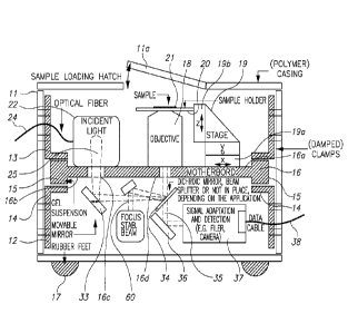
element.
[0007] At least part of the illumination optical system and the return optical
system may be located
in different planes of the compact microscope. Preferably the different planes
are separated by an
opaque part of the primary optical support element or a secondary optical
support element.
[0008] The detection apparatus may be supported by the primary optical support
element.
[0009] At least part of the illumination optical system and the return optical
system may comprise a
secondary optical support element supported by the primary optical support
element.
[0010] The illumination section may comprise a connection to receive an
optical fibre connected to
a light source.
[0011] The detection apparatus may comprise a photodetector.
[0012] The detection apparatus may comprise an imaging apparatus.
[0013] The illumination section may comprise a power meter.
[0014] The illumination optical system may comprise beam shaping optics to
control the shape of
an illumination light beam.
[0015] The illumination optical system may comprise at least one aperture to
control the shape of
an illumination light beam.
[0016] The return optical system may be operable to separate returned light
into at least a first
wavelength band and a second wavelength band.
[0017] Where the compact microscope comprises an imaging apparatus, the return
optical system
may direct returned light in a first wavelength band to a first area of the
imaging apparatus and
returned light in a second wavelength band to a second area of the imaging
apparatus.
[0018] The sample stage may be moveable.
[0019] The compact microscope may further comprise a focus stability beam
optical system to
direct a focus stability beam to the objective lens system.
2
CA 02928712 2016-04-25
WO 2015/059682
PCT/1B2014/065639
[0020] The compact microscope system may comprise a focus controller operable
to receive a
reference image of the focus stability beam from the objective lens system,
receive a subsequent
image of the focus stability beam, and control the sample stage in accordance
with the reference
image and subsequent image.
[0021] Where the illumination light beam is pulsed, the subsequent image may
be obtained
between pulses of the illumination light beam.
[0022] According to a second aspect of the invention there is provided a
microscope system
comprising a compact microscope according to the first aspect of the invention
and an illumination
source module, the illumination optical system of the compact microscope and
the illumination
source module being connected by an optical fibre.
[0023] The illumination source module may comprise a laser source.
[0024] The illumination source module may comprise a first laser source to
generate a first
illumination light beam having a first wavelength and a second laser source to
generate a second
illumination light beam having a second wavelength, and a beam combination
optical system to
transmit the first illumination light beam and second illumination light beam
to the optical fibre.
[0025] The illumination source module may comprise a focus stability beam
laser source and a
focus stability beam optical fibre to transmit the focus stability beam to the
compact microscope.
[0026] According to a third aspect of the invention there is provided a
microscope focus control
system comprising a movable sample stage, an objective lens system, a focus
stability beam optical
system to direct a focus stability beam to the objective lens system, an
imaging apparatus, a return
optical system to return light to the imaging apparatus, and a control system
having a reference
image of the focus stability beam from the objective lens system, the control
system being operable
to receive a subsequent image of the focus stability beam, and control the
sample stage in
accordance with the reference image and subsequent image.
[0027] The control system may be operable to control the sample stage such
that a subsequent
image matches the reference image.
[0028] According to a fourth aspect of the invention there is provided a
method of controlling a
microscope focus, comprising storing a reference image of a focus stability
beam, transmitting a
focus stability beam to an objective lens system of a microscope, receiving a
subsequent image of
3
CA 02928712 2016-04-25
WO 2015/059682 PCT/1B2014/065639
the focus stability beam, and controlling a sample stage of the microscope in
accordance with the
reference image and subsequent image.
[0029] The method may comprise controlling the sample stage such that a
subsequent image
matches the reference image.
Brief Description of the Drawings
[0030] An embodiment of the invention is described by way of example only with
reference to the
accompanying drawings, wherein;
[0031] Figure 1 is a perspective view of a compact microscope embodying the
present invention,
[0032] Figure 2 is a diagrammatic sectional view of the compact microscope of
Figure 1,
[0033] Figure 2a is an illustration of an illumination section of the compact
microscope of Figure 2,
[0034] Figure 2b is a plan view of an alternative support for a primary
optical support element for
the compact microscope of Figure 1,
[0035] Figure 3a is a diagrammatic illustration of an illumination source
module for use with the
compact microscope of Figure 1,
[0036] Figure 3b and 3c are alternative examples of return light paths for
dual-colour wide-field
fluorescence microscopy,
[0037] Figure 4. is a perspective view of beam paths within the compact
microscope of Figure 1 for
use in dual-colour wide-field fluorescence microscopy,
[0038] Figure 5 is a perspective view of further beam paths within the compact
microscope of
Figure 4,
[0039] Figure 6 is an example of an image formed at the imaging apparatus in
the compact
microscope of Figures. 4 and 5,
[0040] Figures. 7a and 7b are perspective view of another compact microscope
embodying the
present invention,
[0041] Figures. 8a to 8d are perspective views of a primary optical support
element of the compact
microscope of figures 7a and 7b,
4
CA 02928712 2016-04-25
WO 2015/059682 PCT/1B2014/065639
[0042] Figures 9a to 9d are diagrammatic views of the configuration of the
compact microscope of
figures 7a and 7b,
[0043] Figure 9e is a perspective view of the optical configurations of
figures 9b to 9d,
[0044] Figure 10a is a perspective view of the secondary optical support
elements of the compact
microscope of figures 7a and 7b,
[0045] Figure 10b is a perspective view of the secondary optical support
elements of the compact
microscope of figures 7a and 7b mounted in the primary optical support element
of figures 8a to 8d,
[0046] Figure 11a is a sectional view of an integral mirror mount of the
compact microscope of
figures 7a and 7b,
[0047] Figure 11b is a sectional view of a further integral mirror mount of
the compact microscope
of figures 7a and 7b,
[0048] Figure 12 is a side view of an objective stage for use with a compact
microscope embodying
the present invention,
[0049] Figure 13 is a diagram illustrating operation of a focus control
system,
[0050] Figure 14 shows a plurality of example reference images for use with
the focus control
system of Figure 13,
[0051] Figure 15 shows examples of detection paths for one- to three-colour
wide-field
fluorescence microscopy,
[0052] Figure 16 is an example of optical paths for dual-colour confocal
fluorescence microscopy,
[0053] Figure 17 is an example of an optical path for fluorescence
polarization microscopy,
[0054] Figure 18 is an example of an optical path for bright-field
interferometric scattering
microscopy, and
[0055] Figures 19a and 19b are example of optical paths for simultaneous dual-
colour wide-field
fluorescence microscopy and dark-field microscopy.
Detailed Description of the Preferred Embodiments
CA 02928712 2016-04-25
WO 2015/059682 PCT/1B2014/065639
[0056] With specific reference now to the drawings in detail, it is stressed
that the particulars
shown are by way of example and for purposes of illustrative discussion of the
preferred
embodiments of the present invention only, and are presented in the cause of
providing what is
believed to be the most useful and readily understood description of the
principles and conceptual
aspects of the invention. In this regard, no attempt is made to show
structural details of the
invention in more detail than is necessary for a fundamental understanding of
the invention, the
description taken with the drawings making apparent to those skilled in the
art how the several
forms of the invention may be embodied in practice.
[0057] Before explaining at least one embodiment of the invention in detail,
it is to be understood
that the invention is not limited in its application to the details of
construction and the arrangement
of the components set forth in the following description or illustrated in the
drawings. The invention
is applicable to other embodiments or of being practiced or carried out in
various ways. Also, it is to
be understood that the phraseology and terminology employed herein is for the
purpose of
description and should not be regarded as limiting.
[0058] First example
[0059] A compact microscope embodying the present application is generally
shown at 10 in Figure
1. The microscope 10 has an enclosure 11, completely enclosing the optical
paths of the microscope.
The enclosure 11 comprises a hatch 11a to provide access to a sample stage as
described below.
Preferably the side and top walls of the enclosure 11 are removable to allow
access to the
components within the microscope 10. In this example, the enclosure 11 is
extremely compact,
having a length of 24cm, a depth of 21.5cm and a height of 15cm, giving the
microscope a footprint
area approximately that of an A4 sheet of paper, and a volume of about 8
litres. It is envisaged that
further reduction of the unused space within the enclosure 11 would allow the
size of the
microscope could be reduced further, to 23cm x 16cm x 15cm without other major
design changes,
and may be even further minimised with suitable design of the components and
optical paths. The
portability allows the microscope to be easily repositioned or relocated, or
even located in a
controlled environment such as a refrigerator, or an incubator with special
atmospheric
compositions, for example controlled levels of CO2 for pH-sensitive mammalian
cell cultures.
[0060] The hatch 11a may be interlocked to cut off the illumination section or
prevent its operation
when the hatch is open. Providing that the enclosure 11 prevents the escape of
light from within the
microscope, the microscope can therefore be a Class I laser product and
therefore may be used
anywhere, and not restricted to for example laser controlled areas.
6
CA 02928712 2016-04-25
WO 2015/059682 PCT/1B2014/065639
[0061] A sectional view of the microscope 10 is shown in Figure 2. Located
within the enclosure 11
is a support element generally shown at 12. In this example, the support
element 12 comprises a
plurality of uprights 13 with mount holders 14 generally at the midpoints of
uprights 13. Although
the support element 12 is a discrete structure in this example, the support
element could be
provided integrally with the enclosure 11.
[0062] To support the optical components, a primary optical support element 16
is provided. The
primary optical support element 16 is preferably a single contiguous and
compact piece of
dimensionally stable material, for example an aluminium, titanium or Invar
block, fabricated from
carbon fibre or otherwise. The primary optical support element 16 in this
example is a generally
rectangular plate, although any other geometry or irregular shape may be used
as appropriate, for
example to accommodate other components or systems within the enclosure 11.
The primary
optical support element 16 may also be cast or machined with holders for the
optical components
already in place or integrally provided, to increase stability and reduce the
possibility of
misalignment of the components.
[0063] The primary optical support element 16 is supported on uprights 13
through vibration
isolating mounts 15 held in the mount holders 14 of the uprights 13. In this
example, the vibration
isolating mounts 15 comprise gel polymer patches, but any suitable mount may
be used, to provide
adequate vibration isolation. In addition, the enclosure 11 is provided with
rubber feet 17 to engage
a supporting surface, to further reduce transmitted vibrations. If desired,
other vibration isolation
components may be provided, or indeed the enclosure 11 and/or the support
element 12 or parts
thereof may comprise vibration isolating material. As shown in the alternative
of Figure 2b, a
primary optical support element 16' is supported on gel-coated titanium rods
15' which are received
in channels 16a' in the primary optical support element 16'. The ends 15a' of
the rods 15' are
received in rubber mounts 17' supported in recesses 18' on the internal face
of enclosure 12'. In a
further alternative, the primary optical support element may be supported by
active vibration
isolation components, such as regulated air pistons.
[0064] In this example, the gel polymer patches 15 act as a low-pass damping
material with a
frequency cut-off of approximately 10 Hz. The length and width of the primary
support structure 16
is chosen to be small while keeping a moderate thickness of the material to
push modal frequencies
to well above 1kHz, for example if aluminium is used for the primary support
structure. The camera
in this embodiment has a full frame readout frequency of 100 Hz. Therefore,
due to the different
order of magnitude of these characteristic frequencies, external forces do not
efficiently excite any
modal frequencies, and the amplitude of any excitations are small due the high
modal frequencies,
7
CA 02928712 2016-04-25
WO 2015/059682 PCT/1B2014/065639
and any vibrations at the modal frequencies are inconsequential for the data
acquisition time scale
of the camera.
[0065] As is apparent from Figure 2, components may be mounted on opposite
sides of the primary
optical support element 16. The primary optical support element has a first,
uppermost side 16a and
a second, lowermost side 16b. Mounted on the first, uppermost side 16a are an
objective lens
system 18 and a sample stage 19. The sample stage 19 is positioned to support
a sample holder 20
above the objective lens 18 to enable a sample 21 to be imaged. The sample
stage 19 has a
transverse positioner 19a, to allow the sample 21 to be moved in the x-y
plane, and a z-axis
positioner 19b to enable the sample holder 20 to be moved vertically relative
to the objective lens
system 18. The positioners 19a, 19b are preferably piezoelectric friction
drives which have low
mechanical drift and backlash, and may be controlled automatically with
nanometre precision over
relatively large distances (several cm), allowing a large number of areas of a
sample to be imaged.
Piezoelectric friction drives allow nanometre scale steps to be made, the
steps being in the range of
approximately 1 to 100 nm depending on the drive used. This makes the
microscope suitable for
automated operation, where the transverse position is operable to successively
bring different areas
of the sample into the field of view of the objective lens to automatically
take a large number of
measurements. In addition to focusing the microscope, control of the z-axis
position also allows
measurements to be taken on different planes, for example through a cell.
[0066] An illumination section is generally shown at 22 and in more detail in
Figure 2a, mounted on
the first side 16a of the primary optical support element 16. The illumination
section 22 may
comprise a laser, an LED or a lamp, or multiple sources, or, as in this
example, may include a
connection to receive an optical fibre for connection to a separate
illumination source module. By
using a separate illumination source module, the microscope can be adapted for
different
techniques or applications by providing different sources. As shown in Figure
2a, optical fibre
connector 23 receives and holds optical fibre 24. Illumination light beam 25
is passed through
cylindrical lenses 26a, 26b to shape and collimate the beam. Adjustable
aperture 27 shapes the
beam further, and wide field lens 28 then provides a converging beam focused
on the rear focal
plane of the objective lens system 18. Beam splitter 29 diverts approximately
<10% of the light to
power meter 30 to allow the beam power to be monitored and noise in the
resulting data caused by
beam intensity fluctuations to be lowered. Mirror 32 then directs the
illumination light beam 25
through aperture 16c. As discussed below, for some applications it is
desirable to adjust or replace
the lens 28 to provide a collimated or divergent illumination beam, or an
illumination beam focused
at some other point in the optical path.
8
CA 02928712 2016-04-25
WO 2015/059682 PCT/1B2014/065639
[0067] On the second, lower side 16b of primary optical support element 16,
mirrors 33, 34 direct
the illumination light beam 25 through aperture 16d to the objective lens
system 18, where the
illumination light beam 25 is focused on the back focal plane of the objective
lens system 18 for wide
field imaging to evenly illuminate the sample 21, or is focused on sample 21
by the objective lens
system 18 via collimated light entering the back aperture of the objective
lens system 18, for
confocal microscopy. The illumination section 22, and mirrors 33, 34 are
referred to collectively as
the illumination optical system. For other applications, other components may
be used in the
illumination optical system. For example, mirror 34 may be a dichroic mirror,
a beam splitter, a
miniaturized mirror, or omitted completely. The mirror 33 can be moveable or
translatable to
change the illumination angle, for example for total internal reflection
microscopy.
[0068] Returned light from the sample 21 is generally shown at 35. The
returned light passes
through mirror 34. If mirror 34 is a dichroic mirror, the characteristics of
the dichroic mirror 34 are
selected such that the wavelengths or wavelength range of the illumination
light beam 25 are
reflected, but returned light passes through. After mirror 34, the returned
light is directed by mirror
36 to detection apparatus 37. Detection apparatus 37 includes a suitable
detector or camera (or
more than one detector or camera as needed) and the appropriate optical
components for the
required application. The detection apparatus has a connection 38 to allow
data to be transmitted to
a control system or computer. The optical components to direct returned light
to the detection
apparatus, and the optical components within the detection apparatus, are
collectively referred to
as a return optical system. For other applications, other components may be
used in the return
optical system.
[0069] In this example, by mounting the components on a single primary optical
support element,
close to the primary optical support element surface, and supporting the
primary optical support
element on vibration isolating mounts, the microscope has greatly reduced
susceptibility to external
forces and variations in temperature and other ambient conditions. Use of both
sides of the primary
optical support element for the optical paths, and using a beam height close
to the primary optical
support element surface, enables the components to be included in a relatively
compact volume. In
the present example, the beam heights are between about 10mm and about 30 mm
from the
primary optical support element surface. Mounting the objective lens and
sample holder on one side
of the primary optical support element and at least part of the illumination
and return optical
systems on the opposite side reduces spurious optical reflections from
entering the return beam
path. The location of optics on both sides of the primary optical support
element allows all
components to remain accessible.
9
CA 02928712 2016-04-25
WO 2015/059682 PCT/1B2014/065639
[0070] The configuration shown in Figures. 2 and 2a is a general configuration
and may be adapted
for use with a desired microscopy technique. By way of example, a
configuration for a dual-colour
wide-field fluorescence microscope will now be described with reference to
Figures. 3a to 6. Figure
3a is a diagram of an illumination source module, Figures 3b and 3c show
alternative simple return
optical systems, and Figures. 4 and 5 are perspective views of the beam paths
within the microscope
10. Elements equivalent to those in Figures. 2 and 2a are labelled with the
same reference number.
[0071] In dual-colour wide-field fluorescence microscopy, sample 21 is
labelled with fluorescent
molecules which absorb light at one of two excitation wavelengths and then
fluoresce. The
illumination light beam is thus more accurately regarded as an excitation
light beam in this
embodiment.
[0072] As seen in Figure 3a, an illumination source module is generally shown
at 40. The
illumination source module 40 comprises a first laser source 41 to generate a
first illumination light
beam 41a having a first wavelength and a second laser source 42 to generate a
second illumination
light beam 42a having a second wavelength. A beam combination optical system
43 combines the
first illumination light beam 41a and second illumination light beam 42a and
couples the light into
the optical fibre 24. In this example the first wavelength is 640 nm and the
second wavelength is 532
nm. The illumination light beams 41a, 42a may be pulsed for alternating laser
excitation microscopy,
with the pulses timed so that the pulses of the beams do not overlap.
Optionally, the illumination
source module 40 may have a power meter in place of, or in addition to, power
meter 27. Figure 3a
also shows a focus stability beam source 59 for generating a focus stability
beam 60, coupled into
single-mode optical fibre 61a as discussed in more detail below. For dual-
colour microscopy, both
illumination beams may be on simultaneously. Excitation clean-up filters are
located in the beam
lines to pass only the main illumination laser wavelengths.
[0073] The detection apparatus in figures 3b to 5 comprises a 2D camera, in
this example a CMOS
camera 37', although a CCD or EMCCD camera may be used depending on the
application. The
return optical path comprises optical elements to separate the returned light
by wavelength. As
shown in Figure 5, the illumination optical path is substantially as shown in
Figure 2a.
[0074] The path of the return light is illustrated in Figures 3b and 4.
Returned light passes through
mirror 34, in this application a dichroic mirror, and is directed by mirror
36. The beam then passes
through separation dichroic mirror 38a. The separation dichroic mirror 38a
reflects the green
fluorescence light with wavelength in the range 545nm ¨ 620nm in the returned
light. The red
wavelengths >656nm pass through and are reflected by mirror 38b. The red and
green returned light
CA 02928712 2016-04-25
WO 2015/059682 PCT/1B2014/065639
beams are directed to different areas of the CMOS camera 37'. In the examples
of Figures. 3b and 4,
the green and red beams are separately reflected towards the CMOS camera 37'
by separate mirrors
39c, 39d, through focusing lens 39e. The green and red beams in this example
cross each other
before the camera 37', so that the beams go through the lens 39e closer to its
centre, causing less
aberration. The mirrors 38a, 38b, 39c, 39d allow the controllable positioning
of the images on
different areas of the CMOS camera 37'. An example of a frame obtained by the
CMOS camera 37' is
shown at 50 in Figure 6, with an image 51 corresponding to one wavelength or
wavelength range at
the left of the frame and an image 52 corresponding to the other wavelength or
wavelength range at
the right of the frame. In this example the signals in the red channel on the
right show less bright
signals as the image shows weak fluorescence resonance energy transfer from
"green" fluorophores
to "red fluorophores. Separation of returned light into different wavelength
bands and different
areas of a detector enables a mix of techniques to be used simultaneously,
such as scattering and
fluorescence. The use of emission clean-up filters blocks light at the
illumination laser wavelengths
from reaching the detection apparatus.
[0075] An alternative configuration of the mirrors 39c, 39d, lens 39e and CMOS
camera 37' is
shown in Figure 3c, in which the optical paths proceed generally left to right
as seen in the Figure, as
opposed to the reflection of 1800 shown in Figure 3b. The Figures are intended
to illustrate that
different optical paths may be selected, depending on the most efficient way
of arranging optical
paths and components on the primary optical support element 16 to obtain a
compact system.
Second Example
[0076] A second embodiment of a compact microscope will now be described with
reference to
figures 7 to 11b. As shown in figures 7a and 7b, a compact microscope shown at
200 comprises a
substantially planar support element 201 which supports primary optical
support element 202. The
primary optical support element 202 is supported by the support element 201
through vibration
isolating mounts 203. In addition, the support element 201 has vibration
isolating feet 204 on its
slow surface to engage the potentially unstable work surface on which the
compact microscope 200
is placed, to further reduce transmitted vibrations.
[0077] As shown in figures 7a and 7b, the compact microscope 200 further
includes an enclosure
205 which is supported by the support element 201 but is physically distinct
from, and not
connected to, the primary optical support element 202. The enclosure 204
serves to isolate the
optics of the microscope from environmental influences and external light, and
to protect the user
11
CA 02928712 2016-04-25
WO 2015/059682 PCT/1B2014/065639
from harmful light intensities within the microscope. As in the first
embodiment, the enclosure 204
may be provided with an interlocked access hatch to provide access to the
sample stage if required.
[0078] The primary optical support element 202 will now be described in more
detail with
reference to Figures 8a to 8d. In these figures, the primary optical support
element 202 is shown
located on the support element 201, but with the enclosure 205 and the
secondary optical support
elements, described below, omitted. In contrast to the embodiment of the
compact microscope
shown in Figures 1 to 6, in this example the primary optical support element
202 is a complex shape
designed to accommodate the optical components in a multi-planar
configuration, permitting a
more compact arrangement than a relatively simple planar primary optical
support element. The
primary optical support element 202 has two sections, a camera support section
generally shown at
210, and an optical support section generally shown at 211. The optical
support section 211
comprises a volume 212 to receive the optical components in a number of
secondary optical support
elements. An upper part 213 of the volume 212 is shaped to receive an
objective stage, described in
more detail below. The primary optical support element 202 further includes
integral mirror mounts
to receive mirrors to direct light from the microscope tube lens to the camera
as discussed in more
detail below. The primary optical support element 202 in this example
comprises four machined
components which are rigidly attached to one another, but any suitable means
of fabrication and
assembly may be used. It would be possibly to fabricate the primary optical
support element as a
single component, but using multiple elements enables modularity (for example
to permit the use of
a different camera, or manipulate the signal between the tube lens and the
camera) and makes
machining the components easier.
[0079] The layout of the compact microscope 200 is shown diagrammatically to
scale in figures 9a
to 9e. Figure 9a is a top view showing the relative arrangement and dimensions
of the optical
arrangement and camera to scale. The locations of the vibration isolating
mounts 203 are shown for
reference, and in this example the total footprint is about 180 mm on each
side. The microscope
optics are laid out so that the dimensions of the optical arrangement are 146
mm x 90 mm, achieved
by stacking multiple layers of optics on top of or next to one another in
close proximity.
[0080] As seen in figures 9a to 9e, the optics are arranged in three planes, a
lower plane 220, an
upper plane 221 and a vertical plane 222. Broadly, the illumination optical
system is located in the
lower plane 220 and the vertical plane 222, and the receiving optical system
is located in upper
plane 221. As seen in Figure 9b, the lower plane 220 comprises a light
receiving section 223, to
receive light from a plurality of optical fibres from a suitable source, for
example an illumination
source module similar to that of Figure 3a. The optical fibres are shown at
225a to 225f. The light
12
CA 02928712 2016-04-25
WO 2015/059682 PCT/1B2014/065639
received from fibres 225c to 225f passes through a respective pair of
cylindrical lenses 226 and an
aperture 227 similar to be aperture 27 of figure 2a. As in the example of
figure 2a, to provide a beam
having a desired profile, in this example a substantially rectangular beam
shape, without requiring
complex and optics and thus reducing the space required. A series of dichroic
mirrors 228 reflects
the beams along a common path, 229 such that the combined beams strike mirrors
230a, 230b and
are directed towards first upwardly reflecting mirror 231 lying in plane 222
which reflects the
combined beams upwards.
[0081] A plurality of photodiodes 232 are located to receive light which
passes through the dichroic
mirrors 228, to enable the respective power in each beam arriving at the
microscope to be
measured. Advantageously, no additional beam splitter is required to allow the
determination of the
power in each beam, maximising the available usable power and reducing the
need for further
components.
[0082] The light from fibres 225a, 225b is received separately from that
fibres 225c to 225f. The
received light is directed through focusing lenses 226a and directed by
mirrors 233a, 233b, 233c to
second upwardly reflecting mirror 234 in the vertical plane 222. Mirror 233b
may be a dichroic
mirror.
[0083] The optics in the vertical plane 222 are shown in Figures 9d and 9e.
The light reflected from
first upwardly reflecting mirror 231 is received by a wide field lens unit
235. The light is focused by
the wide field lens 235a and is directed laterally by mirror 236 towards
dichroic mirror 240 and then
mirror 239 which reflects the converging beam into the objective, such that a
focus is formed in the
rear focal plane of the objective, and collimated light emerges out of the
objective front focal plane.
The wide field lens unit 235 includes a piezo actuator 237 which may be
automatically or manually
controlled to vary the position of lens 235a. The piezo actuator 237
translates the lens 235a and the
mirror 236 so that the distance from the lens 235a to the objective remains
constant. This is in
particular required for a short focal length lens 235a, but a constant
distance to the objective is not
crucial for larger microscopes where a lens with much longer focal length is
used. The piezo actuator
allows the lens 235a to adjust the illumination for total internal reflection
microscopy. Light from
second upwardly reflecting mirror 234 strikes a fused silica window 238 and is
simply directed to
mirror 239 and into the objective stage 300. There are minimal losses for the
light directed from the
first upwardly reflecting mirror 231 because the fused silica window 238 is
transparent in the
relevant wavelength region and is anti-reflection coated. The use of fused
silica minimizes
fluorescence generated by the light transmitted through the window from mirror
231.
13
CA 02928712 2016-04-25
WO 2015/059682 PCT/1B2014/065639
[0084] In this example, minimal transmission losses are needed for the
converging light from
upwardly reflecting mirror 231 which illuminates a large (for example 1201.im
x 60p.m) area in the
sample plane, requiring more power, and high reflection losses are acceptable
for the collimated
light from upwardly reflecting mirror 234 which illuminates a small (for
example 1p.m x 1p.m) area,
requiring less power. For other applications, other components such as
dichroic mirrors may be used
in place of the fused silica window.
[0085] The light returned from the sample and objective lens 301 is then
directed by mirror 239 and
dichroic mirror 240 to the receiving optical system generally shown in Figure
9c. The receiving
optical section includes modular elements in the area 241 which may be removed
or adapted
depending on the technique or function to be used with the microscope. In this
example, module
241 comprises an optical arrangement similar to that of Figure 3c, in which
returned light from a
sample in two wavelength ranges is separated into two spatially offset beams
and directed to tube
lens 250. The output beams are directed by mirror 251, held in integral mount
214 and mirror 252,
held in integral mount 215, to the camera. The offset beams result in an
output image shown as
Figure 6, with images 51 and 52 corresponding to different wavelengths or
wavelength ranges offset
two separate sections of the image frame.
[0086] Again, it will be apparent that the geometry of figures 9b to 9e is
extremely adaptable, in
that only a subset of the input fibres 225a to 225f need be used as required,
and the return optical
module 241 may be replaced as required. The requirement is only that the beams
entering and
leaving the module 241 are focused to infinity to enable easy replacement.
[0087] The secondary optical support elements are illustrated in Figures 10a
and 10b. First
secondary optical support element 261 has connections 261a to receive fibres
225c to 225f and
includes lenses 226, apertures 227 and mirrors 228 and 230a. Second secondary
optical support
element 262 supports first and second upwardly reflecting mirrors 231, 234.
Third secondary optical
support element 263 similarly contains simply comprises connectors (not shown)
to receive fibres
225a, 225b, lenses 232 and mirrors 233a, 233b, 233c. Fourth secondary optical
support element 264
holds the wide field lens unit 235 including the wide field lens 235a, piezo
actuator 237 and mirror
236. Silica window 238 is held in place by a separate secondary optical
support element and mirror
239 is held directly by the primary optical support element 202. Fifth
secondary optical support
element 265 provides the receiving optical system 241 as a unit or a as a
group of sub-modules. As
illustrated in figure 10b, each of these components is received within volume
212 of the primary
optical support element 202, substantially filling the volume 212 thus
providing a rigid and
accurately aligned configuration which is modular and adaptable as required.
If necessary, parts of
14
CA 02928712 2016-04-25
WO 2015/059682 PCT/1B2014/065639
the volume 212 not otherwise occupied by secondary optical support modules may
be occupied with
solid or hollow filler blocks to provide additional rigidity. At the same
time, since many apertures are
required to allow the beams to propagate, the microscope is filled with holes,
making it a naturally
rigid, light-weight, hollow structure, which reduces deformation by self-
weight. Advantageously the
different planes are separated by an opaque part of the primary optical
support element or a
secondary optical support element.
[0088] An integral mirror mount 214 is shown in more detail in figures 11a and
11b. As seen in
figure 11a, integral mirror mount 214 comprises a vertically extending slot
270. A beam aperture is
shown at 271, comprising a right-angled channel. The vertical slot 270 is
shaped such that it has a
depth greater than the depth of the beam aperture 271 to provide a lower
support step 272. The
mirror 251 is sized such that it engages the lower support step 272, extends
across with the beam
aperture 271 and engages an upper lip 274 of the slot 270 above the beam
aperture 271. A spring
275 located between the mirror 251 and a back wall 276 of the slot 270 holds
the mirror in place.
The spring exerts a constant, temperature insensitive force perpendicular to a
surface to which the
optical element aligns to. A similar configuration is used for integral mount
215, with the exception
that the slot 270 is shaped such the mirror 252 is introduced perpendicular to
the alignment surface
rather than in parallel, and is held in place by a suitable locking element
that compresses the spring.
Hence, each integral mirror mount 214, 215 has a surface against which a
respective mirror is held,
which is permanently defined by the machining process and cannot be altered,
making misalignment
impossible. In addition to defining the alignment surface, the machining
process concurrently
creates an access port such that the mirror and the spring can be inserted.
Isolation against dust is
also provided since the mirror makes physical contact with the alignment
surface and seals the
machined apertures in which the light propagates. Although only integral
mirror mounts 214, 215
are shown in the figures, similar integral mirror mounts are used elsewhere in
the primary and
secondary optical support elements where appropriate
Objective stage
[0089] The objective stage 300 is shown in more detail in Figure 12. The
objective stage 300
supports the microscope objective 301. The objective stage 300 comprises an
Invar baseplate 302 on
which is mounted a movement stage 304, primarily made of aluminium, for
example. The movement
stage 304 supports and moves an Invar sample holder 305 and allows the
adjustment of the three-
dimensional position of the sample holder 305 relative to the microscope
objective 301. The stage
304 is connected to Invar baseplate 302 by a plate 303 of the same material as
the stage. A first
mounting point 306 connects the aluminium plate 303 and the Invar baseplate
302 such that
CA 02928712 2016-04-25
WO 2015/059682 PCT/1B2014/065639
mounting point 306 is aligned with the front face 307 of the stage 304. A
second, rearward,
mounting point 308 connecting the aluminium plate 303 and the Invar baseplate
302 allows for
relative sliding movement between the aluminium plate 303 the Invar baseplate
302. The Invar plate
is mounted on the primary optical support structure in recess 213 and fixed to
the primary optical
support structure 202 through a first fixed mounting point 302a which is
aligned with the axis of the
objective, and a second mounting point (not shown) that allows relative
sliding movement between
the aluminium primary optical support structure 202 and the Invar plate 302.
The movement stage
304 has three piezo friction motors, generally shown at 309a, 309b and 309c
respectively.
[0090] The objective 301 is thus held in a fixed position relative to the
primary optical support
structure 202 and the optical systems mounted therein. The configuration of
the objective stage 300
allows for compensation of thermal expansion of the movement stage 304,
expansion of the sample
holder, and of the material connecting the objective and the movement stage
304.
[0091] The compensation for the thermal expansion of the movement stage 304 is
achieved by
locating the fixed mounting 306 of the aluminium plate 303 underneath the
aluminium stage aligned
with the face 307 of the movement stage 304, which is contact with the Invar
sample holder 305.
Therefore, if the aluminium movement stage 304 expands to the left, the
aluminium plate 303 will
expand to the right and the amount of expansion relative to the face 307 is
equal and will therefore
cancel. The compensation will not completely cancel the relative expansions
when the stage 304 is
moved from the centred position toward or away from the objective and the face
307 misaligns with
fixed mounting point 306. However, this geometry will reduce drift to a
minimum, and drift
increases linearly from 0 when the stage 304 is at the default position to a
small maximum at the
extremes of the stage range (which will be used less likely than a position
close to the centered
position).
[0092] In this geometry, thermal expansion of the Invar section between the
objective lens 301 and
face 307 is equal in magnitude but opposite to the thermal expansion of the
sample holder 305, so
that relative movement between the objective 301 and a sample held on the
sample holder 305 is
cancelled. The fixed mounting 302a ensures that the objective 301 remains in
place while the sliding
mounting allows the Invar base plate to expand or contract relative to the
primary optical support
element 202.
[0093] Although the objective stage 300 here comprises Invar and aluminium
components, it will be
apparent that the stage may comprise components fabricated from other
materials as desired. Two
pairs of parts should have matched thermal expansion coefficient, the first
pair being the sample
16
CA 02928712 2016-04-25
WO 2015/059682 PCT/1B2014/065639
holder and the objective mounting plate, and the second pair being the
movement stage and the
plate 303. The objective stage may be used with any other suitable microscope,
not only the
examples described herein, and may comprise one actuator or any number of
actuators as needed
for the desired degrees of freedom of movement of the sample holder.
Focus control
[0094] A focus control system can maintain the axial position of the sample
relative to the focus of
the objective lens 301 by controlling the position of the sample holder 305.
[0095] To achieve this, a focus stability beam is provided from a focus
stability beam fibre
connection. In the first embodiment of the compact microscope, a microscope
focus control system
is illustrated in figures 2, 3a and 4. A focus stability beam 60 is provided
from a focus stability beam
fibre connection 61, connected to in this case a single-mode optical fibre 61a
to transmit light from a
focus stability beam laser source 59 in the illumination source module 40.
Focus stability beam 60 is
collimated by lens 62 and directed to the objective lens system by mirror 63
and dichroic mirror 34.
The mirrors 63, 34 direct the focus stability beam 60 into the objective lens
system 18 at an angle
relative to the optical axis 25. The focus stability beam fibre connection 61,
lens 62, mirror 63 and
dichroic mirror 34 are collectively referred to as the focus stability beam
optical system.
[0096] In the second embodiment of the compact microscope, one of the fibres
225a, 225b may be
used as the focus stability beam fibre connection. In this case, the focus
stability beam is directed to
the objective lens 301 by mirrors 233a, 233b, 233c and 234 as discussed above.
[0097] The wavelength of the focus stability beam can be chosen such that the
sample is not
affected by the light (e.g. off-resonance light if the sample contains
fluorescent molecules). The
focus stability beam is preferably only active when the sample is not being
imaged. The objective
lens system focuses the focus stability beam at the interface between cover
glass and sample
medium, where a part of the light is reflected. The beam enters the objective
on the same path as
the excitation lasers, but it is collimated so that an approximately focused
spot is visible in the image
plane (on the camera) when the glass / sample medium interface is at the front
focal plane of the
objective. The beam is strongly converging due to the high numerical aperture
of the objective lens,
so that any movement of the interface away from the focal plane causes the
reflection image to
widen significantly. Light scattered from the illuminated region will be
returned by the return light
optical system to the detection apparatus, where an image can be captured. The
image of the
reflection will have a size, shape and position that depend on the distance
between objective and
17
CA 02928712 2016-04-25
WO 2015/059682 PCT/1B2014/065639
the interface. To control the focus, the system and method compare a reference
image with a
subsequent image.
[0098] A first way of operating the focus stability system is in a focus lock
mode. After the sample
has been initially correctly positioned, the focus stability beam can be
transmitted to the objective
lens system and a reference image saved. If the position of the sample shifts
relative to the
objective, in a subsequently captured image the illumination region will
change appearance.
Accordingly, after moving to another field of view, a subsequent image is
captured. If this differs
from the reference image, the z-axis positioner is iteratively operated. The z-
axis position will be
varied in accordance with a calculated difference between the reference and
subsequent images,
and a further subsequent image captured. Again, the difference is calculated
and a further z-
positioning step carried out. In this way, the system will converge on the
original focus in a few
steps. The difference between the images may be calculated in any suitable
manner.
[0099] In a second way of operating the focus stability system, storing a
reference image may
include storing a stack of reference images each corresponding to a known
different position vertical
position. This may be used to move the sample to a desired z position or to
determine the z position
of the sample. When it is desired to move the sample to desired z position,
the reference image
corresponding to that z position can be retrieved and an iterative process
similar to that described
above carried out, taking subsequent images and moving the z-axis positioner
in small steps until the
subsequent images converge on the desired reference image. Alternatively, by
capturing a
subsequent image and determining which reference image is the best match for
the subsequent
image, the z position of the sample can be determined.
[0100] A reference image or stack of images may be stored when the device is
manufactured or
calibrated. Additional or replacement reference images may be captured at any
suitable point during
operation of the microscope, for example before starting an acquisition, if a
new focal plane is
required or if the reference image becomes incompatible with the correct focus
due to changes in
the sample, or the microscope system. A subsequent image may serve as a
reference image for
subsequent operation.
[0101] An example method is illustrated at 320 in figure 13. As shown at step
321, an image of the
focus beam is acquired. At step 322 to the reference image with the highest
normalised cross-
correlation maximum (NCCM) is identified. The NCCM is a measure of how similar
the captured and
reference images are to each other, with complete similarity giving a score of
1 and no similarity
18
CA 02928712 2016-04-25
WO 2015/059682 PCT/1B2014/065639
giving a score of 0._Such an algorithm is sensitive to the shape of the
reflection pattern, but not its
intensity or its position on the camera.
[0102] As shown by arrow 323, if the highest NCCM is less than 0.5, then at
step 324 the sample
stage 305 is moved around the current z position at increasing ranges, and
repeated images
captured, until an image with a cross-correlation measure of >0.5 for any
reference image is found.
Once the step is complete, the method moves to step 326, which alternatively
may be moved to
directly from step 322 as shown by arrow 325 if the highest NCCM is >0.5 but
is not from the set
point a reference image. At step 326 the stage is moved in the direction of
the set point using
relatively large steps of 200 nm until the cross-correlation measure is >0.9
or the set point is crossed.
At a fine tuning stage 327, after completing step 326 or directly after step
322 if the highest NCCM is
between 0.5 and 0.99 and belongs to the set point reference image, the NCCM is
maximised using
steps of decreasing length 100 mm to 10 nm. When the NCCM is maximised, the
procedure is
complete as shown in 328. If the NCCM of the acquired and reference images is
initially greater than
.99, as shown by arrow 329, then the autofocus procedure is ended immediately.
A set of example
reference images is shown in figure 14, where the central figure 340a
represents the set point. A
series of figures 340b to 340d represent images from progressively lower
sample positions. Similarly,
reference images 340e to 340g are images resulting from the sample position
said being too high.
Accordingly, as illustrated in Figure 13, the focus apparatus will find the
image that best matches the
acquired image of the focus beam, and then adjust the z position towards the
set point, i.e. the
process will be iterated until the acquired image of the focus beam
effectively corresponds to the
reference set point image 340a. Where the acquired image of the focus beam at
step 321 is not
sufficiently similar to any of the captured images 340a to 340g, this
indicates that the sample
position is outside the range of the captured images, and the process
successively moves the sample
position until an image of the focus beam is acquired which is sufficiently
similar to one of the stored
reference images as shown at 323 and 324.
[0103] The microscope focus control system can thus maintain a stable sample
position throughout
extended operation of the microscope 10. The focusing takes less than 1 second
and achieves
nanometre accuracy. This auto-focus method does not require an additional
photodetector for the
reference beam, and lasers of very low beam quality, power and power stability
can be used. The
power of the focus stability beam in this example is extremely low due to
inefficient single-mode
fibre coupling without a focusing lens to the fibre 61a and as such presents a
minimal hazard. The
absence of a fibre coupling lens also removes the need for (re-)alignment of
the laser with respect to
the single-mode fibre.
19
CA 02928712 2016-04-25
WO 2015/059682 PCT/1B2014/065639
[0104] In experiments where the sample is immobilized on the glass substrate,
it is often possible
and desirable to record independent data sets by moving the field of view to a
new area which has
not been exposed by the excitation lasers. Due to thickness variations of the
cover glass on the order
of a few microns and motion irregularities of the sample stage, a refocussing
step is usually
necessary before the acquisition of a new field of view. The focus control
system allows this
movement and refocussing step to be carried out automatically. For example,
the sample could be
moved in a spiral pattern to record multiple field of views. Before each field
of view is exposed by
the excitation lasers, the focus control brings the sample to a pre-defined
axial position. As the light
for the focus stability beam is emitted from the end of a fibre attached
securely on the principle
optical support element of the microscope, and the collimating lens and beam
steering mirrors for
the focus stability beam lack any degrees of freedom for adjustment, the
angular stability of the
beam is improved. Instead of using a dedicated sensor, the use of the main
camera to detect the
image of the focus stability beam again reduces the number of required
components.
[0105] As a further method of controlling the focus and allowing the movement
to a user desired
plane, the glass / sample medium interface is set as the set point and the
user is not permitted to
change it. The focus control mechanism then moves the interface to the focal
plane, and the
position sensor of the piezo stage (which has ¨1nm precision) is then used to
navigate away from
the interface to a desired position. In other words, the interface is used as
a starting point for
movement relative to it, which could eliminate the need for recording
reference images by the user
and establishes the interface as the origin plane of a well-defined coordinate
system.
Alternative optical configurations
[0106] Examples of how the microscope may be adapted to other applications are
illustrated in
figures 15 to 19b. Advantageously, the compact microscope 200 is adaptable to
any of each of these
configurations by providing an appropriate fifth secondary support module 265.
[0107] Figures 15a to 15c show a configuration of the return optical system
adaptable to single-,
dual- or three-colour wide-field fluorescence microscopy. The general
apparatus is shown at 400 in
figure 15a, in which the beam is focused by lens 401 directly on camera 437.
In figure 15b, which is
equivalent to the optical paths of figures 3b and 3c, dichroic mirror 402
separates the returned light
into first and second wavelength bands. Light in a first wavelength band
passes through dichroic
mirror 402 and is reflected by mirrors 403 and 404 through a first band-pass
filter 405. Light in the
second wavelength range is reflected by dichroic mirror 402 and mirror 406 and
passes through
band pass filter 407. The two resulting beams are spatially offset and are
focused on different areas
CA 02928712 2016-04-25
WO 2015/059682 PCT/1B2014/065639
of the camera 437. In a further variation, shown in figure 15c, mirror 403 is
replaced with a further
dichroic mirror 408. Light in a third wavelength range passes through dichroic
mirrors 402, 408 and
long pass filter 409, and the three resulting beams are focused on separate
offset areas of the
camera 437.
[0108] Figure 16 illustrates use of the microscope for dual-colour
fluorescence confocal microscopy,
and an example optical path is shown at 410. The detection apparatus in this
application comprises a
pair of photodetectors 411a, 411b, and particularly avalanche photodiodes
providing high detection
sensitivity. The excitation beam 412 comprises light in two wavelength bands
as discussed above,
and is collimated. The objective lens system focuses the illumination light on
the sample 21.
Returned fluorescent light passes through dichroic mirror 413 and is directed
to the detection
apparatus by mirror 414. Light in a first wavelength band is reflected by long-
pass dichroic mirror
415, passes through band-pass filter 416 and is focused by lens 417 on the
first photodetector 411a.
Light in a second wavelength band passes through dichroic mirror 415 and is
reflected by mirror 418,
passes through long pass filter 419 and is focused by lens 420 on second
photodetector 411b.
[0109] Figure 17 shows a return optical path for fluorescence polarization
microscopy at 430. The
returned fluorescent light 431 passes through emission filter 432, and the
returned light is separated
into different polarization components by polarizing beam splitter 433.
Mirrors 434, 435, 436 direct
the differently polarized beams through lens 438, and the different beam are
focused on offset
regions of the imaging apparatus 437'.
[0110] Figure 18 shows a configuration for bright field interferometric
scattering (iSCAT) microscopy
at 440. Illumination beam 441 is slightly convergent. The illumination beam
441 passes through
beam splitter 442, which directs some of the illumination beam to objective
lens system 18,
illuminating an area of the sample 21. Beam stop 443 absorbs that part of
illumination beam 441
which passes directly through the beam splitter 442. Illumination light
scattered from the sample 21
interferes with illumination light reflected from an interface of the sample
slide. The reflected light
returns through beam splitter 442 and is reflected by mirror 444 and focused
by lens 445 on imaging
apparatus 437.
[0111] Figures 19a and 19b show optical paths for simultaneous dark field
microscopy and multi-
colour wide-field microscopy, shown at 450a, 450b respectively. In 450b, a
small elliptical mirror 451
reflects the converging illumination light to the rear focal plane of the
objective on the optical axis of
the objective, so that the reflected illumination light returns on the optical
axis and is reflected again
by mirror 451 out of the detection path. In 450a, a small mirror 452 reflects
the converging
21
CA 02928712 2016-04-25
WO 2015/059682 PCT/1B2014/065639
illumination light to the rear focal plane of the objective on the edge of its
back aperture. The
illumination light is total internally reflected (TIR) at the glass / sample
interface and returns at the
diametrically opposite edge of the back aperture, where it is directed by
another small mirror 453
into an absorbing element 454. The latter geometry requires an oil-immersion
objective with
sufficiently high numerical aperture.
[0112] In each alternative, a system of dichroic mirrors and filters separate
the fluorescent light and
scattered light from the returned light and direct the various wavelength
bands to different areas of
the imaging apparatus 437. Dual-band dichroic mirror 455 is selected so that
fluorescent light in first
and second wavelength bands passes through the dichroic mirror 455, while
scattered light is
reflected. The scattered light is reflected by mirror 456, through dual laser-
line pass filter 457 and
lens 458 and is focused on a first area of imaging apparatus 437. Long pass
dichroic mirror 459
separates the fluorescent light into first and second wavelength bands, which
pass through long
pass filter 460 and band pass filter 461 respectively and are focused on
second and third areas of the
imaging apparatus 437.
[0113] It will be apparent that the microscope focus control system will be
suitable for use for any
other type of microscope, not only the embodiments of compact microscopes
described above. If
appropriate, the focus control system may have its own imaging apparatus
separate from the main
detector of the microscope.
Adaptability
[0114] The compact microscope as described herein is advantageous in that it
provides the most
desirable aspects of wide-field imaging: low sample drift, high vibration
stability, single-molecule
detection sensitivity, automation and high-throughput in a form factor that is
compact, robust,
portable and low-cost.
[0115] The optical path was designed to provide the highest detection
efficiency as possible, and to
be as compact as possible. For multi-channel imaging on a single array sensor,
the design does not
require the formation of an image in the plane of a slit aperture which cuts
the image. This is usually
done to fit the image into rectangular regions on the array sensor. In
contrast, the compact
microscopes described above use a shaped incident beam which illuminates only
the area that is
going to be detected. Astigmatic lenses and a suitable aperture can be used to
define the width and
length of the incident beam. Compared to other channel splitting optical
systems, the present
system removes the need for two lenses and an adjustable slit aperture from
the detection path,
increasing detection efficiency and saving both space and component cost.
22
CA 02928712 2016-04-25
WO 2015/059682 PCT/1B2014/065639
[0116] The provision of a separate illumination source module is advantageous
in that it removes
components from the microscope enclosure to permit the enclosure to be
smaller, and prevents
heat from the laser or supporting equipment heating the microscope enclosure
and temperature-
sensitive samples within the enclosure. The separate illumination source
module also provides
adaptability, in that to change the microscope function a different
illumination source module can
be easily provided and connected via an optical fibre link. Multiple
connections to allow connection
of the microscope to multiple illumination sections may be provided, or indeed
multiple microscopes
may be connected to a single illumination source module for parallel operation
or reduced cost.
Provision of laser sources may be continuous-wave or any combination of pulsed
sources, including
dual-laser excitation, triple-laser excitation, or complex pulse sequences,
such as sequences with
alternating single- and dual-laser excitation. Where optical fibres are used,
the fibres may be
manipulated in known manner, for example by heating or mechanical flexing,
stretching or
squeezing to produce homogeneous illumination light. The control electronics
may also be provided
as part of the illumination source module including for example a power
supply, piezo drivers, laser
drivers, signalling and I/0 hardware, and fibre-squeezing piezo drivers. This
means that, for example,
to adapt the microscope for a particular experiment, it is only necessary that
the appropriate
illumination source module and secondary optical support modules (where
needed) are connected
to the compact microscope.
[0117] Alternatively, or in addition, an illumination source module or source
may be provided
within the microscope enclosure if desired. Any suitable detector, or group of
detectors, and
corresponding return optical system, may be provided to adapt the microscope
to a desired
function. Although a single objective lens system is described above, the
microscope may include
two or more objective lens systems if required. Although the microscope
described herein has an
objective lens system, for some applications this may be replaced by other
light collection elements,
such as a reflective objective system. The modular nature of the microscope
optics and objective
stage allow such adaptations.
[0118] Advantageously, the microscope may be provided with sensors to detect
and record
measurement parameters, such as temperature, humidity, pressure, atmosphere
composition,
acceleration, magnetic and electric fields and location. The information from
the sensors can be
used in a feedback system with control systems internal or external to the
microscope enclosure, to
realize the exact measurement conditions desired. For example, a temperature
control unit
mounted on the primary optical support element could heat or cool the entire
microscope (and the
air within the enclosure) to a certain temperature. With appropriate
temperature control, the
23
CA 02928712 2016-04-25
WO 2015/059682 PCT/1B2014/065639
microscope could act as an incubator, particularly where samples must be kept
at a certain
temperature. Known approaches with normal microscopes only use an objective
heater to keep the
sample at the required temperature. This inevitably causes temperature
gradients in the microscope
as well as the sample which lead to drift of the microscope and convection in
the sample. These
problems may be reduced or eliminated by maintaining the entire environment at
the required
temperature. The internal atmosphere may also be regulated. For example,
connecting a CO2 line to
the microscope and a gas regulator with feedback from the environmental
sensors would make the
microscope act as a mammalian cell incubator. The microscope software can also
use sensor
information to judge the quality of the measurement, and if necessary discard
invalid
measurements. Recorded sensor information will also support the
reproducibility of measurements.
[0119] The microscope is very adaptable and flexible in terms of illumination
and detection options,
specimens examined, and concentration regimes. For example, a variety of
illumination sections can
be used: a single-wavelength continuous laser, a pulsed excitation source with
modulation in the
picosecond-to second time domain, complex excitation schemes with multiple
modulated lasers
modulated using different ways of modulation (e.g. electronic on/off
modulation, choppers,
acousto-optical modulators, acousto-optical tunable filters, electro-optical
modulation). In some
cases, a microscope can operate even in the absence of the illumination
section, e.g., in the case of
chemiluminescent compounds (where the state responsible for fluorescence
emission is generated
by a chemical reaction). The microscope can be adapted to accommodate samples
that have many
forms, e.g., a solution containing luminescent compounds, a coverslip with
immobilized molecules, a
flow-cell containing fluorescent molecules, a slide with fixed mammalian cells
or tissue samples.
Although the microscope has the sensitivity to detect single molecules, it can
also operate in a high-
concentration mode that looks at the average intensity in a single or multiple
spectral emission
channels. In terms of detection, formats for point source detection e.g.
confocal microscopy (where
a diffraction-limited volume illuminated by a focused laser beam is focused on
a point detector such
as an avalanche photodiode detector, APD) or wide-field imaging (where a large
area in the
specimen plane is imaged on a 2D detector such a CCD, EMCCD and sCMOS camera)
can be
achieved. In addition to fluorescence and also fluorescence lifetime imaging
capabilities, careful
selection of filters in the emission path and an appropriate geometry can
enable scattering
measurements. The laser light wavelength or wavelength may be selected to
induce changes in the
sample, for example to trigger photochemical or (photo)physical processes in
the sample such as
photoactivation of fluorophores (by a UV laser) or stimulated emission and
control of the chemical
and quantum state of fluorophores in general, and local changes in temperature
(by a focused IR
24
CA 02928712 2016-04-25
WO 2015/059682 PCT/1B2014/065639
laser. The use of multiple sources allows light of several wavelengths to be
directed to a sample as
needed, or to different areas of the same sample.
[0120] The microscope system is very suitable for automation with suitable
control and analysis
software, where the hardware control, data acquisition and storage and
visualisation processes are
closely integrated. Suitable software can provide automated data acquisition,
real-time analysis,
intelligent data analysis, so little or no user interaction is required and
real-time data visualization
and reporting. In a wide range of applications, many signals can be measured
simultaneously in a
single field of view. With the piezo friction drives, the sample can be
translated by several
centimetres, so that thousands of fields of view can be measured in a fully
automated fashion, with
data analysis and interpretation occurring in parallel. An extremely large
data set can be collected
and processed in a very short time. One such microscope or an array of such
microscopes could
therefore be used for high throughput, massively parallel multidimensional
screening applications,
such as for use in a pharmaceutical environment.
[0121] The work leading to this invention has received funding from the
European Research Council
under the European Union's Seventh Framework Programme (FP7/2007-2013) / ERC
grant
agreement n 261227.
[0122] In the above description, an embodiment is an example or implementation
of the invention.
The various appearances of "one embodiment", "an embodiment" or "some
embodiments" do not
necessarily all refer to the same embodiments.
[0123] Although various features of the invention may be described in the
context of a single
embodiment, the features may also be provided separately or in any suitable
combination.
Conversely, although the invention may be described herein in the context of
separate
embodiments for clarity, the invention may also be implemented in a single
embodiment.
[0124] Furthermore, it is to be understood that the invention can be carried
out or practiced in
various ways and that the invention can be implemented in embodiments other
than the ones
outlined in the description above.
[0125] Meanings of technical and scientific terms used herein are to be
commonly understood as
by one of ordinary skill in the art to which the invention belongs, unless
otherwise defined.