Note: Descriptions are shown in the official language in which they were submitted.
CA 02929300 2016-05-09
TRANSDERMAL DRUG DELIVERY PATCH SYSTEM, METHOD OF
MAKING SAME AND METHOD OF USING SAME
This application is a divisional of Canadian patent application Serial No.
2478822
filed internationally on March 11, 2003 and entered nationally on September
10, 2004.
TECHNICAL FIELD
This invention relates to devices and method for the creation of small holes
or
perforations or micropores in biological membranes, such as the outer layers
of the skin or
the mucosal linings, the delivery of drugs or other permeants through the
micropores, the
extraction of biological fluids through the micorpores, the integration within
the device and
method of an assay for selected of analytes in the extracted biological
fluids, and the
increase of flux through these micropores by one or more of pressure
modulation, the
mechanical manipulation or distortion of the microporated tissue and adjacent
tissue,
electro-transport, electro-osmosis, iontophoresis and sonic energy.
BACKGROUND ART
The stratum corneum is chiefly responsible for the barrier properties of skin.
Thus,
it is this layer that presents the greatest barrier to transdennal flux of
drugs or other
molecules into the body and of analytes out of the body. The stratum comeum,
the outer
horny layer of the skin, is a complex structure of compact keratinized cell
remnants
separated by lipid domains. Compared to the oral or gastric mucosa, the
stratum corneum
is much less permeable to molecules either external or internal to the body.
The stratum
corneum is formed from keratinocytes, which comprise the majority of epidennal
cells that
lose their nuclei and become comeocytes. These dead cells comprise the stratum
corneum, which has a thickness of only about 10-30 microns and, as noted
above, is a very
resistant waterproof membrane that protects the body from invasion by exterior
substances
and the outward migration of fluids and dissolved molecules. The stratum
corneum is
continuously renewed by shedding of corneum cells during desquamination and
the
fon-nation of new corneum cells by the keratinization process.
Historically, drugs have been delivered across the skin by injection. However,
this
method of administration is inconvenient and uncomfortable, and it is not
suited for self-
1
CA 02929300 2016-05-09
4 =
administration by members of the general public. Additionally, used needles
continue to pose
a hazard after their use. Therefore, transdermal drug delivery to the body is
particularly
desired.
There are many techniques known in the art for transdermal drug delivery and
monitoring applications. One well-known example of the need in the art for
less painful
puncturing of a biological membrane is in the field of diabetes monitoring.
The current
standard of care for a patient with diabetes includes a recommendation of 3 to
5 painful
finger-stick blood draws per day to allow them to monitor their blood glucose
levels. Other
than the relative size of the lancets decreasing over the last few years, the
use of lancets, and
the resulting finger sensitivity and pain, has not changed for many years.
To enhance transdermal drug delivery, there are known methods for increasing
the
permeability of the skin to drugs. For example, U.S. Patent No. 5,885,211 is
directed to
thermal microporation techniques and devices to form one or more micropores in
a biological
membrane and methods for selectively enhancing outward flux of analytes from
the body or
the delivery of drugs into the body. PCT WO 00/03758, published January 27,
2000 is
directed to methods and apparatus for forming artificial openings in a
selected area of a
biological membrane using a pyrotechnic element that is triggered to explode
in a controlled
fashion so that the micro-explosion produces the artificial opening in the
biological
membrane to a desired depth and diameter. PCT W098/29134, published July
9,1998
discloses a method of enhancing the permeability of a biological membrane,
such as the skin
of an animal, using microporation and an enhancer such as a sonic,
electromagnetic,
mechanical, thermal energy or chemical enhancer. Methods and apparatus for
delivery or
monitoring using microporation also are described in PCT WO 99/44637,
published
September 10, 1999; U.S. Patent No. 6,022,316; PCT WO 99/44508, published
September
10, 1999; PCT WO 99/44507, published September 10, 1999; PCT WO 99/44638,
published
September 10, 1999; PCT WO 00/04832, published February 3, 2000; PCT WO
00/04821,
published February 3, 2000; and POT WO 00/15102, published March 23, 2000.
There remains a need for improved methods and devices for transdermal delivery
of
=
agents such as drugs and monitoring of analytes such as blood components.
2
CA 02929300 2016-05-09
SUMMARY OF THE INVENTION
This invention relates to a transdermal drug delivery device for forming a
drug
delivery patch system comprising: a) an actuator comprising: i) an outer body
defining a top
of said actuator, said outer body containing a cavity; ii) a controller board
comprising driving
electronics and a battery, said controller board being positioned within said
cavity; and iii) an
interface connection port for receiving a porator array, said interface
connection port
containing an anode and a cathode b) said porator array comprising: i) a top
surface, with a
removable adhesive attached to said top surface, said top surface containing
two concentric
electrical contact rings for contacting said interface connection port at said
anode and said
cathode upon removal of said adhesive layer; ii) a bottom surface comprising a
tissue
interface membrane, said tissue interface membrane further comprising a
substrate with at
least one porator contained on or within said substrate, said bottom surface
further
comprising an adhesive layer for attaching said porator array to a tissue
membrane; and iii)
an extension tab laterally and removably attached to said bottom surface, said
extension tab
including an adhesive applied on the bottom thereof, thereby allowing said
extension tab to
remain on said tissue membrane upon removal of said porator array; and iv) a
release liner
removably attached to said bottom surface; and c) a reservoir patch attached
to said extension
tab, said reservoir patch being applied to said microporated area of said
tissue membrane
after poration.
Another embodiment of the present inventive subject matter is directed to an
integrated monitoring and delivery system comprising: a) a delivery and
extraction patch
comprising: i) a first section comprising a first tissue interface layer and a
first reservoir for
storing a permeant composition to be applied to a tissue membrane, said fast
tissue interface
membrane further comprising a substrate with a first porator array contained
on or within
said substrate; ii) a second section comprising a second tissue interface
layer and a second
reservoir for collecting an analyte from said tissue membrane for analysis,
said second tissue
interface membrane further comprising a substrate with a second porator array
contained on
or within said substrate; iii) an adhesive for adhering said patch to said
tissue membrane; b) a
controller for actuating said porator array, thereby forming micropores in
said tissue
membrane; and c) an apparatus for analyzing said analyte, said apparatus
containing an
algorithm to determine a concentration of said analyte and control delivery of
said permeant
composition based on said analyte concentration.
3
CA 02929300 2016-05-09
A further embodiment of the present inventive subject matter is directed to a
flux
enhancement device comprising: a) an outer wall defining a cell cavity, said
outer wall
having an edge which bounds said cell cavity and interfaces with a tissue
membrane having a
micropore, said cell cavity having an opening on at least one end, said
opening interfacing
with said tissue membrane; b) a reservoir defining an inner cavity, said
reservoir being
contained within said cell cavity and having an opening oriented toward said
opening in said
cell cavity; c) a first compliant membrane spanning a gap between said
reservoir and said
outer wall at said membrane interface end of said cell cavity; and d) a second
compliant
membrane forming a pressure chamber defined by a wall of said reservoir, said
outer wall
and said first compliant membrane.
A still further embodiment of the present inventive subject matter is directed
to a
method of delivering a drug to a patient in need thereof, comprising the steps
of: a)
contacting a poration device to a tissue membrane of said patient, said a
poration device
comprising: i) an outer body defining a top of said poration device, said
outer body
containing a cavity; ii) a controller board comprising driving electronics and
a battery, said
controller board being positioned within said cavity; and iii) a tissue
interface layer for
contacting a tissue membrane of an animal, said tissue interface layer
containing at least one
porator, and said tissue interface layer forming the bottom of said poration
device; b)
actuating said poration device to form at least one micropore,in said tissue
membrane; c)
removing said poration device from said tissue membrane; and d) applying a
reservoir drug
patch to said microporated area of said tissue membrane after poration.
An even further embodiment of the present inventive subject matter is directed
to a
method of delivering a drug to a patient in need thereof, comprising the steps
of: a)
contacting a poration device to a tissue membrane of said patient, said
poration device
comprising; i) an actuator comprising: A) an outer body defining a top of said
actuator, said
outer body ,containing a cavity; B) a controller board comprising driving
electronics and a
battery, said controller board being positioned within said cavity; and C) an
interface
connection port for receiving a porator array, said interface connection port
containing an
anode and a cathode; ii) said porator array comprising: A) a top surface, with
a removable
adhesive attached to said top surface, said top surface containing two
concentric electrical
contact rings for contacting said interface connection port at said anode and
said cathode
upon removal of said adhesive layer; B) a bottom surface comprising a tissue
interface
4
CA 02929300 2016-05-09
membrane, said tissue interface membrane further comprising a substrate with
at least one
porator contained on or within said substrate, said bottom surface further
comprising an
adhesive layer for attaching said porator array to a tissue membrane; and C) a
release liner
removably attached to said bottom surface; and b) actuating said poration
device to form at =
least one micropore in said tissue membrane; c) removing said poration device
from said
tissue membrane; and d) applying a reservoir drug patch to said microporated
area of said
tissue membrane after poration.
A still even further embodiment of the present inventive subject matter is
directed to a
method of delivering a drug to a patient in need thereof, comprising the steps
of: a)
contacting a poration device to a tissue membrane of said patient, said a
poration device
comprising: i) an actuator comprising: A) an outer body defining a top of said
actuator, said
outer body containing a cavity; B) a controller board comprising driving
electronics and a
battery, said controller board being positioned within said cavity; and C) an
interface
connection port for receiving a porator array, said interface connection port
containing an
anode and a cathode; ii) said porator array comprising: A) a top surface, with
a removable
adhesive attached to said top surface, said top surface containing two
concentric electrical
contact rings for contacting said interface connection port at said anode and
said cathode
upon removal of said adhesive layer; B) a bottom surface comprising a tissue
interface
membrane, said tissue interface membrane further comprising a substrate with
at least one
porator contained on or within said substrate, said bottom surface further
comprising an
adhesive layer for attaching said porator array to a tissue membrane; and C)
an extension tab
laterally and removably attached to said bottom surface, said extension tab
including an
adhesive applied on the bottom thereof, thereby allowing said extension tab to
remain on said
tissue membrane upon removal of said porator array; and D) a release liner
removably
attached to said bottom surface; b) removing said actuator from said porator
array; c)
removing said porator array from said tissue membrane without removing said
extension tab;
and d) applying a
reservoir drug patch to said microporated area of said tissue membrane,
said reservoir drug patch being attached to said extension tab.
Furthermore, the present inventive subject matter is directed to a method for
enhancing a flux across a biological membrane comprising the steps of: a)
adhering a flux
enhancement cell to said biological membrane, said flux_ enhancement cell
comprising a
compliant portion which interfaces with said biological membrane, a central
portion and a
CA 02929300 2016-05-09
reservoir; b) applying pressure to said central portion, thereby compressing
tissue associated
with said biological membrane; c) pulling said central portion away from said
biological
membrane while keeping said flux enhancement cell attached to said biological
membrane;
d) inducing a permeant composition from said reservoir to flow through a pore
in said
biological membrane; e) returning said flux enhancement cell to its original
state; f)
removing said flux enhancement cell from said biological membrane.
Still further, the present inventive subject matter is directed to a method
for enhancing
a flux across a biological membrane comprising the steps of: a) adhering a
flux enhancement
cell to said biological membrane, said flux enhancement cell comprising a
compliant portion
which interfaces with said biological membrane, a central portion and a
reservoir; b)
applying pressure to said central portion, thereby compressing tissue
associated with said
biological membrane; c) pulling said central portion away from said biological
membrane
while keeping said flux enhancement cell attached to said biological membrane;
d) reducing
pressure in said reservoir, thereby inducing a biological fluid to flow into
said reservoir; c)
returning said flux enhancement cell to its original state; fj removing said
flux enhancement
cell from said biological membrane.
Yet still further, the present inventive subject matter is directed to a
method of
monitoring an analyte extracted from- a patient and delivering a penneant
composition to said
patient, comprising the steps of: a) contacting a poration device to a tissue
membrane of said
patient, said poration device comprising: i) an actuator comprising: A) an
outer body defining
a -top of said actuator, said outer body containing a cavity; B) controller
board comprising
driving electronics and a battery, said controller board being positioned
within said cavity;
and C) an interface connection port for receiving a porator array, said
interface connection
port containing an anode and a cathode; ii) said porator array comprising: A)
a top surface,
with a removable adhesive attached' to said top surface, said top surface
containing two
concentric electrical contact rings for contacting said interface connection
port at said anode
and said cathode upon removal of said adhesive layer; B) a bottom surface
comprising a
tissue interface membrane, said tissue interface membrane further comprising a
substrate
with at least one porator contained on or within said substrate and a
plurality of reservoirs,
said bottom surface further comprising an adhesive layer for attaching said
porator array to a
tissue membrane; and C) a release liner removably. attached to said bottom
surface; b)
actuating poration of said tissue membrane using said at. least one poration
array in said
6
CA 02929300 2016-05-09
poration device; c) extracting an analyte from said microporated tissue
membrane by way of
said at least one micropore array into a first of said reservoirs; d)
analyzing said analyte to
determine concentration of same within said tissue membrane; and e) delivering
a permeant
composition to said tissue membrane by way of said at least one micropore
array for a second
of said reservoirs.
The present inventive subject matter is also directed to a method of
delivering two or
more biologically active compounds to a patient in need thereof by way of a
tissue
membrane, said method comprising the steps of: a) forming at least one
micropore in said
tissue membrane by contacting a poration device with said tissue membrane and
activating
said poration device, thereby forming said at least one micropore, said
poration device
comprising: i) a actuator comprising: A) an outer body defining a top of said
actuator, said
outer body containing a cavity; B) a controller board comprising driving
electronics and a
battery, said controller board being positioned within said cavity; and C) an
interface
connection port for receiving a porator array, said interface connection port
containing an
anode and a cathode; ii) said porator array comprising: A) a top surface, with
a removable
adhesive attached to said top surface, said top surface containing two
concentric electrical
contact rings for contacting said interface connection port at said anode and
said cathode
upon removal of said adhesive layer; B) a bottom surface comprising a tissue
interface
membrane, said tissue interface membrane further comprising a substrate with
at least one
porator contained on or within said substrate and a plurality of reservoirs,
said bottom surface
further comprising an adhesive layer for attaching said porator array to a
tissue membrane;
and C) a release liner removably attached to said bottom surface; b) applying
a first
compound contained in a first of said reservoir of said poration device to
said tissue
membrane by way of said at least one micropore; and c) applying a second
compound
contained in a second of said reservoirs of said poration device to said
tissue membrane by
way of said at least one micropore.
In addition, the present inventive subject matter is directed to a method of
facilitating
passage of biological compounds across a tissue membrane comprising the steps
of: a)
forming at least one micropore in said tissue membrane by contacting a
poration device with
said tissue membrane and activating said poration device, thereby forming said
at least one
micropore, said poration device comprising: i) an actuator comprising: A) an
outer body
defining a top of said actuator, said outer body containing a cavity; B) a
controller board
7
CA 02929300 2016-05-09
comprising driving electronics and a battery, said controller board being
positioned within
said cavity; and C) an interface connection port for receiving a porator
array, said interface
connection port containing an anode and a cathode; ii) said porator array
comprising: A) a
top surface, with a removable adhesive attached to said top surface, said top
surface
containing two concentric electrical contact rings for contacting said
interface connection
port at said anode and said cathode upon removal of said adhesive layer; B) a
bottom surface
comprising a tissue interface membrane, said tissue interface membrane further
comprising a
substrate with at least one poratOr contained an or within said substrate and
a plurality of
reservoirs, said bottom surface further comprising an adhesive layer for
attaching said porator
array to a tissue membrane; and C) a release liner removably attached to said
bottom surface;
b) applying a first compound contained in a first said reservoirs of said
poration device to
said tissue membrane by way of said at least one micropore; and c) extracting
a second
compound from said tissue membrane and storing said second compound in a
second of said
reservoirs in said poration device.
Furthermore, the present inventive subject matter is drawn to a method of
manufacturing a drug delivery patch system comprising the steps of: a)
assembling an
actuator comprising the steps of: i) forming an outer body defining a top of
said actuator, said
outer body containing a cavity; ii) assembling a controller board comprising
'driving
electronics and a battery, and positioning said controller board within said
cavity; and iii)
preparing an interface connection port for receiving a porator array, said
interface connection
port containing an anode and a cathode; b) assembling said porator array
comprising the
steps of: i) applying a removable adhesive layer to a top surface, said top
surface containing
two concentric electrical contact rings for contacting said interface
connection port at said
anode and said cathode upon removal of said adhesive layer; ii) forming a
bottom surface
comprising a tissue interface membrane, said tissue interface membrane further
comprising a
substrate with at least one porator contained on or within said substrate,
said bottom surface
further comprising an adhesive layer for attaching said porator array to a
tissue membrane;
and iii) attaching an extension tab laterally and removably to said bottom
surface, and
applying an adhesive layer to the bottom of said extension tab, thereby
allowing said
extension tab to remain on said tissue membrane upon removal of said porator
array; and iv)
removably attaching a release liner to said bottom surface; and c) attaching a
reservoir patch
8
CA 02929300 2016-05-09
to said extension tab, said reservoir patch being applied to said microporated
area of said
tissue membrane after poration.
Even still furthermore, a preferred embodiment of the present inventive
subject matter
is directed to a method of monitoring an analyte extracted from a patient and
delivering a
.permeant composition to said patient, comprising the steps of: a) contacting
a delivery and
extraction patch to a tissue membrane of said patient; b) actuating poration
of said tissue
membrane using at least one poration array in said delivery and extraction
patch; c)
extracting an analyte from said microporated tissue membrane by way of at
least one
micropore array; d) analyzing said analyte to determine concentration of same
within said
tissue membrane; and e) delivering a permeant composition to said tissue
membrane by way
of at least one micropore array.
Another embodiment of the present inventive subject matter is directed to a
transdermal drug delivery patch system for delivering a drug across a tissue
membrane
comprising: a) an actuator; b) a porator array removably connected to said
actuator, said
porator array comprising at least one microporator which is actuated by said
actuator and
forms at least one rnicropore in said tissue membrane; and c) a reservoir
patch, said reservoir
patch separate from said porator array and applied to said tissue membrane
following
formation of said at least one micropore.
BRIEF DESCRIPTION OF THE FIGURES
Figure 1 is a .general embodiment of a Thin Film Tissue Interface (TFTI)
device
showing an enlarged view of a single resistive element.
Figure 2 shows an example of parallel conductive network and resistive
elements.
Figure 3 illustrates the operation of a simple wire element actuator.
Figure 4 shows a micromachined element actuator.
Figure 5 is an enlargement of a hybrid woven material used as a basis for the
manufacture of an example embodiment.
Figure 6 is the same woven material shown in Figure 5 with screen-printed
conductive traces that form resistive elements along with the wire conductors.
9
CA 02929300 2016-05-09
Figure 7 illustrates a unique screen-printing technique used to manufacture an
example embodiment.
Figure 8 is an enlarged side view of a single poration element in an example
embodiment shovvn during manufacture, completed and after activation.
Figure 9 is a tantalum, parallel conductive network and resistive elements
deposited
in an example embodiment.
Figure 10 is an enlarged side view of a single potation element in an example
embodiment shown during manufacture and in its final form.
Figure .11 is an enlarged side view of a single poration element in an example
embodiment shown, during manufacture and in its final form.
Figure 12 shows a perforated polycarbonate sheet that is the basis for an
example
embodiment.
Figure 13 shows the perforated sheet in Figure 12 with screen-printed
conductive
traces.
Figure 14 shows the perforated sheet and conductive network of Figure 13 with
screen- printed plug material.
Figure 15 shows the device of Figure 14 with a screen-printed resistive
element.
Figure 16 shows the final form of an example embodiment with a screen-printed
skin
sealing adhesive layer.
Figure 17 is an exploded view of one embodiment of an integrated device.
Figure 18 shows one embodiment of the integrated device, with one permcant
chamber and a tissue interface.
Figure 19 shows one embodiment of a totally disposable integrated device.
Figure 20 shows one embodiment of an integrated device where one component of
the device is reusable and the other component is disposable.
Figure 21 shows one embodiment of a single cell flux enhancement device.
Figure 22 shows cross sectional view of an embodiment of a mechanically
actuated
pressure modulation device for transcutaneous drug delivery or analyte
monitoring
applications.
Figure 23 shows cross-sectional views of a pressure modulation device before
activation of poration elements and after activation of poration elements and
actuation of
pressure modulation.
CA 02929300 2016-05-09
Figure 24 shows a close-up view of a single pressure modulation micro-cell
before
activation.
Figure 25 shows an embodiment of an integrated device having a closed loop
delivery
and monitoring system with multi-function capabilities.
Figure 26 shows a photomicrograph of an Actuated Planar array of microporation
elements fabricated by direct laser machining of a tungsten film.
Figure 27 shows a photomicrograph of a series/parallel interconnected planar
array of
microporation elements fabricated by direct laser machining of a tungsten
film.
Figure 28 shows an actuator sectien of a poration device.
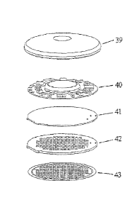
Figure 29 shows a microporator section of a poration device
Figure 30 shows a reservoir patch that is applied to the body tissue after the
poration
is accomplished.
Figure 31 shows a top view of a release liner for use in an embodiment of the
present
inventive subject matter.
Figure 32 depicts a top view of another release liner for protecting the
bottom of a
suitable porator array.
Figure 33 depicts a top view of a porator array.
Figure 34 shows a bottom view of one embodiment of a porator array.
Figure 35 shows a porator array after the poration elements have been removed
from
the locator ring.
Figure 36 depicts a drug reservoir patch applied to the porated area of the
tissue
membrane.
Figure 37 shows reservoir patch following removal of the remaining portions of
the
porator array.
Figure 38 shows a single piece disposable patch design.
DETAILED DESCRIPTION
Definitions
As used herein, "stratum comeurn" refers to the outermost layer of the skin,
consisting of from about 15 to about 20 layers of cells in various stages of
drying out. The
11
CA 02929300 2016-05-09
stratum corneum provides a barrier to the loss of water from inside the body
to the external
environment and from attack from the external environment to the interior of
the body.
As used herein, "tissue' refers to an aggregate of cells of a particular kind,
together
with their intercellular substance, that forms a structural material. At least
one surface of the
tissue must be accessible to the device. The preferred tissue is the skin.
Other tissues suitable
for use with this invention include mucosal tissue and soft organs.
As used herein, the term, "interstitial fluid" is the clear fluid that
occupies the space
between the cells in the body. As used herein, the term "biological fluid" is
defined as a fluid
originating from a biological organism, including blood serum or whole blood
as well as
interstitial fluid.
As used herein, "poration," "microporation," or any such similar term means
the
formation of a small hole or crevice in (defined herein as a "micropore") or
through the
biological membrane, such as skin or mucous membrane, or the outer layer of an
organism to
lessen the barrier properties of this biological membrane the passage of
biological fluids,
such as analytes from below the biological membrane for analysis or the
passage of active
peimeants or drugs from without the biological membrane for selected purposes.
Preferably
the hole or "micropore" so formed is approximately 1-1000 microns in diameter
and would
extend into the biological membrane sufficiently to break the barrier
properties of the'stratum
conieurn without adversely affecting the underlying tissues. It is to be
understood that the
term. "micropore' is used in the singular form for simplicity, but that the
device of the present
invention may form multiple artificial openings. Potation could reduce the
barrier properties
of a biological membrane into the body for selected purposes, or for certain
medical or
surgical procedures. For the purposes of this application, "poration" and
"microporation" are
used interchangeably and mean the same thing.
A "microporator" or "porator" is a component for a microporation device
capable of
microporation. Examples of a microporator or porator include, but are not
limited to, a
heated probe element capable of conductively delivering thermal energy via
direct contact to
a biological membrane to cause the ablation of some portion of the membrane
deep enough
to form a micropore the heated probe may be comprised of an electrically
heated resistive
element capable of ablating a biological membrane or an optically heated
topical
dye/absorber layer, electro-mechanical actuator, a microlancet, an array of
microneedles or
12
CA 02929300 2016-05-09
lancets, a sonic energy ablator, a laser ablation system, and a high pressure
fluid jet
puncturer. As used herein, "microporator" and "porator" are used
interchangeably.
As used herein "penetration" means the controlled removal of cells caused by
the
thermal and kinetic energy released when the pyrotechnic element explodes
which causes
cells of the biological membrane and possibly some adjacent cells to be "blown
away" from
the site. As used herein, "fusible" and "fuse" refer to an element that could
remove itself from
and electrical circuit when a sufficient amount of energy or heat has been
applied to it. e.,
when a resistive, electrically activated poration element is designed to be a
fusible element
this means that upon activation, during or after the formation of the
micropore in the
biological membrane, the element breaks, stopping the current flow through it.
As used herein, "penetration enhancement" or "permeation enhancement" means an
increase in the permeability of the biological membrane to a drug, analyte, or
other chemical
molecule, compound, particle or substance (also called "pemieant"), i.e., so
as to increase the
rate at which a drug, analyte, or other chemical molecule, compound or
particle permeates
the biological membrane and facilitates the increase of flux across the
biological membrane
for the purpose of the withdrawal of analytes out through the biological
membrane or the
delivery of drugs across the biological membrane and into the underlying
tissues.
As used herein, "enhancer", "chemical enhancer," "penetration enhancer,"
permeation
enhancer," and the like includes all enhancers that increase the flux of a
pernaeant, analyte, or
other molecule across the biological membrane, and is limited only by
functionality. In other
words, all cell envelope disordering compounds and solvents and any other
chemical
enhancement agents are intended to be included. Additionally, all active force
enhancer
technologies such as the application of sonic energy, mechanical suction,
pressure, or local
deformation of the tissues, iontophoresis or electroporation are included. For
example,
ammonia may be used as an enhancer for the device of the present invention. In
this example,
the ammonia may increase the permeability of selected tissue structures, such
as the capillary
walls, within the tissues proximate to, or extending some distance from, the
formed
micropore. One or more enhancer technologies may be combined sequentially or
simultaneously. For example, the ammonia enhancer may first be applied to
permealize the
capillary wall and then an iontophoretic or sonic energy field may be applied
to actively drive
a permeant into those tissues surrounding and comprising the capillary bed.
The shock wave
13
CA 02929300 2016-05-09
generated by the detonation of the pyrotechnic element of the present
invention is itself a
sonic permeation enhancer.
As used herein, itransdermal" or "percutaneous" means passage of a peimeant
into
and through the biological membrane to achieve effective therapeutic blood
levels or local
tissue levels of a permeant, or the passage of a molecule or fluid present in
the body
("analyte") out through the biological membrane so that the analyte molecule
maybe
collected on the outside of the body.
As used herein, the term "permeant," "drug," "permeant composition," or
"pharmacologically active agent" or any other similar term means any chemical
or biological
material or compound suitable for transdermal administration by the methods
previously
known in the art and/or by the methods taught in the present invention, that
induces a desired
biological or pharmacological effect, which may include but is not limited to
(1) having a
prophylactic effect on the organism and preventing an undesired biological
effect such as an
infection, (2) alleviating a condition caused by a disease, for exarnple,
alleviating pain or
inflammation caused as a result of disease, and/or (3) either alleviating,
reducing, or
completely eliminating the disease from the organism. The effect may be local,
such as
providing for a local anesthetic effect, or it may be systemic. Such
substances include broad
classes of compounds normally delivered into the body, including through body
surfaCes and
membranes, including skin. In general, this includes but is not limited to:
anti-infectives such
as antibiotics and antiviral agents; analgesics and analgesic combinations;
anorexics;
antihehninthics; antiarthritics; antiasthmatic agents; anticonvulsants;
antidepressants;
antidiabetic agents; antidiarrheals; antihistamines; anti- inflammatory
agents; antimigraine
preparations; antinaus e ants ; anti neopl astic s ; antip arkins onism drugs;
antipruriti cc;
antipsychotics; antipyretics; antispasmodics; anticholinergics;
sympathomimetics; xanthine
derivatives; cardiovascular preparations including potassium and calcium
channel blockers,
beta-blockers, alpha-blockers and. antiarrhythmics; antihypertensives;
diuretics and
antidiuretics; vasodilators including general coronary, peripheral and
cerebral; central
nervous system stimulants; vasoconstrictors; cough and cold preparations,
including
decongestants; hoilliones such as estradiol and other steroids, including
corticosteroids;
hypnotics; immunosuppressives; muscle relaxants; parasympatholytics;
psychostimulants;
sedatives; and tranquilizers. By the method of the present invention, both
ionized and
nonionized drugs maybe delivered, as could drugs of either high or low
molecular weight.
14
CA 02929300 2016-05-09
Additionally, microparticles, DNA, RNA, viral antigens or any combination of
the permeants
listed above may be delivered by the present invention. Examples include
polypeptides,
including proteins and peptides (e.g., insulin); releasing factors, including
Luteinizing
Hormone Releasing Hormone (LERI-I); and carbohydrates (e.g., heparin). Ionized
and
nonionized permeants may be delivered, as could permeants of any molecular
weight
including substances with molecular weights ranging from less than 50 Daltons
to greater
than 1,000,000 Dalions.
As used herein, an "effective" amount of a pharmacologically active agent
means a
sufficient amount of a compound to provide the desired local or systemic
effect and
performance at a reasonable benefit/risk ratio attending any medical
treatment. An
"effective" amount of a permeation or chemical enhancer as used herein means
an amount
selected so as to provide the desired increase in biological membrane
permeability, the
desired depth of penetration, rate of administration, and amount of drug
delivered.
As used herein, a "pyrotechnic element" means any chemical, matter or
combination
of chemicals and/or matters that have an explosive characteristic when
suitably detonated.
The pyrotechnic element of the present invention undergoes very rapid
decomposition (as
combustion) with the production of heat and the formation of more stable
materials (as gases)
which exert pressure as they expand at the high temperature produced thereby
creating a
shock wave with a high peak pressure lasting for a short period o f time.
Thus, the energy
produced by the pyrotechnic element includes both high temperature and high
pressure. One
example of a pyrotechnic element suitable for the present invention includes a
stoichiometric
mixture of zirconium powder and potassium perchlorate combined with a
nitrocellulose
binder of 1 - 5 parts per 100 parts of the stoichiometric mixture as a
suspension in an organic
solvent. Another example would be a gelled form of nitroglycerin, which has
the additional
advantage of already being an approved drug for transdenna1 delivery
applications.
As used herein, a "pyrotechnic ink" means any pyrotechnic element that is
applied in
a liquid foul] and which subsequently cures into the solid or gelled shape of
the pyrotechnic
element.
As used herein, the term "biological membrane" or "tissue membrane" means the
structure separating one area of an organism from another, such as a capillary
wall, lining of
the gut or the outer layer of an organism which separates the organism from
it's external
CA 02929300 2016-05-09
environment, such as epithelial tissue, skin, buccal mucosa or other mucous
membrane. The
stratum corneum of the skin may also be included as a biological membrane.
As used herein, "animal" or "organism" refers to humans and other living
organisms
including plants, to which the present invention maybe applied.
As used herein, "analyte" means any chemical or biological material or
compound
suitable for passage through a biological membrane by the technology taught in
this present
invention, or by technology previously Icnown in the art, of which an
individual might want
to know the concentration or activity inside the body. Glucose is a specific
example of an
analyte because it is a sugar suitable for passage through the skin, and
individuals, for
example those having diabetes, might want to know their blood glucose levels.
Other
examples of analytes include, but are not limited to, such compounds as
sodium, potassium,
bilirubin, urea, ammonia, calcium, lead, iron, lithium, salicylates, and the
like.
As used herein, "transdermal flux rate" is the rate of passage of any analyte
out
through the skin of an individual, human or animal, or the rate of passage of
any pet meant,
drug, pharmacologically active agent, tie, or pigment in and through the skin
of an
organism.
. As used herein,
"artificial opening" or "micropore" means any physical breach of the
biological membrane of a suitable size for delivering or extraction fluid
therethrough,
including micropores. "Artificial opening" or "micropore" or any such similar
term thus
refers to a small hole, opening or crevice created to a desired depth in or
through a biological
membrane. The opening could be formed via the conduction of thermal energy as
described
in U.S. Pat. No. 5,885,211, or through a mechanical process, or through a
pyrotechnic
process. The size of the hole or pore is for example approximately 1-1000
microns in
diameter. It is to be understood that the term micropore is used in the
singular form for
simplicity, but that the devices and methods may form multiple openings or
pores.
As used herein, "use" or "single use" is a single application of the device
that could
last for example, for a few seconds to a few days. An application is denoted
by applying the
device tissue interface to the tissue, the poration process, the delivery or
extraction step, and
the removal of the device tissue interface from the tissue. This "use" or
"single use" could
last for seconds, minutes, or days depending on the nature of the peuneants
delivered, the
biological fluids extracted, and the flux rates desired.
16
CA 02929300 2016-05-09
"Iontophoresis" refers' to the application of an external electrio field to
the tissue
surface through the use of two or more electrodes and delivery of an ionized
form of drug or
aiiunionized drug carried with the water flux associated with ion transport
(elect-so-osmosis) .
into the tissue or the similar extraction of a. biological fluid or analyte,
"Blectroporation" refers to the creation through electric current flow of
openings in
cell walls that are orders of magnitude smaller than micropores. The openings
formed with
electroporation are typically only a 'few nanometers in any dimension.
Electroporation is
useful to facilitate cellular uptake of selected permeants by the targeted
tissues beneath the
outer layers of an organism after the permeant has passed through the
micropores into these
deeper layers of tissue,
"S onophoresis" or ."sonification" refers to sonic energy, which may include
frequencies normally described as ultrasonic, generated by vibrating a
piezoelectric crystal or
other electromechanical element by passing an alternating current through the
material. The
use of sonic energy to increase the permeability of the skin to drug molecules
has been
termed sonophoresis or phonophoresis.
"Integrated device" means a device suitable for forming artificial openings in
tissue
and further suitable for one or. more additional applications, for example,
delivering one or
more permearits into the tissue (preferably through the artificial opentrig0,
and optionally
collecting 'a biological fluid from the tissue (preferably through the
artificial openings) and
optionally analyzing the biological fluid to determine a characteristic
thereof.
As used herein, "non-invasive" means not requiring the entry of a needle,
catheter, or
other invasive medical instrument into apart of the body.
As used herein, "minimally invasive" refers to the use. of mechanical,
hydraulic, or
electrical means that invade the stratum comeum to create a small hole or
rnieropore without
causing substantial damage to the underlying tissues.
As used herein, "pharmaceutically acceptable carrier" refers to a carrier in
which a -
substance such as a = pharmaceutically acceptable drug could be provided for
deliver.
Pharmaceutically acceptable carriers are described in the art, for example, in
"Remington:
The Science and Practice of Pharmacy," Mack Publishing Company, Pennsylvania,
1995.
Carriers could include, for example, water and other aqueous solutions,
saccharides,
polysaccharides, buffers, excipients, and biodegradable polymers such as
polyesters,
17
CA 02929300 2016-05-09
polyanhydrides, polyamino acids, liposomes and mixtures thereof.
As used herein, "reservoir" refers to a designated area or chamber within a
device
which is designed. to contain a perrneant for delivery through an artificial
opening in a
biological meMbrane into, an organism or may be designed to receive a
biological fluid
sample extracted from an organism through an artificial opening in a
biological membrane. A
reservoir could also contain excipient compounds which enhance the effect of a
separately
contained bioactive permeant. Additionally, a reservoir could contain or be
treated with
reactive enzymes or reagents designed to allow the measurement or detection of
a selected
analyte in an extracted biological fluid. A reservoir may be comprised of a
open volume
space, a gel, a fiat planar space which has been coated or treated with a
selected compound
for subsequent release or reaction, or a permeable solid structure such as a
porous polymer.
The present invention comprises a device and a method for painlessly creating
microscopic holes, i.e. micropores, from about 1 to 1000 microns across, in
the stratum .
comeum of human skin. The device .uses thermal energy source, or heat probe,
which is held
in contact with the stratum comeum, for creating micropores. The thermal
micropores are
created using short time-scale (1 microsecond to 50 milliseconds), thermal
energy pulses to
ablate the tissue of biological membranes. This process is described in detail
in U.S. Pat. No.
5,885,211.
The present invention facilitates a rapid and painless method of eliminating
the
barrier function of the stratum comeum to facilitate the transcutaneous
transport of
therapeutic substances into the body when applied topically or to access the
analytes within
the body for analysis. The method utilizes a procedure that begins with the
contact
application of a small area heat source to the targeted area of the stratum
comeum or other
selected biological membrane.
The heat source has the following properties. First, the heat source must be
sized such
that contact with the biological membrane is confined to a small area,
typically about 1 to
1000 p.m in diameter. Second, it must have the capability to Modulate the
temperature of the
stratum comeum at the contact point from ambient skin surface temperature
levels (33 C) to
greater than .123 C (preferably to a temperature greater than 400 C) and then
return to
approximately ambient skin temperature with total cycle times within the I
microsecond to
50 milliseconds range to minimize collateral damage to adjacent viable tissues
and sensation
18
CA 02929300 2016-05-09
to the subject individual. This modulation could be created electronically,
mechanically, or
chemically.
With the heat source placed in contact with the skin, it is cycled through a
series of
one or more modulations of temperature from an initial point of ambient skin
temperature to
a peak temperature in excess of 123 C. to approximately ambient skin
temperature. To
minimize or eliminate the subject's sensory perception of the microporation
process, these
pulses are limited induration, and the interpulse spacing is long enough to
allow cooling of
the viable tissue layers in the skin, and most particularly the enervated
dermal tissues, to
achieve a mean temperature of less than about 45 C. These parameters are based
on the
thermal time constants of the viable epidermal and dermal tissues (roughly 30-
80 ras) located
between the heat probe and the enervated tissue in the underlying dermis. The
result of this
application of pulsed thermal energy is that enough energy is conducted into
the stratum
comeum within -the tiny target spot that the local temperature of this volume
of tissue is
elevated sufficiently higher than the vaporization point of the tissue-bound
volatile
components, such as water and lipids in the stratum comeum. As the temperature
increases
above 100 C., these volatile components of the stratum come= (typically
comprising 5% to
15% within the stratum come=) within this localized spot, are induced to
vaporize and
expand very rapidly, causing a vapor-driven removal of those comeOcytes in the
stratum
comeum located in proximity to this vaporization event. U.S. Pat. No.4,
775,361 teaches that
a stratum corneum temperature of 123 C. represents a threshold at which this
type of flash
vaporization occurs. As subsequent pulses of themial energy are applied,
additional layers of
the stratum come= are removed until a inicropore is formed through the stratum
comeum
down to the next layer of the epidermis, the stratum lucidum. By limiting the
duration of the
heat pulse to less than one thermal time constant of the epidermis and
allowing any heat
energy conducted into the epidermis to dissipate for a sufficiently long
enough time, the
elevation in temperature of the viable layers of the epidermis is minimal.
This allows the
entire microporation process to take place without any sensation to the
subject and no
=
damage to the underlining and surrounding tissues.
One embodiment of this invention relates to designs and manufacturing
techniques
suitable for creating a practical, low cost, Thin Film Tissue Interface (TFTI)
device that
creates micropores using thermal energy produced by the passage of electrical
current
19
CA 02929300 2016-05-09
through resistive elements and methods of manufacturing and functional
operation of the
TFTI devices. fFTI devices create one or more micropores on a wide range of
biological
membranes. TFTIs have applications that include thermal microporation of human
skin for
the enhancement of analyte monitoring and delivery of permeants such as a
therapeutic drug
or a tattoo dye.
TFTIs are characterized by their ability to rapidly and efficiently create a
pattern or
array of micropores on the surface of a biological membrane. The pattern may
be any
geometric spacing of micropores with pore densities as high as one pore every
0.2 square mm
and covering a total porated area ranging from a few square millimeters to
greater than
several hundred square centimeters. TFTI devices are designed to be thin,
flexible,
conformable structures that form the interface between a biological membrane
and the
controller portion of the integrated device that supplies each poration
element or electrode or
other active component such as a piezo-transducer in the TFTI with the
required electrical
signal to effect the poration or other function of the TFTI such as, but not
limited to,
iontoPhoresis, sonophoresis, electroporation, or impedance measurement of the
contacted
tissue. TFTIs are flexible and able to conform to the shape of the targeted
biological
membranes. The I FTIs are fabricated to be very thin, light in weight, and
integrated with a
reservoir and are also connected to the controller, current source through an
umbilical cable
to allow a more user-friendly configuration. When one or more controllable
active additional
flux enhancement features are incorporated into the TFTI, such as, but not
limited to,
pressure modulation, mechanical manipulation, iontophoresis, electro-osmosis,
sonophoresis
or elcctroporation, the activation of this additional flux control feature
could be controlled by
the remote controller module either in a preprogrammed fashion, a user
controlled fashion
via inputs to the controller, or in an automatic, closed loop fashion wherein
the rate of
infusion of a permeant is modulated as a function of the measured level of a
selected analyte
within or other measurable property of the organism. The other measurable
property could
include heart rate, blood pressure, temperature, respiration and skin surface
conductivity. For
example, if would be very useful to control the rate of insulin infusion based
on the real-time
measurement of glucose concentrations in the interstitial fluid or serum of an
organism.
Alternatively, it may be desirable with some therapeutic compounds,
particularly those with
narrower therapeutic windows defining what an effective drug level is versus
when the
negative side effects ,become too intolerable, to modulate the infusion rates
based on the
CA 02929300 2016-05-09
measurable levels of this compound within the organism, thereby allowing a
very accurate,
and self adaptive method for achieving and maintaining the drug concentration
within a
desired therapeutic window regardless of patient body mass or metabolism. In
the design and
manufacture of the TFTI, many of the electrically conductive traces comprising
the TFTI
could be used to serve multiple functions. For example, the traces used to
deliver the short
pulses of current to the resistive poration elements to induce the thermal
cycling, could also
be used as electrodes for an iontophoretic or electro-poration process,
carried out after the
micropores have been formed.
This invention relates to a microporation device, comprising at least one
reservoir and
a tissue interface comprising at least one microporator and a substrate,
wherein the
microporator is located on or within ,the substrate. In one embodiment, the
substrate is
selected from the group consisting of a woven material, a film, a supporting
layer and a sheet.
The woven material comprises conductive fibers and non-conductive fibers. In
another
embodiment, the substrate comprises perforations.
The microporator may be selected from the grail) consisting of a probe element
capable of conductively delivering thermal energy via direct contact to a
biological
membrane to cause the ablation of some portion of the membrane deep enough to
form a
micropore, electro-mechanical actuator, a microlancet, an array of micrci-
needles or lancets, a
sonic energy ablator, a laser ablation system, and a high pressure fluid jet
puncturer; and the
probe element could be selected from the group consisting of an electrically
heated resistive
element capable of ablating a biological membrane, an optically heated topical
dye absorber
layer and optically heated topical dye layer.
In some embodiments of the microporation device of this invention, the probe
element could be selected from the group consisting of a preformed wire
conductor, a
deposited conductive material, a machined conductive material, a laser cut
conductive
material, an adhesive foil, an electroplated material, a screen-printed
material and an etched
conductive material. In some embodiments, the probe element could be destroyed
while
ablating the biological membrane.
In an embodiment of this invention, at least one microporator comprises
multiple
rnicroporators. In another embodiment of the microporation device, the
multiple
microporators are probe elements.
21
CA 02929300 2016-05-09
The microporation device of this invention could comprise diodes for isolating
the
electrical circuits used for activating the probe elements. The microporation
device could
comprise two or more of the probe elements are connected in a parallel circuit
configuration
or a series circuit configuration or a combination thereof.
The microporation device could comprise a material near the microporator,
wherein
the material could be capable of producing an exothermic or endothermic
reaction. The
microporation device could comprise a micro actuator. The mieroacuator could
be selected
from the group consisting of electro-static microactuators, thermal bimorph
microactuators,
piezoelectric microactuators, electromagnetic microactuators, magneto-
restrictive
microactuators and shape memory alloy microactuators.
The microporation device could comprise an electronic circuitry and a power
source:
The probe element could comprise a conductive wire and the substrate could
comprise a
nonconductive fabric. The conductive wire could be woven in the non-conductive
fabric.
The microporation device could comprise a plug material on the perforations.
The
plug material could comprise a volatile material. In one embodiment of the
microporation
device, the substrate could be embossed. The microporation device could
comprise an
enhancer material for enhancing transmembrane or transdermal transport of a
fluid across the
biological membrane.
The microporation device could comprise multiple chambers. The multiple
chambers
could comprise different substances. At least one of the multiple chambers
could be disposed
after a single use of the microporation device. The multiple chambers Could
comprise at least
first and second chambers, the first chamber comprising a first substance and
the second
chamber comprising a second substance. The first and second substances could
be first and
second biologically active agents. The first substance could be a dry
formulation
pharmaceutically active agent, and the second substance could be a diluent for
reconstituting
thee dry formulation into a pharmaceutically acceptable liquid or gel
formulation.
The microporation device could be capable of transdermal delivery of a
substance in
the first chamber or withdrawal of an analyte transdennally into the second
chamber. The
microporation device could be capable of simultaneous transdermal delivery of
a substance
in the first chamber and withdrawal of an analyte transdermally into the
second chamber.
The substance could be insulin and the analyte could be glucose. The
substances could be
selected from the group consisting of bioactive peptides or proteins,
therapeutic drugs,
22
CA 02929300 2016-05-09
vaccines, pain medications, permeation enhancers and pH stabilizers. The
different
substances could be delivered by the microporation device in modulated
amounts. At least
one of the different substances could passively diffuse into the biological
membrane. The
substances, which could be the same or different, could be delivered
simultaneously,
sequentially, alternately, or any combination thereof. The different
substances could be
delivered by the microporation device into the organism in adjacent locations
in the
biological membrane such that the different substances could combine and mix
once they are
within the tissue matrix of the organism.
The microporation device could comprise an analyzer for detecting or
quantitating the
analyte. The microporation device could comprise a control module for
controlling the
delivery of the substance baSed on a quantitative value of the analyte
detected by the
analyzer.
The microporation device could comprise a divider or valve disposed between
the
first and second chambers that prevents mixture of the first and second
substances until the
divider could be removed or the valve could be opened. The divider_ could be a
membrane.
The first substance could be a pharmaceutically active agent, and the second
substance could
a pharmaceutically acceptable carrier.
The microporation device could comprise a flux enhancement microporation
device,
wherein the flux enhancement microporation device enhances a flux rate of a
substance into
the biological membrane. The flux enhancement naicroporation device enhances a
flux rate of
a substan.ce into the biological membrane by a technique selected from the
group consisting
of iontophoresis, electroporation, electro-osmosis, sonophoresis, and
pressurization.
The microporation device could comprise a disposable component or the
microporation device could be for a single use after which the microporation
device could be
discarded. The disposable component could be treated with reagents which react
with a
biological fluid withdrawn from the biological membrane to produce a signal or
measurable
change in properties which could be predictably related to the quantity of an
analyte within
the biological fluid. The disposable component could be treated with one or
any combination
thereof of surfactants, hydrophilic or hydrophobic compounds. The disposable
component
could be treated with antimicrobial or anticoagulent or protease inhibitor
compounds. The
disposable component could comprise stimuli-responsive polar gel sections
comprising a
23
CA 02929300 2016-05-09
material that could be released by a thermal, chemical or electrical stimulus.
The disposable
component could comprise a material that releases a compound when heated.
The-microporation device could comprise a mixer located on or within the
substrate,
the mixer being capable of mixing a substance prior to transdermal delivery of
a substance
into the biological membrane. The microporation device could comprise a closed-
loop
delivery and monitoring system, wherein the closed-loop delivery and
monitoring system is
capable of modulating tansdernaal delivery of a substance through a biological
membrane
based on a value of a property of an animal.
Another embodiment of this invention is a method of manufacturing a
microporation
device, comprising obtaining a substrate and forming a conductive network on
the substrate,
wherein the conductive network provides electrical connections to a
microporator. The
method could comprise bonding an adhesive layer over the conductive network.
The method
could comprise forming a non-conductive plug on the perforations. The method
could
comprise bonding the conductive network to a reservoir.
Another embodiment is a method for forming openings in a biological membrane,
comprising placing a microporation device in close proximity of the biological
membrane
and triggering the microporation device to form at least one opening in the
biological
membrane, the microporation device comprising at least one reservoir and a
tissue interface
comprising at least one microporator and a substrate, wherein the microporator
is located on
or within the substrate. The triggering could transfer heat to the biological
membrane. The
opening could have a diameter of 1-1,000 microns. The opening or artificial
pore could be
formed by a method selected from the group consisting of local heating,
mechanical
puncture, sonic energy, hydraulic puncture, and electroporation. The method
could comprise
anyone or. more of the following: (a) applying an enhancer to the opening; (b)
applying a
perrneant to the opening; (c) collecting a fluid from the opening; (d)
monitoring an analyte in
the fluid; (e) delivering a substance into the biological membrane; (f) mixing
a substance
prior to delivery of a substance into the biological membrane; and (g)
delivering a substance
into the biological membrane and collecting a fluid from the biological
membrane.
An object of this invention is a method for administering a compound through a
biological membrane to an underlying tissue matrix or obtaining a biological
fluid sample
from a tissue matrix under a biological membrane, comprising a) contacting a
flux
enhancement cell with a biological membrane, b) forming a seal between the
outer wall and
24
CA 02929300 2016-05-09
the membrane, wherein the reservoir outlet is in communication with an
artificial pore in the
membrane; c) applying positive pressure to the inner cavity of the reservoir;
d) biasing the
reservoir towards the membrane, thereby producing the compressed state of the
membrane;
e) biasing the reservoir away from the membrane, thereby producing the
relieved state; and i)
the biological membrane having an inner surface in intimate contact with the
tissue matrix
and an outer surface, thereby producing the relieved state, wherein the
biological membrane
has a resting state, a pressurized state in which the outer surface of the
membrane is
depressed to a substantially concave form relative to the resting state and
the underlying
tissue matrix is compressed, and a relieved state, wherein the outer surface
of the membrane
is biased into a substantially convex shape and the underlying tissue matrix
is subjected to
reduced pressure, and ii) wherein the flux enhancement cell comprises an outer
wall, the
outer wall defining a cell cavity, and a reservoir movably contained therein,
the reservoir
comprising an inner cavity and an outlet; the inner cavity containing a
perrneant. One
embodiment of the method for administering a compound through a biological
membrane to
an underlying tissue matrix or obtaining a biological fluid sample from a
tissue matrix
underlying a biological membrane, comprises g) biasing the reservoir towards
the membrane,
thereby producing the compressed state of the membrane; h) biasing the
reservoir away from
the membrane. '
Another object of this invention is a flux enhancement device comprising an
outer
wall, the outer wall defining a cell cavity; and a reservoir comprising an
inner cavity and an
outlet, wherein the reservoir is movably contained within the cell cavity. The
reservoir could
be movably linked to the outer wall with a compliant membrane. The flux
enhancement
device could comprise a microporator. The rnicroporation device or flux
enhancement device
could comprise a closed-loop delivery and monitoring system, wherein the
closed-loop
delivery and monitoring system is capable of transdermal delivery of a
substance through a
biological membrane and withdrawal of an analyte transdemially through the
biological
membrane. The flux enhancement device could comprise could comprise a closed-
loop
delivery and monitoring system, wherein the closed-loop delivery and
monitoring system is
capable of modulating transdermal delivery of a substance through a biological
membrane
based on a value of a property of an animal.
Figure 1 shows the general configuration of a TFTI (1) with plurality of
poration
elements (2). The microporators of a TFTI device are heated probe elements
capable of
CA 02929300 2016-05-09
conductively delivering thermal energy via direct contact to a biological
membrane to cause
the ablation of some portion of the membrane deep enough to form micropores.
In Figure I,
the poration elements (2) are resistive elements.
The resistive elements could take almost any shape, but are typically high
aspect
ratio, straight cylinders or bars with diameters or square cross-sections that
range from 1
micron to 150 microns and lengths from 100 microns to 3000 microns
respectively. When an
electrical current pulse is applied to each element, the pulsed element could
be .controllably
and rapidly brought to a specified high temperature, ranging from 120 C to
greater than
3000 C (the upper limit is really set by the melting point of the material
comprising the
resistive element, for most tungsten alloys this is in excess of 3000 C),
whereupon this
thermal energy could then be delivered to the contacting tissue to effect the
thermal poration
of the tissue.
The patterned array of resistive elements is connected to a conductive network
that
passes electrical energy to each of the resistive elements. The array of
resistive elements are
connected to the current pulse source either individually, as a series
electrical system, parallel
electrical system or some combination thereof. The instantaneous current
required for the
operation of the TFTIs depends mainly on the number of resistive elements in
,a device,
parallel or series network configuration and size of the resistive elements.
Instantaneous
current flowing through the resistive element network could range from 1
rnilliamps to 40
amps, however, as the pulse duration is typically only a few milliseconds
long, and the
impedance of each element is quite low (in practice the typical resistance of
a single tungsten
alloy poration element has been measured to be less than 0.1 ohms) the average
power
requirements are quite modest. For example, in the extreme case of a 40 amp
current pulse of
1 millisecond duration applied to the 0.1 ohm element, the total power
delivered is:
P = Watt x seconds
P = 121V1000¨(40x40)x(0.1)x(0.001), or P=160 milliwatts per poration element.
More common values of power consumption based on the practical parameters (1
amp peak current, 1 millisecond pulse duration, 0.05 ohm poration element
impedance) used
in the preferred embodiments of the invention are:
P = 12(0.05)(0.001)= 50 microwatts per poration element.
26
CA 02929300 2016-05-09
With a power requirement of only 50 microwatts per poration element, for a
typical
delivery patch which utilizes 100 individual poration elements the total power
requirement to
perform the thermal poration process is still only 5 milliwatts, power levels
easily delivered
from very small, low cost batteries.
The resistive elements are arranged in a two-dimensional. pattern that is
transferred
directly to the surface of a biological membrane. The type of pattern produced
is dependent
on the application. For example a set of micropores designed to deliver a
local anesthetic to
an IV insertion site may have a narrow pore pattern beginning at the needle
insertion site and
extending along the expected path of the needle. The desired pore depth is
also dependent on
the application. Using the example above, the pore depths formed maybe
designed to be
relatively shallow at the needle insertion site and deeper along the needles
path within the
body:
Figure 2 shows one embodiment of a parallel conductive network (3) with anode
side
(4), cathode side (5), poration elements (2) and supporting substrate (6).
Each TFTI could be
connected to an external electronic control module to Supply electrical energy
with the
required current and pulse duration parameters.
The mechanism that forms a .micropore is a result of the intimate contact of
the
biological membrane with the resistively heated element. In its most simple
form, the TFTI
would have resistive elements that stayed in contact with the skin before,
during and after the
poration process without moving. This would be known as a non-actuated
poration process
where resistive elements remain passively in the same location within the
apparatus. The
devices using micro-actuation combined with the resistive elements would be
known as
actuated microporation or actuation of poration elements.
The mechanism that forms a micropore is a result of the intimate contact of
the
biological membrane with the resistively heated element. In its most simple
form, the TFTI
of Figure 2 would have resistive elements that stayed in contact with the skin
before during
and after the poration process without moving. This is known as a non-actuated
poration
process where resistive elements remain passively in the same location within
the apparatus.
Another embodiment of this invention uses micro-actuation combined with the
resistive elements and is known as actuated thermal microporation or actuation
of poration
elements. Micro-actuators produce a mechanical actuation of the poration
elements and
achieve greater control over pore depth, act to remove the resistive element
from the
27
CA 02929300 2016-05-09
micropore once it has been formed or perform a function such as opening a
barrier that
isolates a reservoir. An illustrative embodiment of an actuated microporator
is shown in
Figure 3, which shows a wire resistive element in the unheated position (7)
and the heated
position (8).
The actuated microporator of Figure 3 is a straight tungsten wire element.
Figure 3
shows that the straight tungsten wire element undergoes a significant increase
in length from
position (7) to position (8) during the heating pulse as a result of the wires
coefficient of
thermal expansion as it undergoes the dramatic change in temperature of a
typical thermal
poration cycle. The anode side (4) and the cathode side (5) of the wire
element are immobile
and the wire reacts to the heating pulse by bending outward to accommodate its
thermally
induced increased length, away from the original centerline of the element.
The direction of
the wire motion could be designed to be directed away from the substrate (6)
by forming a
small initial bend in the poration element when in the unheated position. With
this
.embodiment of an actuated TFTI device, micropores could be created without
requiring an
initial intimate contact between the biological membrane and the poration
element. That is,
when the poration ,element is heated and subsequently is actuated to move
towards the
biological tissue surface, the necessary contact between the poration element
and the
biological surface could be ensured by designing the geometries of the system
and the
amount of actuation travel to guarantee the required physical contact. The
choice of wire
element length, initial bend and wire temperature could be used to control the
resulting pore
depth in the biological membrane as well. Also, by knowing the actuation
response as it
relates to temperature, and by also knowing the change in impedance of the
resistive poration
element as it relates to temperature, one could monitor dynamically, both the
temperature of
the poration element and the resulting amount .of actuation. Similarly, once
contact is
established with the targeted biological membrane, a detectable shift in the
relationship
between the amount of energy delivered to the poration element and the change
in heat would
occur, adding yet another level of dynamically measurable parameters to the
poration process
which could be used to help ensure the formation of controllably, repeatable
pores at each
poration element. By using these measurable parameters as feedback inputs to
the controller,
current source, the variance in individual poration elements which may result
from the
tolerances of the manufacturing process, could also be .accommodated; allowing
for
28
CA 02929300 2016-05-09
additional cost savings in the manufacturing processes of the TFTI by being
able to accept
looser tolerances;
Another embodiment of an actuated microporator of this invention is shown in
Figure
4, wherein the actuated element is formed from a thin sheet of element
material (9) such as
tungsten or copper. Some of the element material is removed using a process
such as laser
micromachining to produce the resistive element shown in Figure 4. During the
laser
micromachining process, it is possible to dynamically monitor the impedance of
each
poration element as it is formed. By using this sort of dynamically monitored
fabrication
process, a parallel or series array of poration elements could be formed where
it could be
ensured that the current pulse delivered is distributed in a balanced, uniform
manner to each
individual element. The shape of this resistive element was chosen to produce
motion in the
direction perpendicular to the plane of the sheet material during heating. The
physical
expansion of the curved sections (10) of the structure force the tip (11) of
the element to lift
away from the plane of the sheet material. Since the entire element reaches a
high
temperature, the tip (11) ablates tissue as it is forced into the biological
membrane. The
resulting pore depth in this case is controlled by the arc length of the
curved sections (10),
length of the tip region (11) and element temperature.
To additionally ensure the equal distribution of a current pulse to each
poration
element in an array, the specific thermal coefficient of resistance for the
resistive poration
element could be selected or designed such that as the individual element
heats up, its
resistance increases, thereby causing less current to flow in that specific
poration element
within a parallel network and at the same time forcing more current to go to
the other
poration elements in this same network. By using this natural phenomenon a
self-balancing
parallel network of resistive elements could more easily be designed and
manufaCtured. This
is similar to how a standard parallel wiring of a home lighting system
operates when several
incandescent lamps are connected on the same circuit,
In another embodiment of this invention, shape memory alloy (SMA) materials
are
used for the body of the resistive element. The use of SMA materials has the
potential to
maximize the efficiency and effectiveness of actuated poration.
A wide variety of micro-actuators could be used for the purpose of actuated
poration.
Manufacturing methods that employ more advanced processes such as
photolithography are
capable of producing more complex micro-actuators. Some micro-
electromechanical
29
CA 02929300 2016-05-09
systems that could be incorporated into TFTI devices include but are not
limited to electro-
static microactuators, thermal birnorph microactuators, piezoelectric
microactuators,
electromagnetic microactuators and SMA microactuators.
A preferred embodiment of the present inventive subject matter is a
transdermal drug
delivery device for forming a micropore in a tissue membrane of an animal. The
transdermal
delivery devices comprising a tissue interface layer having a substrate and at
least one
porator located on or within said substrate, at least one reservoir in
communication with the
tissue interface layer, and a controller for controlling the formation of the
micropore by the
porator. The porator is constructed of a heat resistive element which deforms
when heated,
thereby allowing the heat resistive element to contact the tissue membrane and
form the
micropore by ablating the tissue membrane. A permeant or analyte is stored
within the
reservoir. The substrate is selected from the group consisting of a woven
material, a film, a
supporting layer and a sheet. In a preferred embodiment, the controller
applies a stimulus to
the porator for forming the pore by deforming the heat resistive element.
Further, the porator
is selected from the group consisting of a wire conductor, a machined
conductive material, a
laser cut conductive material; an adhesive foil, an electroplated material, a
shape memory
alloy material and an etched conductive material. The device may further
comprise an
adhesive layer to bind the device to the tissue membrane. .
The present inventive subject matter is also drawn to a method of using such a
transdermal drug delivery device. In particular, the
present inventive subject matter
contemplates a method of forming at least one micropore in a tissue membrane
of an animal.
The method comprises the steps of: a) providing a poration device; b)
contacting said
poration device with the tissue.membrane; c) providing a stimulus to at least
one porator by
way of a controller, thereby heating the at least one porator and increasing
the length of and
deforming same, causing the at least one porator to come into contact with the
tissue
membrane; d) forming at least one micropore; and e) cooling the porator,
thereby decreasing
the length of same and returning same to its original shape, resulting in the
porator no longer
contacting the tissue membrane. The poration device includes a tissue
interface layer, at least
one reservoir in corinnunication with the tissue interface layer; and a
controller for
controlling the formation of said micropore by said at least one porator. The
tissue interface
layer comprises a substrate and at least one porator. The porator is located
on or within the
substrate and is constructed of a heat resistive element which deforms when
heated. The
=
CA 02929300 2016-05-09
substrate may be selected from the group consisting of a woven material, a
film, a supporting
layer and a sheet. The porator may be selected from the group consisting of a
wire
conductor, a machined conductive material, a laser cut conductive material, an
adhesive foil,
an electroplated material, a shape memory alloy material and an etched
conductive material.
The method may also include the step of applying a permeant composition stored
in the
reservoir to the micropore, or extracting an analyte by way of the micropore
and storing the
analyte in the reservoir.
Fusible Tt.T1 designs are an alternative to actuated and non-actuated poration
schemes. In the case of a fusible design, enough electrical energy is passed
through the
resistive element to destroy the element, taking it out of the electrical
circuit. This also
provides a mechanism of removing the element from the pore site. This
embodiment of the
invention also has the potential to greatly simplify the supporting
electronics requirements. In
the case of resistive elements that do not fuse or break their connection, the
driving
electronics are required to generate a signal of controlled duration and
amplitude for
sensation management. In the case of fusible elements, the thermal pulse
duration could be
controlled mainly by the physical failure properties of the element and the
electronics are
only required to deliver an impulsive signal with uncontrolled duration, as in
the case of a
capacitor discharging. Whereas simply delivering enough energy to the poration
element to
cause the conductive trace to melt or vaporize is one method of blowing the
fuse', n more
preferable method may be to fabricate the substrate holding the element out of
a material
which has been specified to undergo a thermal shrinking or tearing process
when exposed to
the elevation of temperature due to the activation of the poration element.
With suitable
attachment of the poration element trace to this tear-able substrate, when the
substrate tears,
it would also rip the element apart and thereby break the current path while
simultaneously
opening a path into a reservoir adjacent to the poration element. If this now
connected
reservoir contained a permeant for delivery, this permeant would now be
disposed directly
onto the just formed micropore in the biological membrane. By appropriately
selecting the
material for this tear-able substrate, this process could be made to occur at
much lower, and
more biocompatible temperatures, than what might be required if one were to
simply 'blow
the fuse'. Some materials that have this type of desired thermal properties
are the heat-
shrinkable polymers and vinyls commonly used in electrical insulation. To help
ensure that
the tear or 'rip occurs when and where desired, and at the designated
temperature, this
31
CA 02929300 2016-05-09
substrate could be formed with a small etch line, embossed stress point, or
other such feature
to provide the 'flaw' from which the thermally induce tear would originate.
Another
significant advantage of this type of thermally induced tearing is that the
opening of the pore
into a drug or assay containing reservoir could be produced with only a
minimal amount of
temperature for a very short period of time, minimizing the amount of thermal
energy and
peak temperature being presented to the reservoir. This feature is of
particular importance
when The reservoir contains thermally fragile peptides, proteins, assay
enzymes or other
drugs sensitive to thermal stress.
An embodiment of the present inventive subject matter is directed to a
transderraal
drug delivery device for forming a micropore in a tissue membrane of an
animal, comprising
a tissue interface, at least one reservoir in communication with the tissue
interface layer, and
a controller for controlling formation of the n3icropore by the porator. The
tissue interface
layer further comprises a substrate and at least one porator, wherein the
porator is located on
or within the substrate and the porator is constructed of a material in which
the porator is
destroyed upon forming the micropore. A permeant or an analyte may be stored
within the
reservoir. In a preferred embodiment, the controller applies a stimulus to the
porator, and the
stimulus initiates formation of the pore by the porator and then destroying
the porator
following formation of the micropore. The stimulus may be a thermal pulse or
an electrical
pulse.
A further embodiment of the present inventive subject matter is drawn to a
method of
= forming at least one micropore in a tissue membrane of an animal. The
method comprises
the steps of: a) providing a poration device; b) contacting the poration
device with the tissue
membrane; a) providing a thermal or electrical pulse to the porator in the
potation device by
way of a controller, thereby forming the micropore in the tissue membrane;
and, d)
destroying the porator after forming the one micropore by sustaining the
thermal or electrical
pulse for a duration sufficient to destroy the porator. The potation device
includes a tissue
interface layer comprising, at least one reservoir in communication with the
tissue interface
layer; and a controller for controlling the formation of the micropore by the
porator. The
tissue interface layer further comprises a substrate and at least one porator,
wherein the
porator is located on or within the substrate and the porator is constructed
of a material in
which the porator is destroyed upon forming said micropore.
32
CA 02929300 2016-05-09
In another preferred embodiment of the device and methods, the substrate is
constructed of a material which undergoes thermal shrinking when exposed to an
elevated
temperature due to activation of the porator, whereby the thermal shrinking
results in a tear in
the substrate and destruction of the porator. Suitable heat-shrinking
materials have been
previously discussed. In addition, the substrate may be formed with a flaw
from which a tear
would form.
The TFTI devices of this invention could also be enhanced by the addition of a
range
of substances at or near the poration element. This approach also has
particular utility with
elements that are fusible as previously described. The object of these
substances is to produce
a chemical reaction at the pore sites and during the poration process.
This chemical reaction could be tailored to perform a variety of functions.
One
example is coating an element with a pyrotechnic material or other material
that results in an
exothermic reaction. The energy used to ablate tissue would then come mainly
from the
exothermic reaction. This allows a simple way to reduce the electrical energy
required to
trigger poration and thus reduce the overall size of the integrated device. A
second example
is a combined exothermic and endothermic reaction. An initial exothermic
reaction would
produce a micropore and be followed closely by an endothermic reaction to cool
the pore site
and improve sensation experienced by patients.
A chemical reaction at the pore site could also be useful for the byproducts
of the
reaction. With appropriate choice of reactants, byproducts could perform all
or some of the
functions of flux enhancers, anti-clogging agents, permeants, therapeutic
agents, reactants to
drive subsequent reactions or other beneficial purposes.
The TFTIs comprising a resistive element could be manufactured by different
methods. The first method uses a previously formed wire conductor to create
the resistive
element. By the second method, the resistive elements are created by a
deposition of
conductive material. By the third method, the resistive elements are formed by
etching or
machining of the element material. In addition, some manufacturing methods
employ both
deposition and etching. Several examples of TFTI manufacturing processes to
demonstrate
the manufacture of TYIl devices and illustrate the variety of manufacturing
methods
available as shown below, The invention is illustrated in the following non-
limiting
examples.
33
CA 02929300 2016-05-09
Example 1: A Woven Material TFTI Device
Some embodiments of the TFTI devices involve the use of previously
manufactured
wire conductors such as tungsten, tantalum, or tungsten alloy wire as the
resistive element.
There are a variety of methods for incorporating the wire conductors into a
IFFI design.
These methods include, but are not limited to weaving, sewing, bonding,
brazing, spot
welding, connecting with conductive adhesives or resins and laminating to a
thin film or
laminated structure.
The basis of a woven material TFTI device is a hybrid woven fabric such as
what is
shown in Figure 5. Figure 5 is an enlargement of a section of the hybrid woven
fabric and
should be considered as extending outward in two dimensions as a repeating
structure. The
hybrid woven fabric contains a combination of structural fibers (10) and (11)
which are not
electrically conductive (such as polyester, fiberglass, nylon, mylar,
polycarbonate, or the
like) and electrically conductive fibers or strands (12) (such as tungsten or
tantalum or copper
wires, conductive polymers, glass or carbon fibers, or the' like). In this
example, polyester
fibers of 50-micron (10) and 80 micron (11) diameters are woven With 50-micron
diameter
tungsten wire (12).
The electrically conductive fibers or strands are woven into the fabric and
run in only
one of the weave directions, spaced apart by a specific number of structural
fibers depending
on the desired poration element array density. Here the number of polyester
fibers between
two tungsten wires is 28 that would result in an element spacing of about 1.4
millimeters.
The woven material is then processed to apply conductive traces on one side as
shown
in Figure 6, creating the desired conductive network (13) with the interwoven
conductive
fibers forming the resistive elements (14). These traces may be created in a
variety of ways
including: pressure transfer of conductive/self adhesive foils onto this
surface; electroplating
into the desired pattern using either a shadow mask or resist mask to define
the traces; or
simply screen-printing with electrically conductive ink or resins and curing.
Most conductive
inks are designed to allow a certain amount of flexibility after curing which
results in a more
compliant TFTI device. For this example, the conductive network in Figure 6 is
arranged as
a parallel electrical circuit although series or combined series and parallel
configurations
could be accommodated by this design. A silver impregnated epoxy is used to
form the
conductive network that is applied using standard screen-printing techniques.
34
CA 02929300 2016-05-09
An added advantage of the woven material TFTI devices is that proper choice of
conductor thread count would result in resistive elements on both sides of the
TFTI. This
results in the optional use of the TFTI to breach or open a drug reservoir
simultaneously with
the creation of micropores. Areas of the fabric that are not covered by the
conductive
network would then be able to pass a deliverable substance from a drug
reservoir, through the
TFTI and into the micropores.
Once the application of the conductive network to the woven fabric has been
completed, further integration of the could take place
that may include bonding to a
drug reservoir or addition of an adhesive layer to maintain contact between
the TFTI and the
biological membrane to be porated. This design is also conducive to the
integration of other
functional features that include iontophoretic electrodes, flux enhancer
releasing elements,
buffer releasing elements, analyte assay electrodes. The analyte assay process
could also be
accomplished via optical means by looking for a colorimetric shift in response
to the selected
analyte's concentration.
The present inventive subject matter is directed to a transdermal drug
delivery device
for foaming a micropore in a tissue membrane of an animal. The transdermal
drug delivery
device comprises a tissue interface layer. The tissue interface layer further
comprises a
substrate comprising a woven fabric, with the woven fabric comprising
structural fibers and
electrically conductive fibers interwoven together as is discussed above. The
tissue interface
layer also comprises at least one porator, wherein the porator is located on
or within the
substrate and is formed by the electrically conductive fibers acting as a heat
resistive
element. The transderrnal drug delivery device also includes at least one
reservoir in
communication with the tissue interface layer and a controller for controlling
the formation
of the micropore by the porator. The transdermal drug delivery device of the
present
embodiment may also have the electrically conductive fibers connected in
parallel or series
by conductive traces, thereby forming a conductive network. The conductive
traces are
selected from the group consisting of foils, inks, resins, electroplating
products and mixtures
thereof.
The present inventive subject matter is also directed to a method of
manufacturing a
transdermal drug delivery device in accordance with the details set forth
above. The method
comprises the steps of: weaving electrically conductive fibers into a fabric
of non-electrically
conductive fibers to form an electrically conductive fabric; applying
conductive traces to one
CA 02929300 2016-05-09
end of the electrically conductive fabric to form a conductive network; and
connecting the
conductive network with a controller which controls the application of
electricity to the
conductive network.
In another embodiment, the present inventive subject matter includes a method
of
forming at least one micropore in a tissue membrane of an animal. The method
includes the
steps of: providing a poration device, contacting the poration device with the
tissue
membrane and actuating the poration device to form the raicropore in the
tissue membrane.
The poration device includes a tissue interface layer, at least one reservoir
in communication
-with said tissue interface layer and a controller for controlling the
formation of said '
micropore by said at least one porator. The tissue interface layer further
includes a substrate
comprising a woven fabric, said woven fabric comprising structural fibers and
electrically
conductive fibers interwoven together and at least one porator located on or
within the
substrate. The porator is formed by the electrically conductive fibers acting
as a heat
resistive element.
Example 2: A Wire Overlay TFTI Device
This TFTI design utilizes a unique screen-printing process that involves
overlaying
wires on a substrate and then printing conductive traces over the wires to
both form electrical
connections with the conductive network and bond the wires to the substrate.
This example
design also uses SMA wire as the resistive element material to produce an
optimized
actuation of the poration element. The poration elements are designed to alter
their shape
during the poration process and breach a drug reservoir directly over the pore
site.
As shown in Figure 7, multiple lengths of SMA wire (15) such as nitinol are
mounted
in a frame (16) with a spacing given by the desired element density in the
final array. A
. spacing of 1.00 nun between lengths of SMA wire is used. The frame and
mounted wires are
then placed over a thin film substrate (17) and standard screen-printing
techniques are used to
deposit conductive ink (18) onto the substrate and SMA wire combination to
produce an
electronic network. The SMA material chosen for this application should have a
high
) melting point such as nitinol. The substrate material must be non-
conductive and have a low
melting point such as polyester. A good candidate conductive ink should have a
high
conductivity and be flexible after it is fully cured such as a silver/polymer
conductive ink.
36
CA 02929300 2016-05-09
The next step in the manufacturing process is to emboss the array at each of
the
poration element locations. Figure 8a shows an enlarged side view of a single
poration
element after the screen-printing process and before embossing occurs. A
dielectric or
adhesive layer (19) prevents the conductive ink network from making contact
with the skin
or other biological membrane.
Figure 8b shows an element after it has been embossed. It is important that
the
embossing process does not cause the SMA material to anneal or undergo a
change in crystal
structure. This would allow the SMA material to return to its original shape
(straight) when
heated resistively by the conductive network as shown in Figure 8c. As an
element becomes
heated, it initially creates a skin pore due to intimate contact with the
surface of the skin. As
further heating of the element occurs, the SMA material begins to return to
its original shape
and retract from the newly created pore while simultaneously forming an
opening in the
embossed feature (20) of the supporting substrate. This could then open a
pathway between a
reservoir on the opposite side of the substrate and the microscopic pore as
described above.
Some embodiments of the TFTI devices involve resistive elements that are
deposited by
processes such as electro-discharge machining (EDM), sputtering, screen-
printing,
electroplating and chemical vapor deposition (CVD) that are common to the
flexible circuit
and electronic industries. The following section illustrates a TFTI device
that could be
manufactured using any of the above deposition processes.
Example 3: A Sputter Deposited TFTI Device
The first step involved in manufacturing is the deposition of a material such
as
tantalum by sputtering to form the resistive elements and conductive network
on an
appropriate substrate such as 50-micron polyamide. Figure 9 shows the pattern
of deposited
tantalum traces (21) on the polyamide substrate (22). A parallel electrical
configuration is
used for purposes of illustration, however the conductive network could be
designed to
address each poration element single or in a parallel circuit, series circuit
or any combination
of parallel and series circuits.
Depending on the properties of the material used for the conductive network
and
resistive elements, it may be desirable to deposit additional material onto
the pattern
everywhere except for the resistive elements themselves. The additional
material could be
37
CA 02929300 2016-05-09
any other type of compatible conductive material and serves the purpose of
reducing the
resistance of the conductive network and thus reducing the overall power
required to operate
the array of resistive elements, as well as confining more precisely in a
spatial sense those
areas of the TFTI which would undergo the cycling to the ablation temperature
threshold.
Figure 10 shows an enlarged side view of a single resistive element (23) at
different points in
the manufacturing process with adjacent conductive network connections (24).
Figure 10a
shows the element after the initial deposition and an optionally additional
layer over the
conductive network (25).
The next step in the manufacturing process is the placement, screening or
bonding of
an adhesive layer (26) over the conductive network without covering the
resistive elements as
shown in Figure 10b. The purpose of the adhesive layer is to bond the
biological membrane
such as skin to the TFII and ensure that there is intimate 'contact with the
resistive elements.
The final step in the manufacture of the TFTI is optionally embossing in the
area of the
resistive elements as shown in Figure 10c. The purpose of embossing is to move
the resistive
element near or even proud of the adhesive, biological mernbrane
contacting`side of the IITI
and ensure intimate contact between the resistive element and the biological
membrane to be
microporated. The embossing process could also serve to thin the substrate
material in the
area of the resistive element. This may help the resistive element to 'breach
the substrate
material during poration, thus providing a mechanism by which a substance is
introduced to
the pore site for drug delivery applications. Another possible advantage of
embossing for any
design is that the resistive element material would undergo strain hardening
and thus
provide a method for altering the electrical and mechanical properties of the
element.
Additional flexibility in tailoring of properties is achieved by varying the
temp of the
material during the embossing process.
It should also be noted that many deposition techniques are conducive to the
manufacture of complex resistive element geometry's for the purposes of
actuated poration.
Some techniques, commonly used in the mass-production of electronic components
are
capable of depositing structures with feature sizes of 0.5 microns or less.
Some embodiments of the TFTI devices involve resistive elements that are
etched or
machined from a layer or sheet of material by processes such as laser
micromachining and a
range of photolithography techniques COMITIOn to experimental MEMS devices and
the
38
CA 02929300 2016-05-09
electronics industry. The following section illustrates a TFTI device that
could be
manufactured using a micromachining process.
Example 4: A Micromachined TFTI Device
Figure 11 shows an enlarged side view of a single resistive element at
different points
in the manufacturing process. The first step in the manufacturing process is
to laminate thin
films of the resistive element material (27) such as tungsten in a 30 micron
sheet to a
supportive or resistance tailoring layer such as copper (28) in a 50 micron
sheet. These layers
are then micromachined using a. laser from the tungsten side as shown in
Figure 11a. Laser
power, repetition rate and cutting speed are adjusted so that the resistive
elements (29) and
conductive network (30) are produced without cutting through the supportive or
resistive
tailoring layer. Also, during this process of laser mieromachining, the laser
energy could be
used to effectively form the electrical bonds between the tungsten poration
elements and the
resistance-tailoring layer.
The next step shown in Figure 11 b is to bond the tungsten side of the
structure in
Figure lla to a nonconductive layer such as polyester (31). This laminated
structure is then
laser micromachined from the copper side (28). At this point the copper is no
longer needed
as a structural support. The result of this process is to leave copper
material on the conductive
network only and remove it front other locations including over the resistive
elements. Care
is taken in the laser parameter settings to avoid cutting through the
nonconductive layer (31).
The next step in the process is to bond an adhesive layer (32) over the
conductive network
with the resulting structure shown in Figure 11c. The final step in the
manufacturing process
is to emboss the nonconductive layer at the locations of the resistive
elements as shown in
Figure lid.
Example 5: A Simple Screened TFTI Device
The following example utilizes screen-printing almost entirely to faun the =
device. A 20-micron thick polyearbonate sheet (33) is obtained and about 10-20
micron
diameter perforations (34) are made in the sheet as shown in Figure 12. The
perforations (34)
could be made by laser processing, mechanical punching or other method for
perforating a
39
CA 02929300 2016-05-09
sheet. The perforations could be of any shape ranging from 1 micron to several
millimeters.
The perforations are generated in tight groups, with multiple tight groups
forming a larger
array. The next step is to screen-print a conductive network (35) without
elements onto the
polycarbonate sheet as shown in Figure 13. The conductive network may be
formed using
silver conductive ink in a flexible when cured carrier and allowed to cure.
Next a low melting
point, nonconductive plug material such as wax (36) is screened over the
perforations to seal
them as shown in Figure 14. Then additional conductive ink (37) is screened to
form a fine
bridge of material connecting the two sides of the conductive network over
each wax plug as
shown in Figure 15. This is the resistive element that becomes heated during
the poration
process. The conductive ink used to form the resistive poration element may be
the same as
that used to form the conductive network or it maybe selected to be or a
different material,
such as a carbon conductive ink, to be more suitable for this design purpose.
This design
functions by creating a rnicropore initially and then further heating removes
the plug material
by either a melting process or the thermal ripping or tearing process
described previously and
opens a pathway between the micropore and a reservoir. the final step in
manufacturing the
TFTI is to screen an adhesive (38) as shown in Figure 16 to ensure intimate
contact between
each resistive element and the biological membrane to be porated and also to
act as the
principal attachment mechanism of the device to the subject's body.
Any of the TFTI designs discussed here could be designed to allow for
individually
addressable resistive elements. The addition of diodes to the conductive
network would allow
current directional isolation of individual array elements which supports some
schemes by
which individual elements could be activated with a 'row-column' addressing
approach,
similar to how an individual pixel might be toggled in a two dimensional
visual array. An
integrated device design that used separate reservoirs for each poration
element could benefit
from an individually addressable poration element control scheme. Another
advantage of this
approach is an overall reduction in the peak power required to activate the
fl,TIs. The
maximum peak current required to effect poration would be smaller than that if
single
elements were activated one at a time. Also, by having each cell comprising a
poration
element, and its associated micro-reservoir being essentially individual,
independently
) controlled systems, one could program the controller system to only activate
a certain
number of these cells at a time, allowing more control over a drug delivery
profile or when
=
40 =
CA 02929300 2016-05-09
the cells are used to effect the assay of an analyte, individual assays may be
made at various
selected points in time.
A feature of the TFTI designs of this invention is that manufacturing
processes are
used that allow the technology to be scaled down drastically. Techniques such
as
photolithography are able to produce iihTI designs with high densities of
extremely small
poration elements. Scaling down the size of poration elements has potential.
advantages such
as reduced energy required for poration, improved skin surface healing and
improved patient
sensation.
The devices of this invention could be manufactured using micro-
electromechanical
systems (MEMS) manufacturing technology, The micromanufacturing technology is
suitable
for cost effective mass production. In other embodiments of the devices of
this invention,
there could be micromaehines integral to and working with TFTI devices. For
example,
mictoactuators could be designed to deliver permeants by individual pore
microinjeetors.
The microinjectors could be made integrally with the resistive element so that
the
microinjector body thermally ablated tissue, extended into the skin layer and
delivered a
short-duration, high pressure fluid injection on a microscopic level.
Another example of microsystem technology could be applied to TFTI designs is
in
the area of tattoo removal. An array of mieromachines could be designed to
progressively lift
up microscopic flaps of skin and remove dye-bearing tissues. In fact a closed
loop control
scheme could be used where integrated microsensors detect the location of dye
bearing
tissues, a microprocessor then determines the best course of action,
The use of sensors and actuators in the same TFTI device allows the creation
of
extremely sophisticated and intelligent rnicrosystems. A single TFTI device
could- be built
that drew interstitial fluid from pore sites and assayed for a particular
analyte (such as
glucose) and also delivered a substance through other pores (such as insulin)
based on the
results of the analyte measurement.
Example 6: Integrated Tissue Poration and Drug Delivery Device =
The microporation device of this invention could be used as an integrated
device for
the creation of small holes or perforations or micropores in tissue, the
delivery of drugs or
other permeants through the micropores, the extraction of biological fluids
through the
41
CA 02929300 2016-05-09
micropores, and the assaying of analytes in an extracted biological fluid or
perrneants to be
delivered.
The integrated device is a multi-component device comprising a tissue-
interface layer
comprising at least one microporator and at least one reservoir, one or more
distinct
reservoirs, a power supply, batteries, electronics, display and case. Figure
17 shows one
embodiment of a single or a multi-component device of this invention showing a
thin cap
(39) that forms the outer body of the device, a controller board (40) that
contains driving
electronics and a battery, a thin film top plate (41) and reservoir wall (42)
that forms the top
and sides of the chambers that contain the permeant for delivery. Finally a
TFTI device (43)
forms the bottom of the permeant chamber. In this design the top plate (41),
reservoir wall
(42) and TFTI device (43) are bonded together to form the disposable portion
of the device
containing the permeants for delivery. The disposable (41-43) and the
controller board (40)
are designed to fit completely into the thin cap (39) with the TFTI exposed on
the bottom
surface of the device. =
One embodiment of the device is a single, disposable unit. An alternate
embodiment
has a subset of the components incorporated into a disposable portion while
the remainder of
the components is reusable. The device may be manufactured in more than one
version, for
example a personal version or a clinical version, with slightly different
formats but similar
functions. Some versions would be effective with fewer components and a
reduced
functionality. All versions would be discrete and small (on the order of one
half (0.5) to ten
(10) cubic inches).
A further embodiment includes an integrated device for forming a cavity in a
surface
of a tissue of an animal. The integrated device comprises a controller board
connected to an
energy source for actuating at least one porator, a fluid reservoir in fluid
communication with
the tissue; and a tissue interface layer, the tissue interface layer
containing the at least one
porator, the porator in contact with the tissue for forming the cavity. The
reservoir and the
tissue interface layer may be removably attached to the outer body. In a still
further
embodiment, the reservoir patch is separate from the integrated device and
applied to the
porated area of the tissue membrane following poration thereof.
If the case of a separate reservoir patch, the patch may comprise a top layer,
a middle
layer that has at least one cavity for containing a drug or other permeant
composition to be
applied to the membrane, and a bottom layer containing pores through which the
drug is
42
CA 02929300 2016-05-09
applied to the tissue membrane. The bottom layer may contain an adhesive for
attachment of
the reservoir patch to the microporated area of the tissue membrane.
The tissue interface layer comprises some or all of the following: elements
for
effecting the poration of the tissue, adhesive for attaching the device to the
tissue, reservoirs =
containing permeants for delivery, reservoirs for holding extracted biological
fluids, and
reagents fur assaying an analyte. The tissue interface layer could also
include hydrophilic
and hydrophobic surface treatments to act as fluid flow modifiers for
controlling the motion
of liquid permeants or biological fluids collected. The tissue interface layer
may also
incorporate antimicrobial agents to prevent sepsis or anticlotting or
anticoagulents to control
the aggregation of permeants or biological fluids extracted. The tissue
interface layer may
also be treated with permeation enhancers or buffers used for pH
stabilization. The tissue
interface layer may contain stimuli-responsive polYmer gel sections, saturated
with beneficial
permeants, which could be triggered to release the beneficial permeants
through a thermal,
chemical or electrical stimulus. The tissue interface layer may release
beneficial permeants
on demand when heated, for example by the poration elements or other similar
elements on
the tissue interface layer. The tissue interface layer may contain
piezoelectric elements for
delivery of acoustic energy into the tissue Of permeants being delivered or
biological fluids
being extracted. The tissue interface layer is intended to become part of a
disposable as
shown in Figures 18 and 20 or may be peithanently mounted in the integrated
device as in
Figure 19. Figure 18 shows one embodiment of the integrated device showing the
poration
elements 44, conductive traces to the elements 45, the adhesive layer 46 with
holes beneath
the poration elements 44 and a single permeant reservoir 47.
Figure 19 shows one embodiment of the integrated device where the entire
device is
disposable. In this embodiment, intended for single use, the poration
elements, adhesive layer
and permeant reservoir (all represented as 48) are permanently installed in
the device. This
embodiment has two control buttons 49 on the upper surface of the case.
Pressing one button
would initiate the poration process and basal delivery of the permeant.
Pressing the other
button would deliver an additional preset amount of permeant
Figure 20 shows an embodiment of the integrated device having a reusable
component 50 and a disposable component 51. The reusable component 50 contains
a
permeant reservoir 53 and a skin interface 52. Batteries and circuits are
housed in the
reusable component 50. After a single use, the disposable component 51 would
be replaced,
43
CA 02929300 2016-05-09
thereby replenishing the permeant, the poration elements, and the adhesive
which are all parts
of the skin interface 52.
In addition to the poration elements, other conductive traces, or wires may be
,
incorporated into the tissue interface layer to act as all or some of the
electrodes for
electroporation iontophoretically enhanced delivery of a permeant into the
tissue or for the
enhancement of the extraction of biological fluids from the tissue for the
purpose of
monitoring one or more analytes. These electrodes may also be used to provide
all or part of
the current path via which one may deliver pulses of electrical energy into
the tissue for the
purpose of electroporating selected tissues within the current path. These
electrodes may also
be used for sensing through a drop in impedance that poration has occurred.
Electrically
conductive poration elements themselves could be used as one of the electrodes
for either
iontophoresis, or electroporation, or impedance sensing.
The tissue interface layer may comprise one or more reservoirs. In the case of
multiple reservoirs, these reservoirs could be used to keep different and
perhaps incompatible
pernicants separate. Delivery of permeants from the reservoirs could be
simultaneously or
sequentially. A reservoir wall is typically ''porated" to breach the reservoir
membrane and
allow the delivery of the permeant into the tissue. This poration of the
reservoir is
accomplished with the same type of poration eleMents as are used to porate the
tissue. Prior
to the breach of this reservoir, the reservoir could maintain a stable,
sealed, and sterile
environment for the permeant, allowing the entire disposable portion of the
integrated device
= to be manufactured and packaged efficiently and economically. The
breaching of the
reservoir may occur before, coincidentally with or after the poration of the
tissue as required.
Additionally, the flux rate of a permeant from a particular reservoir into the
tissue is
proportional to the area of the rnicropore coupling the reservoir to the
biological membrane,
if all other factors such as micropore density or iontophoretic current are
the same. A
reservoir could initially be empty or contain an absorbent material, in order
to serve as a
storage location for extracted biological fluids. Reagents for the assay of an
analyte in the
biological fluid would typically be located at the entrance to the extracted
biological fluid
storage reservoir.
The electronics for controlling the device are responsible for initiating the
poration
process, controlling the timing and amounts of perrneants delivered, enforcing
limits on the -
44
CA 02929300 2016-05-09
delivery mechanisms, processing the data for analyte assay and environment
sensing, control
of piezoelectric elements, and control of the user interface display if any.
Environment sensing could include temperature, humidity, and pressure. These
values, particularly the temperature, could affect the results of assays
performed by the
device. Battery requirements for electroporation, and iontophoresis are
minimal due to the
large drop in resistance that typically occurs when the tissue is porated.
Batteries of the flat,
coin cell variety are sufficient. Nevertheless, in a clinical environment
where the reusable
component of the integrated device is used frequently, an external power
source could be
used. Some embodiments require or are facilitated by providing information to
the user. In
these embodiments, a display is provided on the top of the case.
Example 6A: Passive Vaccine Delivery Device
This embodiment of the device would be used in a clinical setting, where a
patient
receives a disposable patch that delivers the vaccine by diatsion through the
micropores over
a number of hours or days. The disposable for this embodiment would be simple,
small, thin
and inexpensive. The disposable would consist of a thin sealed reservoir with
thermal
poration elements and adhesive on the bottom and electrical contact pads on
the top. The
contact pads are attached to traces that lead to the thermal poration
elements. The reservoir
contains the vaccine to be delivered. The disposable is inserted into the
reusable component
of the device in a clinical setting. The entire device is placed against the
surface of the skin so
that the adhesive fixes the disposable to the surface of the skin. The thermal
poration
elements are activated, porating the surface of the skin and simultaneously
breaching the
lower surface of the reservoir allowing the vaccine to flow down and into the
micropores,
The reusable component of the device is then removed from the disposable
portion, leaving
-the disposable portion attached to the surface of the skin and precisely
registered to the
micropores, allowing the vaccine to passively diffuse into the skin until the
disposable is
removed and discarded. This method for delivering a vaccine antigen has
particular
advantages in that the portion of the autoimmune system optimally targeted by
an antigen to
) induce the best antibody response is thelangerhans cells or dendritic
cells. These langerhans
cells or dendritic cells exist within the epidermis, exactly those tissues to
which this method
of delivery places the permeant being delivered.
CA 02929300 2016-05-09
Example 6B: On-demand pain medication delivery
This embodiment of the device is entirely disposable. The device comprises a
reservoir for hydromorphone or other suitable opiate, circuitry required to
support the
thermal poration process, circuitry required to support the iontophoretic
delivery of the
hydromorphone, adhesive for attaching the device to the surface of the skin,
thermal poration
elements, a button to initiate delivery and a button for breakthrough pain
dosing. The device
has at least one counter electrode pad that contacts the skin while the device
is used. The
poration elements are used as the delivery electrodes after the poration step.
The device is
placed against the surface of the skin so that the adhesive fixes the device
to the surface of
the skin. The initiation button is pressed, activating the thermal poration
elements, porating
the surface of the skin and simultaneously breaching the lower surface of the
reservoir
allowing the hydromorphone to flow down and into the micropores. lontophoretic
delivery of
the hydromorphone at a basal delivery rate commences. For breakthrough pain,
the patient
presses the other button on the surface of the device that temporarily
increases the
iontophoretic current to deliver a burst of hydromorphone. After many hours or
days, the
entire device is removed and discarded.
Example 6C: Use of multiple reservoirs
This embodiment of the integrated device comprises a reservoir for a drug,
another
reservoir for a capillary permeability enhancer such as NH3, and another
reservoir for a pH-
neutralizing compound. The device includes thermal poration elements,
circuitry required to
support the thermal poration of the tissue, circuitry required to support the
thermal poration
or breaching of the reservoir walls, circuitry required to support the
iontophoretic delivery of
the permeants, and adhesive for attaching the device to the surface of the
skin. The device
has at least one counter-electrode pad which contacts the skin while the
device is being used.
The poration elements are used as the delivery electrodes after the poration
step. The device
is placed against the surface of the skin so that the adhesive fixes the
device to the surface of
the skin. The thermal poration elements are activated, porating the surface of
the skin and
simultaneously breaching the lower surface of the reservoir containing the
NH3. Additional
poration elements are used to heat the NH3 reservoir, creating gaseous 1\1143
and water; After a
46
CA 02929300 2016-05-09
short wait, the drug reservoir is breached and the drug is iontophoretically
delivered. An
iontophoretic current slowly alters the pH of the tissue, possibly interfering
with further
iontophoretic delivery as well as irritating the tissue, so after a period of
minutes the pH
neutralizing reservoir is breached and some pH neutralizer is delivered into
the tissue to bring
the pore interface zone back to hear physiological pH of 7.2. Alternate
delivery of drug and
pH neutralizer continues as necessary to delivery the desired amount of drug.
Example 7: Pressure Modulation and Flux Enhancer
The microporation device of this invention could be used as an integrated
device in
conjunction with a preSsure modulation and flux enhancer. However, the
pressure modulation
and flux enhancer could be used as a stand-alone -device or in conjunction
with any other
device, preferably medical devices. .
The pressure modulation and flux enhancer of this invention utilizes pressure
modulation to increase transmembrane flux through One or more micropores in
the
membrane. Forced compressions followed by forced expansions of the tissue
matrix
underlying the membrane are applied in a coordinated fashion with pressure or
suction from
within the reservoir attached to the outer surface.
Various embodiments of the pressure modulation and flux enhancement device of
this
invention may be used to perform flux enhancement. Preferably, the devices
would have at
least one flux enhancement cell, and certain preferred embodiments would
comprise multiple
cells joined into a single array. In a multi-cell array, the flux cells may be
arranged to work
synchronously (e.g., by "parallel" cell function, delivering the permeant(s)
from a plurality of
cells at the same time), for example by synchronous control of individual
actuators or by use
of actuators which act on multiple cells. Such devices may be used to
:administer a single
perrneant, particularly when a large dose of the permeant is required, or to
administer
different permeants, where combination therapy is desired. Alternately, multi-
cell devices
may be arranged such that the various cells act asynchronously or even perform
different
functions. For example, a multi-cell device may comprise cells with different
drugs which are
administered on different schedules, or may comprise cells :with different
functions, such as a
device comprising cells for delivery of a permeant as well as cells for
sampling of fluid from
the tissue matrix.
47
CA 02929300 2016-05-09
The structure of an embodiment of a single cell of a flux enhancement device
of this
invention is represented in FIG. 21. Generally, a single flux enhancement cell
would have an
outer wall or an outer annulus (61) defining a cell cavity (62), with the
cavity open at least
one end. This open end interfaces with the biological membrane (74) having a
micropore
(73) during use of the device. The outer Wall is typically in the shape of a
hollow cylinder
having at least one open end, although polygonal cross-sections are also
contemplated. The
outer wall is substantially upstanding, and has an edge bounding the cavity
(63, the
"membrane interface 10 edge"). A reservoir (64) defining an inner cavity or a
central portion
(65) is movably contained in the cavity. In devices intended for
administration of a permeant,
the reservoir contains the permeant (66). The reservoir has an outlet (67),
which is oriented
towards the open (membrane interface) end of the cavity. In certain
embodiments, a
compliant membrane (68) spans the gap between the reservoir and the outer wall
at the
membrane interface end of the cavity. An additional compliant membrane (69)
may also be
included to form a pressure chamber defined by the reservoir wall, the outer
wall, and the
compliant membranes. The compliant membrane may additionally be coated with an
adhesive (70), to promote a seal with the biological membrane. In other
embodiments, the
membrane interface edge of the outer wall, and the end of the reservoir with
the outlet are
coated with an adhesive. The reservoir and the outer wall may additionally
comprise
controllable pressure ports (71,72), through which the pressure in the cell
cavity and inner
cavity, respectively, maybe modulated. Underneath the biological membrane (74)
is cell
matrix (75) and biological fluid (76) in the space between the cell matrix
(75) .
The principle of the method of operating a flux enhancement device of this
invention
could be explained by an analogy wherein the skin tissue is replaced by a
porous sponge
upon which one side has had a non-porous, flexible membrane bonded to it. This
membrane
will represent the barrier layer of the skin tissue, which in the human
subject is comprised of
the stratum corneum. If a small hole is formed in the membrane, and then a
liquid reservoir is
placed over this, surely some of this liquid will infuse into the sponge
beneath. However,
once the sponge becomes fully saturated with fluid, a condition analogous to
the ¨90% water
content dermis in human skin, this initial flux will stop and any further
molecular flux from
the outside into the sponge will be driven by diffusion alone due to
concentration differences
of selected compounds between the fluid in the reservoir and that in the
sponge. As
previously mentioned the case of animal (or human) skin, it is fully saturated
with fluid to
48
CA 02929300 2016-05-09
start with, so creating the rnicropore and placing the fluid reservoir over it
limits the flux
through the opening to that due to a concentration gradient driven passive
diffusion process.
In one embodiment of this invention, the flux enhancement device is operated
as
shown sequentially in Figure 22. Figure 22a shows the initial 'neutral' stage
of the systems
pressure modulation cycle, Figure 22a shows a single cell of a flux
enhancement device,
which could be a single-cell or a multiple-cell flux enhancement device. The
single cell is
adhered to the skin surface of the biological membrane by an adhesive.
Figure 22b shows the blanching, or second, stage of the pressure modulation
cycle.
While gradually increasing the pressure in the reservoir, the entire area of
the biological
membrane surrounding the micropore(s) is depressed into the underlying skin
tissue by
pushing the central portion. As the force pushing the central portion
increases, it forces the
device to assume a conical. shape, pressing into the targeted tissue, as shown
in Figure 22h.
This produces two effects. First, by pushing the deVice on the biological
membrane, the seal
between the fluid reservoir and the skin surface becomes stronger, allowing a
higher pressure
to be maintained within this reservoir minimizing the possibility of a fluid
leak. Second, the
cell matrix under the skin tissue is compressed, forcing much of the fluid
trapped within it
between the cells out into the neighboring areas. In the case of human skin,
this second effect
is easily observed as the 'blanching' of the tissue when pressure is applied
and then quickly
removed. This could be easily demonstrated by pressing a fingertip firmly into
the fleshy
underside of ones forearm and then quickly removing it. The site most recently
under
compression is clearly whiter than the surrounding skin on a human subject.
Figure 22c shows the tissue expansion, or third stage of the pressure
modulation
cycle. The central portion of the device is now pulled away from the skin
tissue surface while
the compliant annular portion is kept attached to the surface of the skin by a
suitable
adhesive, a mild pneumatic suction or vacuum, or some combination of these
methods.
Simultaneously, the pressure in the reservoir is dropped to ambient levels to
ensure no leaks
are foimed from the central reservoir holding the drug payload. At this time
the
decompressed state of the recently blanched skin cell tissue matrix directly
beneath the
rnicropore would induce fluid from the drug reservoir to flow through the pore
into these skin
0 tissues beneath the porated surface.
Figure 22d shows return to neutral, or fourth Stage of the pressure modulation
cycle.
The central portion of the device surrounding the micropore(s) is now returned
back to the
49
CA 02929300 2016-05-09
neutral position, while simultaneously increasing the pressure in the
reservoir slightly, as
allowed while ensuring that no leaks occur . At this point, the permeant which
had flowed
into the cell matrix immediately beneath the micropore(s) in the previous
steps, would now
be induced to flow further away from the entry point into the larger volume of
surrounding
tissue and ultimately into contact with the capillaries whereupon it could
then be absorbed
into the blood stream if desired. Repeating this cycle would allow more and
more fluid to be
pumped into the tissue.
Suitable adhesives for attachment to the skin surface could include any one of
the
large number of existing, medical grade adhesives used in bandages, dressings,
and
transdermal patches current being produced. Many manufacturers, such as 3M,
Avery,
Specialty Adhesives, and the like, build adhesives specifically designed for
this sort of
application. Preferably, the adhesive chosen will have enough tackiness to
attach the device
to the tissue surface for the extent of its useful application, which could
range from a few
minutes to several days, and yet allow a painless removal when the system is
spent. By
combining a controlled application of suction to assist in this attachment
process, a much less
aggressive, and more people friendly adhesive can be used. When suction is
used for
assisting the attachment process, the adhesives stickiness properties becomes
less important,
however its ability to form a pneumatic seal, to contain the suction becomes
more important.
Clinical studies have demonstrated that when suction is used in conjunction
with an adhesive,
even very low performance adhesives, such as those used in the 3M product
'Post-Its', could
be used effectively, supporting a completely painless, non-traumatic removal
of the system
whenever desired.
= The compliant portions of the device, designed to interface and attach to
the tissue
surface maybe formed from compounds such as, but not limited to, silicone
rubber, latex,
vinyl, polyurethane, plastic, polyethylene or the like. The less flexible, or
rigid portions of the
device make be from any suitable, formable, material, such as metal, plastic,
ceramic or the
like. Preferably, materials that could be molded have some manufacturing
advantages and,
therefore, end product cost advantages as well. In some case, with a material
such as silicone
rubber, latex, vinyl, polyurethane, plastic, polyethylene or the like, both
the flexible and more
rigid portions of the system=could be fabricated from the same material,
simply by designing
the dimensions of the various portions of the structure to allow the necessary
flexing where
needed and the required stiffness where needed as well. In this same general
manner, a
CA 02929300 2016-05-09
layered process could be utilized wherein similar, but slightly different
compounds are
introduced into the mold sequentially to give more flexibility in some areas
and more
stiffness in others, yet provide a good, seamless connection at the interface
of the different
'mixes' . This type of selective variation in tensile properties could also be
affected during the
manufacturing process by selectively applying curing energy to different
portions of the
whole structure at different rates and amounts. For example, by irradiating
with gamma rays,
or ultraviolet light, one could form a greater number of cross-links in a
polymer compound,
dramatically changing its material properties across the same piece of
material which was
initially formed as a single piece. One commercially available example of a
simple structure
which exhibits both very compliant, and sticky qualities on one side, and much
stiffer, non-
sticky, properties on the other side of a single piece of silicone are the
'Corn Pads'
manufactured and sold by 'Dr. Scholls' as a foot care product
To coordinate the actions of the systems, a pre-programmed controller would
generate the proper sequence of control signals to cycle the system through
these different
steps as many times as desired. The controller may contain a microprocessor
which would
generate the appropriate sequence of control signals to enable the different
functions of the
systein in the desired sequence. A small pump(s), such as a small diaphragm or
peristaltic
pump could be engaged when needed to develop a suction or pressure.
Alternatively, a small
pressure reservoir such as a metal or plastic cylinder or bladder of
compressed gas, or a
pressure produced via the electrolysis of a liquid in a closed chamber,
producing gas, could
be used to supply pressure. Optionally, control over all aspects of the
movement of the
system could easily be achieved with a simple valving mechanism(s) to provide
the
microprocessor coordinated control of reservoir pressure/suction and the
action of a
controllable actuator to provide the requisite movement of the central
reservoir relative to the
outer portions of the structure during the compression/decompression cycles.
With suitable
additional valves and seals, one could utilize the suction and pressure
sources to provide the
depression/withdrawal, action of the central portion from the skin surface. In
this manner, a
single peristaltic pump mechanism, with one or more circuits, could be engaged
in either the
forward or reverse direction, generating either pressure or suction as
required, with the proper
0 design of the swept area of the different pump circuits, and
optionally, appropriately sized
pressure bleed ports and one way valves, the required, coordinated, sequence
of suction,
pressure and mechanical translation could all be performed by a system with a
single
51
CA 02929300 2016-05-09
peristaltic pump based moving part. As peristaltic pumps are by nature, a
positive
displacement mechanism, they are very efficient. Alternatively, these motive
forces could
easily be provided, by a small motor(s) or actuator(s) under microprocessor
control with
appropriate linkage to coordinate movements to the device cycle.
If a suitably strong adhesive is used to attach the system to the tissue
surface, the
entire sequence of tissue compression-expansion could be achieved using only
the
-mechanical deformation of the device and the attached tissue, with
atmospheric pressure
providing the only pressure in the delivery-reservoir/extraction-chamber. In
this case, the
compression cycle would be used to generate a sufficiently high internal
pressure in the
tissue matrix' to exceed the ambient atmospheric pressure and thereby induce
the outflow of
an analyte, such as interstitial fluid, through the pore(s) into the
extraction chamber.
To utilize this idea to extract analytes from an organism, one only need to
apply the
same basic series of steps but while maintaining the reservoir at a reduced
pressure level to
induce the out flux of interstitial fluid through the pore(s) into a sample
chamber. Therefore,
when the skin is distended into the decompression state, the cell matrix will
fill with
interstitial fluid and then when the inward compression portion of the cycle
occurs, this
matrix trapped fluid will be forced out of the tissue at the paths of least
resistance, one of
which will be the micropore(s) leading into the sample chamber. An improvement
on the
extraction application could be made if the downward pressure could be applied
by starting at
the outer reaches of the zone involved and then bring the pressure inward
towards the pores;.
This directed increase in pressure would tend to force more fluid towards the
rnicropore(s),
rather than letting it escape into the surrounding tissue matrix. Similarly, a
reversal of this
radially applied pressure pattern could be used to enhance the delivery mode
described
previously.
To optimize the process for harvesting or delivery, it is beneficial to change
the
relative timing and duration of the different phases of the process. For
example, for a given
subject, it will take a specific amount of time for a given peak distention of
the skin tissue
matrix in the decompression cycle to be fully filled up with interstitial
fluid. This time is
dependent upon the subject's level of hydration, their individual skin tissue
make-up, the
. viscosity of their interstitial fluid and other less obvious factors such as
the local hydraulic
permeability of the tissue matrix, the subject's blood pressure and the like.
52
CA 02929300 2016-05-09
Similarly, optimizing for delivery will involve reversing the radially
directed
variation of pressure from the harvesting sequence described previously, such
that after the
delivery reservoir has been allowed to give up some portion of its fluid
payload into the
micropore(s) and the tissue beneath, if the downward pressure could be applied
sequentially
from the center of the device, it will tend to flush the fluid out into the
surrounding tissue
matrix and away from the micropore(s) in a peristaltic fashion. The device
could also use a
plunger mechanism designed to come down and cover and thereby seal off the
micropore(s),
making this directional forcing even more pronounced. All of these features
could readily be
included in a low cost disposable system.
The manufacture of the entire assembled system of the flux enhancement device
of
this invention is through a single molded component of plastic or silicone or
the like.
Similarly, the size of scale of the system could be varied widely, ranging
from systems which
may contain all of the active elements shown in Figure 21 within a small
assembly only a few
hundred microns across, to scaled up versions wherein these same functional
components
may take up an area up to 10 cm across. For the smaller versions, it may well
be useful to
incorporate a plurality of flux enhancement cells within a single integrated
system, with each
micro-pressure modulation system being deployed over a selected number of
pores through
the skin. Figures 23a and-2313 show a cross-sectional schematic of a multi-
chamber, micro-
cell array that also incorporates a thermal poration element(s) at the skin
contact point for
each micro-cell. The multi-chamber, micro-cell array could operate by the
method and
principle illustrated in Figures 22(a-d).
Figure 24 shows a close-uP of a single micro-cell from that of the multi-
chamber,
micro-cell array of Figure 23. The pressure modulation activation links (a)
are shown
connecting the central portion near the artificial opening and a separate pair
of links
connecting the outer armulus of the cell. By pressing the center links down in
relation to the
outer links, the blanching or compression phase of the cycle is achieved.
Conversely, by
pulling back on these central links while pressing the outer links down into
the subject's skin,
the decompression phase is formed. The permeant reservoir (b) is formed within
the
compliant, molded body of the patch and the pressure within this chamber is
set by the
relative deformation of the Surrounding material as the skin deformation cycle
is going
through. Alternatively, a portal into each of these chambers could be molded
into the patch
body to facilitate and active and independent control of the pressure in the
reservoir. This
53
CA 02929300 2016-05-09
portal could also be used in the manufacturing process for filling the
reservoir with the
selected permeant(s). An adhesive disposed on the skin side of the thin film
backing (c) and
the conductive traces (d) could provide the necessary attachment to the skins
surface. By
using mold based manufacturing techniques, a patch-like system could be built
which could
be made to be Only a few mm thick but covering an area of skin ranging from I
to 20 square
cm. This would allow the total system flux capacities to be scaled for each
selected
therapeutic compound. Also, a system which contains a plurality of micro
reservoirs, each of
which could be isolated from one another, is a needle-less delivery system
able to delivery a
plurality of different drugs, at different, yet controllable/programmable flux
rates. The flux
rates could be controlled or selected by several means including: setting the
number of
micro-pressure modulation cells for each drug, varying the both the rate and
depth of
actuation of various cells containing different drugs, varying the number of
pores accessible
by each cell, and so on.
An embodiment of the present inventive sub] e,ct matter is a transdermal drug
delivery
device for forming a micropore in a tissue membrane of an animal, comprising a
tissue
interface layer, a plurality of reservoirs in communication with the tissue
interface layer, and
a controller for controlling the formation of the micropore by the at least
one porator. The
tissue interface layer includes a substrate and at least one porator, wherein
said porator is
located on or within said substrate. The plurality of reservoirs may include
at least a first
reservoir and a second reservoir. The first reservoir may contain a permeant
composition to
be introduced into the tissue membrane, while the second reservoir may contain
an analyte
extracted from the tissue membrane following poration of same. Further, the
first reservoir
may contain a first drug or therapeutically active agent and the second
reservoir contains a
second drug or therapeutically active agent, or the first reservoir may
contain a drug or
therapeutically active agent and the second reservoir may contain an excipient
or other
biologically safe diluent for reconstituting the drug or therapeutically
active agent into a
pharmaceutically acceptable delivery system. The porator in this embodiment
may be of any
type, material or fonn as has been discussed herein.
In a preferred embodiment, the porator comprises a plurality of porators,
whereby a
single porator is associated with a single reservoir, with the reservoirs
containing a perrneant
composition or an analyte.
54
CA 02929300 2016-05-09
Another embodiment of the present inventive subject matter is drawn to a
method of
delivering two or more biologically active compounds to a patient in need
thereof by way of
a tissue membrane. The method comprises the steps of: a) forming at least one
micropore in
the tissue membrane by contacting a poration device with the tissue membrane
and activating
the poration device, thereby forming the at least one micropore; b) applying a
first compound
contained in a first reservoir of the poration device to the tissue membrane
by way of the at
least one micropore; and c) applying a second compound contained in a second
reservoir of
the poration device to the tissue membrane by way of the at least one
micropore. The first
and second compounds may be administered sequentially or simultaneously to the
membrane. The first and second compounds may be first and second biologically.
active
agents, or the first compound may be a first biologically active agent and the
second
compound may be a pharmaceutically acceptable excipient. Further, the first
and second
compounds may be mixed prior to being applied to the membrane.
. A still further
embodiment of the present inventive subject matter is drawn to a
method of facilitating passage of biological compounds across a tissue
membrane comprising
the steps of: a) forming at least one micropore in the tissue membrane by
contacting a
poration device with the tissue membrane and activating the poration device,
thereby forming
the at least one microp ore; b) applying a first compound contained in a first
reservoir of the
poration device to the tissue membrane by way of the at least one micropore;
and c)
extracting a second compound from the tissue membrane and storing the second
compound
in a second reservoir in the poration device. The steps of applying the first
compound and
extracting the second compound may be executed simultaneously, or the step of
extracting
the second compound from the tissue membrane may be carried out prior to the
step of
applying the first compound to the tissue membrane. Further, the method may
comprise the
step of analyzing the second compound and applying the first compound based on
the
analysis.
The design of the system, and the various structures and embodiments present
as
described also lend themselves to allow additional flux enhancement techniques
to be utilized
and combined with the basic pressure modulation/mechanical manipulation system
such as
electrotransport, electroporation, sonophoresis, chemical enhancers or the
like. For example,
if the body of the molded patch is formed with selected portions of it
containing an
electrically conductive polymer, this material, which will be in direct
contact with the
CA 02929300 2016-05-09
drug/permeant in the reservoir, could be used as the delivery electrode, while
a separate,
adjacent, conductive but electrically isolated portion of the patch could
serve as the counter-
electrode in an electro-transport enhanced deliver mode. By incorporating
appropriate
doping into this molded material to provide the functionality of an ion-
exchange resin with
biocompatible ions, it would also allow the electro-transport process to
proceed without the
concern of delivering unwanted molecules into the skin. These same conductive
components
could be used to electroporate the tissue accessible via the current conduit
formed by the
artificial opening in the skin's surface. The basic idea of combining
electroporation with the
thermal micropores is described in detail in U.S. Pat. No. 6,022,316.
Similarly, with the
conductive traces present on the skin-interface layer of the patch, they also
could be used as
electrodes for electro-transport, electro-poration, or impedance sensing
between pores, a
technique which has been shown to be useful to facilitate a closed loop,
dynamic method for
ascertaining whether each pore has been formed to the desired depth into the
tissue matrix of
the skin. Finally, by including an acoustic source, such as a sheet or layer
of piezo-active or
magneto-restrictive material, coupled to the top of the patch, the acoustic
waves could be
directed towards and through the reservoir, inducing higher drug/permeant flux
rates through
the pore into the skin. With acoustic energy, which could be used at all
frequencies from
sub-sonic to ultra-sonic, the patch material selection, and internal shape of
the reservoir and
other features of the patch could be used to very effectively focus and/or
direct the acoustic
energy as desired. For example, the curved conical shape of the reservoir (b)
shown in
Figure 24, would have the effect of focusing a transverse acoustic wave
propagating from the
top of the figure towards the skin's surface. With the correct curvature, the
acoustic energy
entering the reservoir could be focused into a small spot directly coincident
with the pore
formed at the bottom. Similarly, the mechanical linkage structures (a) shown
in Figure 24
could be used to form acoustic impedance mismatches and thereby direct by
reflection at this
boundary the acoustic waves toward the pores. This type of acoustic energy
focusing could
induce dramatic 'acoustic streaming' effects with local fluid velocities, as
high as 50 cm/sec,
and all directed through the pore and into the skin, with very low average
sonic power levels.
The use of mode of sonic energy to induce acoustic streaming, as a method of
transdennal flux enhancement is significantly different from the traditional
mechanism
attributed to sonic energy for this purpose. Whereas sonic and ultrasonic
energy has been
56
CA 02929300 2016-05-09
experimented with and used clinically for decades to increase the transdermal
delivery of
selected small to moderate molecular weight compounds, the general consensus
amongst the
scientific community regarding the actual mechanism of flux enhancement is
that it is either
inducing cavitation which causes microscopic vesicle openings in the various
membrane and
lipid bi-laycrs in the intact stratum corneurn or that the sonic energy is
inducing a local
hypothermia condition, which is well known to increase the permeability of the
stratum
come= and other skin tissues, particularly if the temperature exceeds the
phase change
point of the solid phase lipid layers in the stratum corneum of roughly 37 C.
With the
micropores present, an open channel with little or no hydraulic resistance is
now presented to
allow the influx of a drug formulation. The acoustic streaming effect allows
high, local
velocities and fluid pressures to be directed down these channels into the
epidermis. It is
noteworthy that this type of directed fluid velocity and, pressure into the
micropores is much
more advantageous than merely increasing the hydrostatic pressure within the
delivery
reservoir for the following reason. If one merely increases the pressure
within the delivery
reservoir, then, to hold this pressure and not induce a leak at the adhesive
based junction
between the patch and the skin surface, the adhesive used must be very
aggressive. In clinical
tests wherein patches have been attached to the subjects with cyanocrylic
'super-glue'
adhesive, the continuous application of even a very low positive pressure of
less than 1 psi,
induces a leak to form within a few minutes. Anyone who has ever inadvertently
glued their
fingers together with this sort of 'super-glue' may find this surprising; as
the inventors did
when these experiments were done. However, upon examining closer where the
leaks
actually formed, the true situation is revealed. The following examples
explain.
Example 7A: Constant Pressure Delivery
A moderately sized patch of l square inch total reservoir to skin area is
applied,
attached via adhesive to clean, dry, healthy human skin, on a non-calloused
area such as the
volar forearm or abdomen. The test patch has been formed from a clear plastic
that allows
continuous visual observation of the reservoir and the sealing surface
occupying the 1/4" wide
outer perimeter of the patch. The reservoir is filled with an aqueous
penneant, which for this
experiment has been dyed a deep blue to assist in detection of any leaks from
the chamber.
The adhesive used is a cyanocrylic anaerobic 'super-glue formulation, which
has been
57
CA 02929300 2016-05-09
applied and held under moderate but firm pressure for 5 minutes. The clear
view afforded of
the adhesive interface to the skin allows a good visual check for the quality
and uniformity of
the attachment. After ascertaining that the glue connection between the patch
and the skin
looks good, the dyed permeant solution is loaded into the delivery reservoir
via an injection
port, with a bleed port held open to allow the filling of the reservoir
without 'generating any
pressure. After ascertaining that there are no leaks present, with the bleed
port closed, the
injection port is now used to gradually apply a constant positive pressure to
the delivery
reservoir of 1 psi. This level of pressure is very low, less than what is
typically present in a
child's party balloon when inflated. Upon initial application of the pressure
head, the skin
beneath the reservoir stretches slightly and is bowed downward into the
subject's body. One
might expect an equilibrium condition to quickly establish itself whereon the
distension of
the skin reaches its maximum limit under this amount of force, and wil stretch
no further,
but what was observed in multiple replications of this study is that the human
skin is
amazingly elastic under these conditions, and over the next few minutes, with
pressure kept
constant at 1 psi, the distension of the skin under the reserVoir continues.
The result of this is
that the skin interface, at the inner surface of the glue attachment, is now
being pulled almost
perpendicularly away from the patch body. At this point, with the mild, but
constant force
pulling on the skin in this fashion, what begins to happen is that the stratum
comeuni itself
begins to peel apart. The outermost layers of the stratum comeum are held
together by a
reinforcing network of the 'super-glue which does penetrate slightly into this
tissue, however,
where this penetration stops, the binding forces holding the stratum comeum
together are
solely due to the natural, lipid based adhesion of the body acting as a
'mortar' between the
'bricks' of the keratinocytes, and it is this attachment which starts to
breakdown and let go.
By allowing the skin to stretch downward, away from the plane of the glue
interface, the
resistance to breaking the attachment is focused on a very few cells within
the stratum
comeum layer, rather than being spread out over a larger area. Once the
stratum comeum
begins to split in this fashion, as the pressure is being held constant, this
split just continues
until a leakage path is established to the outside of the patch. What this
means is that
regardless of how good an adhesive is used to attach this sort of patch to a
human subject, if
constant pressure is applied within the patch, it is almost impossible to stop
the tissue
splitting phenomena just described.
58
CA 02929300 2016-05-09
Example 7B: Constant Pressure Delivery
The same basic procedure of Example 7A is repeated, however, certain
dimensions
are now changed as follows. For the micropore enable delivery, a practical
density of
micropores is to form a pore on 1-millimeter centers. For a 1 inch square
total patch area,
this would equate to 625 pores in a matrix of 25 x 25. Whereas, our
experiments have
indicated that essentially no medium to large molecular weight drug flux will
occur through
the unbroken skin between the pores, it seems wasteful to build a reservoir
that covers the
entire area. Instead, it makes better sense to construct the patch in a manner
wherein each
individual pore has a tiny micro-reservoir located directly over it.
Preferably, if the bottom
surface of the patch is formed such that the adhesive attachment to the skin
runs right up to
the edge of the pore which has been formed in through the stratum corneum
layer, this
provides the maximal total area of adhesive attachment to the skin and at the
same time
minimizes the total area of the skin which will be exposed to the constant
pressure about to
be applied. If each pore formed is 100 microns (.0039 inCh) in diameter, then
the total skin
area exposed to the pressure head is 625 x 3.142 x (.002)A2 = 0.0076 square
inches.
Comparing this number to the area presented by the previous example, of 1.0
square inches,
the area is reduced by a factor, of 130:1. For each microporeimicroreservoir,
if the pressure
head is brought back up to the same 1 psi, the peak force on the skin at each
pore site would
be only 0.000012 pounds, whereas in the first example the skin was being
subjected to a total
force of 1 pound, more than 80,000 time greater peak force. Under these
conditions, it was
found that it is possible to use a mild positive, steady pressure head to
induce fluid flux
through the micropores, for a limited amount of time up to about 20 minutes.
However, even
as in Example 7A, once any tearing away of the adhesive interface begins to
occur, an
avalanche effect comes into play wherein the peak pressure being presented to
the skin starts
to increase geometrically as the area exposed increases, and a leak failure is
certain to. occur.
So, by merely redesigning the geometry of the patch interface to the skin,
with specific
attention to maximizing the attachment area and reducing the amoun. t of un-
porated skin
exposed to the reservoir and the pressure head within the reservoir, a system
could be
constructed whiCh does allow the use of a steady pressure gradient to induce a
controlled
delivery profile via the micro-pores for a period of time sufficient for many
applications.
59
CA 02929300 2016-05-09
Example 7C: Modulated Pressure Delivery
Based on the results of the experiments described in Examples 7A and 7B above,
a
method for increasing the total duration possible of the pressure application
was suggested.
Basically, after examining the visco-elastic properties of the skin tissues,
it was determined
that if the patch design presented in Example 7B were used, but rather than
holding a steady,
constant pressure head overtime, that a cyclical pressure modulation should be
applied. By
allowing the pressure to drop to null periodically, two apparent advantages
are realized.
First, the continual stretching of the skin tissues is much more stressful on
them than a
pulsatile stretching process. By only giving relatively short pulses of
pressure, the skin
tissues themselves and more particularly the glue interface, are not stressed
to the tearing
point. Second, as the pressure induces a fluid flow via the micropore into the
viable tissue
matrix below, by dropping the pressure periodically, it allows the fluid
perfused into these
tissues to spread out into a larger area meaning that at the next pressure
delivery cycle a more
'porous' tissue matrix will be presented. For the human skin, there are some
natural resonant
frequencies for which the time course of this sort of pressure modulation
could be optimized.
While there are clearly inter-subject variances in these resonance modes, our
experimental
work has indicated that varying the pressure cycle over a period of from 0.1
to 10 seconds
works well on most subjects tested. It has also been noted that as the
pressure cycle goes to
shorter on periods, with an asymmetric duty cycle, that the peak pressures
sustainable under
these conditions start to rise dramatically, allowing peak pressures of more
than 10 psi to be
sustained, without tearing of the skin/adhesive interface if the on time is
kept below I second
and ran at less than a 30% duty cycle.
Example 7D:Modulated Pressure Delivery
In addition to all of the embodiments described inExamples7A, 7B and 70, by
incorporating an acoustic flux enhancement, and more particularly an acoustic
streaming and
focused sonic energy, an improved micropore based patch delivery system is
realized. This
improved delivery system uses a plurality of small, micro-reservoir chambers
over each pore
formed, wherein fluid flow and pressure is directed towards the pores, but no
constant, steady
pressure is created in the reservoirs themselves. By pulsing the acoustic
energy focused on
CA 02929300 2016-05-09
the pores with high peak power (0.1 to 100 watts/cm2), short duration (0.0001
to 0.1 seconds)
bursts at relatively low repetition rates (0.1 to 50 Hz), short lived,
transient pressure waves of
several atmospheres, inducing both a radiation pressure fluid movement and
acoustic
streaming effect directing the permeant fluid through the pores and into the
subjects body.,
Also, by applying the pressure to the fluid in this fashion, there is no net,
constant pressure
maintained in the reservoir, working to break down the adhesive attachment
between the
patch and the skin. In addition, whereas the peak power of the acoustic energy
may be as
high as 100 watts/cm2 at the focal point, the low duty cycles used, typically
1% or less, reduce
this level to an average power at this point of only 1 watt/cm2, and keeping
in mind that the
actual area of the focal point is only around 100 microns across, or less than
0.0001 cm2 for a
total average sonic power level of only 100 microwatts actually being
delivered, allowing for
a very low cost, energy efficient system to be built.
All of these synergistic combinations of different active flux enhancement
technologies have been described in detail in the cited granted and co-pending
patents of
these same inventors.
Example 8: Device which Combines Delivery and Monitoring
Figure 25 shows a schematic illustration of a device for applying the
micropore
method simultaneously to both deliver a permeant into the subject and extract
a biological
fluid sample from the subject which is then analyzed for the lever of a
selected permeant.
The particular example shown in the figure is for a closed-loop insulin
delivery/glucose
monitoring application. The disposable patch contains two discrete sections,
one devoted to
the delivery of the insulin which contains all of the desired features of the
various micropore
based delivery methods and apparatus described herein, and the second section
using the
micropores to allow extraction of an interstitial fluid sample from which a
glucose level
could be measured. The controller module could be programmed with an algorithm
designed
to modulate the insulin delivery in a manner responsive to the measured
glucose levels with
the desired clinical goal of stabilizing the subject's glucose levels within
the normal range of
80 to 100 ing,/d1. The delivery algorithm could easily incorporate basal
infusion rates and
even pre-meal bolus delivery cycles just as today's latest insulin pump
systems do in addition
to relying solely on the measurement of the glucose levels. The disposable
patch could be
61
CA 02929300 2016-05-09
designed to last for several hours to several days, with the practical limit
being driven by the
useable life of the glucose sensor and the amount of insulin carried in the
delivery reservoir.
By allowing a direct measurement of the pharmacodynamic effect of the insulin
delivery on
the subject's glucose levels, a true, external, artificial pancreas has been
realized. By using
the micropores to establish both the delivery and extraction conduits, the
system is also
non-invasive as compared to the insulin pump which requires the installation
of a physically
invasive cannula into the subcutaneous layers of the skin and the lancet based
blood draws to
assess glucose levels. Whereas this example is focused on insulin infusion and
glucose
monitoring, the same basic concept can be applied to wide variety of
therapeutic compounds
that could benefit from a dynamically controlled delivery rate designed to
achieve and
maintain a specific pharmacolcinetic/phamiacodynamic profile. Some good
candidates for
this sort of closed loop modulated-delivery system are; many of the
chemotherapies being
used which have a narrow window between when the optimal therapeutic effects
are
achieved and when the negative side effects become to oppressive to the
subject; some of the
psycho active drugs to control seizures; as a monitor on a on-demand patient
controlled
'analgesia using opiate based compounds for treatment of chronic pain where a
safety level
threshold could be set which would not allow the subject to inadvertently over-
medicate.
One embodiment of the present inventive subject matter is an integrated
monitoring
and delivery system comprising a delivery and extraction patch, a controller
for actuating the
porator array, thereby forming micropores in the tissue membrane, and an
apparatus for
analyzing the analyte. The apparatus contains an algorithm to determine a
concentration of
the analyte and control .delivery of the penneant composition based on the
analyte
concentration. The delivery and extraction patch further comprises a first
section comprising
a first tissue interface layer and a first reservoir for storing a permeant
composition to be
applied to a tissue membrane. The first tissue interface membrane further
comprising a
substrate with a first porator array contained on or within the substrate. The
delivery and
extraction patch also includes a second section comprising a second tissue
interface layer and
a second reservoir for collecting an analyte from the tissue membrane for
analysis. The
second tissue interface membrane contains a substrate with a second porator
array contained
on or within the substrate. Optionally, the delivery and extraction patch
includes an adhesive
for adhering said patch to the tissue membrane.
62
CA 02929300 2016-05-09
A preferred embodiment of the present inventive subject matter is directed to
a
method of monitoring an analyte extracted from a patient and delivering a
permeant
composition to the patient. The method comprises the steps of: a)
contacting a delivery
and extraction patch to a tissue membrane of the patient; b) actuating
poration of the tissue
membrane using at least one poration array in the delivery and extraction
patch; c) extracting
an analyte from the microporated tissue membrane by way of at least one
micropore array; d)
analyzing the analyte to determine concentration of same within the tissue
membrane; and e)
delivering a permeant composition to the tissue membrane by way of at least
one micropore
array. In a preferred embodiment, the delivery and extraction patch comprises
a first section
comprising a first tissue interface layer and a first reservoir for storing a
permeant
composition to be applied to a tissue membrane, the first tissue interface
membrane further
comprising a substrate with a first porator array contained on or within the
substrate, a
second section comprising a second tissue interface layer and a second
reservoir for
collecting an analyte from the tissue membrane for analysis, the second tissue
interface
membrane further comprising a substrate with a second pOrator array contained
on or within
the substrate, and an adhesive for adhering the patch to the tissue membrane.
The inventive subject matter contemplates the first and second porator arrays
of the
above apparatus and method being the same porator array, or different porator
arrays. Each
of the porator arrays are each selected from the group consisting of a probe
element, an
clectro-mechanical actuator, a microlancet, an array of micro-needles or
lancets, a thermal
energy ablator, a sonic energy ablator, a laser ablation system, and a high
pressure fluid jet.
puncturer. Further, each of the reservoirs further comprise: a) a top layer;
b) a middle layer
that has at least one cavity for storing a drug or other penneant composition
to be applied to
the membrane in the first reservoir, and for accepting the analyte in the
second reservoir; and
c) a bottom layer, the bottom layer containing pores through which the drug is
applied to the
tissue membrane in the first reservoir, and through which the analyte is
extracted in the
second reservoir. In addition, the porator material may be constructed or
produced as taught
herein.
63
CA 02929300 2016-05-09
Example 9: Direct laser machining of planar arrays of poration elements
Figures 26 and 27 show two different design examples of how a functional
planar
array of poration elements could be fabricated using the direct laser
machining methods
described herein. I n figure 26, the poration elements (82a-82d) could have
been fabricated
with a kinked-loop shape. In general, the poration elements will be of the
shape of any one
of elements (82a to 82d); however, for ease of illustration, the different
shapes are shown on
the same planar array. In addition, other shapes not illustrated herein are
also contemplated,
as the shaped indicated are only for illustrative purposes and are not meant
to be limiting.
The shape will force the element, when heated by passing a current pulse
through it, to bend
upward, away from the supporting substrate, towards the biological membrane to
be porated.
The conducting traces (80 and 81) allow the current source to be delivered to
the poration
elements (82) in a parallel fashion, connecting simultaneously to the three
elements shown in
this figure.
Figure 27 shows a similar array of planar poration'elements (93) however not
of the
actuated design. The conductive traces (90, 91 and 92) connect the poration
elements in this
array in a series parallel circuit. In this fashion all eight poration
elements (93) could be
activated by passing the current pulse from conductive trace (90) to
conductive trace (92),
alternatively, either group of four elements connecting to the central
conductive trace (91)
could be activated as a group of four by selectively applying current' between
either traces
(90) to (91) or between (91) and (92). Both examples 'shown in these
photomicrographs of
these device designs were fabricated by starting with a 50-micron thick
tungsten alloy film,
which was then cut to the final dimensions shown through a direct laser
machining process.
The individual poration elements each have a nominal width of 50 microns. For
the tungsten
alloy used in these devices, a poration element having the roughly square
cross-section of 50
microns by 50 microns could be thermally cycled to greater than 1 000 C by
passing a square
wave current pulse through it having an amplitude of 1 amp, and a duration of
0.001 to 0.003
seconds. Other dimensions are contemplated with different materials, for
example resistive
elements made of copper may have different dimensions based on its conductive
properties.
64
CA 02929300 2016-05-09
Example 10: Disposable Patch System
Figure 28 shows the. actuator section of the device. The actuator section 100
consists a
case 102 that houses a electrical circuit board 104, an actuator button 106,
and a battery, not
shown. The battery is a flat coin shaped cell. The electrical circuit provides
a pulsed
electrical current when the button is pressed. The bottom surface of actuator
section has two
exposed electrodes that are not shown.
= Figure 29 shows the microporator section of the ,device. The top surface
of the
microporator section 108 has two electrical contact areas 110 and 112. The
contact areas are
electrically insulated from each other. The top surface also has an adhesive
area so as to
permit attachment to the actuator section and contact between the actuator
section electrodes
and the contact areas 110 and 112.
On the bottom surface of the microporator section 108;there is exposed an
array of
80 resistive elements, 114, spaced over an area of one square inch. The array
of resistive
elements is connected to the contact areas 110 and 112 So that electrical
energy is passed
from the actuator section to each of the resistive elements. The elements are
expose such that
they can be brought into intimate contact with body tissue without excessive
pressure.
The elements are capable of conductively delivering thermal energy via direct
contact
to the tissue and act as heated probes to cause the ablation of a portion of
the tissue
membrane. The ablation of tissue fOrms micropores in the skin. The micropores
formed have
a diameter of about 50 microns and a depth of about 50 microns.
The resistive elements are straight bars with a cross-sectional area of about
625
square microns and have a length of 450 microns. When an electrical current
pulse is applied
to each element, the pulsed element is rapidly brought to a temperature of
about 450 C. The
array of resistive elements is connected in parallel to the current pulse
source. The pulse
duration is from 1 to 5 milliseconds; The bottom surface of the microporator
section also has
an adhesive area .to facilitate maintaining the resistive elements in intimate
contact with the
body tissue. The microporator section has cover release liners on the
adhesives areas on the
top and bottom surfaces for protection. These covers are removed prior to use.
Figure 30 shows a reservoir patch 116 that is applied to the body tissue after
the
poration is accomplished. The patch consists of top layer 118, a middle layer
120 that has a
cavity or cavities 122 for containing the drug and a bottom adhesive porous
layer (not shown)
CA 02929300 2016-05-09
for attachment to the body tissue over the porated area. The patch has
additionally a cover
layer attached to the bottom porous layer for protection and to retain the
drug within the
cavity behind the porous layer. This cover is removed prior to use.
After porating an area of the skin using this device, the microporator section
108
along with actuator section 100 are removed from over the porated area. The
cover on the
reservoir patch 116 is removed and patch 116 containing the drug is applied to
the porated
area of the skin tissue. The drug moves through porous layer of the patch and
contacts the
outer skin. The drug then diffuses through the micropores in the porated area
of the tissue
into the body over a period of time. This period of time may be minutes or
days as
appropriate for the specific drug and use indication for the drug.
A preferred embodiment is drawn to a poration system comprising a porator
array
having at least one porator and an actuator. The actuator comprises an outer
body defining a
top of the actuator and containing a cavity, a controller board comprising
driving electronics
and a battery, the controller board being positioned within the cavity, and an
interface
connection port for receiving the porator array.
A further embodiment of the present inventive subject matter is an integrated
poration
device as described above. The integrated poration device comprises an
actuator, a porator
array, and a reservoir patch. The reservoir patch is applied to the
microporated area of the
tissue membrane after poration. The actuator further comprises an outer body
containing a
cavity and defining a top of the actuator, a controller board comprising
driving electronics.
= and a battery, and being positioned within the cavity, and an interface
connection port for
receiving the porator array and containing an anode and a cathode. The porator
array
comprises a top surface, with a removable adhesive attached to the top
surface. The top
surface contain' s two concentric electrical contact rings for contacting the
interface
connection port at the anode and the cathode upon removal of the adhesive
layer. The
porator array also comprises a bottom surface comprising a: tissue interfaCe
membrane and a
release liner removably attached thereto.
A further embodiment is drawn to a poration system comprising a porator array
comprising at least one porator and an actuator. The actuator comprises an
outer body
defining a top of the actuator and containing a cavity, a controller board
comprising driving
electronics and a battery, the controller board being positioned within the
cavity, and an
interface connection port for receiving the porator array.
66
CA 02929300 2016-05-09
The tissue interface layer further comprises a substrate with at least one
porator
contained on or within the substrate, and the bottom surface further comprises
an adhesive
layer for attaching the porator array to a tissue membrane.
Additionally, the reservoir patch further comprises a top layer, a middle
layer that has
at least one cavity for containing a drug or other permeant composition to be
applied to the
membrane, and a bottom layer, the bottom layer containing pores through which
the drug is
applied to the tissue membrane, and the bottom layer containing an adhesive
for attachment
of the reservoir patch to the microporated area of the tissue membrane. The
reservoir patch
may also include a cover layer attached to the bottom layer to retain the clmg
in the middle
layer until the patch is applied to the tissue membrane. The device may
include a control
button for initiating poration of the tissue membrane.
The present inventive subject matter is also drawn to a method of using the
above
devices for monitoring of analytes and delivery of perrneant compositions
based on the
-analysis. The method comprises the steps of: a) contacting the above device
to a tissue
membrane of the patient; b) actuating poration of the tis. sue membrane using
at least one
poration array in the delivery and extraction patch; c) extracting an analyte
from the
microporated tissue membrane by way of at least one micropore array; d)
analyzing the
analyte to determine concentration of same within the tissue membrane; .and e)
delivering a
permeant composition to the tissue membrane by way of at least one micropore
array. In an
alternative embodiment, the device may be used to deliver two or more
biological substances
to a patient in need thereof.
In a still further embodiment, the present inventive subject matter is
directed to a
method of manufacturing the above poration device. The method comprises the
steps of: a)
forming an outer body defining a top of the integrated poration device, the
outer body
containing a cavity; b) assembling a controller board comprising driving
electronics and a
battery, and positioning the controller board within the cavity; c) assembling
a reservoir
comprising a top, side walls and a bottom, the top comprising a thin film top
plate abutting a
bottom of the controller board and positioning the reservoir within the
cavity; and d) fowling
a tissue interface layer along the bottom of the reservoir, the tissue
interface layer contacting
a tissue membrane of an animal and containing at least one porator, and the
tissue interface
layer twilling the bottom of the reservoir and of the integrated poration
device.
67
CA 02929300 2016-05-09
Example 11: Two step locator alignment system for positioning a drug delivery
reservoir
over an area of a permeated skin
It advantageous to be able to form a planar array of micro-heaters using
technologies
which suitable for implementation in a high-volume production environment. A
technique
which yields a lower unit cost would be advantageous. Many currently used
deposition
techniques, lithographic techniques, and etching techniques are potential
candidates for this
application. It may be advantageous to form the micro-heaters in a manner
which are
supported on either end, but are not in contact the carrier substrate, which
supports the planar
array, elsewhere. This reduces conductive heat losses into the substrate and
improves the
geometry defining the interface between the heater elements and the outer skin
tissues of an
organism that the array is placed in contact with such that when the heaters
are pulsed with
energy, micropores are formed in these skin tissues, as described in U.S.
Patent 6,022,316.
For using a flexible substrate may also be advantageous both for the end user
comfort
and manufacturing efficiency. A flexible array of micro-heaters, elevated or
otherwise, can
be formed by starting with a thin flexible substrate such as polyethelene,
polycarbonate,
silicone, teflon, kapton, upsilon or other suitable material of this sort.
Apply a layer of
conductive material suitable for use as electric traces such as aluminum,
copper, silver, gold,
carbon, or the like. We have used layers of copper from less than 0.6 microns
thick to more
than 18 microns thick. These materials (ex: copper on kapton) are readily
available from
commercial sources such as Sheldahl, Dupont, Rogers, Gould as off-the-shelf
items, typically
used as a starting point for flexible circuit boards. On top of the conductive
layer, apply a
layer of resistive material such as titanium, titanium nitride, tantalum,
tantalum nitride,
chromium, a carbon compound, or the like. In the final array, the lower
impedance
conductive traces will be used to deliver acurrent pulse to the higher
impedance micro-
heaters will be Rained primarily or the resistive material. 1) The use of
selectively applied
etch resist (photo resist, exposed through a mask could be used for this step)
and an etchant,
or an optical machining station, or other suitable micrornachining techniques
such as
diamond milling, electron beam etching, or the like, to selectively remove
portions of the
conductive layers and resistive layer to create a pattern of feeder traces and
resistors in the
array. The use of a laser may be advantageous in some applications as it only
requires one
step and can be designed to form the programmed patterns rapidly in the
resistive layer, as
68
CA 02929300 2016-05-09
this layer is typically thinner than the conductive layer, and/or more photo-
absorbant. The
conductive traces will typically be several times larger in cross-section than
the
micro-heaters. 2) A final step which allows the fotination of the elevated
micro-heaters can
bp achieved by an etch of the entire array with a chemical which removes the
conductive
material but not the resistive material. This allows the resistive material to
act as a protective
layer (like a photo resist layer) over the traces. The etch time should be
sufficient to remove
all of the conductive material from between the traces, and produce some
undercutting of the
relatively wide conductive traces. This undercutting allows the etchant to
completely remove
the conductive material from beneath the relatively narrow -micro-heaters. In
this way,
micro-heaters which are suspended from the substrate by the thickness of the
conductive
layer are formed.
Alternatively, or additionally, the substrate could be removed from beneath
the
micro-heater regions by applying a photo resist pattern and plasma, etching
the back side of
the array, or by laser ablation with a suitable. laser source which is
sufficiently absorbed by
the targeted materials, i.e., remove the substrate but not the conductive
layer, and .then the
conductive layer could be removed with an etchant which did not affect the
resistive layer.
Alternative to traditional photo resist mask, an adhesive film can be applied
to any
layer, and a laser machining station used to remove material to foiw a mask
for etching.
Alternative to the traditional, photo resist, shadow mask, an adhesive film
can be
applied to any layer, and a laser machining station used to remove material to
form a mask
for etching the desired pat tern in the layers below the exposed portions of
the mask.
Supported, elevated filaments could be formed by creating the conductive
traces,
applying an adhesive film such as kapton or a photo resist layer, then
patterning the film with
a laser machining station or patterning the photo resist with conventional
photo exposure-
developing methods and then etching so that small pads are formed bridging the
gaps in the
conductive traces. Filaments are then deposited through a mask so that they
overlap these
pads and touch the traces on either side. This technique would produce
filaments that were
the tallest items on the array, or rather filaments that protrude slightly
from the surface of the -
array.
Unsupported, elevated filaments could be formed by creating the conductive
traces,
applying an adhesive film such as kapton or a photo resist layer, then
patterning the film with
a laser machining station or patterning the photo resist with conventional
developing/etching
69
CA 02929300 2016-05-09
methods so that small pads are formed bridging the gaps in the conductive
traces. Filaments
are then deposited through a mask so that they overlap these pads and touch
the traces on
either side. The photo resist pads or film pads could then be removed by
chemical or plasma
etching, or by CO, laser ablation from the reverse side of the array, This
technique would
produce filaments that were the tallest items on the array, or rather
filaments that protrude
slightly from the surface of the array.
Micro-heaters could be formed over the conductive layer or over preformed
traces by
sputtering or evaporating the desired thickness of resistive material through
a shadow mask,
for example of a copper or molybdenum foil, in a vacuum chamber.
Micro-heaters could be formed over the conductive layer or over preformed
traces by
depositing the desired thickness of resistive material through a shadow mask,
for example of
a copper or molybdenum foil, through the use of a combustion deposition
technique such as,
but not limited to, that described in U.S. Patent No. 6,013,318.
Micro-heaters could be formed over the conductive layer or over preformed
traces by
conductive inks or powders and applied and formed using direct laser writing
techniques,
laser fusing of powders, electro-deposition, ink jet deposition or screen
printing techniques
which could be cured into a resistive layer to form the high impedance heater
elements.
Micro-heaters could be formed Over the conductive layer or over preformed
traces by
using a pick and place process which pbsitioned individual preformed heater
elements onto
the array, and then mechanically and electrically bonded them as needed. This
process
would support the use of a variety of materials for the heater elements which
may not be as
easily adapted to the three previous process, and it would also allow the
formation of heater
elements which were mounted proud of the conductive traces.
The following ideas are related to the material composition and
fabrication/production of the thermal component in the microporator device.
These ideas are
relevant to all microporation and poration devices discussed within this
application. 1) The
material composition of the said device, can be a bimetal foil such that the
trace material is
different from the microporation elements. 2) The materials can be a host of
metals (and
their alloys) including but = not limited to: copper, aluminum, stainless
steel, chromium,
manganese, tantalum, nickel, platinum, evariohm, tungsten, titanium, gold,
silver, titanium
nitride. 3) The material can be thin films deposited by MEMs processes and
their derivatives
(sputter, electroplate, evaporation, CVD, CCVD, etc). 4) The component can be
made from
CA 02929300 2016-05-09
conductive inks or powders and manufactured using direct laser writing
techniques, laser
fusing of powders, electro-deposition, ink jet type deposition or screen
printing techniques.
5) The substrate for the component can be thermoses (phenolics, polyesters,
epoxies,
urethanes, silicones, etc) or thermoplastic (polyethylene, polypropylene,
polystyrene, PVC,
Polytetrafluorethylene, ABS, Polyamides, polyamides, etc.), ceramic or
stainless steel. 6) The
material can also be in wire form. 7) The component can be manufactured using
a variety of
MEMs processes, including, but not limited to lasers, chemical vapor
deposition, physical
vapor deposition, cOmbustion deposition, etc.
Example 12: Patch system of Figures 31-37
These shapes and figures are merely to be viewed as one representative version
of
these concepts for providing an alignment or registration mechanism which
facilitates the
application of an integrated poration device or a microporation system and
then the
subsequent step of applying a drug reservoir patch over the area in which the
micropores are
formed. The poration system could be thermal, mechanical, optical, chemical,
electrical or
acoustical.
Additionally, the two components comprising the porator array and the drug
reservoir
may be linked on the same substrate wherein a folding process can be utilized
to bring the
drug reservoir into contact with the porated skin area after removal of the
activator, as is
discussed .below with respect to Figure 38. After the reservoir is pressed
into place, the
locator components and the folding mechanism are removed, leaving only the
drug reservoir
behind for minimally affected area on the subject's skin..
Figure 31 depicts a top view of a release liner 130 for protecting the top of
a suitable
porator array. Removal of release liner 130 exposes the top surface of the
porator array
which communicates with a reusable actuator/activator unit (not shown).
Release liner 130
may be constructed of any suitable material which provides protection of the
top of the
porator array until it is time to connect the porator array to the actuator
unit.
Figure 32 depicts a top view of a release liner 132 for protecting the bottom
of a
suitable porator array. Removal of release liner 132 exposes the bottom
surface of the
porator array which is then attached to the tissue membrane to be porated. As
with release
liner 130, release liner 132 may be constructed of any suitable material which
provides
71
CA 02929300 2016-05-09
protection of the bottom of the porator array until it is time to attach the
porator array to the
tissue membrane.
Figure 33 depicts a top view of porator array 140 after the release liner as
shown in
Figure 31 is removed. The top of porator array 140 physically and electrically
connects with
the actuator unit (not shown) . As can be seen in the figure, the top of
porator array 140
contains a pair of concentric electric contact rings 142 and 144. Electric
contact rings 142
and 144 provide electrical communication between the actuator unit and porator
array 140.
The actuator unit contains anode and cathode contact pads on its bottom which
align with
electric contact rings 142 and 144. The electric current from the actuator
unit is delivered to
porator array 140 by way of electric contact rings 142 and 144. In addition,
electric contact
rings 142 and 144 aid in physically aligning porator array 140 with the
actuator unit: '
Figure 34 shows a bottom view of one embodiment of porator array 140, which is
contacted with the tissue membrane to be porated. The bottom surface of
porator array
contains thermal poration elements 148 for effecting microporation of the
tissue membrane.
In this example, poration elements 148 are small filaments interconnecting
wider current
deliver traces 150. After application to the tissue membrane, an electric
current pulse from
the actuator unit (not shown) is delivered to porator array 140, actuating
poration elements
148, and folining micropores in the tissue membrane. Porator array 140 also
contains locator
ring 152, which is a ring perforated in the material surrounding poration
elements 148. The
geometry for porator array 140 in this example is for illustrative Purposes
only and it is
contemplated within the scope of the present inventive subject matter that
other geometries
and materials may be used.
Upon poration of the tissue membrane, poration elements 148 are removed frond
the
tissue membrane by tearing along the locator ring 152. Figure 35 shows porator
array 140
after the poration elements have been removed from locator ring 152. Adhesive
applied to
this remaining portion of porator array 140 is of sufficient strength to cause
the outer portion
of porator array 140 to remain in place when the poration elements are pulled
back from the
tissue membrane. Similarly, the adhesive holding the poration elements to the
tissue
membrane is sufficient to pull away the poration elements away from the skin
while breaking
the perforations along locator ring 152.
-Figure 36 depicts the application of a drug reservoir patch 160 to the
porated area of
the tissue membrane, As can be seen, drug reservoir patch, or reservoir patch,
160 is
72
CA 02929300 2016-05-09
constructed of a size to fit within the area left behind in porator array 140
following removal
of the poration elements. The reservoir patch is constructed of a top layer, a
middle layer that
has at least one cavity for containing a drug or other permeant comPosition to
be applied to
the membrane, and a bottom layer. The bottom layer contains small holes or
pores through
which the drug is applied to the tissue membrane and an adhesive for
attachment of the
reservoir patch to the porated area of the tissue membrane. Figure 37 shows
reservoir patch
160 following removal of the remaining portions of the porator array.
In a preferred embodiment, the actuator unit comprises an outer body
containing a
cavity and defining a top of the actuator, a controller board comprising
driving electronics
and a battery positioned within the cavity, and an interface connection port
for receiving the
porator array with the interface connection port containing an anode and a
cathode.
Example 13: Patch system of Figure 38
Figure 38 shows a single piece disposable patch design which incorporates in
an
integrated manner a poration array 170, which is held in registration to a
drug reservoir patch
or reservoir patch 172. The use of said system would be to first apply the
porator array with'
an applicator device or actuation unit (not shown), upon removal of the
applicator, the
porator array 170 portion of the one-piece system would tear away from the
rest of the
system, leaving the reservoir patch and a folding extension tab 174 tab on the
subject's skin.
Reservoir patch 172 would then be applied over the site where the porator
array had been
applied by simply folding along a pre-formed crease line 180 in extension tab
170 and
pressing reservoir patch 172 onto the porated. site. The fmal step is the
removal of the
extension tab 174, which tears away from reservoir patch 172 along preformed
perforated
; tear lines 176, leaving only reservoir patch 172 remaining on the
subject's skin.
In a preferred embodiment, reservoir patch 172 is constructed of a top layer,
a middle
layer that has at least one cavity for containing a drug or other permeant
composition to be
applied to the membrane, and a bottom layer. The bottom layer contains small
holes or pores
through which the drug is applied to the tissue membrane and an adhesive for
attachment of
) the reservoir patch to the porated area of the tissue membrane.
73
CA 02929300 2016-05-09
Further, the formation of the porations in the tissue membrane by the use of
an
actuation unit or other activation means may be accomplished by any device
described herein
and is not limited to any particular actuation unit.
A preferred embodiment is drawn to a drug delivery patch system comprising an
actuator, a porator array, and a reservoir patch attached to an extension tab.
The reservoir
patch is applied to said microporated area of said tissue membrane after
poration. The
actuator comprises an outer body defining a top and containing a cavity, a
controller board
comprising driving electronics and a battery and being positioned within the
cavity, and an
interface connection port for receiving the porator array and containing an
anode and a
cathode.
. The porator array further comprises a top surface, a bottom surface, an
extension tab
and a release liner removably attached, to the bottom surface. The top surface
includes a
removable adhesive and containing two concentric electrical contact rings for
contacting the
interface connection port at the anode and the cathode upon removal of the
adhesive layer.
The bottom surface contains a tissue interface membrane cOmprising a substrate
with at least
one porator contained on or within the substrate. The bottom surface also has
an adhesive
layer for attaching the porator array to a tissue membrane. The porator array
also includes an
extension tab laterally and removably attached to the bottom surface. The
extension tab
further includes an adhesive applied on the bottom thereof, thereby allowing
the extension
tab to remain on the tissue membrane upon removal of the porator array.
The present inventive subject matter also includes a method for using such a
device
for administering a drug or other permeant to a patient in need thereof.
The advantages for using such a transdermal drug delivery patch system
include:
1. The design eliminates any issues relating to having the porator array in
any
close contact with the reservoir patch:
2. It also ensures proper registration of the reservoir patch over the
porated skin
area after application of the porator array.
3. From the user perspective, what is actually two steps, (first porate,
then apply
the reservoir patch) becomes a single step of applying the porator array, then
folding and tearing along the perforated lines to leave the reservoir patch in
place, =chi ike placing a letter in an envelope, then folding the Bap to seal
it,
74
CA 02929300 2016-05-09
a pair of operations which are so intimately linked that they quickly become a
single process in the minds eye.
4, From a marketing perspective, each application of the reservoir patch
is
inextricably linked to the use of one of the porator array disposables.
5. From a packaging consideration, a single foil pack can be used to
contain the
entire disposable porator array/reservoir patch assembly.
6. For manufacturing, the entire assembly can be formed and ETH/0
sterilized if
needed, then filled with the drug (aseptically if needed) prior to being
sealed
into the hermetic foil pack.
The inventive subject matter being thus described, it will be obvious that the
same
may be varied in many ways. Such variations are not to be regarded as a
departure from the
spirit and scope of the inventive subject matter, and all such modifications
are intended to be
included within the scope of the following claims.