Note: Descriptions are shown in the official language in which they were submitted.
CA 02929402 2016-05-03
WO 2015/075477 PCT/GB2014/053470
ANTIBODIES ANTI MATRIPTASE FOR THE TREATMENT OF CANCER
BACKGROUND
Matriptase degrades extracellular matrix. According to SWISS-PROT, it is
proposed to play a
role in breast cancer invasion and metastasis. It exhibits trypsin-like
activity as defined by cleavage
of synthetic substrates with Arg or Lys as the P1 site. It has an essential
physiological role in
profilaggrin processing, comeocyte maturation and lipid matrix formation
associated with terminal
differentiation of the oral epithelium and the epidermis and is also critical
for hair follicle growth. It is
a type II transmembrane serine protease expressed in most human epithelia and
it is a strictly
epithelial protease. It is expressed in carcinomas of epithelial origin and
not in tumours of
mesenchymal origin. Matriptase has previously been described in W02009/020645.
BRIEF SUMMARY OF THE INVENTION
The present disclosure provides antibodies directed against Matriptase (e.g.,
against human
Matriptase comprising SEQ ID NO:26 or a functional fraction, such as the stem
(SEQ ID No: 21-24)
and related compositions, including nucleic acids encoding the antibodies and
therapeutic proteins,
and host cells comprising such nucleic acids. The invention further provides
methods for preparing
anti-Matriptase antibodies and methods of using the antbodies to treat
diseases, such as the
Matriptase mediated disorders, e.g. human cancers, including gastric cancer,
colorectal cancer,
prostate cancer, breast cancer, ovarian cancer, lung cancer, preferably SCLC,
esophageal cancer,
head and neck cancer, pancreatic cancer, lymphoma preferably non-Hodgkin's
lymphoma and skin
cancer.
In a particular embodiment, the anti-Matriptase antibody (or antigen binding
fragment thereof)
of the invention binds to the extracellular stem region of Matriptase (SEQ ID
NO: 21-24) and is
internalized by a cell expressing Matriptase.
In one aspect, the invention provides an antibody, or an antigen-binding
portion thereof, which:
(a) binds an epitope on Matriptase which is recognized by an antibody
comprising a heavy chain
variable region comprising the amino acid sequence set forth in SEQ ID NO: 1
and a light chain
variable region comprising the amino acid sequence set forth in SEQ ID NO: 2,
or (b) competes for
binding to LY75 with an antibody comprising a heavy chain variable region
comprising the amino
acid sequence set forth in SEQ ID NO: 1, and a light chain variable region
comprising the amino acid
sequence set forth in SEQ ID NO: 2.
In one embodiment, the antibody or antigen-binding portion thereof binds to
human LY75 and
comprises a heavy chain variable region comprising 1, 2 or 3 CDRs selected
from the group
consisting of CDRs comprising SEQ ID NOs: 5, 6, and 7, and/or a light chain
variable region
comprising 1, 2 or 3 CDRs selected from the group consisting of CDRs
comprising SEQ ID NOs: 8,
9 and 10.
In another embodiment, the antibody comprises the heavy and light chain
complementarity
determining regions (CDRs) or variable regions (VRs) of the particular
antibodies described herein
CA 02929402 2016-05-03
WO 2015/075477 PCT/GB2014/053470
2
(e.g., referred to herein as "Matriptase_Al"). Accordingly, in one embodiment,
the antibody
comprises the CDR1, CDR2, and CDR3 domains of the heavy chain variable (VH)
region of
Matriptase_A1 having the sequence shown in SEQ ID NO:1, and/or the CDR1, CDR2
and CDR3
domains of the light chain variable (VL) region of Al having the sequence
shown in SEQ ID NO:2.
In another embodiment, the antibody comprises a heavy chain variable region
comprising a
first vhCDR comprising SEQ ID NO: 5; a second vhCDR comprising SEQ ID NO: 6;
and a third
vhCDR comprising SEQ ID NO:7; and/or a light chain variable region comprising
a first vICDR
comprising SEQ ID NO:8; a second vICDR comprising SEQ 'ID NO: 9; and a third
vICDR comprising
SEQ ID NO:10.
In another embodiment, the antibodies of the invention bind to human
Matriptase and include
a heavy chain variable region including an amino acid sequence SEQ ID NO:1,
and conservative
sequence modifications thereof. The antibody may further include a light chain
variable region
including an amino acid sequence SEQ ID NO:2, and conservative sequence
modifications thereof.
In a further embodiment, the antibodies of the invention bind to human
Matriptase and include
a heavy chain variable region and a light chain variable region including the
amino acid sequences
set forth in SEQ ID NOs:1 and/or 2, respectively, and conservative sequence
modifications thereof.
In will be understood that the conservative sequence modifications can be
amino acid
substitutions, additions or deletions, but are preferably substitutions. As
used herein, the term
conservative sequence modification refers to, for example, the substitution of
an amino acid with an
amino acid having similar characteristics. It is common general knowledge for
one skilled in the art
what such substitutions may be considered conservative. Other modifications
which can be
considered to be conservative sequence modifications include, for example,
glycosylation.
It will be further understood that the conservative sequence modifications may
be present in
one or more of the CDR regions (SEQ ID NOs: 5-10) and/or one or more of the
framework regions
(SEQ ID NOs: 29-36) of the heavy and/or light chain variable regions set forth
in SEQ ID NOs: 1
and/or 2.
In preferred embodiments said antibodies are isolated antibodies.
Isolated antibodies which include heavy and light chain variable regions
having at least 80%,
or at least 85%, or at least 90%, or at least 91%, or at least 92%, or at
least 93%, or at least 94%, or
at least 95%, or at least 96%, or at least 97%, or at least 98%, or at least
99%, or more sequence
identity to any of the above sequences are also included in the present
invention. Ranges
intermediate to the above-recited values, e.g., heavy and light chain variable
regions having at least
80-85%, 85-90%, 90-95% or 95-100% sequence identity to any of the above
sequences are also
intended to be encompassed by the present invention. In one embodiment, the
antibody comprises
a heavy chain variable region comprising SEQ ID NO:1, or a sequence that is at
least 90%, at least
91%, at least 92%, at least 93%, at least 94%, at least 95%, at least 96%, at
least 97%, at least
98%, at least 99% identical to SEQ ID NO: 1. In another embodiment, the
antibody comprises a
light chain variable region comprising SEQ ID NO:2 or a sequence that is at
least 90%, at least 91%,
at least 92%, at least 93%, at least 94%, at least 95%, at least 96%, at least
97%, at least 98%, at
CA 02929402 2016-05-03
WO 2015/075477 PCT/GB2014/053470
3
least 99% identical to SEQ ID NO: 2. In another embodiment, the antibody
comprises a heavy chain
framework region comprising an amino acid sequence that is at least 90%, at
least 91%, at least
92%, at least 93%, at least 94%, at least 95%, at least 96%, at least 97%, at
least 98%, at least 99%
identical to the framework of the heavy chain variable region of SEQ ID NO: 1
as shown in SEQ ID
NOs 29, 30, 31 and 32. In another embodiment, the antibody comprises a light
chain framework
region comprising an amino acid sequence that is at least 90%, at least 91%,
at least 92%, at least
93%, at least 94%, at least 95%, at least 96%, at least 97%, at least 98%, at
least 99% identical to
the framework of the light chain variable region of SEQ ID NO:2, as shown in
SEQ ID NOs: 33, 34,
35, and 36.
Also encompassed by the present invention are isolated antibodies which
compete for binding
to Matripitase with the antibodies of the invention. In a particular
embodiment, the antibody
competes for binding to Matripitase with an antibody comprising heavy and/or
light chain variable
regions comprising the amino acid sequences set forth in SEQ ID NOs:1 and 2,
respectively, or
amino acid sequences at least 80%, at least 85%, at least 90%, at least 91%,
at least 92%, at least
93%, at least 94%, at least 95%, at least 96%, at least 97%, at least 98%, at
least 99% identical
thereto. In another embodiment, the antibody competes for binding to
Matriptase with an antibody
comprising heavy and/or light chain variable regions comprising the amino acid
sequences set forth
in SEQ ID NOs:1 and 2 (Al).
Other antibodies of the invention bind to the same epitope or an epitope on
Matriptase
recognized by the antibodies described herein. In another particular
embodiment, the antibody
binds to an epitope on Matriptase recognized by an antibody comprising heavy
and/or light chain
variable regions comprising the amino acid sequences set forth in SEQ ID NOs:1
and 2,
respectively, or amino acid sequences at least 80%, at least 85%, at least
90%, at least 91%, at
least 92%, at least 93%, at least 94%, at least 95%, at least 96%, at least
97%, at least 98%, at least
99% identical thereto. In another embodiment, the antibody binds to an epitope
on Matriptase
recognized by an antibody comprising heavy and/or light chain variable regions
comprising the
amino acid sequences set forth in SEQ ID NOs:1 and 2 (Al).
In a further embodiment, the antibodies of the invention comprise variable
CDRs as compared
to the parent antibodies described herein. Thus, the invention provides
variant antibodies
comprising variant variable regions of a parent antibody, wherein the parent
antibody comprises a
first vhCDR comprising SEQ ID NO:5, a second vhCDR comprising SEQ ID NO: 6, a
third vhCDR
comprising SEQ ID NO:7, a first vICDR comprising SEQ ID NO:8, a second vICDR
comprising SEQ
ID NO:9 and a third vICDR comprising a SEQ ID NO:10, and wherein the variant
antibody has 1,2,
3, 4, 5 or 6 amino acid substitutions collectively in the set of the first
vhCDR, the second vhCDR, the
third vhCDR, the first vICDR, the second vICDR and the third vICDR, with from
1 to 4, 1 to 3 or 1 to 2
substitutions of particular use, and wherein the antibody retains specific
binding to Matriptase.
The antibodies of the invention can either be full-length, for example, any of
the following
isotypes: IgG1, IgG2, IgG3, IgG4, IgM, IgA1, IgA2, IgAsec, IgD, and IgE.
Alternatively, the
antibodies can be fragments such as an antigen-binding portion or a single
chain antibody (e.g., a
CA 02929402 2016-05-03
WO 2015/075477
PCT/GB2014/053470
4
Fab, F(ab')2, Fv, a single chain Fv fragment, an isolated complementarity
determining region (CDR)
or a combination of two or more isolated CDRs). The antibodies can be any kind
of antibody,
including, but not limited to, human, humanized, and chimeric antibodies.
In other embodiments, the antibodies of the invention are in the form of an
immunoconjugate
(i.e., further include a covalently attached moiety). In a particular
embodiment, the moiety is a drug,
such as a maytansinoid, a hemiasterlin, a dolastatin, an auristatin, a
trichothecene, a calicheamicin,
a CC1065 or derivatives thereof. In a preferred embodiment the drug is an
auristatin, more
preferably MMAE or MMAF.
In other embodiments, the antibodies of the invention are in the form of a
bispecific molecule,
for example, which elicts an antibody dependent cellular cytotoxicity (ADCC)
response in the
presence of effector cells, thus killing Matriptase-expressing cells.
In another aspect, the invention provides nucleic acids encoding heavy and/or
light chain
variable regions of the foregoing antibodies. In one embodiment, the invention
provides an isolated
monoclonal antibody that binds human Matriptase, wherein the antibody
comprises a heavy chain
variable region and a light chain variable region encoded by nucleic acid
sequences comprising SEQ
ID NOs:3 and 4, respectively, or nucleic acid sequences having at least 85%,
86%, 87%, 88%, 89%,
90%, 91%, 92%, 93%, 94%, 95%, 96%, 97%, 98% or 99% identity to the
aforementioned nucleic
acid sequences or sequences which differ from SEQ ID NOs: 3 and 4 due to
degeneracy of the
genetic code.
In another aspect of the present invention there are provided expression
vectors comprising
nucleic acids encoding heavy and/or light chain variable regions of the
antibodies of the invention
operably linked to one or more regulatory elements.
In another aspect, the invention provides host cells containing nucleic acids
encoding heavy
and/or light chain variable regions or theantigen binding portions thereof of
the foregoing antibodies.
Preferably, wherein the host cell expresses said heavy and/or light chain
variable regions or the
antigen binding portions thereof when the host cell is grown under condition
wherein the nucleic
acid(s) is expressed. In other embodiments, a method of recovering one or more
antibodies of the
invention are provided.ln a preferred embodiment the host cell comprises: (i)
an expression vector
according to the present invention; or
(ii) a first expression vector comprising the nucleic acid sequence encoding
the heavy chain of the
antibody of the invention or the antigen-binding protion thereof and a second
expression vector
comprising the nucleic acid sequence encoding the light chain of the antibody
of the invention or the
antigen-binding protion thereof.
In a further aspect of the present invention there is provided of making an
antibody or an
antigen-binding portion thereof, comprising culturing a host cell according to
the present invention
under conditions where the antibody or an antigen-binding portion thereof is
expressed and
optionally isolating the antibody or an antigen-binding portion thereof.
CA 02929402 2016-05-03
WO 2015/075477 PCT/GB2014/053470
In still another aspect, the invention provides a method of treating cancer,
wherein a patient in
need thereof is administered an antibody or antibodies, or binding portions
thereof of the invention.
In a particular embodiment, the patient is administered an antibody which
binds to the extracellular
stem region of Matriptase (SEQ ID NO:21-24). In another embodiment, the
antibody or antibodies of
5 the invention are internalized. In one embodiment the antibody or antigen
binding portion comprises
a covanlently attached drug moiety. In another embodiment, the antibody
comprises a heavy chain
variable region comprising a first vhCDR comprising SEQ ID NO: 5; a second vh
CDR comprising
SEQ ID NO: 6; and a third vhCDR comprising SEQ ID NO: 7 and a light chain
variable region
comprising a first vICDR comprising Seq ID NO: 8, a second vICDR comprising
SEQ ID NO: 9 and a
third vICDR comprising SEQ ID NO: 10.
In a further aspect, there is provided a method of treating cancer, wherein a
patient in need
thereof is adminstered an antibody or antibodies or or an antigen-binding
portion thereof of the
invention and wherein such antibody or antibodies or an antigen-binding
portion thereof of the
invention elicit an ADCC response in the presence of effector cells.
Preferably, the antibody or an
antigen-binding portion thereof comprises a heavy chain variable region
comprising a first vhCDR
comprising SEQ ID NO: 5; a second vhCDR comprising SEQ ID NO: 6; and a third
vhCDR
comprising SEQ ID NO: 7; and a light chain variable region comprising a first
vICDR comprising
SEQ ID NO: 8; a second vICDR comprising SEQ ID NO: 9; and a third vICDR
comprising SEQ ID
NO: 10.
In a further aspect there is provided a method of treating cancer, wherein a
patient in need
thereof is adminstered an antibody or antibodies or or an antigen-binding
portion thereof of the
invention and wherein such antibody or antibodies or or an antigen-binding
portion thereof of the
invention elicit a cytotoxic T-cell response in the presence of effector
cells. Preferably, the antibody
comprises a heavy chain variable region comprising a first vhCDR comprising
SEQ ID NO: 5; a
second vhCDR comprising SEQ ID NO: 6; and a third vhCDR comprising SEQ ID NO:
7; and a light
chain variable region comprising a first vICDR comprising SEQ ID NO: 8; a
second vICDR
comprising SEQ ID NO: 9; and a third vICDR comprising SEQ ID NO: 10.
In a further aspect of the present invention there is provided one or more
antibodies of the
invention for use in the treatment of cancer.
Also provided is the use of one or more antibodies of the invention in the
manufacture of a
medicament for the treatment of cancer.
In one embodiment, the cancer is selected from the group of gastric cancer,
colorectal cancer,
prostate cancer, breast cancer, ovarian cancer lung cancer, preferably SCLC,
esophageal cancer,
head and neck cancer, pancreatic cancer, lymphoma preferably non-Hodgkin's
lymphoma and skin
cancer.
According to a still further aspect of the invention there is provided method
of detecting,
diagnosing and/or screening for or monitoring the progression of a cancer
wherein Matriptase is
expressed in said cancer, or of monitoring the effect of a cancer drug or
therapy directed to said
CA 02929402 2016-05-03
WO 2015/075477 PCT/GB2014/053470
6
cancer, in a subject which comprises detecting the presence or level of
antibodies capable of
innmunospecific binding to Matriptase, or one or more fragments thereof.
Preferably the cancer is selected from the group of gastric cancer, colorectal
cancer, prostate
cancer, breast cancer, ovarian cancer lung cancer, preferably SCLC, esophageal
cancer, head and
neck cancer, pancreatic cancer, lymphoma preferably non-Hodgkin's lymphoma and
skin cancer.
Also within the scope of the invention are kits comprising the compositions
(e.g., antibodies) of
the invention and, optionally, instructions for use. The kit can further
contain a least one additional
reagent or one or more additional antibodies of the invention.
Other features and advantages of the instant invention will be apparent from
the following
detailed description and claims.
BRIEF DESCRIPTION OF THE DRAWINGS
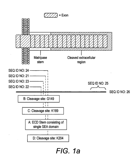
Figures la and lb depict cleavage sites of Matriptase resulting in the stem
regions (SEQ ID
Nos: 21-24).
Figure 2a depicts the alignment of Matriptase_Al heavy chain, the human VH 3-
23 Germline
and the human JH4b Germline. The CDR regions of Matriptase_Al heavy chain are
underlined.
Figure 2b depicts the alignment of Matriptase_Al light chain, the human VK A27
Germline and
the human JK2 Germline. The CDR regions of Matriptase_Al light chain are
underlined.
Figure 3 shows Matriptase_Al binding to the Matriptase-Stem using ELISA.
Figure 4a shows results of flow cytometric anaysis of Matriptase_Al on SNU1
cells.
Figure 4b depicts results of flow cytometric anaysis of Matriptase_Al on HT-29
cells.
Figure 4c depicts results of flow cytometric anaysis of Matriptase_Al on H69
cells.
Figure 5 depicts of Matriptase_Al elicting an antibody dependent cellular
cytotoxicity (ADCC)
response in the presence of effector cells
Figure 6 depicts the internalization of Matriptase_Al by HT-29 and H69 cells,
using MabZAP
assay.
Figure 7a shows a single dose (at 0.3 mole/kg: c.2mg/kg) of toxin conjugated
Matriptase_Al
was found to be curative.
Figure 7b shows the change in body weight over 60 days of dosing of
Matriptase_Al indicating
an amelioration of tumor-induced cachexia.
Figure 7c shows alternate dose groups in the HT-29 ADC xenograft model
revealing a dose
response to treatment.
Figure 7d shows alternate dose groups in the HT-29 ADC xenograft model
revealing a dose
response cachexia amelioration.
Figure 8 depicts the EC50 values for ADC cytoxicity assays using anti-
Matriptase antibodies
conjugated to either MMAE or MMAF in various cancer cell lines.
Figure 9 depicts the efficacy of anti-Matriptase antibodies conjugated to
either MMAE or
MMAF in ovarian adenocarcinoma SCID mouse xenograft model.
CA 02929402 2016-05-03
WO 2015/075477 PCT/GB2014/053470
7
DETAILED DESCRIPTION OF THE INVENTION
In one embodiment, the present disclosure relates to isolated antibodies which
bind
specifically to the stem region of Matriptase described in SEQ ID No: 21-24
with high affinity, as
outlined herein.
In particular embodiments, the Matriptase antibodies of the present invention
may be in the
form of a bispecific molecule, for example, which enhances an antibody
dependent cellular
cytotoxicity (ADCC) response in the presence of effector cells, thus killing
Matriptase-expressing
cells.
In other embodiments, the Matriptase antibodies of the present invention are
internalized
when contacted with cells expressing the Matriptase receptor. As discussed
herein, the Matriptase
receptor is overexpressed and/or differentially expressed on certain cancer
cells, including but not
limited to, tumors of gastric cancer, colorectal cancer, prostate cancer,
breast cancer, ovarian cancer
lung cancer, preferably SCLC, esophageal cancer, head and neck cancer,
pancreatic cancer,
lymphoma preferably non-Hodgkin's lymphoma and skin cancer.
Accordingly, when such Matriptase antibodies of the present invention are
conjugated to drugs
(sometimes referred to herein as "antibody-drug conjugates" or "ADCs"), the
internalization of these
ADC molecules into cancer cells results in cell death and thus tumor
treatment.
Thus, the disclosure provides antibodies particularly isolated antibodies
(which, as outlined
below, includes a wide variety of well known antibody structures, derivatives,
mimetics and
conjugates), nucleic acids encoding these antibodies, host cells used to make
the antibodies,
methods of making the antibodies, and pharmaceutical compositions comprising
the antibodies and
optionally including a pharmaceutical carrier.
Matriptase Proteins
The extracellular stem region of Matriptase has been reported to consist of a
single SEA
domain comprising amino acid residues 86-201 (SEQ ID No: 21). Activation of
Matriptase requires
sequential endoproteolytic cleavages and activation site autocleavage.
Cleavage occurs after amino
acid G1y149, resulting in a stem region comprising amino acid residues 86-149
(Matriptase Stem
Sequence B, SEQ ID No: 22). Further proteolytic cleavage can occur after amino
acid K189, which
results in a stem region comprising amino acid sequences 86-189 (Matriptase
Stem Sequence C,
SEQ ID No: 23) or amino acid K204, which results in a stem region comprising
amino acid
sequences 86-204 (Matriptase Stem Sequence D, SEQ ID No: 24). Matriptase is
then converted into
its active conformation by proteolytic cleavage after Arg614. The catalytic C-
terminal serine protease
domain consists of amino acid residues 615-855 (SEQ ID No: 27) (Figure 1).
See, for example,
Matriptase: Potent Proteolysis on the Cell Surface; List, Bugge and Szabo; Mol
Med 12(1-3)1-7, Jan-
March 2006 and Regulation of the activity of Matriptase on epithelial cell
surfaces by a blood derived
factor; Benaud, Dickson and Lin; EurJ Biochem 268, 1439-1447, 2001 which are
herein
incorporated in their entirety.
Accordingly, the present invention provides isolated anti-Matriptase
antibodies that specifically
bind the stem region of human Matriptase. By "human Matriptase" or "human
Matriptase antigen"
CA 02929402 2016-05-03
WO 2015/075477 PCT/GB2014/053470
8
refers to the protein of SEQ ID NO:26 or a functional fraction such as the
stem (SEQ ID No: 21-24),
as defined herein. In general, Matriptase possesses a short intracytoplasnnic
tail, a transnnennbrane
domain, and an extracellular domain which when cleaved produces the Matriptase
stem region. In
specific embodiments, the antibodies of the invention bind to the stem region
of the Matriptase
protein.
The antibodies of the invention may, in certain cases, cross-react with the
Matriptase from
species other than human. For example, to facilitate clinical testing, the
antibodies of the invention
may cross react with murine or primate Matriptase molecules. Alternatively, in
certain embodiments,
the antibodies may be completely specific for one or more human Matriptase and
may not exhibit
species or other types of non-human cross-reactivity.
Antibodies
The present invention provides anti-Matriptase antibodies, generally
therapeutic and/or
diagnostic antibodies as described herein. Antibodies that find use in the
present invention can take
on a number of formats as described herein, including traditional antibodies
as well as antibody
derivatives, fragments and mimetics, described below. Essentially, the
invention provides antibody
structures that contain a set of 6 CDRs as defined herein (including small
numbers of amino acid
changes as described below).
"Antibody" as used herein includes a wide variety of structures, as will be
appreciated by those
in the art, that at a minimum contain a set of 6 CDRs as defined herein;
including, but not limited to
traditional antibodies (including both monoclonal and polyclonal antibodies),
humanized and/or
chimeric antibodies, antibody fragments, engineered antibodies (e.g. with
amino acid modifications
as outlined below), multispecific antibodies (including bispecific
antibodies), and other analogs
known in the art and discussed herein.
Traditional antibody structural units typically comprise a tetramer. Each
tetramer is typically
composed of two identical pairs of polypeptide chains, each pair having one
"light" (typically having a
molecular weight of about 25 kDa) and one "heavy" chain (typically having a
molecular weight of
about 50-70 kDa). Human light chains are classified as kappa and lambda light
chains. Heavy
chains are classified as mu, delta, gamma, alpha, or epsilon, and define the
antibody's isotype as
IgM, IgD, IgG, IgA, and IgE, respectively. IgG has several subclasses,
including, but not limited to
IgG1, IgG2, IgG3, and IgG4. IgM has subclasses, including, but not limited to,
IgM1 and IgM2. Thus,
"isotype" as used herein is meant any of the subclasses of immunoglobulins
defined by the chemical
and antigenic characteristics of their constant regions. The known human
immunoglobulin isotypes
are IgG1, IgG2, IgG3, IgG4, IgA1, IgA2, IgM1, IgM2, IgD, and IgE. It should be
understood that
therapeutic antibodies can also comprise hybrids of any combination of
isotypes and/or subclasses.
In many embodiments, IgG isotypes are used in the present invention, with IgG1
finding
particular use in a number of applications.
The amino-terminal portion of each chain includes a variable region of about
100 to 110 or
more amino acids primarily responsible for antigen recognition. In the
variable region, three loops
are gathered for each of the V domains of the heavy chain and light chain to
form an antigen-binding
CA 02929402 2016-05-03
WO 2015/075477 PCT/GB2014/053470
9
site. Each of the loops is referred to as a complementarity-determining region
(hereinafter referred to
as a "CDR"), in which the variation in the amino acid sequence is most
significant. "Variable" refers
to the fact that certain segments of the variable region differ extensively in
sequence among
antibodies. Variability within the variable region is not evenly distributed.
Instead, the V regions
consist of relatively invariant stretches called framework regions (FRs) of 15-
30 amino acids
separated by shorter regions of extreme variability called "hypervariable
regions" that are each 9-15
amino acids long or longer.
Each VH and VL is composed of three hypervariable regions ("complementary
determining
regions," "CDRs") and four FRs, arranged from amino-terminus to carboxy-
terminus in the following
order: FR1-CDR1-FR2-CDR2-FR3-CDR3-FR4.
The hypervariable region generally encompasses amino acid residues from about
amino acid
residues 24-34 (LCDR1; "L" denotes light chain), 50-56 (LCDR2) and 89-97
(LCDR3) in the light
chain variable region and around about 31-35B (HCDR1; "H" denotes heavy
chain), 50-65 (HCDR2),
and 95-102 (HCDR3) in the heavy chain variable region; Kabat et al., SEQUENCES
OF PROTEINS
OF IMMUNOLOGICAL INTEREST, 5th Ed. Public Health Service, National Institutes
of Health,
Bethesda, Md. (1991) and/or those residues forming a hypervariable loop (e.g.
residues 26-32
(LCDR1), 50-52 (LCDR2) and 91-96 (LCDR3) in the light chain variable region
and 26-32 (HCDR1),
53-55 (HCDR2) and 96-101 (HCDR3) in the heavy chain variable region; Chothia
and Lesk (1987) J.
Mol. Biol. 196:901-917. Specific CDRs of the invention are described below
Throughout the present specification, the Kabat numbering system is generally
used when
referring to a residue in the variable domain (approximately, residues 1-107
of the light chain
variable region and residues 1-113 of the heavy chain variable region) (e.g,
Kabat et al., supra
(1991)).
The CDRs contribute to the formation of the antigen-binding, or more
specifically, epitope
binding site of antibodies. The term "epitope" or "antigenic determinant"
refers to a site on an
antigen to which an immunoglobulin or antibody specifically binds. Epitopes
can be formed both from
contiguous amino acids or noncontiguous amino acids juxtaposed by tertiary
folding of a protein.
Epitopes formed from contiguous amino acids are typically retained on exposure
to denaturing
solvents, whereas epitopes formed by tertiary folding are typically lost on
treatment with denaturing
solvents. An epitope typically includes at least 3, 4, 5, 6, 7, 8, 9, 10, 11,
12, 13, 14 or 15 amino
acids in a unique spatial conformation. Methods for determining what epitopes
are bound by a given
antibody (i.e., epitope mapping) are well known in the art and include, for
example, immunoblotting
and immunoprecipitation assays, wherein overlapping or contiguous peptides
from Matriptase are
tested for reactivity with the given anti- Matriptase antibody. Methods of
determining spatial
conformation of epitopes include techniques in the art and those described
herein, for example, x-
ray crystallography and 2-dimensional nuclear magnetic resonance (see, e.g.,
Epitope Mapping
Protocols in Methods in Molecular Biology, Vol. 66, G. E. Morris, Ed. (1996)).
The term "epitope
mapping" refers to the process of identification of the molecular determinants
for antibody-antigen
recognition
CA 02929402 2016-05-03
WO 2015/075477 PCT/GB2014/053470
"Epitope" refers to a determinant that interacts with a specific antigen
binding site in the
variable region of an antibody molecule known as a paratope. Epitopes are
groupings of molecules
such as amino acids or sugar side chains and usually have specific structural
characteristics, as well
as specific charge characteristics. A single antigen may have more than one
epitope. In the present
5 invention, the exact epitope is not determinative; rather, the ability of
the antibodies of the invention
to bind to the Matriptase receptor and be internalized or elict an ADCC
response in the presence of
effector cells is important.
The carboxy-terminal portion of each chain defines a constant region primarily
responsible for
effector function. Kabat et al. collected numerous primary sequences of the
variable regions of
10 heavy chains and light chains. Based on the degree of conservation of
the sequences, they
classified individual primary sequences into the CDR and the framework and
made a list thereof (see
SEQUENCES OF IMMUNOLOGICAL INTEREST, 5th edition, NIH publication, No. 91-
3242, E.A.
Kabat et al., entirely incorporated by reference).
In the IgG subclass of immunoglobulins, there are several immunoglobulin
domains in the
heavy chain. By "immunoglobulin (Ig) domain" herein is meant a region of an
immunoglobulin having
a distinct tertiary structure. Of interest in the present invention are the
heavy chain domains,
including, the constant heavy (CH) domains and the hinge domains. In the
context of IgG antibodies,
the IgG isotypes each have three CH regions. Accordingly, "CH" domains in the
context of IgG are
as follows: "CH1" refers to positions 118-220 according to the EU index as in
Kabat. "CH2" refers to
positions 237-340 according to the EU index as in Kabat, and "CH3" refers to
positions 341-447
according to the EU index as in Kabat.
Another type of Ig domain of the heavy chain is the hinge region. By "hinge"
or "hinge region"
or "antibody hinge region" or "immunoglobulin hinge region" herein is meant
the flexible polypeptide
comprising the amino acids between the first and second constant domains of an
antibody.
Structurally, the IgG CH1 domain ends at EU position 220, and the IgG CH2
domain begins at
residue EU position 237. Thus for IgG the antibody hinge is herein defined to
include positions 221
(0221 in IgG1) to 236 (G236 in IgG1), wherein the numbering is according to
the EU index as in
Kabat. In some embodiments, for example in the context of an Fc region, the
lower hinge is
included, with the "lower hinge" generally referring to positions 226 or 230.
Of particular interest in the present invention are the Fc regions. By "Fe" or
"Fc region" or "Fc
domain" as used herein is meant the polypeptide comprising the constant region
of an antibody
excluding the first constant region immunoglobulin domain and in some cases,
part of the hinge.
Thus Fc refers to the last two constant region immunoglobulin domains of IgA,
IgD, and IgG, the last
three constant region immunoglobulin domains of IgE and IgM, and the flexible
hinge N-terminal to
these domains. For IgA and IgM, Fc may include the J chain. For IgG, the Fc
domain comprises
immunoglobulin domains Cy2 and Cy3 (Cy2 and Cy3) and the lower hinge region
between Cy1
(Cy1) and Cy2 (Cy2). Although the boundaries of the Fc region may vary, the
human IgG heavy
chain Fc region is usually defined to include residues C226 or P230 to its
carboxyl-terminus, wherein
the numbering is according to the EU index as in Kabat. In some embodiments,
as is more fully
CA 02929402 2016-05-03
WO 2015/075477 PCT/GB2014/053470
11
described below, amino acid modifications are made to the Fc region, for
example to alter binding to
one or more FcyR receptors or to the FcRn receptor.
In some embodiments, the antibodies are full length. By "full length antibody"
herein is meant
the structure that constitutes the natural biological form of an antibody,
including variable and
constant regions, including one or more modifications as outlined herein.
Alternatively, the antibodies can be a variety of structures, including, but
not limited to,
antibody fragments, monoclonal antibodies, bispecific antibodies, minibodies,
domain antibodies,
synthetic antibodies (sometimes referred to herein as "antibody mimetics"),
chimeric antibodies,
humanized antibodies, antibody fusions (sometimes referred to as "antibody
conjugates"), and
fragments of each, respectively. Structures that rely on the use of a set of
CDRs are included within
the definition of "antibody".
In one embodiment, the antibody is an antibody fragment. Specific antibody
fragments include,
but are not limited to, (i) the Fab fragment consisting of VL, VH, CL and CHI
domains, (ii) the Ed
fragment consisting of the VH and CH1 domains, (iii) the Fv fragment
consisting of the VL and VH
domains of a single antibody; (iv) the dAb fragment (Ward et al., 1989, Nature
341:544-546, entirely
incorporated by reference) which consists of a single variable, (v) isolated
CDR regions, (vi) F(ab')2
fragments, a bivalent fragment comprising two linked Fab fragments (vii)
single chain Fv molecules
(scFv), wherein a VH domain and a VL domain are linked by a peptide linker
which allows the two
domains to associate to form an antigen binding site (Bird et al., 1988,
Science 242:423-426, Huston
et al., 1988, Proc. Natl. Acad. Sci. U.S.A. 85:5879-5883, entirely
incorporated by reference), (viii)
bispecific single chain Fv (WO 03/11161, hereby incorporated by reference) and
(ix) "diabodies" or
"triabodies", multivalent or multispecific fragments constructed by gene
fusion (Tomlinson et. al.,
2000, Methods Enzymol. 326:461-479; W094/13804; Holliger et al., 1993, Proc.
Natl. Acad. Sci.
U.S.A. 90:6444-6448, all entirely incorporated by reference).
Chimeric and Humanized Antibodies
In some embodiments, the antibody can be a mixture from different species,
e.g. a chimeric
antibody and/or a humanized antibody. That is, in the present invention, the
CDR sets can be used
with framework and constant regions other than those specifically described by
sequence herein.
In general, both "chimeric antibodies" and "humanized antibodies" refer to
antibodies that
combine regions from more than one species. For example, "chimeric antibodies"
traditionally
comprise variable region(s) from a mouse (or rat, in some cases) and the
constant region(s) from a
human. "Humanized antibodies" generally refer to non-human antibodies that
have had the variable-
domain framework regions swapped for sequences found in human antibodies.
Generally, in a
humanized antibody, the entire antibody, except the CDRs, is encoded by a
polynucleotide of human
origin or is identical to such an antibody except within its CDRs. The CDRs,
some or all of which are
encoded by nucleic acids originating in a non-human organism, are grafted into
the beta-sheet
framework of a human antibody variable region to create an antibody, the
specificity of which is
determined by the engrafted CDRs. The creation of such antibodies is described
in, e.g., WO
92/11018, Jones, 1986, Nature 321:522-525, Verhoeyen et al., 1988, Science
239:1534-1536, all
CA 02929402 2016-05-03
WO 2015/075477 PCT/GB2014/053470
12
entirely incorporated by reference. "Backmutation" of selected acceptor
framework residues to the
corresponding donor residues is often required to regain affinity that is lost
in the initial grafted
construct (US 5530101; US 5585089; US 5693761; US 5693762; US 6180370; US
5859205; US
5821337; US 6054297; US 6407213, all entirely incorporated by reference). The
humanized
antibody optimally also will comprise at least a portion of an immunoglobulin
constant region,
typically that of a human immunoglobulin, and thus will typically comprise a
human Fc region.
Humanized antibodies can also be generated using mice with a genetically
engineered immune
system. Roque et al., 2004, Biotechnol. Prog. 20:639-654, entirely
incorporated by reference. A
variety of techniques and methods for humanizing and reshaping non-human
antibodies are well
known in the art (See Tsurushita & Vasquez, 2004, Humanization of Monoclonal
Antibodies,
Molecular Biology of B Cells, 533-545, Elsevier Science (USA), and references
cited therein, all
entirely incorporated by reference). Humanization methods include but are not
limited to methods
described in Jones et al., 1986, Nature 321:522-525; Riechmann et al.,1988;
Nature 332:323-329;
Verhoeyen et al., 1988, Science, 239:1534-1536; Queen et al., 1989, Proc Natl
Acad Sci, USA
86:10029-33; He et al., 1998, J. Immunol. 160: 1029-1035; Carter et al., 1992,
Proc Natl Acad Sci
USA 89:4285-9, Presta et al., 1997, Cancer Res. 57(20):4593-9; Gorman et al.,
1991, Proc. Natl.
Acad. Sci. USA 88:4181-4185; O'Connor et al., 1998, Protein Eng 11:321-8, all
entirely incorporated
by reference. Humanization or other methods of reducing the immunogenicity of
nonhuman antibody
variable regions may include resurfacing methods, as described for example in
Roguska et al., 1994,
Proc. Natl. Acad. Sci. USA 91:969-973, entirely incorporated by reference. In
one embodiment, the
parent antibody has been affinity matured, as is known in the art. Structure-
based methods may be
employed for humanization and affinity maturation, for example as described in
USSN 11/004,590.
Selection based methods may be employed to humanize and/or affinity mature
antibody variable
regions, including but not limited to methods described in Wu et al., 1999, J.
Mol. Biol. 294:151-162;
Baca et al., 1997, J. Biol. Chem. 272(16):10678-10684; Rosok et al., 1996, J.
Biol. Chem. 271(37):
22611-22618; Rader et al., 1998, Proc. Natl. Acad. Sci. USA 95: 8910-8915;
Krauss et al., 2003,
Protein Engineering 16(10):753-759, all entirely incorporated by reference.
Other humanization
methods may involve the grafting of only parts of the CDRs, including but not
limited to methods
described in USSN 09/810,510; Tan et al., 2002, J. lmmunol. 169:1119-1125; De
Pascalis et al.,
2002, J. Innnnunol. 169:3076-3084, all entirely incorporated by reference.
In one embodiment, the antibodies of the invention can be multispecific
antibodies, and
notably bispecific antibodies, also sometimes referred to as "diabodies".
These are antibodies that
bind to two (or more) different antigens, or different epitopes on the same
antigen. Diabodies can be
manufactured in a variety of ways known in the art (Holliger and Winter, 1993,
Current Opinion
Biotechnol. 4:446-449, entirely incorporated by reference), e.g., prepared
chemically or from hybrid
hybridomas.
In one embodiment, the antibody is a minibody. Minibodies are minimized
antibody-like
proteins comprising a scFv joined to a CH3 domain. Hu et al., 1996, Cancer
Res. 56:3055-3061,
entirely incorporated by reference. In some cases, the scFv can be joined to
the Fc region, and may
CA 02929402 2016-05-03
WO 2015/075477 PCT/GB2014/053470
13
include some or the entire hinge region. It should be noted that minibodies
are included within the
definition of "antibody" despite the fact it does not have a full set of CDRs.
The antibodies of the present invention are generally isolated or recombinant.
"Isolated,"
when used to describe the various polypeptides disclosed herein, means a
polypeptide that has
been identified and separated and/or recovered from a cell or cell culture
from which it was
expressed. Thus an isolated antibody is intended to refer to an antibody that
is substantially free of
other antibodies having different antigenic specificities (e.g. an isolated
antibody that specifically
binds to the Matriptase is substantially free of antibodies that specifically
bind antigens other than
the Matriptase). Thus, an "isolated" antibody is one found in a form not
normally found in nature (e.g.
non-naturally occuring).
In some embodiments, the antibodies of the invention are recombinant proteins,
isolated
proteins or substantially pure proteins. An "isolated" protein is
unaccompanied by at least some of
the material with which it is normally associated in its natural state, for
example constituting at least
about 5%, or at least about 50% by weight of the total protein in a given
sample. It is understood that
the isolated protein may constitute from 5 to 99.9% by weight of the total
protein content depending
on the circumstances. For example, the protein may be made at a significantly
higher concentration
through the use of an inducible promoter or high expression promoter, such
that the protein is made
at increased concentration levels. In the case of recombinant proteins, the
definition includes the
production of an antibody in a wide variety of organisms and/or host cells
that are known in the art in
which it is not naturally produced. Ordinarily, an isolated polypeptide will
be prepared by at least one
purification step. An "isolated antibody," refers to an antibody which is
substantially free of other
antibodies having different antigenic specificities. For instance, an isolated
antibody that specifically
binds to Matriptase is substantially free of antibodies that specifically bind
antigens other than
Matriptase.
Isolated monoclonal antibodies, having different specificities, can be
combined in a well
defined composition. Thus for example, the antibody of the invention can
optionally and individually
be included or excluded in a formulation, as is further discussed below.
The anti-Matriptase antibodies of the present invention specifically bind
Matriptase (e.g.
Matriptase-stem (SEQ ID Nos: 21-24)). "Specific binding" or "specifically
binds to" or is "specific for"
a particular antigen or an epitope means binding that is measurably different
from a non-specific
interaction. Specific binding can be measured, for example, by determining
binding of a molecule
compared to binding of a control molecule, which generally is a molecule of
similar structure that
does not have binding activity. For example, specific binding can be
determined by competition with
a control molecule that is similar to the target.
Specific binding for a particular antigen or an epitope can be exhibited, for
example, by an
antibody having a KD for an antigen or epitope of at least about 10-4 M, at
least about 10-5 M, at least
about 10-6 M, at least about 10-7 M, at least about 10-8 M, at least about 10-
9 M, alternatively at least
about 10-19 M, at least about 10-11 M, at least about 10-12 M, or greater,
where KD refers to a
dissociation rate of a particular antibody-antigen interaction. Typically, an
antibody that specifically
CA 02929402 2016-05-03
WO 2015/075477
PCT/GB2014/053470
14
binds an antigen will have a KD that is 20-, 50-, 100-, 500-, 1000-, 5,000-,
10,000- or more times
greater fora control molecule relative to the antigen or epitope. However, in
the present invention,
when administering ADCs of the Matriptase antibodies of the invention, what is
important is that the
KD is sufficient to allow internalization and thus cell death without
significant side effects.
Also, specific binding for a particular antigen or an epitope can be
exhibited, for example, by
an antibody having a KA or Ka for an antigen or epitope of at least 20-, 50-,
100-, 500-, 1000-, 5,000-,
10,000- or more times greater for the epitope relative to a control, where KA
or Ka refers to an
association rate of a particular antibody-antigen interaction.
Standard assays to evaluate the binding ability of the antibodies toward
Matriptase can be
done on the protein or cellular level and are known in the art, including for
example, ELISAs,
Western blots, RIAs, BlAcore assays and flow cytometry analysis. Suitable
assays are described in
detail in the Examples. The binding kinetics (e.g. binding affinity) of the
antibodies also can be
assessed by standard assays known in the art, such as by Biacore system
analysis. To assess
binding to Raji or Daudi B cell tumor cells, Raji (ATCC Deposit No. CCL-86) or
Daudi (ATCC Deposit
No. CCL-213) cells can be obtained from publicly available sources, such as
the American Type
Culture Collection, and used in standard assays, such as flow cytometric
analysis.
Matriptase Antibodies
The present invention provides Matriptase antibodies that specifically bind
the stem of human
Matritpase (SEQ ID No: 21-24) and are internalized when contacted with cells
expressing Matriptase
on the cell surface. These antibodies are referred to herein either as "anti-
Matriptase" antibodies or,
for ease of description, "Matriptase antibodies".
The Matriptase antibodies maybe internalized upon contact with cells,
particularly tumor cells that
express Matriptase on the surface. Accordingly, Matriptase antibodies as
defined herein that also
comprise drug conjugates are internalized by tumor cells, resulting in the
release of the drug and
subsequent cell death, allowing for treatment of cancers that exhibit
Matriptase expression.
Internalization in this context can be measured in several ways. In one
embodiment, the Matriptase
antibodies of the invention are contacted with cells, such as a cell line as
outlined herein, using
standard assays such as MAbZap and HuZap. It woud be clear to the skilled
person that the
MabZap assay is representative of the effect that would be expected to be seen
with an antibody-
drug conjugate (ADC). In the latter case, the ADC would be internalised, thus
taking the drug into
the cell. A toxic drug would have the capacity to kill the cell, i.e. to kill
the targeted cancer cell. Data
from MabZap assays are readily accepted by persons of skill in the art to be
representative of ADC
assays (Kohls, M and Lappi, D., [2000] Biotechniques, vol. 28, no. 1, 162-
165). In these in vitro
assay embodiments, the Matriptase antibodies of the invention are added, along
with an anti-
Matriptase antibody comprising a toxin; for example, the Matriptase antibody
may be murine or
humanized and the anti-Matriptase antibody can be anti-murine or anti-
humanized and contain a
toxin such as saporin. Upon formation of the [Matriptase antibody of the
invention]-[anti-Matriptase
antibody-drug conjugate] complex, the complex is internalized and the drug
(e.g. saporin) is
CA 02929402 2016-05-03
WO 2015/075477
PCT/GB2014/053470
released, resulting in cell death. Only upon internalization does the drug get
released, and thus cells
remain viable in the absence of internalization. As outlined below, without
being bound by theory, in
therapeutic applications, the anti-Matriptase antibody contains the toxin, and
upon internalization,
the bond between the antibody and the toxin is cleaved, releasing the toxin
and killing the cell.
5 In
addition, the Matriptase antibody may elict an ADCC response in the presence
of effector
cells, particularly tumor cells that express Matriptase on the surface.
In one embodiment, the antibody comprises the heavy and light chain
complementarity
determining regions (CDRs) or variable regions (VRs) of the particular
antibodies described herein
(e.g., referred to herein as "Matriptase_Al"). Accordingly, in one embodiment,
the antibody
10 comprises the CDR1, CDR2, and CDR3 domains of the heavy chain variable
(VH) region of antibody
Al having the sequence shown in SEQ ID NO:1, and the CDR1, CDR2 and CDR3
domains of the
light chain variable (VL) region of Al having the sequence shown in SEQ ID
NO:2.
In another embodiment, the antibody comprises a heavy chain variable region
comprising a
first vhCDR comprising SEQ ID NO: 5; a second vhCDR comprising SEQ ID NO: 6;
and a third
15 vhCDR comprising SEQ ID NO:7; and a light chain variable region
comprising a first vICDR
comprising SEQ ID NO:8; a second vICDR comprising SEQ ID NO: 9; and a third
vICDR comprising
SEQ ID NO:10. In another embodiment, the antibodies of the invention bind to
human Matriptase
and include a heavy chain variable region including an amino acid sequence
selected from the group
consisting of SEQ ID NOs:1, and conservative sequence modifications thereof.
The antibody may
further include a light chain variable region including an amino acid sequence
selected from the
group consisting of SEQ ID NOs:2, and conservative sequence modifications
thereof.
In a further embodiment, the antibodies of the invention bind to human
Matriptase and include
a heavy chain variable region and a light chain variable region including the
amino acid sequences
set forth in SEQ ID NO5:1 and/or 2, respectively, and conservative sequence
modifications thereof.
Isolated antibodies which include heavy and light chain variable regions
having at least 80%,
or at least 85%, or at least 90%, or at least 91%, or at least 92%, or at
least 93%, or at least 94%, or
at least 95%, or at least 96%, or at least 97%, or at least 98%, or at least
99%, or more sequence
identity to any of the above sequences are also included in the present
invention. Ranges
intermediate to the above-recited values, e.g., heavy and light chain variable
regions having at least
80-85%, 85-90%, 90-95% or 95-100% sequence identity to any of the above
sequences are also
intended to be encompassed by the present invention. In one embodiment, the
antibody comprises
a heavy chain variable region comprising SEQ ID NO:1, or a sequence that is at
least 90%, at least
91%, at least 92%, at least 93%, at least 94%, at least 95%, at least 96%, at
least 97%, at least
98%, at least 99% identical to SEQ ID NO: 1. In another embodiment, the
antibody comprises a
light chain variable region comprising SEQ ID NO:2 or a sequence that is at
least 90%, at least 91%,
at least 92%, at least 93%, at least 94%, at least 95%, at least 96%, at least
97%, at least 98%, at
least 99% identical to SEQ ID NO: 2. In another embodiment, the antibody
comprises a heavy
chain framework region comprising an amino acid sequence that is at least 90%,
at least 91%, at
least 92%, at least 93%, at least 94%, at least 95%, at least 96%, at least
97%, at least 98%, at least
CA 02929402 2016-05-03
WO 2015/075477 PCT/GB2014/053470
16
99% identical to the framework of the heavy chain variable region of SEQ ID
NO: 1 comprising SEQ
ID NOs: 29, 30, 31 and 32. In another embodiment, the antibody comprises a
light chain framework
region comprising an amino acid sequence that is at least 90%, at least 91%,
at least 92%, at least
93%, at least 94%, at least 95%, at least 96%, at least 97%, at least 98%, at
least 99% identical to
the framework of the light chain variable region of SEQ ID NO:2, comprising
SEQ ID NOs: 33, 34, 35
and 36. In one embodiment, the antibody of the invention is an anti-Matriptase
antibody (referred to
herein as "Al" antibody) comprising the following CDRs, as well as variants
containing a limited
number of amino acid variants:
Al SEQ ID NOs
variable heavy CDR1 5
variable heavy CDR2 6
variable heavy CDR3 7
variable light CDR1 8
variable light CDR2 9
variable light CDR3 10
Disclosed herein are also variable heavy and light chains that comprise the
CDR sets of
particular Matriptase antibodies of the invention, such as Al, as well as full
length heavy and light
chains (e.g. comprising constant regions as well). As will be appreciated by
those in the art, the
CDR sets of the invention can be incorporated into murine, humanized or human
constant regions
(including framework regions). As shown for Al and huAl , the amino acid
identity between the
murine and human sequences is about 90%. Accordingly, the present invention
provides variable
heavy and light chains that are at least about 90%-99% identical to the SEQ
IDs disclosed herein,
with 90, 91, 92, 93, 94, 95, 96, 97, 98 and 99% all finding use in the present
invention.
Antibodies that Bind to the Same Epitope as the Matriptase Antibodies of the
Invention
In another embodiment, the invention provides antibodies that bind to the same
epitope on the
human Matriptase as any of the Matriptase monoclonal antibodies of the
invention The term "binds
to the same epitope" with reference to two or more antibodies means that the
antibodies compete for
binding to an antigen and bind to the same, overlapping or encompassing
continuous or
discontinuous segments of amino acids. Those of skill in the art understand
that the phrase "binds
to the same epitope" does not necessarily mean that the antibodies bind to
exactly the same amino
acids. The precise amino acids to which the antibodies bind can differ. For
example, a first antibody
can bind to a segment of amino acids that is completely encompassed by the
segment of amino
acids bound by a second antibody. In another example, a first antibody binds
one or more segments
of amino acids that significantly overlap the one or more segments bound by
the second antibody.
For the purposes herein, such antibodies are considered to "bind to the same
epitope."
CA 02929402 2016-05-03
WO 2015/075477 PCT/GB2014/053470
17
Accordingly, also, encompassed by the present invention are antibodies that
bind to an
epitope on Matriptase which comprises all or a portion of an epitope
recognized by the particular
antibodies described herein (e.g., the same or an overlapping region or a
region between or
spanning the region).
Also encompassed by the present invention are antibodies that bind the same
epitope and/or
antibodies that compete for binding to human Matriptase with the antibodies
described herein.
Antibodies that recognize the same epitope or compete for binding can be
identified using routine
techniques. Such techniques include, for example, an immunoassay, which shows
the ability of one
antibody to block the binding of another antibody to a target antigen, i.e., a
competitive binding
assay. Competitive binding is determined in an assay in which the
immunoglobulin under test
inhibits specific binding of a reference antibody to a common antigen, such as
Matriptase.
Numerous types of competitive binding assays are known, for example: solid
phase direct or indirect
radioimmunoassay (RIA), solid phase direct or indirect enzyme immunoassay
(EIA), sandwich
competition assay (see Stahli etal., Methods in Enzymology 9:242 (1983));
solid phase direct biotin-
avidin EIA (see Kirkland etal., J. lmmunol. 137:3614 (1986)); solid phase
direct labeled assay, solid
phase direct labeled sandwich assay (see Harlow and Lane, Antibodies: A
Laboratory Manual, Cold
Spring Harbor Press (1988)); solid phase direct label RIA using 1-125 label
(see Morel etal., Mol.
Immunol. 25(1):7 (1988)); solid phase direct biotin-avidin EIA (Cheung etal.,
Virology 176:546
(1990)); and direct labeled RIA. (Moldenhauer et al., Scand. J. Immunol. 32:77
(1990)). Typically,
such an assay involves the use of purified antigen bound to a solid surface or
cells bearing either of
these, an unlabeled test immunoglobulin and a labeled reference
immunoglobulin. Competitive
inhibition is measured by determining the amount of label bound to the solid
surface or cells in the
presence of the test immunoglobulin. Usually the test immunoglobulin is
present in excess. Usually,
when a competing antibody is present in excess, it will inhibit specific
binding of a reference antibody
to a common antigen by at least 50-55%, 55-60%, 60-65%, 65-70% 70-75% A 75-
80% 80-85% 85-
90% 90-95% 95-99% or more.
Other techniques include, for example, epitope mapping methods, such as, x-ray
analyses of
crystals of antigen:antibody complexes which provides atomic resolution of the
epitope. Other
methods monitor the binding of the antibody to antigen fragments or mutated
variations of the
antigen where loss of binding due to a modification of an amino acid residue
within the antigen
sequence is often considered an indication of an epitope component. In
addition, computational
combinatorial methods for epitope mapping can also be used. These methods rely
on the ability of
the antibody of interest to affinity isolate specific short peptides from
combinatorial phage display
peptide libraries. The peptides are then regarded as leads for the definition
of the epitope
corresponding to the antibody used to screen the peptide library. For epitope
mapping,
computational algorithms have also been developed which have been shown to map
conformational
discontinuous epitopes.
In a particular embodiment, the antibody competes for binding to Matripitase
with an antibody
comprising heavy and/or light chain variable regions comprising the amino acid
sequences set forth
CA 02929402 2016-05-03
WO 2015/075477 PCT/GB2014/053470
18
in SEQ ID NOs:1 and 2, respectively, or amino acid sequences at least 80%
identical thereto. In
another embodiment, the antibody competes for binding to Matripitase with an
antibody comprising
heavy and/or light chain variable regions comprising the amino acid sequences
set forth in SEQ ID
NOs:1 and 2 (Al).
Other antibodies of the invention bind to an epitope on Matriptase recognized
by the
antibodies described herein. In another particular embodiment, the antibody
binds to an epitope on
Matriptase recognized by an antibody comprising heavy and/or light chain
variable regions
comprising the amino acid sequences set forth in SEQ ID NOs:1 and 2, or amino
acid sequences at
least 80%, at least 85%, at least 90%, at least 95%, at least 96%, at least
97%, at least 98% or at
least 99% identical thereto. In another embodiment, the antibody binds to an
epitope on Matriptase
recognized by an antibody comprising heavy and/or light chain variable regions
comprising the
amino acid sequences set forth in SEQ ID NOs:1 and 2 (Al).
Characterization of Monoclonal Antibodies to Matriptase
Monoclonal antibodies of the invention can be characterized for binding to
Matriptase using a
variety of known techniques. Generally, the antibodies are initially
characterized by ELISA. Briefly,
microtiter plates can be coated with purified Matriptase in PBS, and then
blocked with irrelevant
proteins such as bovine serum albumin (BSA) diluted in PBS. Dilutions of
plasma from Matriptase-
immunized mice are added to each well and incubated for 1-2 hours at 37 C. The
plates are
washed with PBS/Tween 20 and then incubated with a goat-anti-human IgG Fc-
specific polyclonal
reagent conjugated to alkaline phosphatase for 1 hour at 37 C. After washing,
the plates are
developed with ABTS substrate, and analyzed at OD of 405. Preferably, mice
which develop the
highest titers will be used for fusions.
An ELISA assay as described above can be used to screen for antibodies and,
thus,
hybridomas that produce antibodies that show positive reactivity with the
Matriptase immunogen.
Hybridomas that bind, preferably with high affinity, to Matriptase can then be
subcloned and further
characterized. One clone from each hybridoma, which retains the reactivity of
the parent cells (by
ELISA), can then be chosen for making a cell bank, and for antibody
purification.
To purify anti-Matriptase antibodies, selected hybridomas can be grown in
roller bottles, two-
liter spinner-flasks or other culture systems. Supernatants can be filtered
and concentrated before
affinity chromatography with protein A-Sepharose (Pharmacia, Piscataway, NJ)
to purify the protein.
After buffer exchange to PBS, the concentration can be determined by 0D280
using 1.43 extinction
coefficient or preferably by nephelometric analysis. IgG can be checked by gel
electrophoresis and
by antigen specific method.
To determine if the selected anti-Matriptase monoclonal antibodies bind to
unique epitopes,
each antibody can be biotinylated using commercially available reagents
(Pierce, Rockford, IL).
Biotinylated MAb binding can be detected with a streptavidin labeled probe. To
determine the
isotype of purified antibodies, isotype ELISAs can be performed using art
recognized techniques.
For example, wells of microtiter plates can be coated with 10 ..Lg/m1 of anti-
Ig overnight at 4 C. After
CA 02929402 2016-05-03
WO 2015/075477 PCT/GB2014/053470
19
blocking with 5% BSA, the plates are reacted with 10 g/m1 of monoclonal
antibodies or purified
isotype controls, at ambient temperature for two hours. The wells can then be
reacted with either
IgGI or other isotype specific conjugated probes. Plates are developed and
analyzed as described
above.
To test the binding of monoclonal antibodies to live cells expressing
Matriptase, flow
cytometry can be used. Briefly, cell lines and/or human PBMCs expressing
membrane-bound
Matriptase (grown under standard growth conditions) are mixed with various
concentrations of
monoclonal antibodies in PBS containing 0.1% BSA at 4 C for 1 hour. After
washing, the cells are
reacted with Fluorescein-labeled anti- IgG antibody under the same conditions
as the primary
antibody staining. The samples can be analyzed by FACScan instrument using
light and side
scatter properties to gate on single cells and binding of the labeled
antibodies is determined. An
alternative assay using fluorescence microscopy may be used (in addition to or
instead of) the flow
cytometry assay. Cells can be stained exactly as described above and examined
by fluorescence
microscopy. This method allows visualization of individual cells, but may have
diminished sensitivity
depending on the density of the antigen.
Anti-Matriptase IgGs can be further tested for reactivity with the Matriptase
antigen by
Western blotting. Briefly, cell extracts from cells expressing Matriptase can
be prepared and
subjected to sodium dodecyl sulfate polyacrylamide gel electrophoresis. After
electrophoresis, the
separated antigens will be transferred to nitrocellulose membranes, blocked
with 20% mouse serum,
and probed with the monoclonal antibodies to be tested. IgG binding can be
detected using anti-
IgG alkaline phosphatase and developed with BCIP/NBT substrate tablets (Sigma
Chem. Co., St.
Louis, MO).
Methods for analyzing binding affinity, cross-reactivity, and binding kinetics
of various anti-
Matriptase antibodies include standard assays known in the art, for example,
BiacoreTM surface
plasmon resonance (SPR) analysis using a BiacoreTm 2000 SPR instrument
(Biacore AB, Uppsala,
Sweden).
In one embodiment, the antibody specifically binds to human Matriptase
comprising SEQ ID
NO:26 or a functional fraction, such as the stem (SEQ ID Nos: 21-24).
Preferably, an antibody of the
invention binds to human Matriptase with high affinity.
Preferably, an antibody of the invention binds to a Matriptase protein with a
KD of 5 x 1 0-8 M
or less, binds to a Matriptase protein with a KD of 2 x 10⁻⁸ M or less, binds
to a Matriptase protein
with a KD of 5 x 10⁻⁹ M or less, binds to a Matriptase protein with a KD of 4
x 10-9 M or less, binds to a
Matriptase protein with a KD of 3 x 10⁻⁹ M or less, binds to a Matriptase
protein with a KD of 2 x 10⁻⁹
M or less, binds to a Matriptase protein with a KD of 1 x 10⁻⁹ M or less,
binds to a Matriptase protein
with a KD of 5 x 10⁻¹⁰ M or less, or binds to a Matriptase protein with a KD
of 1 x 10-19 M or less.
In one embodiment, antibodies of the invention compete (e.g., cross-compete)
for binding to
Matriptase with the particular anti-Matriptase antibodies described herein
(e.g.,_A1). Such
competing antibodies can be identified based on their ability to competitively
inhibit binding to
Matriptase of one or more of mAbs in standard Matriptase binding assays. For
example, standard
CA 02929402 2016-05-03
WO 2015/075477 PCT/GB2014/053470
ELISA assays can be used in which a recombinant human Matriptase protein is
immobilized on the
plate, one of the antibodies is fluorescently labeled and the ability of non-
labeled antibodies to
compete off the binding of the labeled antibody is evaluated. Additionally or
alternatively, BlAcore
analysis can be used to assess the ability of the antibodies to cross-compete.
The ability of a test
5 antibody to inhibit the binding of an anti-Matriptase antibody of the
invention to human Matriptase
demonstrates that the test antibody can compete with the antibody for binding
to human Matriptase
In one embodiment, the competing antibody is an antibody that binds to the
same epitope on
human Matriptase as the particular anti-Matriptase monoclonal antibodies
described herein
(e.g.,A1). Standard epitope mapping techniques, such as x-ray crystallography
and 2-dimensional
10 nuclear magnetic resonance, can be used to determine whether an antibody
binds to the same
epitope as a reference antibody (see, e.g., Epitope Mapping Protocols in
Methods in Molecular
Biology, Vol. 66, G. E. Morris, Ed. (1996)).
In one embodiment, the antibody that competes for binding to Matriptase and/or
binds to the
same epitope on human Matriptase is a human antibody. Such human monoclonal
antibodies can
15 be prepared and isolated as described in the Examples.
Once a single, archtypal anti-Matriptase mAb has been isolated that has the
desired
properties described herein, it is straightforward to generate other mAbs with
similar properties, e.g.,
having the same epitope, by using art-known methods. For example, mice may be
immunized with
Matriptase as described herein, hybridomas produced, and the resulting mAbs
screened for the
20 ability to compete with the archtypal mAb for binding to Matriptase.
Mice can also be immunized with
a smaller fragment of Matriptase containing the epitope to which the archtypal
mAb binds. The
epitope can be localized by, e.g., screening for binding to a series of
overlapping peptides spanning
Matriptase. Alternatively, the method of Jespers et al., Biotechnology 12:899,
1994 may be used to
guide the selection of mAbs having the same epitope and therefore similar
properties to the
archtypal mAb. Using phage display, first the heavy chain of the archtypal
antibody is paired with a
repertoire of (preferably human) light chains to select a Matriptase-binding
mAb, and then the new
light chain is paired with a repertoire of (preferably human) heavy chains to
select a (preferably
human) Matriptase-binding mAb having the same epitope as the archtypal mAb.
Alternatively
variants of the archetypal mAb can be obtained by mutagenesis of cDNA encoding
the heavy and
light chains of the antibody.
Epitope mapping, e.g., as described in Champe et al. (1995) J. Biol. Chem.
270:1388-1394,
can be performed to determine whether the antibody binds an epitope of
interest. "Alanine scanning
mutagenesis," as described by Cunningham and Wells (1989) Science 244: 1081-
1085, or some
other form of point mutagenesis of amino acid residues in human Matriptase may
also be used to
determine the functional epitope for an anti-Matriptase antibody of the
present invention.
Mutagenesis studies, however, may also reveal amino acid residues that are
crucial to the overall
three-dimensional structure of Matriptase but that are not directly involved
in antibody-antigen
contacts, and thus other methods may be necessary to confirm a functional
epitope determined
using this method.
CA 02929402 2016-05-03
WO 2015/075477
PCT/GB2014/053470
21
The epitope bound by a specific antibody may also be determined by assessing
binding of
the antibody to peptides comprising fragments of human Matriptase. A series of
overlapping
peptides encompassing the sequence of Matriptase may be synthesized and
screened for binding,
e.g. in a direct ELISA, a competitive ELISA (where the peptide is assessed for
its ability to prevent
binding of an antibody to Matriptase bound to a well of a microtiter plate),
or on a chip. Such peptide
screening methods may not be capable of detecting some discontinuous
functional epitopes, i.e.
functional epitopes that involve amino acid residues that are not contiguous
along the primary
sequence of the Matriptase polypeptide chain.
The epitope bound by antibodies of the present invention may also be
determined by
structural methods, such as X-ray crystal structure determination (e.g.,
W02005/044853), molecular
modeling and nuclear magnetic resonance (NMR) spectroscopy, including NMR
determination of the
H-D exchange rates of labile amide hydrogens in Matriptase when free and when
bound in a
complex with an antibody of interest (Zinn-Justin et al. (1992) Biochemistry
31, 11335-11347; Zinn-
Justin et al. (1993) Biochemistry 32, 6884-6891).
With regard to X-ray crystallography, crystallization may be accomplished
using any of the
known methods in the art (e.g. Giege et al. (1994) Acta Crystallogr. D50:339-
350; McPherson (1990)
Eur. J. Biochem. 189:1-23), including microbatch (e.g. Chayen (1997) Structure
5:1269-1274),
hanging-drop vapor diffusion (e.g. McPherson (1976) J. Biol. Chem. 251:6300-
6303), seeding and
dialysis. It is desirable to use a protein preparation having a concentration
of at least about 1 mg/mL
and preferably about 10 mg/mL to about 20 mg/mL. Crystallization may be best
achieved in a
precipitant solution containing polyethylene glycol 1000-20,000 (PEG; average
molecular weight
ranging from about 1000 to about 20,000 Da), preferably about 5000 to about
7000 Da, more
preferably about 6000 Da, with concentrations ranging from about 10% to about
30% (w/v). It may
also be desirable to include a protein stabilizing agent, e.g. glycerol at a
concentration ranging from
about 0.5% to about 20%. A suitable salt, such as sodium chloride, lithium
chloride or sodium citrate
may also be desirable in the precipitant solution, preferably in a
concentration ranging from about 1
mM to about 1000 mM. The precipitant is preferably buffered to a pH of from
about 3.0 to about 5.0,
preferably about 4Ø Specific buffers useful in the precipitant solution may
vary and are well-known
in the art (Scopes, Protein Purification: Principles and Practice, Third ed.,
(1994) Springer-Verlag,
New York). Examples of useful buffers include, but are not limited to, HEPES,
Tris, MES and
acetate. Crystals may be grow at a wide range of temperatures, including 2 C,
4 C, 8 C and 26
C.
Antibody:antigen crystals may be studied using well-known X-ray diffraction
techniques and
may be refined using computer software such as X-PLOR (Yale University, 1992,
distributed by
Molecular Simulations, Inc.; see e.g. Blundell & Johnson (1985) Meth. Enzymol.
114 & 115, H. W.
VVyckoff et al., eds., Academic Press; U.S. Patent Application Publication No.
2004/0014194), and
BUSTER (Bricogne (1993) Acta Cryst. D49:37-60; Bricogne (1997) Meth. Enzymol.
276A:361-423,
Carter & Sweet, eds.; Roversi et al. (2000) Acta Cryst. D56:1313-1323), the
disclosures of which are
hereby incorporated by reference in their entireties.
CA 02929402 2016-05-03
WO 2015/075477 PCT/GB2014/053470
22
Antibody competition assays, as described herein, can be used to determine
whether
an antibody "binds to the same epitope" as another antibody. Typically,
competition of 50% or more,
60% or more, 70% or more, such as 70%, 71%, 72%, 73%, 74%, 75%, 80%, 85%, 90%,
95%, 96%,
97%, 98%, 99% or more, of an antibody known to interact with the epitope by a
second antibody
under conditions in which the second antibody is in excess and the first
saturates all sites, is
indicative that the antibodies "bind to the same epitope." To assess the level
of competition between
two antibodies, for example, radioimmunoassays or assays using other labels
for the antibodies, can
be used. For example, a Matriptase antigen can be incubated with a saturating
amount of a first
anti-Matriptase antibody or antigen-binding fragment thereof conjugated to a
labeled compound
(e.g., 3H, 1251, biotin, or rubidium) in the presence the same amount of a
second unlabeled anti-
Matriptase antibody. The amount of labeled antibody that is bound to the
antigen in the presence of
the unlabeled blocking antibody is then assessed and compared to binding in
the absence of the
unlabeled blocking antibody. Competition is determined by the percentage
change in binding
signals in the presence of the unlabeled blocking antibody compared to the
absence of the blocking
antibody. Thus, if there is a 50% inhibition of binding of the labeled
antibody in the presence of the
blocking antibody compared to binding in the absence of the blocking antibody,
then there is
competition between the two antibodies of 50%. Thus, reference to competition
between a first and
second antibody of 50% or more, 60% or more, 70% or more, such as 70%, 71%,
72%, 73%, 74%,
75%, 80%, 85%, 90%, 95%, 96%, 97%, 98%, 99% or more, means that the first
antibody inhibits
binding of the second antibody (or vice versa) to the antigen by 50%, 60%,
70%, 71%, 72%, 73%,
74%, 75%, 80%, 85%, 90%, 95%, 96%, 97%, 98%, 99% or more (compared to binding
of the
antigen by the second antibody in the absence of the first antibody). Thus,
inhibition of binding of a
first antibody to an antigen by a second antibody of 50%, 605, 70%, 71%, 72%,
73%, 74%, 75%,
80%, 85%, 90%, 95%, 96%, 97%, 98%, 99% or more indicates that the two
antibodies bind to the
same epitope.
Antibody Modifications
The present invention further provides variant antibodies, sometimes referred
to as "antibody
derivatives" or "antibody analogs" as well. That is, there are a number of
modifications that can be
made to the antibodies of the invention, including, but not limited to, amino
acid modifications in the
CDRs (affinity maturation), amino acid modifications in the Fc region,
glycosylation variants, covalent
modifications of other types (e.g. for attachment of drug conjugates, etc.
By "variant" herein is meant a polypeptide sequence that differs from that of
a parent
polypeptide by virtue of at least one amino acid modification. In one
embodiment, the parent
polypeptide is either the full length variable heavy and/or light chains,
listed in SEQ ID Nos: 1 and 2.
Amino acid modifications can include substitutions, insertions and deletions,
with the former being
preferred in many cases.
In general, variants can include any number of modifications, as long as the
function of the
antibody is still present, as described herein. Accordingly, variant
antibodies of the invention, for
CA 02929402 2016-05-03
WO 2015/075477 PCT/GB2014/053470
23
example, should still specifically bind to human Matriptase. Similarly, if
amino acid variants are
generated with the Fc region, for example, the variant antibodies should
maintain the required
receptor binding functions for the particular application or indication of the
antibody.
"Variants" in this case can be made in either the listed CDR sequences, the
framework or Fc
regions of the antibody.
However, in general, from 1, 2, 3, 4, 5, 6, 7, 8, 9 or 10 amino acid
substitutions are generally
utilized as often the goal is to alter function with a minimal number of
modifications. In some cases,
there are from 1 to 5 modifications (e.g. individual amino acid substitutions,
insertions or deletions),
with from 1-2, 1-3 and 1-4 also finding use in many embodiments. The number of
modifications can
depend on the size of the region being modified; for example, in general,
fewer modifications are
desired in CDR regions. However, as shown herein, the CDRs of the Al herein
are similar, such
that a number of amino acid changes can be made and preserve binding.
It should be noted that the number of amino acid modifications may be within
functional
domains: for example, it may be desirable to have from 1-5 modifications in
the Fc region of wild-
type or engineered proteins, as well as from 1 to 5 modifications in the Fv
region, for example. A
variant polypeptide sequence will preferably possess at least about 80%, 85%,
90%, 91%, 92%,
93%, 94%, 95%, 96%, 97%, 98% or 99% identity to the parent sequences (e.g. the
variable regions,
the constant regions, and/or the heavy and light chain sequences Al). It
should be noted that
depending on the size of the sequence, the percent identity will depend on the
number of amino
acids.
For variable region modification within the VH and/or VL CDR1, CDR2 and/or
CDR3 regions,
site-directed mutagenesis or PCR-mediated mutagenesis can be performed to
introduce the
mutation(s) and the effect on antibody binding, or other functional property
of interest, can be
evaluated in in vitro or in vivo assays as described herein and provided in
the Examples. Preferably
conservative modifications (as discussed herein) are introduced. The mutations
can be amino acid
substitutions, additions or deletions, but are preferably substitutions.
Moreover, typically no more
than one, two, three, four or five residues within a CDR region are altered.
Accordingly, in another embodiment, the instant invention provides isolated
anti-Matriptase
monoclonal antibodies, or antigen binding portions thereof, comprising: (a) a
VH CDR1 region
comprising an amino acid sequence selected from the group consisting of SEQ ID
NOs:5 or an
amino acid sequence having one, two, three, four or five amino acid
substitutions, deletions or
additions as compared to SEQ ID NOs:5; (b) a VH CDR2 region comprising an
amino acid sequence
selected from the group consisting of SEQ ID NOs:6, or an amino acid sequence
having one, two,
three, four or five amino acid substitutions, deletions or additions as
compared to SEQ ID NOs:6; (c)
a VH CDR3 region comprising an amino acid sequence selected from the group
consisting of SEQ
ID NOs:7, or an amino acid sequence having one, two, three, four or five amino
acid substitutions,
deletions or additions as compared to SEQ ID NOs:7; (d) a VL CDR1 region
comprising an amino
acid sequence selected from the group consisting of SEQ ID NOs:8, or an amino
acid sequence
having one, two, three, four or five amino acid substitutions, deletions or
additions as compared to
CA 02929402 2016-05-03
WO 2015/075477 PCT/GB2014/053470
24
SEQ ID NOs:8; (e) a VL CDR2 region comprising an amino acid sequence selected
from the group
consisting of SEQ ID NOs:9, or an amino acid sequence having one, two, three,
four or five amino
acid substitutions, deletions or additions as compared to SEQ ID NOs: 9; and
(f) a VL CDR3 region
comprising an amino acid sequence selected from the group consisting of SEQ ID
NOs:10, or an
amino acid sequence having one, two, three, four or five amino acid
substitutions, deletions or
additions as compared to SEQ ID NOs:10.
By "amino acid substitution" or "substitution" herein is meant the replacement
of an amino acid
at a particular position in a parent polypeptide sequence with another amino
acid. For example, the
substitution Si 00A refers to a variant polypeptide in which the serine at
position 100 is replaced with
alanine. By "amino acid insertion" or "insertion" as used herein is meant the
addition of an amino
acid at a particular position in a parent polypeptide sequence. By "amino acid
deletion" or "deletion"
as used herein is meant the removal of an amino acid at a particular position
in a parent polypeptide
sequence.
By "parent polypeptide", "parent protein", "precursor polypeptide", or
"precursor protein" as
used herein is meant an unmodified polypeptide that is subsequently modified
to generate a variant.
In general, the parent polypeptides herein are Al) Accordingly, by "parent
antibody" as used herein
is meant an antibody that is modified to generate a variant antibody.
By "wild type" or "WT" or "native" herein is meant an amino acid sequence or a
nucleotide
sequence that is found in nature, including allelic variations. A WT protein,
polypeptide, antibody,
immunoglobulin, IgG, etc. has an amino acid sequence or a nucleotide sequence
that has not been
intentionally modified.
By "variant Fc region" herein is meant an Fc sequence that differs from that
of a wild-type Fc
sequence by virtue of at least one amino acid modification. Fc variant may
refer to the Fc
polypeptide itself, compositions comprising the Fc variant polypeptide, or the
amino acid sequence.
In some embodiments, one or more amino acid modifications are made in one or
more of the
CDRs of the antibody (any of Al). In general, only 1 or 2 or 3 amino acids are
substituted in any
single CDR, and generally no more than from 4, 5, 6, 7, 8 9 or 10 changes are
made within a set of
CDRs. However, it should be appreciated that any combination of no
substitutions, 1, 2 or 3
substitutions in any CDR can be independently and optionally combined with any
other substitution.
In some cases, amino acid modifications in the CDRs are referred to as
"affinity maturation". An
"affinity matured" antibody is one having one or more alteration(s) in one or
more CDRs which
results in an improvement in the affinity of the antibody for antigen,
compared to a parent antibody
which does not possess those alteration(s). In some cases, although rare, it
may be desirable to
decrease the affinity of an antibody to its antigen, but this is generally not
preferred.
Affinity maturation can be done to increase the binding affinity of the
antibody for the antigen
by at least about 10%, about 20%, about 30%, about 40%, about 50%, about 60%,
about 70%,
about 80%, about 90%, about 100%, about 110%, about 120%, about 130%, about
140%, about
150% or more, or from 1,2, 3,4 to 5 fold as compared to the "parent" antibody.
Preferred affinity
matured antibodies will have nanomolar or even picomolar affinities for the
target antigen. Affinity
CA 02929402 2016-05-03
WO 2015/075477 PCT/GB2014/053470
matured antibodies are produced by known procedures. See, for example, Marks
et al., 1992,
Biotechnology 10:779-783 that describes affinity maturation by variable heavy
chain (VH) and
variable light chain (VL) domain shuffling. Random mutagenesis of CDR and/or
framework residues
is described in: Barbas, et al. 1994, Proc. Nat. Acad. Sci, USA 91:3809-3813;
Shier et al., 1995,
5 Gene 169:147-155; YeIton et al., 1995, J. Immunol. 155:1994-2004; Jackson
et al., 1995, J.
Immunol. 154(7):3310-9; and Hawkins et al, 1992, J. Mol. Biol. 226:889-896,
for example.
Alternatively, amino acid modifications can be made in one or more of the CDRs
of the
antibodies of the invention that are "silent", e.g. that do not significantly
alter the affinity of the
antibody for the antigen. These can be made for a number of reasons, including
optimizing
10 expression (as can be done for the nucleic acids encoding the antibodies
of the invention).
Thus, included within the definition of the CDRs and antibodies of the
invention are variant
CDRs and antibodies; that is, the antibodies of the invention can include
amino acid modifications in
one or more of the CDRs of Al. In addition, as outlined below, amino acid
modifications can also
independently and optionally be made in any region outside the CDRs, including
framework and
15 constant regions as described herein.
In some embodiments, the anti-Matriptase antibodies of the invention are
composed of a
variant Fc domain. As is known in the art, the Fc region of an antibody
interacts with a number of Fc
receptors and ligands, imparting an array of important functional capabilities
referred to as effector
functions. These Fc receptors include, but are not limited to, (in humans)
FcyRI (CD64) including
20 isoforms FcyRla, FcyR1b, and FcyRIc; FcyRII (CD32), including isoforms
FcyRIla (including
allotypes H131 and R131), FcyRIlb (including FcyRIlb-1 and FcyRIlb-2), and
FcyRlIc; and FcyRIII
(CD16), including isoforms FcyRIlla (including allotypes V158 and F158,
correlated to antibody-
dependent cell cytotoxicity (ADCC)) and FcyRIllb (including allotypes FcyR111b-
NA1 and FcyR111b-
NA2), FcRn (the neonatal receptor), C1q (complement protein involved in
complement dependent
25 cytotoxicity (CDC)) and FcRn (the neonatal receptor involved in serum
half-life). Suitable
modifications can be made at one or more positions as is generally outlined,
for example in US
Patent Application 11/841,654 and references cited therein, US 2004/013210, US
2005/0054832,
US 2006/0024298, US 2006/0121032, US 2006/0235208, US 2007/0148170, USSN
12/341,769, US
Patent No. 6,737,056, US Patent No. 7,670,600, US Patent No. 6,086,875 all of
which are expressly
incorporated by reference in their entirety, and in particular for specific
amino acid substitutions that
increase binding to Fc receptors.
In addition to the modifications outlined above, other modifications can be
made. For
example, the molecules may be stabilized by the incorporation of disulphide
bridges linking the VH
and VL domains (Reiter et al., 1996, Nature Biotech. 14:1239-1245, entirely
incorporated by
reference).
In addition, modifications at cysteines are particularly useful in antibody-
drug conjugate (ADC)
applications, further described below. In some embodiments, the constant
region of the antibodies
can be engineered to contain one or more cysteines that are particularly
"thiol reactive", so as to
CA 02929402 2016-05-03
WO 2015/075477 PCT/GB2014/053470
26
allow more specific and controlled placement of the drug moiety. See for
example US Patent No.
7,521,541, incorporated by reference in its entirety herein.
In addition, there are a variety of covalent modifications of antibodies that
can be made as
outlined below.
Covalent modifications of antibodies are included within the scope of this
invention, and are
generally, but not always, done post-translationally. For example, several
types of covalent
modifications of the antibody are introduced into the molecule by reacting
specific amino acid
residues of the antibody with an organic derivatizing agent that is capable of
reacting with selected
side chains or the N- or C-terminal residues.
Cysteinyl residues most commonly are reacted with a-haloacetates (and
corresponding
amines), such as chloroacetic acid or chloroacetamide, to give carboxymethyl
or
carboxyamidomethyl derivatives. Cysteinyl residues may also be derivatized by
reaction with
bromotrifluoroacetone, a-bromo-6-(5-imidozoyl)propionic acid, chloroacetyl
phosphate, N-
alkylmaleimides, 3-nitro-2-pyridyl disulfide, methyl 2-pyridyl disulfide, p-
chloromercuribenzoate, 2-
chloromercuri-4-nitrophenol, or chloro-7-nitrobenzo-2-oxa-1,3-diazole and the
like.
Histidyl residues are derivatized by reaction with diethylpyrocarbonate at pH
5.5-7.0 because
this agent is relatively specific for the histidyl side chain. Para-
bromophenacyl bromide also is useful;
the reaction is preferably performed in 0.1M sodium cacodylate at pH 6Ø
Lysinyl and amino terminal residues are reacted with succinic or other
carboxylic acid
anhydrides. Derivatization with these agents has the effect of reversing the
charge of the lysinyl
residues. Other suitable reagents for derivatizing alpha-amino-containing
residues include
imidoesters such as methyl picolinimidate; pyridoxal phosphate; pyridoxal;
chloroborohydride;
trinitrobenzenesulfonic acid; 0-methylisourea; 2,4-pentanedione; and
transaminase-catalyzed
reaction with glyoxylate.
Arginyl residues are modified by reaction with one or several conventional
reagents, among
them phenylglyoxal, 2,3-butanedione, 1,2-cyclohexanedione, and ninhydrin.
Derivatization of
arginine residues requires that the reaction be performed in alkaline
conditions because of the high
pKa of the guanidine functional group. Furthermore, these reagents may react
with the groups of
lysine as well as the arginine epsilon-amino group.
The specific modification of tyrosyl residues may be made, with particular
interest in
introducing spectral labels into tyrosyl residues by reaction with aromatic
diazonium compounds or
tetranitromethane. Most commonly, N-acetylimidizole and tetranitromethane are
used to form 0-
acetyl tyrosyl species and 3-nitro derivatives, respectively. Tyrosyl residues
are iodinated using 1251
or 1311 to prepare labeled proteins for use in radioimmunoassay, the
chloramine T method
described above being suitable.
Carboxyl side groups (aspartyl or glutamyl) are selectively modified by
reaction with
carbodiimides (R'¨N=C=N--R'), where R and R are optionally different alkyl
groups, such as 1-
cyclohexy1-3-(2-morpholiny1-4-ethyl) carbodiimide or 1-ethy1-3-(4-azonia-4,4-
dimethylpentyl)
CA 02929402 2016-05-03
WO 2015/075477
PCT/GB2014/053470
27
carbodiimide. Furthermore, aspartyl and glutamyl residues are converted to
asparaginyl and
glutanninyl residues by reaction with ammonium ions.
Derivatization with bifunctional agents is useful for crosslinking antibodies
to a water-insoluble
support matrix or surface for use in a variety of methods, in addition to
methods described below.
Commonly used crosslinking agents include, e.g., 1,1-bis(diazoacetyI)-2-
phenylethane,
glutaraldehyde, N-hydroxysuccinimide esters, for example, esters with 4-
azidosalicylic acid,
homobifunctional imidoesters, including disuccinimidyl esters such as 3,3'-
dithiobis
(succinimidylpropionate), and bifunctional maleimides such as bis-N-maleimido-
1,8-octane.
Derivatizing agents such as methyl-3-[(p-azidophenyl)dithio]propioimidate
yield photoactivatable
intermediates that are capable of forming crosslinks in the presence of light.
Alternatively, reactive
water-insoluble matrices such as cynomolgusogen bromide-activated
carbohydrates and the
reactive substrates described in U.S. Pat. Nos. 3,969,287; 3,691,016;
4,195,128; 4,247,642;
4,229,537; and 4,330,440, all entirely incorporated by reference, are employed
for protein
immobilization.
Glutaminyl and asparaginyl residues are frequently deamidated to the
corresponding glutamyl
and aspartyl residues, respectively. Alternatively, these residues are
deamidated under mildly acidic
conditions. Either form of these residues falls within the scope of this
invention.
Other modifications include hydroxylation of proline and lysine,
phosphorylation of hydroxyl
groups of seryl or threonyl residues, methylation of the a-amino groups of
lysine, arginine, and
histidine side chains (T. E. Creighton, Proteins: Structure and Molecular
Properties, W. H. Freeman
& Co., San Francisco, pp. 79-86 [1983], entirely incorporated by reference),
acetylation of the N-
terminal amine, and amidation of any C-terminal carboxyl group.
In addition, as will be appreciated by those in the art, labels (including
fluorescent, enzymatic,
magnetic, radioactive, etc. can all be added to the antibodies (as well as the
other compositions of
the invention).
Bispecific Molecules
In another aspect, the present invention features bispecific molecules
comprising an anti-
Matriptase antibody, or a fragment thereof, of the invention. An antibody of
the invention, or antigen-
binding portions thereof, can be derivatized or linked to another functional
molecule, e.g. another
peptide or protein (e.g. another antibody or ligand for a receptor) to
generate a bispecific molecule
that binds to at least two different binding sites or target molecules. The
antibody of the invention
may in fact be derivatized or linked to more than one other functional
molecule to generate
multispecific molecules that bind to more than two different binding sites
and/or target molecules;
such multispecific molecules are also intended to be encompassed by the term
"bispecific molecule"
as used herein. To create a bispecific molecule of the invention, an antibody
of the invention can be
functionally linked (e.g. by chemical coupling, genetic fusion, noncovalent
association or otherwise)
to one or more other binding molecules, such as another antibody, antibody
fragment, peptide or
binding mimetic, such that a bispecific molecule results.
CA 02929402 2016-05-03
WO 2015/075477 PCT/GB2014/053470
28
Accordingly, the present invention includes bispecific molecules comprising at
least one first
binding specificity for Matriptase and a second binding specificity for a
second target epitope. In a
particular embodiment of the invention, the second target epitope is an Fc
receptor, e.g. human Fcy
RI (CD64) or a human Fca receptor (CD89). Therefore, the invention includes
bispecific molecules
capable of binding both to FcyR or FcaR expressing effector cells (e.g.
monocytes, macrophages or
polymorphonuclear cells (PMNs), and to target cells expressing Matriptase.
These bispecific
molecules target Matriptase expressing cells to effector cell and trigger Fc
receptor-mediated
effector cell activities, such as phagocytosis of Matriptase expressing cells,
antibody dependent cell-
mediated cytotoxicity (ADCC), cytokine release, or generation of superoxide
anion.
In an embodiment of the invention in which the bispecific molecule is
multispecific, the
molecule can further include a third binding specificity, in addition to an
anti-Fc binding specificity
and an anti-Matriptase binding specificity. In one embodiment, the third
binding specificity is an anti-
enhancement factor (EF) portion, e.g. a molecule which binds to a surface
protein involved in
cytotoxic activity and thereby increases the immune response against the
target cell. The "anti-
enhancement factor portion" can be an antibody, functional antibody fragment
or a ligand that binds
to a given molecule, e.g. an antigen or a receptor, and thereby results in an
enhancement of the
effect of the binding determinants for the Fc receptor or target cell antigen.
The "anti-enhancement
factor portion" can bind an Fc receptor or a target cell antigen.
Alternatively, the anti-enhancement
factor portion can bind to an entity that is different from the entity to
which the first and second
binding specificities bind. For example, the anti-enhancement factor portion
can bind a cytotoxic T-
cell (e.g. via 002, CD3, 0D8, CD28, CD4, 0D40, ICAM-1 or other immune cell
that results in an
increased immune response against the target cell).
In one embodiment, the bispecific molecules of the invention comprise as a
binding
specificity at least one antibody, or an antibody fragment thereof, including,
e.g. an Fab, Fab',
F(a13)2, Fv, Fd, dAb or a single chain Fv. The antibody may also be a light
chain or heavy chain
dimer, or any minimal fragment thereof such as an Fv or a single chain
construct as described in US
Patent No. 4,946,778, the contents of which is expressly incorporated by
reference.
In one embodiment, the binding specificity for an Fcy receptor is provided by
a monoclonal
antibody, the binding of which is not blocked by human immunoglobulin G (IgG).
As used herein, the
term "IgG receptor" refers to any of the eight 7-chain genes located on
chromosome 1. These genes
encode a total of twelve transmembrane or soluble receptor isoforms which are
grouped into three
Fey receptor classes: FcyRI (CD64), Fc1RII(0D32), and Fc7RlIl (0D16). In one
preferred
embodiment, the Fcy receptor is a human high affinity FcyRI. The human FcyRI
is a 72 kDa
molecule, which shows high affinity for monomeric IgG (108-10g M-1).
The production and characterization of certain preferred anti-Fcy monoclonal
antibodies are
described in PCT Publication WO 88/00052 and in US Patent No. 4,954,617, the
teachings of which
are fully incorporated by reference herein. These antibodies bind to an
epitope of FcyRI, FcyRII or Fc
yRIII at a site which is distinct from the Fcy binding site of the receptor
and, thus, their binding is not
CA 02929402 2016-05-03
WO 2015/075477 PCT/GB2014/053470
29
blocked substantially by physiological levels of IgG. Specific anti-FcyRI
antibodies useful in this
invention are mAb 22, mAb 32, mAb 44, mAb 62 and mAb 197. The hybridoma
producing mAb 32 is
available from the American Type Culture Collection, ATCC Accession No.
HB9469. In other
embodiments, the anti-Fcy receptor antibody is a humanized form of monoclonal
antibody 22 (H22).
The production and characterization of the H22 antibody is described in
Graziano, R.F. et a/. (1995)
J. Immunol 155 (10): 4996-5002 and PCT Publication WO 94/10332. The H22
antibody producing
cell line was deposited at the American Type Culture Collection under the
designation HA022CL1
and has the accession no. CRL 11177.
In still other preferred embodiments, the binding specificity for an Fc
receptor is provided by
an antibody that binds to a human IgA receptor, e.g. an Fc-alpha receptor
[FcaRI (CD89)], the
binding of which is preferably not blocked by human immunoglobulin A (IgA).
The term "IgA
receptor" is intended to include the gene product of one a-gene (FcaRI)
located on chromosome 19.
This gene is known to encode several alternatively spliced transmembrane
isoforms of 55 to 110
kDa. FcaRI (CD89) is constitutively expressed on monocytes/macrophages,
eosinophilic and
neutrophilic granulocytes, but not on non-effector cell populations. FcaRI has
medium affinity 5 x
107 M-1) for both IgA1 and IgA2, which is increased upon exposure to cytokines
such as G-CSF or
GM-CSF [Morton, H.C. etal. (1996) Critical Reviews in Immunology 16:423-440].
Four FcaRl-
specific monoclonal antibodies, identified as A3, A59, A62 and A77, which bind
FcaRI outside the
IgA ligand binding domain, have been described [Monteiro, R.C. etal. (1992) J.
Immunol. 148:1764].
FcaRI and FcyRI are preferred trigger receptors for use in the bispecific
molecules of the
invention because they are (1) expressed primarily on immune effector cells,
e.g. monocytes, PMNs,
macrophages and dendritic cells; (2) expressed at high levels (e.g. 5,000-
100,000 per cell); (3)
mediators of cytotoxic activities (e.g. ADCC, phagocytosis); and (4) mediate
enhanced antigen
presentation of antigens, including self-antigens, targeted to them.
Antibodies which can be employed in the bispecific molecules of the invention
are murine,
human, chimeric and humanized monoclonal antibodies.
The bispecific molecules of the present invention can be prepared by
conjugating the
constituent binding specificities, e.g. the anti-FcR and anti-Matriptase
binding specificities, using
methods known in the art. For example, the binding specificity of each
bispecific molecule can be
generated separately and then conjugated to one another. When the binding
specificities are
proteins or peptides, a variety of coupling or cross-linking agents can be
used for covalent
conjugation. Examples of cross-linking agents include protein A, carbodiimide,
N-succinimidyl-S-
acetyl-thioacetate (SATA), 5,5'-dithiobis(2-nitrobenzoic acid) (DTNB), o-
phenylenedimaleimide
(oPDM), N-succinimidy1-3-(2-pyridyldithio)propionate (SPDP), and
sulfosuccinimidyl 4-(N-
maleimidomethyl) cyclohaxane-1-carboxylate (sulfo-SMCC) [see e.g. Karpovsky et
al. (1984) J. Exp.
Med. 160:1686; Liu, MA etal. (1985) Proc. Natl. Acad. Sci. USA 82:8648]. Other
methods include
those described in Paulus (1985) Behring Ins. Mitt. No. 78, 118-132; Brennan
etal. (1985) Science
CA 02929402 2016-05-03
WO 2015/075477 PCT/GB2014/053470
229:81-83, and Glennie etal. (1987) J. lmmunol. 139: 2367-2375. Preferred
conjugating agents are
SATA and sulfo-SMCC, both available from Pierce Chemical Co. (Rockford, IL)].
When the binding specificities are antibodies, they can be conjugated via
sulfhydryl bonding
of the C-terminus hinge regions of the two heavy chains. In a particularly
preferred embodiment, the
5 hinge region is modified to contain an odd number of sulfhydryl residues,
preferably one, prior to
conjugation.
Alternatively, both binding specificities can be encoded in the same vector
and expressed
and assembled in the same host cell. This method is particularly useful where
the bispecific
molecule is a mAb x mAb, mAb x Fab, Fab x F(ab')2 or ligand x Fab fusion
protein. A bispecific
10 molecule of the invention can be a single chain molecule comprising one
single chain antibody and a
binding determinant, or a single chain bispecific molecule comprising two
binding determinants.
Bispecific molecules may comprise at least two single chain molecules. Methods
for preparing
bispecific molecules are described for example in US Patent Numbers 5,260,203;
5,455,030;
4,881,175; 5,132,405; 5,091,513; 5,476,786; 5,013,653; 5,258,498; and
5,482,858, all of which are
15 expressly incorporated herein by reference.
Binding of the bispecific molecules to their specific targets can be confirmed
by, for example,
enzyme-linked immunosorbent assay (ELISA), radioimmunoassay (RIA), FACS
analysis, bioassay
(e.g. growth inhibition), or Western Blot assay. Each of these assays
generally detects the presence
of protein-antibody complexes of particular interest by employing a labeled
reagent (e.g. an
20 antibody) specific for the complex of interest. For example, the FcR-
antibody complexes can be
detected using e.g. an enzyme-linked antibody or antibody fragment which
recognizes and
specifically binds to the antibody-FcR complexes. Alternatively, the complexes
can be detected
using any of a variety of other immunoassays. For example, the antibody can be
radioactively
labeled and used in a radioimmunoassay (RIA) (see, for example, Weintraub, B.,
Principles of
25 Radioimmunoassays, Seventh Training Course on Radioligand Assay
Techniques, The Endocrine
Society, March, 1986, which is incorporated by reference herein). The
radioactive isotope can be
detected by such means as the use of a y counter or a scintillation counter or
by autoradiography.
Glycosylation
Another type of covalent modification is alterations in glycosylation. In some
embodiments, the
30 antibodies disclosed herein can be modified to include one or more
engineered glycofornns. By
"engineered glycoform" as used herein is meant a carbohydrate composition that
is covalently
attached to the antibody, wherein the carbohydrate composition differs
chemically from that of a
parent antibody. Engineered glycoforms may be useful for a variety of
purposes, including but not
limited to enhancing or reducing effector function. For example, an
aglycoslated antibody can be
made (i.e. the antibody that lacks glycosylation). Glycosylation can be
altered to, for example,
increase the affinity of the antibody for antigen. Such carbohydrate
modifications can be
accomplished by, for example, altering one or more sites of glycosylation
within the antibody
sequence. For example, one or more amino acid substitutions can be made that
result in elimination
of one or more variable region framework glycosylation sites to thereby
eliminate glycosylation at
CA 02929402 2016-05-03
WO 2015/075477 PCT/GB2014/053470
31
that site. Such aglycosylation may increase the affinity of the antibody for
antigen. Such an approach
is described in further detail in US Patent Nos. 5,714,350 and 6,350,861 by Co
etal., and can be
accomplished by removing the asparagine at position 297.
A preferred form of engineered glycoform is afucosylation, which has been
shown to be
correlated to an increase in ADCC function, presumably through tighter binding
to the FcyRIlla
receptor. In this context, "afucosylation" means that the majority of the
antibody produced in the
host cells is substantially devoid of fucose, e.g. 90-95-98% of the generated
antibodies do not have
appreciable fucose as a component of the carbohydrate moiety of the antibody
(generally attached
at N297 in the Fc region). Defined functionally, afucosylated antibodies
generally exhibit at least a
50% or higher affinity to the FcyRIlla receptor.
Engineered glycoforms may be generated by a variety of methods known in the
art (Umaria et
al., 1999, Nat Biotechnol 17:176-180; Davies et al., 2001, Biotechnol Bioeng
74:288-294; Shields et
al., 2002, J Biol Chem 277:26733-26740; Shinkawa et al., 2003, J Bid l Chem
278:3466-3473; US
6,602,684; USSN 10/277,370; USSN 10/113,929; PCT WO 00/61739A1; PCT WO
01/29246A1;
PCT WO 02/31140A1; PCT WO 02/30954A1, all entirely incorporated by reference;
(POTELLIGENT technology [Biowa, Inc., Princeton, NJ]; GlycoMAb glycosylation
engineering
technology [Glycart Biotechnology AG, Zurich, Switzerland]). Many of these
techniques are based
on controlling the level of fucosylated and/or bisecting oligosaccharides that
are covalently attached
to the Fc region, for example by expressing an IgG in various organisms or
cell lines, engineered or
otherwise (for example Lec-13 CHO cells or rat hybridoma YB2/0 cells, by
regulating enzymes
involved in the glycosylation pathway (for example FUT8 [a1,6-
fucosyltranserase] and/or 131-4- N-
acetylglucosaminyltransferase III [GnT111]), or by modifying carbohydrate(s)
after the IgG has been
expressed. For example, the "sugar engineered antibody" or "SEA technology" of
Seattle Genetics
functions by adding modified saccharides that inhibit fucosylation during
production; see for example
US/2009/0317869, hereby incorporated by reference in its entirety. "Engineered
glycoform"typically
refers to the different carbohydrate or oligosaccharide as compared to the
antibody made in the
absence of the glycosylation technology; thus an antibody can include an
engineered glycoform.
Alternatively, engineered glycoform may refer to the IgG variant that
comprises the different
carbohydrate or oligosaccharide. As is known in the art, glycosylation
patterns can depend on both
the sequence of the protein (e.g., the presence or absence of particular
glycosylation amino acid
residues, discussed below), or the host cell or organism in which the protein
is produced. Particular
expression systems are discussed below.
Glycosylation of polypeptides is typically either N-linked or 0-linked. N-
linked refers to the
attachment of the carbohydrate moiety to the side chain of an asparagine
residue. The tri-peptide
sequences asparagine-X-serine and asparagine-X-threonine, where X is any amino
acid except
proline, are the recognition sequences for enzymatic attachment of the
carbohydrate moiety to the
asparagine side chain. Thus, the presence of either of these tri-peptide
sequences in a polypeptide
creates a potential glycosylation site. 0-linked glycosylation refers to the
attachment of one of the
CA 02929402 2016-05-03
WO 2015/075477 PCT/GB2014/053470
32
sugars N-acetylgalactosamine, galactose, or xylose, to a hydroxyamino acid,
most commonly serine
or threonine, although 5-hydroxyproline or 5-hydroxylysine may also be used.
Addition of glycosylation sites to the antibody is conveniently accomplished
by altering the
amino acid sequence such that it contains one or more of the above-described
tri-peptide sequences
(for N-linked glycosylation sites). The alteration may also be made by the
addition of, or substitution
by, one or more serine or threonine residues to the starting sequence (for 0-
linked glycosylation
sites). For ease, the antibody amino acid sequence is preferably altered
through changes at the
DNA level, particularly by mutating the DNA encoding the target polypeptide at
preselected bases
such that codons are generated that will translate into the desired amino
acids.
Another means of increasing the number of carbohydrate moieties on the
antibody is by
chemical or enzymatic coupling of glycosides to the protein. These procedures
are advantageous in
that they do not require production of the protein in a host cell that has
glycosylation capabilities for
N- and 0-linked glycosylation. Depending on the coupling mode used, the
sugar(s) may be attached
to (a) arginine and histidine, (b) free carboxyl groups, (c) free sulfhydryl
groups such as those of
cysteine, (d) free hydroxyl groups such as those of serine, threonine, or
hydroxproline, (e) aromatic
residues such as those of phenylalanine, tyrosine, or tryptophan, or (f) the
amide group of glutamine.
These methods are described in WO 87/05330 and in Aplin and Wriston, 1981, CRC
Crit. Rev.
Biochem., pp. 259-306, both entirely incorporated by reference.
Removal of carbohydrate moieties present on the starting antibody (e.g. post-
translationally)
may be accomplished chemically or enzymatically. Chemical deglycosylation
requires exposure of
the protein to the compound trifluoromethanesulfonic acid, or an equivalent
compound. This
treatment results in the cleavage of most or all sugars except the linking
sugar (N-acetylglucosamine
or N-acetylgalactosamine), while leaving the polypeptide intact. Chemical
deglycosylation is
described by Hakimuddin et al., 1987, Arch. Biochem. Biophys. 259:52 and by
Edge et al., 1981,
Anal. Biochem. 118:131, both entirely incorporated by reference. Enzymatic
cleavage of
carbohydrate moieties on polypeptides can be achieved by the use of a variety
of endo- and exo-
glycosidases as described by Thotakura et al., 1987, Meth. Enzymol. 138:350,
entirely incorporated
by reference, including removal of fucose residues using a fucosidase enzyme
as is known in the
art. Glycosylation at potential glycosylation sites may be prevented by the
use of the compound
tunicannycin as described by Duskin et al., 1982, J. Biol. Chem. 257:3105,
entirely incorporated by
reference. Tunicamycin blocks the formation of protein-N-glycoside linkages.
Another type of covalent modification of the antibody comprises linking the
antibody to various
nonproteinaceous polymers, including, but not limited to, various polyols such
as polyethylene
glycol, polypropylene glycol or polyoxyalkylenes, in the manner set forth in,
for example, 2005-2006
PEG Catalog from Nektar Therapeutics (available at the Nektar website) US
Patents 4,640,835;
4,496,689; 4,301,144; 4,670,417; 4,791,192 or 4,179,337, all entirely
incorporated by reference. In
addition, as is known in the art, amino acid substitutions may be made in
various positions within the
antibody to facilitate the addition of polymers such as PEG. See for example,
U.S. Publication No.
2005/0114037A1, entirely incorporated by reference.
CA 02929402 2016-05-03
WO 2015/075477 PCT/GB2014/053470
33
In additional embodiments, for example in the use of the antibodies of the
invention for
diagnostic or detection purposes, the antibodies may comprise a label. By
"labeled" herein is meant
that a compound has at least one element, isotope or chemical compound
attached to enable the
detection of the compound. In general, labels fall into three classes: a)
isotopic labels, which may be
radioactive or heavy isotopes; b) magnetic, electrical, thermal; and c)
colored or luminescent dyes;
although labels include enzymes and particles such as magnetic particles as
well. Preferred labels
include, but are not limited to, fluorescent lanthanide complexes (including
those of Europium and
Terbium), and fluorescent labels including, but not limited to, quantum dots,
fluorescein, rhodamine,
tetramethylrhodamine, eosin, erythrosin, coumarin, methyl-coumarins, pyrene,
Malacite green,
stilbene, Lucifer Yellow, Cascade Blue, Texas Red, the Alexa dyes, the Cy
dyes, and others
described in the 6th Edition of the Molecular Probes Handbook by Richard P.
Haugland, hereby
expressly incorporated by reference.
Antibody-Drug Conjugates
In some embodiments, the anti-Matriptase antibodies of the invention are
conjugated with
drugs to form antibody-drug conjugates (ADCs). In general, ADCs are used in
oncology
applications, where the use of antibody-drug conjugates for the local delivery
of cytotoxic or
cytostatic agents allows for the targeted delivery of the drug moiety to
tumors, which can allow
higher efficacy, lower toxicity, etc. An overview of this technology is
provided in Ducry et al.,
Bioconjugate Chem., 21:5-13 (2010), Carter et al., Cancer J. 14(3):154 (2008)
and Senter, Current
Opin. Chem. Biol. 13:235-244 (2009), all of which are hereby incorporated by
reference in their
entirety.
Thus the invention provides anti-Matriptase antibodies conjugated to drugs.
Generally,
conjugation is done by covalent attachment to the antibody, as further
described below, and
generally relies on a linker, often a peptide linkage (which, as described
below, may be designed to
be sensitive to cleavage by proteases at the target site or not). In addition,
as described above,
linkage of the linker-drug unit (LU-D) can be done by attachment to cysteines
within the antibody.
As will be appreciated by those in the art, the number of drug moieties per
antibody can change,
depending on the conditions of the reaction, and can vary from 1:1 to 10:1
drug:antibody. As will be
appreciated by those in the art, the actual number is an average.
Thus the invention provides anti-Matriptase antibodies conjugated to drugs. As
described
below, the drug of the ADC can be any number of agents, including but not
limited to cytotoxic
agents such as chemotherapeutic agents, growth inhibitory agents, toxins (for
example, an
enzymatically active toxin of bacterial, fungal, plant, or animal origin, or
fragments thereof), or a
radioactive isotope (that is, a radioconjugate) are provided. In other
embodiments, the invention
further provides methods of using the ADCs.
Drugs for use in the present invention include cytotoxic drugs, particularly
those which are
used for cancer therapy. Such drugs include, in general, DNA damaging agents,
anti-metabolites,
natural products and their analogs. Exemplary classes of cytotoxic agents
include the enzyme
inhibitors such as dihydrofolate reductase inhibitors, and thymidylate
synthase inhibitors, DNA
CA 02929402 2016-05-03
WO 2015/075477
PCT/GB2014/053470
34
intercalators, DNA cleavers, topoisomerase inhibitors, the anthracycline
family of drugs, the vinca
drugs, the mitonnycins, the bleomycins, the cytotoxic nucleosides, the
pteridine family of drugs,
diynenes, the podophyllotoxins, dolastatins, maytansinoids, differentiation
inducers, and taxols.
Members of these classes include, for example, taxol. methotrexate,
methopterin,
dichloromethotrexate, 5-fluorouracil, 6-mercaptopurine, cytosine arabinoside,
melphalan, leurosine,
leurosideine, actinomycin, daunorubicin, doxorubicin, mitomycin C, mitomycin
A, caminomycin,
aminopterin, tallysomycin, podophyllotoxin and podophyllotoxin derivatives
such as etoposide or
etoposide phosphate, vinblastine, vincristine, vindesine, taxanes including
taxol, taxotere retinoic
acid, butyric acid, N8-acetyl spermidine, camptothecin, calicheamicin,
esperamicin, ene-diynes,
duocarmycin A, duocarmycin SA, calicheamicin, camptothecin, maytansinoids
(including DM1),
monomethylauristatin E (MMAE), monomethylauristatin F (MMAF), and
maytansinoids (DM4) and
their analogues.
Toxins may be used as antibody-toxin conjugates and include bacterial toxins
such as
diphtheria toxin, plant toxins such as ricin, small molecule toxins such as
geldanamycin (Mandler et
al (2000) J. Nat. Cancer Inst. 92(19):1573-1581; Mandler et al (2000)
Bioorganic & Med. Chem.
Letters 10:1025-1028; Mandler et al (2002) Bioconjugate Chem. 13:786-791),
maytansinoids (EP
1391213; Liu et al., (1996) Proc. Natl. Acad. Sci. USA 93:8618-8623), and
calicheamicin (Lode et al
(1998) Cancer Res. 58:2928; Hinman et al (1993) Cancer Res. 53:3336-3342).
Toxins may exert
their cytotoxic and cytostatic effects by mechanisms including tubulin
binding, DNA binding, or
topoisomerase inhibition.
Conjugates of an anti-Matriptase antibody and one or more small molecule
toxins, such as a
maytansinoids, dolastatins, a hemiasterlin, auristatins, a trichothecene,
calicheamicin, and CC1065,
and the derivatives of these toxins that have toxin activity, are
contemplated. In a preferred
embodiment, the toxin ia an auristatin, more preferably MMAE or MMAF.
Examples of Conjugates
Examples of conjugates made with an antibody Z(SH)m of this invention (where m
is 1, 2, 3,
4, or 5) are shown below. Conjugates A 1 to A 6 and A 8 to A 15 are conjugates
in which cleavable
group C comprises a peptide bond. Conjugates A 7 and A 16 are conjugates in
which cleavable
group C is a hydrazone. Conjugates A 17 and A 18 are conjugates in which
cleavable group C is a
disulfide. In conjugates A Ito A 2, A 5 to A 9, A 11 to A 14, and A 16,
partner molecule D is a
cytotoxin having a prodrug moiety attached thereto. Conjugates A 10, A 11, A
14, and A 15 are
conjugates having a self-immolating moiety (two in the case of conjugate A
10). Conjugates A 1
through A 8 and A 10 through A 18 illustrate the use of spacers having modular
segments.
CA 02929402 2016-05-03
WO 2015/075477 PCT/GB2014/053470
o.y NH2 0 s e
I_IN , 74-
0
Hal H ,r,0
\
H= 0 HNA`
1
H
(A-1)
1\ls.ri.N.J.-xN.,,J
0
H 4
R300 1.1 N
/ 0 N 0 0
Me Me
0
0 N
_ H m
_
0 S 430
Y-4N1
0.., NH2 0
Hal
HN
io
0-p-,
NH
\ L'_ 0 (A-2)
F NI N11;11
o H
R 11 140
3 0 1.1 N / 10 Me Me
0
N
_ o H m
_
0
S 0
H2N H r_1\1,
0
7N-4-
0
_ 0
0 8 ,
/ õ . 0 H : H
01
NnxNNO (A-3)
N ill 0
Me Me H
0 N0 0
5 ¨ H m
-
031--4_s 0
H
is Hal\ _ H2N Nic.:I N 0 0
H ii H
el N.y..N N0
0 H H
HO lei N
/ 1110 Me Me
0
0 N
- H m
CA 02929402 2016-05-03
WO 2015/075477
PCT/GB2014/053470
36
_
o
oy NH2 ,71."4¨s e
HN o
is Hal \ 0NH
HO2C -.
_ o (A-5)
H0,,, kil -:,NJ-5c kil ,)
AO 0
', / s ENI 0 Oil H
. 'OH 0 N Me Me
OHo
o m N
H
_
_
0yNH2 o
HN.1 ,I1R¨s 0
0 HaI 0
\T
L' 0
:
Ell
R300 1.1 N N I\I.N (A-6)
kil 410 rH H
0
/ 0 0
0
H2NA N
_ .-
N
_ s 410
0 N 0
Hal \
I\
Me H (A-7)
=-N
H 5H
/ 0 N
0 0 4
R300 N
o
N
5 - H m
_
H
H2N,.,.N.,
II
S
Hal
\ 0
E.- H 7 '' H
-,,JL,NH
H
R3 0 .I N N
o NH 101 NrHN,JtxN
Me Me H (A-8)
0 0
0---.7
7----/ 0
0 s 0
0 0--./--
ct_y___IN---..7-0
0
0 _m
CA 02929402 2016-05-03
WO 2015/075477 PCT/GB2014/053470
37
_
NH2
orr¨s 0
io Hal 0
\
H 0 /
:-.
Ny--,- N,I. (A-9)
kii 0 8 H
R3 0 N / 0
0
0 N
H m
¨
o
0 s-fr,
o Oy NH2
NH
HN (A-10)
?
H Me CO2Me
...., Hal Me.NMe
HN
0 HN
. N
0 4111Me 0
H
' H
Me-Me 0.1r.NNO N
/ igi 0
O Me
0 0 WI m
¨
0
Me Me S
,....--
-
io Hal _
\ HN N 0"N (1
H
H
HN 0 (A-11)
R300 ISI N / 0 N
Y'NHII" NH -.......õ0,......,--.N....0
0 N 0 ..,
0 0 H2N--k-0 4H
- H m
- 0
0
H
H2N IIN )\-Q---S
ii 1,
-: 0 H1\1).L-C) Hal \ 0
H .r0()
H 1
R300
Ny.,N,JLxN.,., .,0,..,..õ NH (A-12)
H
0 N. 0 4
/ 110 N lel Me Me
0
0 N
- H m
-
0
H IIR-S-C)
is, Hal\ , H2N,,,,N
0
II ''
F 0
R300 01 0 0
, HN (A-13)
'll
N
/ 0 yL, .J-Lxi\i.,)
N Oy-
-.., ,.... NH
N 0
0--.
H 4
O N Me Me
H _ m
CA 02929402 2016-05-03
WO 2015/075477
PCT/GB2014/053470
38
ioHal.
R300 1110 N 0
Me Meo
0 H
N/ 0 Nj(0 0 0 ),
H H r
o 0 ..Ø..........õNH
\---N1 4
_ H2N H ¨n
,--- NH
H2NyO
-- ¨
meme 0 40 y meopHA AI z,Hal Me.N,Me
(A-15)
He=H 0
NJI,N H
N
0 N
OMe N 0 71.1 N
0 0
, 0
0
_ m
_
õI Hal
\ 0
H 0
,Ny---0,4 (A-16)
N N
I H
N /
0 4
101 0
S mco
0 N
- H
¨
Hal Me.N,Me
:
0
A it [1 (A-17, R3 = H)
N =0
Me.N O
0 ?MewRm
0..,...õ.õ--..N24-.....õ--)c.s.S / (A-18, R3 = Me)
0
4 H 40 0 0
m
_
Where present in the preceding formulae, Hal is Cl or Br and R30 is the
carboxyesterase-
cleavable carbamate prodrug group shown below:
0
A.
N 1
Me'N)
Preparation of Conjugates
Conjugates of this invention preferably are prepared by first joining partner
molecule D and
linker (XZ)aC(XD)b to form a moiety D (XZ)aC(XD)b R31, where R31 is a
functional group suitable
for reacting with a functional group on antibody Z, to form the conjugate.
Examples of suitable
groups R21 include:
CA 02929402 2016-05-03
WO 2015/075477 PCT/GB2014/053470
39
0
9NH2
, 0 or 1
sr53
R32
¨N=C=O 1¨N=C=S 1¨O¨NH 2 , 8
, and
/ R33
8
Where R32 is CI, Br, F, mesylate, or tosylate and R33 is Cl, Br, I, F, OH, 0 N
succinimidyl, 0
(4-nitrophenyl), 0 pentafluorophenyl, or-0 tetrafluorophenyl. The preparation
of suitable moieties D
(XZ)aC(XD)b R31 is disclosed in Ng et al., US 7,087,600 B2 (2006); Ng et al.,
US 6,989,452 B2
(2006); Ng et al., US 7,129,261 B2 (2006); Ng et al., WO 02/096910 Al (2002);
Boyd et al., US
2006/0024317 Al (2006); Chen et al., US 2006/0004081 Al (2006); Gangwar et
al., US
2006/0247295 Al (2006); Boyd et al., WO 2007/038658 A2 (2007); Gangwar et al.,
WO
2007/051081 Al (2007); Gangwar et al., WO 2007/059404 A2 (2007); Sufi et al.,
WO 2008/083312
A2 (2008); and Chen et al., PCT Application No. PCT/US2008/054362, filed Feb.
20, 2008; the
disclosures of which are incorporated herein by reference.
In a preferred embodiment (formula M), R31 is a maleimide group and the
functional group on
antibody Z is a thiol group as illustrated following, using conjugate A-2
where Hal is Cl and antibody
Z(SH)m:
CA 02929402 2016-05-03
WO 2015/075477 PCT/GB2014/053470
,r1q
oy NH,
HI\H0 Z(SH)m
CI
0
r
).L0 N 0 0
Me2INMe
0 /N
0
S __________________________________________________________
oy NH,
0
HN
0 NH
^
CI
0
H = Ax1R11
0 Ts N
11 8 H
r'N 0
/ Me Me
Me'N-`) 0 N 0
¨ m
Formula M
The following is an illustrative procedure, based on introduction of free
thiol groups into an
5 antibody by reaction of its lysine &amino groups with 2-iminothiolane,
followed by reaction with a
drug-linker moiety D (XZ)aC(XD)b R31, where R31 is maleimide. Initially the
antibody is buffer
exchanged into 0.1 M phosphate buffer (pH 8.0) containing 50 mM NaCI and 2 mM
DTPA and
concentrated to 5-10 mg/mL. Thiolation is achieved through addition of 2-imino-
ithiolane to the
antibody. The amount of 2-iminothiolane to be added can be determined by a
preliminary experiment
10 and varies from antibody to antibody. In the preliminary experiment, a
titration of increasing
amounts of 2-iminothiolane is added to the antibody, and following incubation
with the antibody for 1
h at room temperature, the antibody is desalted into 50 mM pH 6.0 HEPES buffer
using a Sephadex
G-25 column and the number of thiol groups introduced determined rapidly by
reaction with
dithiodipyridine (DTDP). Reaction of thiol groups with DTDP results in
liberation of thiopyridine,
15 which can be monitored spectroscopically at 324 nm. Samples at a protein
concentration of 0.5-1.0
mg/mL are typically used. The absorbance at 280 nm can be used to accurately
determine the
concentration of protein in the samples, and then an aliquot of each sample
(0.9 mL) is incubated
with 0.1 mL DTDP (5 mM stock solution in ethanol) for 10 min at room
temperature. Blank samples
of buffer alone plus DTDP are also incubated alongside. After 10 nnin,
absorbance at 324 nm is
20 measured and the number of thiol groups is quantitated using an
extinction coefficient for
thiopyridine of 19,800 M-1.
Typically a thiolation level of three thiol groups per antibody is desired in
this procedure. For
example, with some antibodies this can be achieved by adding a 15-fold molar
excess of 2-
CA 02929402 2016-05-03
WO 2015/075477 PCT/GB2014/053470
41
iminothiolane followed by incubation at room temperature for 1 h. The antibody
is then incubated
with 2-inninothiolane at the desired molar ratio and then desalted into
conjugation buffer (50 mM pH
6.0 HEPES buffer containing 5 mM glycine and 2 mM DTPA). The thiolated
material is maintained
on ice while the number of thiols introduced is quantitated as described
above.
After verification of the number of thiols introduced, the drug-linker moiety
D (XZ)aC(XD)b R31
is added at a 3-fold molar excess per thiol. The conjugation reaction is
allowed to proceed in
conjugation buffer also containing a final concentration of 5%
dimethylsulfoxide (DMSO), or similar
alternative solvent. Commonly, the drug-linker stock solution is dissolved in
100% DMSO. The stock
solution is added directly to the thiolated antibody, which has enough DMSO
added to bring the final
concentration to 10%, or pre-diluted in conjugation buffer containing a final
concentration of 10%
DMSO, followed by addition to an equal volume of thiolated antibody.
The conjugation reaction mixture is incubated at room temperature for 2 h with
stirring.
Following incubation, the conjugation reaction mixture is centrifuged and
filtered through a 0.2 pm
filter. Purification of the conjugate can be achieved through chromatography
using a number of
methods. In one method, the conjugate is purified using size-exclusion
chromatography on a
Sephacryl S200 column pre-equilibrated with 50 mM pH 7.2 HEPES buffer
containing 5 mM glycine
and 50 mM NaCI. Chromatography is carried out at a linear flow rate of 28
cm/h. Fractions
containing conjugate are collected, pooled and concentrated. In an alternative
method, purification
can be achieved through ion-exchange chromatography. Conditions vary from
antibody to antibody
and should to be optimized in each case. For example, antibody-drug conjugate
reaction mix is
applied to an SP-Sepharose column pre-equilibrated in 50 mM pH 5.5 HEPES
containing 5mM
glycine. The antibody conjugate is eluted using a gradient of 0-1 M NaCI in
equilibration buffer at pH
5.5. Relevant fractions containing the conjugate are pooled and dialyzed
against formulation buffer
(50 mM pH 7.2 HEPES buffer containing 5 mM glycine and 100 mM NaCI).
Those skilled in the art will understand that the above-described conditions
and methodology
are exemplary and non-limiting and that other approaches for conjugating
antibodies are known in
the art and usable in the present invention.
Maytansinoids
Maytansine compounds suitable for use as maytansinoid drug moieties are well
known in the
art, and can be isolated from natural sources according to known methods,
produced using genetic
engineering techniques (see Yu et al (2002) PNAS 99:7968-7973), or maytansinol
and maytansinol
analogues prepared synthetically according to known methods. As described
below, drugs may be
modified by the incorporation of a functionally active group such as a thiol
or amine group for
conjugation to the antibody.
Exemplary maytansinoid drug moieties include those having a modified aromatic
ring, such as:
C-19-dechloro (U.S. Pat. No. 4,256,746) (prepared by lithium aluminum hydride
reduction of
ansamytocin P2); C-20-hydroxy (or C-20-demethyl) +/-C-19-dechloro (U.S. Pat.
Nos. 4,361,650 and
4,307,016) (prepared by demethylation using Streptomyces or Actinomyces or
dechlorination using
CA 02929402 2016-05-03
WO 2015/075477 PCT/GB2014/053470
42
LAH); and C-20-demethoxy, C-20-acyloxy (--OCOR), +/-dechloro (U.S. Pat. No.
4,294,757)
(prepared by acylation using acyl chlorides) and those having modifications at
other positions.
Exemplary maytansinoid drug moieties also include those having modifications
such as: C-9-
SH (U.S. Pat. No. 4,424,219) (prepared by the reaction of maytansinol with H2S
or P2S5); C-14-
alkoxymethyl(demethoxy/CH2OR) (U.S. Pat. No. 4,331,598); C-14-hydroxymethyl or
acyloxymethyl
(CH2OH or CH20Ac) (U.S. Pat. No. 4,450,254) (prepared from Nocardia); C-15-
hydroxy/acyloxy
(U.S. Pat. No. 4,364,866) (prepared by the conversion of maytansinol by
Streptomyces); C-15-
methoxy (U.S. Pat. Nos. 4,313,946 and 4,315,929) (isolated from Trewia
nudlflora); C-18-N-
demethyl (U.S. Pat. Nos. 4,362,663 and 4,322,348) (prepared by the
demethylation of maytansinol
by Streptomyces); and 4,5-deoxy (U.S. Pat. No. 4,371,533) (prepared by the
titanium trichloride/LAH
reduction of maytansinol).
Of particular use are DM1 (disclosed in US Patent No. 5,208,020, incorporated
by reference)
and DM4 (disclosed in US Patent No. 7,276,497, incorporated by reference). See
also a number of
additional maytansinoid derivatives and methods in 5,416,064, WO/01/24763,
7,303,749, 7,601,354,
USSN 12/631,508, W002/098883, 6,441,163, 7,368,565, W002/16368 and
W004/1033272, all of
which are expressly incorporated by reference in their entirety.
ADCs containing maytansinoids, methods of making same, and their therapeutic
use are
disclosed, for example, in U.S. Pat. Nos. 5,208,020; 5,416,064; 6,441,163 and
European Patent EP
0 425 235 B1, the disclosures of which are hereby expressly incorporated by
reference. Liu et al.,
Proc. Natl. Acad. Sci. USA 93:8618-8623 (1996) described ADCs comprising a
maytansinoid
designated DM1 linked to the monoclonal antibody C242 directed against human
colorectal cancer.
The conjugate was found to be highly cytotoxic towards cultured colon cancer
cells, and showed
antitumor activity in an in vivo tumor growth assay.
Chari et al., Cancer Research 52:127-131 (1992) describe ADCs in which a
maytansinoid was
conjugated via a disulfide linker to the murine antibody A7 binding to an
antigen on human colon
cancer cell lines, or to another murine monoclonal antibody TA.1 that binds
the HER-2/neu
oncogene. The cytotoxicity of the TA.1-maytansonoid conjugate was tested in
vitro on the human
breast cancer cell line SK-BR-3, which expresses 3x10⁵ HER-2 surface antigens
per cell. The drug
conjugate achieved a degree of cytotoxicity similar to the free maytansinoid
drug, which could be
increased by increasing the number of maytansinoid molecules per antibody
molecule. The A7-
maytansinoid conjugate showed low systemic cytotoxicity in mice.
Auristatins and Dolastatins
In some embodiments, the ADC comprises an anti-Matriptase antibody conjugated
to
dolastatins or dolostatin peptidic analogs and derivatives, the auristatins
(U.S. Pat. Nos. 5,635,483;
5,780,588). Dolastatins and auristatins have been shown to interfere with
microtubule dynamics,
GTP hydrolysis, and nuclear and cellular division (Woyke et al (2001)
Antimicrob. Agents and
Chemother. 45(12):3580-3584) and have anticancer (U.S. Pat. No. 5,663,149) and
antifungal activity
(Pettit et al (1998) Antimicrob. Agents Chemother. 42:2961-2965). The
dolastatin or auristatin drug
CA 02929402 2016-05-03
WO 2015/075477 PCT/GB2014/053470
43
moiety may be attached to the antibody through the N (amino) terminus or the C
(carboxyl) terminus
of the peptidic drug moiety (WO 02/088172).
Exemplary auristatin embodiments include the N-terminus linked
monomethylauristatin drug
moieties DE and DF, disclosed in "Senter et al, Proceedings of the American
Association for Cancer
Research, Volume 45, Abstract Number 623, presented Mar. 28, 2004 and
described in United
States Patent Publication No. 2005/0238648, the disclosure of which is
expressly incorporated by
reference in its entirety.
An exemplary preferred auristatin embodiment is MMAE (see US Patent No.
6,884,869
expressly incorporated by reference in its entirety).
Another exemplary auristatin embodiment is MMAF, (US 2005/0238649, 5,767,237
and
6,124,431, expressly incorporated by reference in their entirety).
Additional exemplary embodiments comprising MMAE or MMAF and various linker
components (described further herein) have the following structures and
abbreviations (wherein Ab
means antibody and p is 1 to about 8, for example 1, 2, 3, 4, 5, 6, 7 or 8).
Typically, peptide-based drug moieties can be prepared by forming a peptide
bond between
two or more amino acids and/or peptide fragments. Such peptide bonds can be
prepared, for
example, according to the liquid phase synthesis method (see E. Schroder and
K. Lubke, "The
Peptides", volume 1, pp 76-136, 1965, Academic Press) that is well known in
the field of peptide
chemistry. The auristatin/dolastatin drug moieties may be prepared according
to the methods of:
U.S. Pat. No. 5,635,483; U.S. Pat. No. 5,780,588; Pettit et al (1989) J. Am.
Chem. Soc. 111:5463-
5465; Pettit et al (1998) Anti-Cancer Drug Design 13:243-277; Pettit, G. R.,
et al. Synthesis, 1996,
719-725; Pettit et al (1996) J. Chem. Soc. Perkin Trans. 1 5:859-863; and
Doronina (2003) Nat
Biotechnol 21(7):778-784.
Calicheamicin
In other embodiments, the ADC comprises an antibody of the invention
conjugated to one or
more calicheamicin molecules. For example, Mylotarg is the first commercial
ADC drug and utilizes
calicheamicin y1 as the payload (see US Patent No. 4,970,198, incorporated by
reference in its
entirety). Additional calicheamicin derivatives are described in US Patent
Nos. 5,264,586, 5,384,412,
5,550,246, 5,739,116, 5,773,001, 5,767,285 and 5,877,296, all expressly
incorporated by reference.
The calicheamicin family of antibiotics are capable of producing double-
stranded DNA breaks at sub-
picomolar concentrations. For the preparation of conjugates of the
calicheamicin family, see U.S.
Pat. Nos. 5,712,374, 5,714,586, 5,739,116, 5,767,285, 5,770,701, 5,770,710,
5,773,001, 5,877,296
(all to American Cyanamid Company). Structural analogues of calicheamicin
which may be used
include, but are not limited to, y11, a21, a21, N-acetyl- y11, PSAG and 811
(Hinman et al., Cancer
Research 53:3336-3342 (1993), Lode et al., Cancer Research 58:2925-2928 (1998)
and the
aforementioned U.S. patents to American Cyanamid). Another anti-tumor drug
that the antibody can
be conjugated is QFA which is an antifolate. Both calicheamicin and QFA have
intracellular sites of
action and do not readily cross the plasma membrane. Therefore, cellular
uptake of these agents
through antibody mediated internalization greatly enhances their cytotoxic
effects.
CA 02929402 2016-05-03
WO 2015/075477 PCT/GB2014/053470
44
Duocarmycins
CC-1065 (see 4,169,888, incorporated by reference) and duocarmycins are
members of a
family of antitumor antibiotics utilized in ADCs. These antibiotics appear to
work through sequence-
selectively alkylating DNA at the N3 of adenine in the minor groove, which
initiates a cascade of
events that result in apoptosis.
Important members of the duocarmycins include duocarmycin A (US Patent No.
4,923,990,
incorporated by reference) and duocarmycin SA (U.S. Pat. No. 5,101,038,
incorporated by
reference), and a large number of analogues as described in US Patent Nos.
7,517,903, 7,691,962,
5,101,038; 5,641,780; 5,187,186; 5,070,092; 5,070,092; 5,641,780; 5,101,038;
5,084,468,
5,475,092, 5,585,499, 5,846,545, W02007/089149, W02009/017394A1, 5,703,080,
6,989,452,
7,087,600, 7,129,261, 7,498,302, and 7,507,420, all of which are expressly
incorporated by
reference.
Other Cytotoxic Agents
Other antitumor agents that can be conjugated to the antibodies of the
invention include
BCNU, streptozoicin, vincristine and 5-fluorouracil, the family of agents
known collectively LL-
E33288 complex described in U.S. Pat. Nos. 5,053,394, 5,770,710, as well as
esperannicins (U.S.
Pat. No. 5,877,296).
Enzymatically active toxins and fragments thereof which can be used include
diphtheria A
chain, nonbinding active fragments of diphtheria toxin, exotoxin A chain (from
Pseudomonas
aeruginosa), ricin A chain, abrin A chain, modeccin A chain, alpha-sarcin,
Aleurites fordii proteins,
dianthin proteins, Phytolaca americana proteins (PAPI, PAPII, and PAP-S),
momordica charantia
inhibitor, curcin, crotin, sapaonaria officinalis inhibitor, gelonin,
mitogellin, restrictocin, phenomycin,
enomycin and the tricothecenes. See, for example, WO 93/21232 published Oct.
28, 1993.
The present invention further contemplates an ADC formed between an antibody
and a
compound with nucleolytic activity (e.g., a ribonuclease or a DNA endonuclease
such as a
deoxyribonuclease; DNase).
For selective destruction of the tumor, the antibody may comprise a highly
radioactive atom. A
variety of radioactive isotopes are available for the production of
radioconjugated antibodies.
Examples include At211, 1131,1125, Y90, Re186, Re188, Sm153, Bi212, P32, Pb212
and
radioactive isotopes of Lu.
The radio- or other labels may be incorporated in the conjugate in known ways.
For example,
the peptide may be biosynthesized or may be synthesized by chemical amino acid
synthesis using
suitable amino acid precursors involving, for example, fluorine-19 in place of
hydrogen. Labels such
as Tc99m or 1123, Re186, Re188 and In111 can be attached via a cysteine
residue in the peptide.
Yttrium-90 can be attached via a lysine residue. The IODOGEN method (Fraker et
al (1978)
Biochem. Biophys. Res. Commun. 80: 49-57 can be used to incorporate lodine-
123. "Monoclonal
Antibodies in lmmunoscintigraphy" (Chatal, CRC Press 1989) describes other
methods in detail.
For compositions comprising a plurality of antibodies, the drug loading is
represented by p, the
average number of drug molecules per Antibody. Drug loading may range from 1
to 20 drugs (D) per
CA 02929402 2016-05-03
WO 2015/075477 PCT/GB2014/053470
Antibody. The average number of drugs per antibody in preparation of
conjugation reactions may be
characterized by conventional means such as mass spectroscopy, ELISA assay,
and HPLC. The
quantitative distribution of Antibody-Drug-Conjugates in terms of p may also
be determined.
In some instances, separation, purification, and characterization of
homogeneous Antibody-
5 Drug-conjugates where p is a certain value from Antibody-Drug-Conjugates
with other drug loadings
may be achieved by means such as reverse phase HPLC or electrophoresis. In
exemplary
embodiments, p is 2, 3, 4, 5, 6, 7, or 8 or a fraction thereof
The generation of Antibody-drug conjugate compounds can be accomplished by any
technique known to the skilled artisan. Briefly, the Antibody-drug conjugate
compounds can include
10 an anti-Matriptase antibody as the Antibody unit, a drug, and optionally
a linker that joins the drug
and the binding agent.
A number of different reactions are available for covalent attachment of drugs
and/or linkers to
binding agents. This is can be accomplished by reaction of the amino acid
residues of the binding
agent, for example, antibody molecule, including the amine groups of lysine,
the free carboxylic acid
15 groups of glutamic and aspartic acid, the sulfhydryl groups of cysteine
and the various moieties of
the aromatic amino acids. A commonly used non-specific methods of covalent
attachment is the
carbodiimide reaction to link a carboxy (or amino) group of a compound to
amino (or carboxy)
groups of the antibody. Additionally, bifunctional agents such as dialdehydes
or imidoesters have
been used to link the amino group of a compound to amino groups of an antibody
molecule.
20 Also available for attachment of drugs to binding agents is the Schiff
base reaction. This
method involves the periodate oxidation of a drug that contains glycol or
hydroxy groups, thus
forming an aldehyde which is then reacted with the binding agent. Attachment
occurs via formation
of a Schiff base with amino groups of the binding agent. Isothiocyanates can
also be used as
coupling agents for covalently attaching drugs to binding agents. Other
techniques are known to the
25 skilled artisan and within the scope of the present invention
In some embodiments, an intermediate, which is the precursor of the linker, is
reacted with the
drug under appropriate conditions. In other embodiments, reactive groups are
used on the drug
and/or the intermediate. The product of the reaction between the drug and the
intermediate, or the
derivatized drug, is subsequently reacted with an anti-Matriptase antibody of
the invention under
30 appropriate conditions.
It will be understood that chemical modifications may also be made to the
desired compound
in order to make reactions of that compound more convenient for purposes of
preparing conjugates
of the invention. For example a functional group e.g. amine, hydroxyl, or
sulfhydryl, may be
appended to the drug at a position which has minimal or an acceptable effect
on the activity or other
35 properties of the drug.
Linker Units
Typically, the antibody-drug conjugate compounds comprise a Linker unit
between the drug
unit and the antibody unit. In some embodiments, the linker is cleavable under
intracellular or
extracellular conditions, such that cleavage of the linker releases the drug
unit from the antibody in
CA 02929402 2016-05-03
WO 2015/075477 PCT/GB2014/053470
46
the appropriate environment. For example, solid tumors that secrete certain
proteases may serve as
the target of the cleavable linker; in other embodiments, it is the
intracellular proteases that are
utilized. In yet other embodiments, the linker unit is not cleavable and the
drug is released, for
example, by antibody degradation in lysosomes.
In some embodiments, the linker is cleavable by a cleaving agent that is
present in the
intracellular environment (for example, within a lysosome or endosome or
caveolea). The linker can
be, for example, a peptidyl linker that is cleaved by an intracellular
peptidase or protease enzyme,
including, but not limited to, a lysosomal or endosomal protease. In some
embodiments, the peptidyl
linker is at least two amino acids long or at least three amino acids long or
more.
Cleaving agents can include,without limitation, cathepsins B and D and
plasmin, all of which
are known to hydrolyze dipeptide drug derivatives resulting in the release of
active drug inside target
cells (see, e.g., Dubowchik and Walker, 1999, Pharm. Therapeutics 83:67-123).
Peptidyl linkers that
are cleavable by enzymes that are present in Matriptase-expressing cells. For
example, a peptidyl
linker that is cleavable by the thiol-dependent protease cathepsin-B, which is
highly expressed in
cancerous tissue, can be used (e.g., a Phe-Leu or a Gly-Phe-Leu-Gly linker
(SEQ ID NO: X)). Other
examples of such linkers are described, e.g., in U.S. Pat. No. 6,214,345,
incorporated herein by
reference in its entirety and for all purposes
In some embodiments, the peptidyl linker cleavable by an intracellular
protease is a Val-Cit
linker or a Phe-Lys linker (see, e.g., U.S. Pat. No. 6,214,345, which
describes the synthesis of
doxorubicin with the val-cit linker).
In other embodiments, the cleavable linker is pH-sensitive, that is, sensitive
to hydrolysis at
certain pH values. Typically, the pH-sensitive linker hydrolyzable under
acidic conditions. For
example, an acid-labile linker that is hydrolyzable in the lysosome (for
example, a hydrazone,
semicarbazone, thiosemicarbazone, cis-aconitic amide, orthoester, acetal,
ketal, or the like) may be
used. (See, e.g., U.S. Pat. Nos. 5,122,368; 5,824,805; 5,622,929; Dubowchik
and Walker, 1999,
Pharm. Therapeutics 83:67-123; Neville et al., 1989, Biol. Chem. 264:14653-
14661.) Such linkers
are relatively stable under neutral pH conditions, such as those in the blood,
but are unstable at
below pH 5.5 or 5.0, the approximate pH of the lysosome. In certain
embodiments, the hydrolyzable
linker is a thioether linker (such as, e.g., a thioether attached to the
therapeutic agent via an
acylhydrazone bond (see, e.g., U.S. Pat. No. 5,622,929).
In yet other embodiments, the linker is cleavable under reducing conditions
(for example, a
disulfide linker). A variety of disulfide linkers are known in the art,
including, for example, those that
can be formed using SATA (N-succinimidy1-5-acetylthioacetate), SPDP (N-
succinimidy1-3-(2-
pyridyldithio)propionate), SPDB (N-succinimidy1-3-(2-pyridyldithio)butyrate)
and SMPT (N-
succinimidyl-oxycarbonyl-alpha-methyl-alpha-(2-pyridyl-dithio)toluene)- , SPDB
and SMPT. (See,
e.g., Thorpe et al., 1987, Cancer Res. 47:5924-5931; Wawrzynczak et al., In
Immunoconjugates:
Antibody Conjugates in Radioimagery and Therapy of Cancer (C. W. Vogel ed.,
Oxford U. Press,
1987. See also U.S. Pat. No. 4,880,935.)
CA 02929402 2016-05-03
WO 2015/075477 PCT/GB2014/053470
47
In other embodiments, the linker is a malonate linker (Johnson et al., 1995,
Anticancer Res.
15:1387-93), a nnaleinnidobenzoyl linker (Lau et al., 1995, Bioorg-Med-Chem.
3(10):1299-1304), or a
3'-N-amide analog (Lau et al., 1995, Bioorg-Med-Chem. 3(10):1305-12).
In yet other embodiments, the linker unit is not cleavable and the drug is
released by antibody
degradation. (See U.S. Publication No. 2005/0238649 incorporated by reference
herein in its entirety
and for all purposes).
In many embodiments, the linker is self-immolative. As used herein, the term
"self-immolative
Spacer" refers to a bifunctional chemical moiety that is capable of covalently
linking together two
spaced chemical moieties into a stable tripartite molecule. It will
spontaneously separate from the
second chemical moiety if its bond to the first moiety is cleaved. See for
example, WO
2007059404A2, W006110476A2, W0051 12919A2, W02010/062171, W009/017394,
W007/089149, WO 07/018431, W004/043493 and W002/083180, which are directed to
drug-
cleavable substrate conjugates where the drug and cleavable substrate are
optionally linked through
a self-immolative linker and which are all expressly incorporated by
reference.
Often the linker is not substantially sensitive to the extracellular
environment. As used herein,
"not substantially sensitive to the extracellular environment," in the context
of a linker, means that no
more than about 20%, 15%, 10%, 5%, 3%, or no more than about 1% of the
linkers, in a sample of
antibody-drug conjugate compound, are cleaved when the antibody-drug conjugate
compound
presents in an extracellular environment (for example, in plasma).
Whether a linker is not substantially sensitive to the extracellular
environment can be
determined, for example, by incubating with plasma the antibody-drug conjugate
compound for a
predetermined time period (for example, 2, 4, 8, 16, or 24 hours) and then
quantitating the amount of
free drug present in the plasma.
In other, non-mutually exclusive embodiments, the linker promotes cellular
internalization. In
certain embodiments, the linker promotes cellular internalization when
conjugated to the therapeutic
agent (that is, in the milieu of the linker-therapeutic agent moiety of the
antibody-drug conjugate
compound as described herein). In yet other embodiments, the linker promotes
cellular
internalization when conjugated to both the auristatin compound and the anti-
Matriptase antibodies
of the invention.
A variety of exemplary linkers that can be used with the present compositions
and methods
are described in WO 2004-010957, U.S. Publication No. 2006/0074008, U.S.
Publication No.
20050238649, and U.S. Publication No. 2006/0024317 (each of which is
incorporated by reference
herein in its entirety and for all purposes).
Drug Loading
Drug loading is represented by p and is the average number of Drug moieties
per antibody in
a molecule. Drug loading ("p") may be 1, 2, 3, 4, 5, 6, 7, 8, 9, 10, 11, 12,
13, 14, 15, 16, 17, 18, 19,
20 or more moieties (D) per antibody, although frequently the average number
is a fraction or a
decimal. Generally, drug loading of from 1 to 4 is frequently useful, and from
1 to 2 is also useful.
ADCs of the invention include collections of antibodies conjugated with a
range of drug moieties,
CA 02929402 2016-05-03
WO 2015/075477
PCT/GB2014/053470
48
from 1 to 20. The average number of drug moieties per antibody in preparations
of ADC from
conjugation reactions may be characterized by conventional means such as mass
spectroscopy
and, ELISA assay.
The quantitative distribution of ADC in terms of p may also be determined. In
some instances,
separation, purification, and characterization of homogeneous ADC where p is a
certain value from
ADC with other drug loadings may be achieved by means such as electrophoresis.
For some antibody-drug conjugates, p may be limited by the number of
attachment sites on
the antibody. For example, where the attachment is a cysteine thiol, as in the
exemplary
embodiments above, an antibody may have only one or several cysteine thiol
groups, or may have
only one or several sufficiently reactive thiol groups through which a linker
may be attached. In
certain embodiments, higher drug loading, e.g. p>5, may cause aggregation,
insolubility, toxicity, or
loss of cellular permeability of certain antibody-drug conjugates. In certain
embodiments, the drug
loading for an ADC of the invention ranges from 1 to about 8; from about 2 to
about 6; from about 3
to about 5; from about 3 to about 4; from about 3.1 to about 3.9; from about
3.2 to about 3.8; from
about 3.2 to about 3.7; from about 3.2 to about 3.6; from about 3.3 to about
3.8; or from about 3.3 to
about 3.7. Indeed, it has been shown that for certain ADCs, the optimal ratio
of drug moieties per
antibody may be less than 8, and may be about 2 to about 5. See US 2005-
0238649 Al (herein
incorporated by reference in its entirety).
In certain embodiments, fewer than the theoretical maximum of drug moieties
are conjugated
to an antibody during a conjugation reaction. An antibody may contain, for
example, lysine residues
that do not react with the drug-linker intermediate or linker reagent, as
discussed below. Generally,
antibodies do not contain many free and reactive cysteine thiol groups which
may be linked to a drug
moiety; indeed most cysteine thiol residues in antibodies exist as disulfide
bridges. In certain
embodiments, an antibody may be reduced with a reducing agent such as
dithiothreitol (DTT) or
tricarbonylethylphosphine (TCEP), under partial or total reducing conditions,
to generate reactive
cysteine thiol groups. In certain embodiments, an antibody is subjected to
denaturing conditions to
reveal reactive nucleophilic groups such as lysine or cysteine.
The loading (drug/antibody ratio) of an ADC may be controlled in different
ways, e.g., by: (i)
limiting the molar excess of drug-linker intermediate or linker reagent
relative to antibody, (ii) limiting
the conjugation reaction time or temperature, (iii) partial or limiting
reductive conditions for cysteine
thiol modification, (iv) engineering by recombinant techniques the amino acid
sequence of the
antibody such that the number and position of cysteine residues is modified
for control of the number
and/or position of linker-drug attachements (such as thioMab or thioFab
prepared as disclosed
herein and in W02006/034488 (herein incorporated by reference in its
entirety).
It is to be understood that where more than one nucleophilic group reacts with
a drug-linker
intermediate or linker reagent followed by drug moiety reagent, then the
resulting product is a
mixture of ADC compounds with a distribution of one or more drug moieties
attached to an antibody.
The average number of drugs per antibody may be calculated from the mixture by
a dual ELISA
antibody assay, which is specific for antibody and specific for the drug.
Individual ADC molecules
CA 02929402 2016-05-03
WO 2015/075477
PCT/GB2014/053470
49
may be identified in the mixture by mass spectroscopy and separated by HPLC,
e.g. hydrophobic
interaction chromatography.
In some embodiments, a homogeneous ADC with a single loading value may be
isolated from
the conjugation mixture by electrophoresis or chromatography.
Methods of Determining Cytotoxic Effect of ADCs
Methods of determining whether a Drug or Antibody-Drug conjugate exerts a
cytostatic and/or
cytotoxic effect on a cell are known. Generally, the cytotoxic or cytostatic
activity of an Antibody Drug
conjugate can be measured by: exposing mammalian cells expressing a target
protein of the
Antibody Drug conjugate in a cell culture medium; culturing the cells for a
period from about 6 hours
to about 5 days; and measuring cell viability. Cell-based in vitro assays can
be used to measure
viability (proliferation), cytotoxicity, and induction of apoptosis (caspase
activation) of the Antibody
Drug conjugate.
For determining whether an Antibody Drug conjugate exerts a cytostatic effect,
a thymidine
incorporation assay may be used. For example, cancer cells expressing a target
antigen at a density
of 5,000 cells/well of a 96-well plated can be cultured for a 72-hour period
and exposed to 0.5 pCi of
3H-thyrnidine during the final 8 hours of the 72-hour period. The
incorporation of 3H-thynnidine into
cells of the culture is measured in the presence and absence of the Antibody
Drug conjugate.
For determining cytotoxicity, necrosis or apoptosis (programmed cell death)
can be measured.
Necrosis is typically accompanied by increased permeability of the plasma
membrane; swelling of
the cell, and rupture of the plasma membrane. Apoptosis is typically
characterized by membrane
blebbing, condensation of cytoplasm, and the activation of endogenous
endonucleases.
Determination of any of these effects on cancer cells indicates that an
Antibody Drug conjugate is
useful in the treatment of cancers.
Cell viability can be measured by determining in a cell the uptake of a dye
such as neutral red,
trypan blue, or ALAMARTm blue (see, e.g., Page et al., 1993, Intl. J. Oncology
3:473-476). In such an
assay, the cells are incubated in media containing the dye, the cells are
washed, and the remaining
dye, reflecting cellular uptake of the dye, is measured
spectrophotometrically. The protein-binding
dye sulforhodamine B (SRB) can also be used to measure cytoxicity (Skehan et
al., 1990, J. Natl.
Cancer Inst. 82:1107-12).
Alternatively, a tetrazoliunn salt, such as MIT, is used in a quantitative
colorinnetric assay for
mammalian cell survival and proliferation by detecting living, but not dead,
cells (see, e.g.,
Mosmann, 1983, J. Immunol. Methods 65:55-63).
Apoptosis can be quantitated by measuring, for example, DNA fragmentation.
Commercial
photometric methods for the quantitative in vitro determination of DNA
fragmentation are available.
Examples of such assays, including TUNEL (which detects incorporation of
labeled nucleotides in
fragmented DNA) and ELISA-based assays, are described in Biochemica, 1999, no.
2, pp. 34-37
(Roche Molecular Biochemicals).
Apoptosis can also be determined by measuring morphological changes in a cell.
For
example, as with necrosis, loss of plasma membrane integrity can be determined
by measuring
CA 02929402 2016-05-03
WO 2015/075477 PCT/GB2014/053470
uptake of certain dyes (e.g., a fluorescent dye such as, for example, acridine
orange or ethidium
bromide). A method for measuring apoptotic cell number has been described by
Duke and Cohen,
Current Protocols in Immunology (Coligan et al. eds., 1992, pp. 3.17.1-
3.17.16). Cells also can be
labeled with a DNA dye (e.g., acridine orange, ethidium bromide, or propidium
iodide) and the cells
5 observed for chromatin condensation and margination along the inner
nuclear membrane. Other
morphological changes that can be measured to determine apoptosis include,
e.g., cytoplasmic
condensation, increased membrane blebbing, and cellular shrinkage.
The presence of apoptotic cells can be measured in both the attached and
"floating"
compartments of the cultures. For example, both compartments can be collected
by removing the
10 supernatant, trypsinizing the attached cells, combining the preparations
following a centrifugation
wash step (e.g., 10 minutes at 2000 rpm), and detecting apoptosis (e.g., by
measuring DNA
fragmentation). (See, e.g., Piazza et al., 1995, Cancer Research 55:3110-16).
In vivo, the effect of a therapeutic composition of the anti-Matriptase
antibody of the invention
can be evaluated in a suitable animal model. For example, xenogenic cancer
models can be used,
15 wherein cancer explants or passaged xenograft tissues are introduced
into immune compromised
animals, such as nude or SCID mice (Klein et al., 1997, Nature Medicine 3:402-
408). Efficacy can
be measured using assays that measure inhibition of tumor formation, tumor
regression or
metastasis, and the like.
The therapeutic compositions used in the practice of the foregoing methods can
be formulated
20 into pharmaceutical compositions comprising a carrier suitable for the
desired delivery method.
Suitable carriers include any material that when combined with the therapeutic
composition retains
the anti-tumor function of the therapeutic composition and is generally non-
reactive with the patient's
immune system. Examples include, but are not limited to, any of a number of
standard
pharmaceutical carriers such as sterile phosphate buffered saline solutions,
bacteriostatic water, and
25 the like (see, generally, Remington's Pharmaceutical Sciences 16th
Edition, A. Osal., Ed., 1980).
Methods for producing the antibodies of the invention
The present invention further provides methods for producing the disclosed
anti-Matriptase
antibodies. These methods encompass culturing a host cell containing isolated
nucleic acid(s)
encoding the antibodies of the invention. As will be appreciated by those in
the art, this can be done
30 in a variety of ways, depending on the nature of the antibody. In some
embodiments, in the case
where the antibodies of the invention are full length traditional antibodies,
for example, a heavy chain
variable region and a light chain variable region under conditions such that
an antibody is produced
and can be isolated.
The variable heavy and light chains of antibodies A1-A14 are disclosed herein
(both protein
35 and nucleic acid sequences); as will be appreciated in the art, these
can be easily augmented to
produce full length heavy and light chains. That is, having provided the DNA
fragments encoding
VH and VK segments as outlined herein, these DNA fragments can be further
manipulated by
standard recombinant DNA techniques, for example, to convert the variable
region genes to full-
length antibody chain genes, to Fab fragment genes, or to a scFv gene. In
these manipulations, a
CA 02929402 2016-05-03
WO 2015/075477 PCT/GB2014/053470
51
VK- or VH-encoding DNA fragment is operatively linked to another DNA fragment
encoding another
protein, such as an antibody constant region or a flexible linker. The term
"operatively linked", as
used in this context, is intended to mean that the two DNA fragments are
joined such that the amino
acid sequences encoded by the two DNA fragments remain in-frame.
The isolated DNA encoding the VH region can be converted to a full-length
heavy chain gene
by operatively linking the VH-encoding DNA to another DNA molecule encoding
heavy chain
constant regions (CHI, CH2 and CH3). The sequences of murine heavy chain
constant region genes
are known in the art [see e.g. Kabat, E. A., etal. (1991) Sequences of
Proteins of Immunological
Interest, Fifth Edition, US Department of Health and Human Services, NIH
Publication No. 91-3242]
and DNA fragments encompassing these regions can be obtained by standard PCR
amplification.
The heavy chain constant region can be an IgG1, IgG2, IgG3, IgG4, IgA, IgE,
IgM or IgD constant
region, but most preferably is an IgG1 or IgG4 constant region. For a Fab
fragment heavy chain
gene, the VH-encoding DNA can be operatively linked to another DNA molecule
encoding only the
heavy chain CH1 constant region.
The isolated DNA encoding the VL / VK region can be converted to a full-length
light chain
gene (as well as a Fab light chain gene) by operatively linking the VL-
encoding DNA to another DNA
molecule encoding the light chain constant region, CL. The sequences of murine
light chain constant
region genes are known in the art [see, e.g. Kabat, E. A., etal. (1991)
Sequences of Proteins of
Immunological Interest, Fifth Edition, US Department of Health and Human
Services, NIH
Publication No. 91-3242] and DNA fragments encompassing these regions can be
obtained by
standard PCR amplification. In preferred embodiments, the light chain constant
region can be a
kappa or lambda constant region.
To create a scFv gene, the VH- and VL / VK-encoding DNA fragments are
operatively linked to
another fragment encoding a flexible linker, e.g. encoding the amino acid
sequence (G1y4-Ser)3,
such that the VH and VL / VK sequences can be expressed as a contiguous single-
chain protein, with
the VL / VK and VH regions joined by the flexible linker [see e.g. Bird et al.
(1988) Science 242:423-
426; Huston etal. (1988) Proc. Natl. Acad. Sci. USA 85:5879-5883; McCafferty
et al., (1990) Nature
348:552-554].
In general, nucleic acids are provided which encode the antibodies of the
invention. Such
polynucleotides encode for both the variable and constant regions of each of
the heavy and light
chains, although other combinations are also contemplated by the present
invention in accordance
with the compositions described herein. The present invention also
contemplates oligonucleotide
fragments derived from the disclosed polynucleotides and nucleic acid
sequences complementary to
these polynucleotides.
The polynucleotides can be in the form of RNA or DNA. Polynucleotides in the
form of DNA,
cDNA, genomic DNA, nucleic acid analogs, and synthetic DNA are within the
scope of the present
invention. The DNA may be double-stranded or single-stranded, and if single
stranded, may be the
coding (sense) strand or non-coding (anti-sense) strand. The coding sequence
that encodes the
polypeptide may be identical to the coding sequence provided herein or may be
a different coding
CA 02929402 2016-05-03
WO 2015/075477 PCT/GB2014/053470
52
sequence, which sequence, as a result of the redundancy or degeneracy of the
genetic code,
encodes the same polypeptides as the DNA provided herein.
In some embodiments, nucleic acid(s) encoding the antibodies of the invention
are
incorporated into expression vectors, which can be extrachromosomal or
designed to integrate into
the genome of the host cell into which it is introduced. Expression vectors
can contain any number
of appropriate regulatory sequences (including, but not limited to,
transcriptional and translational
control sequences, promoters, ribosomal binding sites, enhancers, origins of
replication, etc.) or
other components (selection genes, etc.), all of which are operably linked as
is well known in the art.
In some cases two nucleic acids are used and each put into a different
expression vector (e.g. heavy
chain in a first expression vector, light chain in a second expression
vector), or alternatively they can
be put in the same expression vector. It will be appreciated by those skilled
in the art that the design
of the expression vector(s), including the selection of regulatory sequences
may depend on such
factors as the choice of the host cell, the level of expression of protein
desired, etc.
In general, the nucleic acids and/or expression can be introduced into a
suitable host cell to
create a recombinant host cell using any method appropriate to the host cell
selected (e.g.,
transformation, transfection, electroporation, infection), such that the
nucleic acid molecule(s) are
operably linked to one or more expression control elements (e.g., in a vector,
in a construct created
by processes in the cell, integrated into the host cell genome). The resulting
recombinant host cell
can be maintained under conditions suitable for expression (e.g. in the
presence of an inducer, in a
suitable non-human animal, in suitable culture media supplemented with
appropriate salts, growth
factors, antibiotics, nutritional supplements, etc.), whereby the encoded
polypeptide(s) are produced.
In some cases, the heavy chains are produced in one cell and the light chain
in another.
Mammalian cell lines available as hosts for expression are known in the art
and include many
immortalized cell lines available from the American Type Culture Collection
(ATCC), Manassas, VA
including but not limited to Chinese hamster ovary (CHO) cells, HEK 293 cells,
NSO cells, HeLa
cells, baby hamster kidney (BHK) cells, monkey kidney cells (COS), human
hepatocellular
carcinoma cells (e.g., Hep G2), and a number of other cell lines. Non-
mammalian cells including but
not limited to bacterial, yeast, insect, and plants can also be used to
express recombinant
antibodies. In some embodiments, the antibodies can be produced in transgenic
animals such as
cows or chickens.
General methods for antibody molecular biology, expression, purification, and
screening are
well known, for example, see US Patent Nos. 4,816,567, 4,816,397, 6,331,415
and 7,923,221, as
well as Antibody Engineering, edited by Kontermann & Dube!, Springer,
Heidelberg, 2001 and 2010
Hayhurst & Georgiou, 2001, Curr Opin Chem Biol 5:683-689; Maynard & Georgiou,
2000, Annu Rev
Biomed Eng 2:339-76; and Morrison, S. (1985) Science 229:1202.
Pharmaceutical Compositions
In another aspect, the present invention provides a composition, e.g. a
pharmaceutical
composition, containing one or a combination of Matriptase antibodies, or
antigen-binding portion(s)
thereof, of the present invention, formulated together with a pharmaceutically
acceptable carrier.
CA 02929402 2016-05-03
WO 2015/075477 PCT/GB2014/053470
53
Such compositions may include one or a combination of (e.g. two or more
different) antibodies, or
innmunoconjugates or bispecific molecules of the invention. For example, a
pharmaceutical
composition of the invention can comprise a combination of antibodies (or
immunoconjugates or
bispecifics) that bind to different epitopes on the target antigen or that
have complementary
activities.
Pharmaceutical compositions of the invention also can be administered in
combination
therapy, i.e. combined with other agents. For example, the combination therapy
can include an anti-
antibody of the present invention combined with at least one other anti-tumor
agent, or an anti-
inflammatory or immunosuppressant agent. Examples of therapeutic agents that
can be used in
combination therapy are described in greater detail below in the section on
uses of the antibodies of
the invention.
As used herein, "pharmaceutically acceptable carrier" includes any and all
solvents, dispersion
media, coatings, antibacterial and antifungal agents, isotonic and absorption
delaying agents, and
the like that are physiologically compatible. Preferably, the carrier is
suitable for intravenous,
intramuscular, subcutaneous, parenteral, spinal or epidermal administration
(e.g. by injection or
infusion). Depending on the route of administration, the active compound, i.e.
antibody,
immunoconjugate, or bispecific molecule, may be coated in a material to
protect the compound from
the action of acids and other natural conditions that may inactivate the
compound.
The pharmaceutical compounds of the invention may include one or more
pharmaceutically
acceptable salts. A "pharmaceutically acceptable salt" refers to a salt that
retains the desired
biological activity of the parent compound and does not impart any undesired
toxicological effects
[see, e.g. Berge, S.M., etal. (1977) J. Pharm. Sci. 66:1-19]. Examples of such
salts include acid
addition salts and base addition salts. Acid addition salts include those
derived from nontoxic
inorganic acids, such as hydrochloric, nitric, phosphoric, sulfuric,
hydrobromic, hydroiodic,
phosphorous and the like, as well as from nontoxic organic acids such as
aliphatic mono- and
dicarboxylic acids, phenyl-substituted alkanoic acids, hydront alkanoic acids,
aromatic acids,
aliphatic and aromatic sulfonic acids and the like. Base addition salts
include those derived from
alkaline earth metals, such as sodium, potassium, magnesium, calcium and the
like, as well as from
nontoxic organic amines, such as N,N'-dibenzylethylenediamine, N-
methylglucamine,
chloroprocaine, choline, diethanolannine, ethylenediannine, procaine and the
like.
A pharmaceutical composition of the invention also may include a
pharmaceutically acceptable
anti-oxidant. Examples of pharmaceutically acceptable antioxidants include:
(1) water soluble
antioxidants, such as ascorbic acid, cysteine hydrochloride, sodium bisulfate,
sodium metabisulfite,
sodium sulfite and the like; (2) oil-soluble antioxidants, such as ascorbyl
palmitate, butylated
hydroxyanisole (BHA), butylated hydroxytoluene (BHT), lecithin, propyl
gallate, alpha-tocopherol,
and the like; and (3) metal chelating agents, such as citric acid,
ethylenediamine tetraacetic acid
(EDTA), sorbitol, tartaric acid, phosphoric acid, and the like.
Examples of suitable aqueous and nonaqueous carriers that may be employed in
the
pharmaceutical compositions of the invention include water, ethanol, polyols
(such as glycerol,
CA 02929402 2016-05-03
WO 2015/075477 PCT/GB2014/053470
54
propylene glycol, polyethylene glycol, and the like), and suitable mixtures
thereof, vegetable oils,
such as olive oil, and injectable organic esters, such as ethyl oleate. Proper
fluidity can be
maintained, for example, by the use of coating materials, such as lecithin, by
the maintenance of the
required particle size in the case of dispersions, and by the use of
surfactants.
These compositions may also contain adjuvants such as preservatives, wetting
agents,
emulsifying agents and dispersing agents. Prevention of presence of
microorganisms may be
ensured both by sterilization procedures, supra, and by the inclusion of
various antibacterial and
antifungal agents, for example, paraben, chlorobutanol, phenol sorbic acid,
and the like. It may also
be desirable to include isotonic agents, such as sugars, sodium chloride, and
the like into the
compositions. In addition, prolonged absorption of the injectable
pharmaceutical form may be
brought about by the inclusion of agents which delay absorption such as
aluminum monostearate
and gelatin.
Pharmaceutically acceptable carriers include sterile aqueous solutions or
dispersions and
sterile powders for the extemporaneous preparation of sterile injectable
solutions or dispersion. The
use of such media and agents for pharmaceutically active substances is known
in the art. Except
insofar as any conventional media or agent is incompatible with the active
compound, use thereof in
the pharmaceutical compositions of the invention is contemplated.
Supplementary active compounds
can also be incorporated into the compositions.
Therapeutic compositions typically must be sterile and stable under the
conditions of
manufacture and storage. The composition can be formulated as a solution,
microemulsion,
liposome, or other ordered structure suitable to high drug concentration. The
carrier can be a solvent
or dispersion medium containing, for example, water, ethanol, polyol (for
example, glycerol,
propylene glycol, and liquid polyethylene glycol, and the like), and suitable
mixtures thereof. The
proper fluidity can be maintained, for example, by the use of a coating such
as lecithin, by the
maintenance of the required particle size in the case of dispersion and by the
use of surfactants. In
many cases, it will be preferable to include isotonic agents, for example,
sugars, polyalcohols such
as mannitol, sorbitol, or sodium chloride in the composition. Prolonged
absorption of the injectable
compositions can be brought about by including in the composition an agent
that delays absorption,
for example, monostearate salts and gelatin.
Sterile injectable solutions can be prepared by incorporating the active
compound in the
required amount in an appropriate solvent with one or a combination of
ingredients enumerated
above, as required, followed by sterilization microfiltration. Generally,
dispersions are prepared by
incorporating the active compound into a sterile vehicle that contains a basic
dispersion medium and
the required other ingredients from those enumerated above. In the case of
sterile powders for the
preparation of sterile injectable solutions, the preferred methods of
preparation are vacuum drying
and freeze-drying (Iyophilization) that yield a powder of the active
ingredient plus any additional
desired ingredient from a previously sterile-filtered solution thereof.
The amount of active ingredient which can be combined with a carrier material
to produce a
single dosage form will vary depending upon the subject being treated, and the
particular mode of
CA 02929402 2016-05-03
WO 2015/075477 PCT/GB2014/053470
administration. The amount of active ingredient which can be combined with a
carrier material to
produce a single dosage form will generally be that amount of the composition
which produces a
therapeutic effect. Generally, out of 100 per cent, this amount will range
from about 0.01 per cent to
about 99 per cent of active ingredient, preferably from about 0.1 per cent to
about 70 per cent, most
5 preferably from about 1 per cent to about 30 per cent of active
ingredient in combination with a
pharmaceutically acceptable carrier.
Dosage regimens are adjusted to provide the optimum desired response (e.g. a
therapeutic
response). For example, a single bolus may be administered, several divided
doses may be
administered over time or the dose may be proportionally reduced or increased
as indicated by the
10 exigencies of the therapeutic situation. It is especially advantageous
to formulate parenteral
compositions in dosage unit form for ease of administration and uniformity of
dosage. Dosage unit
form as used herein refers to physically discrete units suited as unitary
dosages for the subjects to
be treated; each unit contains a predetermined quantity of active compound
calculated to produce
the desired therapeutic effect in association with the required pharmaceutical
carrier. The
15 specification for the dosage unit forms of the invention are dictated by
and directly dependent on (a)
the unique characteristics of the active compound and the particular
therapeutic effect to be
achieved, and (b) the limitations inherent in the art of compounding such an
active compound for the
treatment of sensitivity in individuals.
For administration of the antibody, the dosage ranges from about 0.0001 to 100
mg/kg, and
20 more usually 0.01 to 5 mg/kg, of the host body weight. For example
dosages can be 0.3 mg/kg body
weight, 1 mg/kg body weight, 3 mg/kg body weight, 5 mg/kg body weight or 10
mg/kg body weight or
within the range of 1-10 mg/kg. An exemplary treatment regime entails
administration once per
week, once every two weeks, once every three weeks, once every four weeks,
once a month, once
every 3 months or once every three to 6 months. Preferred dosage regimens for
an anti-Matriptase
25 antibody of the invention include 1 mg/kg body weight or 3 mg/kg body
weight via intravenous
administration, with the antibody being given using one of the following
dosing schedules: (i) every
four weeks for six dosages, then every three months; (ii) every three weeks;
(iii) 3 mg/kg body
weight once followed by 1 mg/kg body weight every three weeks.
In some methods, two or more monoclonal antibodies with different binding
specificities are
30 administered simultaneously, in which case the dosage of each antibody
administered falls within
the ranges indicated. Antibody is usually administered on multiple occasions.
Intervals between
single dosages can be, for example, weekly, monthly, every three months or
yearly. Intervals can
also be irregular as indicated by measuring blood levels of antibody to the
target antigen in the
patient. In some methods, dosage is adjusted to achieve a plasma antibody
concentration of about
35 1-1000 pg /ml and in some methods about 25-300 pg /ml.
Alternatively, antibody can be administered as a sustained release
formulation, in which case
less frequent administration is required. Dosage and frequency vary depending
on the half-life of the
antibody in the patient. In general, human antibodies show the longest half
life, followed by
humanized antibodies, chimeric antibodies, and nonhuman antibodies. The dosage
and frequency of
CA 02929402 2016-05-03
WO 2015/075477 PCT/GB2014/053470
56
administration can vary depending on whether the treatment is prophylactic or
therapeutic. In
prophylactic applications, a relatively low dosage is administered at
relatively infrequent intervals
over a long period of time. Some patients continue to receive treatment for
the rest of their lives. In
therapeutic applications, a relatively high dosage at relatively short
intervals is sometimes required
until progression of the disease is reduced or terminated, and preferably
until the patient shows
partial or complete amelioration of symptoms of disease. Thereafter, the
patient can be administered
a prophylactic regime.
Actual dosage levels of the active ingredients in the pharmaceutical
compositions of the
present invention may be varied so as to obtain an amount of the active
ingredient which is effective
to achieve the desired therapeutic response for a particular patient,
composition, and mode of
administration, without being toxic to the patient. The selected dosage level
will depend upon a
variety of pharmacokinetic factors including the activity of the particular
compositions of the present
invention employed, or the ester, salt or amide thereof, the route of
administration, the time of
administration, the rate of excretion of the particular compound being
employed, the duration of the
treatment, other drugs, compounds and/or materials used in combination with
the particular
compositions employed, the age, sex, weight, condition, general health and
prior medical history of
the patient being treated, and like factors well known in the medical arts.
A "therapeutically effective dosage" of an anti-Matriptase antibody of the
invention preferably
results in a decrease in severity of disease symptoms, an increase in
frequency and duration of
disease symptom-free periods, or a prevention of impairment or disability due
to the disease
affliction. For example, for the treatment of the Matriptase mediated tumors,
a "therapeutically
effective dosage" preferably inhibits cell growth or tumor growth by at least
about 20%, at least about
30%, more preferably by at least about 40%, at least about 50%, even more
preferably by at least
about 60%, at least about 70% and still more preferably by at least about 80%
or at least about 90%
relative to untreated subjects. The ability of a compound to inhibit tumor
growth can be evaluated in
an animal model system predictive of efficacy in human tumors. Alternatively,
this property of a
composition can be evaluated by examining the ability of the compound to
inhibit cell growth, such
inhibition can be measured in vitro by assays known to the skilled
practitioner. A therapeutically
effective amount of a therapeutic compound can decrease tumor size, or
otherwise ameliorate
symptoms in a subject. One of ordinary skill in the art would be able to
determine such amounts
based on such factors as the subject's size, the severity of the subject's
symptoms, and the
particular composition or route of administration selected.
A composition of the present invention can be administered via one or more
routes of
administration using one or more of a variety of methods known in the art. As
will be appreciated by
the skilled artisan, the route and/or mode of administration will vary
depending upon the desired
results. Preferred routes of administration for antibodies of the invention
include intravenous,
intramuscular, intradermal, intraperitoneal, subcutaneous, spinal or other
parenteral routes of
administration, for example by injection or infusion. The phrase "parenteral
administration" as used
herein means modes of administration other than enteral and topical
administration, usually by
CA 02929402 2016-05-03
WO 2015/075477 PCT/GB2014/053470
57
injection, and includes, without limitation, intravenous, intramuscular,
intraarterial, intrathecal,
intracapsular, intraorbital, intracardiac, intradernnal, intraperitoneal,
transtracheal, subcutaneous,
subcuticular, intraarticular, subcapsular, subarachnoid, intraspinal, epidural
and intrasternal injection
and infusion.
Alternatively, an antibody of the invention can be administered via a non-
parenteral route,
such as a topical, epidermal or mucosal route of administration, for example,
intranasally, orally,
vaginally, rectally, sublingually or topically.
The active compounds can be prepared with carriers that will protect the
compound against
rapid release, such as a controlled release formulation, including implants,
transdermal patches, and
microencapsulated delivery systems. Biodegradable, biocompatible polymers can
be used, such as
ethylene vinyl acetate, polyanhydrides, polyglycolic acid, collagen,
polyorthoesters, and polylactic
acid. Many methods for the preparation of such formulations are patented or
generally known to
those skilled in the art [see, e.g. Sustained and Controlled Release Drug
Delivery Systems (1978)
J.R. Robinson, ed., Marcel Dekker, Inc., N.Y].
Therapeutic compositions can be administered with medical devices known in the
art. For
example, in a preferred embodiment, a therapeutic composition of the invention
can be administered
with a needleless hypodermic injection device, such as the devices disclosed
in US Patent Nos.
5,399,163; 5,383,851; 5,312,335; 5,064,413; 4,941,880; 4,790,824; or
4,596,556. Examples of well-
known implants and modules useful in the present invention include: US Patent
No. 4,487,603,
which discloses an implantable micro-infusion pump for dispensing medication
at a controlled rate;
US Patent No. 4,486,194, which discloses a therapeutic device for
administering medicants through
the skin; US Patent No. 4,447,233, which discloses a medication infusion pump
for delivering
medication at a precise infusion rate; US Patent No. 4,447,224, which
discloses a variable flow
implantable infusion apparatus for continuous drug delivery; US Patent No.
4,439,196, which
discloses an osmotic drug delivery system having multi-chamber compartments;
and US Patent No.
4,475,196, which discloses an osmotic drug delivery system. These patents are
incorporated herein
by reference. Many other such implants, delivery systems, and modules are
known to those skilled
in the art.
In certain embodiments, the monoclonal antibodies of the invention can be
formulated to
ensure proper distribution in vivo. For example, the blood-brain barrier (BBB)
excludes many highly
hydrophilic compounds. To ensure that the therapeutic compounds of the
invention cross the BBB (if
desired), they can be formulated, for example, in liposomes. For methods of
manufacturing
liposomes, see, e.g. US Patents 4,522,811; 5,374,548; and 5,399,331. The
liposomes may comprise
one or more moieties which are selectively transported into specific cells or
organs, thus enhance
targeted drug delivery [see, e.g. V.V. Ranade (1989) J. Cl/n. Pharmacol.
29:685]. Exemplary
targeting moieties include folate or biotin (see, e.g. US Patent 5,416,016.);
mannosides [Umezawa
etal. (1988) Biochem. Biophys. Res. Commun. 153:1038]; antibodies [PG. Bloeman
et al. (1995)
FEBS Lett. 357:140; M. Owais etal. (1995) Antimicrob. Agents Chemother.
39:180]; surfactant
protein A receptor [Briscoe et al. (1995)Am. J. Physiol. 1233:134]; p120
[Schreier et al. (1994) J.
CA 02929402 2016-05-03
WO 2015/075477 PCT/GB2014/053470
58
Biol. Chem. 269:9090]; see also K. Keinanen; M.L. Laukkanen (1994) FEBS Lett.
346:123; J.J.
Killion; I.J. Fidler (1994) Immunomethods 4:273.
Uses and Methods
The antibodies, antibody compositions and methods of the present invention
have numerous
in vitro and in vivo diagnostic and therapeutic utilities involving the
diagnosis and treatment of
Matriptase mediated disorders.
In some embodiments, these molecules can be administered to cells in culture,
in vitro or ex
vivo, or to human subjects, e.g. in vivo, to treat, prevent and to diagnose a
variety of disorders. As
used herein, the term "subject" is intended to include human and non-human
animals. Non-human
animals include all vertebrates, e.g. mammals and non-mammals, such as non-
human primates,
sheep, dogs, cats, cows, horses, chickens, amphibians, and reptiles. Preferred
subjects include
human patients having disorders mediated by Matriptase activity. The methods
are particularly
suitable for treating human patients having a disorder associated with the
aberrant Matriptase
expression. When antibodies to Matriptase are administered together with
another agent, the two
can be administered in either order or simultaneously.
Given the specific binding of the antibodies of the invention for Matriptase,
the antibodies of
the invention can be used to specifically detect Matriptase expression on the
surface of cells and,
moreover, can be used to purify Matriptase via immunoaffinity purification.
Furthermore, given the expression of Matriptase on tumor cells, the
antibodies, antibody
compositions and methods of the present invention can be used to treat a
subject with a tumorigenic
disorder, e.g. a disorder characterized by the presence of tumor cells
expressing Matriptase
including, for example gastric cancer, colorectal cancer, prostate cancer,
breast cancer, ovarian
cancer lung cancer, preferably SCLC, esophageal cancer, head and neck cancer,
pancreatic cancer,
lymphoma preferably non-Hodgkin's lymphoma and skin cancer. Matriptase has
been demonstrated
to be internalised on antibody binding as illustrated in Examples 5 and 7
below, thus enabling the
antibodies of the invention to be used in any payload mechanism of action e.g.
an ADC approach,
radioimmunoconjugate, or ADEPT approach.
In one embodiment, the antibodies (e.g. monoclonal antibodies, antibody
fragments,
Nanobodies, multispecific and bispecific molecules and compositions, etc.) of
the invention can be
used to detect levels of Matriptase, or levels of cells which contain
Matriptase on their membrane
surface, which levels can then be linked to certain disease symptoms.
Alternatively, the antibodies,
generally administered as ADCs, can be used to inhibit or block Matriptase
function which, in turn,
can be linked to the prevention or amelioration of certain disease symptoms,
thereby implicating the
Matriptase as a mediator of the disease. This can be achieved by contacting a
sample and a control
sample with the anti-Matriptase antibody under conditions that allow for the
formation of a complex
between the antibody and Matriptase. Any complexes formed between the antibody
and the
Matriptase are detected and compared in the sample and the control.
In another embodiment, the antibodies (e.g. monoclonal antibodies,
multispecific and
bispecific molecules and compositions) of the invention can be initially
tested for binding activity
CA 02929402 2016-05-03
WO 2015/075477 PCT/GB2014/053470
59
associated with therapeutic or diagnostic use in vitro. For example,
compositions of the invention
can be tested using the flow cytonnetric assays described in the Examples
below.
The antibodies (e.g. monoclonal antibodies, multispecific and bispecific
molecules,
immunoconjugates and compositions) of the invention have additional utility in
therapy and diagnosis
of Matriptase related diseases. For example, the monoclonal antibodies, the
multispecific or
bispecific molecules and the immunoconjugates can be used to elicit in vivo or
in vitro one or more
of the following biological activities: to inhibit the growth of and/or kill a
cell expressing Matriptase; to
mediate phagocytosis or ADCC of a cell expressing Matriptase in the presence
of human effector
cells, or to block Matriptase ligand binding to Matriptase.
In a particular embodiment, the antibodies (e.g. monoclonal antibodies,
multispecific and
bispecific molecules and compositions) are used in vivo to treat, prevent or
diagnose a variety of
Matriptase-related diseases. Examples of Matriptase-related diseases include,
among others,
human cancer tissues representing gastric cancer, colorectal cancer, prostate
cancer, breast cancer,
ovarian cancer lung cancer, preferably SCLC, esophageal cancer, head and neck
cancer, pancreatic
cancer, lymphoma preferably non-Hodgkin's lymphoma and skin cancer.
Suitable routes of administering the antibody compositions (e.g. monoclonal
antibodies,
multispecific and bispecific molecules and immunoconjugates) of the invention
in vivo and in vitro
are well known in the art and can be selected by those of ordinary skill. For
example, the antibody
compositions can be administered by injection (e.g. intravenous or
subcutaneous). Suitable dosages
of the molecules used will depend on the age and weight of the subject and the
concentration and/or
formulation of the antibody composition.
As previously described, the anti-Matriptase antibodies of the invention can
be co-
administered with one or other more therapeutic agents, e.g. a cytotoxic
agent, a radiotoxic agent or
an immunosuppressive agent. The antibody can be linked to the agent (as an
immunocomplex) or
can be administered separate from the agent. In the latter case (separate
administration), the
antibody can be administered before, after or concurrently with the agent or
can be co-administered
with other known therapies, e.g. an anti-cancer therapy, e.g. radiation. Such
therapeutic agents
include, among others, anti-neoplastic agents such as doxorubicin
(adriamycin), cisplatin bleomycin
sulfate, carmustine, chlorambucil, and cyclophosphamide hydroxyurea which, by
themselves, are
only effective at levels which are toxic or subtoxic to a patient. Cisplatin
is intravenously
administered as a 100 mg/kg dose once every four weeks and adriamycin is
intravenously
administered as a 60-75 mg/ml dose once every 21 days. Other agents suitable
for co-administration
with the antibodies of the invention include other agents used for the
treatment of cancers, e.g.
gastric cancer, colorectal cancer, prostate cancer, breast cancer, ovarian
cancer or lung cancer,
esophageal cancer, head and neck cancer, pancreatic cancer, lymphoma
preferably non-Hodgkin's
lymphoma and skin cancer, such as Avastin , 5FU and gemcitabine. Co-
administration of the anti-
Matriptase antibodies or antigen binding fragments thereof, of the present
invention with
chemotherapeutic agents provides two anti-cancer agents which operate via
different mechanisms
which yield a cytotoxic effect to human tumor cells. Such co-administration
can solve problems due
CA 02929402 2016-05-03
WO 2015/075477 PCT/GB2014/053470
to development of resistance to drugs or a change in the antigenicity of the
tumor cells which would
render them unreactive with the antibody.
Target-specific effector cells, e.g. effector cells linked to compositions
(e.g. monoclonal
antibodies, multispecific and bispecific molecules) of the invention can also
be used as therapeutic
5 agents. Effector cells for targeting can be human leukocytes such as
macrophages, neutrophils or
monocytes. Other cells include eosinophils, natural killer cells and other IgG-
or IgA-receptor bearing
cells. If desired, effector cells can be obtained from the subject to be
treated. The target-specific
effector cells can be administered as a suspension of cells in a
physiologically acceptable solution.
The number of cells administered can be in the order of 108-109, but will vary
depending on the
10 therapeutic purpose. In general, the amount will be sufficient to obtain
localization at the target cell,
e.g. a tumor cell expressing Matriptase, and to affect cell killing by, e.g.
phagocytosis. Routes of
administration can also vary.
Therapy with target-specific effector cells can be performed in conjunction
with other
techniques for removal of targeted cells. For example, anti-tumor therapy
using the compositions
15 (e.g. monoclonal antibodies, multispecific and bispecific molecules) of
the invention and/or effector
cells armed with these compositions can be used in conjunction with
chemotherapy. Additionally,
combination immunotherapy may be used to direct two distinct cytotoxic
effector populations toward
tumor cell rejection. For example, anti-Matriptase antibodies linked to anti-
Fc-gamma RI or anti-CD3
may be used in conjunction with IgG- or IgA-receptor specific binding agents.
20 Bispecific and multispecific molecules of the invention can also be used
to modulate FcyR or
FcyR levels on effector cells, such as by capping and elimination of receptors
on the cell surface.
Mixtures of anti-Fc receptors can also be used for this purpose.
The compositions (e.g. monoclonal antibodies, multispecific and bispecific
molecules and
immunoconjugates) of the invention which have complement binding sites, such
as portions from
25 IgG1, -2, 01 -3 or IgM which bind complement, can also be used in the
presence of complement. In
one embodiment, ex vivo treatment of a population of cells comprising target
cells with a binding
agent of the invention and appropriate effector cells can be supplemented by
the addition of
complement or serum containing complement. Phagocytosis of target cells coated
with a binding
agent of the invention can be improved by binding of complement proteins. In
another embodiment
30 target cells coated with the compositions (e.g. monoclonal antibodies,
multispecific and bispecific
molecules) of the invention can also be lysed by complement. In yet another
embodiment, the
compositions of the invention do not activate complement.
The compositions (e.g. monoclonal antibodies, multispecific and bispecific
molecules and
immunoconjugates) of the invention can also be administered together with
complement. In certain
35 embodiments, the instant disclosure provides compositions comprising
antibodies, multispecific or
bispecific molecules and serum or complement. These compositions can be
advantageous when the
complement is located in close proximity to the antibodies, multispecific or
bispecific molecules.
Alternatively, the antibodies, multispecific or bispecific molecules of the
invention and the
complement or serum can be administered separately.
CA 02929402 2016-05-03
WO 2015/075477 PCT/GB2014/053470
61
Also within the scope of the present invention are kits comprising the
antibody compositions of
the invention (e.g. monoclonal antibodies, bispecific or multispecific
molecules, or
immunoconjugates) and instructions for use. The kit can further contain one or
more additional
reagents, such as an immunosuppressive reagent, a cytotoxic agent or a
radiotoxic agent, or one or
more additional antibodies of the invention (e.g. an antibody having a
complementary activity which
binds to an epitope in the Matriptase antigen distinct from the first
antibody).
Accordingly, patients treated with antibody compositions of the invention can
be additionally
administered (prior to, simultaneously with, or following administration of an
antibody of the
invention) with another therapeutic agent, such as a cytotoxic or radiotoxic
agent, which enhances or
augments the therapeutic effect of the antibodies.
In other embodiments, the subject can be additionally treated with an agent
that modulates,
e.g. enhances or inhibits, the expression or activity of Fey or Fey receptors
by, for example, treating
the subject with a cytokine. Preferred cytokines for administration during
treatment with the
multispecific molecule include of granulocyte colony-stimulating factor (G-
CSF), granulocyte-
macrophage colony-stimulating factor (GM-CSF), interferon-y (IFN-y), and tumor
necrosis factor
(TN F).
The compositions (e.g. antibodies, multispecific and bispecific molecules) of
the invention can
also be used to target cells expressing FeyR or Matriptase, for example, for
labeling such cells. For
such use, the binding agent can be linked to a molecule that can be detected.
Thus, the invention
provides methods for localizing ex vivo or in vitro cells expressing Fc
receptors, such as FcyR, or
Matriptase. The detectable label can be, e.g. a radioisotope, a fluorescent
compound, an enzyme, or
an enzyme co-factor.
In a particular embodiment, the invention provides methods for detecting the
presence of the
Matriptase antigen in a sample, or measuring the amount of the Matriptase
antigen, comprising
contacting the sample, and a control sample, with a monoclonal antibody, or an
antigen binding
portion thereof, which specifically binds to Matriptase, under conditions that
allow for formation of a
complex between the antibody or portion thereof and Matriptase. The formation
of a complex is then
detected, wherein a difference complex formation between the sample compared
to the control
sample is indicative the presence of the Matriptase antigen in the sample.
In other embodiments, the invention provides methods for treating an
Matriptase mediated
disorder in a subject, e.g. human cancers, including gastric cancer,
colorectal cancer, prostate
cancer, breast cancer, ovarian cancer lung cancer, preferably SCLC, esophageal
cancer, head and
neck cancer, pancreatic cancer, lymphoma preferably non-Hodgkin's lymphoma and
skin cancer.
All references cited in this specification, including without limitation all
papers, publications,
patents, patent applications, presentations, texts, reports, manuscripts,
brochures, books, internet
postings, journal articles, periodicals, product fact sheets, and the like,
one hereby incorporated by
reference into this specification in their entireties. The discussion of the
references herein is intended
to merely summarize the assertions made by their authors and no admission is
made that any
CA 02929402 2016-05-03
WO 2015/075477 PCT/GB2014/053470
62
reference constitutes prior art and Applicants' reserve the right to challenge
the accuracy and
pertinence of the cited references.
Although the foregoing invention has been described in some detail by way of
illustration and
example for purposes of clarity of understanding, it will be readily apparent
to those of ordinary skill
in the art in light of the teachings of this invention that certain changes
and modifications may be
made thereto without departing from the spirit or scope of the dependant
claims.
The present invention is further illustrated by the following examples which
should not be
construed as further limiting.
Example 1: Generation of Human Monoclonal Antibodies Against Matriptase-
Antigen
The Matriptase-stem protein is known to be abundantly expressed in MCF7 cells.
These cells
were used in the following protocol for immunization.
Transgenic HuMAb Mouse strains
Fully human monoclonal antibodies to Matriptase were prepared using HC012
strains of the
transgenic HuMAb Mouse , which express human antibody genes. In these mouse
strains, the
endogenous mouse kappa light chain gene has been honnozygously disrupted as
described in Chen
etal. (1993) EMBO J. 12:811-820 and the endogenous mouse heavy chain gene has
been
homozygously disrupted as described in Example 1 of PCT Publication WO
01/09187.
HuMab Immunizations
To generate fully human monoclonal antibodies to Matriptase, HuMab mice of the
HCo12
Mouse strains were immunized with Matriptase-expressing MCF7 cells. General
immunization
schemes for these mice are described in Lonberg, N. et al (1994) Nature
368(6474): 856-859;
Fishwild, D. etal. (1996) Nature Biotechnology 14: 845-851 and PCT Publication
WO 98/24884.
The mice were 6-16 weeks of age upon the first infusion of Matriptase-
expressing MCF7 cells.
Transgenic mice were immunized with the Matriptase-expressing MCF7 cells in
Ribi
adjuvant either intraperitonealy (IF), subcutaneously (Sc) or via footpad (FP)
in 3-21 days intervals
(up to a total of 9 immunizations). The immune response was monitored by
retroorbital bleeds. The
plasma was screened by ELISA (as described below), and mice with sufficient
titers of anti-
Matriptase human immunogolobulin were used for fusions. Mice were boosted
intravenously with
antigen 3 and 2 days before sacrifice and removal of the spleen. Typically, 10-
20 fusions for each
antigen were performed. Several dozen mice were immunized for each antigen.
Selection of a HuMab Mouse Animal Producing Anti-Matriptase Antibodies
To select a HuMab Mouse animal producing antibodies that bound Matriptase,
sera from
immunized mice was tested by ELISA as described by Fishwild, D. etal.
(1996)(supra). Briefly,
microtiter plates were coated with purified recombinant Matriptase at 1-2 pg
/ml in PBS, 50 p1/wells
incubated 4 C overnight then blocked with 200 p1/well of 5% chicken serum in
PBS/Tween (0.05%).
Dilutions of plasma from Matriptase-immunized mice were added to each well and
incubated for 1-2
hours at ambient temperature. The plates were washed with PBS/Tween and then
incubated with a
goat-anti-human IgG Fc polyclonal antibody conjugated with horseradish
peroxidase (HRP) for 1
CA 02929402 2016-05-03
WO 2015/075477
PCT/GB2014/053470
63
hour at room temperature. After washing, the plates were developed with ABTS
substrate (Moss
Inc, product: ABTS-1000) and analyzed by spectrophotometer at OD 415-495. Mice
that developed
the highest titers of anti-Matriptase antibodies were used for fusions.
Fusions were performed as
described below and hybridoma supernatants were tested for anti- Matriptase
activity by ELISA and
FACS.
Generation of Hybridomas Producing Human Monoclonal Antibodies to Matriptase
The mouse splenocytes, isolated from a HuMab mouse , were fused with a mouse
myeloma cell line using electric field based electrofusion using a Cyto Pulse
large chamber cull
fusion electroporator (Cyto Pulse Sciences, Inc., Glen Burnie, MD). Briefly,
single cell suspensions
of splenic lymphocytes from immunized mice were fused to equal number of Sp2/0
nonsecreting
mouse myeloma cells (ATCC, CRL 1581). Cells were plated at a density of
approximately
2x10⁴/well in flat bottom microtiter plates, which were then incubated in
selective medium containing
10% fetal bovine serum, 10% P388D1 (ATCC, CRL TIB-63) conditioned medium, 3-5%
origen
(IGEN) in DMEM (Mediatech, CRL 10013, with high glucose, L-glutamine and
sodium pyruvate) plus
5 mM HEPES, 0.055 mM 2-mercaptoethanol, 50 mg/ml gentamycin and lx HAT (Sigma,
CRL P-
7185). After 1-2 weeks, cells were cultured in medium in which the HAT was
replaced with HT.
Approximately 10-14 days after cell plating, supernatants from individual
wells were screened first
for whether they contained human g,k antibodies. The supernatants which were
scored positive for
human g,k were then subsequently screened-by ELISA and FACS (described above)
for human anti-
Matriptase monoclonal IgG antibodies. The antibody secreting hybridomas were
transferred to 24
well plates, screened again and, if still positive for human anti-Matriptase
IgG monoclonal antibodies,
were subcloned at least twice by limiting dilution. The stable subclones were
then cultured in vitro to
generate small amounts of antibody in tissue culture medium for further
characterization.
Hybridoma clone coding for Matriptase_A1 generated from a HuMab Mouse , was
selected
for further analysis.
Example 2: Structural Characterization of Monoclonal Antibodies to Matriptase
The cDNA sequences encoding the heavy and light chain variable regions of the
Matriptase_A1 monoclonal antibodies were obtained using standard PCR
techniques and were
sequenced using standard DNA sequencing techniques.
The antibody sequences may be mutagenized to revert back to germline residues
at one or
more residues.
The nucleotide and amino acid sequences of the heavy chain variable region of
Matriptase_A1
are set forth in SEQ ID NOs:3 and 1, respectively.
The nucleotide and amino acid sequences of the light chain variable region of
Matriptase_A1
are set forth in SEQ ID NOs:4 and 2, respectively.
Comparison of the Matriptase_A1 heavy chain immunoglobulin sequence to the
known human
germline immunoglobulin heavy chain sequences demonstrated that the
Matriptase_A1 heavy chain
utilizes a VH segment from murine germline VH 3-23 and a JH segment from human
germline JH
CA 02929402 2016-05-03
WO 2015/075477 PCT/GB2014/053470
64
JH4b. Further analysis of the Matriptase_Al VH sequence using the Kabat system
of CDR region
determination led to the delineation of the heavy chain CDR1, CDR2 and CDR3
regions as shown in
SEQ ID NOs: 5, 6 and 7, respectively. The alignments of the Matriptase_A1
CDR1, CDR2 and
CDR3 VH sequences to the germline VH 3-23 and germline JH JH4b sequence are
shown in Figure
2a.
Comparison of the Matriptase_A1 light chain immunoglobulin sequence to the
known murine
germline immunoglobulin light chain sequences demonstrated that the
Matriptase_A1 light chain
utilizes a VK segment from human germline VK A27 and a JK segment from human
germline JK JIQ.
Further analysis of the Matriptase_A1 VK sequence using the Kabat system of
CDR region
determination led to the delineation of the light chain CDR1, CDR2 and CDR3
regions as shown in
SEQ ID NOs:8, 9 and 10, respectively. The alignments of the Matriptase_A1
CDR1, CDR2 and
CDR3 VK sequences to the germline VK A27 and germline JK JK2 sequences are
shown in Figure
2b.
Example 3: Screening Antigen Specific Antibody Using Enzyme-Linked
Immunosorbent Assay
(ELISA)
The specificity of Matriptase_A1 to the extra-cellular stem region of
matriptase was determined
by Enzyme-Linked Immunosorbent Assay (ELISA).
31Oug/m1 of Matripase Stem protein was diluted in ELISA coating buffer (100 mm
Sodium
Carbonate/Bicarbonate) to 0.1ug/ml, transferred into wells at 10Ouland
incubated at 4 C overnight.
After incubation, the wells were washed three times with PBST. 200u1 of
SuperBlock (Thermo
Scientific) was then added to each well and incubated for 30min at 25 C,
afterwhich the wells were
washed three more times with PBST.
Matriptase_A1, serially diluted from 0.01 to 30 umol/L in PBS and 0.1% BSA,
was transferred
into wells at 100u1 and incubated at 25 C for lhr, the wells were then washed
three times with
PBST. Goat-anti-human IgG kappa specific-HRP (Lot# NG1876246) and Goat-anti-
human IgG FC
specific (Lot# 96091) were serially diluted from 0.01 to 30 umol/L and
transferred at 10Oulto each of
the wells, which were then incubated at 25 C for 1hr and then washed again
three times with PBST.
100 ul of TMB substrate was then added to each well and incubated again at 25
C for approximately
1hr, afterwhich 100 ul of IN HCL was added to each well.
The results were read using Thermo Varioskan at 450nm (See Figure 3). These
show that
Matriptase_A1 binds to the stem of matriptase.
Example 4: Immunohistochemistry Using Monoclonal Antibody to Matriptase
Using the following Reference Protocol, Matriptase_A1 was used in IHC
experiments at
2Oug/ml. Under these conditions significant staining was observed in
colorectal cancer, gastric
cancer, prostate cancer and breast cancer for tissue sections prepared as FFPE
or frozen formats.
The same conditions were used to test binding of Matriptase_A1 on normal human
tissues
Deparaffinisation and Rehydration
CA 02929402 2016-05-03
WO 2015/075477
PCT/GB2014/053470
Slides were heated for 2hr at 60 C in 50 ml Falcons in a water bath with no
buffer. Each
Falcon had one slide or two slides back-to back with long gel loading tip
between them to prevent
slides from sticking to each other. Slides were deparaffinised in EZ-DeWax
(BioGenex, CA, USA) for
5 min in black slide rack, then rinsed well with the same DeWax solution using
1 ml pipette, then
5 washed with water. Slides were placed in a coplin jar filled with water
until the pressure cooker was
ready; the water was changed a couple of times.
Antigen Retrieval
Water was exchanged for antigen retrieval solution = 1 x citrate buffer, pH 6
(DAKO). Antigen
was retrieved by the pressure cooker method. The slides in the plastic coplin
jar in antigen retrieval
10 solution were placed into a pressure cooker which was then heated up to
position 6 (the highest
setting). 15-20 min into the incubation, the temperature was reduced to
position 3 and left at that
(when the temperature inside the pressure cooker was 117 C) for another 20-25
min. Then the hob
was switched off and the cooker was placed onto the cold hob and the pressure
was released by
carefully moving the handle into the position between "open" and "closed". The
whole system was
15 left to release the pressure and to cool down for another 20 min. The
lid was opened and samples
taken out to rest on the bench. The slides were washed 1x5nnin with PBS-3T
(0.5 L PBS + 3 drops of
Tween-20) and the slides were placed in PBS.
Staining
After antigen retrieval, slides were mounted in the Shandon Coverplate system.
Trapping of air
20 bubbles between the slide and plastic coverplate was prevented by
placing the coverplate into the
coplin jar filled with PBS and gently sliding the slide with tissue sections
into the coverplate. The
slide was pulled out of the coplin jar while holding it tightly together with
the coverplate. The
assembled slide was placed into the rack, letting PBS trapped in the funnel
and between the slide
and coverplate to run through. Slides were washed with 2x2 ml (or 4x1 ml) PBS-
3T and 1x2 ml PBS,
25 waiting until all PBS had gone through the slide and virtually no PBS
was left in the funnel.
Endogenous peroxide blockade was performed using peroxidase blocking reagent
(32001,
DAKO). 1-4 drops of peroxide solution was used per slide and incubated for 5
minutes. The slides
were rinsed with water and then once with 2 ml PBS-3T and once with 2 ml PBS;
it was important to
wait until virtually no liquid was left in the funnel before adding a new
portion of wash buffer.
30 The
primary antibody was diluted with an Antibody diluent reagent (DAKO). Optimal
dilution
was determined to be 0.5pg/ml. 50-200 pl of diluted primary antibody was
applied to each section
and/or tissue microarray; taking care to cover the whole tissue. The slide was
gently tapped to
distribute the antibody evenly over the section or a pipette tip was used over
the top of the section.
The slide was incubated for 45 min in a moist chamber at room temperature.
Slides were washed
35 with 2x2 ml (or 4x1 ml) PBS-3T and then 1x2 ml PBS, waiting until all
PBS had gone through the
slide and virtually no PBS was left in the funnel. The corresponding donkey
anti-goat IgG:HRP
(1500P, 1 mg/ml, Serotec) was applied at 1:1000 and incubated for 35 min at
room temperature.
The slides were washed as above. The DAB substrate was made up in dilution
buffer; 2 ml
containing 2 drops of substrate was enough for 10 slides. The DAB reagent was
applied to the slides
CA 02929402 2016-05-03
WO 2015/075477 PCT/GB2014/053470
66
by applying a few drops at a time. All of the DAB was distributed between the
slides. The slides were
incubated for 10 min. The slides were washed 1x2 ml (or 2x1 ml) with PBS-3T
and 1x2 nnl (or 2x1
ml) with PBS, waiting until all PBS had gone through the slide and virtually
no PBS was left in the
funnel. Hematoxylin (DAKO) was applied; 1 ml was enough for 10 slides and
slides were incubated
for 1 min at room temperature. The funnels of the Shandon Coverplate system
were filled with 2 ml
of water and let to run through. When slides were clear of the excess of
hematoxylin, the system
was disassembled, tissue sections and/or arrays were washed with water from
the wash bottle and
placed into a black slide rack. Tissues were rehydrated by incubating in EZ-
DeWax for 5 min and
then in 95% ethanol for 2-5 min. Slides were left to dry on the bench at room
temperature and then
mounted in mounting media and covered with coverslip.
Results
Expression of Matriptase was found in 95%-100% of colorectal cancer (Table 2)
clinical
samples, 97% of gastric cancer (Table 1) samples, 100% of prostate cancer
samples and 75% of
breast cancer samples tested.
In the epithelial origin cells where the target was observed, detected
expression levels were
significantly lower than comparable tumor tissues. Normal expression was
observed sporadically in
kidney tubules and also the gastrointestinal tract.
Table 1. IHC with Matriptase_A1 in Gastric cancer: The table below summarizes
the expression of
the stem protein on the cell surface of an array of 41 stomach cancer samples
and 8 normal
stomach samples.
Pathology Grade TMN Type Result
Pathology Grade TMN Type Result
Adenocarcinoma 1 T2NOMO Malignant ++ Adenocarcinoma
2 T2NOMO Malignant ++
Adenocarcinoma 1 T2N1M0 Malignant o Adenocarcinoma 2
T3N1 MO Malignant +++
Adenocarcinoma 1 T3N2M0 Malignant +++ Adenocarcinoma 2 --
3 T3N1 MO Malignant +
Adenocarcinoma 1 T3N1M0 Malignant +++ Adenocarcinoma 2
T2N3M0 Malignant o
Adenocarcinoma 1 T3N4M1 Malignant +++
Adenocarcinoma 2 T3N1 MO Malignant ++
Adenocarcinoma 1 T3NOMO Malignant +++ Adenocarcinoma
3 T3NOMO Malignant +++
Adenocarcinoma 1 T3NOMO Malignant ++ Adenocarcinoma 2 --
3 T2N1 MO Malignant +++
Adenocarcinoma 1 T3N1M0 Malignant + Adenocarcinoma
3 T3N2M0 Malignant ++
Adenocarcinoma 1 T3NOMO Malignant +++ Adenocarcinoma 3
T3N1 MO Malignant -
Papillary
1 --2 T3N1M0 Malignant ++ Adenocarcinoma 3
T3NOMO Malignant +
adenocarcinoma
Adenocarcinoma 1 -- 2 T3N3M0 Malignant ++
Adenocarcinoma 3 T3NOMO Malignant +++
Adenocarcinoma 1 T3N1M0 Malignant ++ Adenocarcinoma
3 T2NOMO Malignant ++
Adenocarcinoma 1 T3N1M0 Malignant +++ Adenocarcinoma 3
T3N2M0 Malignant +
Papillary
2 T3N1M0 Malignant +++ Adenocarcinoma 3 T3NOMO
Malignant +++
adenocarcinoma
Adenocarcinoma 1 T3N1M0 Malignant ++ Adenocarcinoma 3
T3N2M0 Malignant +
Adenocarcinoma 2 T3N1M0 Malignant ++ Adenocarcinoma 3
T3N1 MO Malignant ++
Normal gastric
Adenocarcinoma 2 T3N1M0 Malignant ++ tissue (smooth- -
Normal -
muscle tissue)
CA 02929402 2016-05-03
WO 2015/075477 PCT/GB2014/053470
67
Normal gastric
Adenocarcinoma 1 --2 T3N1 MO Malignant ++ -
Normal -
tissue
Normal gastric
Adenocarcinoma 2 T2N1 MO Malignant ++ -
Normal +
tissue
Papillary Normal gastric
2 T3N1 MO Malignant ++ -
Normal +
adenocarcinoma tissue
Normal gastric
Adenocarcinoma 2 - Malignant +++ -
Normal +
tissue
Normal gastric
Adenocarcinoma 2 T3N1 MO Malignant +++ -
Normal -
tissue
Papillary Normal gastric
2 T3N2M0 Malignant +++ - Normal -
adenocarcinoma tissue
Normal gastric
Adenocarcinoma 2 T3N3M0 Malignant +++ - Normal -
tissue
Table 2. IHC with Matriptase_Al in colorectal cancer: The table below
summarizes the expression of
the stem protein on the cell surface of an array of 54 colon cancer samples.
Pathology Grade Type TMN Result Pathology Grade Type TMN Result
Papillary
1 T3NOMO Malignant ++ Adenocarcinoma 2
T1 NOMO Malignant ++
adenocarcinoma
Papillary
1 T3NOMO Malignant ++ Adenocarcinoma 2
T1 NOMO Malignant ++
adenocarcinoma
Tubular papillary
1 T3NOMO Malignant ++ Adenocarcinoma 2
T1 NOMO Malignant ++
adenocarcinoma
Adenocarcinoma 1 T3N1 MO Malignant +
Adenocarcinoma 2 T3NOMO Malignant +++
Adenocarcinoma 1 T3N1 MO Malignant +
Adenocarcinoma 2 T3NOMO Malignant +++
Adenocarcinoma 1 T3N1 MO Malignant +
Adenocarcinoma 2 T3NOMO Malignant +++
Adenocarcinoma 1 T3N1 MO Malignant +
Adenocarcinoma 3 T3N1 MO Malignant +
Adenocarcinoma 1--2 T3N1 MO Malignant +
Adenocarcinoma 3 T3N1 MO Malignant ++
Adenocarcinoma 1 T3N1 MO Malignant +
Adenocarcinoma 3 T3N1 MO Malignant ++
Adenocarcinoma 2 T2NOMO Malignant ++
Adenocarcinoma 2--3 T3N1 MO Malignant +
Adenocarcinoma 2 T2NOMO Malignant ++
Adenocarcinoma 2--3 T3N1 MO Malignant +
Adenocarcinoma 2 T2NOMO Malignant +++
Adenocarcinoma 2--3 T3N1 MO Malignant +
Adenocarcinoma 1 T3N1 MO Malignant +
Adenocarcinoma 3 T3N1 MO Malignant +
Adenocarcinoma 1 T3N1 MO Malignant +
Adenocarcinoma 3 T3N1 MO Malignant +
Adenocarcinoma 1 T3N1 MO Malignant +
Adenocarcinoma 3 T3N1 MO Malignant +++
Adenocarcinoma 1 T3 NO Malignant +++
Adenocarcinoma 2 T3N1 MO Malignant ++
Adenocarcinoma 1 T3 NO Malignant ++ Adenocarcinoma 2
T3N1 MO Malignant 0
Adenocarcinoma 1 T3 NO Malignant ++ Adenocarcinoma 2--
3 T3N1 MO Malignant +++
Adenocarcinoma
Adenocarcinoma 2 T2N1 MO Malignant ++ 2
T3NOMO Malignant ++
(sparse)
Adenocarcinoma
Adenocarcinoma 2 T2N1 MO Malignant ++-
T3NOMO Malignant 0
(sparse)
Adenocarcinoma 2 T2N1 MO Malignant ++
Adenocarcinoma 2 T3NOMO Malignant ++
Mucinous
2 T3N1 MO Malignant + Adenocarcinoma 3
T3N1 MO Malignant +
adenocarcinoma
CA 02929402 2016-05-03
WO 2015/075477 PCT/GB2014/053470
68
Mucinous
2 T3N1 MO Malignant Adenocarcinoma 3 T3N1 MO
Malignant
adenocarcinoma
Mucinous
2 T3N1 MO Malignant ++ Adenocarcinoma 3 T3N1 MO
Malignant
adenocarcinoma
Adenocarcinoma 2 T3NOMO Malignant ++ Adenocarcinoma 3 T2NOMO Malignant
Adenocarcinoma
Adenocarcinoma 2 T3NOMO Malignant ++ 3 T2NOMO Malignant
with necrosis
Adenocarcinoma 2 T3NOMO Malignant ++ Adenocarcinoma 3 T2NOMO Malignant
Example 5: Specificity of Monoclonal Antibodies to Matriptase. Determined by
Flow Cytometry
Analysis
The specificity of antibodies against the Matriptase selected in Example 1 was
tested by flow
cytometry. To test the ability of the antibodies to bind to the cell surface
Matriptase protein, the
antibodies were incubated with the Matriptase-expressing cells. Cells were
washed in FACS buffer
(DPBS, 2% FBS), centrifuged and resuspended in 100p1of the diluted primary
Matriptase antibody
(also diluted in FACS buffer). The antibody-cell line complex was incubated on
ice for 60 min and
then washed twice with FACS buffer as described above. The cell-antibody
pellet was resuspended
in 100p1 of the diluted secondary antibody (also diluted in FACS buffer) and
incubated on ice for 60
min on ice. The pellet was washed as before and resuspended in 200p1 FACS
buffer. The samples
were loaded onto the BD FACScanto II flow sytometer and the data analyzed
using the BD
FACSdiva software.
The results of the flow cytometry analysis demonstrated that the monoclonal
antibody
Matriptase_Al bound effectively to the cell-surface human Matriptase expressed
in HT-29 and H69
cells (Figure 4b). In addition to those cell-lines, Matriptase_Al was also
found to bind with high
affinity to a number of other cell-lines including SNU-1 (Figure 4a), A431
(epidermoid carcinoma),
SW620 (colorectal adenocarcinoma), SKOV-3 (ovarian adenocarcinoma), MCF-7
(breast cancer)
and A549 (human alveolar adenocarcinoma).
Example 6: Antibody-Dependent Cellular Cytotoxicity Mediated by Matriptase Al
Following standard procedures, the CytoTox 96 Non-Radioactive Cytotoxicity
Assay
(Promega UK Ltd) was used to determine the ability of Matriptase_Al anti-
Matriptase mAb to kill
Matriptase-expressing cells in the presence of effector cells via antibody
dependent cellular
cytotoxicity (ADCC with HT-29 and OVCAR8 cells).
Using an antibody known to incite cell-kill via ADCC as a positive control and
a human IgG1
isotype control as a negative control, the results show Matriptase_Al was
capable of eliciting ADCC
on HT-29 cells. Figure 5 shows Matriptase_Al eliciting ADCC on HT-29 cells.
Example 7: Internalization of Matriptase Al monoclonal antibodies by HT-29 and
H69 cells.
Matriptase_Al antibodies were shown to be internalized by HT-29 cells (human
colon
carcinoma cell line) and H69 cells (human small cell lung cancer cell line),
upon binding to the cells
CA 02929402 2016-05-03
WO 2015/075477 PCT/GB2014/053470
69
using MabZAP assays. The MabZAP antibodies were bound to the primary
antibodies. Next, the
MabZAP complex was internalized by the cells. The entrance of Saporin into the
cells resulted in
protein synthesis inhibition and eventual cell death.
The MabZAP assay was conducted as follows. Each of the cells was seeded at a
density of
5x10³ cells per well. The anti-Matriptase monoclonal antibodies or an isotype
control human IgG
were serially diluted then added to the cells. The MabZAP were then added at a
concentration of 50
pg/ml and the plates allowed to incubate for 48 and 72 hours. Cell viability
in the plates was detected
by CellTiter-Glo0 Luminescent Cell Viability Assay kit (Promega, G7571) and
the plates were read
at 490nM by a Luminomitor (Tuner BioSystems, Sunnyvale, CA). The data was
analyzed by Prism
(Graphpad).
Figure 6 shows that Matriptase_A1 was efficiently internalized by HT-29 and
H69 cells, with an
EC50 of 0.1829 and 0.07398, respectively. These results demonstrate an
increase in cytotoxic
activity of Matriptase_A1 proportional to antibody concentration and other
anti-Matriptase antibodies.
Example 8: Anti- Matriptase antibody-drug conjugate inhibits HT-29 cell growth
in a mouse
xenograft model
The effect of a Matriptase_A1 conjugate according to formula M (hereinafter
referred to as"
Matriptase_A1-Formula M conjugate") on the growth of colorectal carcinoma
derived HT-29 cells in a
mouse xenograft model was examined. In this xenograft model, SCID mice (CB17,
from Charles
River Laboratories, Hollister, CA) were implanted with 2.5 x 10⁶ HT-29 cells /
mouse and the HT-29
cells were allowed to grow for ca. 30 days. The mice were then randomized and
treated
intraperitoneally (i.p.) with Matriptase_A1-Formula M conjugate (0.03, 0.1 and
0.3 pmole/kg). DT,
and anti diptheria toxin antibody, was used as a non binding isotype control.
The results show treatment with the Matriptase_A1-Formula M conjugate
significantly inhibited
tumor growth rate in a dose dependent fashion. Figure 7a shows a single dose
(at 0.3 pmole/kg:
c.2mg/kg) of toxin conjugated mAb was found to be curative. Figure 7b shows
the change in body
weight over 60 days of dosing indicating amelioration in tumor-induced
cachexia. Figure 7c shows
alternate dose groups in the HT-29 ADC xenograft model revealing a dose
dependent response to
treatment. Figure 7d shows alternate dose groups in the HT-29 ADC xenograft
model revealing a
dose dependent cachexia amelioration.
Example 9: Efficacy of MMAE-Conjugated and MMAF-Conjugated Anti-Matriptase
Monoclonal
Antibodies in Cancer Cell Lines
Materials
Cell stripper (Non-enzymatic cell dissociation) (MT-25-056CI) from Fisher
Scientific, PA, USA.
PBS pH 7.4 (1X) (SH30028LS) from Fisher Scientific, PA, USA.
RPMI 1640 Media (MT-10-041-CM) from Fisher Scientific, PA, USA.
Cell Titer Glo (G7572) from Promega, WI, USA.
CA 02929402 2016-05-03
WO 2015/075477 PCT/GB2014/053470
Method
Cells were dissociated using cell stripper and counted. 5e3 cells/well were
spun down into a
pellet (for suspension cells, more can be used depending on the doubling time
of the cells, such as
10e3 cells/well). The pellet was resuspended in culture media to a
concentration of 1e5 cells/mL.
5 50u1/well cell suspension was added to wells of a 96-well white sided,
clear bottomed plate.
Antibodies were diluted and titrated to 8 points (3-fold titrations)
corresponding to concentrations
between 0-20 nM (twice the test concentrations). Diluted antibodies or media
(for untreated
samples) (50u1/well) were added to the appropriate wells. Excess media
(200u1/well) was added to
the outside rows and columns of the plate to prevent evaporation. The plate
was incubated for 72h
10 at 37C.
The plate was removed from the incubator and incubated at room temperature for
30
minutes. Meanwhile Cell Titer Glo solution was thawed. The plate was flicked
and washed lx with
100u1/well PBS (for suspension cells, plate is centrifuged first to pellet
cells). 100u1/well PBS and
100u1 Cell titer glo was added to each well and triturated to mix. The plate
was incubated in the dark
15 at room temperature for 15 minutes and visualized by microscopy to
ensure efficient cell lysis
occurred. The plate was then read on a Glonnax lunninometer.
Results
Table 3 shows the EC50 values for a number of human tumour cell lines in an
ADC
cytoxicity assay using anti-Matriptase antibodies conjugated to MMAE. The cell
lines examined
20 were Coav-4 (Human ovarian adenocarcinoma), C0V644 (Human ovarian
epithelial-mucinous
carcinoma), OVCAR-3 (Human ovarian adenocarcinoma), 0V90 (Human papillary
serous
adenocarcinoma), HCC70 (Human breast ductal carcinoma ER negative, PR negative
and Her2
negative), HCC1143 (Human breast ductal carcinoma ER negative, PR negative and
Her2
negative), BT474 (Human breast ductal carcinoma), PC-3 (Human prostate
adenocarcinoma), N87
25 (Human gastric adenocarcinoma), SNU16 (Human gastric adenocarcinoma),
KAT0111 (Human
gastric adenocarcinoma), ST16 (Human gastric adenocarcinoma), HT-29 (Human
colorectal
adenocarcinoma) and DMS79 (lung small cell carcinoma) cells. Table 3 also
shows the EC50
values for the anti-Matriptase antibodies conjugated to MMAF in Coav-4,
C0V644, OVCAR-3,
0V90, PC-3, N87, SNU16 and KATO!!! cell lines. These results demonstrate
cytotoxic activity of
30 anti-Matriptase antibodies conjugated to MMAE and anti-Matriptase
antibodies conjugated to MMAF
at >1nM (see Figure 8).
Table 3. Summary for EC50 values for ADC Cytoxicity Assay against cancer cell
lines
Matriptase_Al -vc-M MAE Matriptase_Al -mc-MMAF
Cell line
(EC50 nM) (EC50 nM)
Coav-4 0.196 0.151
C0V644 0.166 0.102
OVCAR-3 0.039 0.035
CA 02929402 2016-05-03
WO 2015/075477 PCT/GB2014/053470
71
0V90 0.094 0.185
HCC70 0.248 Not Calculated
HCC1143 0.061 Not Calculated
BT474 0.118 Not Calculated
PC-3 0.579 0.389
N87 0.196 0.194
SNU16 0.082 0.073
KATO!!! 0.068 0.261
ST16 0.086 Not Calculated
HT-29 0.139 Not Calculated
DMS79 0.444 Not Calculated
Example 10: Efficacy of MMAE-Conjugated and MMAF-Conjugated Anti-Matriptase
Monoclonal
Antibodies in Ovarian Cancer Xenog raft Models
The efficacy of Matriptase_A1_MMAE and Matriptase_A1_MMAF were tested in the
OVACAR-3 nude mouse xenograft model.
lmmunodeficient athymic nude mice were inoculated subcutaneously with OVACAR-3
(human
ovarian adenocarcinoma) tumour cells. Tumours were allowed to establish and
mice were sorted
into seven treatment groups of 5-8 mice per group. When the mean tumour volume
reached an
average size of 167 mm3 per group, each group was treated with one of the
following compounds,
administered intravenously at the indicated dosages: Group 1 (Vehicle; 20 mM
sodium succinate, pH
5.0, 6% trehalose, 0.04% polysorbate; n=8); Group 2 (Matriptase_A1_MMAE; 1
mg/kg; n=8), Group
3 (Matriptase_A1_MMAE; 3 mg/kg; n=8), Group 4 (Isotype control-MMAE; 3 mg/kg;
n=5), Group 5
(Matriptase_A1_MMAF; 1 mg/kg; n=8), Group 6 (Matriptase_A1_MMAF; 3 mg/kg;
n=8), Group 7
(Isotype control-MMAF; 3 mg/kg; n=5). Body weights (BVV) were monitored, the
mice were examined
frequently for health and adverse side effects, and tumours were measured
twice weekly. Mice were
euthanized 82 days after tumour inoculation. Efficacy was determined from anti-
tumour activity
(mean tumour size in treatment group/mean tumour size in control group x 100)
and the increase in
mean time-to-endpoint (TTE) in ADC-treated versus PBS-treated mice. The five
largest tumours in
vehicle-treated control mice on day 71 post inoculation were sampled processed
by formalin fixation
and paraffin embedded.
Results
Figure 9 shows Matriptase_A1_MMAE and Matriptase_A1_MMAF each demonstrated
dramatic anti-tumour activity in the OVACAR-3 nude mouse xenograft model
compared to controls.
Matriptase_A1_MMAE at 3 mg/kg yielded 8/8 survivors at day 61 of study, eight
complete
regressions, the maximum possible tumor growth delay (85%), and significant
survival extension
compared to its isotype control and vehicle control groups. Matriptase_A1_MMAF
produced 68%
TGD at 3 mg/kg, with two survivors, three partial regressions and one complete
regression. All
treatments were well-tolerated and no clinical signs of toxicity were
observed. These data suggest
CA 02929402 2016-05-03
WO 2015/075477 PCT/GB2014/053470
72
the potential for ADCs directed towards Matriptase, for example
Matriptase_A1_MMAE and
Matriptase_A1_MMAF, to provide clinical benefit in the treatment of human
triple negative breast
cancer patients. As can be seen from these results Matriptase_A1_MMAE produced
a surpisingly
superior effect in this xenograft model.
Example 11: Toxicity of MMAE-Conjugated and MMAF-Conjugated Anti-Matriptase
Monoclonal
Antibodies in Cynomolgus Monkeys
Ten monkeys were assigned to the study with one monkey/sex/group. Either
vehicle (PBS),
Matriptase_A1_MMAE or Matriptase_A1_MMAF was administered twice (on Day 1 and
Day 29) by a
15-minute intravenous infusion at 0 mg/kg/dose (PBS, vehicle), 1 mg/kg/dose
(Matriptase_A1_MMAE), 3 mg/kg/dose (Matriptase_A1_MMAE), 3 mg/kg/dose
(Matriptase_A1_MMAF), 6 mg/kg/dose (Matriptase_A1_MMAF) as presented in Table
4. Blood
samples were collected for toxicokinetic evaluations prior to dose initiation
(Day 1), and 1, 2, 3, 7,
14, 21 and 28 days post each dose. Blood samples for clinical pathology
analyses were collected
prior to dose initiation (Day 1), and 1, 3, 7, 14,21 and 28 days post each
dose (28 days post the 1st
dose was also served as the pre-dose time point for the 2nd dose). All study
animals were
euthanized and necropsied following the final blood collection on Day 57. The
plasma separated
from each blood draw was isolated, frozen and shipped to Oxford
BioTherapeutics, Inc. to be
analyzed for ADC concentration by ELISA.
Table 4.
Dose Infusion
Dose Level Conc. No. of
Group Treatment Volume rate
(mg/kg/dose) (mg/mL) Animals
(mL/kg) (mL/kg/min)
1 PBS 0 0 2.0 0.1333 1M/1F
2 Matriptase_A1_MMAE 1.0 0.50 2.0 0.1333 1M/1F
3 Matriptase_A1_MMAE 3.0 1.50 2.0 0.1333 1M/1F
4 Matriptase_A1_MMAF 3.0 1.50 2.0 0.1333 1M/1F
5 Matriptase_A1_MMAF 6.0 3.00 2.0 0.1333 1M/1F
Conclusion
Two doses of PBS, Matriptase_A1_MMAE (cleavable) up to 3.0 mg/kg/dose or
Matriptase_A1_MMAF (non-cleavable) up to 6.0 mg/kg/dose, respectively, via 15-
minute
intravenous infusion in cynomolgus monkeys was well tolerated. There was no
mortality or
moribundity observed during the course of the study. No changes in clinical
signs, body weights and
body weight changes or food consumption were observed in the animals treated
with
Matriptase_A1_MMAE or Matriptase_A1_MMAF.
CA 02929402 2016-05-03
WO 2015/075477 PCT/GB2014/053470
73
Increases in AST values were noted only on the day after each infusion for one
Group 3
(Matriptase_A1_MMAE 3 mg/kg/day) male; both Group 4 (Matriptase_A1_MMAF 3
mg/kg/day)
monkeys; and, both Group 5 (Matriptase_A1_MMAE 6 mg/kg/day) monkeys. The LDH
values on
the day after each infusion for the Group 3 male were also increased. There
were no increases in
either AST or LDH at any other time point. These findings were not associated
with
histopathological findings in the limited number of tissues examined. The
increases are test article-
related but not adverse. Increased BUN values for the Group 3
(Matriptase_A1_MMAE 3 mg/kg/day)
male and female may be associated with test article administration, but given
the lack of correlation
with other study parameters and the magnitude of the increases, they are not
toxicologically
significant. There were no test article-related effects on hematology,
coagulation or urinalysis
parameters. There were no treatment related gross pathology findings or
changes in absolute and
relative organ weights. Histopathologically, the prominent number of
lymphocytes in the spleen
periarteriolar sheaths for the Group 2 (Matriptase_A1_MMAE 1 mg/kg/day)
female; the Group 4
(Matriptase_A1_MMAE 3 mg/kg/day) female; and, the Group 5 (Matriptase_A1_MMAE
6 mg/kg/day)
male are likely test article-related, but of no toxicological significance.
SUMMARY OF SEQUENCE LISTING
SEQ ID
No Description Sequence
EVQLLESGGGLVQPGGSLRLSCAASGFTFRNYDMSVVVRQAPGKGLE
1 VH_Al aa VVVSSISYSGGSTYYADSVKGRFTISRDNSKNTLSLQMNSLRAEDTAV
YYCAKRGATPFDYWGQGSLVTVSS
EIVLTQSPGTLSLSPGERATLSCRASQSVSSSYLAWYQQKPGQAPRL
2 VK_Al aa LIYGASSRATGIPDRFSGSGSGTDFTLTISRLEPEDFAVYYCQQYGSS
PYTFGQGTKLEIK
GAGGTGCAACTGTTGGAGTCTGGGGGAGGCTTGGTACAGCCTGG
GGGGTCCCTGAGACTCTCCTGTGCAGCCTCTGGATTCACCTTTAG
GAACTATGACATGAGCTGGGTCCGCCAGGCTCCAGGGAAGGGGC
TGGAGTGGGTCTCAAGTATTAGTTATAGTGGTGGTAGCACATACTA
3 VHA1 nt
_
CGCAGACTCCGTGAAGGGCCGGTTCACCATCTCCAGAGACAATTC
CAAGAATACGCTGTCTCTGCAAATGAACAGCCTGAGAGCCGAGGA
CACGGCCGTTTATTACTGTGCGAAAAGGGGGGCTACCCCATTTGA
CTACTGGGGCCAGGGATCCCTGGTCACCGTCTCCTCA
GAAATTGTGTTGACGCAGTCTCCAGGCACCCTGTCTTTGTCTCCAG
GGGAAAGAGCCACCCTCTCCTGCAGGGCCAGTCAGAGTGTTAGC
AGCAGTTACTTAGCCTGGTACCAGCAGAAACCTGGCCAGGCTCCC
AGGCTCCTCATCTATGGTGCATCCAGCAGGGCCACTGGCATCCCA
4 VKAl nt
_
GACAGGTTCAGTGGCAGTGGGTCTGGGACAGACTTCACTCTCACC
ATCAGCAGACTGGAGCCTGAAGATTTTGCAGTGTATTACTGTCAGC
AGTATGGTAGCTCACCGTACACTTTTGGCCAGGGGACCAAGCTGG
AGATCAAA
5 VH_A1_CDR1 NYDMS
CA 02929402 2016-05-03
WO 2015/075477
PCT/GB2014/053470
74
aa
VH Al CDR2
6 _ _
SISYSGGSTYYADSVKG
aa
VH Al CDR3
7 _ _
RGATPFDY
aa
VK Al CDR1
8 _ _
RASQSVSSSYLA
aa
VK Al CDR2
9 _ _
GASSRAT
aa
VK Al CDR3
_ _
QQYGSSPYT
aa
VH Al CDR1
11 _ _
AACTATGACATGAGC
nt
VH Al CDR2 AGTATTAGTTATAGTGGTGGTAGCACATACTACGCAGACTCCGTGA
12 _ _
nt AGGGC
VH Al CDR3
13 _ _
AGGGGGGCTACCCCATTTGACTAC
nt
VK Al CDR1
14 _ _
AGGGCCAGTCAGAGTGTTAGCAGCAGTTACTTAGCC
nt
VK Al CDR2
_ _
GGTGCATCCAGCAGGGCCACT
nt
VK Al CDR3
16 _ _
CAGCAGTATGGTAGCTCACCGTACACT
nt
EVQLLESGGGLVQPGGSLRLSCAASGFTFSSYAMSWVRQAPGKGLE
3-23 Germline
17 VVVSAISGSGGSTYYADSVKGRFTISRDNSKNTLYLQMNSLRAEDTAV
(V)
YYCAK
JH4b Germline
18 RGATPFDYVVGQGTLVTVSS
(J)
EIVLTQSPGTLSLSPGERATLSCRASQSVSSSYLAWYQQKPGQAPRL
A27 Germline
19 LIYGASSRATGIPDRFSGSGSGTDFTLTISRLEPEDFAVYYCQQYGSS
(V) P
JK2 Germline
YTFGQGTKLEIK
(J)
VQKVFNGYMRITNENFVDAYENSNSTEFVSLASKVKDALKLLYSGVPF
Matriptase
21 LGPYHKESAVTAFSEGSVIAYYWSEFSIPQHLVEEAERVMAEERVVM
Stem A
LPPRARSLKSFVVTSVVAFPT
22 Matriptase VQKVFNGYMRITNENFVDAYENSNSTEFVSLASKVKDALKLLYSGVPF
Stem B LGPYHKESAVTAFSEG
VQKVFNGYMRITNENFVDAYENSNSTEFVSLASKVKDALKLLYSGVPF
Matriptase
23 LGPYHKESAVTAFSEGSVIAYYWSEFSIPQHLVEEAERVMAEERVVM
Stem C
LPPRARSLK
VQKVFNGYMRITNENFVDAYENSNSTEFVSLASKVKDALKLLYSGVPF
Matriptase
24 LGPYHKESAVTAFSEGSVIAYYWSEFSIPQHLVEEAERVMAEERVVM
Stem D
LPPRARSLKSFVVTSVVAFPTDSK
CA 02929402 2016-05-03
WO 2015/075477
PCT/GB2014/053470
VVGGTDADEGEWPWQVSLHALGQGHICGASLISPNWLVSAAHCYID
DRGFRYSDPTQVVTAFLGLHDQSQRSAPGVQERRLKRIISHPFFNDFT
Matriptase FDYDIALLELEKPAEYSSMVRPICLPDASHVFPAGKAIWVTGWGHTQY
ECD GGTGALILQKGEIRVINQTTCENLLPQQITPRMMCVGFLSGGVDSCQG
DSGGPLSSVEADGRIFQAGVVSWGDGCAQRNKPGVYTRLPLFRDWI
KENTGV
MGSDRARKGGGGPKDFGAGLKYNSRHEKVNGLEEGVEFLPVNNVK
KVEKHGPGRVVVVLAAVLIGLLLVLLGIGFLVWHLQYRDVRVQKVFNG
YMRITNENFVDAYENSNSTEFVSLASKVKDALKLLYSGVPFLGPYHKE
SAVTAFSEGSVIAYYWSEFSIPQHLVEEAERVMAEERVVMLPPRARSL
KSFVVTSVVAFPTDSKTVQRTQDNSCSFGLHARGVELMRFTTPGFPD
SPYPAHARCQWALRGDADSVLSLTFRSFDLASCDERGSDLVTVYNTL
SPMEPHALVQLCGTYPPSYNLTFHSSQNVLLITLITNTERRHPGFEATF
FQLPRMSSCGGRLRKAQGTFNSPYYPGHYPPNIDCTWNIEVPNNQH
Matriptase VKVRFKFFYLLEPGVPAGTCPKDYVEINGEKYCGERSQFVVISNSNKI
26 Full-length TVRFHSDQSYTDTGFLAEYLSYDSSDPCPGQFTCRTGRCIRKELRCD
protein GWADCTDHSDELNCSCDAGHQFTCKNKFCKPLFWVCDSVNDCGDN
SDEQGCSCPAQTFRCSNGKCLSKSQQCNGKDDCGDGSDEASCPKV
NVVTCTKHTYRCLNGLCLSKGNPECDGKEDCSDGSDEKDCDCGLRS
FTRQARVVGGTDADEGEWPWQVSLHAQGHICGASLISPNWLVSAAH
CYIDDRGFRYSDPTQVVTAFLGLHDQSQRSAPGVQERRLKRIISHPFF
NDFTFDYDIALLELEKPAEYSSMVRPICLPDASHVFPAGKAIWVTGWG
HTQYGGTGALILQKGEIRVINQTTCENLLPQQITPRMMCVGFLSGGVD
SCQGDSGGPLSSVEADGRIFQAGVVSWGDGCAQRNKPGVYTRLPLF
RDWIKENTGV
VVGGTDADEGEWPWQVSLHALGQGHICGASLISPNWLVSAAHCYID
Matriptase
DRGFRYSDPTQVVTAFLGLHDQSQRSAPGVQERRLKRIISHPFFNDFT
catalytic C-
FDYDIALLELEKPAEYSSMVRPICLPDASHVFPAGKAIWVTGWGHTQY
27 terminal serine
GGTGALILQKGEIRVINQTTCENLLPQQITPRMMCVGFLSGGVDSCQG
protease
DSGGPLSSVEADGRIFQAGVVSWGDGCAQRNKPGVYTRLPLFRDWI
domain
KENTGV
VQKVFNGYMRITNENFVDAYENSNSTEFVSLASKVKDALKLLYSGVPF
LGPYHKESAVTAFSEGSVIAYYWSEFSIPQHLVEEAERVMAEERVVM
LPPRARSLKSFVVTSVVAFPTASGSGIEGRGLEPKSSDKTHTCPPCPA
Matriptase
PELLGGPSVFLFPPKPKDTLMISRTPEVTCVVVDVSHEDPEVKFNVVY
28 stem Fc-fusion
VDGVEVHNAKTKPREEQYNSTYRVVSVLTVLHQDWLNGKEYKCKVS
protein
NKALPAPIEKTISKAKGQPREPQVYTLPPSRDELTKNQVSLTCLVKGF
YPSDIAVEWESNGQPENNYKTTPPVLDSDGSFFLYSKLTVDKSRWQ
QGNVFSCSVMHEALHNHYTQKSLSLSPGK
29 VH FR1 EVQLLESGGGLVQPGGSLRLSCAASGFTFR
VH FR2 VVVRQAPGKGLEVVVS
31 VH FR3 RFTISRDNSKNTLSLQMNSLRAEDTAVYYCAK
32 VH FR4 WGQGSLVTVSS
33 VK FR1 EIVLTQSPGTLSLSPGERATLSC
34 VK FR2 VVYQQKPGQAPRLLIY
CA 02929402 2016-05-03
WO 2015/075477
PCT/GB2014/053470
76
35 VK FR3 GIPDRFSGSGSGTDFTLTISRLEPEDFAVYYC
36 VK FR4 FGQGTKLEIK