Note: Descriptions are shown in the official language in which they were submitted.
CA 02929498 2016-05-03
WO 2015/063609
PCT/1B2014/002992
SURGICAL STAPLER
Cross-Reference to Rt!isted Application
[001] This application claims the benefit of U.S. Appl. No. 61/899,654,
filed November 4, 2013, which is hereby incorporated by reference in its
entirety.
Field
[002] This application relates to methods and devices for carrying out
surgical stapling within the human body and, in particular, methods and
devices for
accessing a chamber within the human heart and performing a minimally invasive
surgical procedure, such as repairing pathology of a heart valve within a
cardiac
chamber by stapling while the heart is still beating.
Background
[003] Various devices have been developed for carrying out percutaneous
minimally invasive surgery within the human body, and various stapling devices
have been developed for such procedures. Many of these, such as that shown in
U.S. Patent No. 6,312,447 to Grimes, employ a shape memory staple. Other
tools,
such as those disclosed in U.S. Patent No. 8,157,719 to Ainsworth et al., have
been
proposed for percutaneous minimally invasive heart surgery where a stapling
device
would be operated within a chamber of a human heart. Although there have been
significant advances in this art in the last decade, still further
improvements in such
devices continue to be sought.
Summary
[004] Advantageously, the stapler can allow for having a hinge and/or
rotation connection of a holder of the stapler that is further away from the
handle
than the holder of the stapler. Further, such a configuration can
advantageously
allow for insertion of a staple in a direct generally directed away from the
hinge
and/or rotation connection and toward the handle. The relatively distant hinge
allows for force to be applied to the stapler head such that a large part of
that force
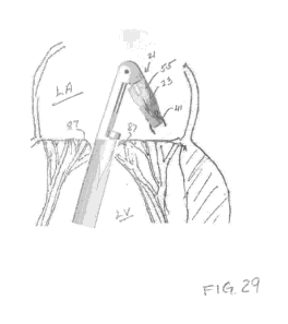
CA 02929498 2016-05-03
WO 2015/063609
PCT/1B2014/002992
2
is more or generally parallel but opposite to the forward direction of the
handle. In
other words, the force pulls the stapler head generally toward the user. This
can
allow for staples to be applied to tissue such that their prongs are closer to
the user
than their crown or upper part, which can be advantageous due to the entry
point
into the heart in the apex, at its bottom, and the staples can be inserted so
that their
prongs are in the direction of the apex.
[005] The surgical stapler can be made of sufficiently small proportions
that it can be effectively delivered to a desired chamber of a patient's
heart, either
via transapical passage or via entrance through some other opening created in
the
heart wall. For example, delivery of a stapling device into the left atrium of
the
heart can be carried out preliminary to effecting annuloplasty of the rnitral
valve.
The surgical stapler is preferably delivered to the desired heart chamber
encased
within an encompassing sheath along a guide wire that was earlier inserted.
Once
the desired location is reached, a stapling device is caused to emerge from
the
sheath within the heart chamber where it can affect the desired procedure,
e.g.
stapling to constrict tissue of the mitral valve annulus and thus
advantageously
changing the shape of the valve to minimize regurgitation and render it again
fully
operative.
[006] The stapler design comprises a stapling head that is hinged to a body
at the distal end of an elongated barrel that extends distally from a handle.
The head
includes mechanism for holding one generally M-shaped surgical staple having
two
legs terminating in stiff, sharpened prongs that point back at the proximal
end of the
handle. Preferably, the barrel of the handle contains a magazine of surgical
staples
which can thereafter be individually loaded into the stapling head.
[007] The illustrated stapler is adapted to implant M-shaped surgical
staples of the type generally shown in U.S. Patent No. 5,725,554 to Simon et
al.
where the application of force against a pair of shoulders presses the
undersurface of
the crown connector against an anvil; the fundamentals of this stapler design
may be
adapted to construct a device that would use shape memory staples. More
particularly, the illustrated stapler is adapted to implant staples of the
general type
shown in U.S. Patent No. 8,475,491 having a ring connector extending laterally
CA 02929498 2016-05-03
WO 2015/063609
PCT/1B2014/002992
3
from at least one leg that become interconnected with one another to form a
chain.
[008] The stapling head is pivotable away from the longitudinal axis of the
barrel of the handle sufficiently to expose the prongs of the staple in a
position
where they can be caused to penetrate the tissue of the annulus; in an
annuloplasty
procedure, the prongs will remain in an orientation where they are pointed
proximally, i.e. generally back toward the handle. This is because the pivot
point of
the stapling head is located more distal from the handle than is the stapling
head.
The surgeon manipulates the stapling device to position the staple at the
desired
location along the valve annulus, where it is to be implanted in a manner so
as to
constrict the valve tissue. There is further benefit in using interconnected
staples,
such as shown in the '491 patent, which not only constrict the annulus, but
prevent
subsequent remodeling of tissue that can occur if non-interconnecting staples
are
used because the tissue between staples can elongate over time.
[009] Optionally, the distance between the staple legs can be altered
before
their penetration into the tissue, e.g. while already loaded into the stapling
head, in
order to set the desired amount of constriction of tissue that will occur upon
implantation of that particular staple. Preferably, the stapling head is
reloadable
while in place within the heart chamber, e.g. by pivoting back to its initial
at rest
position, generally aligned with the longitudinal axis of the handle, where a
single
staple can be grasped from the magazine by a pusher and slid generally along
the
longitudinal axis and delivered into the stapling head. In addition to the
stapling
head being pivotable, preferably for at least about 80 from its initial at
rest
orientation in juxtaposition with the body part at the distal end of the
handle, the
head, once pivoted, can also be rotated about its axis in either direction, as
can the
barrel of the stapler itself.
[0010] In one particular aspect, a surgical stapler comprises a control
handle
having a forward-extending barrel having a central axis, a stapling device at
the
distal end of said barrel, said stapling device comprising a stapling head
part, a
stapler body part, a hinge connection which pivotally interconnects said two
parts,
and a generally M-shaped surgical staple having two legs with prongs at the
respective ends, said stapling head part comprising mechanism which holds one
CA 02929498 2016-05-03
WO 2015/063609
PCT/1B2014/002992
4
such surgical staple and implants said staple in tissue by causing said two
legs to
move toward each other, after puncturing a patient's tissue, to reach a secure
final
position constricting said tissue, and drive mechanism which pivots said
stapling
head part to at least about 15 from an at rest position, juxtaposed with said
stapler
body part, to an active position for implantation of the staple into tissue in
a
generally proximal direction.
[0011] In another particular aspect, the invention provides a surgical
stapler
which comprises a control handle having a forward-extending barrel having a
central axis, a stapling device at the distal end of said barrel, said
stapling device
comprising a stapler body part, a stapling head part pivotably binged to said
body
part, and a surgical staple having two legs with prongs at the respective
ends,
oriented to point proximally, said stapling head part comprising mechanism
which
holds one such surgical staple and implants such staple in tissue by causing
said two
legs to move toward each other, after puncturing a patient's tissue, to a
secure final
position constricting said tissue, and drive mechanism which can pivot said
head.
part to at least about 15 from an at rest position, juxtaposed with said body
part, to
an active position for implantation of the staple into tissue in a generally
proximal
direction.
[0012] In a further particular aspect, the invention provides a surgical
stapler
which comprises a handle having a forward-extending barrel, a stapling device
at
the distal end of said barrel, said stapling device comprising a stapler body
part, and
a stapling head part pivotably hinged to said stapler body part, said stapling
head
part comprising mechanism which holds one surgical staple, having two legs
with
prongs at respective ends being positioned at a location proximal of the pivot
point,
and implants such staple in tissue by causing said two legs to move toward
each
other, after puncturing a patient's tissue, to a secure final position
constricting said
tissue, a magazine in said barrel containing a plurality of surgical staples,
mechanism which can withdraw one of the surgical staples from said magazine
and
deliver such, with its prongs pointed in the direction of said handle, into
said
stapling head part at a time when said stapling head part is pivoted into its
at rest
juxtaposition adjacent said stapler body part, and mechanism which can pivot
said
CA 02929498 2016-05-03
WO 2015/063609
PCT/1B2014/002992
stapling head part to at least about 15' from an at rest position juxtaposed
with said
stapler body part to an active position for implantation of the staple into
tissue in a
generally proximal direction.
[010] In a still further particular aspect, the invention provides a method of
repairing a patient's leaking mitral valve, which method comprises the steps
of (a)
inserting an endoscopic surgical stapler, having a handle with an elongated
body
and having a stapling head part, which carries a surgical staple with its
prongs
oriented in a direction pointing toward the handle disposed, in folded
condition with
said elongated body, into the left ventricle of a patient's heart through a
transapical
passageway, (b) moving said stapler through the valve opening between the
leaflets
of the mitral valve into the left atrium, (c) unfolding said stapler to expose
the
stapling head part, (d) implanting said staple into the valve annulus adjacent
the
posterior leaflet with its prongs still oriented in a direction generally
toward the
handle, whereby the tissue is constricted where the staple is implanted, (e)
refolding
said stapler and reloading another staple into said stapling head part, (f)
unfolding
the reloaded stapler and implanting another staple adjacent the implanted
staple, (g)
repeating steps (e) and (f) to adequately change the shape of the mitral valve
annulus so as to effect improved closing of the mitral valve, and (h)
refolding said
stapler and withdrawing it from the heart of the patient.
Brief Description of the Drawines
[011] FIG. 1 is a perspective view showing one embodiment of a surgical
stapler incorporating various features of the invention, illustrated in
connection with
a guide-wire, such as is usually first inserted and then used to guide a
surgical
stapler to an operative location.
[012] FIG. IA is a perspective view like FIG. 1 shown from the opposite
side.
[013] FIG. 2 is a perspective view of the surgical stapler of FIG. 1
without
the guide-wire and with the flanged sheath shown in its retracted position.
[014] FIG. 3 is a fragmentary perspective view, enlarged in size, of the
distal end of the surgical stapler of FIG. 2 with the barrel rotated slightly
clockwise.
CA 02929498 2016-05-03
WO 2015/063609
PCT/1B2014/002992
6
[015] FIG. 4 is a perspective view similar to FiG. 3 with the barrel
rotated
further clockwise, showing the distal end of the stapler with the stapling
head
pivoted at an angle of about 200 to the axis of the elongated barrel of the
surgical
stapler.
[016] FIG. 5 is a perspective view similar to FIG. 4 showing such pivoting
to an angle of about 40 .
[017] FIG. 6 is a perspective view similar to FIG. 5 showing such pivoting
to an angle of about 65 .
[018] FIG. 7 is a perspective view similar to FIG. 6 showing the end
section of the stapling head rotated clockwise about 20 .
[019] FIG. 8 is a view similar to FIG. 7 showing the end section rotated
further clockwise to about 60 .
[020] FIG. 9A is a view similar to FIG. 8 with the end section shown
rotated counterclockwise about 90 .
[021] FIG. 9B is a perspective view of the distal end of the stapler as
seen
in FIG. 9A shown inverted and taken looking at the opposite surface.
[022] FIG. 10 is a view similar to FIG. 9B showing the stapling head
withdrawn slightly following implantation and schematically showing the
implanted
staple.
[023] FIG. 11 is a view similar to FIG. 10 showing the end section rotated
back to its zero position and pivoted back part way toward its at rest
position,
continuing to show the schematic location of the implanted staple while also
schematically showing the location in the magazine of the most distal staple.
[024] FIG. 12 is a view similar to FIG. 11 with the stapling head
juxtaposed with the body portion and in the process of receiving the staple
from the
magazine for reloading.
[025] FIG. 13 is a view similar to FIG. 12 showing the reloaded stapling
head.
[026] FIG. 14 is a view similar to FIG. 5 showing the stapling head
carrying the reloaded staple after having pivoted from the at rest position.
[027] FIG. 15 is a view of the distal end of the stapler shown in FIG. 14
CA 02929498 2016-05-03
WO 2015/063609
PCT/1B2014/002992
7
with the end section rotated and the stapling head pivoted to align one prong
of the
staple that it carries with the ring on the right hand side of the implanted
staple.
[028] FIG. 16 is a view similar to FIG. 15 showing the position where there
would be initial penetration of the prongs of the staple into the heart valve
tissue
with one prong having passed through the ring.
[029] FIG. 17 is a view similar to FIG. 10 schematically showing the two
implanted staples and the stapling head withdrawn slightly.
[030] FIG. 18 is a view having the orientation of FIG. 11 showing the
stapling head reloaded with a staple oriented so that its lateral ring lies in
the
opposite orientation to that shown in FIGS. 11-14.
[031] FIG. 19 is a view similar to FIG. 16 showing the staple being
implanted with its leg passing through the ring on the left hand side of the
original
implanted staple.
[032] FIG. 20 is a view similar to FIG. 17 showing the 3rd staple having
been implanted and the stapling head withdrawn slightly.
[033] FIG. 21 is a view showing the stapling head having been pivoted
back to the at rest position following implantation and the relative
retraction of the
stapling device distal end of the barrel to a location partially within the
sheath.
[034] FIG. 22A is a fragmentary perspective of the sheath seen in FIG. 1 ,
showing the tunnel along one lateral edge through which the guide-wire passes.
[035] FIG. 223 is a perspective view looking at the proximal end of the
flanged sheath of FIG. 22A showing the small entrance into the tunnel and the
interior unidirectional valve near the proximal end of the sheath which would
prevent the outflow of blood were the barrel end of the stapler to be
withdrawn
while the sheath remained in a transapical passageway.
[036] FIG. 23 is an enlarged fragmentary perspective view of the stapler of
FIG. 1 showing the barrel schematically.
[037] FIG. 24 is a schematic view illustrating the entry of the distal end
of
the stapler of FIG. 1 through a transapical opening into the left ventricle
(LV) of the
human heart.
[038] FIG. 25 is a view similar to FIG. 24 where the stapler has been
CA 02929498 2016-05-03
WO 2015/063609
PCT/1B2014/002992
8
further inserted so the leading guide-wire extends between the leaflets of the
mitral
valve into the left atrium (LA).
[039] FIG. 26 is a view similar to FIG. 25 where the distal tip of the
sheath
has been inserted to pass into the LA between the leaflets of the mitral valve
and the
guide-wire has been withdrawn a substantial distance into a tunnel along the
side of
the sheath.
[040] FIG. 27 is an enlarged fragmentary view similar to FIG. 26 wherein
the guide-wire has been withdrawn completely into the tunnel and relative
movement of the sheath on the barrel of the stapler causes the distal end of
the
stapler to emerge within the LA.
[041] FIG. 28 is a view similar to FIG. 27 showing sufficient relative
movement of the sheath along the barrel of the stapler so that the stapling
device is
totally exposed within the LA.
[042] FIG. 29 is a view similar to FIG. 28 showing the stapling head
pivoted into an operative position similar to that shown in FIG. 9B.
Detailed Description
[043] Illustrated in FIG. 1 is a surgical stapler 11 which comprises a
control handle 13 that is formed with a grip portion 15 at its proximal end
and an
elongated barrel portion 17 which extends forward therefrom. A flanged sheath
or
introducer 19 is preferably carried on the barrel portion 17 to envelop and
shield it
as the surgical instrument is inserted percutaneously into the chest of the
patient and
into the heart. The surgical stapler 11 is designed for insertion through an
incision
in the chest of the patient, where it is passed through the apex of the heart,
for
example, into the left ventricle (LV), and then through the mitral valve into
the left
atrium (LA). It will be understood that this basic stapling device can be used
for
other stapling operations within the heart and that it can likewise be
developed for
other desired particular endoscopic stapling procedures. if adapted for use
within a
catheter, the sheath may not be employed.
[044] Encased protectively within the sheath 19 is a stapling device 21 at
the distal end of the barrel 17 which comprises a stapling head 23 that is
pivotably
CA 02929498 2016-05-03
WO 2015/063609
PCT/1B2014/002992
9
connected to a stapler body 25 by a hinge region or connection 27. When
relative
movement is effected so that the flanged sheath 19 becomes retracted in a
proximal
direction, its pointed, split tip 29 is spread apart to cause the emergence of
the
stapling device 21. A flat flange 31 aligned perpendicular to the longitudinal
axis of
the sheath is located at its proximal end. The flange 31 allows manipulation
of the
sheath relative to the elongated barrel 17 of the stapler; however, other
devices for
manipulating the sheath relative to the barrel of the stapler could
alternatively be
employed. The sheath 19 is also formed with a tunnel 33 extending along its
entire
length through which a guide-wire 34 can be conveniently passed, and a small
entrance 35 to the tunnel 33 can be seen at the proximal end of the flanged
sheath
(see FIG. 22B). The sheath also includes an interior unidirectional valve 37
the
purpose of which will be explained hereinafter.
[045] From FIG. 1, it can be seen that the elongated barrel 17 of the
stapler
is stepped at the location 39, and the sheath 19 is slidably received on the
lesser
diameter, distal portion of the barrel. Thus, the sheath 19 can be withdrawn
no
further proximally than the orientation shown in FIG. 2 where it abuts the
step 39; at
this relative location, the stapling mechanism has been exposed to its
operative
position. Moreover, the length of the sheath 19 and the barrel are used as a
safety
feature so as to prevent the insertion of the distal end of the stapler too
far through
the mitral valve where it might cause injury at the upper end of the LA; for
example,
the length of the sheath might be sized such that the flange 31 might abut the
skin of
the patient and thereby limit the insertion of the distal tip of the stapler.
Although a
stapler might be designed to interconnect a guide-wire with the barrel and
exclude a
sheath, a protective sheath or introducer is preferred.
[046] The stapling head 23 is designed to hold and implant a single staple
41. After such implantation has been accomplished, the head 23 is returned to
an
"at rest" position in juxtaposition with the body 25 where it is preferably
reloaded
with a staple 47 from a magazine 43 of staples contained within a hollow
region 45
within the elongated barrel 17 located proximal of the body part 25 that is
constructed at the distal end. The magazine 43 contains a series of staples 47
within
a cartridge of cylindrical exterior surface that resides in the cylindrical
hollow
CA 02929498 2016-05-03
WO 2015/063609
PCT/1B2014/002992
region 45 provided in the elongated barrel 17 of the stapler, as best seen in
FIG. 23.
The illustrated staples 47 which are loaded into the magazine are of a design
similar
to the staple 41 but have a ring extending laterally from only one leg of the
staple,
the reason for which being explained hereinafter. The generally M-shaped
staples
47 are formed with an essentially planar or flat body that comprises a pair of
rigid
legs each of which ends in a sharpened prong and a crown connecter. In its
initial
configuration, the crown connector has the general shape of a U; its U-shape
is
flattened to a substantially straight configuration upon implantation. In the
illustration shown in FIG. 23, the ring of the staple 47 is shown affixed to
the left
hand leg of each of the staples in the magazine, the bodies of which lie in a
common
plane aligned to include the centerline of the barrel, with the ring being
perpendicular to that plane. The very first staple 41 that is initially
implanted has
two symmetrical 0-rings, as well seen in Figs. 3-10. This first staple 41 may
be
mounted in the stapling head 23 by the manufacturer; the remaining staples 47
from
the magazine 43 will all have one 0-ring only. This first staple having two
symmetrical 0-rigs is preferably deployed first at the middle of the annulus
of the
posterior mitral leaflet. The following staples can be deployed alternatively
on each
side, one at a time, allowing thus symmetrical shortening of the annulus of
the
posterior leaflet.
[047] FIGS. 1 and IA show a surgical stapler ii embodying various
features of the present invention from both sides. The stapler 11 is
illustrated with
the flanged sheath 19 at its distal end and with a guide-wire 34 extending
through
the tunnel 33 along the side of the sheath and protruding from its distal end.
FIG. 2
shows the surgical stapler 11 without the guide-wire and with its distal end
protruding from the split end 29 of the sheath as a result of relative
movement
between the sheath 19 and the elongated barrel 17 of the stapler. Such
movement is
facilitated by the flat flange 31 at the proximal end of the sheath, and it
may be
accomplished by manual withdrawal, or by pressing the flange against the flesh
of
the patient's chest adjacent the percutaneous entry slit through which the
distal end
29 of the sheath is inserted. Insertion of the surgical stapler 11 will
usually follow
along a guide-wire 34 that was previously put in place, as explained in more
detail
CA 02929498 2016-05-03
WO 2015/063609
PCT/1B2014/002992
11
hereinafter.
[048] In FIG. 2, the sheath 19 is shown as having been retracted to the
step
39 in the elongated barrel portion 17 of the handle so it exposes the stapling
device
21 at the distal end of the barrel. The stapling device 21 comprises the
stapling head
part 23 that is connected to the stapler body part 25 at the end of the barrel
by a
hinge connection 27 so that it can be pivoted outward from its at rest
position shown
in FIG. 2 where it juxtaposes with the stapler body part. FIG. 3 is an
enlarged
fragmentary perspective of the distal end of the surgical stapler, taken from
a
different angle than that in FIG. 2, which shows a staple 41 that is held in
the
stapling head 23 with its prongs at the end of its two legs pointed proximally
back at
the grip portion 15 of the handle.
[049] FIG. 4 shows the stapling head 23 having pivoted about 15 from its
juxtaposed position with the stapler body part 25, exposing the stapling head
and the
prongs at the ends of the staple 41. FIG. 5 shows the pivoting further to an
angle of
about 30 to the longitudinal centerline of the barrel, and it illustrates a
cavity 49
within the stapler body part 25 where the staple 41 and a portion of the
stapling
bead are received in the at rest position. The pivoting movement of the
stapling
head 23 is effected by the control handle by rotation of a knurled wheel 51 on
the
grip portion 15 of the control handle. This knurled wheel 51 at the upper
ridge of
the grip portion 15 of the control handle connects via mechanism that
traverses the
length of the elongated barrel 17 to cause the pivoting of the stapling head
23.
[050] In FIG. 6, the stapling head 23 is shown as having pivoted to about
60' from its at rest position juxtaposed with the body part. It is believed
that the
stapler should be designed to pivot the stapling head 23 at least about 60",
and
preferably at least about 80' to facilitate the desired annuloplasty procedure
for
which it has been designed. However, the stapler might also be designed so as
to
pivot the stapling head up to 180 , i.e. so that it extends straight distally
from the
barrel, if desired for some particular endoscopic stapling procedure.
Generally, the
operation of the surgical stapler 11 will be such that pivoting of the
stapling head 23
for at least about 15 and preferably for at least about 25 will be effected
so as to
space the staple sufficiently offset from the elongated barrel to allow the
staple to be
CA 02929498 2016-05-03
WO 2015/063609
PCT/1B2014/002992
12
implanted in a generally proximal direction without interference from the
presence
of the adjacent barrel.
[051] The stapling head 23 is formed with a base section 53 and a rotatable
end section 55, as can be seen in FIG. 7 where the end section has been
rotated
about 200 clockwise from its initial zero position in the at rest orientation
of the
stapling head. The rotation of the end section 55, which carries the staple
41,
relative to the base section 53 is controlled by another knurled wheel 57
located on
the left hand side of the grip portion 15 of the control handle, which
likewise
contains linkage extending through the barrel 17 to the hinge region 27 at the
distal
end of the surgical stapler. FIG. 8 shows further rotation clockwise to about
600
from the zero position. Rotation can be in either direction, clockwise or
counter-
clockwise, from the zero position, and FIG. 9A shows rotation of the end
section 55
of the stapling head 23 about 900 in the counter-clockwise direction from the
zero
position. FIG. 9B illustrates the distal end of the stapler with an inverted
orientation
to that in FIG. 9A, and it is noted that the staple 41 in the rigid stapling
head 23
remains oriented with its prongs pointing proximally, i.e. in the general
direction
back toward the grip portion 15 of the control handle. With this orientation
of the
rigid head 23 when stapling into the tissue, force can controllably be applied
to the
staple so that its prongs are pushed against the tissue with precise force.
With the
stapling head 23 being generally pointed toward the handle, the surgeon can
carefully pull back on the control handle 13 with steady exact motion when the
stapling head is adjacent the tissue and at a sharp (but inverted) angle to
the barrel
centerline. As a result, the desired force vector is readily created,
essentially pulling
the rigid stapling head 23 and the loaded staple towards the tissue with the
staple
oriented transverse, and preferably generally perpendicular, to the tissue
surface.
[052] FIG. 10 illustrates the distal end of the surgical stapler after the
staple
41 has been implanted into the tissue and the stapling head 23 has been
slightly
withdrawn. The implanted staple 41 is shown schematically with its central
crown
connector 61 now straightened and with its legs 59 crimped so that the prongs
lie
adjacent each other. The staple 41 has two rings 63 extending laterally
respectively
from the two legs 59 which are employed to form an interconnected chain of
staples
CA 02929498 2016-05-03
WO 2015/063609
PCT/1B2014/002992
13
that effects the desired annulopla,sty as disclosed in the '491 patent. The
stapling
head 23 in FIG. 10 is shown in its open position exposing its holder region 65
wherein an M-shaped staple is received and held within the holder by a
pivoting
clamp 67. The clamp 67 is shown in its release position to which it is moved
following the implantation of the staple.
[053] In FIG. II. the stapler is illustrated where the stapling head end
portion 55 has been rotated back to its zero position and where the head 23
has been
pivoted toward the body part 25 as it is returning to its at rest, juxtaposed
location.
The clamp 67 remains in its release or open position during return to the at
rest
position.
[054] As best seen in FIG. 23, the hollow barrel 17 includes a plurality of
staples 47 disposed in a cylindrical holder or magazine 43 and aligned so that
each
staple lies with its body in a common plane that preferably includes the
central axis
of the barrel. The staples 47 in the magazine have only a single ring disposed
laterally from one leg. In FIG. 23, the magazine is oriented so that the ring
is
attached to the leg at the left hand side of the staple within the barrel. For
convenience of the surgeon, the stapler 11 is designed so that the magazine 43
can
be rotated 180" so that the ring-carrying leg of the staple is at the right
hand edge of
the staple in the barrel of the stapler. Rotation of the magazine for 180' is
effected
by a slide 69 near the distal end of the grip portion 15 of the control
handle. The
slide 69 can be moved transversely across the diameter of the grip portion 15
and is
arranged so that when the end of the slide 69 protrudes from the left hand
side of the
grip portion, the staples are so orientated as seen in FIG. 23 with the ring-
carrying
leg to the left. When the slide is pressed inward to the right so that it
protrudes from
the right hand surface of the grip portion, the magazine has been rotated 180'
so that
the ring is now attached to the leg on the right. This can result in omitting
the need
of rotating the entire, fixed delivery device in 180" in order to enable
symmetrical
deployment on alternative sides of the initial, double 0-ring, first staple.
[055] FIG. 11 schematically illustrates the most distal staple 41 in the
magazine with its ring carrying leg oriented on the left hand side of the
staple. The
illustrated staple is a M-shaped staple wherein the crown connector is formed
in an
CA 02929498 2016-05-03
WO 2015/063609
PCT/1B2014/002992
14
essentially U-shape, in which shape it initially exists until it is implanted
by action
thereupon by a former mechanism as explained hereinafter.
[056] FIG. 12 schematically shows the stapling device 21 having been
returned to its at rest position. In FIG. 12, the barrel has been rotated 180'
from the
orientation of FIG. 1. Rotation of the entire barrel 17 relative to the grip
portion 15
for 360 is accomplished by turning the transversely oriented knurled wheel 72
at
the distal end of the grip portion. In this orientation, the ring-carrying leg
is on the
right hand side relative to the grip portion 15. The staple 47 is shown in the
process
of being delivered into the holder region 65 of the stapling head 23 by an
extraction
and loading mechanism 73 that includes a pusher which engages a proximal
facing
surface of the staple. In this position, the clamp 67 remains in the open
position so
as to receive the staple 47.
[057] In FIG. 13, the delivery of the staple 47 has been completed, and the
extraction and loading mechanism 73, which is operated by a lever 74 located
in a
slot in the undersurface of the grip portion 15, has closed the spring-loaded
clamp
67 and loaded the spring-loaded former mechanism 71 so that it is cocked and
ready
to implant the staple 47. The clamp 67 includes an anvil section 75 at its end
which
is moved into abutment with the facing surface of the holder region 65 in the
loaded
condition so that it lies adjacent the undersurface of the U-shaped crown
connector
of the staple 47.
[058] FIG. 14 shows the stapling head 23 (with the staple 47 loaded)
pivoted away from the body part 25 to expose the prongs of the staple which
are
pointed proximally relative to the grip portion 15. FIG. 15 shows further
pivoting of
the stapling head 23 and rotation of the end section 55 relative to the base
53 to
align the staple 47 so that its leg which does not carry the ring is aligned
with the
center of the ring 63 on the right hand side of the implanted staple 41. FiG.
16
shows subsequent movement by the surgeon of the surgical stapler 11 so that
the leg
of the staple 47 passes through the ring 63 and now penetrates the heart valve
tissue
of the patient.
[059] Sensors are located in the stapling head 23 so as to determine that
both prongs of the staple 47 are symmetrically penetrating into the heart
valve tissue
CA 02929498 2016-05-03
WO 2015/063609
PCT/1B2014/002992
as indicated by having surpassed a minimal determined pressure threshold. Such
sensors may be mechanical or electronic and are designed to send a signal to a
trigger mechanism 77. The trigger mechanism 77 includes a pair of triggers 79
disposed substantially coaxially on opposite sides of the grip portion 15 of
the
handle, and simultaneous pressing of the two oppositely disposed triggers 79
is
required to actuate the trigger mechanism 77. This spatial disposition of a
pair of
coaxial triggers 79 positively guards against the surgeon inadvertently
slightly
moving the control handle at the movement of implantation. The trigger
mechanism
77 actuates the spring-loaded former mechanism 71 which presses the upper
surfaces of the shoulders of the M-shaped staple 47 forward, causing the U-
shaped
crown connector with its undersurface resting on the anvil 75 to be reshaped
into a
straight connector, as the two legs move past one another to the orientation
shown in
FIG. 17 where the staple 47 has become interconnected with the staple 41 that
was
first implanted.
[060] FIG. 18 shows the stapling head 23 having been returned to
juxtaposition with the body part 25 at the distal end of the barrel and loaded
with
another staple 47. In FIG. 18, the staple is oriented at 180' from the staple
47
carried in the stapling head in FIG. 14. Thus, the staple 47 was loaded into
the
stapling head 23 from the magazine 43 after it had been rotated 180' so that
the ring
of the staple is attached to the leg on the opposite side compared to that
shown in
FIGS. 12 and 13.
[061] FIG. 19 depicts the stapler 11 being manipulated by the surgeon so
that a leg of the staple 47 protrudes through the ring 63 on the left hand
side of the
initially implanted staple 41 and ready to be implanted in the heart valve
annulus.
FIG. 20 shows the completion of the implantation and the slight removal of the
stapling head 23; it provides a good view of the holder region 65 where the
initial
M-shaped staple is received and also shows the track 81 along which a part of
the
former mechanism 71 moves as it deforms the M-shaped staple to the final
implanted shape. Finally, FIG. 21 shows the stapling head 23 having been moved
to
its juxtaposed, at rest position, and after initial proximal withdrawal
movement of
the barrel. As a result, the flanged sheath or introducer 19 is beginning the
CA 02929498 2016-05-03
WO 2015/063609
PCT/1B2014/002992
16
encasement of the stapling head preparatory to extracting the distal end of
the
stapler from between the leaflets of the mitral valve.
[062] The surgical stapler 11, as indicated hereinbefore, can be adapted
for
a variety of endoscopic uses; the concept of being able to implant a staple
with its
prongs oriented proximally, i.e. in a direction generally back toward the
handle grip
of the stapler, and particularly at an angle of about 45' or less to the
longitudinal
axis of the stapler, is believed to be unique. However, the surgical stapler
11
illustrated in the drawings, which embodies various features of this inventive
concept, is particularly designed for effecting annuloplasty of a heart valve,
particularly the mitral valve, which is a hi-leaflet valve that is prone to
suffer a
pathological condition. The particular design of the illustrated surgical
stapler Ills
such that providing access for it to a beating heart through its apex into the
left
ventricle (LV) is advantageous. To prepare for such entry, a guide-wire 34 is
usually first inserted in the left ventricle (LV) through a hollow needle
passed
through the apex; for example, a device such as that disclosed in Published
International Application No. WO 2013/027107 may be used. With the guide-wire
34 in place, its proximal end is fed through the tunnel 33 provided in the
side wall of
the flanged sheath 19 which extends from a location near the pointed tip of
the
sheath to the opening 35 in the proximal flange 31.
[063] With the sheath 19 in place encasing the stapling device 21 at the
distal end of the surgical stapler, it is inserted through the apex of the
heart into the
left ventricle as schematically shown in FIG. 24. The guide-wire 34 is formed
to
have a memory such that the distal tip 85 of the wire bends over upon itself
to
provide a curved forward-facing surface to assure smooth passage between the
leaflets 87 of the mitral valve and into the left atrium. Insertion of the
stapler so that
the curved distal tip 85 of the guide-wire 34 passes into the left atrium (LA)
is
depicted in FiG. 25. All such movement is guided by X-ray fluoroscopy or
preferably by continuous 3D real-time echocardiography.
[064] Next, the stapler Ills caused to travel along the guide-wire 34, and
the pointed tip of the sheath slides between the leaflets 87 to enter the left
atrium, as
illustrated in FIG. 26. Once the pointed tip 29 of the sheath resides in the
left
CA 02929498 2016-05-03
WO 2015/063609
PCT/1B2014/002992
17
atrium, the guide-wire is withdrawn into the tunnel 33 that extends along the
side of
the sheath, as shown in FIG. 26 where such partial withdrawal is depicted.
With the
guide-wire 34 withdrawn, the elongated barrel 17 of the stapler is moved
relative to
the sheath 19, as shown in FIG. 27 so that the stapling device 21 begins to
emerge
from the distal end of the opened split tip 29 of the sheath 19.
[065] FIG. 28 shows the flanged sheath 19 withdrawn to a position where
the flange 31 would reside at about the stepped portion 39 of the barrel, at
which
location the stapling device 21 is fully exposed and residing in the left
atrium. The
tips of the split end 29 of the sheath are located at about the region of the
mitral
valve leaflets 87. Having reached this location, the stapling head 23 is
pivoted away
from its at rest juxtaposition with the stapler body 25, and the end portion
55 is
rotated so as to position the staple 41, carrying two rings 63, so it is
aligned with the
mitral valve annulus at about the midpoint of the posterior leaflet. The
staple 41 is
implanted in this location in the manner earlier described with respect to
FIGS. 98
and 10, and the stapling head 23 is then reloaded with one staple 47 at a time
from
the magazine 43 to create a chain of interconnected staples taking steps as
shown in
FIGS. 15-20.
[066] Although the implantation of only 3 staples is illustrated, it should
be
understood that the surgeon will implant a desired number of staples 47 on
each side
of the central staple 41 along the valve annulus to achieve the desired amount
of
constriction of the annulus, as described generally in U.S. Patent No.
8,123,801 and
in the '491 patent. The objective of the procedure is to counteract the
pathological
condition of the mitral valve in order that the leaflets 87 again co-apt to
effectively
close the valve and prevent, or at least minimize, regurgitation during the
pumping
stroke of the left ventricle.
[067] In this respect, the surgical stapler 11 is optionally equipped with
a
crimping mechanism 89 which can effectively change the spacing between the
sharpened prongs at the end of the two legs of a staple. The mechanism 89 is
located in association with the holder region 65 in the end section 55 of the
stapling
head 23, and it is designed to apply inward lateral pressure to the exterior
lateral
surfaces of the stiff legs of the staple to deform them toward each other. For
CA 02929498 2016-05-03
WO 2015/063609
PCT/1B2014/002992
18
example, the staples may be made from stainless steel or from Co-Cr alloy of
comparable stiffness. For instance, the staples 41 and 47 initially loaded in
the
surgical stapler might be formed so the sharpened tips are spaced from each
other
about 7.5 mm, and the crimping mechanism 89 can reduce spacing, for example to
about 5.5 mm, which might be about the length of the crown connector in its
straightened implanted form. The crimping mechanism is operated by a slide 91
located on the grip portion 15 of the handle in the region between the knurled
wheels 51 and 57, as best seen perhaps in FIG. 23. Movement of the slide 91
proximally from its at rest position shown in FIG. 23 effects laterally inward
bending of the legs of the staple then loaded in the stapling head 23 via
linkage that
extends through the elongated barrel portion 17 of the handle. This inclusion
within
the surgical stapler of such a crimping mechanism allows a surgeon to achieve
the
precise amount of tissue constriction desired with each staple to effect the
reshaping
of the mitral valve annulus and create an effective annuloplasty, as each
staple can
be set with its prongs any desired distance between about 7.5 mm and 5.5 mm
for
example. Although the annuloplasty operation is illustrated and described for
a
procedure where a first implanted staple 47 having two rings 63 is located in
the
annulus centrally of the posterior leaflet, it should be understood that a
staple could
be, if desired, positioned at either end of the desired chain near a trigon
and that
only staples 47 having a single ring might be used. For example, a chain may
be
begun near one trigonal region and extend arcuately along the mitral valve
annulus
in one direction until the desired amount of constriction was obtained by the
surgeon.
[068] Moreover, it should be understood that the surgical instrument is
such that if, while operation on a beating heart is being performed, it
becomes
desired to interrupt the staple implantation procedure because the patient
ceases to
tolerate too long a period of mitral incompetence, such can be accommodated to
permit revival of the natural blood flow throughout the patient. After
implantation
of several staples 47 from the magazine, the stapling device can be retracted
within
the sheath (as shown in FIG. 21) and withdrawn into the left ventricle for a
time
sufficient to effect such revival. Thereafter, the guide-wire 34 can be again
CA 02929498 2016-05-03
WO 2015/063609
PCT/1B2014/002992
19
extended from its location in the tunnel 33 and directed between the mitral
valve
leaflets and into the left atrium as seen in FIG. 25, preparatory to guiding
the stapler
back into its operative position. If for whatever reason it might be necessary
to
remove a stapler midway through a surgical procedure, the elongated barrel
could
be removed, leaving the sheath 19 extending into the 1_,V and the guide-wire
34 in
place; the interior unidirectional valve 37 will block the outflow of blood
while a
substitute stapler is prepared and then inserted along the guide-wire and
through the
sheath.
[0691 Although the invention has been described and illustrated in terms
of
the best mode presently understood by the inventors to perform such an
annuloplasty, it should be understood that various changes and modifications
to the
devices illustrated made be made without departing from the scope of the
invention,
which is defined in the claims appended hereto. Furthermore, various features
of
the invention are emphasized in the claims that follow.