Note: Descriptions are shown in the official language in which they were submitted.
81796660
Tissue Scaffold Materials for Tissue Regeneration and Methods of Making
CROSS-REFERENCE TO RELATED APPLICATIONS
[0001] This application claims priority to U.S. provisional application
61/906,131, filed
November 19, 2013.
BACKGROUND OF THE DISCLOSURE
[0002] The optimization of cell guidance through autologous or artificial
tissue scaffolds has
long been a topic of great interest. The most prevalent and thus far the most
successfully
applied off-the-shelf "tissue-engineered" products were all originally
intended to serve as
dermal replacement scaffolds. Commercially available scaffolds are acellular
and thus share
the common requirements of host cell invasion and vascularization to achieve
durable
incorporation. Because this process is prolonged, requiring a minimum of
several weeks for
completion and necessitating obligatory dressing changes, wound
immobilization, and
nursing care, there is significant interest in developing better scaffolds
that could optimize the
rate of cellular invasion. (Eppley, Plast Reconstr Surg. 107:757-762 (2001);
Wong et al.,
Plast Reconstr Surg. 121:1144-1152 (2008)).
[0003] Currently available acellular dermal replacements can be categorized
into two broad
groups: products derived from decellularized den-nis, and synthetic products
based on
naturally-derived hydrogels (Truong et al. J. Burns Wounds 4:e4 (2005)).
[0004] Commercially available decellularized dermal products are made of
decellularized
cadaveric porcine or human dennis. As a result of the decellularization
process, these
products contain an internal network of microchannels with an intact basement
membrane
that are the remnants of the native dermal microvasculature.
[0005] INTEGRA (Integra LifeScienees, Plainsboro, NJ), another commonly
applied dermal
regeneration template., is comprised of a synthetic "dermal" porous layer of
cross-linked type
I bovine collagen and chondroitin-6-sulfate covered by an "epidermal" semi-
permeable
silicone sheet. Following implantation, the silicone sheet is replaced with
split-thickness
autograft once the dermal layer has vascularized (Yannas et al., Science
215:174-176
TM
(1982)). Unlike decellularized dermal products, INTEGRA is representative of
products
1
Date recu/Date Received 2020-04-20
CA 02929611 2016-05-03
WO 2015/077300 PCT/US2014/066344
without an internal vascular structure and is instead characterized by its
random porosity
(mean pore diameter 30-120pm) (van der Veen et al., Burns 36:305-321 (2010)).
[0006] The use of currently available tissue replacement scaffolds is not
without substantial
associated cost. For example, the production of decellularized dermal products
requires
tissue acquisition and harvesting, as well as decellularization and
sterilization processes (Ng
et al., Biomaterials 25:2807-2818 (2004)). In addition, commercially available
tissue
scaffolds are avascular and prone to high failure rates when used in complex
settings, such as
irradiated wounds or those with exposed hardware or bone. In such complex
settings,
neovascularization is insufficient using existing tissue replacement products.
[0007] Improved tissue scaffolds that promote optimal cellular invasion and
vascularization
of new and surrounding tissue are highly desired in the art.
BRIEF SUMMARY OF THE DISCLOSURE
[0008] Disclosed herein is a type of tissue scaffold material made of a
hydrogel with
embedded microspheres. In the disclosed tissue scaffolds, the microspheres
have a different
or greater density (w/v) of polymer relative to the density of the hydrogel,
which differential
density facilitates cellular invasion into the tissue scaffold. In one
embodiment, the hydrogel
includes a first polymer and the microspheres include a second polymer, the
microspheres are
embedded in the hydrogel, and the microspheres have a greater density than the
hydrogel.
[0009] The first and second polymers can be independently selected from the
group
consisting of of collagen, gelatin, elastin, hyaluronate, cellulose,
fibrinogen, poly(l actic-co-
glycolic acid) (PLGA), poly(glycolic acid) (PGA), poly(lactic acid) (PLA),
poly(caprolactone), poly(butylene succinate), poly(trimethylene carbonate),
poly(p-
dioxanone), and poly(butylene terephthalate); a polyester amide, a
polyurethane,
poly[(carboxyphenoxy) propane-sebacic acid], poly[bis(hydroxyethyl)
terephthalate-ethyl
orthophosphorylate/ terephthaloyl chloride], a poly(ortho ester), a poly(alkyl
cyanoacrylate),
poly(ethylene glycol), a microbial polyester, poly(I3-hydroxyalkanoate), and a
tyrosine
derived polycarbonate. In examples, the microspheres can contain 0.2% to 2.0%.
0.4% to
1.2%, 0.6% to 1.0%, or 1.0% w/v of the second polymer. In a particular
example, the second
polymer is collagen. The microspheres can be between 50-250 pm in diameter.
The
microspheres can fill at least about 50%, 60%, or 70% by volume of the tissue
scaffold
2
CA 02929611 2016-05-03
WO 2015/077300 PCT/US2014/066344
material. The microspheres can also contain bioactive factors, but in some
embodiments, the
microspheres do not contain bioactive factors. The bioactive factors can
promote one or
more of cellular invasion, cellular growth, or vascularization.
[0010] In one example, the hydrogel contains collagen. In some examples, the
hydrogel
contains collagen in an amount of 0.1% to 0.6%, 0.2 to 0.4%, or 0.3% w/v. The
tissue
scaffold material can have microspheres with 0.2% to 2.0%, 0.4% to 1.2%, 0.6%
to 1.0%, or
1.0% w/v collagen, embedded in a hydrogel with 0.1% to 0.6%, 0.2 to 0.4%, or
collagen w/v. In one embodiment, the tissue scaffold material has microspheres
with 0.6-
1.0% w/v collagen, embedded in a hydrogel containing 0.3% w/v collagen.
[0011] The tissue scaffold material of any of the above embodiments can be in
the form of a
sheet or in a flowable form. The material can be, for example, in the form of
a sheet with a
depth of 0.5-3.0 mm, or about 1.0-2.0 mm. The disclosed tissue scaffold
materials can be
used in a method of wound healing or tissue regeneration in a subject.
[0012] Further disclosed herein are methods to promote wound healing or tissue
regeneration
in a subject in need thereof, by applying the tissue scaffold material as
disclosed above or
herein to a wound or tissue of the subject. The tissue scaffold material can
be applied, for
example, to an area of the subject with exposed bone, hardware, or necrotic
tissue.
[0013] Also disclosed herein are methods of making a tissue scaffold material.
The methods
involve the steps of: (a) providing a first composition with microspheres, and
a second
composition with a polymer material, the first composition having a different
density than the
second composition; (b) mixing the first and second compositions; and (c)
causing
crosslinking of the polymer material in said mixture, to form a hydrogel with
embedded
microspheres. The first and second compositions can each contain collagen,
such as human
or bovine collagen, as a polymer. The collagen can be neutralized collagen.
The
microspheres can contain 0.4% to 1.2%, or 0.6-1.0% w/v of collagen. The
microspheres can
further contain bioactive factors. The second composition can contain 0.1% to
0.6%, or 0.3%
w/v of collagen. Crosslinking can be accomplished, for example, by thermal
methods.
[0014] Also disclosed are tissue scaffold materials produced by the methods
provided above
and further disclosed herein, and wound dressings comprising such tissue
scaffold materials.
3
81796660
[0014a] The present disclosure as claimed relates to:
- a tissue scaffold material comprising a hydrogel and microspheres, said
hydrogel comprising
a first polymer and said microspheres comprising a second polymer, wherein
said
microspheres are embedded in said hydrogel and have a density that is at least
25% greater
than the density of said hydrogel;
- use of the tissue scaffold material as described herein to promote wound
healing or tissue
regeneration in a subject in need thereof;
- a method of making a tissue scaffold material, comprising the steps of:
a. providing a first
composition comprising polymeric microspheres, and a composition comprising a
second
polymer material, the first composition having a density that is at least 25%
greater than the
density of said second composition; b. mixing the first and second
compositions; and c.
causing crosslinking of the polymer materials in said mixture, to form a
hydrogel with
embedded microspheres;
- a tissue scaffold material produced by a method comprising the steps of:
a. providing a first
composition comprising polymeric microspheres, and a second composition
comprising a
polymer material, the first composition having a density that is at least 25%
greater than the
second composition; b. mixing the first and second compositions; and c.
causing crosslinking
of the polymer material in said mixture, to form a hydrogel with embedded
polymeric
microspheres; and
- a dressing comprising the tissue scaffold material as described herein.
3a
Date Recue/Date Received 2020-10-14
81796660
BRIEF DESCRIPTION OF THE FIGURES
[0015]
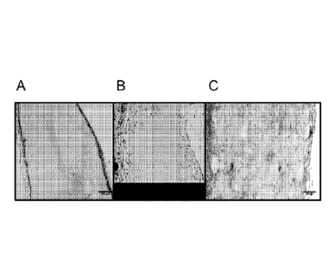
[0016] FIGS. IA-1C. At seven days post-implantation, cells infiltrate MSS
scaffolds (C) but
do. not infiltrate 1% bulk alone (A) and poorly infiltrate 0.3% bulk (B).
[0017] FIGS. 2A-2C. At fourteen days post-implantation, cells show excellent
infiltration of
MSS scaffolds (C) but do not infiltrate beyond outer portion of 1% bulk (A)
and show only
modest infiltration of 0.3% bulk (B).
[0018] FIGS. 3A-3D. At seven days post-implantation, cells show more complete
infiltration
of MSS scaffolds with 1% microspheres in 0.3% bulk (C), and 0.6% microspheres
in 0.3%
bulk (D), with less infiltration of 0.4% microspheres in 0.6% bulk (A) and
0.4% microspheres
in 0.2% bulk (B).
[0019] FIGS. 4A-4B. At seven and fourteen days post-implantation, cellular
infiltration of
1% microspheres in 0.3% bulk (blue staining, DAPI) includes endothelial
precursor CD31+
cells (red staining).
[00201 FIGS. 5A-5D. Seven days post-implantation. (A-C), identification of
MSS, 0.3%
bulk, 1% bulk and INTEGRA scaffolds in mouse. (D), relative sizes of scaffolds
after
implantation.
[0021] FIGS 6A-6C. Seven days post-implantation, cells infiltrate MSS scaffold
all the way
to the center of the scaffold (A) but do not infiltrate 1% bulk except where
scaffold is split
(B) and poorly infiltrate 3% bulk (C).
[0022] FIG. 7. Seven days post-implantation. DAPI nuclear staining (blue)
demonstrating
cell invasion to the center of the MSS and CD31+ endothelial precursors (red).
[0023] FIGS. 8A-8E. Fourteen days post-implantation. (A-D), identification of
MSS, 0.3%
bulk, 1% bulk and INTEGRA scaffolds in mouse. (E), relative sizes of scaffolds
after
implantation.
4
Date recu/Date Received 2020-04-20
CA 02929611 2016-05-03
WO 2015/077300 PCT/US2014/066344
[0024] FIGS. 9A-9D. Fourteen days post-implantation. (A), significant cellular
invasion in
MSS scaffold. (B), 1% collagen with minimal invasion (except along fissures).
(C), 0.3%
collagen scaffold with sparse invasion. (D), INTEGRA at 14 days also with less
robust
appearing invasion.
[0025] FIG. 10. Cell count per unit scaffold area shows that significantly
more cells invaded
the MSS scaffold at 7 and 14 days (approximately 7 cells and 10 cells per
area, respectively)
relative to 1% hydrogel (approximately 3 and 5 cells per unit area) and 0.3%
hydrogel
(approximately 3 and 7 cells per unit area).
[0026] FIGS. II A- -1 ID. Twenty eight days post-implantation. (A-C),
identification of MSS,
0.3% bulk, l % bulk and INTEGRA scaffolds in mouse. (D), relative sizes of
scaffolds after
implantation. Note 0.3% hydrogel is significantly shrunken.
[0027] FIGS. 12A-12D. Twenty eight days post-implantation. (A), excellent
cellular
invasion in MSS scaffold. (B), 1% collagen maintains minimal invasion (except
along
fissures). (C), 0.3% collagen scaffold shows uniform moderate invasion. (D),
INTEGRA
also shows reasonable invasion.
[0028] FIG. 13.Scanning electron microscopy of microspheres.
DETAILED DESCRIPTION OF THE DISCLOSURE
[0029] Disclosed herein are tissue scaffold materials with improved ability to
facilitate
cellular invasion and vascularization for wound healing and tissue
regeneration. The
inventors have found that materials having components with different densities
promotes
invasion of cells, including desirable cells such as fibroblasts and
endothelial precursor cells,
into the material.
[0030] The tert-ns "tissue scaffold". "tissue scaffold material", "dermal
substitute", "dermal
substitute material" and "material" are used interchangeably herein to refer
to a cell growth
support structure made of biocompatible polymer. These materials are capable
of
regenerating damaged tissues by providing a biocompatible template that
promotes cellular
invasion and tissue regeneration.
[0031] The tissue scaffold materials disclosed herein are composed of a
hydrogel support,
which is filled with microspheres. The microspheres have a density (the
density being
CA 02929611 2016-05-03
WO 2015/077300 PCT/US2014/066344
measured as weight by volume or w/v) that differs from the density of the
hydrogel in which
the microspheres are embedded. In a preferred embodiment, the microspheres
have a greater
density than the hydrogel. However, the microspheres can have a lower density
than the
hydrogel.
[0032] ] Throughout this application, the terms "about" and "approximately"
indicate that a
value includes the inherent variation of error for the device, the method
being employed to
determine the value, or the variation that exists among the study subjects. In
one non-limiting
embodiment the terms are defined to be within 10%, preferably within 5%, more
preferably
within 1%, and most preferably within 0.5%.
Polymers
[0033] The microspheres, hydrogels, and compositions disclosed herein contain
polymers.
The microspheres and hydrogels can contain the same polymer, or can contain
different
polymers from one another. A "polymer" is a macromolecule composed of
repeating
subunits. Suitable polymer materials for tissue engineering include natural
polymers, such as
collagen, gelatin, elastin, hyaluronate, and cellulose; fibrinogen; and
synthetic polymers,
including polyesters such as poly(lactic-co-glycolic acid) (PLGA),
poly(glycolic acid) (PGA),
poly(lactic acid) (PLA), poly(caprolactone), poly(butylene succinate),
poly(trimethylene
carbonate), poly(p-dioxanone), and poly(butylene terephthalate); polyester
amides, such as
HYBRANE S1200 (DSM, The Netherlands); polyurethanes, such as DEGRAPOL
(Abmedica, Italy); polyanhydrides, such as poly[(carboxyphenoxy) propane-
sebacic acid];
polyphosphoesters, such as poly[bis(hydroxyethyl) terephthal ate-ethyl
orthophosphorylate/
terephthaloyl chloride]; poly(ortho esters); poly(alkyl cyanoacrylates);
polyethers, such as
poly(ethylene glycol); microbial polyesters, such as poly(I3-
hydroxyalkanoate); and
poly(amino acids), such as tyrosine derived polycarbonate (for review, see
Mann et al., Int. J.
Nanomed. 8:3071-3091 (2013)). In one embodiment, the polymer is selected from
the group
consisting of collagen, hyaluronic acid, poly(lactic-co-glycolic acid) (PLGA),
poly(glycolic
acid) (PGA), and poly(lactic acid) (PLA). Preferred polymers are collagen and
collagen-based
biomaterials, including collagen types I, II, III, IV, and V. Particularly
preferred for use in
human subjects are human and bovine collagens, such as human or bovine type I
collagen.
Microspheres
6
CA 02929611 2016-05-03
WO 2015/077300 PCT/US2014/066344
[0034] "Microspheres" are small particles, made of a polymer. The term
"microspheres" as
used herein encompasses small particles that can be spherical or non-
spherical; accordingly,
any reference to "microspheres" in this application can be used
interchangeably with the term
"microstructures". as the microspheres disclosed herein include both spherical
and non-
spherical small particles. Although microspheres can encompass any diameter
from 11.1M- 1
mm, microspheres as disclosed herein typically have a diameter of between 10-
500 um in
diameter, between 50-250 um in diameter, between 50-150 um in diameter, or
between 100-
200 m in diameter, for example. In one embodiment, microspheres in a tissue
scaffold
material are fairly uniform in size and shape, for example, all the
microspheres in a given
scaffold can be roughly spherical and have a diameter of about 50-150 p m, or
about 100-200
l_tm in diameter. In another embodiment, microspheres in a given scaffold can
differ in
shape, for example, some can be flattened, curved, oblong, or irregularly
shaped, while others
can be spherical. In another embodiment, microspheres in a given scaffold can
differ in size,
for example, differing in size from 10-500 um, or even 1-1000 um in diameter.
[0035] In some examples, the microspheres are made of 0.2% to 2.0%, 0.4% to
1.2%, 0.4%
to 0.8%, or 0.2%, 0.3%, 0.4%, 0.5%, 0.6%, 0.7%, 0.8%, 0.9%, or 1.0% w/v of a
polymer
selected from collagen, gelatin, elastin, hyaluronate, cellulose, fibrinogen,
poly(lactic-co-
glycolic acid) (PLGA), poly(glycolic acid) (PGA), poly(lactic acid) (PLA),
poly(caprolactone), poly(butylene succinate), poly(trimethylene carbonate),
poly(p-
dioxanone), and poly(butylene terephthalate); a polyester amide, a
polyurethane,
poly[(carboxyphenoxy) propane-sebacic acid], poly[bis(hydroxyethyl)
terephthalate-ethyl
orthophosphorylate/ terephthaloyl chloride], a poly(ortho ester), a poly(alkyl
cyanoacrylate),
poly(ethylene glycol), a microbial polyester, poly(13-hydroxyalkanoate), and a
tyrosine
derived polycarbonate. In a specific embodiment, the polymer is collagen.
[0036] The microspheres can further include bioactive factors in addition to
the polymer. A
"bioactive factor" can be a small organic molecule, a nucleic acid, or a
polypeptide that can
stimulate or promote one or more of cellular invasion, cellular growth,
angiogenesis,
vascularization, nerve regeneration, or cellular differentiation. The
bioactive factor can be,
for example, a growth factor contained within the microsphere or mixed with
the polymer
matrix of the microsphere prior to preparing the tissue scaffold material. In
one example, the
bioactive factor is a growth factor selected from the group consisting of
nerve growth factor
(NGF), vascular endothelial growth factor (VEGF), platelet derived growth
factor (PDGF),
7
CA 02929611 2016-05-03
WO 2015/077300 PCT/US2014/066344
neurotrophin-3 (NT-3), brain derived growth factor (BDNF), acidic and basic
fibroblast
growth factor (FGF), pigment epithelium-derived factor (PEDF), glial derived
growth factor
(GDNF), angiopoietin, and erythropoietin (EPO). In another example, the
bioactive factor is
a nucleic acid, such as antisense siRNA molecule. In other embodiments, the
microspheres
do not include other bioactive factors.
Hydro gels
[0037] The term "hydrogel" refers to a broad class of polymeric materials
which are swollen
extensively in water, but which do not dissolve in water. Generally, hydrogels
are formed by
polymerizing a hydrophilic monomer in an aqueous solution under conditions
where the
polymer becomes crosslinked so that a three dimensional polymer network is
formed which
is sufficient to gel the solution. Hydrogels are described in more detail in
Hoffman, D. S.,
"Polymers in Medicine and Surgery," Plenum Press, New York, pp 33-44 (1974).
[0038] The hydrogels disclosed herein can be composed of the polymers provided
above. In
examples, the hydrogel contains 0.1% to 0.6%, 0.2 to 0.4%, or 0.3% w/v of a
polymer
selected from the group consisting of collagen, gelatin, elastin, hyaluronate,
cellulose,
fibrinogen, poly(lactic-co-glycolic acid) (PLGA), poly(glycolic acid) (PGA),
poly(lactic acid)
(PLA), poly(caprolactone), poly(butylene succinate), poly(trimethylene
carbonate), poly(p-
dioxanone), and poly(butylene terephthalate); a polyester amide, a
polyurethane,
poly[(carboxyphenoxy) propane-sebacic acid], poly[bis(hydroxyethyl)
terephthalate-ethyl
orthophosphorylate/ terephthaloyl chloride], a poly(ortho ester), a poly(alkyl
cyanoacrylate),
poly(ethylene glycol), a microbial polyester, poly(I3-hydroxyalkanoate), and a
tyrosine
derived polycarbonate. In one example, the hydrogel contains collagen. In some
examples,
the hydrogel contains collagen in an amount of 0.1% to 0.6%, 0.2 to 0.4%, or
0.3% w/v.
Methods of making tissue scaffold materials
[0039] Also disclosed herein are methods of making a tissue scaffold material.
The methods
involve the steps of: (a) providing a first composition with microspheres, and
a second
composition with a polymer material, the first composition having a different
density than the
second composition; (b) mixing the first and second compositions; and (c)
causing
crosslinking of the polymer material in said mixture, to form a hydrogel with
embedded
microspheres.
8
CA 02929611 2016-05-03
WO 2015/077300 PCT/US2014/066344
[0040] To make the scaffolds, suitable polymers are incorporated into
compositions for
production. Suitable polymers include natural polymers, such as collagen,
gelatin, elastin,
hyaluronate, and cellulose; fibrinogen; and synthetic polymers, including
polyesters such as
poly(lactic-co-glycolic acid) (PLGA), poly(glycolic acid) (PGA), poly(lactic
acid) (PLA),
poly(caprolactone), poly(butylene succinate), poly(trimethylene carbonate),
poly(p-
dioxanone), and poly(butylene terephthalate); polyester amides, such as
HYBRANE S1200
(DSM, The Netherlands); polyurethanes, such as DEGRAPOL (Abmedica, Italy);
polyanhydrides, such as poly[(carboxyphenoxy) propane- sebacic acid];
polyphosphoesters,
such as poly[bis(hydroxyethyl) terephthalate-ethyl orthophosphorylate/
terephthaloyl
chloride]; poly(ortho esters); poly(alkyl cyanoacrylates); polyethers, such as
poly(ethylene
glycol); microbial polyesters, such as poly(I3-hydroxyalkanoate); and
poly(amino acids), such
as tyrosine derived polycarbonate (for review, see Mann et al., Int. J.
Nanomed. 8:3071-3091
(2013)) . In one embodiment, the polymer is selected from the group consisting
of collagen,
hyaluronic acid, poly(lactic-co-glycolic acid) (PLGA), poly(glycolic acid)
(PGA), and
poly(lactic acid) (PLA). Preferred polymers are collagen and collagen-based
biomaterials,
including collagen types I, II, III, IV, and V. Particularly preferred are
human and bovine
collagens. Bovine type I collagen is commercially available, for example, from
Life
Technologies, Inc. Human type I collagen is available, for example, in
lyophilized form or
solution, as VITROCOL (Advanced Biomatrix, Inc., San Diego, California).
Recombinant
human collagen is available, for example, as COLLAGE Collagen (CollPlant Ltd.,
Ness-Ziona,
Israel).
[0041] Collagen can be derived from various sources, such as human or bovine
tissue.
Collagen can be autologous to the subject for whom the tissue scaffold is to
be administered,
and can be extracted, for example, from the skin of the subject. Once a
suitable biological
sample (such as skin, placenta, tendon, or cultured cells) is procured,
collagen can be
extracted from the sample by known techniques to form a stock solution. See,
for example,
Epstein, J. Biol. Chem. 249:3225-3231 (1974). Stock solutions of collagen can
include
collagen in a suitable solution, containing, for example, 0.1% acetic acid, or
Earle's or Hank's
salts, L-glutamine, HEPES, and sodium bicarbonate. An example of a suitable
medium is a
Medium 199 (M199)-based medium. Such media are commercially available, for
example,
from Sigma-Aldrich. Life Technologies, and other cell culture media vendors.
Collagen is
generally kept at a stock concentration higher than the final concentration,
such as
concentrations of 0.2%4.6% collagen, preferably 0.3-0.5% collagen for the
hydrogel, and
9
CA 02929611 2016-05-03
WO 2015/077300 PCT/US2014/066344
0.6-2.0% collagen for the microspheres. Collagen suitable for use in the
disclosed methods is
also commercially available.
[0042] In some embodiments, collagen is neutralized before use. Collagen can
be neutralized
by mixing a stock solution of collagen with sodium hydroxide to reach a pH of
7.2-7.6,
preferably pH 7.4. This mixture can be overlayed with oil, such as mineral
oil, preferably at
least 5 volumes of oil per volume of collagen with NaOH, and stored with
refrigeration until
use.
[0043] To make microspheres, a polymer (e.g., collagen) composition with oil
overlay is
mixed at high speed to form an oil-in water emulsion. The polymer composition
can further
contain at least one type of bioactive factor as disclosed hereinabove. The
emulsion is then
subject to repeated washings with increasing concentrations of ethanol, for
example, a first
wash with 50% ethanol, a second wash with 80% ethanol, and a third through
fifth wash with
100% ethanol. The first wash comprises mixing (such as by stirring at 800-1500
rpm for 20-
40 minutes) with at least 5 volumes of ethanol per volume of collagen
solution, centrifuging
the mixture at 2500-3500 rpm for 5-10 minutes, and removing the oil and
alcohol layers.
Subsequent washes include mixing with at least 5 volumes of ethanol per volume
of collagen
solution, centrifuging the mixture at 2500-3500 rpm for 5-10 minutes, and
removing the
alcohol layer. After the alcohol washes, the collagen is then washed three to
five times with
at least 5 volumes of cold saline, such as phosphate buffered saline (PBS).
After removal of
the final saline wash, the collagen microsphere composition formed by the
washes is ready
for use.
[0044] The polymer used for the hydrogel can be the same or different from the
polymer
used to make the microspheres. In some embodiments, the polymer for the
hydrogel is the
same as the polymer for the microspheres. In other embodiments, the polymer
for the
hydrogel is different from the polymer for the microspheres. In a preferred
embodiment, the
polymer used for both the microspheres and the hydrogel is collagen. However,
whether the
microspheres and hydrogel have the same or different polymer, the density of
the polymer
(w/v) in the microspheres will differ from the density of polymer (w/v) in the
hydrogel
"bulk".
CA 02929611 2016-05-03
WO 2015/077300 PCT/US2014/066344
[0045] To make collagen hydrogel "bulk" scaffolds, a collagen stock solution
is mixed with
sodium hydroxide to reach a pH of 7.2-7.6, preferably pH 7.4. This collagen
composition is
then ready for use.
[0046] To make the tissue scaffold materials, the first composition,
containing microspheres,
is added to a mold or shaping platform. The second composition that will form
the hydrogel,
containing a polymer material, is added to the first composition. The
compositions are
mixed, such as by stirring or pipetting, to achieve uniform mixing. The
mixture is then cross-
linked by standard methods suitable for crosslinking polymers, such as by
thermal
(incubating at 35-45 C, preferably 37 C, for 20-40 minutes) or chemical
methods.
Following cross-linking, the tissue scaffold material can be used immediately
or stored for
future use.
Tissue Scaffolds and Dressings
[0047] Also disclosed are tissue scaffold materials produced by the methods
provided herein.
The microspheres and hydrogel making the tissue scaffold each contain a
polymer selected
from the group consisting of collagen, gelatin, elastin, hyaluronate,
cellulose, fibrinogen,
poly(lactic-co-glycolic acid) (PLGA), poly(glycolic acid) (PGA), poly(lactic
acid) (PLA),
poly(caprolactone), poly(butylene succinate), poly(trimethylene carbonate),
poly(p-
dioxanone), and poly(butylene terephthalate); a polyester amide, a
polyurethane,
poly[(carboxyphenoxy) propane-sebacic acid], poly[bis(hydroxyethyl)
terephthalate-ethyl
orthophosphorylate/ terephthaloyl chloride], a poly(ortho ester), a poly(alkyl
cyanoacrylate),
poly(ethylene glycol), a microbial polyester, poly(I3-hydroxyalkanoate), and a
tyrosine
derived polycarbonate. In one embodiment, the microspheres and hydrogel of the
disclosed
tissue scaffold material each contain collagen, such as human or bovine
collagen, as a
polymer. The collagen can be neutralized collagen. The tissue scaffold
material can be in a
flowable form suitable for injection into a subject, or in a sheet form, for
example, a sheet
with a depth of 0.5-3.0 mm, or 1-2 mm.
[0048] In particular examples, the tissue scaffold material can have
microspheres with 0.2%
to 2.0%, 0.4% to 1.2%, 0.6% to 1.0%, or 1.0% w/v collagen, embedded in a
hydrogel with
0.1% to 0.6%, 0.2 to 0.4%, or 0.3%% collagen w/v. Microspheres have a density
different
from, typically great than, that of the hydrogel. The difference between the
densities should
be at least 25%. In some embodiments, the difference is at least 30%, 40%,
50%, 60%, 70%,
11
CA 02929611 2016-05-03
WO 2015/077300 PCT/US2014/066344
80%, 90%, 100%, 150%. 200%, or more, when comparing the density of
microspheres
relative to the density of collagen. In one embodiment, the tissue scaffold
material has
microspheres with 0.6-1.0% w/v collagen, embedded in a hydrogel containing
0.3% w/v
collagen. In another embodiment, the microspheres fill at least about 50%, 60%
or 70% of
the volume of the tissue scaffold material. In a further embodiment, the
microspheres contain
bioactive factors, such as growth factors.
[0049] Further disclosed are wound dressings and medical products into which
the disclosed
tissue scaffold material is integrated. The tissue scaffold material may be
embedded into the
dressing, or deposited on one side of the dressing. The dressing can further
include one or
more of silicone, gauze, or other covering, and/or an antibiotic, anti-
inflammatory or pain
reducing agent or other ointment to facilitate healing or reduce pain.
[0050] The tissue scaffold product can be further suitably packaged, such as
in sterile
packaging, for use in wound healing or tissue regeneration.
Methods' of treatment
[0051] Further disclosed herein are methods to promote wound healing or tissue
regeneration
in a subject in need thereof, by applying the tissue scaffold material as
disclosed herein to a
wound or tissue of the subject. The tissue scaffold material can be applied,
for example, to
any area of the subject in which tissue regeneration is desired, such as
application to an open
wound or during the course of a surgical procedure. In preferred embodiments,
the disclosed
tissue scaffolds are applied to areas of the body with exposed bone, hardware,
or necrotic
tissue.
[0052] The tissue scaffolds disclosed herein can be removed or remain in
place. The
polymer can be biodegradable and in such cases will gradually dissolve,
leaving behind a
new network of cells and vasculature formed from the subject's cells.
[0053] As used herein, the terms "subject" and "patient" are used
interchangeably and refer to
an animal, including mammals such as non-primates (e.g., cows, pigs, horses,
cats, dogs, rats
etc.) and primates (e.g., monkey and human).
[0054] The present disclosure is further illustrated by the following non-
limiting examples.
12
CA 02929611 2016-05-03
WO 2015/077300 PCT/US2014/066344
EXAMPLES
Example 1. Production of microsphere/hydrogel scaffolds.
[0055] Collagen type I was extracted from rat tail samples using standard
techniques. Skin
was removed from rat tails using sharp dissection and discarded. Then,
starting from the
distal end of the tail, tendons were extracted by breaking a joint within the
vertebrae and
pulling upward on the distal vertebrae until the distal vertebrae with
attached tendon
separated from the remaining proximal tail. The vertebrae was then sharply
dissected from
the tendon and discarded. Next, the tendon was placed in 70% ethanol. This was
repeated
until all joints within the tail were broken and tendons extracted. The
extracted tendons were
collected, weighed and placed in a sterile 1 L container. Thereafter, 0.1%
acetic acid was
added to the tendons to reach a final concentration of 75 ml of acetic acid/ g
of tendon in
order to arrive at a stock collagen solution of 15 mg/mL (1.5% w/v) type I
collagen. The
collagen stock was then stored at 4 C and agitated for approximately 1 minute
daily for at
least 72 hours.
[0056] After 72 hrs, the collagen stock was aliquoted into 50 mL conical
tubes, centrifuged at
4 C and 8800 rpm for 90 minutes, and any pellet removed and discarded. The
final 15
mg/mL (1.5 % w/v) collagen stock was then placed in a standard lyophilizer and
lyophilized
for at least 72 hours. Following lyophilization, collagen stock was stored at -
4 C until use.
Upon use, this lyophilized collagen was resuspended in 0.1% acetic acid to a
concentration of
mg/mL (1% w/v). This resuspended collagen was agitated daily (for
approximately 1
min) for 3 days prior to use. Stock solutions of 1.5% (w/v) collagen and
0.384% (w/v)
collagen were used to create rnicrospheres and 0.3% hydrogels, respectively.
[0057] To neutralize collagen to make 1% microspheres, 2 ml of 1.5% collagen
was mixed
with 656 pi of 1X M199 medium (Gibco/Life Technologies, Inc.), 300 pl of 10X
M199
medium, and 44 plNaOH (or more NaOH as needed to adjust pH to 7.4), on ice.
This
mixture was overlayed with at least 5 times volume (e.g., 15 ml) of mineral
oil, and stored at
4 C until use.
[0058] To produce microspheres, neutralized collagen with oil overlay was
mixed by high-
speed vortexing for about 5 minutes to create a water-in-oil emulsion. The
emulsion was
then poured into a flask, combined with at least 5 volumes of 50% ethanol per
volume of
collagen solution minus oil, and stirred with a stir bar at 1100 rpm for 30
minutes. The
13
CA 02929611 2016-05-03
WO 2015/077300 PCT/US2014/066344
stirred mixture was then poured into a 50 ml tube, and centrifuged at 3200 rpm
at 4 C for 7
minutes to form oil and ethanol layers with a thin layer of collagen between
the oil and
alcohol layers. The oil and alcohol layers were removed, the collagen layer
was washed with
volumes of 80% ethanol. vortexed and centrifuged as above, alcohol layer
removed,
washed with 5 volumes of 100% ethanol. vortexed and centrifuged, and the
alcohol layer
removed. The collagen was then washed for three rounds with 5 volumes of cold
PBS,
vortexed and centrifuged, and PBS removed. During this process, collagen
microspheres are
formed.
[0059] To prepare collagen "bulk" for hydrogels, 391 n1 of 0.384% collagen was
mixed with
50.8 Ill of 1X M199 medium, 50 .1 of 10X M199 medium. and 8.61.11 NaOH (or
more NaOH
as needed to adjust pH to 7.4), on ice. This mixture can then be used to make
scaffolds, as
follows.
[0060] To make the scaffolds, molds were used with a diameter of 7 mm and a
depth of 2.5
mm to create a scaffold of approximately 96 mtin. To make microsphere
scaffolds,
microspheres produced by the methods above were pipetted into each well to
fill each well
about half full. One drop of the collagen bulk was added to each well, and
mixed with the
microspheres by stirring, to form a hydrogel embedded with microspheres. The
scaffolds
were then cured at 37 C for 30 minutes. Phosphate buffered saline (PBS) was
overlayed on
the cured scaffolds to prevent further drying. To make "bulk" scaffolds,
collagen bulk was
added to the molds, without microspheres, to approximately the same level as
the scaffolds
with microspheres. The scaffolds were cured as above and overlayed with PBS.
[0061] According to Kepler's conjecture of close-packed spheres, approximately
74% of the
volume of the scaffold should be comprised of higher density microspheres,
with the
remaining volume taken up by the bulk collagen hydrogel.
Example 2. Microsphere containing scaffolds promote cellular infiltration.
[0062] Scaffolds were produced one day prior to implantation. Scaffolds were
implanted
subcutaneously in the dorsa of 8 week old wild-type C57b1/6 mice. 3 mice were
implanted
with 4 total scaffolds as follows: Two 1% microspheres in 0.3% bulk scaffolds;
one 1% bulk
scaffold as a control; one 0.3% bulk scaffold as a control. All mice were
sacrificed and
harvested for histological analysis after 7 or 14 days. Hematoxylin and eosin
(H&E) staining
14
CA 02929611 2016-05-03
WO 2015/077300 PCT/US2014/066344
was performed on tissue samples embedded in optimal cutting temperature
compound (OCT)
medium, to identify cellular infiltration into scaffolds.
[0063] After 7 days of implantation, the microsphere scaffolds (MSS) show
substantial and
uniform cellular invasion spanning the entire depth of the scaffold (FIG. 1C).
Comparatively,
cells sporadically and only partially invaded the 0.3% control scaffolds (FIG.
1B), and failed
to invade the 1% control scaffolds, instead proliferating along the periphery
of the scaffolds
(FIG. 1A).
[0064] After 14 days of implantation, MSS revealed robust cellular invasion
spanning the
scaffold depth (FIG. 2C). Comparatively, cells sporadically invaded 0.3% (w/v)
collagen
scaffolds (FIG. 2B) and failed to invade 1% (w/v) collagen scaffolds
altogether, instead
remaining confined to the periphery (FIG. 2A).
Example 3. Different densities of microspheres relative to hydrogel density
promote cellular
infiltration.
[0065] Microsphere scaffolds with different densities (w/v) of collagen in
microsphere (MS)
and hydrogel (H) were prepared as follows: (A) 1% collagen MS in 0.3% H; (B)
0.6% MS/
0.3% H; (C) 0.4% MS/ 0.2% H; (D) 0.4% MS/ 0.6% H. See, Table 1.
Table 1. Densities of Microsphere Scaffolds
Microsphere Collagen Density (w/v) Bulk Collagen Density (w/v)
1% 0.3%
0.6% 0.3%
0.4% 0.2%
0.4% 0.6%
[0066] MSS were implanted subcutaneously in the dorsa of adult mice and
harvested for
immunohistochemistry at 7 and 14 days after implantation. Immunohistochemical
analysis
identified cellular infiltration in all MSS (FIGS. 3A-3D), with greatest
infiltration seen in 1%
CA 02929611 2016-05-03
WO 2015/077300 PCT/US2014/066344
MS/ 0.3% H, and 0.6% MS 0.3% H (FIGS. 3C- 3D). In addition, CD31 expression
was seen
in all MSS after 7 and 14 days of implantation (FIGS 4A-4B), indicative of
invading
endothelial precursors and the formation of neovasculature.
Example 4. MSS promotes cellular infiltration over 28 day implantation.
[0067] Eighteen mice received four subcutaneous implants (A-D) per mouse as
follows: (A)
MSS (1% collagen microspheres in 0.3% collagen bulk), (B) 1% bulk collagen
hydrogel
control, (C) 0.3% collagen hydrogel control, and (D) 7 mm diameter section of
INTEGRA
Dermal Regeneration Template (Integra LifeSciences, Plainsboro, NJ). Mice were
sacrificed
at 7, 14, and 28 days post-implantation (6 mice per time point).
[0068] At 7 days after implantation (FIGS. 5A-5D), MSS, 1% collagen control,
and
INTEGRA scaffolds retained similar size and morphology relative to pre-
implantation, while
0.3% collagen control was noticeably reduced in size (FIG. 5D). H&E staining
of MSS 1
week after implantation reveals invasion of cells all the way to the center of
the scaffold
(FIG. 6A). By comparison, there is no invasion of the 1% collagen scaffolds
(FIG. 6B),
except along cracks where the material has split. There was also minimal
invasion into the
shrunken 0.3% collagen scaffold (FIG. 6C). Fluorescent staining of the MSS
template with
CD31 antibodies (to identify endothelial progenitor cells) and DAPI (to
identify infiltrating
cells) shows that multiple cell types, including endothelial progenitor cells,
are already
infiltrating the MSS scaffold at 7 days (FIG. 7). CD31+ cells were not
observed within 1%
and 0.3% hydrogel controls (data not shown).
[0069] After 14 days (FIGS. 8A-8E), the MSS, 1% collagen control, and INTEGRA
scaffolds are still close to pre-implantation size, while 0.3% collagen
control is dramatically
reduced in size (FIG. 8E). The MSS scaffold shows significant cellular
invasion (FIG. 9A),
the 1% collagen displays minimal invasion except along fissures (FIG. 9B), and
the 0.3%
collagen scaffold shows sparse invasion (FIG. 9C). The INTEGRA scaffold showed
less
robust invasion than in the MSS scaffold (FIG. 9D); the dense structure of the
INTEGRA
scaffold led to shearing of the scaffold during sectioning for H&E staining.
[0070] A comparison of cell count per unit scaffold area (FIG. 10) shows that
significantly
more cells invaded the MSS scaffold at 7 and 14 days (approximately 7 cells
and 10 cells per
area, respectively) relative to 1% hydrogel (approximately 3 and 5 cells per
unit area) and
0.3% hydrogel (approximately 3 and 7 cells per unit area).
16
CA 02929611 2016-05-03
WO 2015/077300
PCT/US2014/066344
[0071] At 28 days post-implantation (FIGS. 11A-11D), the MSS, 1% collagen
control, and
INTEGRA scaffolds are slightly smaller than pre-implantation size, while 0.3%
collagen
control is smaller than at 7 or 14 days (FIG. 11D). The MSS scaffold at 28
days shows good
cellular invasion (FIG. 12A), the 1% collagen displays essentially no invasion
(FIG. 12B),
and the 0.3% collagen scaffold shows invasion despite its small size (FIG.
12C). The
INTEGRA scaffold showed some invasion as well (FIG. 12D).
Example 5. Scanning electron microscopy of microspheres.
[0072] Microspheres were prepared as in Example 1 and prepared for scanning
electron
microscopy (SEM). As seen in FIG. 13, microspheres can vary in size (between
50-300 ii.tm)
and in shape (some are highly spherical, while others are irregular in
morphology).
17