Note: Descriptions are shown in the official language in which they were submitted.
CA 02929741 2016-05-10
ASSAY FOR DETECTING AND QUANTIFYING HIV-1
Sequence Listing in Electronic Form
This description contains a sequence listing in electronic form in ASCII text
format. A
copy of the sequence listing in electronic form is available from the Canadian
Intellectual Property
Office.
Field
The present disclosure relates to the field of biotechnology. More
specifically, this
disclosure relates to diagnostic assays for detecting and quantifying the
nucleic acids of HIV-1.
Background
Advances in the clinical management of individuals infected with the human
immunodeficiency virus type 1 (HIV-1) have been able to reduce viral titers
below the detection
limits of some early-generation HIV-1 assays. More specifically, highly active
anti-retroviral drug
therapy (HAART) can reduce the viral load down to a level approaching 50 HIV-1
RNA
copies/ml, a level substantially below the 400-500 copies/ml threshold of some
previous detection
assays. This fact, together with a desire to monitor and maintain low viral
titers, necessitated the
development of improved quantitative assays for measuring HIV-1 RNA. (Elbeik
et al., J. Clin.
Micro. 38:1113-1120 (2000)) Complicating matters, however, is the fact that
useful quantitative
assays must be capable of accurately measuring a range of genetically diverse
HIV-1 variants.
Three classes of HIV-1 have developed across the globe: M (major), 0
(outlying) and N
(new). Among the M group, which accounts for greater than 90% of reported
HIV/AIDS cases,
viral envelopes have diversified so greatly that this group has been
subclassified into nine major
clades including A-D, F-H, J and K, as well as several circulating recombinant
forms. Subtypes
within the HIV-1 0 group are not clearly defined, and the diversity of
sequences within the 0
group is nearly as great as the diversity of sequences in the HIV-1 M group.
Phylogenetic analyses
of the gag and env genes have failed to reveal clades of 0 group viruses as
clearly as the clades
detected in the M group. Subtypes and sub-subtypes of the HIV-1 M group are
thought to have
diverged in humans following a single chimpanzee-to-human transmission event.
In contrast, the
HIV-1 0 and N groups are each thought to have resulted from separate
chimpanzee-to-human
1
CA 02929741 2016-05-10
s
transmission events. Of the completely sequenced HIV-1 genomes, nearly 20%
have a mosaic
structure consisting of at least two subtypes, yet another potential
complication for ultrasensitive
HIV-1 detection assays. (Spira et al., I Antimicrobial Chemotherapy 51:229
(2003).)
Most viral load monitoring is currently performed in the developed Western
World where
the clade B (i.e., Asubtype 13:=_ hereafter), which represents only about 3%
of HIV infections
worldwide, predominates. Importantly, the HIV-1 viral subtypes are expanding
in different
geographical regions, thereby imposing an additional requirement for broad
detection capacity on
detection and viral load monitoring assays. Accordingly, there is a need for
ultrasensitive HIV-1
detection assays which are capable of accurately measuring the full range of
HIV-1 subtypes. The
present invention addresses this need.
An example quantitative HIV-1 assay, performed using real-time monitoring of a
nucleic
acid amplification reaction, has been described in published International
Patent Application WO
2003106714.
Summary
A first aspect disclosed herein relates to a reaction mixture useful for
amplifying either
HIV-1 M group nucleic acids or HIV-1 0 group nucleic acids. The reaction
mixture ordinarily
includes first and second amplification primers. The first amplification
primer includes a first
primer target-hybridizing sequence that can independently hybridize to a first
strand of HIV-1 M
group nucleic acids, and to a first strand of HIV-1 0 group nucleic acids. The
second
amplification primer includes a second primer target-hybridizing sequence that
hybridizes to an
enzymatic extension product of the first amplification primer using as a
template either the first
strand of HIV-1 M group nucleic acids or the first strand of HIV-1 0 group
nucleic acids. The
second primer target-hybridizing sequence consists essentially of SEQ ID
NO:33. In a preferred
embodiment, the second primer target-hybridizing sequence consists essentially
of SEQ ID NO:2.
When this is the case, the first primer target-hybridizing sequence may
consist essentially of SEQ
ID NO:13. Alternatively, the first primer target-hybridizing sequence may
consist essentially of
SEQ ID NO:15. In a different preferred embodiment, the second primer target-
hybridizing
sequence consists essentially of SEQ ID NO:5. When this is the case, the first
primer target-
hybridizing sequence may consist essentially of SEQ ID NO:15. In yet another
preferred
embodiment, the reaction mixture further includes a hybridization probe. In
some instances, the
2
CA 02929741 2016-05-10
hybridization probe is a molecular beacon hybridization probe or a molecular
torch hybridization
probe. Regardless of whether the hybridization probe is a molecular beacon or
a molecular torch,
it is preferred in certain embodiments that no more than two primers and a
single probe are used
for amplifying and detecting the HIV-1 M group nucleic acids or the HIV-1 0
group nucleic acids.
A second aspect disclosed herein relates to a method of quantifying the
combined amount
of an HIV-1 M group nucleic acid and an HIV-1 0 group nucleic acid that may be
present in a
biological sample. The method involves steps for: (a) combining in a single
reaction vessel the
biological sample, a first amplification primer, a second amplification
primer, and a hybridization
probe; (b) amplifying, with substantially equal efficiency, any of the HIV-1 M
group nucleic acid
and the HIV-1 0 group nucleic acid present in the biological sample using an
in vitro amplification
reaction that relies on enzymatic extension of the first amplification primer
using a first strand of
the HIV-1 M group nucleic acid or the HIV-1 0 group nucleic acid as a first
template to create a
first primer extension product, and enzymatic extension of the second
amplification primer using
the first primer extension product as a second template, whereby there are
produced HIV-1 M
group amplicons if the biological sample contained HIV-1 M group nucleic
acids, and HIV-1 0
group amplicons if the biological sample contained HIV-1 0 group nucleic
acids; (c) monitoring
amplicon production in the in vitro amplification reaction as a function of
time by a process that
includes detection of a signal from the hybridization probe, whereby time-
dependent quantitative
data is obtained; and (d) quantifying the combined amount of the HIV-1 M group
nucleic acid and
the HIV-1 0 group nucleic acid present in the biological sample using the time-
dependent
quantitative data obtained in the monitoring step. In accordance with this
aspect of the invention,
neither the first amplification primer nor the second amplification primer is
fully complementary to
the HIV-1 M group nucleic acid or the complement thereof, or to the HIV-1 0
group nucleic acid
or the complement thereof. Further in accordance with this aspect of the
invention, the
hybridization probe hybridizes to both HIV-1 M group amplicons and HIV-1 0
group amplicons.
Notably, the method also is contemplated for use in detecting and quantifying
HIV-1 N group
nucleic acids. In a preferred embodiment, the in vitro amplification reaction
is an isothermal in
vitro amplification reaction that does not require temperature cycling to
achieve some degree of
exponential amplification. More preferably, the isothermal in vitro
amplification reaction is a
transcription associated amplification reaction that is either a TMA reaction
or a NASBA reaction.
In an alternative preferred embodiment, the signal detected in the monitoring
step is a fluorescent
3
CA 02929741 2016-05-10
signal, such as a fluorescent signal produced by a molecular torch
hybridization probe. In a highly
preferred embodiment, the first amplification primer includes a first primer
target-hybridizing
sequence that consists essentially of SEQ ID NO:15. More preferably, the
second amplification
primer includes a second primer target-hybridizing sequence that consists
essentially of SEQ ID
NO:5. In accordance with another preferred embodiment, no more than two
primers and a single
probe are used for amplifying and detecting both the HIV-1 M group nucleic
acid and the HIV-1 0
group nucleic acid. In a highly preferred embodiment, the in vitro
amplification reaction is an
isothermal in vitro amplification reaction. In an alternative highly preferred
embodiment, the
quantifying step involves comparing a quantitative result with no more than a
single standard
curve.
A third aspect disclosed herein relates to a method of establishing a point on
a standard
curve that can be used for quantifying HIV-1 M group nucleic acids and HIV-1 0
group nucleic
acids in a single reaction. The method involves steps for: (a) providing a
known amount of an
HIV-1 standard; (b) amplifying in an in vitro amplification reaction the HIV-1
standard using a
first primer and a second primer in the presence of a hybridization probe to
produce HIV-1
standard amplicons, wherein the amplification reaction amplifies HIV-1 M group
nucleic acids and
HIV-1 0 group nucleic acids with substantially equal efficiency; (c)
monitoring production of
HIV-1 standard amplicons synthesized in the in vitro amplification reaction as
a function of time
by a process that involves detection of a signal from the hybridization probe,
whereby quantitative
data is obtained; and (d) establishing from the quantitative data a point on
the standard curve. In a
preferred embodiment, the first amplification primer includes a first primer
target-hybridizing
sequence that independently hybridizes to a first strand of HIV-1 M group
nucleic acids and to a
first strand of HIV-1 0 group nucleic acids, wherein the second amplification
primer includes a
second primer target-hybridizing sequence that hybridizes to an enzymatic
extension product of the
first amplification primer using as a template either the first strand of HIV-
1 M group nucleic acids
or the first strand of HIV-1 0 group nucleic acids. In accordance with this
embodiment, (a)
neither the first primer target-hybridizing sequence nor the second primer
target-hybridizing
sequence is fully complementary to HIV-1 M group or HIV-1 0 group nucleic
acids or the
complements thereof, and (b) the hybridization probe is able to hybridize
either to HIV-1 M group
nucleic acids and HIV-1 0 group nucleic acids, or to their complements. In one
preferred
embodiment, the hybridization probe is a molecular torch. More preferably,
when the
4
CA 02929741 2016-05-10
=
=
hybridization probe is a molecular torch, the HIV-1 standard is an HIV-1 M
group nucleic acid
standard. Still more preferably, when the hybridization probe is a molecular
torch, and when the
HIV-1 standard is an HIV-1 M group nucleic acid standard, there can be an
additional step for
using the standard curve to quantify an HIV-I 0 group nucleic acid contained
in a biological
sample. In a different preferred embodiment, the HIV-1 standard is an HIV-1 0
group nucleic
acid standard. When this is the case, there can be a further step for using
the standard curve to
quantify an HIV-1 M group nucleic acid contained in a biological sample. In
still another different
embodiment, the in vitro amplification reaction in the amplifying step can be
an isothermal in vitro
amplification reaction. When this is the case, the isothermal in vitro
amplification reaction can be
a transcription associated amplification reaction, such as a TMA reaction or a
NASBA reaction. In
such an instance, the step for monitoring can involve measuring a fluorescent
signal.
A fourth aspect disclosed herein relates to a method of preparing a reaction
mixture for
amplifying either or both of HIV-1 M group nucleic acids and HIV-1 0 group
nucleic acids. The
method includes steps for: (a) selecting a first amplification primer that
includes a sequence that
independently hybridizes to a first strand of either HIV-1 M group target
nucleic acids or HIV-1 0
group target nucleic acids; (b) selecting a second amplification primer that
includes a sequence that
hybridizes to enzymatic extension products of the first amplification primer
using the first strand of
either HIV-1 M group target nucleic acids or HIV-1 0 group target nucleic
acids as a template; (c)
selecting a hybridization probe that hybridizes to amplicons synthesized by
the use of the first and
the second amplification primers, wherein neither the first primer target-
hybridizing sequence nor
the second primer target-hybridizing sequence is fully complementary to the
HIV-1 M group or
HIV-1 0 group nucleic acids or the complements thereof, and wherein the first
amplification
primer, the second amplification primer, and the hybridization probe are
further selected to amplify
in an in vitro amplification reaction HIV-I M group nucleic acids and HIV-1 0
group nucleic
acids with substantially equal efficiencies; and (d) combining in a single
reaction vessel the first
amplification primer, the second amplification primer, and the hybridization
probe. In a preferred
embodiment, the reaction mixture includes no more than two primers and a
single hybridization
probe for amplifying and detecting the HIV-1 M group nucleic acids and HIV-1 0
group nucleic
acids. More preferably, the in vitro amplification reaction is an isothermal
in vitro amplification
reaction. Still more preferably, the isothermal in vitro amplification
reaction is a transcription
associated amplification reaction that is either a TMA reaction or a NASBA
reaction.
CA 02929741 2016-05-10
A fifth aspect disclosed herein relates to a composition for amplifying HIV-1
M group
target nucleic acids and HIV-1 0 group target nucleic acids. The composition
includes: (a) a first
amplification primer that includes a first primer target-hybridizing sequence
that independently
hybridizes to a first strand of HIV-1 M group target nucleic acids and to a
first strand of HIV- l 0
group target nucleic acids; and (b) a second amplification primer that
includes a second primer
target-hybridizing sequence that hybridizes to enzymatic extension products of
the first
amplification primer using the first strand of either HIV-1 M group target
nucleic acids or HIV-1
0 group target nucleic acids as a template. In accordance with this aspect of
the invention, neither
the first primer target-hybridizing sequence nor the second primer target-
hybridizing sequence is
fully complementary to the HIV-1 M group or HIV-1 0 group target nucleic acids
or the
complements thereof. In a preferred embodiment, the composition also includes
a hybridization
probe that hybridizes to an amplification product produced in an in vitro
amplification reaction by
the combined activity of the first and second amplification primers using as a
template either HIV-
1 M group target nucleic acids or HIV-1 0 group target nucleic acids. More
preferably, the
composition amplifies HIV-1 M group target nucleic acids and HIV-1 0 group
target nucleic acids
in the in vitro nucleic acid amplification reaction with substantially equal
efficiency. Still more
preferably, the first primer target-hybridizing sequence consists essentially
of SEQ ID NO:! 5, and
the second primer target-hybridizing sequence consists essentially of SEQ ID
NO:5. In an
alternative preferred embodiment, the hybridization probe is a molecular torch
or a molecular
beacon. In certain instances, it is preferred for the hybridization probe to
be a molecular torch. In
other preferred embodiments, the second primer target-hybridizing sequence
consists essentially of
SEQ ID NO:5. When this is the case, the first primer target-hybridizing
sequence may consist
essentially of SEQ ID NO:15. In still other preferred embodiments, when the
composition
includes the above-mentioned hybridization probe, the first primer target-
hybridizing sequence
may consist essentially of SEQ ID NO:! 5, and the second primer target-
hybridizing sequence may
consist essentially of SEQ ID NO:5. More preferably, the hybridization probe
is a molecular torch.
A sixth aspect disclosed herein and the subject matter of the claimed
invention relates to a
reaction mixture for amplifying either HIV-1 M group nucleic acids or HIV-1 0
group nucleic
acids. This reaction mixture includes: (a) a first amplification primer that
includes a first primer
target-hybridizing sequence that independently hybridizes to a first strand of
HIV-1 M group
nucleic acids and a first strand of HIV-1 0 group nucleic acids; (b) a second
amplification primer
6
CA 02929741 2016-05-10
that includes a second primer target-hybridizing sequence that hybridizes to
an enzymatic
extension product of the first amplification primer, using as a template
either the first strand of
HIV-1 M group nucleic acids or the first strand of HIV-1 0 group nucleic
acids; and (c) a
molecular torch hybridization probe that hybridizes to an amplicon synthesized
by the combined
activity of the first amplification primer and the second amplification
primer. In accordance with
this aspect, neither the first primer target-hybridizing sequence nor the
second primer target-
hybridizing sequence is fully complementary to the HIV-1 M group or HIV-1 0
group nucleic
acids or the complements thereof. Significantly, the HIV-1 M group nucleic
acids and HIV-1 0
group nucleic acids amplify in the reaction mixture with substantially equal
efficiency.
Definitions
The following terms have the following meanings for the purpose of this
disclosure, unless
expressly stated to the contrary herein.
As used herein, a "biological sample" is any tissue or polynucleotide-
containing material
obtained from a human, animal or environmental sample. Biological samples in
accordance with
the invention include peripheral blood, plasma, serum or other body fluid,
bone marrow or other
organ, biopsy tissues or other materials of biological origin. A biological
sample may be treated to
disrupt tissue or cell structure, thereby releasing intracellular components
into a solution which
may contain enzymes, buffers, salts, detergents and the like.
As used herein, "polynucleotide" means either RNA or DNA, along with any
synthetic
nucleotide analogs or other molecules that may be present in the sequence and
that do not prevent
hybridization of the polynucleotide with a second molecule having a
complementary sequence.
As used herein, a "detectable label" is a chemical species that can be
detected or can lead
to a detectable response. Detectable labels in accordance with the invention
can be
7
CA 02929741 2016-05-10
WO 2006/039564 PCT/US2005/035318
linked to polynucleotide probes either directly or indirectly, and include
radioisotopes,
enzymes, haptens, chromophores such as dyes or particles that impart a
detectable color (e.g.,
latex beads or metal particles), luminescent compounds (e.g., bioluminescent,
phosphorescent
or chemiluminescent moieties) and fluorescent compounds.
A "homogeneous detectable label" refers to a label that can be detected in a
homogeneous fashion by determining whether the label is on a probe hybridized
to a target
sequence. That is, homogeneous detectable labels can be detected without
physically
removing hybridized from unhybridized forms of the label or labeled probe.
Homogeneous
detectable labels are preferred when using labeled probes for detecting HIV-1
nucleic acids.
Examples of homogeneous labels have been described in detail by Arnold et al.,
U.S. Patent
No. 5,283,174; Woodhead et al., U.S. Patent No. 5,656,207; and Nelson et al.,
U.S. Patent No.
5,658,737. Preferred labels for use in homogenous assays include
chemiluminescent
compounds (e.g., see Woodhead et al., U.S. Patent No. 5,656,207; Nelson et
al., U.S. Patent
No. 5,658,737; and Arnold, Jr., et al., U.S. Patent No. 5,639,604). Preferred
5 chemiluminescent labels are acridinium ester ("AE") compounds, such as
standard AE or
derivatives thereof (e.g., naphthyl-AE, ortho-AE, 1- or 3-methyl-AE, 2,7-
dimethyl-AE, 4,5-
dimethyl-AE, ortho-dibromo-AE, ortho-dimethyl-AE, meta-dimethyl-AE, ortho-
methoxy-AE,
ortho-methoxy(cinnamy1)-AE, ortho-methyl-AE, ortho-fluoro-AE, 1- or 3-methyl-
ortho-
fluoro-AE, 1- or 3-methyl-meta-difluoro-AE, and 2-methyl-AE).
0 A "homogeneous assay" refers to a detection procedure that does not
require physical
separation of hybridized probe from non-hybridized probe prior to determining
the extent of
specific probe hybridization. Exemplary homogeneous assays, such as those
described herein,
can employ molecular beacons or other self-reporting probes which emit
fluorescent signals
when hybridized to an appropriate target, chemiluminescent acridinium ester
labels which can
be selectively destroyed by chemical means unless present in a hybrid duplex,
and other
homogeneously detectable labels that will be familiar to those having an
ordinary level of skill
in the art.
As used herein, "amplification" or "amplifying" refers to an in vitro
procedure for
obtaining multiple copies of a target nucleic acid sequence, its complement or
fragments
;0 thereof.
By "target nucleic acid" or "target" is meant a nucleic acid molecule
containing a
target nucleic acid sequence.
8
CA 02929741 2016-05-10
WO 2006/039564 PCT/US2005/035318
By "target nucleic acid sequence" or "target sequence" or "target region" is
meant a
specific deoxyribonucleotide or ribonucleotide sequence comprising all or part
of the
nucleotide sequence of a single-stranded nucleic acid molecule, and possibly
comprising
(when specified) the deoxyribonucleotide or ribonucleotide sequence
complementary thereto.
In general, a target nucleic acid sequence that is to be amplified will be
positioned between
two oppositely disposed primers, and will include the portion of the target
nucleic acid
molecule that is partially or fully complementary to each of the primers. In
the context of the
invention, a target nucleic acid molecule may be, for example, an HIV-1
nucleic acid
molecule. The portion of this target nucleic acid molecule to be amplified in
an in vitro
0 nucleic acid amplification reaction would be referred to as the "target
nucleic acid sequence"
to be amplified.
By "transcription associated amplification" is meant any type of nucleic acid
amplification that uses an RNA polymerase to produce multiple RNA transcripts
from a
nucleic acid template. One example of a transcription associated amplification
method, called
5 "Transcription Mediated Amplification" (TMA), generally employs an RNA
polymerase, a
DNA polymerase, deoxyribonucleoside triphosphates, ribonucleoside
triphosphates, and a
promoter-template complementary oligonucleotide, and optionally may include
one or more
analogous oligonucleotides. Variations of TMA are well known in the art as
disclosed in
detail in Burg et al., U.S. Patent No. 5,437,990; Kacian et al., U.S. Patent
Nos. 5,399,491 and
.0 5,554,516; Kacian et al., PCT No. WO 93/22461; Gingeras et al., PCT No.
WO 88/01302;
Gingeras et al., PCT No. WO 88/10315; Malek et al., U.S. Patent No. 5,130,238;
Urdea et al.,
U.S. Patent Nos. 4,868,105 and 5,124,246; McDonough et al., PCT No. WO
94/03472; and
Ryder et al., PCT No. WO 95/03430. The methods of Kacian et al. are preferred
for
conducting nucleic acid amplification procedures of the type disclosed herein.
Another
t5 example of a transcription associated amplification method is the
Nucleic Acid Sequence-
Based Amplification (NASBA) method disclosed in U.S. Patent No. 5,554,517.
As used herein, an "oligonucleotide" or "oligomer" is a polymeric chain of at
least
two, generally between about five and about 100, chemical subunits, each
subunit comprising
a nucleotide base moiety, a sugar moiety, and a linking moiety that joins the
subunits.
30 Common nucleotide base moieties are guanine (G), adenine (A), cytosine
(C), thymine (T)
and uracil (U), although other rare or modified nucleotide bases, including
nucleotide analogs,
able to hydrogen bond are well known to those skilled in the art.
Oligonucleotides may
9
CA 02929741 2016-05-10
WO 2006/039564
PCT/US2005/035318
optionally include analogs of any of the sugar moieties, the base moieties,
and the backbone
constituents. Preferred oligonucleotides of the pre sent invention fall in a
size range of about
to about 100 residues. Oligonucleotides may be purified from naturally
occurring sources,
but preferably are synthesized using any of a variety of well known enzymatic
or chemical
5 methods.
As used herein, a "hybridization probe" is an oligonucleotide that hybridizes
specifically to a target sequence in a nucleic acid, preferably in an
amplified nucleic acid,
under conditions that promote hybridization, to form a detectable hybrid. A
probe optionally
may contain a detectable moiety which either may be attached to the end(s) of
the probe or
0 may be internal. The nucleotides of the probe which combine with the
target polynucleotide
need not be strictly contiguous, as may be the case with a detectable moiety
internal to the
sequence of the probe. Detection may either be direct (i.e., resulting from a
probe hybridizing
directly to the target sequence or amplified nucleic acid) or indirect (i.e.,
resulting from a
probe hybridizing to an intermediate molecular structure that links the probe
to the target
5 sequence or amplified nucleic acid). The "target" of a probe generally
refers to a sequence
contained within an amplified nucleic acid sequence which hybridizes
specifically to at least a
portion of a probe oligonucleotide using standard hydrogen bonding (i.e., base
pairing). A
probe may comprise target-specific sequences and optionally other sequences
that are non-
complementary to the target sequence that is to be detected. These non-
complementary
0 sequences may comprise a promoter sequence, a restriction endonuclease
recognition site, or
sequences that contribute to three-dimensional conformation of the probe
(e.g., as described in
Lizardi et al., U.S. Patent Nos. 5,118,801 and 5,312,728). Sequences that are
"sufficiently
complementary" allow stable hybridization of a probe oligonucleotide to a
target sequence
that is not completely complementary to the probe' s target-specific sequence.
5 As used herein, an "amplification primer" is an oligonucleotide that
hybridizes to a
target nucleic acid, or its complement, and participates in a nucleic acid
amplification
reaction. For example, amplification primers, or more simply "primers," may be
optionally
modified oligonucleotides which are capable of hybridizing to a template
nucleic acid and
which have a 3' end that can be extended by a DNA. polymerase activity. In
general, a primer
will have a downstream HIV- 1 complementary sequence, and optionally an
upstream
sequence that is not complementary to HIV-I nucleic acids. The optional
upstream sequence
may, for example, serve as an RNA polymerase promoter or contain restriction
endonuclease
CA 02929741 2016-05-10
WO 2006/039564 PCT/US2005/035318
cleavage sites.
By "substantially homologous," "substantially corresponding" or "substantially
corresponds" is meant that the subject oligonucleotide has a base sequence
containing an at
least 10 contiguous base region that is at least 70% homologous, preferably at
least 80%
homologous, more preferably at least 90% homologous, and most preferably 100%
homologous to an at least 10 contiguous base region present in a reference
base sequence
(excluding RNA and DNA equivalents). Those skilled in the art will readily
appreciate
modifications that could be made to the hybridization assay conditions at
various percentages
of homology to permit hybridization of the oligonucleotide to the target
sequence while
0 preventing unacceptable levels of non-specific hybridization. The degree
of similarity is
determined by comparing the order of nucleobases making up the two sequences
and does not
take into consideration other structural differences which may exist between
the two
sequences, provided the structural differences do not prevent hydrogen bonding
with
complementary bases. The degree of homology between two sequences can also be
expressed
5 in terms of the number of base mismatches present in each set of at least
10 contiguous bases
being compared, which may range from 0-3 base differences.
By "substantially complementary" is meant that the subject oligonucleotide has
a base
sequence containing an at least 10 contiguous base region that is at least 70%
complementary,
preferably at least 80% complementary, more preferably at least 90%
complementary, and
!O most preferably 100% complementary to an at least 10 contiguous base
region present in a
target nucleic acid sequence (excluding RNA and DNA equivalents). (Those
skilled in the art
will readily appreciate modifications that could be made to the hybridization
assay conditions
at various percentages of complementarity to permit hybridization of the
oligonucleotide to
the target sequence while preventing unacceptable levels of non-specific
hybridization.) The
?5 degree of complementarity is determined by comparing the order of
nucleobases making up
the two sequences and does not take into consideration other structural
differences which may
exist between the two sequences, provided the structural differences do not
prevent hydrogen
bonding with complementary bases. The degree of complementarity between two
sequences
can also be expressed in terms of the number of base mismatches present in
each set of at least
30 10 contiguous bases being compared, which may range from 0-3 base
mismatches.
By "sufficiently complementary" is meant a contiguous nucleic acid base
sequence
that is capable of hybridizing to another base sequence by hydrogen bonding
between a series
11
CA 02 92 9741 2 01 6-05-10
WO 2006/039564
PCT/US2005/035318
of complementary bases. Complementary base sequences may be complementary at
each
position in the base sequence of an oligonucleotide using standard base
pairing (e.g., G:C, A:T
or A:U pairing) or may contain one or more residues that are not complementary
using
standard hydrogen bonding (including abasic "nucleotides"), but in which the
entire
complementary base sequence is capable of specifically hybridizing with
another base
sequence under appropriate hybridization conditions. Contiguous bases are
preferably at least
about 70%, more preferably at least about 80%, still more preferably at least
about 90%, and
most preferably about 100% complementary to a sequence to which an
oligonucleotide is
intended to specifically hybridize. Appropriate hybridization conditions are
well known to
0 those skilled in the art, can be predicted readily based on base sequence
composition, or can
be determined empirically by using routine testing (e.g., See Sambrook et al.,
Molecular
Cloning, A Laboratory Manual, 2'1 ed. (Cold Spring Harbor Laboratory Press,
Cold Spring
Harbor, NY, 1989) at 1.90-1.91, 7.37-7.57, 9.47-9.51 and 11.47-11.57
particularly at
9.50-9.51, 11.12-11.13, 11.45-11.47 and 11.55-11.57).
5 By
"capture oligonucleotide" is meant at least one nucleic acid oligonucleotide
that
provides means for specifically joining a target sequence and an immobilized
oligoaucleotide
due to base pair hybridization. A capture oligonucleotide preferably includes
two binding
regions: a target sequence-binding region and an immobilized probe-binding
region, usually
contiguous on the same oligonucleotide, although the capture oligonucleotide
may include a
!O target sequence-binding region and an immobilized probe-binding region
which are present on
two different oligonucleotides joined together by one or more linkers. For
example, an
immobilized probe-binding region may be present on a first oligonucleotide,
the target
sequence-binding region may be present on a second oligonucleotide, and the
two different
oligonucleotides are joined by hydrogen bonding with a linker that is a third
oligonacleotide
?.5 containing sequences that hybridize specifically to the sequences of
the first and second
oligonucleotides.
By "immobilized probe" or "immobilized nucleic acid" is meant a nucleic acid
that
joins, directly or indirectly, a capture oligonucleotide to an immobilized
support. An
immobilized probe is an oligonucleotide joined to a solid support that
facilitates separation of
30 bound target sequence from unbound material in a sample.
By "separating" or "purifying" is meant that one or more components of the
biological
sample are removed from one or more other components of the sample. Sample
components
12
CA 02 92 9741 2 01 6-05-10
WO 2006/039564
PCT/US2005/035318
include nucleic acids in a generally aqueous solution phase which may also
include materials
such as proteins, carbohydrates, lipids and labeled probes. Preferably, the
separating or
purifying step removes at least about 70%, more preferably at least about 90%
and, even more
preferably, at least about 95% of the other components present in the sample.
By "RNA and DNA equivalents" or "RNA and DNA equivalent bases" is meant
molecules, such as RNA and DNA, having the same complementary base pair
hybridization
properties. RNA and DNA equivalents have different sugar moieties (i.e.,
ribose versus
deoxyribose) and may differ by the presence of uracil in RNA and thymine in
DNA. The
differences between RNA and DNA equivalents do not contribute to differences
in homology
0 because the equivalents have the same degree of complementarity to a
particular sequence.
As used herein, an "in vitro amplification reaction" is an enzyme-catalyzed
reaction
that results in the synthesis of multiple copies of a target nucleic acid
sequence, its
complement or fragments thereof. Examples of some useful amplification methods
that can
be used for preparing in vitro amplification reactions are given below. An
"isothermal in vitro
5 amplification reaction" is an in vitro amplification reaction that can
synthesize multiple copies
of a target nucleic acid sequence, its complement or fragments thereof at a
constant
temperature (i.e., without thermal cycling).
As used herein, the term "amplicons" refers to the nucleic acid amplification
products
of an in vitro amplification reaction. Amplicons may comprise DNA or RNA,
depending on
,0 the nature of the in vitro amplification reaction used to produce the
amplicons.
As used herein, the "target-hybridizing sequence" of a hybridization probe or
an
amplification primer refers to the base sequence of the probe or primer which
participates in a
duplex structure upon hybridization to an appropriate target nucleic acid. In
the case of a
promoter-primer that includes a downstream sequence complementary to the
target nucleic
acid and an upstream T7 promoter sequence which is not complementary to the
target nucleic
acid, the non-complementary promoter sequence of the primer would not be
considered a
target-hybridizing sequence. Conversely, a downstream primer sequence
sufficiently
complementary to the target nucleic acid to be able to form a duplex structure
upon
hybridization to the target nucleic acid would be a target-hybridizing
sequence. If the target-
hybridizing sequence of the primer contains occasional mismatches to the
target nucleic acid
sequence, then it would not be fully complementary to the target nucleic acid
sequence within
the target nucleic acid molecule.
13
CA 02 92 9741 2 01 6-05-10
WO 2006/039564
PCT/US2005/035318
By "fully complementary" is meant 100% base complementarity between two
polynucleotide molecules over the length of the target-hybridizing sequence.
As used herein, monitoring amplicon production "as a function of time" refers
to the
process of taking periodic measurements of the amount of amplicon present in
an in vitro
amplification reaction, and associating that measured amount with the time
elapsed since
initiating the in vitro amplification reaction. For example, periodic
measurements can be
taken at the same point of different cycles of an amplification reaction, or
at periodic time
intervals (such as every 20 seconds) during a reaction that does not involve
physical cycling of
reaction steps.
As used herein, a "standard curve" is a representation that relates (1) a pre-
amplification amount of a polynucleotide, and (2) some time-dependent indicia
of a post-
amplification amount of a corresponding amplicon. For example, a standard
curve can be a
graph having known numbers of input template molecules plotted on the x-axis,
and a time
value required for the amplification reaction to achieve some level of
detectable amplicon
production plotted on the y-axis. Standard curves typically are produced using
control
polynucleotide standards containing known numbers of polynucleotide templates.
Standard
curves can be stored in electronic form or can be represented graphically. The
pre-
amplification amount of an analyte polynucleotide in a test sample can be
determined by
comparing a measured time-dependent value obtained for the test sample with a
standard
0 curve, as will be familiar to those having an ordinary level of skill in
the art.
By an "HIV-I standard" is meant a known number of copies of an HIV-I
polynucleotide, without specifying the HIV-1 genotype.
By an "HIV-1 M group standard" is meant a known number of copies of an I-IIV-1
M
group polynucleotide.
5 By an "HIV-1 0 group standard" is meant a known number of copies of an
HIV-1 0
group polynucleotide.
As used herein, the process step of "selecting" an amplification primer or
hybridization
probe means choosing an amplification primer or hybridization probe having
certain specified
features.
,0 As used herein, two different nucleic acid targets are said to amplify
with
"substantially equal efficiency" when the rates of amplicon synthesis are
substantially equal in
in vitro amplification reactions conducted using similar numbers of the two
different nucleic
14
CA 02929741 2016-05-10
WO 2006/039564
PCT/US2005/035318
acid targets as templates. Practically speaking, it is not necessary to
amplify all species of
HIV-1 nucleic acids with identical efficiencies to achieve the benefits of the
invention.
Instead, it is only necessary to use primers and a probe that will yield
substantially equal
amplification efficiencies. By this it is meant that, for independent in vitro
amplification
reactions conducted using HIV-1 M group and 0 group nucleic acid templates at
starting
levels of 1,000 copies/reaction, the difference between the average number of
starting
copies/reaction determined for each target and the actual number of starting
copies/reaction is
no greater than 1.0 log10 copies/reaction, more preferably no greater than 0.7
log10
copies/reaction, and still more preferably no greater than 0.5 log10
copies/reaction.
As used herein, requiring that two primers and a probe are "selected to
amplify in an in
vitro amplification reaction HIV-1 M group nucleic acids and HIV-1 0 group
nucleic acids
with substantially equal efficiencies" means that, after screening different
combinations of
primers and probes, particular combinations are chosen for the characteristic
of amplifying
HIV-1 M group and HIV-1 0 group nucleic acids in in vitro amplification
reactions with
substantially equal efficiencies.
By "an amplification product produced by the combined activity of said first
and
second amplification primers using as a template either HIV-1 M group target
nucleic acids or
HIV-1 0 group target nucleic acids" is meant any amplicon synthesized using a
combination
of two primers, where each of the primers is able to use HIV-1 M group target
nucleic acids or
0 HIV-1 0 group target nucleic acids, or the complements thereof, as
templates.
By "consisting essentially of" is meant that additional component(s),
composition(s)
or method step(s) that do not materially change the basic and novel
characteristics of the
present invention may be included in the compositions or kits or methods of
the present
invention. Such characteristics include the ability to selectively detect HTV-
1 nucleic acids in
5 biological samples such as whole blood or plasma. Any component(s),
composition(s), or
method step(s) that have a material effect on the basic and novel
characteristics of the present
invention would fall outside of this term.
Brief Description of the Drawings
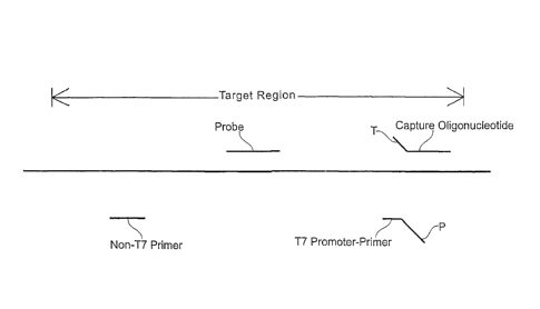
0 Figure 1 is a schematic diagram illustrating the various
polynucleotides that can be
used for detecting a target region within the HIV-1 nucleic acid (represented
by a thick
horizontal line). Positions of the following nucleic acids are shown relative
to the target
CA 02929741 2016-05-10
WO 2006/039564
PCT/US2005/035318
region: "Capture Oligonucleotide" refers to the nucleic acid used to hybridize
to and capture
the target nucleic acid prior to amplification, where "T" refers to a tail
sequence used to
hybridize an immobilized oligonucleotide having a complementary sequence (not
shown);
"Non-T7 Primer" and "T7 Promoter-Primer" represent two amplification primers
used for
conducting TMA, where "P" indicates the promoter sequence of the Ti promoter-
primer; and
"Probe" refers to the probe used for detecting amplified nucleic acid.
Figure 2 is a line graph relating the amount of HIV-1 standard input into a
real-time
nucleic acid amplification reaction (x-axis) and the time-of-emergence of the
measured
fluorescent signal above a background threshold (y-axis). Results are shown
for trials
conducted using the primer of SEQ ID NO:1 in combination with a promoter-
primer having
the target-hybridizing sequence of SEQ ID NO:13 (open squares/solid line), and
using the
primer of SEQ ID NO:2 in combination with a promoter-primer having the target-
hybridizing
sequence of SEQ ID NO:13 (open triangles/dashed line).
Figure 3 is a line graph relating the amount of HIV-1 standard input into a
real-time
5 nucleic acid amplification reaction (x-axis) and the time-of-emergence of
the measured
fluorescent signal above a background threshold (y-axis). Results represent
time-dependent
amplification of HIV-1 subtype B (open triangles/solid line) and HIV-1 0 group
(open
diamonds/dashed line) templates using a first-strand promoter-primer that
included the target-
hybridizing sequence of SEQ ID NO:13 and a second-strand primer having the
sequence of
,0 SEQ ID NO:2.
Figures 4A-4B are line graphs relating the amount of HIV-1 standard input into
a real-
time nucleic acid amplification reaction (x-axis) and the time-of-emergence of
the measured
fluorescent signal above a background threshold (y-axis). Results represent
time-dependent
amplification of HIV-1 subtype B (Figure 4A) and HIV-1 0 group (Figure 4B)
templates
using a first-strand promoter-primer that included the target-hybridizing
sequence of SEQ ID
NO:13 and a second-strand primer having the sequence of SEQ ID NO:2 (open
triangles/dashed lines), or a first-strand promoter-primer that included the
target-hybridizing
sequence of SEQ ID NO:15 and a second-strand primer having the sequence of SEQ
ID NO:2
(open diamonds/solid lines).
30 Figures 5A-5B are bar graphs representing the time-of-emergence of
measured
fluorescent signals above a background threshold (y-axis) for different HIV-1
variants at 1,000
copies/reaction. Figure 5A shows results for reactions conducted using a first-
strand
16
CA 02929741 2016-05-10
WO 2006/039564 PCM1S2005/035318
promoter-primer that included the target-hybridizing sequence of SEQ ID N0:13
and a
second-strand primer having the sequence of SEQ lD NO:2. Figure 5B shows
results for
reactions conducted using a first-strand promoter-primer that included the
target-hybridizing
sequence of SEQ ID NO:15 and a second-strand primer having the sequence of SEQ
ID N0:2.
Figures 6A-6B show a series of bar graphs representing results for time-
dependent
amplification of numerous HIV-1 variants. Amplification primers had the target-
hybridizing
sequences of SEQ ID NO:5 and SEQ ID NO:15. The molecular torch hybridization
probe
used in the procedure had the target-hybridizing sequence of SEQ ID N0:23.
Results are
shown for amplification reactions conducted using 1,000 copies/reaction of the
different HIV-
1 subtypes. Figure 6A identifies the HIV-1 nucleic acid input into a real-time
nucleic acid
amplification reaction (x-axis) and the time-of-emergence of the measured
fluorescent signal
above a background threshold (y-axis). Numerical values shown above each bar
indicate the
time-of-emergence. Figure 6B presents the same data shown in Figure 6A, but
plots the
average log10 copy number on the y-axis. Numerical values shown above each bar
indicate the
determined average log10 copy number.
Detailed Description of the Invention
Disclosed herein are compositions, methods and kits for selectively detecting
the
nucleic acids of HIV-1 in biological samples such as blood, serum, plasma or
other body fluid
ZO or tissue. The probes, primers and methods of the invention can be used
either in diagnostic
applications, viral-load testing applications, or for screening donated blood
and blood
products or other tissues that may contain infectious particles.
Introduction and Overview
The present invention includes compositions (nucleic acid capture
oligonucleotides,
2.5 amplification oligonucleotides and probes), methods and kits that are
particularly useful for
detecting HIV-1 nucleic acids in a biological sample. To design
oligonucleotide sequences
appropriate for such uses, known HIV-1 nucleic acid sequences were first
compared to
identify candidate regions of the viral genome that could serve as reagents in
a diagnostic
assay. As a result of these comparisons, the capture oligonucleotides, primers
and probes
30 shown schematically in Figure 1 were selected for use in an amplified
assay. Portions of
sequences containing relatively few variants between the compared sequences
were chosen as
starting points for designing synthetic oligonucleotides suitable for use in
capture,
17
CA 02929741 2016-05-10
amplification and detection of amplified sequences.
Based on these analyses, the capture oligonucleotide, amplification primer and
probe
sequences presented below were designed. Those having an ordinary level of
skill in the art will
appreciate that any primer sequences specific for an HIV-1 target, with or
without a T7 promoter
sequence, may be used as primers in the various primer-based in vitro
amplification methods
described below. It is also contemplated that oligonucleotides having the
sequences disclosed
herein could serve alternative functions in assays for detecting HIV-1 nucleic
acids. For example,
the capture oligonucleotides disclosed herein could serve as hybridization
probes, the hybridization
probes disclosed herein could be used as amplification primers, and the
amplification primers
disclosed herein could be used as hybridization probes in alternative
detection assays.
Useful Amplification Methods
Amplification methods useful in connection with the present invention include:
Transcription Mediated Amplification (TMA), Nucleic Acid Sequence-Based
Amplification
(NASBA), the Polymerase Chain Reaction (PCR), Strand Displacement
Amplification (SDA), and
amplification methods using self-replicating polynucleotide molecules and
replication enzymes
such as MDV-1 RNA and Q-beta enzyme. Methods for carrying out these various
amplification
techniques respectively can be found in U.S. Patent No. 5,399,491, U.S. Patent
No. 5,554,517,
U.S. Patent No. 4,965,188, U.S. Patent No. 5,455,166, U.S. Patent No.
5,472,840 and Lizardi et
al., BioTechnology 6:1197 (1988). These documents are referred to for their
descriptions of how
to perform nucleic acid amplification reactions.
In a highly preferred embodiment of the invention, HIV-1 nucleic acid
sequences are
amplified using a TMA protocol. According to this protocol, the reverse
transcriptase which
provides the DNA polymerase activity also possesses an endogenous RNase H
activity. One of the
primers used in this procedure contains a promoter sequence positioned
upstream of a sequence
that is complementary to one strand of a target nucleic acid that is to be
amplified. In the first step
of the amplification, a promoter-primer hybridizes to the HIV-1 target RNA at
a defined site.
Reverse transcriptase creates a complementary DNA copy of the target RNA by
extension from the
3' end of the promoter-primer. Following interaction of an opposite strand
primer with the newly
synthesized DNA strand, a second strand of DNA is synthesized from the end of
the primer by
reverse transcriptase, thereby creating a double-stranded DNA
18
CA 02929741 2016-05-10
WO 2006/039564
PCT/ITS2005/035318
molecule_ RNA polymerase recognizes the promoter sequence in this double-
stranded DNA
template and initiates transcription. Each of the newly synthesized RNA
amplicons re-enters
the TWIA process and serves as a template for a new round of replication,
thereby leading to
an exponential expansion of the RNA amplicon. Since each of the DNA templates
can make
100-1000 copies of RNA amplicon, this expansion can result in the production
of 10 billion
amplicons in less than one hour. The entire process is autocatalytic and is
performed at a
constant temperature.
Structural Features of Primers
As indicated above, a "primer" refers to an optionally modified
oligonucleotide which
0 is capable of participating in a nucleic acid amplification reaction.
Preferred primers are
capable of hybridizing to a template nucleic acid and which has a 3' end that
can be extended
by a DNA polymerase activity. The 5' region of the primer may be non-
complementary to the
target nucleic acid. If the 5' non-complementary region includes a promoter
sequence, it is
referred to as a "promoter-primer." Those skilled in the art will appreciate
that any
5 oligonucleotide that can function as a primer (i.e., an oligonucleotide
that hybridizes
specifically to a target sequence and has a 3' end capable of extension by a
DNA polymerase
activity) can be modified to include a 5' promoter sequence, and thus could
function as a
promoter-primer. Similarly, any promoter-primer can be modified by removal of,
or synthesis
without, a promoter sequence and still function as a primer.
!O Nucleotide base moieties of primers may be modified (e.g., by the
addition of propyne
groups), as long as the modified base moiety retains the ability to form a non-
covalent
association with G, A, C, T or U, and as long as an oligonucleotide comprising
at least one
modified nucleotide base moiety or analog is not sterically prevented from
hybridizing with a
single-stranded nucleic acid. As indicated below in connection with the
chemical composition
7-5 of useful probes, the nitrogenous bases of primers in accordance with
the invention may be
conventional bases (A, G, C, T, U), known analogs thereof (e.g., inosine or
"I" having
hypoxanthine as its base moiety; see The Biochemistry of the Nucleic Acids 5-
36, Adams et
al., ed., 1 1 th ed., 1992), known derivatives of purine or pyrimidine bases
(e.g., N4-methyl
deoxyga-unosine, deaza- or aza-purines and deaza- or aza-pyrimidines,
pyrimidine bases
30 having s-ubstituent groups at the 5 or 6 position, purine bases having
an altered or a
replacement substituent at the 2, 6 or 8 positions, 2-amino-6-
methylaminopurine, 06-
methylguanine, 4-thio-pyrimidines, 4-amino-pyrimidines, 4-dimethylhydrazine-
primidines,
19
CA 02929741 2016-05-10
and 04-alkyl-pyrimidines (see, Cook, PCT Int'l Pub. No. WO 93/13121) and
"abasic" residues
where the backbone includes no nitrogenous base for one or more residues of
the polymer (see
Arnold et al., U.S. Patent No. 5,585,481). Common sugar moieties that comprise
the primer
backbone include ribose and deoxyribose, although 21-0-methyl ribose (0Me),
halogenated sugars,
and other modified sugar moieties may also be used. Usually, the linking group
of the primer
backbone is a phosphorus-containing moiety, most commonly a phosphodiester
linkage, although
other linkages, such as, for example, phosphorothioates, methylphosphonates,
and non-
phosphorus-containing linkages such as peptide-like linkages found in "peptide
nucleic acids"
(PNA) also are intended for use in the assay disclosed herein.
Useful Probe Labeling Systems and Detectable Moieties
Essentially any labeling and detection system that can be used for monitoring
specific
nucleic acid hybridization can be used in conjunction with the present
invention. Included among
the collection of useful labels are radiolabels, enzymes, haptens, linked
oligonucleotides,
chemiluminescent molecules, fluorescent moieties (either alone or in
combination with "quencher"
moieties), and redox-active moieties that are amenable to electronic detection
methods. Preferred
chemiluminescent molecules include acridinium esters of the type disclosed by
Arnold et al., in
U.S. Patent No. 5,283,174 for use in connection with homogenous protection
assays, and of the
type disclosed by Woodhead et al., in U.S. Patent No. 5,656,207 for use in
connection with assays
that quantify multiple targets in a single reaction. Preferred electronic
labeling and detection
approaches are disclosed in U.S. Patent Nos. 5,591,578 and 5,770,369, and the
published
international patent application WO 98/57158. Redox active moieties useful as
labels in the
present invention include transition metals such as Cd, Mg, Cu, Co, Pd, Zn, Fe
and Ru.
Particularly preferred detectable labels for probes in accordance with the
present invention
are detectable in homogeneous assay systems (i.e., where, in a mixture, bound
labeled probe
exhibits a detectable change, such as stability or differential degradation,
compared to unbound
labeled probe). Examples of homogeneously detectable labels include
fluorescent labels,
electronically detectable labels, and chemiluminescent compounds (e.g., as
described by
Woodhead et al., in U.S. Patent No. 5,656,207; by Nelson et al., in U.S.
Patent No. 5,658,737; or
by Arnold et al., in U.S. Patent No. 5,639,604).
In some applications, probes exhibiting at least some degree of self-
complementarity are
desirable to facilitate detection of probe:target duplexes in a test sample
without first requiring the
CA 02929741 2016-05-10
removal of unhybridized probe prior to detection. By way of example,
structures referred to as
"Molecular Beacons" comprise nucleic acid molecules having a target
complementary sequence,
an affinity pair (or nucleic acid arms) holding the probe in a closed
conformation in the absence of
a target nucleic acid sequence, and a label pair that interacts when the probe
is in a closed
conformation. Hybridization of the target nucleic acid and the target
complementary sequence
separates the members of the affinity pair, thereby shifting the probe to an
open conformation.
The shift to the open conformation is detectable due to reduced interaction of
the label pair, which
may be, for example, a fluorophore and a quencher (e.g., DABCYL and EDANS).
Molecular
beacons are fully described in U.S. Patent No. 5,925,517. Molecular beacons
useful for detecting
HIV-1 specific nucleic acid sequences may be created by appending to either
end of one of the
probe sequences disclosed herein, a first nucleic acid arm comprising a
fluorophore and a second
nucleic acid arm comprising a quencher moiety. In this configuration, the HIV-
1 specific probe
sequence disclosed herein serves as the target-complementary "loop" portion of
the resulting
molecular beacon, while the self-complementary "arms" of the probe represent
the "stem" portion
of the probe.
Another example of a self-complementary hybridization assay probe that may be
used in
conjunction with the invention is a structure commonly referred to as a
"Molecular Torch." These
self-reporting probes are designed to include distinct regions of self-
complementarity (coined "the
target binding domain" and "the target closing domain") which are connected by
a joining region
and which hybridize to one another under predetermined hybridization assay
conditions. When
exposed to an appropriate target or denaturing conditions, the two
complementary regions (which
may be fully or partially complementary) of the molecular torch melt, leaving
the target binding
domain available for hybridization to a target sequence when the predetermined
hybridization
assay conditions are restored. Molecular torches are designed so that the
target binding domain
favors hybridization to the target sequence over the target closing domain.
The target binding
domain and the target closing domain of a molecular torch include interacting
labels (e.g.,
fluorescent/quencher) positioned so that a different signal is produced when
the molecular torch is
self-hybridized as opposed to when the molecular torch is hybridized to a
target nucleic acid,
thereby permitting detection of probe :target duplexes in a test sample in the
presence of
unhybridized probe having a viable label associated therewith. Molecular
torches are fully
described in U.S. Patent No. 6,361,945.
21
CA 02929741 2016-05-10
Molecular torches and molecular beacons preferably are labeled with an
interactive pair of
detectable labels. Examples of detectable labels that are preferred as members
of an interactive
pair of labels interact with each other by FRET or non-FRET energy transfer
mechanisms.
Fluorescence resonance energy transfer (FRET) involves the radiationless
transmission of energy
quanta from the site of absorption to the site of its utilization in the
molecule, or system of
molecules, by resonance interaction between chromophores, over distances
considerably greater
than interatomic distances, without conversion to thermal energy, and without
the donor and
acceptor coming into kinetic collision. The "donor" is the moiety that
initially absorbs the energy,
and the "acceptor" is the moiety to which the energy is subsequently
transferred. In addition to
FRET, there are at least three other "non-FRET" energy transfer processes by
which excitation
energy can be transferred from a donor to an acceptor molecule.
When two labels are held sufficiently close that energy emitted by one label
can be
received or absorbed by the second label, whether by a FRET or non-FRET
mechanism, the two
labels are said to be in "energy transfer relationship" with each other. This
is the case, for
example, when a molecular beacon is maintained in the closed state by
formation of a stem duplex,
and fluorescent emission from a fluorophore attached to one arm of the probe
is quenched by a
quencher moiety on the opposite arm.
Highly preferred label moieties for the invented molecular torches and
molecular beacons
include a fluorophore and a second moiety having fluorescence quenching
properties (i.e., a
"quencher"). In this embodiment, the characteristic signal is likely
fluorescence of a particular
wavelength, but alternatively could be a visible light signal. When
fluorescence is involved,
changes in emission are preferably due to FRET, or to radiative energy
transfer or non-FRET
modes. When a molecular beacon having a pair of interactive labels in the
closed state is
stimulated by an appropriate frequency of light, a fluorescent signal is
generated at a first level,
which may be very low. When this same probe is in the open state and is
stimulated by an
appropriate frequency of light, the fluorophore and the quencher moieties are
sufficiently separated
from each other that energy transfer between them is substantially precluded.
Under that
condition, the quencher moiety is unable to quench the fluorescence
22
CA 02929741 2016-05-10
WO 2006/039564 PCT/US2005/035318
from the fluorophore moiety. If the fluorophore is stimulated by light energy
of an appropriate
wavelength, a fluorescent signal of a second level, higher than the first
levol, will be
generated. The difference between the two levels of fluorescence is detectable
and
measurable. Using fluorophore and quencher moieties in this manner, the
molecular beacon is
only "on" in the "open" conformation and indicates that the probe is bound to
the target by
emanating an easily detectable signal. The conformational state of the probe
alters the signal
generated from the probe by regulating the interaction between the label
moieties.
Examples of donor/acceptor label pairs that may be used in connection with the
invention, making no attempt to distinguish FRET from non-FRET pairs, include
fluorescein/tetramethylrhodamine, IAEDANS/fluororescein, EDANS/DABCYL,
coumarin/DABCYL, fluorescein/fluorescein, BODIPY FL/BODIPY FL,
fluorescein/DABCYL, lucifer yellow/DABCYL, BODIPY/DABCYL, eosine/DABCYL,
erythrosine/DABCYL, tetramethylrhodamine/DABCYL, Texas Red/DAB CYL, CY5/BH1,
CY5/BH2, CY3/BH1, CY3/BH2 and fluorescein/QSY7 dye. Those having an ordinary
level
5 of skill in the art will understand that when donor and acceptor dyes are
different, energy
transfer can be detected by the appearance of sensitized fluorescence of the
acceptor or by
quenching of donor fluorescence. When the donor and acceptor species are the
same, energy
can be detected by the resulting fluorescence depolarization. Non-fluorescent
acceptors such
as DABCYL and the QSY 7 dyes advantageously eliminate the potential problem of
0 background fluorescence resulting from direct (i.e., non-sensitized)
acceptor excitation.
Preferred fluorophore moieties that can be used as one member of a donor-
acceptor pair
include fluorescein, ROX, and the CY dyes (such as CY5). Highly preferred
quencher
moieties that can be used as another member of a donor-acceptor pair include
DAB CYL and
the BLACK HOLE QUENCHER moieties which are available from Biosearch
Technologies,
Inc., (Novato, CA).
Synthetic techniques and methods of bonding labels to nucleic acids and
detecting
labels are well known in the art (e.g., see Sambrook et al., Molecular
Cloning, A Laboratory
Manual, 2nd ed. (Cold Spring Harbor Laboratory Press, Cold Spring Harbor, NY,
1989),
Chapter 10; Nelson et al., U.S. Patent No. 5,658,737; Woodhead et al., U_S.
Patent No.
30 5,656,207; Hogan et al., U.S. Patent No. 5,547,842; Arnold et al., U.S.
Patent No. 5,283,174;
KourilsIcy et al., U.S. Patent No. 4,581,333), and Becker et al., European
Patent App. No. 0
747 706.
23
CA 02929741 2016-05-10
WO 2006/039564 PCT/US2005/035318
Chemical Composition of Probes
Probes in accordance with the invention comprise polynucleotides or
polynucleotide
analogs and optionally may carry a detectable label covalently bonded thereto.
Nucleosides or
nucleoside analogs of the probe comprise nitrogenous heterocyclic bases, or
base analogs,
where the nucleosides are linked together, for example by phospohdiester bonds
to form a
polynucleotide. Accordingly, a probe may comprise conventional ribonucleic
acid (RNA)
and/or deoxyribonucleic acid (DNA), but also may comprise chemical analogs of
these
molecules. The "backbone" of a probe may be made up of a variety of linkages
known in the
art, including one or more sugar-phosphodiester linkages, peptide-nucleic acid
bonds
0 (sometimes referred to as "peptide nucleic acids" as described by Hyldig-
Nielsen et al., PCT
Intl Pub. No. WO 95/32305), phosphorothioate linkages, methylphosphonate
linkages or
combinations thereof. Sugar moieties of the probe may be either ribose or
deoxyribose, or
similar compounds having known substitutions, such as, for example, 2'-0-
methyl ribose and
2' halide substitutions (e.g., 2'-F). The nitrogenous bases may be
conventional bases (A, G, C,
5 T, U), known analogs thereof (e.g., inosine or "I"; see The Biochemistry
of the Nucleic Acids
5-36, Adams et al., ed., 11th ed., 1992), known derivatives of purine or
pyrimidine bases (e.g.,
2\14-methyl deoxygaunosine, deaza- or aza-purines and deaza- or aza-
pyrimidines, pyrimidine
bases having substituent groups at the 5 or 6 position, purine bases having an
altered or a
replacement substituent at the 2, 6 or 8 positions, 2-amino-6-
methylaminopurine, 06-
.0 methylguanine, 4-thio-pyrimidines, 4-amino-pyrimidines, 4-
dimethylhydrazine-pyrimidines,
and 04-alkyl-pyrimidines (see, Cook, PCT Int'l Pub. No. WO 93/13121) and
"abasic" residues
where the backbone includes no nitrogenous base for one or more residues of
the polymer (see
Arnold et al., U.S. Patent No. 5,585,481). A probe may comprise only
conventional sugars,
bases and linkages found in RNA and DNA, or may include both conventional
components
t5 and substitutions (e.g., conventional bases linked via a methoxy
backbone, or a nucleic acid
including conventional bases and one or more base analogs).
While oligonucleotide probes of different lengths and base composition may be
used
for detecting HIV-1 nucleic acids, preferred probes in this invention have
lengths of up to 1 00
nucleotides, and more preferably have lengths of up to 60 nucleotides.
Preferred length ranges
30 for the invented oligonucleotides are from 10 to 100 bases in length, or
more preferably
between 15 and 50 bases in length, or still more preferably between 15 and 30
bases in length.
However, the specific probe sequences described below also may be provided in
a nucleic
24
CA 02929741 2016-05-10
acid cloning vector or transcript or other longer nucleic acid and still can
be used for detecting
HIV-1 nucleic acids.
Selection of Amplification Primers and Detection Probes Specific for HIV-1
Useful guidelines for designing amplification primers and probes with desired
characteristics are described herein. The optimal sites for amplifying and
probing HIV-1 nucleic
acids contain two, and preferably three, conserved regions each greater than
about 15 bases in
length, within about 200 bases of contiguous sequence. The degree of
amplification observed with
a set of primers or promoter-primers depends on several factors, including the
ability of the
oligonucleotides to hybridize to their complementary sequences and their
ability to be extended
enzymatically. Because the extent and specificity of hybridization reactions
are affected by a
number of factors, manipulation of those factors will determine the exact
sensitivity and specificity
of a particular oligonucleotide, whether perfectly complementary to its target
or not. The effects of
varying assay conditions are known to those skilled in the art, and are
described by Hogan et al., in
U.S. Patent No. 5,840,488.
The length of the target nucleic acid sequence and, accordingly, the length of
the primer
sequence or probe sequence can be important. In some cases, there may be
several sequences from
a particular target region, varying in location and length, which will yield
primers or probes having
the desired hybridization characteristics. While it is possible for nucleic
acids that are not perfectly
complementary to hybridize, the longest stretch of perfectly homologous base
sequence will
normally primarily determine hybrid stability.
Amplification primers and probes should be positioned to minimize the
stability of the
oligonucleotide:nontarget (i.e., nucleic acid with similar sequence to target
nucleic acid) nucleic
acid hybrid. It is preferred that the amplification primers and detection
probes are able to
distinguish between target and non-target sequences. In designing primers and
probes, the
differences in these Tm values should be as large as possible (e.g., at least
2 C and preferably
C.).
The degree of non-specific extension (primer-dimer or non-target copying) can
also affect
amplification efficiency. For this reason, primers are selected to have low
self- or cross-
complementarity, particularly at the 3' ends of the sequence. Long homopolymer
tracts and high
GC content are avoided to reduce spurious primer extension. Commercially
available computer
software can aid in this aspect of the design. Available computer programs
CA 02929741 2016-05-10
WO 2006/039564
PCT/US2005/035318
include MacDNASISTM 2.0 (Hitachi Software Engineering American Ltd.) and OLIGO
ver.
6.6 (Molecular Biology Insights; Cascade, CO).
Those having an ordinary level of skill in the art will appreciate that
hybridization
involves the association of two single strands of complementary nucleic acid
to form a
hydrogen bonded double strand. It is implicit that if one of the two strands
is wholly or
partially involved in a hybrid, then that strand will be less able to
participate in formation of a
new hybrid. By designing primers and probes so that substantial portions of
the sequences of
interest are single stranded, the rate and extent of hybridization may be
greatly increased. If
the target is an integrated genomic sequence, then it will naturally occur in
a double stranded
form (as is the case with the product of the polymerase chain reaction). These
double-stranded
targets are naturally inhibitory to hybridization with a probe and require
denaturation prior to
the hybridization step.
The rate at which a polynucleotide hybridizes to its target is a measure of
the thermal
stability of the target secondary structure in the target binding region. The
standard
5 measurement of hybridization rate is the C0t12 which is measured as
moles of nucleotide per
liter multiplied by seconds. Thus, it is the concentration of probe multiplied
by the time at
which 50% of maximal hybridization occurs at that concentration. This value is
determined
by hybridizing various amounts of polynucleotide to a constant amount of
target for a fixed
time. The C0tu2 is found graphically by standard procedures familiar to those
having an
0 ordinary level of skill in the art.
Preferred Amplification Primers
Primers useful for conducting amplification reactions can have different
lengths to
accommodate the presence of extraneous sequences that do not participate in
target binding,
and that may not substantially affect amplification or detection procedures.
For example,
promoter-primers useful for performing amplification reactions in accordance
with the
invention have at least a minimal sequence that hybridizes to the HIV-1 target
nucleic acid,
and a promoter sequence positioned upstream of that minimal sequence. However,
insertion
of sequences between the target binding sequence and the promoter sequence
could change
the length of the primer without compromising its utility in the amplification
reaction.
Additionally, the lengths of the amplification primers and detection probes
are matters of
choice as long as the sequences of these oligonucleotides conform to the
minimal essential
requirements for hybridizing the desired complementary sequence.
26
CA 02929741 2016-05-10
WO 2006/039564 PCT/US2005/035318
Tables 1 and 2 present specific examples of oligonucleotide sequences that
were used
as primers for amplifying HIV-1 nucleic acids in the poi region. Table 1
presents the
sequences of primers that were complementary to HIV-1 sequences on one strand
of nucleic
acid. Table 2 presents the sequences of both the HIV-1 target-complementary
primers and the
full sequences for promoter-primers that were used during development of the
invention.
Notably, the oligonucleotide sequences in Table 1 and Table 2 are
complementary to opposite
strands of the HIV-1 nucleic acid.
Table 1
Polynucleotide Sequences of Amplification Primers
0 Sequence SEQ ID NO:
ACAGCAGTACAAATGGCAG 1
CCACAATTTTAAAAGAAAAGGG 2
CCACAA 1 TAAGAGAAAAGGG 3
CCACAATTTTAGAAGAAAAGGG 4
5 CCACAATTTTGAAAGAAAAGGG 5
CCACAA 1-1. 1 TAAAGGAAAAGGG 6
CCACAAT'TTGAAAAGAAAAGGG 7
CCACAGTTTTAAAAGAAAAGGG 8
CCACAATTTTGAAAGAAAAGGGG 9
0 CCACAATATTAAAAGAAAAGGG 10
CCACAAITITAAAAGAGAAGGGGGGATTGG 11
CCACAATTTTAAAAGGAAAGGGGGGATTGG 12
Table 2 presents HIV-1 target-complementary oligonucleotide sequences and the
corresponding promoter-primer sequences that were used for amplifying HIV-1
nucleic acid
sequences in the H1V-1 poi region. As indicated above, promoter-primers that
are to be used
for practicing the invention include sequences complementary to an HIV-1
target sequence at
their 3' ends, and a T7 promoter sequence (presented in lowercase) at their 5'
ends.
27
CA 02929741 2016-05-10
WO 2006/039564
PCT/US2005/035318
Table 2
Polynucleotide Sequences of Amplification Primers
=
Sequence SEQ ID NO:
AGTTTGTATGTCTGTTGCTATTATGTCTA 13
AGTTTGTGTGTCTGTTGCTGTTATGTCTA 14
AG iii GTATGTCTGATGCTATrATGTCTA 15
AGTTTGTATGTCTGGTGCTATTATGTCTA 16
5'-aatttaatacgactcactatagggag- 17
AGTTTGTATGTCTGTTGCTATTATGTCTA-3'
0 5'-aatttaatacgactcactatagggag- 18
AGTTTGTGTGTCTGTTGCTGTTATGTCTA-3'
5'-aatttaatacgactcactatagggag- 19
AGTTTGTATGTCTGATGCTATTATGTCTA-3'
5'-aatttaatacgactcactatagggag- 20
5 AGM GTATGTCTGGTGCTATTATGTCTA-3'
Preferred sets of primers for amplifying HIV-1 sequences in the pol region
include a
first primer that hybridizes an HIV-1 target sequence (such as one of the
primers listed in
Table 2) and a second primer complementary to the sequence of an extension
product of the
0 first primer (such as one of the primer sequences listed in Table 1). In
a highly preferred
embodiment, the first primer is a promoter-primer that includes a T7 promoter
sequence at its
5' end.
Preferred Detection Probes
Another aspect of the invention relates to hybridization probes for detecting
HIV-1
:5 nucleic acids. Methods for amplifying a target nucleic acid sequence
present in the nucleic
acid of HIV-1 can include an optional further step for detecting amplicons.
This method
includes a step for contacting a test sample with a hybridization assay probe
that preferentially
hybridizes to the target nucleic acid sequence, or the complement thereof,
thereby forming a
probe:target duplex that is stable for detection. Next there is a step for
determining whether
the hybrid is present in the test sample as an indication of the presence or
absence of HIV-1
nucleic acids in the test sample. This may involve detecting the probe:target
duplex, and
preferably involves homogeneous assay systems.
Hybridization assay probes useful for detecting HIV-1 nucleic acid sequences
include
a sequence of bases substantially complementary to an HIV-1 target nucleic
acid sequence.
28
CA 02929741 2016-05-10
WO 2006/039564 PCT/US2005/035318
Thus, probes of the invention preferably hybridize one strand of an HIV-1
target nucleic acid
sequence, or the complement thereof. These probes may optionally have
additional bases
outside of the targeted nucleic acid region which may or may not be
complementary to the
HIV-1 nucleic acid.
Certain highly preferred probes are able to hybridize to HIV-1 target nucleic
acids
under conditions suitable for performing a nucleic acid amplification
reaction, such as those
described herein. Examples of particularly preferred probes useful in
connection with this
aspect of the invention include molecular beacons and molecular torches.
Other preferred probes are sufficiently homologous to the target nucleic acid
to
0 hybridize under stringent hybridization conditions corresponding to about
60 C when the salt
concentration is in the range of 0.6-0.9 M. Preferred salts include lithium
chloride, but other
salts such as sodium chloride and sodium citrate also can be used in the
hybridization
solution. Example high stringency hybridization conditions are alternatively
provided by 0.48
M sodium phosphate buffer, 0.1% sodium dodecyl sulfate, and 1 mM each of EDTA
and
5 EGTA, or by 0.6 M LiC1, 1% lithium lauryl sulfate, 60 mM lithium
succinate and 10 mM each
of EDTA and EGTA.
Probes in accordance with the invention have sequences complementary to, or
corresponding to a portion of the HIV-1 genome. Certain probes that are
preferred for
detecting HIV-1 nucleic acid sequences have a probe sequence, which includes
the target-
complementary sequence of bases together with any base sequences that are not
complementary to the nucleic acid that is to be detected, in the length range
of from 10-100
nucleotides. Certain specific probes that are preferred for detecting HIV-1
nucleic acid
sequences have target-complementary sequences in the length range of from 10-
50, from 10-
20, or from 10-15 nucleotides. Of course, these target-complementary sequences
may be
linear sequences, or may be contained in the structure of a molecular beacon,
molecular torch
or other construct having one or more optional nucleic acid sequences that are
non-
complementary to the HIV-1 target sequence that is to be detected. As
indicated above,
probes may be made of DNA, RNA, a combination of DNA and RNA, a nucleic acid
analog,
or contain one or more modified nucleosides (e.g., a ribonucleoside having a
2'-0-methyl
substitution to the ribofuranosyl moiety).
Certain highly preferred probes include a detectable label. In one embodiment,
the
detectable label is a fluorescent label which may, optionally, be used in
combination with a
29
CA 02929741 2016-05-10
quencher moiety. In other embodiments, the label is an acrid inium ester
joined to the probe by
means of a non-nucleotide linker. For example, detection probes can be labeled
with
chemiluminescent acridinium ester compounds that are attached via a linker
substantially as
described in U.S. Patent No. 5,585,481; and in U.S. Patent No. 5,639,604,
particularly as
described at column 10, line 6 to column 11, line 3, and in Example 8. Of
course, highly preferred
probes for use in time-dependent amplicon detection include molecular beacons
and molecular
torches.
Table 3 presents the target-complementary base sequences, and full sequences
of some of
the hybridization probes that were used for detecting HIV-1 amplicons. Since
alternative probes
for detecting HIV-1 nucleic acid sequences can hybridize to the opposite-sense
strand of HIV-1,
the present invention also includes oligonucleotides that are complementary to
the sequences
presented in the table. The target-hybridizing sequence of SEQ ID NO:21 was
incorporated into
the molecular beacon having the sequence of SEQ ID NO:22. The target-
hybridizing sequence of
SEQ ID NO:23 was incorporated into the molecular torch having the sequence of
SEQ ID NO:24.
Both the molecular beacon and the molecular torch appearing in Table 3 were
labeled with a
fluorescein moiety at its 5' end, and with a DABCYL quencher moiety at its 3'
end.
Table 3
Polynucleotide Sequences of HIV-1 Detection Probes
Sequence SEQ ID NO:
UGGIGGGUACAGUGC 21
CCGUGGIGGGUACAGUGCCACGG3' 22
GGIGGGUACAGUGC 23
CGGIGGGUACAGUGC (C9) CCCCG 24
As indicated above, any number of different backbone structures can be used as
a scaffold
for the nucleobase sequences of the invented hybridization probes. In certain
highly preferred
embodiments, the probe sequence used for detecting HIV-1 amplicons includes a
methoxy
backbone, or at least one methoxy linkage in the nucleic acid backbone.
CA 02929741 2016-05-10
=
WO 2006/039564
PCT/US2005/035318
Selection and Use of Capture Oligonucleotides
Preferred capture oligonucleotides include a first sequence that is
complementary to an
HIV-1 sequence (i.e., an "HIV-1 target sequence") covalently attached to a
second sequence
(i.e., a "tail" sequence) that serves as a target for immobilization on a
solid support. Any
backbone to link the base sequence of a capture oligonucleotide may be used.
In certain
preferred embodiments the capture oligonucleotide includes at least one
methoxy linkage in
the backbone. The tail sequence, which is preferably at the 3' end of a
capture
oligonucleotide, is used to hybridize to a complementary base sequence to
provide a means for
capturing the hybridized target HIV-1 nucleic acid in preference to other
components in the
biological sample.
Although any base sequence that hybridizes to a complementary base sequence
may be
used in the tail sequence, it is preferred that the hybridizing sequence span
a length of about 5-
50 nucleotide residues. Particularly preferred tail sequences are
substantially homopolymeric,
containing about 10 to about 40 nucleotide residues, or more preferably about
14 to about 30
residues. A capture oligonucleotide according to the present invention may
include a first
sequence that specifically binds an HIV-1 target polynucleotide, and a second
sequence that
specifically binds an oligo(dT) stretch immobilized to a solid support.
Using the components illustrated in Figure 1, one assay for detecting HIV-1
sequences
in a biological sample includes the steps of capturing the target nucleic acid
using the capture
oligonucleotide, amplifying the captured target region using at least two
primers, and
detecting the amplified nucleic acid by first hybridizing the labeled probe to
a sequence
contained in the amplified nucleic acid and then detecting a signal resulting
from the bound
labeled probe.
The capturing step preferably uses a capture oligonucleotide where, under
hybridizing
conditions, one portion of the capture oligonucleotide specifically hybridizes
to a sequence in
the target nucleic acid and a tail portion serves as one component of a
binding pair, such as a
ligand (e.g., a biotin-avidin binding pair) that allows the target region to
be separated from
other components of the sample. Preferably, the tail portion of the capture
oligonucleotide is a
sequence that hybridizes to a complementary sequence immobilized to a solid
support particle.
0 Preferably, first, the capture oligonucleotide and the target nucleic
acid are in solution to take
advantage of solution phase hybridization kinetics. Hybridization produces a
capture
oligonucleotidelarget nucleic acid complex which can bind an immobilized probe
through
31
CA 02929741 2016-05-10
WO 2006/039564 PCT/US2005/035318
hybridization of the tail portion of the capture oligonucleotide with a
complementary
immobilized sequence. Thus, a complex comprising a target nucleic acid,
capture
oligonucleotide and immobilized probe is formed under hybridization
conditions. Preferably,
the immobilized probe is a repetitious sequence, and more preferably a
homopolymeric
3 sequence (e.g., poly-A, poly-T, poly-C or poly-G), which is complementary
to the tail
sequence and attached to a solid support. For example, if the tail portion of
the capture
oligonucleotide contains a poly-A sequence, then the immobilized probe would
contain a
poly-T sequence, although any combination of complementary sequences may be
used. The
capture oligonucleotide may also contain "spacer" residues, which are one or
more bases
located between the base sequence that hybridizes to the target and the base
sequence of the
tail that hybridizes to the immobilized probe. Any solid support may be used
for binding the
target nucleic acid:capture oligonucleotide complex. Useful supports may be
either matrices
or particles free in solution (e.g., nitrocellulose, nylon, glass,
polyaerylate, mixed polymers,
polystyrene, silane polypropylene and, preferably, magnetically attractable
particles).
Methods of attaching an immobilized probe to the solid support are well known.
The support
is preferably a particle which can be retrieved from solution using standard
methods (e.g.,
centrifugation, magnetic attraction of magnetic particles, and the like).
Preferred supports are
paramagnetic monodisperse particles (i.e., uniform in size about 5%).
Retrieving the target nucleic acid:capture oligonucleotide:immobilized probe
complex
0 effectively concentrates the target nucleic acid (relative to its
concentration in the biological
sample) and purifies the target nucleic acid from amplification inhibitors
which may be
present in the biological sample. The captured target nucleic acid may be
washed one or more
times, further purifying the target, for example, by resuspending the
particles with the attached
target nucleic acid:capture oligonucleotide:immobilized probe complex in a
washing solution
and then retrieving the particles with the attached complex from the washing
solution as
described above. In a preferred embodiment, the capturing step takes place by
sequentially
hybridizing the capture oligonucleotide with the target nucleic acid and then
adjusting the
hybridization conditions to allow hybridization of the tail portion of the
capture
oligonucleotide with an immobilized complementary sequence (e.g., as described
in PCT No.
;0 WO 98/50583). After the capturing step and any optional washing steps
have been
completed, the target nucleic acid can then be amplified. To limit the number
of handling
steps, the target nucleic acid optionally can be amplified without releasing
it from the capture
32
CA 02929741 2016-05-10
WO 2006/039564 PCT/US2005/035318
oligonucleotide.
Useful capture oligonucleotides may contain mismatches to the above-indicated
sequences, as long as the mismatched sequences hybridize to the HIV-1 nucleic
acid
containing the sequence that is to be amplified. Each. capture oligonucleotide
described herein
included one of the HIV-1 sequences presented in Table 4 linked to a poly-(dA)
tail at its 3'
end. All of the capture oligonucleotides also included three optional
thymidine nucleotides
interposed between the HIV-1 complementary sequence and the poly-(dA) tail.
The presence
of these thymidine nucleotides is not believed to be essential for success of
the capture
procedure. The three thymidine nucleotides and the poly-(dA) tail were
synthesized using
DNA precursors, while the HIV-1 complementary portions of the oligonucleotides
were
synthesized using 2'-0Me nucleotide analogs.
Table 4
HIV-I Complementary Portions of Capture Oligonucleotides
Sequence SEQ ID NO:
5 GCUGGAAUAACUUCUGCUUCUAU 25
GCUGGAAUAGCUUCUGCUUCUAU 26
UCUGCUGUCCCUGUAAUAAAC CCG 27
UCUGCUGUCCCUGUGAUAAAC CCG 28
0 Preferred Methods for Amplifying and Detecting HIV-1 Polynucleotide
Sequences
Preferred methods of the present invention are described and illustrated by
the
Examples presented below. Figure 1 schematically illustrates one system that
may be used for
detecting a target region of the HIV-1 genome (shown by a thick solid
horizontal line). This
system includes four oligonucleotides (shown by the shorter solid lines): one
capture
5 oligonucleotide that includes a sequence that hybridizes specifically to
an HIV-1 sequence in
the target region and a tail ("T") that hybridizes to a complementary sequence
immobilized on
a solid support to capture the target region present in a biological sample;
one T7 promoter-
primer which includes a sequence that hybridizes specifically to an 11IV-1
sequence in the
target region and a T7 promoter sequence ("P") which, when double-stranded,
serves as a
;0 functional promoter for T7 RNA polymerase; one non-T7 primer which
includes a sequence
that hybridizes specifically to a first strand cDNA made from the target
region sequence using
the T7 promoter-primer; and one labeled probe which includes a sequence that
hybridizes
33
CA 02929741 2016-05-10
WO 2006/039564 PCT/US2005/035318
specifically to a portion of the target region that is amplified using the two
primers.
As indicated above, amplifying the captured target region using the two
primers can be
accomplished by any of a variety of known nucleic acid amplification reactions
that will be
familiar to those having an ordinary level of skill in the art. In a preferred
embodiment, a
transcription-associated amplification reaction, such as TMA, is employed. In
such an
embodiment, many strands of nucleic acid are produced from a single copy of
target nucleic
acid, thus permitting detection of the target by detecting probes that are
bound to the amplified
sequences. Preferably, transcription-associated amplification uses two types
of primers (one
being referred to as a promoter-primer because it contains a promoter
sequence, labeled "P" in
0 Figure 1, for an RNA polymerase) two enzymes (a reverse transcriptase and
an RNA
polymerase), and substrates (deoxyribonucleoside triphosphates, ribonucleoside
triphosphates)
with appropriate salts and buffers in solution to produce multiple RNA
transcripts from a
nucleic acid template.
Referring to Figure 1, during transcription-mediated amplification, the
captured target
5 nucleic acid is hybridized to a first primer shown as a T7 promoter-
primer. Using reverse
transcriptase, a complementary DNA strand is synthesized from the T7 promoter-
primer using
the target DNA as a template. A second primer, shown as a non-T7 primer,
hybridizes to the
newly synthesized DNA strand and is extended by the action of a reverse
transcriptase to form
a DNA duplex, thereby forming a double-stranded T7 promoter region. T7 RNA
polymerase
then generates multiple RNA transcripts by using this functional T7 promoter.
The
autocatalytic mechanism of TMA employs repetitive hybridization and
polymerization steps
following a cDNA synthesis step using the RNA transcripts as templates to
produce additional
transcripts, thereby amplifying target region-specific nucleic acid sequences.
The detecting step uses at least one detection probe that binds specifically
to the
amplified RNA transcripts or amplicons described above. Preferably, the
detection probe is
labeled with a label that can be detected using a homogeneous detection
system. For example,
the labeled probe can be labeled with an acridinium ester compound from which
a
chemiluminescent signal may be produced and detected, as described above.
Alternatively,
the labeled probe may comprise a fluorophore or a combination of ftuorophore
and quencher
moieties. Molecular beacons and molecular torches are alternative embodiments
of such
labeled probes that may be used in homogeneous detection systems.
34
CA 02 92 9741 2 01 6-05-10
WO 2006/039564 PCT/US2005/035318
Use of a Standard Curve ¨ Quantifying Pre-Amplification Amounts of Analyte
Polynucleotide
In general, the invented methods can involve the step of consulting a standard
curve
that relates pre-amplification amounts of analyte polynucleotide and post-
amplification
amounts of analyte amplicon.
Since real-time amplification reactions advantageously feature quantitative
relationships between the number of analyte polynucleotides input into the
reaction and the
number of analyte amplicons synthesized as a function of time, the number of
analyte
polynucleotides present in a test sample can be determined using a standard
curve. For
0 example, a plurality of amplification reactions containing known amounts
of a polyraucleotide
standard can be run in parallel with an amplification reaction prepared using
a test sample
containing an unknown number of analyte polynucleotides. Alternatively, a
standard curve
can be prepared in advance so that it is unnecessary to prepare a curve each
time an analytical
procedure is carried out. Such a curve prepared in advance can even be stored
electronically
5 in a memory device of a testing instrument. A standard curve having pre-
amplification
amounts of the polynucleotide standard on a first axis and some indicia of the
time required to
effect a certain level of nucleic acid amplification (such as a time-of-
emergence above a
background signal) on a second axis is then prepared. The post-amplification
amount of
analyte amplicon measured for the test reaction is then located on the post-
amplification axis
:0 of the standard curve. The corresponding value on the other axis of the
curve represents the
pre-amplification amount of analyte polynucleotide that was present in the
test reaction. Thus,
determining the number of molecules of analyte polynucleotide present in the
test sample is
accomplished by consulting the standard curve, or more particularly by
comparing the
quantitative results obtained for the test sample with the standard curve, a
procedure that will
be familiar to those having an ordinary level of skill in the art.
The procedures described herein can easily be used to quantify analyte
polynucleotides
present in a test sample. Indeed, if a plurality of standard control
amplification reactions are
initiated using known numbers of an analyte polynucleotide standard, and if a
test reaction
that includes an unknown number of analyte polynucleotide molecules is carried
alit, then it
30 becomes possible after measuring the time required to effect a certain
level of amplification in
each reaction to determine the number of analyte polynucleotide molecules that
must have
been present in the test sample. The relationship between the number of
analyte
CA 02929741 2016-05-10
WO 2006/039564
PCT/US2005/035318
polynucleotide molecules input into standard amplification reaction and the
time required to
effect a certain level of amplification is conveniently established using a
graph. Determining
the number of analyte polynucleotide molecules present in a test sample is
simply a matter of
determining from the standard graph the number of analyte polynucleotide
molecules that
correspond to a measured analyte amplicon signal strength. This illustrates
how analyte
polynucleotide standards can be used in connection with polynucleotide
amplification
reactions to quantify pre-amplification amounts of analyte polynucleotide
contained in test
samples.
0 Kits for Detecting HI-1 Nucleic Acids
The present invention also embraces kits for performing polynucleotide
amplification
reactions using viral nucleic acid templates. Certain preferred kits will
contain a hybridization
assay probe that includes a target-complementary sequence of bases, and
optionally primers or
other ancillary oligonucleotides for amplifying the target that is to be
detected. Other
5 preferred kits will contain a pair of oligonucleotide primers that may be
used for amplifying
target nucleic acids in an in vitro amplification reaction. Exemplary kits
include first and
second amplification oligonucleotides that are complementary to opposite
strands of an HIV-1
nucleic acid sequence that is to be amplified. The kits may further contain
one or more
oligonucleotide detection probes. Still other kits in accordance with the
invention may
,0 additionally include capture oligonucleotides for purifying HIV-1
template nucleic acids away
from other species prior to amplification.
The general principles of the present invention may be more fully appreciated
by
reference to the following non-limiting Examples. These Examples describe the
development
of quantitative nucleic acid amplification assays characterized by
substantially linear
relationships between the time required to yield a positive amplification
signal and the initial
amount of HIV-1 template nucleic acid included in the reaction. The invented
assays are
further characterized by high levels of precision in the quantitation of1-11V-
1 targets at low
copy numbers, and by accurate detection of different H1V-1 subtypes, including
M group and
0 group variants.
30 Oligonucleotide primers disclosed in published international
application WO
2003106714, together with a molecular beacon, served as a starting point for
the development
of the invented assay. As indicated by the evidence presented in Example 1,
modifying the
36
CA 02 92 9741 2 01 6-05-10
WO 2006/039564
PCT/US2005/035318
initial primer set by substituting one of the primers dramatically improved
the quantitative
capacity of the assay by increasing the detectability of low levels of the HIV-
1 template. In all
cases, positive amplification was indicated by the time-dependent appearance
of a fluorescent
signal in homogeneous assays.
Analysis of the experimental data was performed using a computer-implemented
algorithm to establish a substantially linear relationship between the number
of HIV-1
template copies included in an amplification reaction and the time at which
the fluorescent
signal exceeded a background value (i.e., "time-of-emergence" above
background).
Essentially identical analyses were conducted for all of the time-dependent
assays disclosed
0 herein.
As confirmed by the results presented below, similar procedures can be used
for
quantifying analyte target amounts present in a test sample. More
specifically, when known
amounts of an analyte polynucleotide are used as calibration standards, it is
possible to
determine the amount of analyte present in a test sample by comparing the time-
dependent
5 appearance of a fluorescent signal measured for the test sample with a
standard curve.
Example 1 describes procedures wherein a molecular beacon probe labeled with
an
interactive fluorophore/quencher pair was used for monitoring time-dependent
amplicon
production in nucleic acid amplification reactions. Although the molecular
beacons described
in this Example hybridized to only one strand of the amplified nucleic acid
product, probe
,0 sequences complementary to the HIV-1 nucleic acid on the opposite strand
also fall within the
scope of the invention. Results from these procedures indicated that the
choice of
oligonucleotide primers profoundly affected the quantitative capacity of the
assay.
Example 1
Time-Dependent Monitoring of HIV-1 M Group.,
Subtype B Amplicon Production
An in vitro synthesized transcript of known concentration included the
sequence
GGTACCAGCACACAAAGGAATTGGAGGAAATGAACAAGTAGATAAATTAGTCAGTGCTGGAAT
CAGGAAAGTACTATTTTTAGATGGAATAGATAAGGCCCAAGATGAACATGAGAAATATCACAG
TAATTGGAGAGCAATGGCTAGTGATTTTAACCTGCCACCTGTAGTAGCAAAAGAAATAGTAGC
CAGCTGTGATAAATGTCAGCTAAAAGGAGAAGCCATGCATGGACAAGTAGACTGTAGTCCAGG
AATATGGCAACTAGATTGTACACATTTAGAAGGAAAAGTTATCCTGGTAGCAGTTCATGTAGC
CAGTGGATATATAGAAGCAGAAGTTATTCCAGCAGAAACAGGGCAGGAAACAGCATATTTTCT
TTTAAAATTAGCAGGAAGATGGCCAGTAAAAACAATACATACTGACAATGGCAGCAATTT CAC
CGGTGCTACGGTTAGGGCCGCCTGTTGGTGGGCGGGAATCAAGCAGGAATTTGGAATTCCCTA
35 CAATCCCCAAAGTCAAGGAGTAGTAGAATCTATGAATAAAGAATTAAAGAAAATTATAGGACA
GGTAAGAGATCAGGCTGAACATCTTAAGACAGCAGTACAAATGGCAGTATTCATCCACAATTT
37
CA 02929741 2016-05-10
TAAAAGAAAAGGGGGGATTGGGGGGTACAGTGCAGGGGAAAGAATAGTAGACATAATAGCAAC
AGACATACAAACTAAAGAAT TACAAAAACAAAT TACAAAAATTCAAAATTTTCGGGT TTAT TA
CAGGGACAGCAGAAATCCACTTTGGAAAGGACCAGCAAAGCTCCTCTGGAAAGGTGAAGGGGC
AGTAGTAATACAAGATAATAGTGACATAAAAGTAGTGCCAAGAAGAAAAGCAAAGATCAT TAG
GGATTATGGAAAACAGATGGCAGGTGATGATTGTGTGGCAAGTAGACAGGATGAGGAT (SEQ
ID NO:29),
and served as the source of H1 V-1 subtype B template sequences in
amplification reactions that
employed paired sets of primers. This in vitro transcript contained portions
of the HIV-1 genome
that included sequences substantially corresponding to, or substantially
complementary to, each of
the primers used in the procedure. Nucleic acid amplification reactions were
performed using a
TMA protocol, and were carried out essentially as described by Kacian et al.,
in U.S. Patent No.
5,399,491. Promoter-primers used in the TMA reactions included a T7 promoter
sequence
AATTTAATACGACTCACTATAGGGAG (SEQ ID NO:30) appended upstream of an HIV-1
complementary sequence. The sequence of the T7 promoter is absent from the HIV-
1 analyte
polynucleotide, and so was not complementary to the HIV-1 template.
Amplification reactions
were conducted using variable amounts of the HIV-1 in vitro transcript, and
about 0.07-0.12
pmoles/ 1 of each primer in 30 I reaction volumes.
A molecular beacon capable of hybridizing to the HIV-1 amplicons was
synthesized by
standard solid-phase phosphite triester chemistry using 3' quencher-linked
controlled pore glass
(CPG) and 5' fluorophore-labeled phosphoramidite on a Perkin-Elmer (Foster
City, CA)
EXPEDITE model 8909 automated synthesizer. Fluorescein was used as the
fluorophore,
DABCYL was used as the quencher, and 2'-methoxy nucleotide analogs were used
for
construction of the molecular beacon. The CPG and phosphoramidite reagents
were purchased
from Glen Research Corporation (Sterling, VA). Following synthesis,
deprotection and cleavage
from the solid support matrix, the probes were purified using polyacrylamide
gel electrophoresis
followed by HPLC using standard procedures that will be familiar to those
having an ordinary
level of skill in the art. The target-complementary sequence contained in the
molecular beacon,
allowing for the substitution of a single inosine nucleotide analog at
position four, and the
substitution of uricil for thymine bases, was TGGGGGGTACAGTGC (SEQ ID NO:31).
Notably,
the target-hybridizing sequences of the molecular beacon and molecular torch
hybridization probes
described herein were not fully complementary to the HIV-1 0 group nucleic
acids, or HIV-1 0
group amplicons synthesized using the primers disclosed herein. Additionally,
the target-
hybridizing sequences of these probes were not fully
38
CA 02929741 2016-05-10
WO 2006/039564
PCT/US2005/035318
complementary to the nucleic acids or amplicons for HIV-1 M group members
represented by
subtypes A, E and F. Conversely, these target-hybridizing sequences were fully
complementary to HIV-1 subtype B amplicons. The overall sequence of the
molecular beacon
probe used in the procedure was given by SEQ ID NO:22.
Individual wells in a multiwell plate each contained 30 ul of a Tris-buffered
solution
that included potassium and magnesium salts, N-Acetyl-L-Cysteine,
ribonucleotide
triphosphates, nucleotide triphosphates and other reagents, a target
polynucleotide, and a
molecular beacon. The target polynucleotide was included in amounts ranging
from 5 to
5x10⁶ copies/reaction. The first-strand promoter-primer for amplifying the HIV-
1 template
had the target-hybridizing sequence of SEQ ID NO:13 (which was contained
within the
sequence of SEQ ID NO:17). Second-strand primers had the sequence of either
SEQ ID NO:1
or SEQ ID NO:2. Samples were incubated for 10 minutes at 60 C to facilitate
primer
annealing, and then incubated at 42 C for at least 5 minutes. Aliquots of an
enzyme reagent
that included both MMLV reverse transcriptase and T7 RNA polymerase enzymes
were added
to each of the tubes using a repeat pipettor. Amplification reactions were
carried out at 42 C,
and fluorescence readings were taken every 19.4 seconds using a CHROM04 REAL-
TIME
DETECTOR (MJ Research; Reno, NV), or every 30 seconds using an OPTICON 2 (MJ
Research; Reno, NV) real-time instrument essentially according to the
manufacturer's
instructions. Reactions were performed in replicates of 4-8.
0 The results presented in Figure 2 showed how the substitution of one
amplification
primer for another dramatically increased the quantitative capacity and
precision of the assay.
Time-dependent amplification signals obtained using a first-strand promoter-
primer that
included the target-hybridizing sequence of SEQ ID NO:13 and a second-strand
primer having
the target-complementary sequence SEQ ID NO:1 showed reduced precision, as
judged by the
:5 increased spread among individual data points, and substantial
divergence from linearity when
the level of template used in the reaction fell below 500 copies. This primer
combination was
preferred for assays intended to quantify HIV-1 nucleic acids at levels
greater than 500
copies/reaction. Conversely, time-dependent amplification signals obtained
using a first-
strand primer that included the target-complementary sequence SEQ ID NO:13 and
a second-
strand primer having the target-complementary sequence of SEQ ID NO:2 showed
improved
precision and excellent linearity over the range of from 25 to 5 x 10⁶
copies/reaction of the
nucleic acid template. This latter primer combination advantageously enhanced
the low-end
39
CA 02 92 9741 2 01 6-05-10
WO 2006/039564 PCT/US2005/035318
quantitative capacity of the assay by 20 fold, a dramatic result that could
not have been
predicted in advance of this showing.
Although not illustrated in Figure 2, there was a failure to amplify when
using a first-
strand promoter-primer having the target-complementary sequence SEQ ID NO:13
and a
second-strand primer having the target-complementary sequence SEQ ID NO:1 when
using
the HIV-1 0 group template at levels less than or equal to 50,000
copies/reaction. This
demonstrated that the observed loss of precision at low levels of input
template was a
characteristic of the primer combination, and was independent of the template
being
amplified. Conversely, the results presented in the following Example
confirmed that the
combination of a first-strand promoter-primer having the target-hybridizing
sequence of SEQ
ID NO:13 and a second-strand primer having the sequence of SEQ ID NO:2
advantageously
gave linear relationships between input template amounts and the time-
dependent
amplification signals over an extended range with good precision for templates
representing
multiple HIV-1 subtypes.
5 Example 2 demonstrates that Firv-1 M group, subtype B and HIV-1 0
group templates
could amplify with good precision over an input template range that extended
from 25 to 5 x
105 copies/reaction. Notably, the two templates amplified with somewhat
different kinetics.
Example 2
Different Kinetic Profiles Characterize Amplification
,0 of HIV-1 Variants
Amplification reactions were performed essentially as described under Example
1 with
the following modifications. Parallel reactions were conducted using a first-
strand promoter-
primer having the target-hybridizing sequence of SEQ ID NO:13, a second-strand
primer
having the target-complementary sequence of SEQ ID NO :2, and variable amounts
of either
the subtype B template described in Example 1, or an 0 group template that
included the
sequence
AGTGGGTTCATAGAAGCAGAAGTGATACCAGCAGAAACAGGACAAGAAACTGCCTACTTCCTG
TTAAAACTGGCTGCAAGATGGCCTGTTAAAGTAATACATACAGACAACGGGCCTAATTTTACA
AGTACAACTATGAAGGCTGCATGTTGGTGGGCCAACATACAACATGAGTTTGGAATACCATAT
30 AATCCACAAAGTCAAGGAGTAGTAGAAGCCATGAATAAGGAATTAAAATCAATTATACAGCAG
GTGAGGGACCAAGCAGAACACTTAAGAACAGCAGTACAAATGGCAGTATTTGTTCACAATTTT
AAAAGAAAAGGGGGGATTGGGGGGTACACTGCAGGAGAAAGGATAATAGACATATTAGCATCA
CAAATACAAACAACAGAATTACAAAAACAAATTTTAAAANTTCACAAATTTCGGGTCTATTAC
AGAGACAGCAGAGACCCTAT (SEQ ID N0:32).
35 Like the subtype B template, the 0 group template was also an in vitro
transcript prepared
CA 02 92 9741 2 01 6-05-10
WO 2006/039564 PCT/US2005/035318
using materials and procedures that will be familiar to those having an
ordinary level of skill
in the art. Templates were included in the reactions in amounts ranging from
50 to 5x10⁵
copies/reaction.
The results presented in Figure 3 confirmed that advantages associated with
the
combination of a first-strand primer that included the target-hybridizing
sequence of SEQ ID
NO:13 and a second-strand primer having the target-complementary sequence of
SEQ ID
NO:2 extended to amplification of HIV-1 0 group templates. More specifically,
reactions
conducted using this primer combination advantageously exhibited good
precision among data
points at low levels of HIV-1 template, and linearity of the time-dependent
amplification
signal. Moreover, the beneficial characteristics of this primer combination
were observed for
both fly-I subtype B and 0 group templates. As indicated above, tests
conducted using the
HIV-1 0 group template, a first-strand promoter-primer having the target-
hybridizing
sequence SEQ ID NO:13 and a second-strand primer having the target-
complementary
sequence SEQ ID NO:1 failed to yield useful amplification signals when the
number of input
5 copies of template was below 50,000 copies.
Interestingly, the different 1-1W-1 template species used in the procedure
gave rise to
substantially parallel lines on the graph shown in Figure 3, with the HIV-1 0
group template
yielding somewhat slower amplification kinetics. For example, a reaction
conducted using
5,000 copies of the HIV-1 subtype B template required about 15 minutes to
achieve a positive
:0 result, but a similar reaction conducted using the HIV-1 0 group
template required an
additional four minutes to achieve the same result. In a quantitative assay
that measures the
time to achieve a positive result, such a difference conceivably could
compromise
interpretation of the results and lead to an erroneous conclusion.
Despite the benefits of the primer combination used in this Example, the
results
indicated that different HIV-1 variants amplified with different kinetic
profiles. In the
instance illustrated in Figure 3, detection of a positive amplification signal
at 15 minutes
would ambiguously indicate the presence of 5,000 copies of the subtype B
template, or
500,000 copies of the 0 group template. A desire to perform assays using a
single calibrator,
or set of calibrators for quantifying multiple HIV-1 species in a single
reaction rendered
30 preferable a close relationship between the amplification profiles of
the HIV-1 variants to be
detected. Thus, to improve the quantitative capacity of the assay even
further, reaction
conditions were sought to normalize amplification kinetics for different HIV-1
subtypes.
41
CA 02929741 2016-05-10
WO 2006/039564 PCT/US2005/035318
The following Example discloses amplification primers containing mismatches to
both
HIV-1 subtype B and HIV-1 0 group templates, and use of these primers to
normalize the
amplification kinetics of HIV-1 variants. The approach used in this procedure
was to
substitute nucleotides within the sequence of the first-strand primer such
that the substitution
was complementary to a position contained in the 0 group template, yet non-
complementary
to the sequence contained in the subtype B template. The object of this
approach was to
enhance the amplification kinetics of the 0 group template relative to the
subtype B template.
Example 3 describes methods that identified a first-strand primer which
enhanced
amplification kinetics of H1V-1 0 group templates. Contrary to what might have
been
0 expected, there was substantially no effect on the amplification kinetics
for the HIV-1 subtype
B template.
Example 3
Enhancement of Amplification Kinetics of HIV-1 0 Group Templates
Parallel sets of amplification reactions were prepared to compare the effects
of two
5 different primer combinations on the kinetics of amplification of HIV-1
subtype B and HIV-1
0 group templates. In each instance, a first-strand promoter-primer having the
target-
hybridizing sequence of SEQ ID NO:13 or SEQ ID NO:15 was used in combination
with a
second-strand primer having the target-complementary sequence of SEQ ID NO:2.
Notably,
the sequence of SEQ ID NO:15 differed from the sequence of SEQ ID NO:13 by the
;0 substitution of adenine for thymidine at position 15 in the target-
hybridizing portion of the
primer. This substitution corresponds to position 41 of the promoter-primers
identified by
SEQ ID NO:19 and SEQ ID NO:17. Amounts of HIV-1 templates used in the
reactions
ranged from 5 to 5x10⁴ copies/reaction. Amplification reactions were prepared
and monitored
using materials and procedures essentially as described above.
The results presented in Figures 4A-4B indicated that substitution of the
primer having
the target-hybridizing sequence of SEQ ID NO:15 for the primer having the
target-
hybridizing sequence of SEQ ID N0:13 had different effects on the
amplification kinetics of
the different templates. More specifically, Figure 4A shows that the different
primer sets
amplified the HIV-1 subtype B template with substantially identical kinetics.
However,
30 Figure 4B shows that the HIV-1 0 group template amplified with somewhat
more rapid
kinetics over the full range of input template values tested when using the
primer having the
target-hybridizing sequence of SEQ ID NO:15 instead of SEQ ID N0:13.
Accordingly, the
42
CA 02929741 2016-05-10
WO 2006/039564 PCT/US2005/035318
combination of primers that included the target-hybridizing sequences SEQ ID
NO:15 and
SEQ ID NO:2 advantageously amplified the different HIV-1 template species with
kinetics
that more closely approximated each other when compared with the combination
of primers
that included the target-hybridizing sequences SEQ ID NO:13 and SEQ ID NO:2.
In a related procedure, the different primer combinations were used to amplify
independent templates representing HIV-1 subtypes A-C, E-F, G/A, H and the HIV-
1 0
group. The time required to yield a positive amplification signal was
determined for input
levels of template equaling 1,000 copies/reaction. Reactions were performed
using replicates
of six.
0 The bar graphs in Figures 5A-5B demonstrated that the new primer
combination
advantageously reduced differences between the times needed to achieve
positive
amplification results for numerous HIV-1 subtypes. Figure 5A indicates that
3.4 minutes
distinguished the times to achieve positive amplification results for the
subtype B and 0 group
templates when using primers that included the target-hybridizing sequences of
SEQ ID
5 NO:13 and SEQ ID NO:2. In contrast, Figure 5B shows that this difference
was reduced to
only 1.3 minutes when the primers included the target-hybridizing sequences of
SEQ ID
NO:15 and SEQ ID NO:2. Additionally, differences between the times needed to
achieve
positive results for several subtypes also appeared to be minimized in
reactions conducted
using these primers. Despite these improvements, the amplification kinetics
for the HIV-1
:0 subtype E and subtype F templates appeared somewhat retarded compared
with the
amplification kinetics observed for the other samples.
It also was shown that, when paired with the primer of SEQ ID NO:2, the primer
having the target-hybridizing sequence of SEQ ID NO:16 advantageously improved
accuracy
of quantitation for the HIV-1 0 group template when compared with the
combination of the
primers having the target-hybridizing sequences of SEQ ID NO:2 and SEQ ID
NO:13. Thus,
the primer represents a preferred embodiment of the invention, particularly
when paired with
the primer of SEQ ID NO:2.
Finally, each of the primers identified by SEQ ID NOs:3-6 and 8-12 in Table 1,
when
paired with the primer of SEQ ID NO:15, and when compared with results
obtained using the
30 primer of SEQ ID NO:2 in combination with the primer of SEQ ID NO:15,
behaved
substantially equivalently. This pattern was demonstrated using the molecular
beacon
disclosed herein with the primers from Table 1 identified by SEQ ID NOs:3-9,
and using the
43
CA 02 92 9741 2 01 6-05-10
WO 2006/039564
PCT/US2005/035318
molecular torch disclosed herein with the primers from Table 1 identified by
SEQ ID NO:5
(see below), SEQ ID NO:8, and SEQ ID NOs:10-12. For a reason that is unclear,
equally
good results were not achieved using the combination of primers having the
target-hybridizing
sequences of SEQ ID NO:7 and SEQ ID NO:15_ Thus, any combination of the primer
of SEQ
ID NO:15 with any of the primers identified by SEQ ID NOs:3-6 and 8-12
represents a
preferred combination of primers for amplifying HIV-1 nucleic acids. These
combinations are
particularly preferred when further combined with a molecular beacon
hybridization probe, or
with a molecular torch hybridization probe.
The foregoing procedures identified a primer combination that advantageously
was
capable of amplifying several HIV-1 subtypes with substantially equivalent
kinetic profiles.
Notably, HIV-1 subtypes E and F exhibited somewhat delayed amplification
kinetics
compared with the other targets used in the testing procedure. Having already
modified the
first- and second-strand primers, a different approach investigated the
effects of modifying the
detection probe used in the fluorescent monitoring protocol.
5 Example
4 describes procedures that identified oligonucleotide primers and a probe
that yielded substantially equivalent amplification kinetics for all of the
different HIV-1
variants.
Example 4
Time-Dependent Monitoring of Amplicon Synthesis
0 Using a Molecular Torch
Parallel amplification reactions were prepared essentially as described in the
preceding
Examples with the following modifications. A first-strand primer having the
target-
hybridizing sequence of SEQ ID NO:15 positioned downstream from a T7 promoter
sequence
(i.e., the promoter-primer of SEQ ID NO:19) was used in combination with a
second-strand
primer having the sequence SEQ ID NO:5. Additionally, a molecular torch having
the
sequence of SEQ ID NO:24 (i.e., having the target-hybridizing sequence given
by SEQ ID
NO:23) was substituted for the molecular beacon having the sequence of SEQ ID
NO:22. The
molecular torch was labeled at its 5' end with a fluorescein fluorophore, and
at its 3' end with a
DABCYL quencher moiety. Finally, in vitro transcripts representing HIV-1
subtypes A-C, E-
;0 F, G/A, H and 0 group were used as templates at 50 and 1,000
copies/reaction.
A standard curve was prepared from data obtained in trials conducted using the
HIV-I
subtype B templates as illustrative HIV-1 M group standards at 50 and 1,000
copies/reaction.
44
=
CA 02929741 2016-05-10
WO 2006/039564 PCT/US2005/035318
Reactions were carried out in replicates of six. The time required to effect
detectable levels
of amplification above background were plotted on the y-axis, and the number
of
copies/reaction of the standard plotted on the x-axis of the standard curve.
The average time
required to effect detectable levels of amplification in each reaction
performed using the
> different HIV-1 subtypes was determined, and those time values used to
establish average
log10 copy values by comparison with the standard curve.
The results presented in Figures 6A-6B showed that all of HIV-1 variants
advantageously were amplified with substantially equal efficiency when using
the specified
combination of amplification primers, and when a molecular torch was
substituted for the
molecular beacon hybridization probe. The maximum difference among the times
required to
achieve positive amplification signals at the 1,000 copy/reaction level was
reduced to only 1.3
minutes (0.7 log10 copies/reaction). Indeed, the difference between the
determined number of
HIV-1 subtype F templates (i.e., the species exhibiting the slowest
amplification kinetics
among the HIV-1 M group) did not exceed 0.7 log10 copies/reaction when
reactions were
initiated using 1,000 template copies/reaction. Likewise, the determined
number of HW-1 0
group templates differed from the actual number of template copies/reaction by
no more than
0.5 log10 copies/reaction when reactions were initiated using 1,000 template
copies/reaction.
The nature of real-time amplification systems, such as those disclosed herein,
gives improved
precision at increasing copy levels. Accordingly, differences between the
actual number of
0 HIV-1 template copies/reaction and the determined number of template
copies/reaction will be
less than 0.7 logla, copies/reaction for reactions carried out using greater
than 1,000
copies/reaction of the HIV-1 subtype F species, and will be less than 0.5
log10 copies/reaction
for reactions carried out using greater than 1,000 copies/reaction of the HIV-
1 0 group
template.
,5 The fact that the different HIV-1 subtypes gave more normalized time
values in this
procedure was attributed to the substitution of a molecular torch for a
molecular beacon (as
illustrated in the previous Example), because it was independently established
that the second-
strand primers of SEQ ID N0:2 and SEQ ID NO:5 performed essentially
equivalently in the
amplification reactions. Notably, the target-hybridizing sequences of SEQ ID
N0:2 and SEQ
;0 ID N0:5 both conform to the consensus CCACAA FITIRAAAGAAAAGGG (SEQ ID
N0:33). Further, our finding demonstrates that molecular torches can have
advantages over
molecular beacons when used as probes for real-time monitoring of isothermal
amplification
CA 02929741 2016-05-10
reactions, particularly when the target binding portion of the probe is
required to hybridize to
amplicons that are not fully complementary. As indicated above, the target-
hybridizing sequence
of the molecular torch was not fully complementary to the HIV-1 0 group
nucleic acid or
amplicon, or to the nucleic acids or amplicons of HIV-1 M group subtypes A, E
and F. Although
not shown in the figure, all of the different subtypes were easily detected
when present at the level
of 50 copies/reaction, thereby demonstrating robustness of the amplification
system.
Using a combination of primers and a probe that amplify HIV-1 M group and HIV-
1 0
group nucleic acids with substantially equal efficiency in a real-time
amplification protocol, it is
preferred to employ a polynucleotide of a single HIV-1 subtype as a
calibration standard for assays
capable of quantifying numerous different HIV-1 subtypes. For example, it is
preferred to use an
HIV-1 M group standard, such as a known amount of an HIV-1 subtype B nucleic
acid, as a
calibration standard. This HIV-1 M group nucleic acid standard can be used for
establishing a
point on a standard curve, and the resulting standard curve can be used for
quantifying both HIV-1
M group and HIV-1 0 group nucleic acids. Of course, it is also possible to
employ a collection of
HIV-1 M group standards, each having a different known amount of HIV-1 M group
nucleic acids,
to establish several points on a standard curve, and to use the resulting
standard curve for
quantifying the various HIV-1 M group and 0 group nucleic acids.
Alternatively, instead of using
the HIV-1 M group nucleic acid standard, HIV-1 0 group standards can be
employed instead. In
this instance known amounts of an HIV-1 0 group nucleic acid are employed as
standards to
create a standard curve by amplifying the nucleic acids using a combination of
amplification
primers and hybridization probe that amplify the HIV-1 M group and HIV-1 0
group nucleic acids
with substantially equal efficiencies. The resulting standard curve can be
used for quantifying both
HIV-1 0 group and M group nucleic acids. It is even contemplated that a
chimeric standard
nucleic acid which is not strictly an HIV-1 M group nucleic acid or an HIV-1 0
group nucleic acid
could be used as a standard for quantifying both HIV-1 M group and 0 group
nucleic acids.
The claimed invention has been described with reference to specific examples
and
embodiments thereof. Of course, a number of different embodiments of the
claimed invention will
suggest themselves to those having ordinary skill in the art upon review of
the present application.
46