Note: Descriptions are shown in the official language in which they were submitted.
CA 02930304 2016-05-10
WO 2015/073680 PCT/US2014/065469
Post-Natal Hematopoeitic Endothelial Cells and Their Isolation and Use
CROSS-REFERENCE TO RELATED APPLICATIONS
[0001] This application claims priority to U.S. provisional application
61/956,194, filed
November 13, 2013, which is incorporated herein in its entirety.
BACKGROUND OF THE DISCLOSURE
[0002] During murine development, definitive hematopoietic stem cells (HSCs)
originate in the
dorsal aorta within the aorta-gonad-mesonephros (AGM) region (North, T. E. et
al., Immunity
16:661-672 (2002); de Bruijn, M. F. et al., EMBO J 192:465-2474 (2000);
Medvinsky, A. et al.,
Cell 86:897-906 (1996)). In vertebrates, including zebra fish, murine, and
possibly human,
HSCs are believed to emerge from the layer of hemogenic vascular cells lining
the dorsal aorta
floor and umbilical arteries (Zovein, A. C. et al., Cell Stem Cell 3:625-636
(2008); Boisset, J. C.
et al., Nature 464:116-120 (2010); Bertrand, J. Y. et al., Nature 464:108-111
(2010); Kissa, K. et
al., Nature 464:112-115 (2010)). Close association of developing endothelial
cells (ECs) and
HSC precursor cells in the embryo has led to an EC-hematopoietic transition
theory of
hematopoiesis (Zovein, A. C. et al., Cell Stem Cell 3:625-636 (2008)).
Although it is known that
I-ISCs and definitive erythroid/myeloid progenitors (EMPs) arise from multiple
sites containing
hemogenic ECs, it has been difficult to characterize the molecular programs
driving the
spontaneous ontogenetic transition of primitive hemogenic ECs to hematopoietic
progenitors
(Chen, M. J. et al., Nature 457:887-891 (2009); North, T. E. et al., Cell
137:736-748 (2009)).
However, it is commonly accepted that de-novo hematopoiesis does not occur
post-natally.
[0003] It has been shown that during development of mammals, transitioning
ECs/HECs are
CD144 CD45 , but that expression of CD144 (also called VE-cadherin) was
downregulated soon
after the emergence of HSCs from embryonic HECs (North, T. E. et al., Immunity
16:661-672
(2002)). Kim et al. (Blood 106:903-905 (2005)) further identified CD144
expression as present
on murine fetal liver HSCs at embryonic day E 13.5, declining in expression at
embryonic day
E16.5, and absent in HSCs in liver and bone marrow by adulthood. CD144 CD45+
transitioning
ECs/HECs are thus only known to be present in the embryo and not shown to be
present after
birth. Whether hemogenic endothelial cells exist anywhere within the organism
after birth and
1
CA 02930304 2016-05-10
WO 2015/073680 PCT/US2014/065469
whether post-natal endothelium is capable of giving rise to new HSCs and/or
multi-potent
hematopoietic progenitors are unknown.
[0004] Identification of cells with hemogenic potential in post-natal mammals
would open up
new possibilities for regeneration of the hematopoietic system.
BRIEF SUMMARY OF THE DISCLOSURE
[0005] Disclosed herein are methods of isolating post-natal hemogenic
endothelial cells (HECs),
by isolating CD144 CD45+ cells from a post-natal subject. Such HECs are
capable of generating
hematopoietic cells following transplantation into a recipient. In one
embodiment,
CD144 CD45+ cells are isolated from the liver, spleen, bone marrow, blood,
umbilical cord,
skin, kidney, muscle, or lung of a subject, preferably from the liver of the
subject. HECs can be
isolated to form a substantially pure population of CD144 CD45+ cells.
[0006] In other embodiments, the isolation step further includes selection of
cells based on
expression of at least one additional marker selected from CD180, chemokine C-
X-C motif
receptor 2 (CXCR2), chemokine C-X-C motif receptor 3 (CXCR3), chemokine C-X3-C
motif
receptor 1 (CX3CR1), chemokine C-C motif receptor 9 (CCR9), G protein-coupled
receptor 141
(GPR141), G protein-coupled receptor 174 (GPR174), Signaling Lymphocyte
Activation
Molecule Family member 7 (SLAMF7), Signaling Lymphocyte Activation Molecule
Family
member 7 (SLAMF9), integrin alpha L (ITGAL), integrin alpha X (ITGAX), and
membrane-
spanning 4-domain, subfamily A, member 6D (MS4A6D).
[0007] This disclosure further contemplates compositions incorporating a
substantially pure
population of CD144 CD45+ post-natal HECs. In one embodiment, the HECs are
autologous to
a subject for whom administration of the composition is contemplated. In
another embodiment,
the HECs are allogeneic to a subject for whom administration of the
composition is
contemplated. In further embodiments, CD144 CD45+ post-natal HECs are in
admixture with a
pharmaceutically acceptable carrier, or in admixture with a suitable culture
medium, or in
admixture with a cryoprotective agent.
2
CA 02930304 2016-05-10
WO 2015/073680 PCT/US2014/065469
[0008] Further disclosed herein are methods of treatment for an
immunodeficiency disorder,
where the treatment includes administering a substantially pure population of
CD144 CD45+
post-natal hemogenic endothelial cells to a subject in need thereof. The
immunodeficiency
disorder can be selected from a T-cell deficiency, a B-cell deficiency, a
combined T-cell/B-cell
deficiency, an antibody deficiency, a complement deficiency, leukemia,
lymphoma, anemia,
neutropenia, lymphopenia, lupus, and Wiskott-Aldrich syndrome. The
immunodeficiency
disorder can arise or result from administration of an immunosuppressive or
cytotoxic agent, or
from infection with human immunodeficiency virus (HIV) or hepatitis.
[0009] Further disclosed herein are methods of identifying hemogenic
endothelial cells (HECs)
in a post-natal subject, involving identifying cells that are CD144 CD45+ in
the subject.
Methods of identifying HECs can further include identifying cells expressing
at least one
additional marker selected from CD180, chemokine C-X-C motif receptor 2
(CXCR2),
chemokine C-X-C motif receptor 3 (CXCR3), chemokine C-X3-C motif receptor 1
(CX3CR1),
chemokine C-C motif receptor 9 (CCR9), G protein-coupled receptor 141
(GPR141), G protein-
coupled receptor 174 (GPR174), Signaling Lymphocyte Activation Molecule Family
member 7
(SLAMF7), Signaling Lymphocyte Activation Molecule Family member 7 (SLAMF9),
integrin
alpha L (ITGAL), integrin alpha X (ITGAX), and membrane-spanning 4-domain,
subfamily A,
member 6D (MS4A6D).
BRIEF DESCRIPTION OF THE FIGURES
[0010] FIGS 1A-1B. Temporally restricted genetic tracing of endothelial cells
in post-natal
mice. A. Transgenic mice with tamoxifen-inducible cre-recombinase CreERT2
under the control
of endothelial specific VE-cadherin promoter (VCC-CreERT2 mice) were crossed
with ACTB-
loxp-tdTomato-STOP-loxp-EGFP reporter mice to generate inducible VCC-EGFP
reporter mice
(iVCC-EGFP). B. Tamoxifen injections induced EGFP expression in VE-cadherin
expressing
endothelial cells lining vascular beds. Left panel shows expression of
tdTomato (red) in the skin
of iVCC-EGFP mice. Middle panel shows EGFP expression (green) for the same
area as shown
in the left panel after tamoxifen induction (3 weeks post-natal). Right-hand
panel shows overlap
of the left and middle panels. Only the vasculature is showing EGFP
expression.
3
CA 02930304 2016-05-10
WO 2015/073680 PCT/US2014/065469
[0011] FIGS 2A-2C. CD144+CD45+ cells are found in tissues including liver,
spleen, lung and
bone marrow. iVCC-EGFP mice were induced with tamoxifen at 4 weeks post-
natally. A.
Analysis of several organs harvested from the induced mice revealed the
presence of
CD144+CD45+ endothelial cells in the lung, liver, spleen, and bone marrow. B.
Bright-field
image of sorted CD144+CD45+ cells. Scale bar, 400 lam. C. Green fluorescent
image of same
field of view as B, showing that CD144+CD45+ cells are also GFP+. As seen in
B. and C., the
sorted cells did not attach to the plate surface, as regular endothelial cells
would, and did not
expand in the presence of hematopoietic cytokines, as regular hematopoietic
progenitor cells
would.
[0012] FIGS 3A-3C. CD144+CD45+ endothelial cells are capable of functional
reconstitution of
the hematopoietic system. SSC, side scattered light. FSC, forward scattered
light. BM, bone
marrow. PB, peripheral blood. A. iVCC-EGFP mice were induced with tamoxifen at
6 weeks
after birth (adult mice) and used for experiments two weeks post-induction.
Mice were injected
with anti-CD144+ antibody and sacrificed eight minutes post-injection. Liver
was digested and
post-stained with antibodies against CD31 and CD45. CD144+GFP+CD31+CD45+ cells
(red
areas) were sorted using FACS and transplanted into imuno-compromised sub-
lethally irradiated
mice (NOD- SCID-IL2g ("NSG") mice). B. Twelve weeks post-transplantation, mice
were
tested for the presence of the donor cells in their peripheral blood. A
significant portion of their
peripheral blood (>35%) was composed of the donor GFP+CD45.2+ cells (green
areas) (NSG
mice express CD45.1 surface protein). C. GFP+CD45.2+ cells were isolated from
the bone
marrow of the primary recipients and used for secondary transplantations into
a sub-lethally
irradiated (650 RAD) CD45.1 expressing mice (non-NSG). Nineteen weeks post-
transplantation,
CD45.2+GFP+ cells (green areas) were detected in the peripheral blood of
secondary recipients.
These experiments prove that CD144+CD45+ endothelial cells are hemogenic (HEC)
and capable
of reconstitution of hematopoietic system when transplanted in vivo.
[0013] FIGS 4A-4D. Whole-transcriptome deep sequencing reveals the hemogenic
signature of
CD144+CD45+ endothelial cells. A. Six week-old (adult) iVCC-EGFP mice were
induced with
tamoxifen and injected with anti-CD144+ antibody two weeks after induction.
Liver was
digested and post-stained with anti-CD45 antibodies. CD144+GFP+CD45+ (red gate
in the left
4
CA 02930304 2016-05-10
WO 2015/073680 PCT/US2014/065469
plot) and CD144 GFP CD45- (blue gate in the left plot) endothelial cells were
sorted using
FACS (green gate in the middle plot). Sorted cells were used for total RNA
extraction. RNA was
used for whole-transcriptome deep sequencing (RNA-Seq). B. Comparison of whole-
transcriptome sequences of CD144 GFP CD45+ and CD144 GFP CD45- endothelial
cells
revealed a cluster of genes (red dots) that are upregulated (log2[CD144 GFP
CD45 /
CD144 GFP CD45-1>3) in CD144 GFP CD45+ endothelial cells. C. Analysis of the
upregulated genes (minimum expression, blue; maximum expression, red) revealed
a set of cell-
surface expressed proteins (showing maximum expression in CD144 CD45+ cells,
minimum
expression in CD144 CD45- cells) representing additional post-natal HEC
surface markers. D.
Analysis of the additional post-natal HEC surface markers revealed that these
markers are
typically upregulated in emerging hematopoietic cells/hemogenic endothelial
cells during
development in the areas known to be associated with definitive hematopoiesis,
particularly
AGM (showing maximum/red expression pattern).
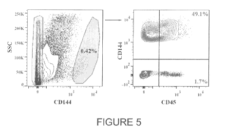
[0014] FIG 5. CD144 CD45+ cells are found and isolated from a sample of human
liver. A
human liver biopsy was analyzed for the presence of CD144 CD45+ and CD144 CD45-
cells. A
large portion of CD144+ cells was also CD45+ similar to mice livers. Red gate
in the left graph
corresponds to the red contour plot in the right graph. Blue gate in the left
graph corresponds to
the blue plot in the right graph.
DETAILED DESCRIPTION OF THE DISCLOSURE
[0015] This disclosure provides a previously unknown reservoir of hemogenic
endothelial cells
(HEC) in post-natal mammals that can give rise to hematopoietic cells, and
surface markers that
allows separation of HECs from other cell types. HECs are found in the
endothelial cell layers
of several organs and have the ability to reconstitute the immune system for
the treatment of
hematopoietic disorders.
Hemogenic endothelial cells
[0016] "Hemogenic endothelial cells" (HECs) are endothelial cells that have
the capacity to
generate hematopoietic cells, including hematopoietic stem cells. HECs as
disclosed herein have
the ability to engraft (establish residency) and provide long term
repopulation of hematopoietic
CA 02930304 2016-05-10
WO 2015/073680 PCT/US2014/065469
cells following transplantation into a recipient, such as an immunocompromised
subject. The
disclosed HECs are also capable of subsequent engraftment from one recipient
to one or more
additional recipients, and thus retain the ability to regenerate the immune
system. Capacity for
long term engraftment (e.g., for 4 weeks, 8 weeks, 12 weeks, 16 weeks, or 20
weeks or longer
post-transplantation) and secondary engraftment are highly desirable in a cell
population for
treatment of hematopoietic disorders.
[0017] HECs disclosed herein can be defined by expression of the markers CD45
(cluster of
differentiation 45, also known as Protein tyrosine phosphatase, receptor type
C or leukocyte
common antigen/LCA) and CD144 (cluster of differentiation 144, also known as
vascular-
endothelial cadherin or VE-Cadherin). CD144 CD45+ expression defines HECs, and
this
combination of markers has not been previously identified in any cell type of
a post-natal
subject.
[0018] HECs can further optionally be defined by expression, in addition to
CD144 CD45 , of
one or more additional markers selected from CD180, chemokine C-X-C motif
receptor 2
(CXCR2), chemokine C-X-C motif receptor 3 (CXCR3), chemokine C-X3-C motif
receptor 1
(CX3CR1), chemokine C-C motif receptor 9 (CCR9), G protein-coupled receptor
141 (GPR141),
G protein-coupled receptor 174 (GPR174), Signaling Lymphocyte Activation
Molecule Family
member 7 (SLAMF7), Signaling Lymphocyte Activation Molecule Family member 7
(SLAMF9), integrin alpha L (ITGAL), integrin alpha X (ITGAX), and membrane-
spanning 4-
domain, subfamily A, member 6D (MS4A6D). Preferably, the cells show positive
expression of
one or more of these additional markers. HECs can be defined by expression of
1, 2, 3, 4, 5, 6,
7, 8, or all of these additional markers, in addition to CD144 CD45+
expression. In one
embodiment, HECs are defined as CD144 CD45 SLAMF9+CXCR1+ cells.
[0019] HECs have some characteristics of endothelial cells (ECs), for example,
expression of the
EC marker CD144 and optionally, expression of the EC marker CD31 (also known
as platelet
endothelial cell adhesion molecule-1 or PECAM-1). HECs can also be found in
normal
endothelial cell layers in organs and tissues, adjacent to ECs. However,
unlike normal ECs,
which adhere to gelatin-coated and other coated surfaces in culture, HECs do
not adhere to
coated surfaces, such as gelatin-coated surfaces, in culture.
6
CA 02930304 2016-05-10
WO 2015/073680 PCT/US2014/065469
[0020] "Hematopoeitic stem cells" (HSCs) are cells that can generate
hematopoietic cells (HCs).
HSCs can be defined by expression of Lin-CD34 CD38-CD90+CD45RA-CD45+ (human
HSCs)
and Lin-cKit+Scal+Flk2-CD34-Slamf1+ (murine HSCs).
[0021] "Hematopoeitic cells" encompass myeloid lineage cells, which include
erythrocytes,
monocytes, macrophages, megakaryocytes, myeloblasts, dendritic cells, and
granulocytes
(basophils, neutrophils, eosinophils, and mast cells); and lymphoid lineage
cells, which include T
lymphocytes/ T cells, B lymphocytes/B cells, and natural killer cells. The
HECs disclosed herein
have the ability to generate HSCs and HCs including myeloid lineage cells and
lymphoid lineage
cells.
[0022] HECs have some characteristics of HSCs, for example, HECs can generate
new HCs.
However, unlike HCs and HSCs, HECs have not been found to expand under typical
conditions
for culturing HSCs using standard concentrations of hematopoietic cytokines,
such as SCF (stem
cell factor), TPO (thrombopoietin), FLT3L (Flt-3 ligand), and IL-3
(interleukin-3).
Identifying HECs
[0023] Disclosed herein are methods of identifying post-natal hemogenic
endothelial cells
(HECs) in a subject. The methods involve identifying CD144 CD45+ cells in a
post-natal
subject. The methods can further involve identifying cells that are CD144
CD45+ and also show
expression of one or more additional markers, in particular expression of one,
two, three, four,
five, six, seven, eight, or more markers selected from CD180, CXCR2, CXCR3,
CX3CR1,
CCR9, GPR141, GPR174, SLAMF7, SLAMF9, ITGAL, ITGAX, and MS4A6D. Identification
can be in vitro or in vivo, for example, using antibodies or antigen-binding
fragments thereof that
bind to cell surface markers, conjugated to an imaging moiety such as a
fluorescent or magnetic
agent, a radioisotope, or other suitable imaging agent.
[0024] In one embodiment, a specifically bound and labeled antibody can be
detected in an
individual using an in vivo imaging method, including, but not limited to,
radionuclide imaging,
positron emission tomography, computerized axial tomography, X-ray or magnetic
resonance
imaging method, fluorescence detection, and chemiluminescent detection.
7
CA 02930304 2016-05-10
WO 2015/073680 PCT/US2014/065469
Isolation of HECs
[0025] Disclosed herein are methods of isolating post-natal HECs. The methods
involve
isolating CD144 CD45+ cells from at least one tissue of a post-natal subject.
The methods can
further involve isolating cells that are CD144 CD45+ and also show expression
of one or more
additional markers, in particular expression of one, two, three, four, five,
six, seven, eight, or
more markers selected from CD180, CXCR2, CXCR3, CX3CR1, CCR9, GPR141, GPR174,
SLAMF7, SLAMF9, ITGAL, ITGAX, and MS4A6D.
[0026] The terms "isolated" and "purified" are used interchangeably herein to
refer to a material
that is substantially or essentially removed from or concentrated in its
natural environment. For
example, a cell is isolated if it is substantially removed from other
endogenous cell types, tissues,
and materials which the cell would normally be found in proximity to in a
subject. Methods for
purification and isolation of cell types according to expression of cell-
surface markers are
documented methodologies. A "substantially isolated" cell or cell population
is a cell or cell
population that is at least 50%, 60%, 70%, 80%, 85%, 90%, 95%, 98%, or 99% or
more isolated
from other cell types, tissues, or materials found in the tissue of a subject.
Also, a cell or cell
population is "substantially purified" when at least 50%, 60%, 70%, 80%, 85%,
90%, 95%, 98%,
or 99% or more of the cells in a cell sample express the cell-surface markers
of interest.
[0027] HECs can be isolated from tissues and organs throughout the body
including, but not
limited to, the liver, spleen, bone marrow, blood, umbilical cord, skin,
kidney, muscle, or lung.
Preferred tissues include liver, blood, umbilical cord, and skin. In one
embodiment, HECs are
isolated from a biological sample obtained by biopsy of a tissue or organ. In
another
embodiment, HECs are isolated from a blood or plasma sample. Autologous HECs
are isolated
from the same subject to whom the HECs are to be administered. Allogeneic HECs
are isolated
from at least one individual of the same species as the subject to whom the
HECs are to be
administered. Xenogeneic HECs are isolated from at least one individual of a
different species
from the subject to whom the HECs are to be administered. Non-autologous HECs
(that is,
allogeneic or xenogeneic HECs) can be derived from pre-natal, post-natal, or
post-mortem
tissues or organs. Preferred HECs are autologous. Preferred non-autologous
HECs are
allogeneic post-natal HECs. The most preferred HECs are autologous and post-
natal.
8
CA 02930304 2016-05-10
WO 2015/073680 PCT/US2014/065469
[0028] As a first step, HECs are isolated from a tissue, organ, or biological
sample, according to
CD144 CD45+ expression, optionally in combination with one or more additional
markers as
disclosed above. Methods to isolate or separate cells according to expression
of cell surface
markers include: fluorescence activated cell sorting by the use of e.g.
antibodies or fragments
thereof directed to CD144 and CD45, and optionally using additional antibodies
or fragments
thereof directed to additional markers; magnetic separation, using e.g.
antibody-coated magnetic
beads; affinity chromatography using antibodies or fragments thereof;
"panning" with antibodies
or fragments thereof attached to a solid matrix, e.g., a plate or other solid
matrix; or other
techniques as are known and used in the art for separation of cells based on
cell surface marker
expression. In preferred embodiments, fluorescence activated cell sorting or
magnetic separation
is used to isolate HECs from non-HECs.
[0029] Isolation of HECs generates a substantially pure population of CD144
CD45+ cells. As
defined herein, a "substantially pure population" of CD144 CD45+ cells means
more than 50%,
more than 60%, more than 70%, more than 75%, more than 80%, more than 85%,
more than
90%, more than 95%, more than 98%, more than 99%, or even 100% of the cells
following the
isolation/separation step are CD144 CD45 .
Culture, maintenance, and cryopreservation of HECs
[0030] Following isolation, HECs can be maintained in culture for up to one
week with standard
human/mammalian cell culture media, such as RPMI1640, Minimal Essential Medium
(MEM),
or Dulbecco's Modified Eagle Medium (DMEM) (each of these and similar media
available for
example through Gibco/ Life Technologies), supplemented with serum, such as 5-
30%,
preferably 10-25%, most preferably 20% serum such as fetal bovine/calf serum
(FBS or FCS),
and one or more growth supplements for endothelial cells, such as Endothelial
Cell Growth
Supplement (ECGS) at 2-200 [tg/mL. In a preferred embodiment, HECs are
maintained in RPMI
1640 media with 20% FCS and ECGS at 2-200 [t.g/m1 for 1 to 7 days, preferably
between 4 to 72
hours or 1, 2, 3, or 4 days, and then frozen for storage until needed using
the cryopreservation
methods disclosed herein.
9
CA 02930304 2016-05-10
WO 2015/073680 PCT/US2014/065469
[0031] Culture of HECs requires particular conditions, as these cells survive
and proliferate
poorly or not at all using standard methods for culturing endothelial or
hematopoietic cells.
Preferred culture conditions can include culturing HECs on a layer of feeder
cells, such as bone-
marrow stroma or fetal/embryonic organ specific (liver) fibroblasts.
[0032] Isolated HECs can be cryopreserved using techniques known in the art
for cell
cryopreservation. HECs can be frozen for storage, either directly after
isolation, or following
maintenance in culture conditions as described above, or after proliferation
in culture.
Accordingly, in one embodiment, HECs can be removed from a subject and frozen,
until such
time that it is determined that a subject is in need of treatment for an
immunodeficiency disorder,
at which point the HECs can be thawed and transplanted back into the subject
(for autologous
transplantation) or thawed and transplanted to a different subject in need of
treatment (for non-
autologous transplantation).
[0033] Isolated HECs can be prepared for cryogenic storage by addition of one
or a combination
of cryoprotective agents such as dimethyl sulfoxide (DMSO), glycerol,
polyvinylpyrrolidine,
polyethylene glycol, albumin, dextran, sucrose, ethylene glycol, i-erythritol,
D-ribitol, D-
mannitol, D-sorbitol, i-inositol, D-lactose, choline chloride, amino acids,
methanol, acetamide,
glycerol monoacetate, and inorganic salts. Addition of plasma or serum (e.g.,
to a concentration
of 20-25%) may augment the protective effect of DMSO. HECs can be frozen, for
example, in
60-40% growth media as disclosed above (e.g., RPMI 1650, MEM or DMEM) with 40-
60%
serum and 5-20% DMSO. In one embodiment, HECs are frozen in 50% growth media,
50%
FCS (fetal calf serum) with 10% DMSO.
[0034] Isolated HECs admixed with cryoprotective agents should be cooled at a
controlled rate
for cryogenic storage. Different cryoprotective agents and different cell
types have different
optimal cooling rates. Considerations and procedures for the manipulation,
cryopreservation,
and long-term storage of HSC sources are known in the art. Considerations in
the thawing and
reconstitution of frozen cell sources are also known in the art.
CA 02930304 2016-05-10
WO 2015/073680 PCT/US2014/065469
Isolated and purified populations of HECs and compositions thereof
[0035] Further encompassed by the subject disclosure is a substantially pure
population of
CD144 CD45+ post-natal hemogenic endothelial cells, wherein more than 80%,
more than 85%,
more than 90%, more than 91%, more than 92%, more than 93%, more than 94%,
more than
95%, more than 96%, more than 97%, more than 98%, more than 99%, more than
99.5%, or
even 100% of the cells following the isolation/separation step are CD144 CD45
.
[0036] Also contemplated are compositions that include a substantially pure
population of
CD144 CD45+ post-natal HECs. In one embodiment, the substantially pure
population of
CD144 CD45+ post-natal HECs is included in a composition with at least one
cryoprotective
agent, such as disclosed above. The cells in this composition can be in a
frozen or unfrozen
state. In another embodiment, the substantially pure population of CD144 CD45+
post-natal
HECs is included in a composition with a suitable culture medium, such as the
culture media
disclosed for maintenance of HECs in vitro. In another embodiment, the
substantially pure
population of CD144 CD45+ post-natal HECs is included in a composition with a
pharmaceutically acceptable carrier suitable for administration to a subject.
[0037] As used herein the phrase "pharmaceutically acceptable" means the
carrier, or vehicle,
does not cause an adverse reaction when administered to a mammal. Such
carriers are non-toxic
and do not create an inflammatory or anergic response in the body.
Pharmaceutically acceptable
carriers for practicing the invention include well known components such as,
for example,
culture media and phosphate buffered saline. Additional pharmaceutically
acceptable carriers
and their formulations are well-known and generally described in, for example,
Remington's
Pharmaceutical Science (18th Ed., ed. Gennaro, Mack Publishing Co., Easton,
Pa., 1990) and the
Handbook of Pharmaceutical Excipients (4th ed., Ed. Rowe et al. Pharmaceutical
Press,
Washington, D.C.), each of which is incorporated by reference.
[0038] Examples of compositions of CD144 CD45+ post-natal hemogenic
endothelial cells
include liquid preparations for parenteral, subcutaneous, intradermal,
intramuscular, or
intravenous administration (e.g., injectable administration), such as sterile
suspensions or
emulsions. Such compositions may be in admixture with a suitable carrier,
diluent, or excipient
11
CA 02930304 2016-05-10
WO 2015/073680 PCT/US2014/065469
such as sterile water, physiological saline, glucose or the like. The
compositions can also be
lyophilized. The compositions can contain auxiliary substances such as wetting
or emulsifying
agents, pH buffering agents, gelling or viscosity enhancing additives,
preservatives, flavorings,
colors, and the like, depending upon the route of administration and the
preparation desired.
[0039] A prefilled injection vial, ampoule or infusion bag of in unit dose
form, encompassing the
isolated HECs is also provided. The injection vial, ampoule or infusion bag
can include 1 x 10⁴
to lx 1010 HECs, lx 105 to lx 109 HECs, or lx 106 to lx 108 HECs.
[0040] The compositions of the present invention are administered in a manner
compatible with
the dosage formulation, and in a therapeutically effective amount. The
quantity to be
administered depends, for instance, on the subject and debilitation to be
treated, capacity of the
subject's organ, cellular and immune system to accommodate the therapeutic
composition, and
the nature of the cell or tissue therapy, etc. Precise amounts of therapeutic
composition required
to be administered depend on the judgment of the practitioner and are peculiar
to each individual.
However, suitable dosages of the therapeutic composition of the present
invention may range
from about 1 x 10⁴-1 x 10¹⁰ HECs per dose, or about 1 x 10⁵-1 x 10⁹ HECs per
dose, or about 1 x
106-1 x 10⁸ HECs per dose, depending on the route of administration. Suitable
regimes for initial
administration and follow on administration are also variable, but can include
an initial
administration followed by repeated doses at one or more hour, or day,
intervals by a subsequent
injection or other administration.
Methods of treatment
[0041] Further provided herein are methods of treatment for an
immunodeficiency disorder, the
method including administering a composition with a substantially pure
population of
CD144 CD45+ HECs to a subject in need thereof. The HECs of the present
invention can be
used for reconstituting the full range of hematopoietic cells in an
immunocompromised subject
following therapies such as, but not limited to, radiation treatment and
chemotherapy.
Administration of the disclosed HECs, such as by infusion or transplantation
into a subject, can
augment or replace stem or progenitor cells of, for example, the liver,
pancreas, kidney, lung,
12
CA 02930304 2016-05-10
WO 2015/073680 PCT/US2014/065469
nervous system, muscular system, bone, bone marrow, thymus, or spleen. It is
appreciated that it
may be necessary to treat the host to reduce immunological rejection of the
donor cells.
[0042] Preferred conditions treatable by the disclosed methods include
immunodeficiency
disorders characterized by an inadequate amount or activity of immune cells.
The
immunodeficiency disorder may be primary or secondary. In one embodiment, the
immunodeficiency disorder is a primary immunodeficiency disorder selected
from: a T-cell, B-
cell, or combined T-cell/B-cell immunodeficiency, such as severe combined
immunodeficiency
(SCID); an antibody deficiency, such as agammaglobulinemia; a complement
deficiency, such as
lupus; leukemia; lymphoma; an anemia, such as severe aplastic anemia;
neutropenia;
lymphopenia; or any condition associated with immune deficiency, such as
Wiskott-Aldrich
syndrome. In another embodiment, the immunodeficiency disorder is a secondary
immunodeficiency disorder associated with an infectious disease including
human
immunodeficiency virus (HIV) or hepatitis. In another embodiment, the
immunodeficiency
disorder is a secondary immunodeficiency disorder associated with the
administration of an
immunosuppressive agent, such as fluorouracil, vincristine, cisplatin,
oxoplatin, methotrexate, 3'-
azido-3'-deoxythymidine, paclitaxel, doxetaxel, an anthracycline antibiotic,
or mixtures thereof
having a secondary immunosuppressive effect.
[0043] As used herein, the terms "subject" and "patient" are used
interchangeably and refer to an
animal, including mammals such as non-primates (e.g., cows, pigs, horses,
cats, dogs, rats etc.)
and primates (e.g., monkey and human). In particular embodiments, the subject
is post-natal,
that is, the subject is, for example, a newborn animal, a young animal, an
adolescent animal, an
adult animal, or an aged animal.
[0044] As used herein, "treatment" refers to clinical intervention in an
attempt to alter the disease
course of the individual or cell being treated. Therapeutic effects of
treatment include without
limitation, preventing recurrence of disease, alleviation of symptoms,
diminishment of any direct
or indirect pathological consequences of the disease, decreasing the rate of
disease progression,
amelioration or palliation of the disease state, and remission or improved
prognosis. As used
herein, the terms "therapeutically effective amount" and "effective amount"
are used
13
CA 02930304 2016-05-10
WO 2015/073680 PCT/US2014/065469
interchangeably to refer to an amount of a composition of the invention that
is sufficient to treat
the immunological condition.
[0045] With respect to administering the HECs provided herein to a patient, an
effective amount
of cells may range from as few as several hundred or fewer to as many as
several million or
more. It will be appreciated that the number of HECs to be administered will
vary depending on
the specifics of the disorder to be treated, including but not limited to size
or total volume to be
treated, as well as the needs and condition of the recipient, among other
factors familiar to the
medical professional. In some embodiments, between 104 and 1010 cells per 100
kg person are
administered or transplanted into the subject or individual. HECs provided
herein can be
administered or transplanted, for example, by intravenous infusion or by
direct grafting, using
methods known in the art.
[0046] In one embodiment, HECs are used to augment or replace bone marrow
cells in bone
marrow transplantation. Human autologous and allogenic bone marrow
transplantations are
currently used as therapies for diseases such as leukemia and lymphoma. The
drawback of these
procedures, however, is that a large amount of donor bone marrow must be
removed to insure
that there are enough cells for engraftment. The present invention reduces or
eliminates the need
for large bone marrow donation, by substituting or supplementing a marrow
donation with HECs
for transplantation into a recipient. The HECs can be autologous to the
subject, or allogeneic to
the subject, or xenogeneic to the subject.
[0047] In another embodiment, HECs are administered to the bloodstream by
infusion.
[0048] In another embodiment, HECs are administered by transplantation to an
organ, such as
the liver, spleen, kidney, lung, eye, central nervous system, muscle, skin,
bone, ovary, testis,
heart, blood vessel, intestine, or lymph node.
[0049] In some embodiments, a single administration of cells is provided. In
other
embodiments, multiple administrations are used. Multiple administrations can
be provided over
periodic time periods such as an initial treatment regime of 3 to 7
consecutive days, and then
repeated at other times.
14
CA 02930304 2016-05-10
WO 2015/073680 PCT/US2014/065469
[0050] Further contemplated are methods involving co-administration, that is,
administration of
a composition of the invention before, after, or contemporaneously with
administration of a
treatment that may deplete the immune system or immune response of a subject.
Such methods
involve administering a composition with HECs to a subject before, during,
and/or after
treatments including cancer treatment and treatment with immunosuppressive
agents. The term
"cancer treatment" includes administration of any cancer agent including
radioactive isotopes
and cytotoxic agents. Examples of cytotoxic agents include, but are not
limited to
maytansinoids, yttrium, bismuth, ricin, ricin A-chain, doxorubicin,
daunorubicin, taxol, ethidium
bromide, mitomycin, etoposide, tenoposide, vincristine, vinblastine,
colchicine, dihydroxy
anthracin dione, actinomycin, diphtheria toxin, Pseudomonas exotoxin (PE) A,
PE40, abrin,
abrin A chain, modeccin A chain, alpha-sarcin, gelonin, mitogellin,
retstrictocin, phenomycin,
enomycin, curicin, crotin, calicheamicin, sapaonaria officinalis inhibitor,
and glucocorticoid and
other chemotherapeutic agents. Examples of immunosuppressive agents include
cyclosporine,
GAD65 antibodies, fluorouracil, cisplatin, oxoplatin, methotrexate, 3'-azido-
3'-deoxythymidine,
paclitaxel, doxetaxel, an anthracycline antibiotic, or mixtures thereof having
a secondary
immunosuppressive effect. Several cytotoxic agents as indicated herein are
also
immunosuppressive agents.
[0051] The present disclosure is further illustrated by the following non-
limiting examples.
EXAMPLES
Example 1. Generation of mice with inducible expression of Cre-recombinase
under
control of the CD144 promoter. Transgenic mice with tamoxifen-inducible Cre-
recombinase
CreERT2 under the control of endothelial specific VE-cadherin promoter (VCC-
CreERT2 mice)
( Pitulescu M. et al., Nat. Protoc. 5:1518-1534 (2010)) were crossed with ACTB-
loxp-tdTomato-
STOP-loxp-EGFP reporter mice (Muzumdar M. et al., Genesis 45:593-605 (2007))
(Figure 1A).
The ACTB-loxp-tdTomato-STOP-loxp-EGFP reporter consists of a chicken f3-actin
core
promoter with a CMV enhancer (pCA) driving a loxP flanked coding sequence of
membrane-
targeted tandem dimer Tomato (tdTomato) resulting in membrane localized
tdTomato (a red
fluorescent protein) expression. In progeny from the cross (inducible VCC-EGFP
reporter mice
or "iVCC-EGFP mice"), injection of tamoxifen induces Cre-recombinase
expression in cells in
CA 02930304 2016-05-10
WO 2015/073680 PCT/US2014/065469
which VE-cadherin/CD144 expression is activated. Cre-recombinase mediates
intra-
chromosomal recombination, causing excision of tdTomato at the loxP sites (and
loss of red
fluorescence) in CD144 expressing cells. Excision of tdTomato allows the pCA
promoter to
drive expression of membrane-targeted enhanced green fluorescent protein, thus
identifying cells
in which CD144 expression is turned on subsequent to tamoxifen induction by
green
fluorescence. This allowed tracing of endothelial cells in a temporally
restricted manner.
[0052] iVCC-EGFP mice were induced with tamoxifen at 4 weeks post-natally
(mice are
considered to be adult mice at weaning age of 3 weeks). As seen in Figure 1B,
the tamoxifen
injections induced EGFP expression in VE-cadherin expressing cells. The left
panel of Figure
1B shows expression of tdTomato (red) in the skin of iVCC-EGFP mice. The
middle panel
shows EGFP expression (green) for the same area as shown in the left panel
after tamoxifen
induction. The right-hand panel shows the overlap of the left and middle
panels. Only the
vasculature is showing EGFP expression.
[0053] Example 2. Post-natal CD144 CD45+ cells isolated from liver, spleen,
lung and bone
marrow. Analysis of several organs harvested from iVCC-EGFP mice induced with
tamoxifen
at 4 weeks post-natally revealed the presence of CD144 CD45+ endothelial cells
in the lung,
liver, spleen, and bone marrow (Figure 2A).
[0054] CD144 (GFP )CD45+ cells were sorted using standard techniques and
plated for in vitro
culture on gelatin-coated tissue culture plates using RPMI 1640 media,
supplemented with 20%
FCS, endothelial cell growth supplement (ECGS) at 2-200 lug/m1, and cytokines
(SCF, TPO,
FLT3L, IL6, and IL3) used for expansion of hematopoietic cells (Figure 2B).
The sorted cells
did not attach to the gelatin-coated tissue culture surfaces, as regular
endothelial cells would.
The sorted cells also did not respond to or expand in the presence of
hematopoietic cytokines, as
regular hematopoietic progenitors would. This suggested that the isolated
cells were not true
endothelial cells, nor were they standard hematopoietic cells.
[0055] Example 3. CD144 CD45+ endothelial cells are capable of functional
reconstitution
of the hematopoietic system. iVCC-EGFP mice were induced with tamoxifen at 6
weeks after
birth (adult mice) and used for experiments two weeks post-induction. To label
CD144+
16
CA 02930304 2016-05-10
WO 2015/073680 PCT/US2014/065469
endothelial cells (arterial, venal, and capillary) mice were injected with
anti-CD144+ antibody
peri-orbitally. Injected mice were sacrificed eight minutes post-injection to
exclude labeling of
the lymphatic vascular bed. This allowed double labeling of CD144+ endothelial
cells ¨
genetically (by tamoxifen induction of VE-cadherin promoter driven expression
of CRE
recombinase) and by intra-vital staining of CD144+ endothelial cells using
anti CD144+
antibodies. Liver was digested as described in Nolan D. J. et al., Dev Cell
26:204-219 (2013)).
Briefly, liver was minced and incubated with Collagenase A (25 mg/ml), Dispase
11 (25 mg/ml),
and DNase (250 mg/ml) (Roche) at 37 C for 20-30 min to create a single cell
suspension.
Following digestion, cells were post-stained with anti-CD45 antibodies and
antibodies to CD31,
an additional endothelial specific surface marker.
[0056] CD144+GFP+CD31+CD45+ cells were sorted using FACS and transplanted into
imuno-
compromised sub-lethally irradiated mice (NOD-SCID-IL2g or "NSG" mice) (Figure
3A).
Twelve weeks post-transplantation, transplanted mice were tested for the
presence of the donor
cells in their peripheral blood. A significant portion of their peripheral
blood (>35%) was
composed of the donor GFP+CD45.2+ cells (NSG mice express CD45.1 surface
protein) (Figure
3B). GFP+CD45.2+ cells were isolated from the bone marrow of the primary
recipients and used
for secondary transplantations into a sub-lethally irradiated (650 RAD) CD45.1
expressing mice
(non-NSG). Nineteen weeks post-transplantation CD45.2+GFP+ cells were detected
in peripheral
blood of secondary recipients. These experiments prove that CD144+CD45+
endothelial cells are
hemogenic (HEC) and capable of reconstitution of the hematopoietic system when
transplanted
in vivo (Figure 3C).
[0057] Example 4. Identification of additional markers that distinguish HECs.
To identify
additional surface markers that could further delineate HECs from donor
tissues including, but
not limited to liver, whole-transcriptome profiles of CD144+CD45+ liver cells
were compared to
CD144+CD45 liver cells. iVCC-EGFP mice were induced with tamoxifen at 6 weeks
after birth
(adult mice) and used for experiments two weeks post-induction. To label
CD144+ endothelial
cells, mice were injected with anti-CD144+ antibody peri-orbitally and
sacrificed eight minutes
post-injection. Liver was digested as described above and post-stained with
anti-CD45
antibodies. CD144+GFP+CD45+ and CD144+GFP+ CD45- endothelial cells were sorted
using
17
CA 02930304 2016-05-10
WO 2015/073680 PCT/US2014/065469
FACS. Sorted cells were used for total RNA extraction. RNA was used for whole-
transcriptome
deep sequencing (RNA-Seq) (Figure 4A).
[0058] Comparison of whole-transcriptome sequences of CD144 GFP CD45+ and
CD144 GFP CD45- endothelial cells revealed a separate cluster of genes (red
dots in the Figure
4B) overexpressed (log2[CD144 GFP CD45 / CD144 GFP CD451>3) in CD144 GFP
CD45+
endothelial cells. Analysis of the overexpressed genes (Figure 4B) revealed a
set of additional
cell-surface expressed proteins that can optionally be used to further define
post-natal
CD144 CD45+ HECs (Figure 4C).
[0059] Analysis of the novel post-natal HEC surface markers revealed that
these markers are
typically overexpressed in emerging hematopoietic cells/hemogenic endothelial
cells during
development in the areas known to be associated with definitive hematopoiesis,
specifically, the
placenta, AGM, and fetal liver (McKinney-Freeman S. et al., Cell Stem Cell
11:701-14 (2012))
(Figure 4D).
[0060] Example 5. Isolation of CD144 CD45+ cells from human liver. The
presence of
CD144 CD45+ HECs was investigated in adult human liver. Human liver tissue
samples from
three unidentified adult individuals were analyzed for the presence of CD144
CD45+ and
CD144 CD45- cells. Surprisingly, a large portion of CD144+ liver cells were
also CD45 ,
similar to mice livers (Figure 5). This identifies HECs as present in adult
humans.
18