Note: Descriptions are shown in the official language in which they were submitted.
CA 02930441 2016-05-12
WO 2015/070351
PCT/CA2014/051094
1
BIONANOFLUID FOR USE AS A CONTRAST, IMAGING, DISINFECTING
AND/OR THERAPEUTIC AGENT
FIELD OF THE INVENTION
The present invention relates to the field of nanotechnology and more
particularly
concerns a bionanofluid comprising a mono-dispersed carbon-based
nanomaterial as a contrast, imaging, disinfecting and/or therapeutic agent.
BACKGROUND
Over the past few years, there has been tremendous interest in exploiting
nanotechnology materials and devices in the diagnostic and/or treatment of
biological problems or diseases, including the treatment of infections and/or
human cellular diseases. However, so far the interactions between carbon
nanomaterials and cellular physiology have been characterized as an issue of
biochemical mechanisms involving molecular transport, cellular adhesion, etc.
Ultrasound imaging is a widely applied technique in clinical research and
treatment, where sound waves are projected towards an object and the reflected
waves are analysed. Ultrasound imaging, however, has some major drawbacks.
Achieving high axial and spatial resolution comes at the price of penetration
depth. Frequencies of 30 to 55 MHz are typically used, providing an image
which
is highly resolved, but shallow. For deep structures, lower frequencies in the
range of 1-18 MHz are applied, enabling a greater penetration depth, but with
a
limited image resolution in either axis. Several strategies have been
developed to
resolve this issue, such as image reconstruction techniques to lower
background
noise, the creation of synthetic apertures, and, finally, by contrast-enhanced
ultrasound approaches.
Contrast-enhanced ultrasound (CEUS) is a technique where contrast agents
having ligands allowing them to bind to the cells of interest are injected in
the
patient. The technique is however limited to entities that can cause an
intense
CA 02930441 2016-05-12
WO 2015/070351
PCT/CA2014/051094
2
reflection or generate significant echogenicity, i.e., the ability to reflect
sound
waves. Currently, the major and only commercial contrast agents are
microbubbles, which are filled with a gas, usually using perfluorcarbons. The
microbubbles oscillate in the presence of the ultrasonic field, generating the
backscatter that can be detected with a strong contrast to surrounding
tissues.
One drawback of microbubbles is the use of perfluorocarbons, which last but a
few short minutes in the blood and are highly expensive, prohibiting their
widespread use. The presence of microbubbles is also detrimental to patient
health, resulting in head pains, nausea and other side effects of use in a
significant number of patients, which provides an incentive to avoid their use
from
a clinical prospective.
Nanofluids comprising nanoparticles dispersed in a fluid where the physical
material is defined as nano and is dispersed. Nanoparticles such as carbon
nanotubes (CNT), carbon nanoparticles and hybrid particle systems can be
modified by physical or chemical processes to enhance their dispersibility in
the
fluid. However, these nanofluids are not suitable to enable or elicit bio-
specific
biological responses. Nanofluids that are not biomodified, are not able of
delivering specific targeted effects. Bionanofluid definitions require the
inclusion
of bio-related or bio-molecular functionalization that is specific to a
desired
application.
SUMMARY
Bionanofluids can be developed from carbon-based nanomaterials by addition to
the nanomaterials surface of bio-affinity agents or biological molecules,
these
bio-affinity agents or biological molecules are also referred to as targeting
moieties. By the addition of biological molecules and/or other biologic
modifications, the materials' properties are harnessed and improved. These
bionanofluids can deliver a platform for imaging and/or therapeutic action.
Formulations of bionanofluids can be prepared for specific applications and
each
formulation is a new product designed for a specific function. The range of
CA 02930441 2016-05-12
WO 2015/070351
PCT/CA2014/051094
3
applications for the bionanofluids is broad as the applications for targeted
cell
death can encompass fields such as cancer treatment and/or infection control.
The bio-modification of the nanomaterial allows preventing non-specific
association with non-targeted entities. In one embodiment, the specific cell
targeting that can be achieved with the bionanofluid, combined with the
bionanofluid's photonic properties allows enhancing cellular function
disruption to
the point where cell viability is impossible. Thanks to the combination of its
targeting and photonic properties, the bionanofluid can thus be used as a
therapeutic agent and/or disinfecting agent. But, the combination of the
targeting
and photonic properties also allows using the bionanofluid in other
applications
which do not necessarily involve cell disruption, such as an imaging agent or
as a
contrast agent for ultrasound.
In one aspect, there is provided a bionanofluid comprising a carbon-based
nanomaterial substantially mono-dispersed in a fluid, wherein the carbon-based
nanomaterial is surface modified with polar groups when the fluid is polar or
with
non-polar groups when the fluid is non-polar, and functionalized with
targeting
moieties to allow specific association of the carbon-based nanomaterial to
targeted entities.
In one embodiment, there is provided a bionanofluid comprising a carbon-based
zo nanomaterial substantially mono-dispersed in a fluid, wherein the
carbon-based
nanomaterial is surface modified with a polar group when the fluid is polar or
with
a non-polar group when the fluid is non-polar, and functionalized with a
targeting
moiety to allow specific association of the carbon-based nanomaterial to a
targeted entity.
In one embodiment, there is provided a hybrid bionanofluid comprising the
bionanofluid described therein, wherein the carbon-based nanomaterial is
further
modified with a hybrid nanoparticle which comprises an alloy, transition
metal,
semi-conductor, semi-metal, polymer based nanoparticle or a combination
thereof.
CA 02930441 2016-05-12
WO 2015/070351
PCT/CA2014/051094
4
In one embodiment, there is provided a hydrogel comprising the bionanofluid
described therein or the hybrid bionanofluid described therein, and gelatin.
In one embodiment, there is provided a foam comprising the bionanofluid
described therein or the hybrid bionanofluid described therein, and silica or
a
derivative thereof.
In one embodiment, there is provided a cream or a spray comprising the
bionanofluid described therein or the hybrid bionanofluid described therein.
In one embodiment, there is provided a dried product obtained by drying the
bionanofluid described therein or the hybrid bionanofluid described therein.
In one embodiment, there is provided a use of the bionanofluid described
therein
or the hybrid bionanofluid described therein to create disruption of the
targeted
entity upon application of an external energy including light, ultrasound or
radiowaves, when the targeting moiety is associated with the targeted entity,
preferably a prokaryote or an eukaryote, more preferably a cell, virus,
bacteria,
spore, a fungus or a small multi-cellular organism such as a microscopic worm.
In one embodiment, there is provided a use of the bionanofluid described
therein
or the hybrid bionanofluid described therein as an antiseptic agent, wherein
the
targeted entity is an infectious agent, preferably a prokaryote or an
eukaryote,
preferably a cell, bacteria, spore, virus, prion, fungus, or a small multi-
cellular
zo .. organism such as a worm.
In one embodiment, there is provided a use of the bionanofluid described
therein
or the hybrid bionanofluid described therein as a contrast agent for
ultrasound,
preferably ultrasound imaging.
In one embodiment, there is provided a use of the bionanofluid described
therein
or the hybrid bionanofluid described therein as an imaging agent.
Other features as aspects of the invention will be better understood upon
reading
of preferred embodiments thereof with reference to the appended figures.
CA 02930441 2016-05-12
WO 2015/070351
PCT/CA2014/051094
BRIEF DESCRIPTION OF THE DRAWINGS
Figures 1 to 49 are images and diagrams illustrating various embodiments.
Figure 1. Coupling chemistry used with Thiol-PEG-CNTs to attach bio-affinity
molecules. PEGylation of the Thiol-CNT particles is described in PEG
treatment.
5 Figure 2.
Coupling chemistry used with Mal-PEG-CNTs to attach bio-affinity
molecules. Initially EDC/NHS chemistry is used to link maleimide-PEG-amide to
COOH-functionalized bionanofluid in Reaction 1. Reaction 2 allows for the
coupling of bio-affinity molecule to maleimide component of PEG.
Figure 3. Total antibody load using an amine-Mal-PEG-Cysteine coupling to
carbon-derived bionanofluid, creating antibody labeled chemi-luminescent
applications. Horse-radish peroxidase (HRP)-Ab conjugates prepared as for
conventional antibody linkage. Conjugates were washed three times to exclude
non-specific carry-over of residual non-covalently linked HRP-Ab. None was
detected in C-PEG control. Total loading of Ab via thioester coupling assessed
by
presence of reporter HRP-Ab on surface. Possible pre-concentration of HRP-Ab
via covalent linkage as serial dilutions generate lower signal at 11500p1
(v/v)
dilution than 0.5p1Ab conjugate.
zo Figure 4.
Image of Bacterial streak on Agar plate obliterated by use of Carbon-
derived BioNanofluid Hydrogel and laser cell killing. Region of bacterial
killing
indicated by black box.
Figure 5. Crystalline purified Carbon Dots (quantity 1 gram) produced from
green
materials. Material is ready for re-suspension in water and immediately mono-
disperse.
Figure 6. Photoluminescence of Carbon Dots under UV light illumination. A.
BSA¨Carbon-Dots against water B. Comparison of BSA-Carbon-Dots, Glucose-
CA 02930441 2016-05-12
WO 2015/070351
PCT/CA2014/051094
6
Carbon-Dots and PEG-Carbon-dots against water, under UV illumination. 1m1 of
dots on in the left hand tube and compared to water. Concentration is
0.5mg/ml.
Figure 7. Comparison of magnetized-a-TSHR-Thiol-PEG-CNT (left) vs. non-
magnetized-a-TSHR-Thiol-PEG-CNTs CNT (right) particles on a magnetic
separator.
Figure 8. a-TSHR-Magnetized-CNT on BCPAP cells exposed to magnetized field.
Arrows indicate stationary cells, and yellow boxes indicate movement of cells
attached with a-TSHR-magnetized-CNTs over a period of time. A. 3 seconds, B.
seconds, and C. 20 seconds.
Figure 9. Antibody-Magnetized-CNT on LNCaP and BCPAP cells. A. A single
LNCaP cell before and after exposure to laser, targeted with magnetized-a-
15 PSMA-Thiol-PEG-CNTs. B. LNCaP and BCPAP cells prior to laser exposure to
illustrate the adherence of antibody-magnetized-CNTs on the cells.
Figure 10. Carbon-derived bionanofluid: Gelatin hydogel (heating at different
laser powers. At laser powers >1W, the observation that the hydrogel becomes
liquefied.
Figure 11. Carbon-derived bionanofluid: Gelatin hydrogel heating increasing
BioNanofluid concentration (1g/L) leads to rapid heating, laser 2W.
Figure 12. Carbon-derived bionanofluid foam at different magnifications. A.
Spot
of bionanofluid foam, with size marker. B. 5X magnification, C. 10X
magnification,
D. 20X magnification.
Figure 13.
Scanning Electron Microscopy (SEM) images of carbon-derived
bionanofluids. A. COOH-functionalized Bionanofluid. B/C. Gold loaded MWCNT,
gold particles having defined spherical structure, at two different
magnifications.
CA 02930441 2016-05-12
WO 2015/070351
PCT/CA2014/051094
7
Figure 14. Carbon Dots on STEM mesh substrate. Size varies from 100 nm to 5
nm and below. A. PEG-Dots. B. BSA-Dots.
Figure 15. UVNIS of Size controlled carbon nanotubes derived bionanofluid. All
particles are below 220 nm length, 50 nm diameter.
Figure 16. Evidence on resonant Au Particles bound to Carbon nanotubes.
Figure 17. COOH-CNT bionanofluid, size range 0.001 ¨2 pM.
Figure 18. UVNis spectra of Carbon Dots, A. PEG-Carbon dots, B. Glucose-
Carbon dots and C. BSA-Carbon dots.
Figure 19. Carbon Dot Near Infra Red Photo-luminescence.
Figure 20. Demonstration of the effectiveness of PEG-Thiol-CNT Bionanofluid as
an ultrasound contrast agent. A. without and, B. with bionanofluid.
Figure 21. Thiol-PEG-CNT Bionanofluid encased in an agarose gel. Image of
bionanofluid taken above the plane of ultrasound probe set to 30 mHz. The
reflection is caused by the interaction with the bionanofluid above the plane
of the
gel can be noted.
Figure 22. Thiol-PEG-CNT Bionanofluid end oriented longitudinally to the probe
face, covered with gel and brought into contact with the probe face. A. The
interaction at the bottom of the tube waves was analyzed. B. Same experiment
with tube 2, left blank and lacking modified bionanofluid.
Figure 23. Potential to agitate/manipulate the PEG-Thiol-CNT bionanofluid by
non-contact movement of ultrasound probe at a distance of 2-5 cm.
Figure 24. Ultrasound contrast of Thiol-PEG-CNT Bionanofluid in the Vena Cava
of a mouse. Arrows indicate contrast image of the bionanofluid, over a time
CA 02930441 2016-05-12
WO 2015/070351
PCT/CA2014/051094
8
course. A. saline injection, B. Bionanofluid injection time point t= 1 minute,
C.
Bionanofluid injection time point t = 10 minutes.
Figure 25. Ultrasound contrast of Thiol-PEG-CNT Bionanofluid in the Aortic
Arch
of a mouse. Arrows indicate contrast image of the bionanofluid, over a time
course. A. saline injection, B. Bionanofluid injection time point t= 1 minute,
C.
Bionanofluid injection time point t = 2 minutes.
Figure 26. Ultrasound contrast of Thiol-PEG-CNT Bionanofluid in the bladder of
lo a mouse. Box shows a magnified area of the urethra and arrows indicate
contrast image of the bionanofluid of the expulsion of the bionanofluid over a
period of time. Images represent frame capture of 200 frames total (time
course), post bionanofluid injection. A. Frame 7/200, B. Frame 69/200, C.
Frame 85/200, and D. Frame 151/200
Figure 27. Ultrasound contrast of Thiol-PEG-CNT Bionanofluid in the kidney of
a
mouse. White Arrows indicate the bionanofluid movement in the Kidney over a
period of time. Yellow arrowheads indicate the needle tip. Images represent
frame capture of 200 frames total (time course), post bionanofluid injection.
A.
Frame 20/200, B. Frame 85/200, and C. Frame 110/200
Figure 28. HEK293 cells mixed with carbon-derived Thiol-PEG-CNT bionanofluid
and exposed to laser for different periods of time, A. 5 seconds, B. 10
seconds
and C. 20 seconds. After exposure cells were reseeded onto 6-well plates with
DMEM+10% FBS growth media. Cells were allowed to grow for 5 days and a
picture of the cells in the plate was taken. No live cells were present after
20sec
exposure.
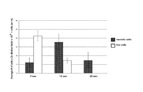
Figure 29. HEK283 cells mixed with Thiol-PEG-CNT carbon-derived bionanofluid
and exposed to laser for different periods of time (5, 10, 20 sec). After
exposure
cells were mixed with Trypan blue, and counted. Trypan Blue labels
necrotic/blue
CA 02930441 2016-05-12
WO 2015/070351
PCT/CA2014/051094
9
(or dead) cells and live/white cells. Experiment performed n=4. Cells counted
using a haemocytometer.
Figure 30. 650,000 HEK283 cells mixed with Thiol-PEG-CNT carbon-derived
bionanofluid and exposed to laser for different periods of time (5, 10, 20
sec).
After exposure cells were mixed with Trypan blue, and counted. Trypan Blue
labels necrotic/blue (or dead) cells blue and live/white cells.
Ablated cell
numbers were determined from a per-count of cells prior to exposure to the
laser.
As the amount of heat generated in the tube, is so high, cells "literally"
explode.
Experiment performed n=1. Cells counted using a haemocytometer.
Figure 31. Cell ablation studies using a-PSMA-Thiol-PEG-CNTs, to determine
concentration of cells to CNT particle. Cells were mixed with a-PSMA-Thiol-
PEG-CNT-bionanofluid at 37 C for lhr, cells were washed 5X with PBS and re-
suspended in PBS and exposed to 532nm laser for 30sec. An aliquot of cells
were removed to give a pre-count of cells. After laser exposure, cells were
mixed
with Trypan blue, and white cells or live cells were counted. Results given as
%
Alive and experiment performed n=6. 2:1 ratio cells to bionanofluid was
determined optimal, as higher concentrations of bionanofluid results in bulk-
heating. Cells counted using a haemocytometer.
Figure 32. Cell ablation studies using a-PSMA-Thiol-PEG-CNT, to exposure time
of cells to laser. Cells were mixed with a-PSMA-Thiol-PEG-CNT-bionanofluid at
37 C for 1hr, cells were washed 5X with PBS and resuspended in PBS and
exposed to 532nm laser for 30sec. An aliquot of cells were removed to give a
pre-count of cells. After laser exposure (20, 25, 30, 35 sec) cells were mixed
with
Trypan blue, and white cells or live cells were counted. Results given as %
Alive
and experiment performed n=6. 30 sec exposure of the cells to the bionanofluid
was determined optimal, as longer exposure results in bulk-heating. Cells
counted using a haemocytometer.
CA 02930441 2016-05-12
WO 2015/070351
PCT/CA2014/051094
Figure 33. Cell ablation studies using a-PSMA-Thiol-PEG-CNT, of PSMA positive
LNCaP cells vs. PSMA negative-PC3 cells. Cells were mixed with a-PSMA-
Thiol-PEG-CNT bionanofluid at 37 C for 1hr, cells were washed 5X with PBS
and re-suspended in PBS and exposed to 532nm laser for 305ec. An aliquot of
5 cells were removed to give a pre-count of cells. 2:1 cell:bionanofluid,
and 30sec
exposure was used. Experiment was repeated n=4. Cells counted using a
haemocytometer.
Figure 34. Cell ablation studies using PC3 cells with SELEX isolated PC3-
10 specific aptamer and Doxyrubicin-linked carbon dots. PC3 cells were
mixed with
respective bionanofluid or PBS/control at 37 C for 1hr, cells were washed 5X
with PBS and resuspended in PBS and exposed to 532nm laser for 30sec. An
aliquot of cells were removed to give a pre-count of cells. 2:1 cell:Mal-PEG-
bionanofluid, and 30sec exposure was used, unless noted otherwise.
Experiment was repeated n=4. 5a73 is a known DNA aptamer for PC3 cells,
whereas 2Vis is the SELEX-DNA aptamer isolatedby the inventors, which show
higher and more significant (p=0.0124) cell killing than the commercially
available
product. Carbon dots were also coupled to doxorubicin and exposed to cells.
Observation of a synergistic cell ablation of doxorubucin-dots vs. BSA-dots.
Cells
counted using a haemocytometer.
Figure 35. Cell ablation studies using a-TSHR-Thiol-PEG-CNTs, to determine
concentration of cells to CNT particle. Cells were mixed with a-TSHR-Thiol-PEG-
CNT-bionanofluid at 37 C for 1hr, cells were washed 5X with PBS and re-
suspended in PBS and exposed to 532nm laser for 30sec. An aliquot of cells
were removed to give a pre-count of cells. After laser exposure, cells were
mixed
with Trypan blue, and white cells or live cells were counted. Results given as
%
Alive and experiment performed n=6. 2:1 ratio cells to bionanofluid was
determined optimal, as higher concentrations of bionanofluid results in bulk-
heating. Cells counted using a haemocytometer.
CA 02930441 2016-05-12
WO 2015/070351
PCT/CA2014/051094
11
Figure 36. Cell ablation studies using a-TSHR-Thiol-PEG-CNTs, to exposure time
of cells to laser. Cells were mixed with a-TSHR-Thiol-PEG-CNT-bionanofluid at
37 C for 1hr, cells were washed 5X with PBS and re-suspended in PBS and
exposed to 532nm laser for 305ec. An aliquot of cells were removed to give a
.. pre-count of cells. After laser exposure (20, 30, 40 sec) cells were mixed
with
Trypan blue, and white cells or live cells were counted. Results given as %
Alive
and experiment performed n=6. 30 sec exposure of the cells to bionanofluid was
determined optimal, as longer exposure results in bulk-heating. Cells counted
using a haemocytometer.
Figure 37. Cell ablation studies using a-TSHR-Thiol-PEG-CNTs, of TSHR
positive BCPAP cells vs. TSHR negative-NSC34 cells. Cells were mixed with a-
TSHR-Thiol-PEG-CNT-bionanofluid at 37 C for 1hr, cells were washed 5X with
PBS and re-suspended in PBS and exposed to 532nm laser for 30sec. An
aliquot of cells was removed to give a pre-count of cells. 2:1
cell:bionanofluid,
and 30sec exposure was used. Experiment was repeated n=4. Cells counted
using a haemocytometer.
Figure 38. Cell ablation studies using a-TSHR-Thiol-PEG-CNTs, of TSHR
positive BCPAP cells to determine stability carbon-derived bionanofluid, at A.
4 C
or B. -20 C or -80 C. Cells were mixed with TSHR-bionanofluid at 37 C for
lhr,
cells were washed 5X with PBS and re-suspended in PBS and exposed to
532nm laser for 30sec. An aliquot of cells was removed to give a pre-count of
cells. 2:1 cell:bionanofluid, and 30sec exposure was used. Experiment was
repeated n=4. Cells counted using a haemocytometer.
Figure 39. Multiple treatment cell ablation studies using a-TSHR-Thiol-PEG-
CNTs. Cells were mixed with a-TSHR-Thiol-PEG-CNT-bionanofluid at 37 C for
1hr, cells were washed 5X with PBS and re-suspended in PBS and exposed to
532nm laser for 30sec. An aliquot of cells was removed to give a pre-count of
cells. After laser exposure, cells were mixed with Trypan blue, and white
cells or
CA 02930441 2016-05-12
WO 2015/070351
PCT/CA2014/051094
12
live cells were counted.
Afterwards, cells were reseed on 6-well plates
RMPI1640+10%FBS for 48 hrs. After which cells were collected again And
exposed to a-TSHR-Thiol-PEG-CNT-bionanofluid as described previously.
Results given as % Alive and experiment performed n=6. 2:1 ratio cells to
bionanofluid and 30 sec exposure was determined optimal.
Figure 40. Cell ablation studies using either Thyrogen (TSH recombinant)- or
thyrotropin (TSH purifiied)-Thiol-PEG-CNTs, to determine concentration of
cells
to CNT particle. Cells were mixed with TSH-bionanofluid at 37 C for 1hr, cells
lo were washed 5X with PBS and re-suspended in PBS and exposed to 532nm
laser for 30sec. An aliquot of cells was removed to give a pre-count of cells.
After laser exposure, cells were mixed with Trypan blue, and white cells or
live
cells were counted. Results given as % Alive and experiment performed n=6.
2:1 ratio cells to bionanofluid was determined optimal, as higher
concentrations
of bionanofluid results in bulk-heating. Cells counted using a haemocytometer.
Figure 41. Cell ablation studies using either Thyrogen (TSH recombinant)- or
thyrotropin (TSH purifiied)-Thiol-PEG-CNTs, to determine concentration of
cells
to CNT particle. Cells were mixed with TSH-Thiol-PEG-bionanofluid at 37 C for
1hr, cells were washed 5X with PBS and re-suspended in PBS and exposed to
532nm laser for 20, 30, or 40sec. An aliquot of cells was removed to give a
pre-
count of cells. After laser exposure, cells were mixed with Trypan blue, and
white
cells or live cells were counted. Results given as % Alive and experiment
performed n=6. 30
seconds laser exposure of cells to bionanofluid was
determined optimal, as higher concentrations of bionanofluid results in bulk-
heating. Cells counted using a haemocytometer.
Figure 42. Cell ablation studies using either Thyrogen (TSH recombinant)- or
thyrotropin (TSH purified)-Thiol-PEG-CNTs, against TSHR-positive BCPAP cell
lines vs. TSHR-negative NSC34 cell lines. Cells were mixed with TSH-Thiol-
PEG-bionanofluid at 37 C for 1hr, cells were washed 5X with PBS and re-
CA 02930441 2016-05-12
WO 2015/070351
PCT/CA2014/051094
13
suspended in PBS and exposed to 532nm laser for 30sec. An aliquot of cells
was removed to give a pre-count of cells. After laser exposure, cells were
mixed
with Trypan blue, and white cells or live cells were counted. Results given as
%
Alive and experiment performed n=4. Cells counted using a haemocytometer.
Figure 43. Cell ablation studies using different amounts of TSHR antibody
conjugated to Mal-PEG-CNTs. Cells were mixed with a-TSH-Mal-PEG
bionanofluid at 37 C for lhr, cells were washed 5X with PBS and re-suspended
in PBS and exposed to 532 nm laser for 30 sec. An aliquot of cells was removed
to give a pre-count of cells. After laser exposure, cells were mixed with
Trypan
blue, and white cells or live cells were counted. Results given as % Alive and
experiment performed n=3. Cells counted using a haemocytometer.
Figure 44. Cell ablation studies using different amounts of SNRNP antibody
conjugated to Mal-PEG-CNTs. SNRNP is an intracellular protein, thus showed
negligible effects on cell killing. Cells were mixed with a-SNRNP-Mal-PEG-
bionanofluid at 37 C for lhr, cells were washed 5X with PBS and re-suspended
in PBS and exposed to 532nm laser for 305ec. An aliquot of cells was removed
to give a pre-count of cells. After laser exposure, cells were mixed with
Trypan
blue, and white cells or live cells were counted. Results given as % Alive and
experiment performed n=3. Cells counted using a haemocytometer.
Figure 45. Cell ablation studies using different ratios of BCPAP cells to TSHR
antibody conjugated to Au-Thiol-PEG-CNTs. Cells were mixed with a-TSHR-Au-
nanoparticles at 37 C for lhr, cells were washed 5X with PBS and re-suspended
in PBS and exposed to 532nm laser for 30sec. An aliquot of cells was removed
to give a pre-count of cells. After laser exposure, cells were mixed with
Trypan
blue, and white cells or live cells were counted. Results given as % Alive and
experiment performed n=3. Cells counted using a haemocytometer.
CA 02930441 2016-05-12
WO 2015/070351
PCT/CA2014/051094
14
Figure 46. Cell ablation studies using either a-TSHR-Mail-PEG-CNT vs. a-TSHR-
Thiol-PEG-Au particles. Cells were mixed with a-TSHR-Mail-PEG-CNT or a-
TSHR-Thiol-PEG-Au-particles bionanofluid at 37 C for 1hr, cells were washed 5X
with PBS and re-suspended in PBS and exposed to 532nm laser for 305ec. An
aliquot of cells was removed to give a pre-count of cells. After laser
exposure,
cells were mixed with Trypan blue, and white cells or live cells were counted.
Results given as % Alive and experiment performed n=3. Cells counted using a
haemocytometer.
Figure 47. Western blot analysis of PSMA (prostate specific membrane antigen)
from LNCaP and PC3 prostate cancer cell lines. Commercially available PSMA
from ProSci (Poway, CA, USA) and Abcam (Cambridge, MA, USA), were blotted
on 30 pg of total protein lysate. Predicted molecular weight of PSMA is
approximately 100 kDa. PSMA antibody from Abcam was chosen for all
subsequent cell ablation studies
Figure 48. Western blot analysis of TSHR (thyroid stimulating hormone
receptor)
from a number of cancer cell lines. Commercially available TSHR from Novus
Biologicals (Littleton, CO, USA), were blotted on 30 pg of total protein
lysate.
Predicted molecular weight of TSHR is approximately 80 kDa. Papillary thyroid
cancer cell line BCPAP and NSC34 cell lines were used in all subsequent
experiments.
Figure 49. Bio-distribution analysis. Hybrid-BioNanofluids labelled with IRDye-
800CW were injected into the tail vein of a mouse. The distribution was
followed
over 60minutes. Particles are shown to distribution through the major organs
before removed by fecal and renal excretion. No observable retention of
functional ligands for tissues of the heart and lungs.
CA 02930441 2016-05-12
WO 2015/070351
PCT/CA2014/051094
DETAILED DESCRIPTION OF EMBODIMENTS
As mentioned above, embodiments of the invention relate to a bionanofluid, an
hybrid bionanofluid, a hydrogel, foam, cream or spray containing thereof and
their
use thereof as ultrasound, imaging, disinfecting and/or therapeutic agent.
5 The
bionanofluid contains a carbon-based nanomaterial substantially mono-
dispersed in a fluid. The carbon-based nanomaterial is surface modified with
polar groups when the fluid is polar or with non-polar groups when the fluid
is
non-polar. Moreover, the carbon-based nanomaterial is functionalized with
targeting moieties to allow specific association of the carbon-based
nanomaterial
10 to
targeted entities. Hybrid bionanofluids are an additional variant upon carbon
derived particles where additional nanomaterials are joined with the
biofunctionalized system to extend applications.
Bionanofluid
Generally speaking, the bionanofluid consists of a stable colloidal suspension
of
15 nanometer-
sized particles in a base fluid. In other words, the bionanofluid can be
defined as a base fluid having nanometer-sized particles uniformly dispersed
therein. The carbon based-nanomaterial is substantially mono-dispersed in the
fluid. Hence, the carbon based-nanomaterial can be substantially homogenously
or uniformly dispersed in the fluid. In other words, the carbon-based
nanomaterial
do not substantially aggregates in the fluid. The dispersibility of the carbon-
based
nanomaterial is possible thanks to the functional groups present at the
surface
thereof. As detailed therein, the carbon-based nanomaterial can be
functionalized
with polar or non-polar groups, thereby providing an overall electric charge
at the
surface of the carbon-based nanomaterial relative to solvent and biological
fluid.
A point of differentiation from nanofluids is the balance of surface charge
and
interaction with biological molecules. The presence of groups with overall
negative or positive charges at the surface of the carbon-base nanomaterials
cause repulsion between them that lead to mono-dispersibility. Mono-
dispersibility can depend on the concentration of the carbon-based
nanomaterial
CA 02930441 2016-05-12
WO 2015/070351
PCT/CA2014/051094
16
in the fluid, as for the solubility of any chemical species in solution which
can
eventually reach saturation, where the repulsive charge between particles
cannot
overcome aggregation. If required, aggregation can be avoided for increased
nanomaterial concentration by using chemically-attached detergent molecules
and/or unbound detergent as additives in the fluid. Moreover, aggregation can
also occur if charge is added to the carbon-based nanomaterial in non-polar
solutions and if charge is cancelled in polar solutions. Using suitable
combinations of groups on the surface of the carbon-based nanomaterial and
fluid can avoid this issue. Suitable combinations can include carbon-based
nanomaterial with polar groups dispersed in a polar fluid or carbon-based
nanomaterial with non-polar groups dispersed in a non-polar fluid, optionally
including a detergent for more concentrated fluids.
The bionanofluid includes carbon-based nanomaterials, such as
Carbon NanoTubes (C NT) and/or carbon nanoparticles. Nanometer-sized
materials can be defined as materials with at least one dimension below 100
nm.
However, materials not meeting this threshold, but still of a small enough
size to
exhibit properties typically associated with nanoparticles, may however still
be
considered within the scope of the present bionanofluid. Materials that are
larger
than the 100 nm limit as part of hybrid carbon-based nanomaterial bionanofluid
formulations that are dispersed within a fluid are also included within the
scope.
The bionanofluid can be defined as a fluid containing carbon-based materials
modified to produce biological specificity and control interactions broadly in
biological systems. This can be achieved through the addition of a biological
targeting moiety including bio-affinity molecules, polymers and/or ligands
that
interact with biomolecules to adhere the bionanofluid to a specific target or
biological function. The bionanofluid can be useful in multimodal imaging
(photo-
luminescence, luminescence, photo-acoustic, MRI, ultrasound) and/or cellular
targeting.
The concentration of bionanofluid can be adapted upon application and
biological
need. It is possible to exceed 1 gram per liter concentrations, although,
CA 02930441 2016-05-12
WO 2015/070351
PCT/CA2014/051094
17
dependent upon functional biomodifications and nanomaterial size, the
concentrations can vary substantially from nanograms per liter to grams per
liter.
The bionanofluid can be dried, and more particularly air-dried or freeze-
dried.
Fluid
The nanofluid requires a base fluid in which the bio-functionalized carbon-
based
nanomaterial can be dispersed, e.g. mono-dispersed. The fluid or solvent can
be
polar or non-polar. More particularly, the fluid can be a polar fluid or
solvent when
the carbon-based nanomaterial is provided with polar groups or it can be a non-
polar fluid or solvent when the carbon-based nanomaterial is provided with non-
polar groups.
In one embodiment, the polar fluid is a polar solvent. It can be a polar
aprotic or a
polar protic solvent. For instance, the polar fluid can be a polar aprotic
solvent
such as a non O-H or N-H containing solvent with a dielectric constant between
5-20 and highly polar bonds, preferably dichloromethane, tetrahydrofuran or
ethyl
acetate. The polar solvent can also be an aprotic polar solvent such as a non
O-
H or N-H containing solvent with a dielectric constant over 20 and highly
polar
bonds, preferably acetone, N,N-dimethylformamide, acetontrilie or dimethyl
sulfoxide. The polar fluid can also be a polar protic solvent that has a high
dielectric constant and possesses 0-H and N-H bonds, such as ammonia,
zo butanol, propanol, ethanol, methanol, acetic acid or water. The polar
fluid can
also be a combination of any of these polar solvents.
Deionized or reverse osmosis water may preferably be used as the base fluid
for
biological applications.
In another embodiment, the non-polar fluid can be a non-polar solvent
including a
liquid alkane such as butane, pentane or hexane; a cyclic alkanes such as
cyclohexane, a substituted or unsubstituted aromatic such as toluene; an
halogenated alkane such as choloromethane, or diethyl ether. The non-polar
fluid
can also be any combination of these non-polar solvents.
CA 02930441 2016-05-12
WO 2015/070351
PCT/CA2014/051094
18
Moreover, for some specific applications, the fluid making up the bionanofluid
has
to be biocompatible and sterile. The bionanofluid should be appropriately
buffered to maintain a physiological pH and ionic strength such that the bio-
modifications of the carbon-based nanomaterial are maintained in native and
functional structures. Example of buffers can include detergents, glycols, or
organic polymers such as chitosan.
Carbon-based nanomaterial
The principal nanoscale materials dispersed in the base fluid can be carbon-
based nanomaterials that are generally understood to be allotropes of carbon
having a cylindrical or spherical structure defining an outer surface and an
inner
surface or planar structure of singular/stacked graphene sheet.
The carbon-based nanomaterial can include a variety of carbon-based
nanostructures known in the art. The bionanofluid can also include a
combination
of different types of carbon-based nanostructures. For instance, the carbon-
is based
nanomaterial can be tubes, spheres and derivative structures including
carbon atoms. Hence, the carbon-based nanomaterial can contain carbon-based
nanoparticles, nanotubes, nanobuds, graphite-like stacked sheets or any
mixtures thereof. In addition, the nanomaterial usually has at least one
dimension
below 100 nm.
zo Spherical structures, such as carbon-based nanoparticles can be carbon-
based
spherical graphitic particles or carbon dots. Tubular structures such as
carbon-
based nanotubes can include single walled, double walled or multiple walled
carbon nanotubes, but also nanotubes with fractured walls or enclosed
structures, and fractured carbon nanotubes with non-linear geometries.
25 In the
case of tubes, length can be between 50 microns and 1 nm, with diameter
varying over the same range. Spheres such as carbon dots, graphite-like
stacked
sheets, or structures of carbon 60 and multiples (e.g. graphene flakes) can
vary
between 1 nm to 300 nm. In some cases, they can be much larger in one
dimension than another, such to exceed 1-10 microns.
CA 02930441 2016-05-12
WO 2015/070351
PCT/CA2014/051094
19
All carbon-derived geometries are highly variable and performance can be
dictated by the dominant structure. Geometries of the carbon-based
nanomaterial
can include straight structures, but for fractured tubes where the rigidity of
concentric graphene walls is diminished, bent, curled and/or waved tubes are
also possible. The carbon nanotubes geometries can also be dictated by the
orientation of the hexagonal lattice which can exhibit different rchirality'
e.g.
armchair or zig-zag.
Carbon-based nanoparticles/nanotubes having broad range of sizes and
geometries can be used in the bionanofluid. The broad range of sizes and
geometries can confer a broadband interactivity with acoustic waves utilised
by
ultrasound for imaging purposes. Moreover, light over the
UltraVioletNisible/NearInfraRed (UV/VIS/NIR) proportion of the spectrum is
absorbed by the bionanofluid, as the carbon-base nanomaterial has excellent
photonic properties over this range when interacting with suitable laser/light
emitting diode (LED)/lamp sources.
Geometric and dimensional properties of the carbon-based nanomaterial can be
altered via fractionation using chemical oxidation/reduction, pyrolysis,
filtration,
controlled growth, or fracturing using high intensity ultrasonic probes. While
broad range of sizes and geometries of carbon-based nanomaterials can be used
to make various bionanofluids, more closely grouped size distributions can be
preferred for some applications. Such grouped size distributions can be
obtained
by size exclusion filtration, either by dialysis or membrane filtration, of
the carbon-
based nanomaterials. For instance, gels and foams to be applied externally on
a
surface (e.g. a surface to be cleaned or disinfected) can be prepared using
bionanofluids with carbon-based materials within the micrometer length.
Indeed,
for such an application, the carbon-based nanomaterials will not need to enter
or
be required to enter the circulatory system, or be absorbed through membranes
(vessel walls, blood brain barrier, etc.). For tumors treatment or imaging
purposes, where intravenous injection is the preferred method of delivery,
smaller
particles and close groupings can prevent issues of clotting and aggregation,
CA 02930441 2016-05-12
WO 2015/070351
PCT/CA2014/051094
aiding rapid distribution through the circulatory system and lymphatic system.
Moreover, the use of carbon-based materials having a size range in the low
nanometer range can be preferable compared to larger size range materials
which can be prevented from passing through the blood brain barrier without
the
5 .. aid of active transport.
Polar and non-polar groups
As mentioned above, the carbon-based nanomaterials of the bionanofluid
possess polar or non-polar groups, more particularly provided on their surface
(outer surface in the case of carbon-based nanotubes or carbon-based
10 nanoparticles). The presence of such groups enhances dispersibility of
the
carbon-based nanomaterials in the base fluid. More particularly, when the
nanomaterial is provided with polar groups, it will be dispersed in a polar
fluid and
when the nanomaterial is provided with non-polar groups, a non-polar fluid
will be
required for its dispersion. Moreover, the polar or non-polar groups on the
15 carbon-based nanomaterial's surface can act as attachments means for the
targeting moieties and/or for spacer molecules if such spacer molecules are
required.
Hydrophilic carbon-based nanomaterials can be achieved by the presence of
polar groups that allow hydrogen bonding to occur. Groups added to the carbon-
20 based nanomaterials surface for hydrophilic modification can be
alcohols, e.g.
methanol, ethanol, propanol, tert-butyl alcohols, cyclodextrins, sugars
residues,
ionic molecules, molecule or portion therein with polar functional groups
(e.g.
hydroxyl, thiol, carbonyl) or polar charged groups (amino, carboxyl,
phosphate),
or polar substituted ring structures. Other examples of polar groups that can
be
present on the carbon-based nanomaterial include alkene, polyene, ketone,
aromatic, ether, alkyl halide, aldehyde, ester, amine, alcohol, carboxylic
acid,
cyclodextrin, glycoside, protein or sugar residues. Combinations of such polar
groups can also be present on the carbon-based nanomaterial.
CA 02930441 2016-05-12
WO 2015/070351
PCT/CA2014/051094
21
Groups added for hydrophobic or non-polar dispersion can be alkanes, alkenes,
oils, fats, saturated fatty acids, waxes, molecules or portion therein with
non-polar
groups (e.g. methyl or phenyl groups), silicones, fluorocarbons.
Moreover, addition of halogens can also be considered for bio-fouling
prevention.
Targeting moieties
Another aspect of the bionanofluid that enable its use as a contrast agent,
disinfecting agent, imaging agent and/or therapeutic agent is the ability of
the
carbon-based nanomaterial contained therein to be functionalized with a
targeting moiety. As explained therein, functionalization of the carbon-based
nanomaterial can provide for their binding to target entities, for example
cells.
The targeting moieties can include a variety of biological and/or chemical
ligands
to enable biological specificity. One can also refer to a bio-affinity
molecule or
agent. For in vivo and/or in vitro biological applications, such as
ultrasound,
imaging, disinfecting and/or therapeutic applications, the carbon-based
nanomaterial can be modified by the addition of biological and/or chemical
ligands specific to each application.
Targeting moieties can be biomolecules as those found native and genetically
modified from native molecules found in organisms. They can be molecules that
are found to bind to small drug molecules, nucleotide (DNA, RNA and
derivatives), protein (structural and globular), co-factors (e.g. NADH, FADH),
lipid
or glycoside entities. Antibodies, aptamers, oligo-peptides, hormones and
small
molecules with strong affinity for proteins (for example biotin) can be used
for
biological recognition and can be defined as targeting moieties or bio-
affinity
agents.
In an embodiment, targeting moieties can include nucleic acids (DNA, RNA and
derivatives), oligonucleotides, peptides (e.g. oligo-peptides), proteins, or
any
other biocompatible ligands capable to be attached to the groups provided on
the
surface of the carbon-based nanomaterial.
CA 02930441 2016-05-12
WO 2015/070351
PCT/CA2014/051094
22
Other examples of targeting moieties can be antibodies, such as a-TSHR
(thyroid
stimulating hormone receptor), a-PSMA (prostate specific membrane antigen) or
any other monoclonal or polyclonal antibody, nucleic acid (DNA or RNA)
aptamers or glycosides. However, the targeting moieties can also be smaller
molecules including for example thyrotropin/TSH (recombinant or purified), a-
snRNP (small nuclear ribonucleic protein), polyethyleneglycol (PEG) and/or
small
drug molecules.
Inclusion by physical absorption or chemi-adsorption of bio-compatible
molecules
such as polyethylene glycol as targeting moieties can enhance dispersibility
of
the bionanofluid in plasma fluid.
In an embodiment, the carbon-based nanoparticles/tubes are reacted with
polyethylene glycol (PEG) to attach PEG to their surface. The PEG molecular
weight may range from 250 to 100,000 g/mol, preferably from 1,000 to 50,000
g/mol. A particular PEG has a molecular weight of 5,000 g/mol, but PEG with
any
other molecular weight can be used as targeting moieties.
In another embodiment, the targeting moieties can include molecules having a p
orbital over a delocalized system, such as a doxorubicin, or any molecule
where
preservation of the sp2 system and molecular structure is possible. Hence a
bionanofluid involving pi-stacking interactions can be prepared. In this
embodiment, a combined localized cell killing can be enabled with the action
of a
suitable light with appropriate light power illuminating the cells, thanks to
the
presence of the carbon-based nanomaterial modified with such targeting
moieties
which allows specificity to cell and small drug loading, e.g. doxorubicin,
through
pi-pi interactions.
In another embodiment, the targeting moieties can be attached to the carbon-
based nanomaterial either directly or through a "spacer" molecule or ligand.
Figure 1 shows the preparation of a carbon-based nanomaterial wherein the
targeting moiety is attached directly to the nanomaterial. Figures 2 and
Figure 3
CA 02930441 2016-05-12
WO 2015/070351
PCT/CA2014/051094
23
show carbon-based nanomaterial with the targeting moiety attached directly to
the nanomaterial through a spacer.
Spacer
As mentioned above, in some embodiments, the targeting moieties can be
attached to the carbon-based nanomaterial through a "spacer" ligand. The
"spacer" ligand can be defined as an extended molecule that moves the
biological recognition molecule (the targeting moieties) further from the
surface of
carbon-based nanomaterial. The spacer can be used for example to control the
targeting biomolecule attachment and/or provide the best spatial orientations
while limiting steric hindrance.
In an embodiment, the spacer can be polyethyleneglycol (PEG), PEG-maleimide,
amine-PEG-maleimide, a protein or combinations thereof. Example of proteins
can be derivatives of protein A or G that binds globulin, avidin, streptavidin
or any
avidin based protein variant that binds to biotin.
In an embodiment, the spacer can include polyethylene glycol (PEG) of a
molecular weight ranging from 250 to 100,000 g/mol, preferably from 1,000 to
50,000 g/mol. A particular PEG has a molecular weight of 5,000 g/mol, but PEG
with any other molecular weight can be used as spacer ligand. One will
understand that when the targeting moiety itself is PEG, as mentioned above, a
zo different spacer can be used. Otherwise, PEG directly attached to the
carbon-
based nanomaterial forms the targeting moiety.
Figures 2 and 3 show the preparation of carbon-based nanomaterials (e.g.
carbon nanotubes, CNT) functionalized with targeting moieties attached to the
nanotubes through a spacer.
Targeted entities
The bionanofluid is characterized in that it is designed to target various
biological
entities thanks to the targeting moieties attached to the carbon-based
nanomaterial. The targeted entities are preferably biological entities such as
CA 02930441 2016-05-12
WO 2015/070351
PCT/CA2014/051094
24
prokaryotes or eukaryotes, preferably organs, tissues, cells, virus, bacteria,
spores or fungi. More particularly, the targeting moieties can be associated
and/or interact with specific sites on the targeted entities. Hence,
localization to
the targeted entities is enabled by the bio-modification with the targeting
moieties
on the carbon-based nanomaterial, and will ensure internal or external
targeting
of the moieties. The bionanofluid's photonic properties can then allow
disrupting
of the function of the targeted entities to the point where their viability is
impossible.
In an embodiment, the targeted entities can include a variety of biological
entities
such as cells (eukaryotic or prokaryotic cells, mammalian cells), bacteria,
virus,
spores or fungi.
In an embodiment, the targeting moiety is designed for intracellular or
extracellular targeting of the targeted cells.
Hybrid bionanofluid
The carbon-based nanomaterial making up the bionanofluid can be further
functionalized by attachment of other nanoparticles, referred herein to as
hybrid
nanoparticles, through chemical linkage to the hexagonal lattice of the gross
carbon structure. With the additional targeting moieties present on the carbon-
based nanomaterial and due to their dispersibility in a biologically-
compatible
solvent, the hybrid bionanofluid can be used in various interesting
applications
including for example imaging applications.
The hybrid bionanofluid can include hybrid carbon-based nanomaterial having
sizes ranging from 1 to 100 nm. However, hybrid carbon-based nanomaterial of
100 nm to 10 microns can also be present, and one could then refer to a hybrid
micro-fluid if the hybrid carbon-based material remains mono-dispersed in the
fluid.
The hybrid nanoparticles can include an alloy, transition metal, semi-
conductors,
semi-metal, polymer based nanoparticle or any combination thereof. In one
CA 02930441 2016-05-12
WO 2015/070351
PCT/CA2014/051094
embodiment, the hybrid nanoparticles can include a noble metal and/or a metal
of the II to VI group elements forming the semiconductor sub-groups. Semi-
conducting materials can also include those nanoparticles or thin films
created
either by doping or junction formation and the carbon semi-conductor classes.
In
5 another
embodiment, the hybrid nanoparticles can include iron (Fe), nickel (Ni),
manganese (Mn), silver (Ag), gold (Au), silica and derivative thereof,
titanium
oxide and derivatives thereof or a combination thereof. Preferred hybrid
nanoparticles include gold (Au), iron (Fe), nickel (Ni) or manganese (Mn).
Hybrid bionanofluid can be useful as imaging agents. For instance, multi-
walled
10 carbon
nanotubes (MWCNT) can be functionalized with the targeting moieties to
target specific entities and the presence of gold, semi-conducting, dye-loaded
core-shell particles and/or plasmonically enhanced nanoparticles can enhance
the imaging properties of the bionanofluid.
Hybrid bionanofluid containing iron, nickel or manganese as hybrid
nanoparticles
15 can present magnetic or paramagnetic properties and can be conjugated to
proteins for example. The resulting paramagnetic bionanofluid can be
particularly
useful in cell capture and purification methods.
Bionanofluid and/or hybrid bionanofluid preparation
Carbon nanotubes and other carbon-derived nanoparticles can be prepared by a
20 number of
chemical methods, inclusive of chemical vapor deposition (CVD),
purification from soot, arc discharge,
electrochemically, laser
ablation/vaporisation, extraction via purification (electrophoresis, size
exclusion
chromatography from carbonized waste, fracture from graphite by
ultrasonication/milling and/or green chemistry approaches. These methods are
25 well known in the art. All methods commonly have a carbon source and an
addition of energy to produce fragments of the starting carbon source,
followed
by recombination of the carbon atoms as graphene/allotropes. Synthesis may or
may not include a metal or chemical catalyst.
CA 02930441 2016-05-12
WO 2015/070351
PCT/CA2014/051094
26
Some carbon derived particles are formed as part of caramelization processes
such as sugar/sweet manufacture where carbon is broken down to elemental
form, or fractioning of the initiating carbon source and recombined as ultra-
small
carbon particles. Carbon-derived particles formed in this manner contain
functional groups common to the starting material. The same chemical
decomposition can be achieved using strong acid, high temperature and/or
pressure in specific measures
Addition of functional groups to carbon-based nanoparticles/tubes can be
performed by wet chemistry, plasma and/or physical adsorption of metallic
(i.e.
gold, silver, platinum, copper, iron and other elements from transition metal
section of periodic table or polymer composition) or non-metallic material to
the
surface of the nanoparticles/tubes. Non-metallic materials can be inclusive of
elemental and polymer compositions (e.g. Teflon TM) and other pure polymer and
co-polymer mixes. Physically adsorbed material can be used to further modify
the
carbon-derived particles by forming a partial or complete capping layer.
In an embodiment, functionalizing can be achieved with a primary amine on
biological or chemical entities. Usually, the reaction is carried out using N-
hydroxysuccinim ide (NHS) in the presence of ethyl (dimethylaminopropyl)
carbodiimide (EDC) as coupling reagent (NHS/EDC coupling method). The
primary amine can be any compound in which the amino group is directly bonded
to a carbon atom linked to the nanoparticles chemical sub-structure.
In another embodiment, a bionanofluid containing predominantly small carbon-
derived nanoparticles or predominantly carbon dots (size 1-40 to 100 nm
diameter) and having pronounced photo-luminescence due to semi-conductor
properties, which is modified with bio-specific molecules for cell targeting
can be
prepared. Functionalization with bio-specific molecules can be achieved by
attachment of the bio-specific molecules to polar or non-polar groups and/or
spacers on the carbon-based nanomaterial surface, described therein.
CA 02930441 2016-05-12
WO 2015/070351
PCT/CA2014/051094
27
PEG groups can be attached to the carbon-based nanomaterial through either
amide bond formation, thiol esterification of carboxylic acid groups (e.g.
carboxylic acid groups) or via the formation of Au-S linkages to the carbon-
based
nanomaterial. The resulting PEG-modified bionanofluid improves suspension in
plasma fluid and the carbon-based nanomaterial can be further modified to
enable further applications in photothermal treatment and in targeted cancer
therapies, as explained further below. In an embodiment, the PEG can serve as
a
"spacer" ligand to which other targeting moieties can be attached.
Alternatively,
bi-functional PEGs (e.g. amine-PEG-maleimide) can be substituted for the basic
PEG ligand to control biomolecule addition and present biomolecules/small
molecule ligands in the best orientations and with limited steric hindrance.
In another embodiment, the carbon-based nanomaterial can be functionalized
with biomolecules by thio-esterification. The reaction can involve the use of
sulfur-containing biomolecules (thiolated molecules) or the thiol function
involved
in the thio-esterification reaction can be present on the carbon derived
nanoparticles themselves.
Examples of biomolecules that can be attached to the carbon-based
nanomaterial using this thio-esterification method include proteins. They can
be
attached as the targeting moieties or as a spacer to which other biomolecules
can be attached. For example, carbon-based nanomaterial functionalized with
streptavidin or any avidin-based protein variant that binds biotin can be
prepared.
In this case, the streptavidin or avidin-based protein variant can act as
spacers
and biotin is attached thereto as the targeting moiety. However, it is also
possible
that the carbon-based nanomaterials is functionalized with biotin as a spacer
and
avidin-based protein variants can then be attached to the biotin as targeting
moieties.
In another embodiment, hybrid bionanofluids are created by attachment of
hybrid
nanoparticles, such as paramagnetic nanoparticles to the carbon-based
nanomaterial. While hybrid nanoparticles can be attached or deposited at the
surface of the carbon-based nanomaterial, in some other embodiments, the
CA 02930441 2016-05-12
WO 2015/070351
PCT/CA2014/051094
28
hybrid nanoparticles can be attached to targeting moieties which are
themselves
bounded to the carbon-based nanomaterial through an amide bond, a thioester
bond (e.g. a thioester biotin) or by any other known coupling method. Magnetic
particles include ferrous particles, nickel, manganese or any variant that
possesses magnetic or paramagnetic properties and can be conjugated to
proteins. The resulting paramagnetic bionanofluid can be particularly useful
in
cell capture and/or purification methods and conforms to the definition of
bionanofluid expanded therein. Moreover, antibodies or other bio-affinity
agents
can be further attached to the carbon-based nanomaterial of the so-obtained
paramagnetic bionanofluid which can then be used for capture of RNA/DNA
using oligonucleotide probes.
Other hybrid bionanofluid can be prepared with carbon-based nanoparticles/
tubes having hybrid nanoparticles attached thereto through chemical linkage to
the hexagonal lattice of the gross carbon structure. The hybrid nanoparticles
can
include gold, silver, other noble metals, semi-conducting nanoparticles such
as
quantum dots and other III-V nanomaterials, just to name a few. Other
transitional metal particles such as iron, nickel and manganese or polymer
based
particles such as silica or titania can also be attached as hybrid
nanoparticles.
Attachment can be either through chemi-sorption or physical adsorption,
zo generation of nanoparticles can be either through ablative or wet
chemical
methods. Dispersal in a suitable solvent allows obtaining a mono-dispersed
hybrid bionanofluid.
A variety of "tailored to purpose" bionanofluids can thus be prepared by
targeted
functional ization of the carbon-based nanomaterial.
Hydrogen foam, cream, spray, dried product
In an embodiment, the bionanofluids or the hybrid bionanofluids can be
incorporated in various types of products which can be used for external
applications. For example, the bionanofluids or the hybrid bionanofluids can
be
used to make hydrogels, silica foams, creams or sprays. Moreover, the
CA 02930441 2016-05-12
WO 2015/070351
PCT/CA2014/051094
29
bionanofluids or the hybrid bionanofluids can be dried to produce dried
products,
e.g. freeze-dried or air-dried products, for storage and/or transport
convenience.
In an embodiment, the hydrogel can contain the bionanofluid which contain
water
and gelatin. The foam can contain the bionanofluid and silica or a derivative
thereof.
In another embodiment, the hydrogels, foams, creams or sprays can be used for
disinfection using photothermal heating. They can be used for sterilization of
any
type of surface, but preferably to disinfect the skin. The product can be
applied
topically to the skin or to a surface to be sterilized and then a laser
applied to the
product can destroy pathogens. Figure 4 shows a streak of DH5alpha bacteria,
streaked on a LB-agar plate. A bionanofluid-containing hydrogel was added to a
region and exposed to a laser. The plate was re-incubated at 37 C overnight.
However, the bacteria could not regrow in the area of the bionanofluid-
containing
hydrogel, thus resulting in a killing/sterilization of the area.
EXAMPLES
Synthesis of carbon-derived nanobarticlesItubes and bionanofluids
1. Carbon nanotubes (single, double, or multi-walled) are synthesized by
plasma/pulsed/AC arc discharge, laser ablation and/or chemical vapour
deposition. Material synthesized can have a variety of lengths, but the base
material is structurally allotropes of carbon 60/graphene.
2. Other carbon particles, e.g. spherical particles, are synthesised by
decomposition, such as carbon dots. Caramelization, hydrolysis, pyrolysis,
microwave assisted, acid catalyzed, hydrothermal, laser ablation, arc
discharge and/or chemical vapour deposition are examples of methods of
synthesis.
3. Creation of a size controlled carbon bionanofluid utilizes carbon-derived
particles/nanotubes prepared as mentioned above, in solid form, solubilized in
CA 02930441 2016-05-12
WO 2015/070351
PCT/CA2014/051094
a fluid, e.g. an aqueous solution, with or without detergents (e.g. ionic/non-
ionic detergent).
4. Improved size of tubes and particles can be achieved by ultrasonication
with
tip probes. 0.5g of carbon derived nanomaterial is placed in aqueous solution
5 and
sonicated for 2 and half hours causing tube and particle fracture. Length is
reduced to below 1 micron or length appropriate to application based upon
time of sonication period. Size control is further improved via filtration
through
a 0.45 micron filter and or dialysis against a membrane with molecular weight
specified by application. For some applications, the length of tubes can be
10 tailored
to reach a specific size range. For example, the length of the tubes
can be adapted to ensure circulation in the blood. Resultant filtered, size
and
length controlled nanotubes-nanoparticles are dispersed in the fluid (e.g.
water) to form the bionanofluid. The dispersion is stable at room temperature
(at 15-30`C), preferably for about 10 months.
15 5. Carbon dots were synthesized by various methods as detailed below.
a. Glucose dots: 0.1 g of glucose in 100 ml dd H20 (double distillated water)
was microwaved (800 W) for 2 mins causing caramelization and dot
formation. Purification was via 32,000 dalton dialysis of remaining
solution. Dots were dried in 60cC oven to form a crystalline product
20 (Figure 5). The bionanofluid was formed by suspension in pure water.
b. Bovine serum albumin (BSA): Dots were created via acid catalyzed
fracture of amide bonds in starting protein. 0.5 g BSA (100%) plus 5 ml
concentrated sulphuric acid were mixed in PyrexTm vessel. Formation of
brown suspension occurred immediately. BSA was ground to a powder to
25 increase
surface area and solution was mixed as the acid was gradually
introduced to ensure even treatment of the starting material. Reduction to
elemental carbon and carbon + residual functional groups from protein
occurred within seconds. The presence of dots was confirmed using a
UV lamp. Purification involved neutralization of sulphuric acid using
CA 02930441 2016-05-12
WO 2015/070351
PCT/CA2014/051094
31
NaOH and dialysis against ddH20 (500 ml per 10 ml of neutralized dot
solution). Dialysis of neutralized solution removed salts and
concentration of dots was performed by evaporation of water, to form
dried crystalline dots. Bionanofluid was formed by resuspension in pure
water (Figure 6A).
c. PEG dots. A solution of PEG (MW 5,000) was dissolved in dd water and
micro-waved for 5 minutes total (in 1 minute intervals). Resulting polymer
gel was ground to 1 micron particles and washed using excess water.
The presence of dots was confirmed with a UV lamp. The starting
material could not glow, but by formation of dots, PEG polymer and
ground material glowed blue (Figure 6B). Bionanofluid was formed by
suspension in pure water.
Carbon-based particle functionalization
Carbon dots synthesized as mentioned above have the same functional groups
as their precursor carbon donating molecules. Carbon nanotubes or spherical
derivatives require either plasma treatment or wet chemical modification to
add
functional groups. Many known methods exist for functionalizing graphene-based
nanomaterials. The addition of functional groups to carbon nanotubes using
standard chemistry requires additions to the carbon at breaks in the hexagonal
lattice of carbon atoms. Functional groups can include nitrogen containing
groups, carboxylic acids, alcohols, polymers, thiols, benzyl rings, extended
rings
structure (phenyalanine, anthracene) among others.
PEG functionalization
PEG treatment (1)
= Vortex 0.03-1 g/L carbon-based nanomaterial conjugated to gold. Gold
modified entity formed by citrate reduction of gold chloride, method involved
dispersal of carbon in 8% citrate solution then dilution to 1% citrate with
the
addition of 8 times the volume of gold chloride solution , concentration 200mg
per
CA 02930441 2016-05-12
WO 2015/070351
PCT/CA2014/051094
32
decaliter. Addition initiates gold particle formation on tubes. Forming a
hybrid
bionanofluid solution with requires CNT-gold mixture being added to an aqueous
thiolated PEG 5000 MW solution in equal volumes, incubate at 30eC for 2 hrs
(elevated temperature decreases reaction time for formation of carbon-based
nanomaterial-thiol-PEG complex). Centrifuge down the particles at 13000 RPM,
remove supernatant. Add MilliQ water for wash step, re-suspend particles by
vortexing and centrifuge again to form a highly coloured pellet (repeat 5
times).
(see Figure 1).
= Store at 4 CC in enclosed light tight box.
PEG treatment (2)
Bifunctional amine-PEG-maleimide was reacted with carboxyl groups of carbon-
based nanomaterial (tubes or dots) in the presence of NHS/EDC to form an
amide bond. Bond formation occurs within half an hour, sufficient to create an
even functionalization on the carbon-based nanomaterial surface. The even
functionalization allows for movement of any additional biological targeting
moieties (e.g. attached through a maleimide group spacer). The great degree of
freedom can enable proper binding with any other biomolecule or biological
entities (Figure 2 and Figure 3).
Formulation of paramagnetic bionanofluid
400 microliters of 0.01 mg per liter bionanofluid is mixed with 1 microliter
of
streptavidin-modified particles, concentration 1 mg per liter (quantities
scale
dependent upon starting carbon-derived nanotubes mass using the ratio 0.1
mg:1 microliter of 20 mg/m I streptavidin particles). In addition, 4
microliters of 1
mg per microliter avidin is added and mixed in. Additional avidin is provided
for
conjugation to bionanofluid to increase the number of available biotin binding
sites to enable functionalization with oligonucleotides and other biotinylated
molecules. 91 microliters of NHS is added and then 91 microliters of EDO; the
volume of 1 mg/ml NHS or EDC scale with the carbon-derived nanoparticles'
CA 02930441 2016-05-12
WO 2015/070351
PCT/CA2014/051094
33
mass and volume. Solution is then mixed again using a vortex. The reaction
reaches completion in 3 minutes, and is evidenced by the growth of the
particle
size, as the paramagnetic particles are conjugated through the primary amine
to
form an ester bond through the carboxylic acid. The individual carbon-
nanotubes
act as a scaffold for the attachment of the paramagnetic particles.
Purification is
achieved by applying a magnet and particles are re-suspended in phosphate-
buffered saline. Figures 7, 8 and 9 contain images of the magnetized
bionanofluid being used to capture cells after further modification with
antibodies
to confer specificity for individual cell types.
Paramagnetic particles can also be formed in a highly efficient manner using
the
PEG treatment (2) using the PEG maleimide functionality to bind avidin.
Sterilization of bionano fluids
In some embodiment, the bionanofluids can be sterilized before to be used.
This
can involve combinations of autoclaving, ionising radiation, heat, excess UV
treatment to crosslink any nucleotides present, or preparation of all
solutions with
MilliQTM autoclaved water. Sterilization can also be simply done by applying a
laser or light source of sufficient power to induce the photothermal
conversion of
light to heat.
Synthesis of bionanofluid-containing hydro gel
Take 0.05 grams per liter bionanofluid (made biocompatible using PEGylation or
alternative grafting of biocompatible polymers), add 0.0089 grams gelatin per
20
microliters of solution. Mix vigorously until the gel is formed. Gel can be
applied
immediately or dried for later rehydration using the same volume as used to
formulate gel. Carbon-based nanomaterial can be tubes, spherical or planar
variants of the carbon-based nanoparticles or auxiliary hybrid particles.
Heating
rate of gel is controlled by particles' absorbance cross-section,
concentration of
particles in gel and solubility in the hydrogel of the bionanofluid (see
Figure 10
and Figure 11).
CA 02930441 2016-05-12
WO 2015/070351
PCT/CA2014/051094
34
Synthesis of bionanofluid-containinq foam
In a 25 ml flask, add 665 microliters of 28% ammonia and 80 microliters of 99%
purity trimethoxysilane, 20 microliters of aminopropyltrimethoxysilane and
stir
vigorously. Take bionanofluid (PEGylated using maleimide amino PEGylation
protocol, with any of the carbon-based nanomaterials described therein)
(concentration 0.01 g/L, volume 1000 microliters) and add to stirred mixture.
Add
8 mL of 100% ethanol and 5 mL MilliQ water and 0.015 grams of sodium dodecyl
sulfate. Continue to mix in a closed environment (place rubber stopper on
vessel
to prevent evaporation). Leave for 12 hrs. The resulting foam is stable for 6
months. Figure 12 shows images of the foam at different magnifications.
Structural features and geometries of the carbon-based nanomaterial
As previously mentioned, the bionanofluid can include a complex and broad
distribution of structural features and geometries of the carbon-based
nanomaterials. This is also evidenced in Figure 13 and Figure 14 representing
SEM and TEM images of the carbon-based nanomaterials showing the variety of
their lengths and structures.
UVNIS spectroscopy was performed to confirm optical absorbance over the
UVNIS/NIR spectrum. Figure 15 demonstrates the persistence of absorbance
zo over the
UVNIS/NIR spectrum. NIR measurements are limited by spectrometers
sensitivity, as heating with 808 nm laser source has proved as effective as
visible
sources in heat generation. Size tailoring to below 0.22 micron does not
affect
the broad band absorbance of the bionanofluid.
To enhance wavelength absorbance at specific wavelengths, the bionanofluids
can be converted into hybrid entities through wet chemical modification with
controlled growth of noble metal particles, such as gold. This is demonstrated
by
Figure 16, where in comparison with Figure 17 an additional peak has been
created in the bionanofluid spectrum, pertaining to a plasmonic resonance. The
CA 02930441 2016-05-12
WO 2015/070351
PCT/CA2014/051094
resonance is supported by the gold particles attached and grown in situ as
evidence by Figure 13B/C. In Figure 13B/C the gold nanoparticles are shown on
the carbon-based nanomaterial and have clear defined spherical shape.
Figure 17 shows the UVNIS/NIR spectrum of a filtered bionanofluid including -
5 COOH functionalized carbon-based nanomaterial. Filtration does not affect
the
absorbance spectrum. Large particles above 2 micron are excluded. Size range
can be restricted by lowering the upper limit of the filter to 1 micron and
below.
Dialysis options based on molecular weight can also be applied. Both are more
practical solutions that gradient centrifugation.
10 .. Carbon Dots are an attractive green chemistry synthesized formulation of
the
bionanofluid concept, where excitation wavelength is matched to a red shifting
emission (Figure 18). The higher the wavelength the further the emission moves
into the red portion of the spectrum. UVNIS spectrums are shown for carbon
dots synthesised from PEG, glucose and BSA precursors. Each has different
15 main peaks in the UV portion of the spectrum with a tail extending into
the visible.
To illustrate the red shift of the carbon dots, the BSA dots were excited at
450nm
and a NIR emission was recorded at 740nm (Figure 19).
Ultrasound imaging
In another embodiment, the bionanofluid is used as a contrast agent for
20 ultrasound imaging.
A suitable contrast agent for ultrasound imaging should oscillate strongly in
response to acoustic waves. The bionanofluid can support harmonic vibrations.
In particular, the multi-wall variety may have a great number of possible
vibrational modes due to oscillations in inner and outer walls.
25 It has been found that a bionanofluid having a range of geometries such
as
described therein provides broadband echogenicity, and can therefore be used
as a contrast agent for ultrasound imaging applications.
CA 02930441 2016-05-12
WO 2015/070351
PCT/CA2014/051094
36
A hypothesis is that a wide range of lengths and diameters confers a broadband
interactivity with acoustic waves used for imaging purposes and thus supports
various harmonic modes that relate to the acoustic properties of the
bionanofluid.
Another hypothesis that could explain the improved echogenic properties of the
bionanofluid could be the shape range of the graphitic walls of the
bionanofluid.
As mentioned therein, different bionanofluid structures are produced when
growing the nanotubes including, for example, straight carbon nanotubes,
bamboo-type carbon nanotubes, waved carbon nanotubes, coiled carbon
nanotubes, hybrid particles and branched carbon nanotubes. It may be possible
that this range of shapes plays a role in making the tubes more echogenic.
Another aspect of the bionanofluid that enables their use as contrast agent
for
ultrasound imaging is their ability to be functionalized. As explained
therein,
functionalization of the bionanofluid can provide for their binding to target
cells.
The bio-functionalized bionanofluid can therefore be directed to intended
tissues
via bio-distribution, and also contribute to slowing the rate of excretion
from the
body. Advantageously, ultrasound imaging using the bionanofluid as contrast
agent can be performed using standard equipment and methodology.
A first demonstration of the effectiveness of the bionanofluid as an
ultrasound
contrast agent was made at an ultrasound frequency of about 3 MHz, using a
commercial ultrasound imager from General Electric (GE) commonly used for
investigating internal organs of humans. Figure 20 compares the obtained image
of the same vessel with the bionanofluid (Figure 20B) and without the
bionanofluid (Figure 20A). The contrast is very clearly visible.
Preliminary trial experiments to test the bionanofluid response to acoustic
waves
were performed. An agarose gel is used to encase the bionanofluid and imaging
was attempted at different probe frequencies (30, 40, 55 MHz). Figure 21 shows
the results at 30 MHz taken from above the plane of the gel surface. The image
label shows the gel surface and positions. The reflections caused by the
interaction with the nanomaterials above the plane of the gel can be noted.
CA 02930441 2016-05-12
WO 2015/070351
PCT/CA2014/051094
37
This work was also supported by an investigation with lower frequency
ultrasound with harmonic capacity at 10 and 12 MHz. In moving to lower
frequency, greater penetration depth means sacrificing spatial resolution of
the
higher frequency systems. An advantage of lower frequencies is that it moves
near to suggested first harmonic resonances of around 1-5 MHz, reflecting
greater signal. Harmonics of these resonances are also considered alternative
measurement ranges. Imaging of the contrast at 12 MHz was achieved with 25
pL of bionanofluid within a 0.2 pL tube. Figure 22 shows that the nanotubes'
ends are orientated longitudinally to the probe face, covered with gel and
brought
into contact with the probe face. At this point the interaction at the bottom
of the
tube becomes clear in addition to vibrations. When the bionanofluids are
removed from tube 2 no signal is seen, demonstrating that tubes are not
contributing to the image and the contrast is purely a function of the
bionanofluid
interaction with the ultrasound.
The bionanofluid in a tube can be agitated by the non-contact movement of the
ultrasonic probe. Figure 23 shows the agitation of the bionanofluid with an
ultrasonic probe. In this demonstrative image, the black and white ultrasound
image has been transposed into false color to show intensity of acoustic
reflected
signal from the echogenic bionanofluid. The image was taken across the glass
vessel contacting the bionanofluid mimicking the dimension of a large blood
vessel. Looking at the increase in the red portion of the spectra, this
demonstrates firstly that the bionanofluid can be manipulated by sounds waves,
but it is also highly echogenic, making it an ideal system for image contrast
in
cardiovascular disease, tracking of nanoparticle therapeutics and as a broad
clinical tool.
Referring to Figure 23 the potential to agitate or manipulate the bionanofluid
by
non-contact movement of the probe at a distance of 2-5 cm from the glass
vessel
they were contained in was demonstrated. To perform the measurements, the
probe was moved away from the tube, moved up and down over the length of the
tube (8 cm) and returned to a stable contact position. Without agitation the
image
CA 02930441 2016-05-12
WO 2015/070351
PCT/CA2014/051094
38
had high contrast, but by creating movement in the vessel, using ultrasound
non-
contact agitation, the acoustic field intensity was increased due to local
movement and collection of particles within the probe field, in effect
inducing an
oscillation. The increase in signal reflects a localized concentration
returning
more signal to the probe than when particles are static and in a non-moving
acoustic field. The implication is that particles that collect at either
organs or at
points of restricted blood flow such as in arthrosclerosis. The modification
with
biomolecules and ligands extends the sensitivity of bionanofluids as sono-
acoustic particles that are sensitive to ultrasound. The inventors demonstrate
here the ability to further cause motion through the interaction of the probes
acoustic emission without direct contact with the bionanofluid or container.
Post
agitation increases are observed in the red and purple regions denoting
increased intensity in the profile image taken at 12 MHz and with harmonic
enhancement using the GE Ultrasound machine.
The investigation of ultrasound contrast enhancement by bionanofluid aids the
imaging of prostate morphology (as well as other hard tissues or organs to
define
structures by ultrasound) and structure within mice, and has wider
applications in
ultrasound-guided investigations in larger animals and, ultimately, humans.
The
first step is to determine with mouse studies the ideal concentrations and
effect of
flow rates upon ability to image (see Figures 24, 25, 26, 27). It is clear at
lower
frequencies nearer the 1-5 MHz acoustic resonance of the bionanofluid that
interaction with acoustic waves is stronger.
Studies using ultrasound experiments were conducted with intravenous injection
to the tail vein of a mouse. The ultrasonic probe was position and above the
longitudinal axis of the vena cava prior to injection (Figure 24). A saline
injection
of 200 microliters was performed and no change in reflection, hence contrast
was
observed within the vessel. When the bionanofluid (carbon derived and modified
for biocompatibility) was injected reflections within the blood flow become
apparent with the vessel (indicated by arrows on Figure 24). Biomodification
of
base materials for bionanofluids is essential as dispersion has to be achieved
CA 02930441 2016-05-12
WO 2015/070351
PCT/CA2014/051094
39
within blood (red blood cells and plasma), unmodified carbon tubes lacking bio-
compatibility cannot be used and will aggregate in solution. This is a
specific
claim of bionanofluids that they must be bio-compatible to elicit function in
vivo,
failure to modify is not a bionanofluid.
Further experiments to investigate circulation of the carbon derived
bionanofluids
were performed using injection into the jugular vein, through a stent (Figure
25).
The purpose was to study the progress of the bionanofluid with the circulatory
system and observe its passage through the heart's major chambers before
exiting through the aortic arch. The bionanofluid loaded blood stream must
pass
through the jugular vein, right atrium and right ventricle before exiting
through the
pulmonary arteries to the lungs. The injection then has to return to the heart
before exiting the left atrium and ventricle through the aorta. The aortic
arch is
branched and represents a complex flow system. To firstly deliver the
bionanofluid loaded blood to the arch involves interaction with two major
organs
and large blood vessels, in Figure 25 the absence of injection is seen in the
furthest left image (aortic arch indicated by arrow). The passage of the
injection is
shown in the following images, again by increased reflective contrast caused
by
the bionanofluid dispersed in the blood stream.
Injections of the bionanofluid were also studied by direct injection through
the
mouse bladder (Figure 26). Injection was made into the urine and the flow out
of
the urethra observed. Figure 30 enlarges the section where the bionanofluid
passes through with the urine.
Studies were continued with consideration of smaller vessels. A needle was
introduced to the kidney, such that ultrafiltration of the bionanofluid could
be
considered (Figure 27). Careful size modification of the carbon derived
bionanofluid differentiates it from base nanofluid both by length, diameter
and
bio-compatibility. Size modification relates to filtration and dialysis
treatments to
tailor size. As stated previously size ranges have different biological
impacts and
size range relates to area/targeted region of application. Specifically for
tumour
perfusion below 0.22 micron, for external surface of tumour retention 0.22 to
1
CA 02930441 2016-05-12
WO 2015/070351
PCT/CA2014/051094
micron is favoured and for external/topic treatments such as for gel/foams
where
no issues of perfusion, or circulation within the blood stream is not
required.
Further, longer fibrils of any material are known to cause fibrosis and
general skin
irritation. Any nanomaterial preparation, liquid, solid, composite or
otherwise, is
5 carefully designed paying attention to size and geometry. In the
bionanofluid
formulation for cancer treatments, size is tailored to avoid short rigid
tubules that
can puncture cells, analogous to transfection by electrophoresis. Tubes that
are
too long are also avoided, as they can induce non-specific mechanical abrasive
effects leading to cell death. Consideration is given to evidencing the
benefit of
10 this approach in control experiments of Figures 28, 29, 30, 31, 32, 33,
34, 35,
36, 37, 38, 39, 40, 41, 42, 43 and Figure 44. Where controls are applied, the
modification performed to avoid non-specific adsorption also protect against
mechanical abrasive damage by prevent cell adhesion. If size related cell
damage was present, these controls would show significantly worse cell death,
15 the opposite is true where retention of bionanofluids is so slow, longer
laser
exposure times (Figures 32, 36 and Figure 40 ) have to be applied to limit
damage to cell numbers reflecting very low non-specific retention and
mechanical
damage. The bionanofluid is seen exiting the tip of the needle before re-
concentrating post ultrafiltration in the kidney vein with the solution moving
20 above and to the top right of the image.
It will therefore be readily understood by one skilled in the art that the
bionanofluid can be used as a contrast agent for a variety of ultrasound
imaging
applications, at a wide range of frequencies and that biomolecule-driven
adhesion/retention at site of relevance to the practitioner will increase the
25 echogenic effect.
Therapeutic Applications
In accordance with another aspect, a bionanofluid as described therein can
further be used in the destruction of targeted entities, preferably biological
entities
such as prokaryotes or eukaryotes, preferably, organs, tissues, bacteria,
viruses,
CA 02930441 2016-05-12
WO 2015/070351
PCT/CA2014/051094
41
fungi or cells such as cancer cells. This application is not limited to cancer
but
includes other human cellular diseases and/or infections.
In one embodiment, cancer serves as an application specific tool. Generally
speaking, the challenges for developing a cancer treatment include: (1)
preparing
a therapeutic agent as a stabilized and dispersed bionanofluid; (2) imaging to
follow the therapeutic agent; (3) reaching the target (tissue perfusion,
biodistribution, solubility and retention in plasma) such as cancer cells; and
(4)
killing this target (immediately or initiating the process of apoptosis).
With respect to the preparation of a bionanofluid therapeutic agent, the
challenges to overcome are: (1) stabilizing the nanoparticles, which tend to
agglomerate due to their high surface area and the large Van der Waals forces
present between the particles, while balancing excess negative and positive
surface charges that can lead to dipole:dipole association with biomolecules
in an
uncontrolled nonspecific way; (2) using fabrication methods which will not
degrade the particles and thus destroy the property which is desired for the
bionanofluid. One skilled in the art will readily understand that the
preparation of
a bionanofluid such as explained therein can readily meet these requirements.
With respect to imaging, as explained therein, only microbubbles are currently
used to image blood flow using ultrasound. However, presence of microbubbles
zo is
detrimental to patient health, resulting in head pain, nausea and other side
effects in a significant number of patients. This provides an incentive to
avoid
their use from a clinical prospective. The bionanofluid described herein
represents an interesting alternative tool.
In some embodiments, because with declining or lower sound frequencies the
interaction with the bionanofluid becomes greatly increased based on
observations documented herein, it is proposed that HIFU (High Intensity
Focused Ultrasound) will be greatly enhanced on a local scale by the presence
of
particles that will oscillate on a nanoscale, causing acoustic-mechanical
damage
to cells to which they have been targeted by surface modification.
CA 02930441 2016-05-12
WO 2015/070351
PCT/CA2014/051094
42
With respect to reaching the target, the Applicants have found that
modifications
of the bionanofluid as described therein are useful for directing the
bionanofluid
to specific tissues via bio-distribution, but also in slowing the rate of
excretion
from the body. In addition, the potential to image blood is advanced by the
presence of the bionanofluid within the flow, coupled with a greater
persistence in
the blood, reflecting modifications.
The bionanofluid conjugated with antibodies/short peptides/small drug
molecules
present a platform where quantitative accumulation can be measured by portable
ultrasound machines, and treatment initiated.
When developing bionanofluids for heat absorption, the bionanofluid, as well
as
carbon nanotubes (CNTs) in general, offer a great advantage by being high
absorbers of heat and having great thermal conductivity. The inventors found
that
the bionanofluid described herein could be used as a photothermal agent for
cancer treatments, by heating and killing cells, such as cancer cells.
Light from a laser beam at a suitable wavelength and power is absorbed at the
surface of the bionanofluid, transitioning electrons to an excited, higher
energy
state. As the electron relaxes from an excited state, returning to the ground
state,
a predominantly non-radiative transfer takes place. It has been found that the
process is mainly non-radiative as the emission of a photon through
zo photoluminescence in carbon nanotubes occurs at a lower quantum
efficiency at
UVNis and increases with near IR and more so at telecommunication
wavelengths. The energy is therefore emitted as heat, which relates directly
to a
temperature increase at the surface of the carbon nanotube. Heat is then
dispersed by conduction when in contact with cells and by convention to the
solution.
Two methods using the photothermal conversion of laser light to heat can cause
cell death.
The first method is the ablative bulk heating method. The second method is the
localized nanoscale heating of the bionanofluid in contact with or adhering to
CA 02930441 2016-05-12
WO 2015/070351
PCT/CA2014/051094
43
cells through an antibody or other cell specific ligands and delivering high
temperatures (between 45`C and 200CC) to cell membr anes causing lethal
damage through heat denaturation of cellular components (lipids, proteins).
Ablative bulk heating method
In this variant, ablative killing relies on the bulk heating of a tissue.
Ablative
treatment in clinical fields requires a high powered laser that heats the
tissue,
causing wide spread damage to include adjacent non-cancerous tissue. To aid
the process and reduce power, nanoparticle mediators that couple light into
heat
are used to produce more local effects. The bionanofluid can be effectively
used
to achieve this. In the demonstration here, the effect of laser exposure time
with
the bionanofluid present and after 20 seconds showing complete cell death is
presented (Figure 28). Ablative killing relies upon having a high
concentration of
the bionanofluid in the bulk solution or tissue.
Experiments were carried out using both a near-infrared laser source at 808 nm
and a 532 nm laser source, to initiate the photothermal effect. Time in the
tables
refers to exposure time with a 2.7 W 532nm laser, while 808 nm laser could
also
be used. It will however be readily understood that other wavelengths can be
used without departing from the scope of the invention.
Before evaluating the bionanofluid on cancer cells, experiments were performed
zo on various
cell lines, such as Human Embryonic Kidney Cells, such as HEK 293
cells.
Figure 28 shows the effect of short exposures of the bionanofluid:HEK 293
cells
to the laser light. The result is rapid cell death; after 20 seconds, the
cells are all
dead. Figure 29 and Figure 30 shows the obvious qualitative differences in
cell
numbers between HEK 293 cell suspensions in the presence of the bionanofluids
after different exposure lengths (5, 10 and 20 seconds) to the laser (2.7 W).
Few
cells are evident at exposure time of 20 seconds and these remaining cells are
necrotic. To more accurately quantify the effect of exposure, cell counting
was
employed in combination with the trypan blue cell viability method in Figure
29.
CA 02930441 2016-05-12
WO 2015/070351
PCT/CA2014/051094
44
In Figure 29, "Blue" refers to necrotic/dead cells that internalized trypan
blue and
are stained blue; and "White" refers to non-necrotic/live cells that did not
internalize methylene blue and remained white. Dead cells are not observed as
no colour change can be determined. Conversion of cells counted by
haemocytometer to reflect cell culture concentration: average # cells X
dilution
(X2) x 104 = cells per mL.
Cell death calculation via haemocytometer involves sampling two squares from
each repeat sample at 3 different exposure times, at a constant optical power
of
2.7 W. Controls showed no blue cells, hence no cell necrosis or death.
Averages
for blue and white counted cells are produced and used to calculate cells of
each
possible state (dead/uncountable, necrotic and live). Figure 30 shows the cell
concentrations for each exposure time and condition. Dead or uncountable cells
are calculated as the remaining fraction of the starting quantity (650,000
cells per
mL).
A clear trend develops indicating that cell death increased, as does necrotic
cell
count, with increased exposure time. The inversion of the initial white to
blue cell
ratio reflects the relationship between exposure time and cell death. Cell non-
viability extends to the point where so the few cells are left unlysed, with
few
intact necrotic cells remaining.
zo To compare between cell lines, breast cancer cells were trialled by the
same
approach, using HEK293 as a control. Again all cells were sent into at least a
necrotic non-viable state or destroyed.
Table 1. Trypan Blue viability test comparing HEK293 cells with MCF7 breast
cancer cell line (2.7W power, 20 second exposure, n=4)
stock Blue White Blue White Blue White Blue White
MCF7 12000 7000 0 5000 1000 4000 1000 12000 0
HEK293 61000 8000 0 11000 0 7000 1000 7000 0
CA 02930441 2016-05-12
WO 2015/070351
PCT/CA2014/051094
Blue denotes necrotic and white is dead or unidentifiable under the trypan
blue
viability test.
This method is consistent in cultures with different tissue culture/eukaryotic
cell
lines.
5 Directed Therapeutic Methods
Ablative bulk therapy is not appropriate in all cases and it is wise to look
at
localized killing methods that do not damage the healthy cells.
This approach involves confirming specificity on cells using carefully
designed
controls. In this approach, without modification of the bionanofluid,
specificity for
10 cell death cannot be achieved, even when the bionanofluid are present at
the
same concentration.
In one embodiment, thyroid cancer, prostate cancer and breast cancer were
studied. Cancer application using each formulation is seen as a separate
embodiment. The methods of targeting each cancer type and combination of
15 physical and chemical treatments are unique when treated as a whole.
Thyroid cancer incidence is increasing. The current treatment consists of
thyroidectomy and radio-iodine ablation, followed by thyroid hormone
replacement and TSH suppression. However, there are many side effects and
recurrence rates are high. TSH Receptor (TSH-R) was studied as a Target.
zo Modifications of the carboxylic acid groups of the bionanofluid to form
thioesters
with modified tetrazole compounds and addition of cellular recognition through
attachment of thyroid specific hormone (TSH) and TSH-R antibody have been
evaluated.
Prostate cancer is the second most common cancer in men in the US. About 1
25 man in 6 will be diagnosed with prostate cancer during his lifetime. 60%
of the
patients are above 65 years of age. Treatment options are surgery, radiation
therapy, hormone therapy and more. Prostate specific membrane antigen
(PSMA) was studied as a target. It is situated on prostatic cells, it is a
sensitive
CA 02930441 2016-05-12
WO 2015/070351
PCT/CA2014/051094
46
and highly specific marker for prostate cancer, and its expression correlates
with
cancer aggressiveness and represents an independent indicator of poor
prognosis.
Selection of the targeting antibody using western blot analysis was performed.
Negative and positive cell lines were determined. Antibody-conjugated carbon-
derived nanotubes were prepared. Cells were incubated with the conjugated
bionanofluid, followed by washes to remove non-specifically bound
bionanofluid.
Cells were then exposed to laser light, creating localized photothermal damage
of
cells. Comparison of treated and untreated cells was performed. Cell killing
was
completed repeatedly and with controls, while controls of experimental
variables
were approached systematically.
Referring to Figure 47 and Figure 48, antibody selection for PSMA (prostate
cancer) and TSH-R (thyroid) is shown, respectively. In these Figures:
= LNCaP is a human prostate adenocarcinoma cell line, derived from left
supraclavicular lymph node metastasis, expressing PSMA.
= PC3 is an aggressive human prostate cancer cell line, it is a prostate
adenocarcinoma cell line, derived from bone metastasis.
= BCPAP is a TSH-R positive cell line, a poorly differentiated human
papillary thyroid cancer cell line.
= NSC-34 is a TSH-R negative cell line, an hybrid cell line derived from the
fusion of mouse neuroblastoma cells with mouse motor neuron spinal cord
cells.
= TPC-1 is a human thyroid tumour cell line.
= T47D is a human ductal breast epithelial cell line.
= MCF10A is a human breast cancer cell line.
= Hep38 is a human hepato cell line.
= HS578 is a human pancreatic cancer cell line.
= Ntera2 is a human neuronal cancer cell line.
CA 02930441 2016-05-12
WO 2015/070351
PCT/CA2014/051094
47
Selection was optimized by western blot analysis against the available cell
lines.
Antibody and protein mass were carefully analyzed to confirm the correct
target
molecule was being selected for. Laser treatment was optimized with respect to
exposure time, conjugate ratio and protein loading.
Figures 31, 32, 33, 34 (prostate cancer cell ablation evaluation) and Figures
35,
36, 37, 38, 39, 40, 41, 42, 43, 44, 45, 46 (thyroid cancer cell ablation
evaluation)
contain images and data related to cell death.
Fluorescent Imaging
Application of the detection of the labeled-bionanofluid will be determined by
using 710nm excitation and emission at 780nm (IVIS requires a 50nm band-gap
between filter peak wavelengths). The uptake in the tumor, as well as
different
organs such as heart, liver, lung, kidneys, spleen, stomach, small intestines,
bone muscle, brain and also blood, urine, and feces will be measured. Figure
49
highlights an initial proof-of-principle distribution experiment of IRDye-
800CW-
labeled Hybrid-bionanofluid injected into the tail vain of a control animal
and
monitored using an IVIS Infrared imager over a 60 minute period. These results
indicate that the distribution of the labeled nanoparticles can be visualized
in
various organs including the liver, kidney and bladder prior to excretion.
Magnetized Particle Cell Capturing
Figures 7, 8, 9 relate to Magnetic Cell Capture. Cells were mixed with
antibody-
conjugated magnetic bionanofluid. By specific attachment to cell surface
ligands,
cells are associated to the magnetic bionanofluid. Application of a magnet
isolates the magnetized carbon nanotubes from the solution. Cells were also
subject to the photothermal effect and cell killing resulted.
Disinfecting application
In another embodiment, carbon derived bionanofluids have also been
incorporated in hydrogels and silica foams. The applications are for topic
disinfection using photothermal heating, where the foam or gel are applied
48
topically to skin or to surface to be sterilized and the laser applied to
destroy
pathogens. Figure 4 shows a streak of DH5alpha bacteria, streaked on a LB-
agar plate. The bionanofluid hydrogel was added to a region, exposed to the
laser. The plate was reincubated at 37 C overnight. However, the bacteria
could
not regrow in the area of the hydrogel bionanofluid, thus resulting in a
killing/sterilization of the area.
Numerous modifications could be made to the embodiments above without
departing from the scope of the invention. The scope of the claims should not
be
limited by the preferred embodiments set forth in the Examples, but should be
lo given the broadest interpretation consistent with the description as a
whole.
Date Recue/Date Received 2021-04-22