Note: Descriptions are shown in the official language in which they were submitted.
- 1-
REMOVAL OF INFLUENZA NUCLEAR PROTEIN (NP) FROM INFLUENZA VIRUS
PREPARATIONS
[1] This paragraph has been intentionally deleted.
FIELD OF THE INVENTION
[2] This invention relates to production of proteins in host cells and
improved purification methods
thereof.
BACKGROUND OF THE INVENTION
[3] Various methods for producing vaccines and other biologics in cell
cultures have been pre-
described. If continuous cell lines are used for the production, there is the
risk that residual DNA of the
cell line could be oncogenic. It is therefore required to destroy and remove
residual DNA from
therapeutic proteins of interests. For viral vaccines the FDA currently
recommends a DNA amount of
less than lOng/dose and a fragment size of less than 200 base pairs (Guidance
of Industry.
Characterization and Qualification of Cell Substrates and other Biological
Materials Used in the
Production of Viral Vaccines for Infections Disease Indications. FDA/CBER Feb
2010.
[4] Several methods for removing residual DNA from cell culture derived
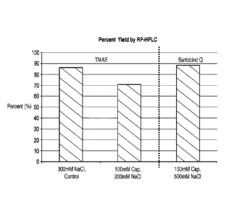
vaccines have been
described. U.S. Patent 5,948,410 describes a method for producing flu vaccines
derived from cell culture
in which a DNAse treatment is combined with a splitting step using CTAB. WO
2007/052163 describes
a method for producing flu vaccines derived from cell culture in which beta
propiolactone (BPL) is used
to inactive the virus and to degrade the residual DNA. Afterwards, the virus
is split, e.g., by treatment
with CTAB. The fragmented DNA is then removed from the virus preparation.
Nevertheless, there is
still the need to further improve removal of residual cellular DNA from
influenza virus preparation or
from other products of interest produced in continuous cell lines.
[5] The use of caprylic acid in combination with ion exchange
chromatography for the removal of
cellular DNA from antibodies produced in cell culture has been disclosed in US
2012-0101262.
However, US 2012-0101262 requires the use of caprylic acid under conditions
that induce precipitation of
residual DNA and contaminating proteins (in particular at low pH). Afterwards,
the precipitate and the
protein of interest can be separated, and the latter is further purified via
ion exchange chromatography.
Date Recue/Date Received 2022-02-02
CA 02930634 2016-05-13
WO 2015/071177 PCT/EP2014/073986
- 2-
[6] Various other methods of removing impurities and aggregates derived
from cell cultures using
caprylic acid or caprylate salts have been described in the art. Steinbuch
(Steinbuch, M. et al.
Arch.Biochem.Biophys. 134:279-94 (1969) describes recovering IgG from human
plasma by caprylate
precipitation of nonenveloped and enveloped viruses therein. U.S.
Patent 7,553,938 describes
purification of antibodies from a starting solution by adding caprylate or
heptanoate ions at pH 4.6 to
about 4.95 and filtering the solution through at least one anion exchange
resin. U.S. Patent 5,886,154
describes a process for purification of antibodies from human plasma involving
suspension of antibodies
at pH 3.8 to 4.5 followed by addition of caprylic acid at pH 5.0 to 5.2 to
precipitate contaminating
proteins and lipids while the antibodies remain in solution. The use of
caprylic acid is employed in
antibody purification because short fatty acids form insoluble complexes with
alpha and beta globulins
and at acidic pH whereas the gamma globulins are not as readily precipitated
(Chanutin et. al., 1960).
Thus the gamma globulin can easily be separated. Yet, none of these
disclosures teach or suggest using
an anionic detergent to remove residual DNA from viral proteins under
conditions which prevent
precipitation as taught herein.
SUMMARY OF THE INVENTION
[7] The present invention relates to manufacturing of proteins and improved
purification methods
thereof. In particular, the invention provides methods for removing cellular
contaminants, such as
residual nucleic acids, from protein products produced in a suitable host
(e.g., host cells). Accordingly,
the invention also encompasses related compositions prepared by such methods.
[8] The invention thus includes methods, which increase the yield, purity
and/or safety of biological
products produced from cell culture. Biological products prepared by methods
of the invention may
include, but are not limited to: biopharmaceuticals, proteins,
polysaccharides, viral antigens, and
antibodies. In a particular aspect, the method provides a biological product
substantially free of residual
DNA.
[9] The inventors have surprisingly found that purification of sample
comprising protein and DNA
derived from cell culture is significantly improved by an addition of an
anionic detergent to a solution
comprising the proteins and cellular DNA, followed by a purification step
comprising an ion exchange
matrix. The problem to be solved might relate to the inefficiency of
separation of negatively charged
DNA impurities from proteins of interest on a positively charged ion exchange
matrix. It can be assumed
that secondary interactions were playing a role in the diminished ability of
anion exchange
chromatography to adsorb the negatively charged DNA impurity. The present
invention now achieves an
enriched product and increased yield thereof, by contacting residual DNA with
an anionic detergent
solution and processing the residual DNA by adsorption over an anion exchange
matrix. The inventors
have unexpectedly found that the present invention also removes influenza
nucleoproteins. Efficient
removal of contaminants as achieved by the present invention allows higher
yields of product, because
CA 02930634 2016-05-13
WO 2015/071177 PCT/EP2014/073986
- 3-
incubation and infection times can be increased so that a higher amount of the
protein of interest can be
obtained. The invention also encompasses the recognition that a particularly
effective removal of residual
DNA from influenza viruses produced in cell culture can be achieved, if the
virus preparation is purified
via an ion exchange chromatography in the presence of an anionic detergent,
such as fatty acid detergents
(e.g., sodium caprylate).
[10] Surprisingly, the inventors have also found the process described herein
substantially removes the
influenza Nucleoprotein (NP). Advantageously, the invention is not restricted
to influenza virus derived
from cell culture, but is also applicable to influenza viruses produced in
eggs, if removal of NP is desired.
[11] Unlike methods described in prior art (see the "Background" above), the
present invention employs
an anionic detergent under conditions that do not precipitate the proteins of
interest or DNA. According
to the invention, the preparation of the protein of interest is separated from
contaminating DNA/proteins
through ion exchange chromatography. By using the process described herein,
the amount of impurities
(e.g., residual DNA) in the sample can be dramatically reduced. This invention
can be used to remove
residual cellular DNA from any samples containing one or more proteins of
interest produced in host
cells, such as cell culture.
[12] Accordingly, the present invention provides a method for removing
residual cellular DNA from a
sample comprising a protein of interest produced in host cells, such as cell
cultures, comprising adding an
anionic detergent to a solution comprising the protein of interest under non-
precipitating conditions and
passing the solution through an ion exchange matrix to remove residual
cellular DNA. The methods of
the invention are not limited by a particular protein of interest. Non-
limiting examples of proteins that
can be purified in accordance with the present invention include, but are not
limited to: therapeutic
proteins, antigens (e.g., immunogenic proteins), antibodies or fragments
thereof.
[13] According to the invention, a suitable starting material which can be
subjected to the methods
provided herein may be a solution comprising a protein of interest. Such
solution may be a crude cell or
tissue preparation, a partially purified preparation, culture media in which
cells were grown, or cell
culture supernatant, etc., but is likely to contain residual cellular
contaminants desired to be removed.
[14] The protein of interest may be grown in a suitable host cell system and
can be purified or clarified
from cell impurities by common separation techniques known in the art.
Optionally, further steps may be
taken prior to the passage of protein through the ion exchange matrix,
preferably prior to the addition of
the anionic detergent. For example the protein of interest may first be
purified from cell culture
impurities to produce a solution which has been clarified. The eluate or flow-
through obtained from the
method of the present invention can be subjected to further processing steps,
such as purifying the protein
of interest and formulating it into a vaccine. In some embodiments of the
invention, the anionic detergent
is added to the clarified solution by contacting the solution comprising the
protein of interest and cell
culture impurities with an anionic detergent solution under non-precipitating
conditions and passing the
CA 02930634 2016-05-13
WO 2015/071177 PCT/EP2014/073986
- 4-
solution through an ion exchange matrix. Non-precipitating conditions are
conditions under which no
substantial precipitation or proteins or DNA occurs.
[15] Thus, the present invention is suitable for the production of viral
proteins. Viral proteins of interest
may be produced in a suitable host (such as cultured cells) infected with the
virus. In some embodiments,
the process of viral protein production may include splitting of virions,
which typically involves the use
of a splitting agent or another detergent. In some embodiments, the anionic
detergent used in the methods
described herein is not the splitting agent or the detergent used in the
splitting process.
[16] Alternatively or additionally, the invention provides a method for
decreasing residual cellular DNA
by passing a solution comprising proteins, cellular DNA in the presence of an
anionic detergent through
an ion exchange matrix under non-precipitating conditions and adsorbing
substantially all of the cellular
DNA on the ion exchange matrix. In a preferred aspect the anionic detergent is
not the splitting agent or
another detergent used in process. Further steps may be taken prior to the
passage of proteins and cellular
DNA through the ion exchange matrix, preferably prior to the addition of the
anionic detergent. For
example the virus may be split with a splitting agent and the proteins may be
separated from cell culture
debris comprising the split virus to produce a solution which has been
clarified. The eluate or flow-
through obtained from the ion exchange matrix produced by the present
invention may be subjected to
further processing steps such as further purifying the viral protein and
formulating it into a vaccine.
[17] The present invention is in particular applicable for the preparation of
viral proteins for vaccine
production. In another embodiment the present invention provides a method for
removing residual
cellular DNA from a sample comprising viral protein produced in cell culture,
comprising adding an
anionic detergent to a solution comprising the protein of interest under non-
precipitating conditions,
passing the solution through an ion exchange matrix, whereby the residual
cellular DNA is bound to the
ion exchange resin. In a preferred aspect the anionic detergent is not the
splitting agent or another
detergent used in process. Optionally further steps may be taken prior to
passage through the ion
exchange matrix, preferably prior to the addition of an anionic detergent. For
example the virus may first
be split with a splitting agent followed by separation of the split virus from
cell culture debris to produce
a solution which has been clarified. The eluate or flow-through obtained from
the ion exchange matrix
produced by the present invention may be subjected to further processing steps
such as further purifying
the viral protein and formulating it into a vaccine.
[18] A particularly effective purification method for biological products
derived from cell culture should
make it possible to optimally remove impurities such as host cell DNA, while
at the same time achieving
a maximum yield of product. To this end, the present invention provides
products substantially free of
impurities and enriched for the immunogenic protein. According to the
invention, residual DNA and
impurities derived from host cells such as cell culture propagation may be
removed from the intended
CA 02930634 2016-05-13
WO 2015/071177 PCT/EP2014/073986
- 5-
product by passage in a solution comprising an anionic detergent, which is
subsequently processed
through an ion exchange matrix.
[19] Accordingly, the present invention provides a method for preparing a
vaccine composition
comprising proteins of interest derived from a cell culture comprising adding
a fatty acid detergent (as
defined below) to a solution comprising proteins of interest under non-
precipitating conditions and
processing the protein of interest on an ion exchange matrix. The present
invention may be useful for
biopharmaceutical vaccine products.
[20] In a preferred aspect, the invention provides a method for producing an
influenza vaccine
composition comprising immunogenic proteins derived from a virus derived from
cell cultures
comprising adding a fatty acid detergent to a solution comprising immunogenic
proteins under non-
precipitating conditions and processing the immunogenic proteins on an ion
exchange matrix. The
immunogenic proteins include hemagglutinin, neuraminidase, and nucleoproteins
obtained from an
influenza virus which has been subjected to inactivation and splitting agents.
Additional steps may be
taken prior to processing the immunogenic protein on the ion exchange matrix,
preferably prior to the
addition of a fatty acid detergent. For example the influenza virus may first
be split with a splitting agent
followed by separation of the split virus from cell culture debris to produce
a solution which has been
clarified. The eluate or flow-through obtained from the ion exchange matrix
produced by the present
invention may be subjected to further processing steps such as further
purifying the viral protein and
formulating it into a vaccine. In a preferred aspect the fatty acid detergent
is not the splitting agent or
another detergent used in process.
[21] As mentioned above, the present invention also encompasses the surprising
finding that the process
described herein substantially removes influenza nucleoprotein (NP).
Advantageously, the invention is
not restricted to influenza virus derived from cell culture, but is also
applicable to influenza viruses
produced in eggs, if removal of NP is desired.
[22] Thus, the invention provides a method for removing viral nucleoproteins
from viral proteins of
interest. An anionic detergent is added to a solution comprising viral
nucleoproteins under non-
precipitating conditions. In some embodiments, the anionic detergent is not
the splitting agent or another
detergent used in process. The nucleoproteins can then be bound to an ion
exchange matrix to produce an
eluate (or flow-through) comprising the proteins of interest which are
substantially free of viral
nucleoproteins and cellular DNA. In some embodiments, a suitable anionic
detergent solution used for
the present invention does not include deoxycholate, sodium lauryl sulfate, or
combination thereof.
[23] Accordingly the present invention provides a method for removing
influenza nucleoproteins from
an influenza virus preparation derived from cell culture or embryonated eggs
comprising adding a anionic
detergent to the virus preparation under non-precipitating conditions, and
processing the virus preparation
through an anion exchange matrix, whereby the nucleoprotein is bound to the
anion exchange matrix.
- 6-
Additional steps may be taken prior to processing the virus preparation on the
ion exchange matrix,
preferably prior to the addition of an anionic detergent. For example the
influenza virus may first be split
with a splitting agent followed by separation of the split virus from cell
culture debris to produce a
solution which has been clarified. The eluate or flow-through obtained from
the ion exchange matrix
produced by the present invention may be subjected to further processing steps
such as further purifying
the viral protein and formulating it into a vaccine. In a preferred aspect the
anionic detergent is not the
splitting agent or another detergent used in process.
[24] The present invention provides an influenza vaccine produced by the
method of the present
invention which is substantially free of residual DNA, and nucleoprotein. The
influenza vaccine can be
formulated in a subvirion particle form, for example HA and NA proteins may be
purified subunit
proteins or bound to portions of influenza viral structures.
BRIEF DESCRIPTION OF THE FIGURES
[25] FIG. 1 provides a bar graph comparing percent yield of HA protein
processed on TMAE and
TM
SARTOBIND Q using different chaotropic agents.
[26] FIG. 2 provides a denaturing gel comparing samples from chromatography
runs with and without
capryliate as a detergent.
[27] FIG. 3 provides a bar graph comparing ratio of DNA/protein recovered from
ion exchange matrices
run with different amounts of caprylate.
[28] FIG. 4 provides percent yield of protein recovered from ion exchange
matrices run with different
amounts of caprylate.
[29] FIG. 5 provides the downstream process for obtaining a cell culture based
subunit influenza
vaccine, as described in Onions et al., 2010.
DETAILED DESCRIPTION OF CERTAIN EMBODIMENTS
Protein of interest
[30] The methods of the invention can be used to purify any protein of
interest derived from a host cell
source (such as cell cultures) from residual host cell contaminations, such as
cellular DNA. Modern virus
production methods as described here have much in common with bioprocessing of
recombinant protein
or monoclonal antibody production. Thus, in a particular aspect, the methods
are employed to purify
proteins of interest, e.g., therapeutic proteins, immunogenic proteins or
antigens, antibodies or fragments
thereof, generated in host cells, such as eukaryotic (e.g., mammalian, avian,
insect, plant, fungal, etc.) cell
cultures and prokaryotic (e.g., bacterial) cell cultures, cell lysates
thereof, clarified bulk (e.g., clarified
cell culture supernatant), or animal derived protein mixtures or extracts.
Date Recue/Date Received 2020-12-17
CA 02930634 2016-05-13
WO 2015/071177 PCT/EP2014/073986
- 7-
[31] In certain embodiments, the methods comprise effectively removing host
cell contaminants (e.g.,
impurities) from a mixture (host cell-derived preparation, e.g., a cell
culture, cell lysate, clarified bulk,
etc.) containing one or more proteins of interest. In some embodiments,
suitable starting materials for the
methods described herein include host cell-derived preparations (such as
sample solutions and cell
lysates) comprising one or more proteins of interest and residual host cell
contaminants in an amount that
is undesirable for intended purposes. In some embodiments, such starting
materials are crude cell lysates.
In some embodiments, such starting materials are cell culture supernatants
comprising secreted proteins
(for example, cell culture media in which host cells are grown). In some
embodiments, such starting
materials are presented as a partially purified form.
[32] Thus one aspect of the present invention provides a method for removing
residual cellular DNA
from a sample comprising a protein of interest produced in a suitable system,
such as cell culture,
comprising steps of adding at least one anionic detergent to a solution
comprising the protein of interest
under non-precipitating conditions; passing the solution through an ion
exchange matrix, whereby the
residual cellular DNA is bound to the ion exchange resin, so as to separate
the protein of interest from the
residual DNA (e.g., in an eluate or flow-through); and, optionally, further
purifying the protein of interest
and formulating it into a product. In some embodiments, the resulting purified
protein of interest is
suitable for use in the manufacture of pharmaceutical compositions. Thus, such
protein or proteins may
be formulated as a pharmaceutical product, such as biologic therapeutics and
vaccines.
[33] Accordingly, the methods described herein are useful for the preparation
of viral proteins produced
in a suitable host. In some embodiments, such viral proteins are viral
immunogenic proteins (i.e., viral
antigens) suitable for vaccine production.
[34] Immunogenic proteins suitable for use in the invention may be derived
from any virus which is the
target of a vaccine. The immunogenic proteins may be formulated as inactivated
(or killed) virus,
attenuated virus, split virus formulations, purified subunit formulations,
viral proteins which are isolated,
purified or derived from a virus, and virus like particles (VLPs).
[35] If during vaccine production, a splitting step is to be used, the
splitting agent may be different from
the anionic detergent of the methods described herein. Preferably the
splitting step or splitting agent is
added prior to the ion exchange chromatography on which the residual cellular
DNA is bound or
separated from the protein of interest.
[36] The immunogenic proteins of the invention are viral antigens which
preferably include epitopes
which are exposed on the surface of the virus during at least one stage of its
life cycle. Viruses may be
non-enveloped or, preferably, enveloped. Viruses are preferably RNA viruses,
and more preferably
ssRNA viruses. They may have a sense or, preferably, an antiscnse genome.
Their gcnomcs may be non-
segmented or, preferably, segmented. Preferred viruses of the invention
include influenza virus
comprising viral antigens such as neuraminidase (NA) and hemagglutinin (HA)
proteins.
CA 02930634 2016-05-13
WO 2015/071177 PCT/EP2014/073986
- 8-
Virus culture
[37] The invention provides a method of preparing an influenza virus, and
removal of residual DNA or
impurities generated during the processing of a viral antigens for vaccine
production. Accordingly, the
invention provides a method for removing nucleoproteins from an influenza
virus preparation. Influenza
virus may be cultured in a host and purification steps taken to isolate and
purify NA and HA proteins.
Thus in one aspect of the present invention relates to a method for removing
Influenza Nuclear Protein
(NP) from a preparation comprising virus proteins of interest, comprising
splitting a virus preparation
obtained from cell culture or eggs, contacting the virus preparation with a
anionic surfactant under non-
precipitating conditions and processing the preparation through an ion
exchange matrix, whereby the
nuclear protein is bound to the anion exchange resin, and optionally further
purifying the viral protein and
formulating it into a vaccine.
[38] The culture host may be cells or embryonated hen eggs, which are suitable
for producing a vaccine
that can be used for administration to humans. Non-limiting examples of
suitable cells which have been
approved for vaccine manufacture include MDCK cells, CHO cells, Vero cells and
PER.C6 cells. For
the embodiments of the inventions involving the use of eggs, the viruses may
also be propagated in eggs.
The current standard method for influenza virus growth for vaccines uses
embyronated SPF hen eggs,
with virus being purified from the egg contents (allantoic fluid). It is also
possible to passage a virus
through eggs and subsequently propagate it in cell culture and vice versa.
Methods for purification of
vaccine products cultivated in embryonated eggs is described, for example, in
GB 1498261.
[39] Preferably, the cells are cultured in the absence of serum, to avoid a
common source of
contaminants. Various serum-free media for eukaryotic cell culture are known
to the person skilled in the
art, e.g., Iscove's medium, ultra CHO medium (BioWhittaker), EX-CELL (JRH
Biosciences).
Furthermore, protein-free media may be used, e.g., PF-CHO (JRH Biosciences).
Otherwise, the cells for
replication can also be cultured in the customary serum-containing media
(e.g., MEM or DMEM medium
with 0.5% to 10% of fetal calf serum).
[40] Virus may be grown on cells in adherent culture or in suspension.
Microcarrier cultures can be
used. In some embodiments, the cells may thus be adapted for growth in
suspension. The suspension
may first be clarified using any method known in the art. The clarification
step serves to remove cells,
cell debris, and host cell impurities from the sample. In some embodiments,
clarification may be
performed via one or more centrifugation steps. Centrifugation of the sample
may be performed by
routine methods known in the art. For example, centrifugation may be performed
using a normalized
loading of about 1x10⁻⁸ mis and a gravitational force of about 5,000 x g to
about 15,000 x g.
Purification
[41] In another aspect, the suspension may be clarified via one or more depth
filtration techniques.
Depth filtration refers to a method of removing particles from solution using
a series of filters, arranged in
CA 02930634 2016-05-13
WO 2015/071177 PCT/EP2014/073986
- 9-
sequence, which have decreasing pore size. A depth filter three-dimensional
matrix creates a maze-like
path through which the sample passes. The principle retention mechanisms of
depth filters rely on
random adsorption and mechanical entrapment throughout the depth of the
matrix. In various aspects, the
filter membranes or sheets may be wound cotton, polypropylene, rayon
cellulose, fiberglass, sintered
metal, porcelain, diatomaceous earth, or other known components. In certain
aspects, compositions that
comprise the depth filter membranes may be chemically treated to confer an
electropositive charge, i.e., a
cationic charge, to enable the filter to capture negatively charged particles,
such as DNA, host cell
proteins, or aggregates.
[42] The methods according to the invention also include harvesting and
isolation of viruses or the
proteins generated from cell culture. During isolation of viruses or proteins,
the cells are separated from
the culture medium by standard methods such as separation, filtration or
ultrafiltration. The viruses or the
proteins are then concentrated according to methods sufficiently known to
those skilled in the art, such as
gradient centrifugation, filtration, precipitation, chromatography, etc., and
then purified. It is also
preferred according to the invention that the viruses are inactivated during
or after purification. Virus
inactivation can occur, for example, by I3-propiolactone or formaldehyde at
any point within the
purification process.
[43] Any depth filtration system available to one of skill in the art may be
used throughout the steps of
present invention. In a particular embodiment, clarification and purification
by depth filtration may be
accomplished with a MILLISTAK+ Pod depth filter system, XOHC media, available
from Millipore
Corporation. In another aspect, the depth filtration step may be accomplished
with a ZETA PLUS Depth
Filter, available from 3M Purification Inc.
Vaccine production
[44] Vaccines are generally based either on live virus or on inactivated
virus. Inactivated vaccines may
be based on whole virions, 'split' virions, or on purified surface antigens.
Antigens can also be presented
in the form of virosomes. The invention can be used for manufacturing any of
these types of vaccines. It
is particularly suitable for manufacturing influenza vaccines, however, which
generally comprise residual
DNA and nucleoprotein in a detectable amount. Such influenza vaccines include
live virus, whole virion
or split virion influenza vaccines. Where the vaccine is formulated in a
subvirion form, the viral antigens
can be found in a split virus form, where the viral lipid envelope has been
dissolved or disrupted, or in the
form of one or more purified viral proteins.
[45] As a further alternative, the vaccine may include a whole virus, e.g., a
live attenuated whole virus,
an inactivated whole virus, etc. Methods for inactivating or killing viruses
to destroy their ability to infect
mammalian cells are known in the art. Such methods include both chemical and
physical means.
Chemical means for inactivating a virus include treatment with an effective
amount of one or more of the
following agents: detergents, formaldehyde, fonnalin, BPL, and UV light.
Additional chemical means for
CA 02930634 2016-05-13
WO 2015/071177 PCT/EP2014/073986
- 10-
inactivation include treatment with methylene blue, psoralen, carboxyfullerene
(C60) or a combination of
any thereof Other methods of viral inactivation are known in the art, such as
for example binary
ethylamine, acetyl ethylencimine, or gamma irradiation. Preferably, the virus
is inactivated with BPL.
[46] Residual DNA may be inactivated with an alkylating agent that cleaves the
DNA into portions
small enough so that it is unable to code for a functional protein.
Preferably, the length of degraded
residual cell culture DNA is less than 500 base pairs. More preferably, the
length of degraded residual
cell culture DNA is less than 200 base pairs. Preferably, the use of an
alkylating agent such as
betapropiolactone (BPL) in the invention provides the additional benefit of
reducing aggregation and
contaminants. Vaccine formulations with reduced aggregates may also have
improved immunogenicity.
US 2009-0304729 teaches the treatment of functional residual DNA with
alkylating agents. Prior to the
use of the anionic detergent in combination with ion exchange chromatography,
parts of the fragmented
residual DNA can be removed by precipitation with a cationic detergent like
CTAB as described in
Onions et al. (2010; Biologicals, 38(5): 544-551). The whole downstream
process of Onions is shown in
Figure 5. In some embodiments, the present invention can be applied as part of
the Onions process.
[47] Methods of splitting viruses, such as influenza viruses, are well known
in the art, e.g., see
International Patent Publications: WO 02/28422, WO 02/067983, WO 02/074336, WO
01/21151, etc.
Splitting of the virus is carried out by disrupting or fragmenting whole
virus, whether infectious (wild-
type or attenuated) or non-infectious (e.g., inactivated), with a disrupting
concentration of a splitting
agent. Splitting agents generally include agents capable of breaking up and
dissolving lipid membranes,
typically with a hydrophobic tail attached to a hydrophilic head. A preferred
splitting agent is
cetyltrimethylammoniumbromide (CTAB). The disruption results in a full or
partial solubilization of the
virus proteins, altering the integrity of the virus. Preferred splitting
agents are non-ionic and ionic (e.g.,
cationic) surfactants, e.g., alkylglycosides, alkylthioglycosides, acyl
sugars, sulphobetaines, betains,
polyoxyethylenealkylethers, N,N-dialkyl-Glucamides, Hecameg, alkylphenoxy-
polyethoxyethanols,
quaternary ammonium compounds, sarcosyl, CTABs (cetyl trimethyl ammonium
bromides), tri-N-butyl
phosphate, C etavl on , rnyristyltr imethylammon i um salts, lipofeetin, 1
ipofeetam in e, and DOT-MA, the
octyl- or nonylphenoxy polyoxyethanols (e.g., the Triton surfactants, such as
Triton X-100 or Triton
N101), polyoxyethylene sorbitan esters (the Tween surfactants),
polyoxyethylene ethers, polyoxyethylene
esters, etc.
[48] One useful splitting procedure uses the consecutive effects of sodium
deoxycholate and
formaldehyde, and splitting can take place during initial virion purification
(e.g., in a sucrose density
gradient solution). Thus a splitting process can involve clarification of the
virion-containing material (to
remove non-virion material), concentration of the harvested virions (e.g.,
using an adsorption method,
such as CaHPO4 adsorption), separation of whole virions from non-virion
material, splitting of virions
using a splitting agent in a density gradient centrifugation step (e.g., using
a sucrose gradient that contains
a splitting agent such as sodium deoxycholate), and then filtration (e.g.,
ultrafiltration) to remove
CA 02930634 2016-05-13
WO 2015/071177 PCT/EP2014/073986
-11-
undesired materials. Split virions can usefully be resuspended in sodium
phosphate-buffered isotonic
sodium chloride solution.
[49] A composition (such as a vaccine) that is "substantially free of residual
DNA" refers to a
composition or formulation, wherein residual DNA fragments of less than 200
basepairs are detectable at
less than 10 ng per 0.5 ml, as determined by capillary electrophoresis (see,
e.g., WO 2009/118420). The
total amount of residual DNA in compositions of the invention is preferably
less than 20 ng/ml, e.g., <10
ng/ml, <5 ng/ml, <1 ng/ml, <100 pg/ml, <10 pg/ml, etc.
[50] Accordingly, an assay used to measure residual DNA will typically be a
validated assay (Guidance
for Industry: Bioanalytical Method Validation. U.S. Department of Health and
Human Services Food and
Drug Administration Center for Drug Evaluation and Research (CDER) Center for
Veterinary Medicine
(CVM). May 2001; Lundblad (2001) Biotechnology and Applied Biochemistry 34:195-
197). Three
principle techniques for DNA quantification can be used: hybridization
methods, such as Southern blots
or slot blots (Ji et al. (2002) Biotechniques. 32:1162-7); immunoassay
methods, such as the
THRESHOLD System (Briggs (1991) J Parenter Sci Technol. 45:7-12; and
quantitative PCR (Lahijani et
al. (1998) Hum Gene Then 9:1173-80). These methods are all familiar to the
skilled person, although the
precise characteristics of each method may depend on various factors such as
choice of probes for
hybridization, the choice of primers and/or probes for amplification, etc.
[51] In another aspect, the invention provides methods for preparing influenza
vaccine compositions
which have reduced levels of nucleoproteins (NP). Preferably, NP makes up less
than 15% by mass of
the total influenza virus protein in the vaccine, e.g., <12%, <10%, <8%, <7%,
<6%, <5%, <4%, <3%,
<2%, or <1%. The vaccine may comprise less than 3 qg NP per 10 pig of HA, less
than 2.5 pg NP per 10
p,g of HA, less than 2 pig NP per 10 qg of HA, less than 1.5 pg NP per 10
1..tg of HA, less than 1 qg NP
per 10 qg of HA, less than 0.5 pg NP per 10 pg of HA or less than 0.1 pg NP
per 10 14 of HA. Most
preferably, the vaccine is substantially free of NP. This is understood as
having less than 0.1 g NP per
qg of HA. In some embodiments, the methods provided herein may achieve at
least 10-fold reduction
in the amount of NP in a preparation, e.g., at least 10-fold, at least 12-
fold, at least 15-fold, at least 20-
fold, at least 25-fold, at least 30-fold, at least 40-fold, at least 50-fold,
at least 75-fold, or at least 100-fold
reduction in the amount of NP in a flow-through (or dilate) as compared to the
starting material subjected
to the purification methods of the invention.
[52] Methods to determine the amount of protein in a composition are known to
the skilled person in the
art. However, since NP and NA have virtually the same molecular weight (around
60 kD), they usually
co-migrate in non-reducing gels. Classic SDS gel-electrophoresis might
therefore not be an appropriate
way to determine the amount of NP (see Chaloupka et al., 1996, Eur J Clin
Microbiol Infect Dis. 1996
Feb;15(2):121-7.). One way to determine the amount of NP in a vaccine bulk
might be a 2 dimensional
electrophoresis with a subsequent clensitometry. Preferred, however is isotope
dilution mass spectrometry
CA 02930634 2016-05-13
WO 2015/071177 PCT/EP2014/073986
- 12-
using an isotopically labeled synthetic peptide as described, for example, in:
Williams et al., Vaccine 30
(2012) 2475-2482. Such method uses liquid chromatography¨tandem mass
spectrometry (LC-MS/MS)
using isotope dilution in conjunction with multiple reaction monitoring (MRM).
This method quantifies
targeted peptides released by proteolytic digestion of the sample as a
stoichiometric representative of the
analyte protein. A stable isotope-labeled reference peptide is spiked into the
sample as an internal
standard (IS). Quantification of NP is achieved by comparing the peak area of
the isotopically labeled
reference peptide with that of the endogenous target peptide. This method
allows simultaneous
quantification of multiple proteins, provided labeled peptides are included
for each specific target.
[53] Alternatively, label free mass spectrometry (LC/MSE) is used for the
quantification, preferably in
quadrupole time-of-flight (Q-Tof) mass spectrometers (Getie-Kebtie et al.,
(2013): Influenza and Other
Respiratory Viruses 7(4), 521-530). For this method, alternating scans of low
collision energy and
elevated collision energy during LC/MS analysis are used to obtain both
protein identity and quantity in a
single experiment. Quantification is based on the experimental data showing
that the average signal
intensity measured by LC/MSE of the three most intense tryptic peptides for
any given protein is constant
at a given concentration, regardless of protein type and size. As the signal
intensity is proportional to
concentration, the amount of any protein in the mixture can be estimated.
[54] The present invention also includes influenza vaccines based on viruses
grown in cell culture
(preferably mammalian or avian cells), whereby the vaccines have an amount of
residual cellular DNA of
less than 5 ng /dose (e.g., less than 4 ng, less than 3 ng, less than 2 ng or
less than 1 ng per dose) at a
fragment size of less than 200 base pairs, and whereby the vaccine contains
less than 1 lag NP per 10 14
of HA, less than 0.5 tig NP per 10 14 of HA, or less than 0.1 pg NP per 10 ig
of HA. Most preferably,
the vaccine is substantially free of NP. This is understood as having less
than 0.1 14 NP per 10 pg of
HA. In particular the influenza vaccine is contains less than 1 ng residual
DNA per dose at a fragment
size of less than 200 base and less than 0.5 dg NP per 10 jig of HA. This
vaccine is most preferably free
from mercury-containing preservatives and antibiotics. The vaccine is most
preferably a tetravalent
seasonal or monovalent pandemic influenza vaccine with an amount of residual
cellular DNA of less than
1 ng per dose at a fragment size of less than 200 base pairs and less than 0.5
tg NP per 10 ttg of HA.
[55] Such vaccine preparations can be obtained, for example, by the following
process, which is a
particularly preferred embodiment: A method for producing an influenza virus
vaccine in which the
following steps are conducted: Influenza viruses are grown in cell culture,
e.g., in MDCK suspension
cells (WO 1997/037000). The viruses are harvested, purified and concentrated
by 0.45 micrometer
filtration and CS chromatography. After addition of detergent (such as
polysorbates, e.g., Tweent 80),
the virus preparation is treated with BPL. Afterwards the viruses are split
with CTAB. After an
ultracentrifugation and adsorption step the viral protein preparation is
subject to ion exchange
chromatography, using TMAE or Sartobind Q as a resin. The chromatography is
done in the presence of
sodium caprylate (about 50 m1VI for Sartobind; 100 mM for TMAE) and sodium
chloride (400 mM for
- 13-
Sartobind), and 200 mM for TMAE). Afterwards the protein preparation is
concentrated by a suitable
means, such as ultrafiltration. The proteins might be optionally blended with
other virus preparation (in
the case of tri- or tetravalent seasonal vaccines), and optionally sterile
filtrated, filled and packaged. The
invention thus includes influenza vaccine obtainable by this process.
[56] It will bc evident to the artisan that the measure of the residual host
cell DNA content is not meant
as a limitation or defining feature of this methodology. Instead, these data
in the examples support the
essence of the present invention: a large-scale methodology for the generation
of virus particles that
results in a highly purified product that may be utilized in clinical and
commercial settings. It can be
noted that the importance of achieving particular DNA levels in the final
product is product-specific.
Viral products produced using continuous cell lines for parenteral use in
humans will require the most
stringent purity standards but, even in that case, the goals may vary from 100
pg per dose up to 10 ng per
dose (WHO Requirements for the Use of Animal Cells as in vitro Substrates for
the Production of
Biologicals Requirements for Biological Substances No. 50), WHO Technical
Report Series, No. 878,
1998) or higher, and are likely to be adjusted depending on the product's
indication.
Detergents
[57] The anionic detergents used in the present invention are detergents which
are added as an extra
substance for carrying out ion exchange. Accordingly, the detergent itself is
not removed by the ion
exchange process nor precipitates the substances processed through the matrix
but serves to interact with
the hydrophobic regions of the residual DNA and/or the virus or viral
proteins, particularly the HA
subunit. In some embodiments, the anionic detergent used for the present
invention excludes
deoxycholate and/or sodium lauryl sulfate.
[58] In preferred embodiments, one or more anionic detergents are used. In
prefened embodiments,
fatty acid detergents are used (as defined below). In particularly preferred
embodiments, eight-carbon
fatty acids are used. For example, in some embodiments, caprylic acids (e.g.,
sodium caprylate) are used.
[59] In one aspect, an anionic detergent solution is added to a solution
having the proteins of interest. If
used for viral preparations, the anionic detergent is preferably added
following inactivation or splitting of
viruses, whereby inactivation may be performed before or after splitting
steps. In one aspect, the anionic
detergent, is added during or prior to an ion exchange step. The addition of a
anionic detergent
significantly improves clearance of residual DNA by at least 10%, 20%, 30%,
40% or 50%, as compared
to clearance of residual DNA without treatment.
[60] Preferred anionic detergents are cholates, deoxycholates, 1-
decanesulfonates, and lauryl sulfate.
Other suitable detergents include cetylpyridinium bromide, alkyl
benzyklimethylmmnonium chloride,
Date Recue/Date Received 2020-12-17
CA 02930634 2016-05-13
WO 2015/071177 PCT/EP2014/073986
- 14-
tetradecyltrimethylammonium chloride, hexatecylammonium chloride, and
orinthinyl-cysteinyl-
tetradecylamide.
[61] In some embodiments, suitable anionic detergents are fatty acid
detergents. In the context of the
present disclosure, fatty acid detergents are understood to be salts of fatty
acid, particularly carboxylic
fatty acids selected from C4-C18 carbon chains, preferably C6-C10 carbon
chains, e.g., C6, C8 and C10.
Preferably, the fatty acids are linear and saturated. In some embodiments,
suitable fatty acid detergent is
sodium caprylate or a similar salt of caprylic acid. As described herein, the
addition of caprylate (sodium
caprylate) (C8) at neutral pH has been shown to improve protein recovery and
prevents protein
aggregation or nonspecific binding. In certain embodiments, the final
concentration of caprylate acid
solution comprising a protein or proteins of interest (such as antibodies or
fragments thereof, antigens,
therapeutic proteins, toxins, peptides, etc.) has a suitable concentration of
detergent is between 25 mIVI
and 300 mM, preferred between 50 mM and 250 mM, particularly preferred between
75 mM and 200
mM. The concentration might be about 25 mM, about 50 mM, about 75 mM, about
100 mM, about 125
mM, about 150 mM, about 175 mM, 200 mM about 250mM or about 300mM, depending
on the ion
exchange resin or chromatography conditions. A skilled person in the art will
be able determine the most
suitable fatty acid detergent and empirically elucidate the concentration of
fatty acid detergents to make a
solution. For example, it is known that carboxylic acid detergents having
lower carbon chains will have
less detergent characteristics, while higher carbon chains will have reduced
solubility. Typically, higher
detergent concentration is seen as providing a more robust process across
different strains of influenza,
due to disruption of any hydrophobic interactions and higher reduction of
impurities as illustrated in
Figure 2. However, it is important that the amount of fatty acid detergents be
present in an amount and at
a pH to prevent precipitation of the proteins and residual DNA in solution.
[62] In another aspect, the pH of the solution comprising proteins is
maintained at a pH at which no (or
an insignificant amount of) precipitation occurs of the proteins,
nucleoproteins and residual DNA. For
example, for caprylic acid, this is neutral pH. The optimum pH required to
prevent protein precipitation
can readily be determined empirically by the skilled person in the art.
Preferably, the final pH of the
mixture should be maintained to be between about 7.0 and 9Ø In some
embodiments, the final pH of the
mixture is maintained between about 7.2 and 7.5, e.g., between about 7.2-7.4,
between about 7.2-7.3,
between about 7.3-7.5, between about 7.4-7.5. In some embodiments, the final
pH of the mixture is
maintained at greater than or equal to about 7 (such as between about 7-9,
e.g., about 7.0, about 7.5, about
8.0, about 8.5, about 9.0, etc.). In some embodiments, the pH of the solution
comprising the proteins,
residual DNA and caprylate should not be reduced to about 6.0 or less (e.g.,
about 5, 4, and 3). The pH
can be adjusted before and/or after the addition of an anionic detergent
(e.g., caprylate) to the sample. In
some embodiments, the pH of the mixture could be adjusted before the addition
of an anionic detergent
(e.g., caprylate). In general, any art-recognized acids or buffers can be used
to alter or adjust the pH of a
mixture, including, for example, phosphate- and tris-containing buffers.
CA 02930634 2016-05-13
WO 2015/071177 PCT/EP2014/073986
- 15-
[63] The method of the present invention may also be applied to partially
purified protein samples to
farther remove DNA or undesired impurities by contacting the mixture with an
anionic detergent solution
under conditions which prevent precipitation of the proteins in the mixture
and passing the mixture
through an ion exchange matrix. The methods of the invention effectively
remove host cell DNA
contaminants to a concentration of <10 ng DNA per dose as recommend by WHO for
continuous cell
lines and nucleoprotein to a concentration of less than 0.5 tg NP per 10 pg
HA. In a particular aspect,
the amount of nucleoprotein removed by the present invention is at least 10%,
15%, 20%, 25%, 30%,
35%, 40%, 50%, 60%, 70%, 80% and 90% as determined by SDS-PAGE.
[64] The composition comprising proteins and residual DNA in a solution of an
anionic detergent is
further processed to recover the desired product. Residual DNA is better
adsorbed on an anion exchange
membrane in the presence of the anionic detergent. Surprisingly, influenza
nucleoproteins are also
captured on the anion exchange membrane as identified by the inventors by
electrophoretic analysis of
adsorption pools. The inventors identified that nucleoproteins run with the
residual DNA when contacted
with a solution of anionic detergent. This finding has not been shown before
and results in an enriched
influenza product which is substantially free of residual DNA.
[65] Accordingly, after residual host cell contaminants are removed by
treatment of the contaminant-
containing sample (e.g., cell culture and clarified bulk mixtures) with an
anionic detergent and subsequent
purification step in accordance with the methods described herein, such sample
can contain no more than
about 10000 ng/mg (e.g., no more than about 10000, 5000, 1000, 500, 200, 100,
50, 25, or 10 ng/mg) of
protein contaminants. In some embodiments, such protein contaminants include
no more than about
10000 ng/mg nucleoproteins, e.g., no more than about 10000, 5000, 1000, 500,
200, 100, 50, 25, or 10
ng/mg nucleoproteins.
[66] Thus, any influenza product which comprises residual DNA and
nucleoprotein can be enriched for
HA and NA proteins by contact with an anionic detergent solution and processed
through an ion
exchange matrix. The person skilled in the art will be able to apply the
methods of the present invention
to influenza products generated from cell culture or egg culture.
Chromatography
[67] The present invention may be used in commercial scale processing
techniques that utilize ion
exchange chromatography to produce bulk quantities of the finished product. It
is lmown that during
large scale manufacturing, the effect of the binding affinity between the
residual DNA and virus particles
or viral proteins may be further compounded during the concentration of the
virus particles because the
DNA may become physically trapped during the aggregation of the virus
particles. Once the DNA is
bound specifically or nonspecifically to the virus, or otherwise entrapped by
aggregates of the virus or
proteins, the use of ion exchange matrices as described in the art becomes
relatively ineffective as a
means for efficiently removing the DNA. Accordingly, the present invention
relates to a purification
- 16-
process to remove residual DNA by purification with an anionic detergent
solution and/or a suitable
concentration or ionic strength provided by a salt buffer over a
chromatography matrix.
TM
[68] An anionic Q membrane chromatography capsule may comprise a Mustang Q
membrane a
chromatography capsule (available from Pall Corporation) or Sartobind Q (a
strongly basic anion
exchanger membrane, available from Sartorius Stedim Biotcch GmbH). Any
positively charged ligand
attached to the solid phase suitable to form the anionic exchange resin can be
used, such as quaternary
amino groups. Commercially available anion exchange resins include DEAE
cellulose, POROST.MP1 20,
PI 50, HQ 10, HQ 20, HQ 50, D 50 from Applied Biosystems, SARTOBIND. Q from
Sartorius, MONO
TM TM TM
Q, MINI Q, Source 15Q and 30Q, Q, DEAE and ANX SEPHAROSE. FAST FLOW, Q
SEPHAROSE
high Performance, QAE SEPHADEX. and FAST Q SEPHAROSE (GE Healthcare), WP PEI,
WP
DEAM, WP QUAT from J. T. Baker, HYDROCELL DEAE and HYDROCELL QA from BioChrom
TM TM
Labs Inc., UNOSPHERE Q, MACRO-PREP DEAE and MACRO-PREP High Q from Bio-Rad,
Ceramic
TM TM TM
HyperD Q, ceramic HyperD DEAE, TRISACRYL M and LS DEAE, Spherodex LS DEAE, QMA
TM TM
SPHEROSIL LS, QMA SPHEROSIL M and MUSTANG Q from Pall Technologies, DOWEX Fine
Mesh
Strong Base Type 1 and Type 11 Anion Resins and DOWEX MONOSPHERE 77, weak base
anion from
Dow Liquid Separations, INTERCEPT Q membrane, MATREX CELLUFINE A200, A500,
Q500, and
Q800, from Millipore, FRACTOGELTmEMD TMAE, FRACTOGEL. EMD DEAE and FRACTOGEL
TM
EMD DMAE from EMD, AMBERLITE weak strong anion exchangers type I and II, DOWEX
weak and
TM
strong anion exchangers type I and II, DIAION weak and strong anion exchangers
type I and
TM
DUOLITETTrom Sigma-Aldrich, TSKgel Q and DEAE 5PW and 5PW-HR, TOYOPEARL SUPERQ-
650S, 650M and 650C, QAE-550C and 650S, DEAE-650M and 650C from Tosoh, QA52,
DE23, DE32,
TM
DE51, DE52, DE53, EXPRESS-Ion D and EXPRESS-Ion Q from Whatman.
[69] Chromatographic separation over the ion exchange matrix is operated in
flow-through mode. The
specific methods used for the chromatography capture step, including flow of
the sample through the
column, wash, and elution, depend on the specific column and resin used and
are typically provided by
the manufacturers or are known in the art.
[70] In an alternative aspect, modulation of ionic strength may also be
employed during the
chromatography step. The ionic strength of buffer solution may be determined
from both molar
concentration and charge numbers of all the ions present in the solution. The
ionic strength, I, may be
calculated using following formula:
15,
j cizi
Date Recue/Date Received 2020-12-17
CA 02930634 2016-05-13
WO 2015/071177 PCT/EP2014/073986
- 17-
where ci is the molar concentration of ion i (mol=dm-3), zi is the charge
number of that ion, and the sum is
taken over all ions in the solution. Generally a 1:1 electrolyte such as NaCl,
the ionic strength is equal to
its molar concentration, while multivalent ions contribute more to the ionic
strength in the solution, for
example, the ionic strength of the 2:2 electrolyte MgSO4 is four times that of
NaCl.
[71] The preferred ionic strength will optimize the balance between removing
the unwanted residual
DNA while maintaining a high viral or protein yield that retains the
antigenicity of the virus in a cost
effective manner.
[72] The person skilled in art will be able to design a chromatographic
separation program depending
on, for example, sample characteristics, chromatograph matrix properties and
efficiency of fractionation.
A saline buffer is preferably provided at or near a neutral pH such as about
7Ø 7.1, 7.2, 7.3, 7.4, 7.5, 7.6,
7.7 and 7.8. The pH should not be reduced below about 6 as the proteins of
interest may lose their
activity, aggregate or precipitate in the presence of an anionic detergent
(e.g., fatty acid detergents).
Suitable concentrations of buffer (e.g., sodium chloride buffer) may be
between about 100 mM and 1M,
such as 100 mM, 150 mM, 200 mM, 300 mM, 400 mM, 500 mM, 600 mM, 700 mM, 800
mM, 900 mM,
and 1M. The optimum salt concentration depends on the ion exchange
chromatography resin that is used.
The person skilled in the art can easily determine the optimum salt
concentration by routine test. For a
TMAE resin the best sodium chloride concentration is about 300 mIVI, for
SARTOBIND Q it is greater
than 400 mM (for the use of caprylate as detergent; see Examples below).
[73] As used herein the term "chromatography" refers to the process by which a
solute of interest, e.g., a
protein of interest, in a mixture is separated from other solutes in the
mixture by percolation of the
mixture through an adsorbent, which adsorbs or retains a solute more or less
strongly due to properties of
the solute, such as pI, hydrophobicity, size and structure, under particular
buffering conditions of the
process. In a method of the present invention, chromatography can be used to
remove contaminants after
the precipitate is removed from a mixture, including without limitation, a
cell culture or clarified cell
culture supernatant.
[74] The term "impurities" as used herein generally refers to residual host
cell DNA, empty viral
particles, aggregated proteins or matter other than the intended component(s)
of a product.
[75] "Processing" or "processed" used in the context of the invention refers
to a downstream step or
steps performed after clarification or the initial starting materials
comprising cellular byproduct and
debris, colloidal particulates, large biomass and high cell densities.
Techniques used in processing steps
include isolation, purification, concentration, centrifugation, filtration,
formulation, inactivation, splitting
and various analytical operations performed for sterile biological products.
"Processed" also may
describe the steps of flowing or passing a sample through a chromatography
column, resin, membrane,
filter, or other mechanism, and can include a continuous flow through each
mechanism as well as a flow
that is paused or stopped between each mechanism.
CA 02930634 2016-05-13
WO 2015/071177 PCT/EP2014/073986
- 18-
[76] Absent explicit teaching, a process comprising a step of mixing two or
more components does not
require any specific order of mixing. Thus, components can be mixed in any
order. Where there are
three components then two components can be combined with each other, and then
the combination may
be combined with the third component, etc.
[77] The phrase "ion exchange material" refers to a solid phase that is
negatively charged (e.g., a cation
exchange resin) or positively charged (e.g., an anion exchange resin). In one
embodiment, the charge can
be provided by attaching one or more charged ligands (or adsorbents) to the
solid phase, e.g., by covalent
linking. Alternatively, or in addition, the charge can be an inherent property
of the solid phase (e.g., as is
the case for silica, which has an overall negative charge).
[78] Accordingly, the present invention encompasses, but is not limited to,
the following embodiments:
1. A method comprising a step of subjecting a first solution containing a
protein of interest and an
anionic detergent to an ion exchange matrix under a non-precipitating
condition, so as to obtain a second
solution containing the protein of interest, wherein the second solution
contains less residual cellular
contaminants than the first solution.
2. The method of embodiment 1, wherein the first solution is selected from
the group consisting of:
cell or tissue lysates, culture media, cell culture supernatants, plasma, and
partially purified protein
solutions.
3. The method of any one of the preceding embodiments, wherein the protein
of interest is selected
from the group consisting of: therapeutic proteins, immunogenic proteins
(e.g., viral antigens), and
antibodies or antigen-binding fragments thereof.
4. The method of any one of the preceding embodiments, wherein the anionic
detergent is selected
from the group consisting of: fatty acid detergents.
5. The method of any one of the preceding embodiments, wherein the anionic
detergent is different
from any other detergent used in a process of protein purification.
6. The method of any one of the preceding embodiments, wherein the anionic
detergent does not
include deoxycholate and/or sodium lauryl sulfate.
7. The method of any one of the preceding embodiments, wherein the ion
exchange matrix
comprises a basic anion exchanger membrane.
8. The method of any one of the preceding embodiments, wherein the non-
precipitating condition
comprises at or near neutral pH.
9. The method of any one of the preceding embodiments, wherein the second
solution is an eluate.
10. The method of any one of the preceding embodiments, further comprising
a step of further
purification.
CA 02930634 2016-05-13
WO 2015/071177 PCT/EP2014/073986
- 19-
11. The method of any one of the preceding embodiments, further comprising
a step of carrying out
sterile filtration.
12. The method of any one of the preceding embodiments, further comprising
a step of formulating
the protein of interest into a pharmaceutical composition.
13. The method of any one of the preceding embodiments, further comprising
a step of carrying out
sterile filtration.
14. The method of embodiment 12 or 13, wherein the pharmaceutical
composition is a prophylactic
composition, therapeutic composition, or combination thereof.
15. The method of any one of embodiments 12-14, wherein the pharmaceutical
composition further
comprises a pharmaceutically acceptable excipient.
16. The method of any one of embodiments 12-15, wherein the pharmaceutical
composition further
comprises and adjuvant.
17. The method of any one of embodiments 12-16, further comprising a step
of packaging the
phaimaceutical composition into a sterile closed system.
18. The method of embodiment 17, wherein the sterile closed system is
selected from the group
consisting of: vials, syringes, and containers.
19. The method of embodiment 17 or 18, wherein the sterile closed system is
plastic or glass.
20. The method of any one of embodiments 17-19, wherein the sterile closed
system comprises a
siliconizal surface.
21. A use of the pharmaceutical composition of any one of embodiments 12-
20, for the manufacture
of a medicament for administering a subject in need thereof.
22. The pharmaceutical composition of any one of embodiments 12-20 for use
as a medicament for
administering to a subject.
23. A method comprising administering to a subject the phaimaceutical
composition of any one of
embodiments 12-20.
24. A viral vaccine comprising no more than 5 ng of residual DNA and no
more than 1.0 pig
nucleoprotein per dose.
25. The viral vaccine of embodiment 24, comprising no more than 1 ng of
residual DNA and no more
than 0.5 pig nucleoprotein per dose.
26. The viral vaccine of embodiment 24, comprising no more than 1 ng of
residual DNA and no more
than 0.1 pig nucleoprotein per dose.
-20-
27. The viral vaccine of any one of embodiments 24-26, wherein the viral
vaccine is an influenza
vaccine.
28. The viral vaccine of any one of embodiments 24-27, further comprising
an adjuvant.
29. The viral vaccine of embodiment 28, wherein the adjuvant is selected
from the group consisting
of: alum adjuvants, oil-in-water adjuvants, virosomes and Toll-like receptor
(TLR) agonists.
[79] This invention is further illustrated by the following examples, which
should not be construed as
limiting.
EXAMPLES
[80] An H5N1 virus was propagated in MDCK suspension cells, harvested and
processed as described
in Onions et al., 2010. The split virus preparation was subjected to ion
exchange chromatography using a
SARTOBIND Q (Sartorius) or a FRACTOGEL TMAE (EMD Millipore) membrane. The
optimal salt
concentration found for TMAE was determined to be approximately 300 mM, while
the optimal
concentration found for SARTOBIND Q was greater than 400 mM. Preparations with
different detergents
and chatoropric reagents were conducted. The pH of the final compositions was
7.5, except for the
arginine compositions, which had a pH of 7.2. DNA reduction was assessed by
Picogeen and protein
yield was assessed by the BCA assay. Overall, SARTOBINDQ performed better than
TMAE in DNA
reduction; however, all runs utilizing sodium caprylate showed significant
increase in DNA reduction
compared to just NaCl. Robust results could be obtained with 50 mM caprylate
and 400 mM NaCl on a
SARTOBINDQ membrane. BCA values for the arginine might not be exact due to
interference of
arginine with the BCA assay. Arginine was not further investigated for yield
due to insufficiently
removing DNA. BCA and DNA data for these conditions are shown in Figures 3 and
4.
[81] Samples from the following three runs were further examined by RP-HPLC
for HA content: (i)
Control - 50 mM phosphate, 300 mM NaCl, pH 7.5; (ii) 50 mM phosphate, 100 mM
sodium caprylate,
200 mM NaCl, pH 7.5; (iii) 50 mM phosphate, 100 mM sodium caprylate, 500 mM
NaCl, pH 7.5. These
runs were considered to be the best case for the conditions examined. Higher
caprylate concentration is
seen as providing a more robust process across strains based on the idea that
a higher concentration of
caprylate would more effectively disrupt any hydrophobic interactions and
overall lead to a higher
reduction of impurities. The yields by RP-HPLC were calculated and plotted in
Figure 1.
[82] Material from these three runs was also analyzed by SDS-PAGE and can be
seen in Figure 2. The
samples required sample prep prior to running on gels due to low protein
concentration. The samples
were concentrated 2.5 fold to ensure protein concentrations high enough to be
visualized by SDS-PAGE.
TM
The samples were concentrated using a 15mL Amicon ULTRA SPIN Tub with 10,000
MWCO
membrane. The adsorption pool was also diluted in buffer and concentrated 2.5
fold to ensure that low
molecular weight contaminants were not lost during the concentration process.
This is demonstrated by
Date Recue/Date Received 2020-12-17
CA 02930634 2016-05-13
WO 2015/071177 PCT/EP2014/073986
-21-
comparing lanes labeled Adsorption and Adsorption, Concentrated. A dramatic
difference in purity can
be seen by comparing the control runs and the runs containing caprylate. The
nucleoprotein in the
caprylate runs is significantly diminished compared to runs just utilizing
NaCl to optimize yield
performance.
[83] These experiments have successfully shown that secondary interactions,
likely hydrophobic in
nature, are playing a role in the diminished ability of AEX chromatography to
adsorb the negatively
charged impurity DNA. Without wishing to be bound by theory, the addition of
caprylate to the
adsorption pool likely disrupts this hydrophobic interaction and allows the
binding of DNA and
Nucleoprotein to the membrane or resin.
[84] It should be understood that the invention has been described by way of
example only and
modifications may be made whilst remaining within the scope and spirit of the
invention.
[85] The various features and embodiments of the present invention,
referred to in individual sections
above apply, as appropriate, to other sections, mutatis mutandis. Consequently
features specified in one
section may be combined with features specified in other sections, as
appropriate.
[86] Throughout the specification, including the claims, where the context
permits, the term
"comprising" and variants thereof such as "comprises" or "comprising" are to
be interpreted as including
the stated element (e.g., integer) or elements (e.g., integers) without
necessarily excluding any other
elements (e.g., integers).
[87] Those skilled in the art will recognize, or be able to ascertain using no
more than routine
experimentation, many equivalents to the specific embodiments of the invention
described herein. Such
equivalents are intended to be encompassed by the following claims.