Note: Descriptions are shown in the official language in which they were submitted.
HIV-1 ENV DNA VACCINE PLUS PROTEIN BOOST
CROSS-REFERENCE TO RELATED APPLIATIONS
This application claims priority to U.S. Provisional Patent Application No.
61/904,416,
filed November 14, 2013.
FIELD OF THE INVENTION
The present invention relates to treating and preventing symptoms of an
associated HIV
infection using a priming vaccine containing a DNA encoding the antigen, and a
second vaccine
for boosting the response to the first vaccine using the same or different
antigen than the first
vaccine.
BACKGROUND OF THE INVENTION
There is an urgent need for improved vaccination approaches against HIV that
induce
improved humoral and cellular immune responses. It is generally agreed upon
that strong T-cell
responses and breath in neutralizing antibodies will likely play a role in the
development of a
protective vaccine. Though DNA platforms in the past have been poor inducers
of
seroconversion, recent improvements in construct design, improved delivery,
and improved
formulations have enhanced the immune potency of this approach. We have
recently reported
the induction of strong HIV/SIV-specific cellular immune responses in mice,
macaques and
humans using consensus DNA immunogens delivered via electroporation (EP).
While these
studies have confirmed the induction of a potent and broad cell-mediated
response, the ability of
this improved DNA-EP platform to induce or prime for neutralizing antibodies
(NAbs) is
unknown. Due to a heightened interest in trying to improve immune responses to
HIV included
by DNA prime-protein boost vaccination strategies, here we studied this
combination focused
on increasing binding titers and neutralization capacity in vivo.
There is a need in the art to study the immunogenicity of a synthetic
consensus DNA
vaccine encoding gp140 constructs derived from individual HIV-1 subtypes A, B,
C and D in a
DNA prime-protein boost regimen. These consensus DNA constructs can be
optimized using
the following plasmid-enhancement techniques: codon optimization, RNA
optimization, leader
sequence addition, plasmid production at high concentrations and the DNA was
delivered by
adaptive EP as previously described. The DNA prime can be followed by a
protein boost with
recombinant HIV gp120. Immune responses were measured by ELISA, B-cell
ELISpot, T-cell
1
Date Recue/Date Received 2020-12-29
ELISpot, and in a TZM-bl neutralization assay. The combination approach
increased T cell and
antibody functionality over these observed with either independent modality.
SUMMARY OF THE INVENTION
The present invention is directed to a composition comprising (a) a first
vaccine and (b) a
second vaccine. The first vaccine may comprise at least one, at least two, at
least three, or at
least four nucleic acids, wherein each nucleic acid may encode an antigen, and
wherein the at
least one, at least two, at least three, or at least four nucleic acids may be
selected from the group
consisting of a nucleic acid encoding a HIV-1 subtype A consensus antigen, a
nucleic acid
encoding a HIV-1 subtype B consensus antigen, a nucleic acid encoding a HIV-1
subtype C
consensus antigen, a nucleic acid encoding a HIV-1 subtype D consensus
antigen, and any
combination thereof. The second vaccine may comprise at least one, at least
two, at least three,
or at least four antigenic peptides, wherein the at least one, at least two,
at least three, or at least
four antigenic peptides may be selected from the group consisting of a HIV-1
subtype A
consensus peptide, a HIV-1 subtype B consensus peptide, a HIV-1 subtype C
consensus peptide,
a HIV-1 subtype D consensus peptide, and any combination thereof.
The present invention is also directed to a method of immunizing a subject in
need
thereof against HIV-I. The method comprises administering a composition
comprising (a) a
first vaccine and (b) a second vaccine to the subject. The first vaccine may
be administered
independently of the second vaccine. The first vaccine may comprise at least
one, at least two, at
least three, or at least four nucleic acids, wherein each nucleic acid may
encode an antigen, and
wherein the at least one, at least two, at least three, or at least four
nucleic acids may be selected
from the group consisting of a nucleic acid encoding a HIV-1 subtype A
consensus antigen, a
nucleic acid encoding a HIV-1 subtype B consensus antigen, a nucleic acid
encoding a HIV-1
subtype C consensus antigen, a nucleic acid encoding a HIV-1 subtype D
consensus antigen, and
any combination thereof. The second vaccine may comprise at least one, at
least two, at least
three, or at least four antigenic peptides, wherein the at least one, at least
two, at least three, or at
least four antigenic peptides may be selected from the group consisting of a
HIV-1 subtype A
consensus peptide, a HIV-1 subtype B consensus peptide, a HIV-1 subtype C
consensus peptide,
a HIV-1 subtype D consensus peptide, and any combination thereof.
In some embodiments there is provided a combination prime-boost vaccine
comprising:
(a) a prime vaccine, wherein the prime vaccine comprises a multi-clade vaccine
comprising a
nucleic acid molecule encoding a consensus HIV-1 subtype A peptide, a nucleic
acid molecule
encoding a consensus HIV-1 subtype B peptide, a nucleic acid molecule encoding
a consensus
2
Date Recue/Date Received 2020-12-29
HIV-1 subtype C peptide, and a nucleic acid molecule encoding a consensus HIV-
1 subtype D
peptide; and (b) a boosting vaccine, wherein the boosting vaccine comprises a
single clade
vaccine comprising a HIV-1 subtype B gp120 consensus peptide, said combination
prime-boost
vaccine for use in generation of a therapeutically effective immune response
against HIV-1 in a
subject in need thereof, wherein the priming vaccine is formulated for
administration
independently of the boosting vaccine and wherein the boosting vaccine
increases the immune
response to the priming vaccine.
BRIEF DESCRIPTION OF THE DRAWINGS
Figure 1. Optimized HIV-1 Env protein expression. (A) Jurkat T-cells were
transfected with an HIV-1 Env-expressing plasmid and expression was determined
by FAGS.
Cells were stained with anti-HIV core or control antibodies followed by PE-
conjugated goat
anti-mouse and human CD4-FITC treatment. (B) Immunofluorescent analysis of
optimized
envelope expression. Human RD cells were transfected with vaccine constructs
and stained with
monoclonal antibodies against MHC-I and HIV-1 Env prior to confocal analysis.
MHC-I was
used as a marker for cell surface expression as it is ubiquitously expressed
on the surface of
nucleated mammalian cells.
Figure 2. HIV-1 Env vaccines are potent inducers of cell-mediated immune
response.
(A) Design of animal groups for DNA prime-protein boost immunization study in
BALB/c. The
antigen-specific T-cell responses from a single plasmid (B) or combined
plasmid (C) formulation
were assessed by the IFN-y ELIspot. Splenic T-cells were stimulated with
BALB/c
immunodominant Env peptide and IFN-y spot forming cells were enumerated after
overnight
incubation. Results shown are the mean number of spot forming cells (SFC) SD
for four
animals/group with control SFC counts with background peptide subtracted.
Figure 3. IFN-y and IL-2 production in response to HIV-1 Env antigen.
Intracellular
cytokine staining with flow cytometry analysis of IFN-y and IL-2 expressing,
Env-specific
CD8+/CD4+ splenic T cells stimulated with Env peptides. Mice were immunized
with indicated
Env constructs, and splenocytes were collected and cultured. (A)
Representative flow cytometry
data for splenocytes harvested from mice immunized with combined DNA and
stimulated for
five hours with the envelope peptide. (B&C) Bar graph showing the number of
Env-specific
IFN-y and IL-2 expressing (B) CD4+ and (C) CD8+T-cell responses generated by
in vitro
stimulation as described in Materials & Methods. Results are the mean SD for
4 mice per
3
Date Recue/Date Received 2020-12-29
group (n=4). Data presented in this figure are from one experiment
representative of two
performed.
Figure 4. Characterization of antisera directed against HIV-1 Env. Binding of
mouse
antisera from DNA prime-protein boosts with subtypes A, B C and D envelope DNA
and
subtype B proteins. ELISA plates were coated with recombinant gp120 (subtype
B) envelope
glycoproteins. (A&B) End-point antigp120 IgG titers obtained from mice (n=4)
immunized with
different Env immunogens as indicated, data shown titers at day 35, one week
after the second
protein boost. C) Correlation between the binding antibody titers and SFU
obtained by T-cell
ELISpot assay.
Figure 5. Detection of antibody secreting cells (ASCs). Groups of mice (n=4)
were
immunized with the indicated constructs. (A) 96-well plates were coated with
goat anti-mouse
IgG in PBS and blocked overnight at 4 C. Approximate number of IgG producing B-
cells was
determined by ELISpot assay. (B) Representative plots of two individual
experiments are shown;
error bars represent standard deviation of at least three replicate wells. (C)
Correlation between
the binding antibody titers and SFU obtained by B-cell ELISpot assay.
Figure 6. Guinea pig Immunization and antibody binding. (A) Timeline for the
DNA
prime-protein boost immunization study in guinea pigs. Serum samples from the
immunized and
control guinea pigs were obtained as indicated. (B&C) Anti-gp120 antibody-
binding titers were
determined by ELISA two weeks after the first protein boost (11=5). Data are
presented as the
mean endpoint titers SD. (D) Specificity of anti-gp120 IgG analyzed by
Western blot analysis
using sera from multi-clade prime and recombinant protein boost immunized
guinea pigs. Cell
lysates from 293T cells transiently transfected with HIV-1 Env plasmid (pHxB2)
and were
loaded onto 10% SDS-PAGE and were analyzed by Western blot using sera from HIV-
1 Env as
indicated immunized guinea pigs as the primary antibody at a dilution of
1:500. Sera were
collected two weeks after the final protein immunization.
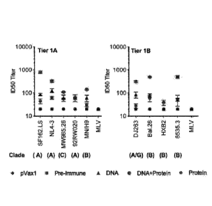
Figure 7. Neutralizing antibody titers against HIV-1 from immunized guinea pig
sera.
Guinea pig sera were collected two weeks after the last protein immunization
for testing. The
neutralization experiment was conducted in TZM-bl cells using a panel of
Envelope tier 1
pseudo viruses as described in Materials and Methods. Neutralization titers
were defined by the
sera dilution that achieves 50% inhibition of viral isolates (ID 50).
DETAILED DESCRIPTION
The inventors have made the surprising discovery of a synthetic consensus DNA
vaccine encoding HIV antigenic constructs derived from individual HIV-1
subtypes A, B, C and
4
Date Recue/Date Received 2020-12-29
D in a DNA prime-protein boost regimen. These consensus DNA constructs can be
optimized
using the following plasmid-enhancement techniques: codon optimization, RNA
optimization,
leader sequence addition, plasmid production at high concentrations and the
DNA was delivered
by adaptive EP as previously described. The DNA prime can be followed by a
protein boost
with recombinant HIV gp120. Immune responses were measured by ELISA, B-cell
ELISpot, T-
cell ELISpot, and in a TZM-bl neutralization assay. The combination approach
increased T cell
and antibody functionality over these observed with either independent
modality.
1. Definitions.
The terminology used herein is for the purpose of describing particular
embodiments
only and is not intended to be limiting. As used in the specification and the
appended claims,
the singular forms "a," "an" and "the" include plural referents unless the
context clearly dictates
otherwise.
For recitation of numeric ranges herein, each intervening number there between
with the
same degree of precision is explicitly contemplated. For example, for the
range of 6-9, the
numbers 7 and 8 are contemplated in addition to 6 and 9, and for the range 6.0-
7.0, the numbers
6.0, 6.1, 6.2, 6.3, 6.4, 6.5, 6.6, 6.7, 6.8, 6.9, and 7.0 are explicitly
contemplated.
"Consensus" or "Consensus Sequence" as used herein may mean a synthetic
nucleic
acid sequence, or corresponding polypeptide sequence, constructed based on
analysis of an
alignment of multiple subtypes of a particular antigen. The sequence may be
used to induce
broad immunity against multiple subtypes or sertypes of a particular antigen.
Synthetic antigens,
such as fusion proteins, may be manipulated to consensus sequences (or
consensus antigens).
A "peptide" or "polypeptide" is a linked sequence of amino acids and can be
natural,
synthetic, or a modification or combination of natural and synthetic.
"Treatment" or "treating," when referring to protection of an animal from a
disease,
means preventing, suppressing, repressing, or completely eliminating the
disease. Preventing the
disease involves administering a composition of the present invention to an
animal prior to onset
of the disease. Suppressing the disease involves administering a composition
of the present
invention to an animal after induction of the disease but before its clinical
appearance.
Repressing the disease involves administering a composition of the present
invention to an
animal after clinical appearance of the disease.
"Substantially identical" can mean that a first and second amino acid sequence
are at least
60%, 65%, 70%, 75%, 80%, 85%, 90%, 95%, 96%, 970/s, 98%,or 99% over a region
of 1, 2, 3, 4,
Date Recue/Date Received 2020-12-29
5, 6, 7, 8, 9, 10, 11, 12, 13, 14, 15, 16, 17, 18, 19, 20, 21, 22, 23, 24, 25,
30, 35, 40, 45, 50, 55, 60,
65, 70, 75, 80, 85, 90, 95, 100, 200, 300, 400, 500, 600, 700, 800, 900, 1000,
1100 amino acids.
A "variant" can mean means a peptide or polypeptide that differs in amino acid
sequence
by the insertion, deletion, or conservative substitution of amino acids, but
retain at least one
biological activity. Representative examples of "biological activity" include
the ability to be
bound by a specific antibody or to promote an immune response. Variant can
also mean a
protein with an amino acid sequence that is substantially identical to a
referenced protein with an
amino acid sequence that retains at least one biological activity. A
conservative substitution of
an amino acid, i.e., replacing an amino acid with a different amino acid of
similar properties (e.g.,
hydrophilicity, degree and distribution of charged regions) is recognized in
the art as typically
involving a minor change. These minor changes can be identified, in part, by
considering the
hydropathic index of amino acids. See Kyte et al., J. Mol. Biol. 157:105-132
(1982). The
hydropathic index of an amino acid is based on a consideration of its
hydrophobicity and charge.
It is known in the art that amino acids of similar hydropathic indexes can be
substituted and still
retain protein function. In one aspect, amino acids having hydropathic indexes
of 2 are
substituted. The hydrophilicity of amino acids can also be used to reveal
substitutions that
would result in proteins retaining biological function. A consideration of the
hydrophilicity of
amino acids in the context of a peptide permits calculation of the greatest
local average
hydrophilicity of that peptide, a useful measure that has been reported to
correlate well with
antigenicity and immunogenicity, as discussed in U.S. Patent No. 4,554,101.
Substitution of
amino acids having similar hydrophilicity values can result in peptides
retaining biological
activity, for example immunogenicity, as is understood in the art.
Substitutions can be performed
with amino acids having hydrophilicity values within 2 of each other. Both
the hyrophobicity
index and the hydrophilicity value of amino acids are influenced by the
particular side chain of
that amino acid. Consistent with that observation, amino acid substitutions
that are compatible
with biological function are understood to depend on the relative similarity
of the amino acids,
and particularly the side chains of those amino acids, as revealed by the
hydrophobicity,
hydrophilicity, charge, size, and other properties.
2. Composition for Priming and Boosting HIV Immunological Response
Provided herein is a composition comprising a first vaccine and a second
vaccine for
priming and boosting an immune response to HIV. The first vaccine is comprised
of at least 1,
at least 2, at least 3, at least 4, at least 5, at least 6, at least 7, at
least 8, at least 9, or at least 10
nucleic acids comprising an antigen. The second vaccine comprises at least 1,
at least 2, at least
6
Date Recue/Date Received 2020-12-29
3, at least 4, at least 5, at least 6, at least 7, at least 8, at least 9, or
at least 10 antigenic peptides.
The first vaccine is administered day 1 of the vaccination regimen. The first
vaccine may be
given in multiple doses. The first vaccine can be administered a second time
within 12 hours, 24
hours, 36 hours, or 48 hours of the first administration of the first vaccine.
The first vaccine
may be a priming vaccination.
The second vaccine can be administered to boost the first vaccine. The second
vaccine
maybe administered a first time 48 hours, 60 hours, 72 hours, 84 hours, 90
hours, 1.5 weeks, 2
weeks, 2.5 weeks, 3.0 weeks, 3.5 weeks, 4.0 weeks, 4.5 weeks, 5.0 weeks, 5.5
weeks, 6.0 weeks,
6.5 weeks, 7.0 weeks, 7.5 weeks, 8.0 weeks, 8.5 weeks, 9.0 weeks, 9.5 weeks,
10.0 weeks, 10.5
weeks, 11.0 weeks, 11.5 weeks, 12.0 weeks, 12.5 weeks, 13.0 weeks, 13.5 weeks,
14.0 weeks, 14.5
weeks or 15.0 weeks after the administration of the first vaccine. The second
vaccine may be in
administered in multiple doses. The second vaccine may be administered a
second time after 60
hours, 72 hours, 84 hours, 90 hours, 1.5 weeks, 2 weeks, 2.5 weeks, 3.0 weeks,
3.5 weeks, 4.0
weeks, 4.5 weeks, 5.0 weeks, 5.5 weeks, 6.0 weeks, 6.5 weeks, 7.0 weeks, 7.5
weeks, 8.0 weeks,
8.5 weeks, 9.0 weeks, 9.5 weeks, 10.0 weeks, 10.5 weeks, 11.0 weeks, 11.5
weeks, 12.0 weeks,
12.5 weeks, 13.0 weeks, 13.5 weeks, 14.0 weeks, 14.5 weeks or 15.0 weeks, or
15.5 weeks after
the first administration of the second vaccine.
The vaccine can induce antigen-specific T cells that inhibit antigen-specific
T cell
function. The combination of the first vaccine comprising a DNA encoding the
antigen and a
second vaccine comprising antigen for boosting the immune response to the
first vaccine
induces an immunological response efficiently against specific antigens far
better than either a
vaccine comprising an antigen or its corresponding DNA alone. The vaccine can
further
enhance MHC Class II presentation and expression for iTreg cell induction.
a. Antigen
The composition may comprise an antigen. The antigen is encoded by a nucleic
acid
sequence. The nucleic acid sequence may be DNA or RNA. The nucleic acid may
encode an
antigen or a variant thereof. The antigen can be the same antigen or a
different antigen between
the first and the second vaccine. The antigen can be an antigen isolated from
human
immunodeficiency virus (HIV). The HIV antigens can include modified consensus
sequences
for immunogens. Genetic modifications including codon optimization, RNA
optimization, and
the addition of a high efficient immunoglobin leader sequence to increase the
immunogenicity of
constructs can be included in the modified consensus sequences. The novel
immunogens can be
7
Date Recue/Date Received 2020-12-29
designed to elicit stronger and broader cellular immune responses than a
corresponding codon
optimized immunogens.
The antigen of the first vaccine may be the same antigen across different
subtypes of
HIV. The first vaccine may comprise 1 or more, 2 or more, 3 or more, 4 or
more, or 5 or more
DNA sequences encoding a particular protein sequence isolated from HIV
subtypes A, B, C, D,
or other HIV subtypes, or a combination or variant thereof. The antigen of the
first vaccine may
be the same antigen across different subtypes of HIV. The first vaccine may
comprise 1 or
more, 2 or more, 3 or more, 4 or more, or 5 or more consensus DNA sequences
encoding a
particular protein sequence isolated from HIV subtypes A, B, C, D, Of other
HIV subtypes, or a
combination or variant thereof. The first vaccine may comprise a DNA sequence
encoding a
particular protein sequence isolated from HIV subtype A, a second DNA sequence
encoding a
particular protein sequence isolated from HIV subtype B, a third DNA sequence
encoding a
particular protein sequence isolated from HIV subtype C, a fourth DNA sequence
encoding a
particular protein sequence isolated from HIV subtype D, or a combination
thereof. The first
vaccine may comprise a consensus DNA sequence encoding a particular protein
sequence
isolated from HIV subtype A, a second consensus DNA sequence encoding a
particular protein
sequence isolated from HIV subtype B, a third consensus DNA sequence encoding
a particular
protein sequence isolated from HIV subtype C, a fourth consensus DNA sequence
encoding a
particular protein sequence isolated from HIV subtype D, or a combination
thereof. The first
vaccine may comprise a consensus DNA sequence or variant thereof encoding a
particular HIV
subtype A protein sequence or variant thereof, a second consensus DNA sequence
or variant
thereof encoding a particular HIV subtype B protein sequence or variant
thereof, a third
consensus DNA sequence or variant thereof encoding a particular HIV subtype C
protein
sequence or variant thereof, a fourth consensus DNA sequence or variant
thereof encoding a
particular HIV subtype D protein sequence or variant thereof.
The second vaccine may comprise 1 or more, 2 of more, 3 or more, 4 or more, 5
or
more antigenic peptide sequences isolated from HIV subtypes A, B, C, D, or
other HIV
subtypes, or a combination or variant thereof. The second vaccine may comprise
1 or more, 2
or more, 3 or more, 4 or more, 5 or more consensus antigenic peptide sequences
isolated from
HIV subtypes A, B, C, D, or other HIV subtypes, or a combination or variant
thereof. The
second vaccine may comprise a antigenic peptide that is the same or different
from the DNA
encoded peptides of the first vaccine. The second vaccine may comprise a
particular protein
sequence isolated from HIV subtype A, subtype B, subtype C, subtype D or other
HIV subtypes.
8
Date Recue/Date Received 2020-12-29
The second vaccine may comprise a particular consensus protein sequence
isolated from HIV
subtype A, subtype B, subtype C, subtype D or other HIV subtypes.
In some embodiments, the HIV antigen can be a subtype A consensus envelope
DNA sequence construct, an IgE leader sequence linked to a consensus sequence
for Subtype A
envelope protein, or a subtype A consensus Envelope protein sequence.
In other embodiments, the HIV antigen can be a subtype B consensus envelope
DNA
sequence construct, an IgE leader sequence linked to a consensus sequence for
Subtype B
envelope protein, or an subtype B consensus Envelope protein sequence.
In still other embodiments, the HIV antigen can be a subtype C consensus
envelope
DNA sequence construct, an IgE leader sequence linked to a consensus sequence
for subtype C
envelope protein, or a subtype C consensus envelope protein sequence.
In further embodiments, the HIV antigen can be a subtype D consensus envelope
DNA sequence construct, an IgE leader sequence linked to a consensus sequence
for Subtype D
envelope protein, or a subtype D consensus envelope protein sequence.
In some embodiments, the HIV antigen can be a subtype A Nef-Rev consensus
envelope DNA sequence construct, an IgE leader sequence linked to a consensus
sequence for
Subtype A Nef-Rev protein, or a Subtype A Nef-Rev consensus protein sequence.
In some embodiments, the HIV antigen can be a subtype B Nef-Rev consensus
envelope DNA sequence construct, an IgE leader sequence linked to a consensus
sequence for
Subtype B Nef-Rev protein, or a Subtype B Nef-Rev consensus protein sequence.
In some embodiments, the HIV antigen can be a subtype C Nef-Rev consensus
envelope DNA sequence construct, an IgE leader sequence linked to a consensus
sequence for
Subtype C Nef-Rev protein, or a Subtype C Nef-Rev consensus protein sequence.
In some embodiments, the HIV antigen can be a subtype D Nef-Rev consensus
envelope DNA sequence construct, an IgE leader sequence linked to a consensus
sequence for
Subtype D Nef-Rev protein, or a Subtype D Nef-Rev consensus protein sequence.
In other embodiments, the HIV antigen can be a Gag consensus DNA sequence of
subtype A, B, C and D DNA sequence construct, an IgE leader sequence linked to
a consensus
sequence for Gag consensus subtype A, B, C and D protein, or a consensus Gag
subtype A, B, C
and D protein sequence.
In still other embodiments, the HIV antigen can be a MPol DNA sequence or a
MPol
protein sequence. The HIV antigen can be nucleic acid or amino acid sequences
of Env A, Env
B, Env C, Env D, B Nef-Rev, , Gag, or any combination thereof.
9
Date Recue/Date Received 2020-12-29
In other embodiments, the HIV antigen may be a DNA sequence or consensus
sequence of subtype A, B, C, or Dencoding gp140 or consensus gp140 protein. In
other
embodiments, the HIV antigen may be a DNA sequence or consensus sequence of
subtype A,
B, C, or D encoding gp140 or consensus gp120 protein. In other embodiments,
the HIV
antigen gp140 peptide sequence or gp140 consensus peptide sequence of subtype
A, B, C, or D.
In other embodiments, the HIV antigen gp120 peptide sequence or gp140
consensus peptide
sequence of subtype A, B, C, or D.
The antigen can affect a mammal, which can be a human, chimpanzee, dog, cat,
horse,
cow, mouse, or rat. The antigen can be contained in a protein from a mammal,
which can be a
human, chimpanzee, dog, cat, horse, cow, pig, sheep, mouse, or rat.
b. DNA
The composition may comprise the DNA. Also provided herein is a DNA that
encodes
the antigen as described above. The DNA can include an encoding sequence that
encodes the
antigen. The DNA can also include additional sequences that encode linker or
tag sequences that
are linked to the antigen by a peptide bond.
c. Vector
The composition may comprise a vector that includes the DNA encoding the
antigen.
The vector can be capable of expressing the antigen. The vector may be an
expression construct,
which is generally a plasmid that is used to introduce a specific gene into a
target cell. Once the
expression vector is inside the cell, the protein that is encoded by the gene
is produced by the
cellular-transcription and translation machinery ribosomal complexes. The
plasmid is frequently
engineered to contain regulatory sequences that act as enhancer and promoter
regions and lead
to efficient transcription of the gene carried on the expression vector. The
vectors of the present
invention express large amounts of stable messenger RNA, and therefore
proteins.
The vectors may have expression signals such as a strong promoter, a strong
termination
codon, adjustment of the distance between the promoter and the cloned gene,
and the insertion
of a transcription termination sequence and a PTIS (portable translation
initiation sequence).
i. Expression vectors
The vector may be circular plasmid or a linear nucleic acid vaccine. The
circular plasmid
and linear nucleic acid are capable of directing expression of a particular
nucleotide sequence in
an appropriate subject cell. The vector may have a promoter operably linked to
the antigen-
Date Recue/Date Received 2020-12-29
encoding nucleotide sequence, which may be operably linked to termination
signals. The vector
may also contain sequences required for proper translation of the nucleotide
sequence. The
vector comprising the nucleotide sequence of interest may be chimeric, meaning
that at least one
of its components is heterologous with respect to at least one of its other
components. The
expression of the nucleotide sequence in the expression cassette may be under
the control of a
constitutive promoter or of an inducible promoter which initiates
transcription only when the
host cell is exposed to some particular external stimulus. In the case of a
multicellular organism,
the promoter can also be specific to a particular tissue or organ or stage of
development.
Circular and Linear Vectors
The vector may be circular plasmid, which may transform a target cell by
integration into
the cellular genome or exist extrachromosomally (e.g. autonomous replicating
plasmid with an
origin of replication).
The vector can be pVAX, pcDNA3.0, or provax, or any other expression vector
capable
of expressing the DNA and enabling a cell to translate the sequence to a
antigen that is
recognized by the immune system. The vector can be combined with antigen at a
mass ratio of
between 5:1 and 1:5, or of between 1:1 and 2:1.
Also provided herein is a linear nucleic acid vaccine, or linear expression
cassette
("LEG"), that is capable of being efficiently delivered to a subject via
electroporation and
expressing one or more desired antigens. The LEG may be any linear DNA devoid
of any
phosphate backbone. The DNA may encode one or more antigens. The LEG may
contain a
promoter, an intron, a stop codon, a polyadenylation signal. The expression of
the antigen may
be controlled by the promoter. The LEG may not contain any antibiotic
resistance genes and/or
a phosphate backbone. The LEG may not contain other nucleic acid sequences
unrelated to the
desired antigen gene expression.
The LEG may be derived from any plasmid capable of being linearized. The
plasmid
may be capable of expressing the antigen. The plasmid may be pNP (Puerto
Rico/34) or pM2
(New Caledonia/99). See Figure 1. The plasmid may be pVAX, pcDNA3.0, or
provax, or any
other expression vector capable of expressing the DNA and enabling a cell to
translate the
sequence to a antigen that is recognized by the immune system.
The LEG may be perM2. The LEG may be perNP. perNP and perMR may be derived
from pNP (Puerto Rico/34) and pM2 (New Caledonia/99), respectively. See Figure
34. The
LEG may be combined with antigen at a mass ratio of between 5:1 and 1:5, or of
between 1:1 to
2:1.
11
Date Recue/Date Received 2020-12-29
Promoter, Intron, Stop codon, and Polyadenylation signal
The vector may have a promoter. A promoter may be any promoter that is capable
of
driving gene expression and regulating expression of the isolated nucleic
acid. Such a promoter is
a cis-acting sequence element required for transcription via a DNA dependent
RNA polymerase,
which transcribes the antigen sequence described herein. Selection of the
promoter used to
direct expression of a heterologous nucleic acid depends on the particular
application. The
promoter may be positioned about the same distance from the transcription
start in the vector as
it is from the transcription start site in its natural setting. However,
variation in this distance may
be accommodated without loss of promoter function.
The promoter may be operably linked to the nucleic acid sequence encoding the
antigen
and signals required for efficient polyadenylation of the transcript, ribosome
binding sites, and
translation termination. The promoter may be a CMV promoter, 5V40 early
promoter, 5V40
later promoter, metallothionein promoter, murine mammary tumor virus promoter,
Rous
sarcoma virus promoter, polyhedrin promoter, or another promoter shown
effective for
expression in eukaryotic cells.
The vector may include an enhancer and an intron with functional splice donor
and
acceptor sites. The vector may contain a transcription termination region
downstream of the
structural gene to provide for efficient termination. The termination region
may be obtained
from the same gene as the promoter sequence or may be obtained from different
genes.
d. Other Components of Vaccine-Adjuvants, Excipients
The composition may further comprise a pharmaceutically acceptable excipient.
The
pharmaceutically acceptable excipient can be functional molecules as vehicles,
adjuvants, carriers,
or diluents. The pharmaceutically acceptable excipient can be a transfection
facilitating agent,
which can include surface active agents, such as immune-stimulating complexes
(ISCOMS),
Freunds incomplete adjuvant, LPS analog including monophosphoryl lipid A,
muramyl peptides,
quinone analogs, vesicles such as squalene and squalene, hyaluronic acid,
lipids, liposomes,
calcium ions, viral proteins, polyanions, polycations, or nanoparticles, or
other known
transfection facilitating agents.
The transfection facilitating agent is a polyanion, polycation, including poly-
L-glutamate
(LGS), or lipid. The transfection facilitating agent is poly-L-glutamate, and
the poly-L-glutamate
is may be present in the vaccine at a concentration less than 6 mg/ml. The
transfection
facilitating agent may also include surface active agents such as immune-
stimulating complexes
(ISCOMS), Freunds incomplete adjuvant, LPS analog including monophosphoryl
lipid A,
12
Date Recue/Date Received 2020-12-29
muramyl peptides, quinone analogs and vesicles such as squalene and squalene,
and hyaluronic
acid may also be used administered in conjunction with the genetic construct.
The DNA plasmid
vaccines may also include a transfection facilitating agent such as lipids,
liposomes, including
lecithin liposomes or other liposomes known in the art, as a DNA-liposome
mixture (see for
example W09324640), calcium ions, viral proteins, polyanions, polycations, or
nanoparticles, or
other known transfection facilitating agents. The transfection facilitating
agent is a polyanion,
polycation, including poly-L-glutamate (LGS), or lipid. Concentration of the
transfection agent
in the vaccine is less than 4 mg/ml, less than 2 mg/ml, less than 1 mg/ml,
less than 0.750
mg/ml, less than 0.500 mg/ml, less than 0.250 mg/ml, less than 0.100 mg/ml,
less than 0.050
mg/ml, or less than 0.010 mg/ml.
The pharmaceutically acceptable excipient can be an adjuvant. The adjuvant can
be
other genes that are expressed in alternative plasmid or are delivered as
proteins in combination
with the plasmid above in the vaccine. The adjuvant may be selected from the
group consisting
of: a-interferon(IFN- a), 3-interferon (IFN-3), ''-interferon, platelet
derived growth factor
(PDGF), TNFa, TNF3, GM-CSF, epidermal growth factor (EGF), cutaneous T cell-
attracting
chemokine (CTACK), epithelial thymus-expressed chemokine (TECK), mucosae-
associated
epithelial chemokine (MEG), IL-12, IL-15, MHC, CD80,CD86 including IL-15
having the signal
sequence deleted and optionally including the signal peptide from IgE. The
adjuvant can be IT ,-
12, IL-15, IL-28, CTACK, TECK, platelet derived growth factor (PDGF), TNFa,
TNFI3, GM-
CSF, epidermal growth factor (EGF), IT ,1, IL-2, IL-4, IL-5, IL-6, IL-10, IL-
12, IL-18, or a
combination thereof.
Other genes that can be useful adjuvants include those encoding: MCP-1, MIP-
la, MIP-
1p, IL-8, RANTES, L-selectin, P-selectin, E-selectin, CD34, GlyCAM-1, MadCAM-
1, LFA-1,
VLA-1, Mac-1, p150.95, PECAM, ICAM-1, ICAM-2, ICAM-3, CD2, LFA-3, M-CSF, G-
CSF,
IT ,-4, mutant forms of IT ,-18, CD40, CD4OL, vascular growth factor,
fibroblast growth factor,
11,-7, nerve growth factor, vascular endothelial growth factor, Fas, TNF
receptor, Flt, Apo-1,
p55, WSL-1, DR3, TRAMP, Apo-3, AIR, LARD, NGRF, DR4, DRS, KILLER, TRAIL-R2,
TRICK2, DR6, Caspase ICE, Fos, c-jun, Sp-1, Ap-1, Ap-2, p38, p65Rel, MyD88,
IRAK,
TRAF6, IkB, Inactive NIK, SAP K, SAP-1, JNK, interferon response genes, NFkB,
Bax,
TRAIT ,, TRAILrec, TRAIT ,recDRC5, TRAIL-R3, TRAIL-R4, RANK, RANK LIGAND,
0x40,
0x40 LIGAND, NKG2D, MICA, MICB, NKG2A, NKG2B, NKG2C, NKG2E, NKG2F,
TAP1, TAP2 and functional fragments thereof.
The vaccine may further comprise a genetic vaccine facilitator agent as
described in U.S.
Serial No. 021,579 filed April 1,1994.
13
Date Recue/Date Received 2020-12-29
The vaccine can be formulated according to the mode of administration to be
used. An
injectable vaccine pharmaceutical composition can be sterile, pyrogen free and
particulate free.
An isotonic formulation or solution can be used. Additives for isotonicity can
include sodium
chloride, dextrose, mannitol, sorbitol, and lactose. The vaccine can comprise
a vasoconstriction
agent. The isotonic solutions can include phosphate buffered saline. Vaccine
can further
comprise stabilizers including gelatin and albumin. The stabilizers can allow
the formulation to
be stable at room or ambient temperature for extended periods of time,
including LGS or
polycations or polyanions.
3. Method of vaccination
Provided herein is a method of vaccinating a patient to treat or prevent HIV
infection
using the composition. The method of vaccinating a patient comprises
administering the
composition to a subject in need thereof. The first vaccine can efficiently
deliver antigen to a
subject in need thereof for immune stimulation via a priming vaccination. A
subject's immune
system may be efficiently induced against specific antigens by administering a
priming DNA that
encodes the antigen followed by a boost antigenic peptide. The DNA vaccine,
which may
contain an antigen-encoding circular plastnid or a linear nucleic acid, can
elicit antigen-specific
antibody responses, which are sustainable for longer periods of time as
compared to plasmid-
based vaccines, when efficiently delivered to a subject. The second vaccine
may be a boosting
vaccine to the first vaccine and comprise the same or different antigenic
peptide as the first
vaccine.
The herein described composition may be administered to a subject so as to
provide a
longer lasting antigen-specific immune response that is well tolerated by the
subject population.
The present invention is also directed to a number of antigens that can be
expressed from the
DNA.
The dose of the first and second vaccine can be between 1 g to 10 mg active
component/kg
body weight/time, and can be 20 pig to 10 mg component/kg body weight/time.
The first vaccine is
administered day 1 of the vaccination regimen. The first vaccine may be given
in multiple doses. The
first vaccine can be administered a second time within 12 hours, 24 hours, 36
hours, or 48 hours of the
first administration of the first vaccine. The first vaccine may be a priming
vaccination.
The second vaccine can be administered to boost the first vaccine. The second
vaccine
maybe administered a first time 48 hours, 60 hours, 72 hours, 84 hours, 90
hours, 1.5 weeks, 2
weeks, 2.5 weeks, 3.0 weeks, 3.5 weeks, 4.0 weeks, 4.5 weeks, 5.0 weeks, 5.5
weeks, 6.0 weeks,
6.5 weeks, 7.0 weeks, 7.5 weeks, 8.0 weeks, 8.5 weeks, 9.0 weeks, 9.5 weeks,
10.0 weeks, 10.5
14
Date Recue/Date Received 2020-12-29
weeks, 11.0 weeks, 11.5 weeks, 12.0 weeks, 12.5 weeks, 13.0 weeks, 13.5 weeks,
14.0 weeks, 14.5
weeks or 15.0 weeks after the administration of the first vaccine. The second
vaccine may be in
administered in multiple doses. The second vaccine may be administered a
second time after 60
hours, 72 hours, 84 hours, 90 hours, 1.5 weeks, 2 weeks, 2.5 weeks, 3.0 weeks,
3.5 weeks, 4.0
weeks, 4.5 weeks, 5.0 weeks, 5.5 weeks, 6.0 weeks, 6.5 weeks, 7.0 weeks, 7.5
weeks, 8.0 weeks,
8.5 weeks, 9.0 weeks, 9.5 weeks, 10.0 weeks, 10.5 weeks, 11.0 weeks, 11.5
weeks, 12.0 weeks,
12.5 weeks, 13.0 weeks, 13.5 weeks, 14.0 weeks, 14.5 weeks or 15.0 weeks, or
15.5 weeks after
the first administration of the second vaccine.
The number of vaccine doses for effective treatment can be 1, 2, 3, 4, 5, 6,
7, 8, 9, or 10.
a. Administration
The composition can be formulated in accordance with standard techniques well
known to those
skilled in the pharmaceutical art. Such compositions can be administered in
dosages and by techniques
well known to those skilled in the medical arts taking into consideration such
factors as the age, sex,
weight, and condition of the particular subject, and the route of
administration. The subject can be a
mammal, such as a human, a horse, a cow, a pig, a sheep, a cat, a dog, a rat,
or a mouse.
The composition can be administered prophylactically or therapeutically. In
prophylactic
administration, the vaccines can be administered in an amount sufficient to
induce iTreg responses. In
therapeutic applications, the vaccines are administered to a subject in need
thereof in an amount
sufficient to elicit a therapeutic effect. An amount adequate to accomplish
this is defined as
"therapeutically effective dose." Amounts effective for this use will depend
on, e.g., the particular
composition of the vaccine regimen administered, the manner of administration,
the stage and severity
of the disease, the general state of health of the patient, and the judgment
of the prescribing physician.
The composition can be administered by methods well known in the art as
described in
Donnelly et al. (Ann. Rev. Immunol. 15:617-648 (1997)); Feigner et al. (U.S.
Pat. No. 5,580,859, issued
Dec. 3, 1996); Feigner (U.S. Pat. No. 5,703,055, issued Dec. 30, 1997); and
Carson et al. (U.S. Pat. No.
5,679,647, issued Oct. 21, 1997). The DNA of the vaccine can be complexed to
particles or beads that
can be administered to an individual, for example, using a vaccine gun. One
skilled in the art would
know that the choice of a pharmaceutically acceptable carrier, including a
physiologically acceptable
compound, depends, for example, on the route of administration of the
expression vector.
The composition can be delivered via a variety of routes. Typical delivery
routes include
parenteral administration, e.g., intradermal, intramuscular or subcutaneous
delivery. Other routes include
oral administration, intranasal, and intravaginal routes. For the DNA of the
vaccine in particular, the
vaccine can be delivered to the interstitial spaces of tissues of an
individual (Feigner et al., U.S. Pat. Nos.
Date Recue/Date Received 2020-12-29
5,580,859 and 5,703,055). The vaccine can also be administered to muscle, or
can be administered via
intradermal or subcutaneous injections, or transdermally, such as by
iontophoresis. Epidermal
administration of the vaccine can also be employed. Epidermal administration
can involve mechanically
or chemically irritating the outermost layer of epidermis to stimulate an
immune response to the irritant
(Carson et al., U.S. Pat. No. 5,679,647).
The composition can also be formulated for administration via the nasal
passages. Formulations
suitable for nasal administration, wherein the carrier is a solid, can include
a coarse powder having a
particle size, for example, in the range of about 10 to about 500 microns
which is administered in the
manner in which snuff is taken, i.e., by rapid inhalation through the nasal
passage from a container of
the powder held close up to the nose. The formulation can be a nasal spray,
nasal drops, or by aerosol
administration by nebulizer. The formulation can include aqueous or oily
solutions of the vaccine.
The composition can be a liquid preparation such as a suspension, syrup or
elixir. The vaccine
can also be a preparation for parenteral, subcutaneous, intradermal,
intramuscular or intravenous
administration (e.g., injectable administration), such as a sterile suspension
or emulsion.
The composition can be incorporated into liposomes, microspheres or other
polymer matrices
(Feigner et al., U.S. Pat. No. 5,703,055; Gregoriadis, Liposome Technology,
Vols. Ito III (2nd ed. 1993)).
Liposomes can consist of phospholipids or other lipids, and can be nontoxic,
physiologically acceptable
and metabolizable carriers that are relatively simple to make and administer.
The composition can be administered via electroporation, such as by a method
described in U.S.
Patent No. 7,664,545. The electroporation can be by a method and/or apparatus
described in U.S.
Patent Nos. 6,302,874; 5,676,646; 6,241,701; 6,233,482; 6,216,034; 6,208,893;
6,192,270; 6,181,964;
6,150,148; 6,120,493; 6,096,020; 6,068,650; and 5,702,359. The electroporation
may be carried out via a
minimally invasive device.
The minimally invasive electroporation device ("MID") may be an apparatus for
injecting the
vaccine described above and associated fluid into body tissue. The device may
comprise a hollow
needle, DNA cassette, and fluid delivery means, wherein the device is adapted
to actuate the fluid
delivery means in use so as to concurrently (for example, automatically)
inject DNA into body tissue
during insertion of the needle into the said body tissue. This has the
advantage that the ability to inject
the DNA and associated fluid gradually while the needle is being inserted
leads to a more even
distribution of the fluid through the body tissue. The pain experienced during
injection may be reduced
due to the distribution of the DNA being injected over a larger area.
The MID may inject the composition into tissue without the use of a needle.
The MID may
inject the vaccine as a small stream or jet with such force that the vaccine
pierces the surface of the
tissue and enters the underlying tissue and/or muscle. The force behind the
small stream or jet may be
16
Date Recue/Date Received 2020-12-29
provided by expansion of a compressed gas, such as carbon dioxide through a
micro-orifice within a
fraction of a second. Examples of minimally invasive electroporation devices,
and methods of using
them, are described in published U.S. Patent Application No. 20080234655; U.S.
Patent No. 6,520,950;
U.S. Patent No. 7,171,264; U.S. Patent No. 6,208,893; U.S. Patent NO.
6,009,347; U.S. Patent No.
6,120,493; U.S. Patent No. 7,245,963; U.S. Patent No. 7,328,064; and U.S.
Patent No. 6,763,264.
The MID may comprise an injector that creates a high-speed jet of liquid that
painlessly pierces
the tissue. Such needle-free injectors are commercially available. Examples of
needle-free injectors that
can be utilized herein include those described in U.S. Patent Nos. 3,805,783;
4,447,223; 5,505,697; and
4,342,310.
A desired composition in a form suitable for direct or indirect
electrotransport may be
introduced (e.g., injected) using a needle-free injector into the tissue to be
treated, usually by contacting
the tissue surface with the injector so as to actuate delivery of a jet of the
agent, with sufficient force to
cause penetration of the vaccine into the tissue. For example, if the tissue
to be treated is mucosa, skin
or muscle, the agent is projected towards the mucosal or skin surface with
sufficient force to cause the
agent to penetrate through the stratum comeum and into dermal layers, or into
underlying tissue and
muscle, respectively.
Needle-free injectors are well suited to deliver vaccines to all types of
tissues, particularly to skin
and mucosa. In some embodiments, a needle-free injector may be used to propel
a liquid that contains
the vaccine to the surface and into the subject's skin or mucosa.
Representative examples of the various
types of tissues that can be treated using the invention methods include
pancreas, larynx, nasopharynx,
hypopharynx, oropharynx, lip, throat, lung, heart, kidney, muscle, breast,
colon, prostate, thymus, testis,
skin, mucosal tissue, ovary, blood vessels, or any combination thereof.
The MID may have needle electrodes that electroporate the tissue. By pulsing
between multiple
pairs of electrodes in a multiple electrode array, for example set up in
rectangular or square patterns,
provides improved results over that of pulsing between a pair of electrodes.
Disclosed, for example, in
U.S. Patent No. 5,702,359 entitled "Needle Electrodes for Mediated Delivery of
Drugs and Genes" is an
array of needles wherein a plurality of pairs of needles may be pulsed during
the therapeutic treatment.
In that application, needles were disposed in a circular array, but have
connectors and switching
apparatus enabling a pulsing between opposing pairs of needle electrodes. A
pair of needle electrodes
for delivering recombinant expression vectors to cells may be used. Such a
device and system is
described in U.S. Patent No. 6,763,264. Alternatively, a single needle device
may be used that allows
injection of the DNA and electroporation with a single needle resembling a
normal injection needle and
applies pulses of lower voltage than those delivered by presently used
devices, thus reducing the
electrical sensation experienced by the patient.
17
Date Recue/Date Received 2020-12-29
The MID may comprise one or more electrode arrays. The arrays may comprise two
or more
needles of the same diameter or different diameters. The needles may be evenly
or unevenly spaced
apart. The needles may be between 0.005 inches and 0.03 inches, between 0.01
inches and 0.025 inches;
or between 0.015 inches and 0.020 inches. The needle may be 0.0175 inches in
diameter. The needles
may be 0.5 mm, 1.0 mm, 1.5 mm, 2.0 mm, 2.5 mm, 3.0 mm, 3.5 mm, 4.0 mm, or more
spaced apart.
The MID may consist of a pulse generator and a two or more-needle vaccine
injectors that
deliver the vaccine and electroporation pulses in a single step. The pulse
generator may allow for flexible
programming of pulse and injection parameters via a flash card operated
personal computer, as well as
comprehensive recording and storage of electroporation and patient data. The
pulse generator may
deliver a variety of volt pulses during short periods of time. For example,
the pulse generator may
deliver three 15 volt pulses of 100 ms in duration. An example of such a MID
is the Elgen 1000 system
by Inovio Biomedical Corporation, which is described in U.S. Patent No.
7,328,064
The MID may be a CELLECTRA (Inovio Pharmaceuticals, Blue Bell PA) device and
system,
which is a modular electrode system, that facilitates the introduction of a
macromolecule, such as a
DNA, into cells of a selected tissue in a body or plant. The modular electrode
system may comprise a
plurality of needle electrodes; a hypodermic needle; an electrical connector
that provides a conductive
link from a programmable constant-current pulse controller to the plurality of
needle electrodes; and a
power source. An operator can grasp the plurality of needle electrodes that
are mounted on a support
structure and firmly insert them into the selected tissue in a body or plant.
The macromolecules are then
delivered via the hypodermic needle into the selected tissue. The programmable
constant-current pulse
controller is activated and constant-current electrical pulse is applied to
the plurality of needle electrodes.
The applied constant-current electrical pulse facilitates the introduction of
the macromolecule into the
cell between the plurality of electrodes. Cell death due to overheating of
cells is minimized by limiting
the power dissipation in the tissue by virtue of constant-current pulses. The
Cellectra device and system
is described in U.S. Patent No. 7,245,963.
The MID may be an Elgen 1000 system (Inovio Pharmaceuticals). The Elgen 1000
system may
comprise device that provides a hollow needle; and fluid delivery means,
wherein the apparatus is
adapted to actuate the fluid delivery means in use so as to concurrently (for
example automatically) inject
fluid, the described vaccine herein, into body tissue during insertion of the
needle into the said body
tissue. The advantage is the ability to inject the fluid gradually while the
needle is being inserted leads to
a more even distribution of the fluid through the body tissue. It is also
believed that the pain
experienced during injection is reduced due to the distribution of the volume
of fluid being injected over
a larger area.
18
Date Recue/Date Received 2020-12-29
In addition, the automatic injection of fluid facilitates automatic monitoring
and registration of
an actual dose of fluid injected. This data can be stored by a control unit
for documentation purposes if
desired.
It will be appreciated that the rate of injection could be either linear or
non-linear and that the
injection may be carried out after the needles have been inserted through the
skin of the subject to be
treated and while they are inserted further into the body tissue.
Suitable tissues into which fluid may be injected by the apparatus of the
present invention
include tumor tissue, skin or liver tissue but may be muscle tissue.
The apparatus further comprises needle insertion means for guiding insertion
of the needle into
the body tissue. The rate of fluid injection is controlled by the rate of
needle insertion. This has the
advantage that both the needle insertion and injection of fluid can be
controlled such that the rate of
insertion can be matched to the rate of injection as desired. It also makes
the apparatus easier for a user
to operate. If desired means for automatically inserting the needle into body
tissue could be provided.
A user could choose when to commence injection of fluid. Ideally however,
injection is
commenced when the tip of the needle has reached muscle tissue and the
apparatus may include means
for sensing when the needle has been inserted to a sufficient depth for
injection of the fluid to
commence. This means that injection of fluid can be prompted to commence
automatically when the
needle has reached a desired depth (which will normally be the depth at which
muscle tissue begins). The
depth at which muscle tissue begins could for example be taken to be a preset
needle insertion depth
such as a value of 4 mm which would be deemed sufficient for the needle to get
through the skin layer.
The sensing means may comprise an ultrasound probe. The sensing means may
comprise a
means for sensing a change in impedance or resistance. In this case, the means
may not as such record
the depth of the needle in the body tissue but will rather be adapted to sense
a change in impedance or
resistance as the needle moves from a different type of body tissue into
muscle. Either of these
alternatives provides a relatively accurate and simple to operate means of
sensing that injection may
commence. The depth of insertion of the needle can further be recorded if
desired and could be used to
control injection of fluid such that the volume of fluid to be injected is
determined as the depth of
needle insertion is being recorded.
The apparatus may further comprise: a base for supporting the needle; and a
housing for
receiving the base therein, wherein the base is moveable relative to the
housing such that the needle is
retracted within the housing when the base is in a first rearward position
relative to the housing and the
needle extends out of the housing when the base is in a second forward
position within the housing.
This is advantageous for a user as the housing can be lined up on the skin of
a patient, and the needles
can then be inserted into the patient's skin by moving the housing relative to
the base.
19
Date Recue/Date Received 2020-12-29
As stated above, it is desirable to achieve a controlled rate of fluid
injection such that the fluid is
evenly distributed over the length of the needle as it is inserted into the
skin. The fluid delivery means
may comprise piston driving means adapted to inject fluid at a controlled
rate. The piston driving means
could for example be activated by a servo motor. However, the piston driving
means may be actuated by
the base being moved in the axial direction relative to the housing. It will
be appreciated that alternative
means for fluid delivery could be provided. Thus, for example, a closed
container which can be squeezed
for fluid delivery at a controlled or non-controlled rate could be provided in
the place of a syringe and
piston system.
The apparatus described above could be used for any type of injection. It is
however envisaged
to be particularly useful in the field of electroporation and so it may
further comprises means for
applying a voltage to the needle. This allows the needle to be used not only
for injection but also as an
electrode during, electroporation. This is particularly advantageous as it
means that the electric field is
applied to the same area as the injected fluid. There has traditionally been a
problem with
electroporation in that it is very difficult to accurately align an electrode
with previously injected fluid
and so user's have tended to inject a larger volume of fluid than is required
over a larger area and to
apply an electric field over a higher area to attempt to guarantee an overlap
between the injected
substance and the electric field. Using the present invention, both the volume
of fluid injected and the
size of electric field applied may be reduced while achieving a good fit
between the electric field and the
fluid.
The present invention has multiple aspects, illustrated by the following non-
limiting
examples.
4. Examples
Example 1¨Materials and Methods
Mice were housed and treated in a temperature-controlled, light-cycled
facility in
accordance with the guidelines of the National Institutes of Health (Bethesda,
MD, USA) and
the University of Pennsylvania (Philadelphia, PA, USA) Institutional Animal
Care and Use
Committee (IACUC #801577).
Female Dunkin-Hartley guinea pigs weighing between 350 and 450 g and free of
intercurrent infection were obtained from Charles River (Wilmington, MA) and
were housed at
Bio-Quant, Inc., (San Diego, CA) through collaboration with Inovio
Pharmaceuticals Inc., PA.
The protocol was approved by the Committee on the Ethics of Animal Experiments
at the Bio-
Quant, Inc. In collaboration with the animal resource dept. of Inovio
Pharmaceuticals Inc.,
Date Recue/Date Received 2020-12-29
(Permit Number: 08-021). At all locations, animals were handled based on the
recommendations
in the Guide for the Care and Use of Laboratory Animals of the National
Institutes of Health. In
accordance with the Weatherall report, animal welfare was ensured and steps
were taken to
ameliorate or minimize.
Cells and Reagents: HeLa, 293T and Jurkat (ATCC, Manassas, VA) and
HeLa-CD4-TZM-b1 (NIH-AIDS Reagent Program, MD) cells were maintained in
Dulbecco's
modified Eagle's medium (DMEM; Gibco-Invitrogen) supplemented with 10% Fetal
Bovine
Serum (FBS) and antibiotics and passaged upon confluence. Recombinant HIV-1
envelope
gp120 proteins were obtained from Protein Sciences Corporation, Meriden, CT
and peroxidise-
conjugated streptavidin from Jackson Laboratory. HIV-1 envelope polyclonal
antibodies and
other viral reagents were obtained through the AIDS Research and Reference
Reagent Program,
Division of AIDS, NIAID, NIH (Germantown, MD).
Construction, Expression and Immunization: The HIV-1 envelope consensus
sequences were previously described. Briefly, consensus sequences of HIV-1
envelope from
subtype clades A, B, C and D were constructed with modifications as discussed.
An IgE leader
sequence was added to all envelope antigen sequences to improve expression,
and the
cytoplasmic tail was truncated to prevent envelope recycling. The resulting
optimized HIV-Env
DNA immunogens were codon and RNA optimized, and balanced for GCAT content and
synthesized, and cloned into the modified pVax1 expression vector. Production
of DNA
constructs was carried out by Aldevron (Fargo, ND), and purified plasmid DNA
was formulated
in water for immunization as described.
To test the expression, cells were transfected for expression analysis of
synthetic
envelopes using the non-liposomal FuGENETM transfection reagent (Roche Applied
Science,
Indianapolis, IN) as suggested by the manufacturer. Briefly, cells were seeded
at 70% confluence
(50,000 cells per well in 6-well plates) a day before and trans fected with 5n
of the HIV-1
envelope plasmids. Cells were harvested 48 to 72 hrs post-transfection in lx
RIPA buffer (50
mM Tris/HC1 (pH 7.4), 150 mM NaCl, 1% Tritonm4 X-100, 1% sodium deoxycholate,
and 0.1%
SDS, supplemented with a complete protease inhibitor cocktail from Roche
Applied Science,
Indianapolis, IN) and mixed with SDS sample buffer (0.08M Tris (pH 6.8); 2.0%
SDS, 10%
glycerol, 0.1M dithiothreitol, 0.2% bromophenol blue) before boiling for 5
minutes.
For envelope expression study by immunoblot, specific sera or Abs were diluted
1:100
in PBS and reacted with individual strips for 1h. Subsequently, strips were
washed four times
with Tris-buffered saline- 0.2% TweenTm, reacted with a peroxidase-coupled
antiserum against
mouse IgG (Sigma, St Louis, MO), and incubated with diaminobenzidine substrate
(Sigma, St.
21
Date Recue/Date Received 2020-12-29
Louis, MO). For immunofluorescent analysis of optimized envelope expression,
RD cells were
transfected with vaccine constructs and stained with monoclonal antibodies
against HIV-1
envelope (4E10; NIH-AIDS Reagent Program, MD) and against MHC-I prior to
confocal
analysis. All images were captured by Zeiss 710 LSM Meta microscope system
observed in PBS.
Mice and Guinea Pip Immunizations: Study 1 was performed in the BALB/c
mice, using four groups of animals (4 animals per group/repeated 3 times)
which received DNA
or protein immunogens alone or in prime-boost combinations as indicated in
Fig. 2A. Study 2,
for guinea pig study (5 animals/group; repeated 2 times), HIV-1 Env plasmids
were immunized
as indicated in Figure 6A. Animals in prime-boost and recombinant groups
received HIV-1 clade
B protein (50g) formulated with TiterMaxIm adjuvant (Sigma-Aldrich, St Louis,
MO) at weeks 8
and 11. A sham immunization group received an empty pVax1 vector and sham
protein (PBS)
formulated with TiterMaxIm.
At various time points, small amounts of peripheral blood was harvested from
the
mice and guinea pigs for analysis of the humoral immune response using ELISA
assay as
indicated in Figure 2A and 6A. For analysis of the cellular immune response,
the mice were
humanely sacrificed at week 7 and their spleens were used as a source of
lymphocytes for the
ELISpot and flow cytometry immune analysis. Immunizations were delivered into
the quadriceps
muscles by in vivo Cellectra0 -adaptive EP as described previously. All
procedures were
performed in accordance with the guidelines of the National Institutes of
Health (Bethesda, MD,
USA) and the University of Pennsylvania (Philadelphia, PA, USA) Institutional
Animal Care and
Use Committee.
T-Cell Elispot Assay: We determined antigen-specific T-cell responses via IFN-
y
ELISpot. Briefly, ELISpot 96-well plates (EMD Millipore Corporation,
Billerica, MA) were
coated with anti-mouse IFN-y capture Abs and incubated for 24h at 4 C (R&D
Systems,
Minneapolis, MN). The following day, plates were washed and blocked for 2h
with 1% BSA.
Splenocytes from the immunized mice were added to each well and stimulated
overnight at 37
C in 5% CO2 in the presence of RPMI 1640 (negative control), Concanavalin A
(positive
control), or specific peptide pools (10 g/m1). Peptide pools consist of 15-mer
peptides
overlapping by 11 amino acids. After 24h of stimulation, the cells were washed
and incubated for
24 h at 4 C with biotinylated anti-mouse IFN-y Abs (R&D Systems, Minneapolis,
MN). The
plates were washed, and streptavidin¨alkaline phosphatase (R&D Systems,
Minneapolis, MN)
was added to each well and incubated for 2h at room temperature. The plates
were then washed
and 5-bromo-4-chloro-3'-indolylphosphate p-toluidine salt and nitro blue
tetrazolium chloride
(chromogen color reagent; R&D Systems, Minneapolis, MN) were added to each
well. The plates
22
Date Recue/Date Received 2020-12-29
were then rinsed with distilled water and dried at room temperature. Spots
were counted by an
automated ELISpot reader (CTL Limited, Shaker Heights, OH).
B-Cell Elispot Assay: When testing for antibody-secreting B cells (ASCs),
splenocytes were not stimulated prior to detection by ELISpot assay, but
instead were tested
directly after isolation from the spleen. MultiScreenIm-IP plates (Millipore,
Billerica, MA) were
coated with affinity-purified goat anti-mouse IgG (KPL, Gaithersburg, MD) in
PBS. Plates were
washed six times with PBS and blocked with RPMI with 10% FCS for 2h at room
temperature.
Splenocytes (105) were added to each well of the ELISpot plate in at least
100kil of medium and
incubated overnight at 37 C. Plates were washed six times in PBS with 0.25%
Tween 20 (Sigma-
Aldrich, St. Louis, MO) (PBS-T) and incubated with 100kil of 1:5,000 biotin-
IgG (Jackson
ImmunoResearch Laboratories, Inc., West Grove, PA) for 1 h at room
temperature. Plates were
then washed and incubated with 100kil of 1:60 streptavidin-AP (R&D Research
Systems,
Minneapolis, MN) for 1h at room temperature. The plates were washed with PBS-
T, PBS, and
distilled water and developed with 100kil of BCIP/NBT (R&D Research Systems,
Minneapolis,
MN) for 20 min at room temperature; the reaction was stopped with distilled
water. Spots were
counted by an automated ELISpot reader (CTL Limited, Shaker Heights, OH). Raw
values were
determined and multiplied by the appropriate factor so that the data could be
represented as
ASCs per million splenocytes.
Intracellular Cytokine Staining: The phenotype of the responding T cells were
analyzed by ICS fluorescence activated cell sorting (FACS) analysis as
described. Splenocytes
(5x10⁶) were stimulated with a 15-mer HIV-1 MN Env peptide pool (2 pig/m1 of
each peptide;
NIH AIDS Research & Reference Reagent Program, MD). Golgi plug (BD
Biosciences, San
Jose, CA) was added during the final 4 h of incubation. Cells were then
stained with anti-mouse
CD16/32 (Fc block) antibody, followed by surface staining. Cells were surface
stained with
CD3-FITC, CD4-Alexa 700, CD8-PerCP (BD Biosciences, San Jose, CA). Cells were
then fixed,
permeabilized with cytofix cytoperm (BD Biosciences, San Jose, CA) and stained
with anti- IFN-
y -PE and anti-II,2-FITC. One million cells per sample were acquired on a BD
LSRII flow
cytometer (BD Biosciences, San Jose, CA) and CD4+ and CD8+ events (gated
previously on
CD3+ cells) were gated versus IFN- and TI-2. Sample analysis was performed
using FlowJoTM
software (Tree Star, Ashland, OR).
Antibody Binding Assay: Elisa: For antibody detection, mouse serum samples
were
collected 7 days after the last immunization. Standard ELISAs were performed
using
recombinant GP120 as the antigen source, which was prepared as previously
described.
Antibody binding assays were carried out with either individual animal or
pooled sera from the
23
Date Recue/Date Received 2020-12-29
mice or Guinea pig in each group immunized with DNA or DNA+gp120 from
different clades
of HIV. Briefly, high-binding polystyrene Corning 96 well plates (Sigma, St
Louis, MO) were
coated overnight at 4 C with recombinant envelope protein (MN clade B) (2g/ml)
(Protein
Sciences Corporation, Meriden, CT), which was diluted in 50 mM carbonate
buffer (pH 9.6) and
stored overnight at 4 C.
The next day, plates were washed with PBS-T (PBS, 0.05% Tween 20) and blocked
for 1h with 3% BSA in PBS-T. Bound IgG was detected using goat anti-mouse IgG-
HRP
(Sigma, St Louis, MO) at a dilution of 1:5,000. Bound enzyme were detected by
the addition of
the chromogen substrate solution TMB (R&D Systems, Minneapolis, MN). The
enzymatic
reaction was stopped with 1N H2SO4 and plates were read at a 450-nm wavelength
on a Biotek
EL312e Bio-Kinetics reader (BioTek, Winooski, VI). All samples were assayed in
triplicate. To
determine the titers of antibodies after the last immunization, the sera from
mice within a group
were pooled, serially diluted, and analyzed by ELISA as described above. All
samples were
assayed in triplicate. End-point titers were calculated as the highest
dilution, resulting in a reading
of two OD above the value of a negative control serum.
HIV-1 Single Round Pseudovirus Production: The HIV-1 proviral infectious
DNA construct pNL4-3/AEnv, and primary clade specific envelope isolates were
obtained
through the AIDS Research and Reference Reagent (RRR)-Program, National
Institute of
Health (NIH). Pseudoviruses were produced using the pNL4-3/AEnv DNA plasmid
encoding
the HIV backbone and a plasmid encoding multiple primary as well as laboratory
viruses,
including clade A (SF162 LS; NL4-3 and 92RW020) clade B (MN/H9; Ba1.26; 6535.3
and
HXB2); clade C (MW965.26); and clade A/G (DJ263) envelope variants. HIV-1
pseudoviral
particles were generated by transfection with pNL4-3/AEnv alone or co-
transfection with a
panel of envelope plasmids by FuGENETM 6 transfection in 293T cells. Two to
three days after
transfection, virion-containing culture supernatants were harvested,
precleared by centrifugation
at 1,200 rpm for 7 min and filtered through a 0.45-um pore-size membrane.
Cleared culture
supernatants were then treated with DNase I (Roche Applied Science,
Indianapolis, IN) at a final
concentration of 20 g/m1 at 37 C for 1 h and aliquots in 300-ul fractions were
saved at -80 C
until needed. The p24 concentrations of the virus stocks were quantified by
HIV-1 p24 antigen
ELISA as described. Titration of pseudotyped virus was determined using the
50% tissue culture
infectious dose (TC-ID5c) assay in TZM-BI cells.
HIV-1 Neutralization Assay: Neutralization titers were measured as a function
of
the reduction in luciferase reporter gene expression after a single round of
viral infection in
TZM-B1 cells as previously described [40]. TZM-B1 cells were obtained through
the NIH AIDS
24
Date Recue/Date Received 2020-12-29
Research and Reference Reagent Program. These cells are engineered to express
CD4 and CCR5
and contain integrated reporter genes for firefly luciferase and E. coli p-
galactosidase under
control of an HIV-1 LTR.
Guinea pig sera were heat-inactivated at 56 C for lh prior to the assay.
251al of sera
from the four groups were diluted in 125111 of PBS. The diluted sera were
further diluted
threefold in a 96-well plate. Fifty microliters of a cell-free virus (200
TCID50) were added to each
well. After lb of incubation, a 10,000 TZM-Bl cell suspension was added to
each well. The plates
were incubated for 48h, after which 20111 of lysis buffer (Cell Culture Lysis
Reagent, Promega.
Madison, WI) was added to each well at room temperature for 10min and followed
by 100111 of
BrightGloTM substrate and buffer (Luciferase Assay system, Promega, Madison,
WI). The plate
was read immediately with GlomaxTM Luminometer (Promega, Madison, WI). The
percentages
of RLU reduction were calculated as (1¨ (average RLU of duplicates with sample
sera¨control
wells)/ (average RLU from mock control sera¨control wells)) X 100%.
Neutralizing antibody
titers were expressed as the reciprocal of the serum dilution required to
reduce the RLU by 50%.
Statistical Analysis: Group analyses were completed by matched, two-tailed,
unpaired t-test and all values are mean SEM. Statistical analyses were
performed by GraphPad
PrismTM Software (La Jolla, CA). Statistically significant differences between
groups were defined
as *p < 0.1, **P < 0.01, ***P < 0.001, and ****P < 0.0001.
Example 2: Designs of HIV-1 Envelope Consensus DNA Vaccines
HIV-1-Envelope was constructed. Several modifications were made in
constructing
the plasmid including addition of a highly efficient IgE leader peptide
sequence to facilitate
expression. The resulting Env plasmid expressed at high levels in tissue
culture as shown by
FAGS analysis in Jurkat cells (Figure 1A). To verify the expression of the
consensus HIV-1 Env
plasmids, an indirect immuno fluorescence assay (IFA) with confocal microscopy
of transfected
RD cells was performed, using a anti-HIV-1 Env 4E10 mAbs antibody, we observed
colocalization of the specific cell membrane expression of Env with MHC-1
expression (Figure
1B). No expression was detected in control vector (pVax1) transfected cells.
Example 3: Improved CD8 T-Cell Responses by Optimized HIV-1 Envelope
Plasmid
Vaccine immunogenicity was assessed in vivo following immunization performed
in
mice. Mice were immunized with 25ug each Env plasmid DNA by using adaptive EP
at week 0
Date Recue/Date Received 2020-12-29
and 2 (Figure 2A). Mice receiving the protein boost were then immunized
intramuscularly with
5011g of HIV-1 MN gp120 formulated TiterMaXim X-Gold adjuvant at weeks four
and six. One
week post final immunization, mice were sacrificed and splenocytes were
isolated to test the T-
cell responses using an ELISpot assay, using the corresponding envelope
peptide pools for
stimulation. An increased number of Env-specific IFN-y producing cells were
observed in
vaccinated animals, whereas control (pVax1) group splenocytes showed no
response to Env
peptide (Figure 2B). While DNA vaccination alone was superior to recombinant
protein alone,
the combination induced the best response in all cases. Interestingly, dade A,
C and D group,
DNA primes were boosted by a mismatched clade B antigen. Furthermore, mice
immunized
with the combined multi-dade DNA vaccine developed considerably higher IFN-y
production
by CD8 T cells than mice injected with single-dade vaccines. There did not
appear to be any
antigen competition between envelopes (Figure 2C).
Example 4: Antigen Specific T- Cells Produce IFN-6 and IL-2 After DNA Prime-
Protein Boost Immunization
Given the importance of polyfunctional T cell responses in controlling HIV-1
infection, we measured the ability of multidade-specific T cell populations
from immunized mice
to secrete IFN-y and IL-2 in response to envelope peptide pool stimulation.
Functionally
divergent Ag-specific T-cell populations are commonly defined by secretion of
11,-2 by CD4 T
cells populations and IFN-y by CD8 T cell populations. Therefore, we
characterized Ag-specific
CD8 + and CD4+ T-cell responses by simultaneous measurement of both IFN-y and
IL-2
secretion following peptide stimulation as described in Materials and Methods.
We further
analyzed the phenotype of the adaptive immune response elicited in DNA+protein
immunization groups by polychromatic flow cytometry, using FACS- based ICS, we
evaluated
IFN-y and IL-2 after in vitro stimulation with HIV-1 Env peptide pools that
covered the entire
Env sequence. Our gating strategy for intracellular cytokine flow cytometric
analysis is depicted
in Figure 3A; this strategy allowed us to separate CD8 + or CD4+ T cells into
subsets based on
their ability to produce one or more cytokines. As shown in Figure 3B a multi-
clade DNA prime
and protein boost induced higher levels of IFN-',/TL-2 secretion in CD8+/CD4+
T-cells (Figure
3B & C). We observed that the percentage of Env-specific IFN-y+ -secreting CD8
+ Tcells was
greater as that of IL-2+ secreting CD4+ T-cells, suggesting the induction of a
more dominant
CD8 + T-cell response observed in multi dade vaccine. Another effect of the
DNA prime-protein
boost strategy was an increase in the CD8 + /IFN-y+ and CD4+/TT 2+ T-cell
populations as
compared to the CD8 + /if 2+ and CD4+/IFN-y+ populations. Consistent with the
results
26
Date Recue/Date Received 2020-12-29
observed in the IFN-y ELISpot, the combined DNA prime-protein boost generated
more
robust Env specific T-cell responses. This result supports the conclusion that
optimized DNA,
in combination with EP delivery and vaccination with a protein boost, is able
to induce
expansion of functional Env-specific CD8+ and CD4+ T-cells.
Example 5: Effects of Multiple Clade Formulations and Protein Boost Regimens
on ABS Elicitation and B-Cell Activation
Next, we wanted to test whether immunization with multiple Env variants
induced
higher titers of antibodies than immunization with any single construct alone.
We measured the
levels of Env-specific IgG production using pooled serum from the groups of
mice by binding
ELISA to HIV-1 Env. Surprisingly, DNA alone elicited higher antibody binding
titers than
single recombinant protein alone. However, a DNA prime-protein boost, induced
more robust
binding titers than either DNA or protein alone (Figure 4). More importantly,
the multiple-clade
immunization elicited higher titers than did immunization with any single Env
variant alone
(Figure 4B). Titers from the multiple-clade immunization were further enhanced
with the
addition of a protein boost. These studies illustrate that an improved humoral
response is
generated with a multi-clade DNA prime followed by recombinant protein boost.
To further understand the mechanisms underlying antibody production, B-cell
ELISpots were performed to measure the frequency of vaccine-induced antibody
secreting cells
(ASCs). As indicated in Figure 5A and Figure 5B, we observed higher levels of
HIV-1-specific
IgG producing B-cells following a DNA prime than protein boost. Also these
results
demonstrate that a DNA vaccine combined with EP can activate a robust humoral
response as
measured by Env-specific IgG production by B-cell ELIspot. The B-cell response
was enhanced
by approximately 50% with the inclusion of a recombinant protein boost. Taken
together, these
data support the conclusion that optimized DNA alone can induce a strong
humoral response
and the effect is further enhanced by the addition of a protein boost.
Further, we also observed a
positive correlation between antibody titer to B-cell ELIspot in the multi-
clade DNA prime
followed by a recombinant protein boost immunization strategies (Figure 5C).
Example 6-Antibody Levels in Guinea Pigs After DNA Prime Protein Boost
Immunization
We further characterized the humoral response induced by the DNA prime-protein
boost regimen in guinea pigs using three 2511g DNA immunizations with EP
followed by two 50
lag protein boosts (Figure 6A). Guinea pigs allow for additional investigation
of antibody
27
Date Recue/Date Received 2020-12-29
responses since neutralization assays using TZM-Bl cells can be easily
performed without the
isolation of IgG as would be necessary in mouse serum. As seen in mice, the
DNA prime-
protein boost immunized guinea pigs exhibited the highest level of antibody
responses as judged
by binding ELISA. Similarly consistent with our mouse studies was the fact
that multi-clade
DNA alone resulted in superior levels of binding antibodies as compared to
recombinant protein
alone (Figure 6B&C).
To further characterize Env-specific responses induced in immunized guinea
pigs,
sera from DNA prime and protein boost animals were tested for seroconversion
using Western
blot analysis to confirm specificity. Protein lysates were isolated from HIV-1
clade Env
transfected 293T cells and resolved using SDS-PAGE. Figure 6D shows that sera
from multi
clade Env-immunized guinea pigs bound to Env protein. Consistent with antibody
analysis by
ELISA, guinea pigs vaccinated with a multi-clade DNA vaccine exhibited a high
level of cross
reactivity with envelope proteins from different clades and demonstrate that
the multi-clade
DNA prime and protein boost vaccination procedure provided a technical
platform to allow us
to develop more effective HIV vaccines by using polyvalent Env formulations.
Example 7-Neutralizing Antibody Response in Guinea Pigs
Sera from immunized guinea pigs were analyzed for the level and specificity of
NAbs
activity to define antibody functionality induced by a multi-clade DNA prime-
protein boost.
Guinea pigs sera were used because serum IgG does not need to be isolated as
in mice for
neutralization studies. The serum neutralization titer was determined by
assessing whether sera
could neutralize 50% of the virus infection using the TZM-bl cell assay
system. Sera were tested
at in triplicate (1:50 dilution) to test specificity.
We observed that immunization with a combination of Env clades A, B, C and D
primed and protein boost was capable of generating NAbs using a standardized
tierl a and tier lb
panel of reference Env pseudoviruses for Nab assessment (Figure 7). Out of the
nine viruses
from different clades that were tested, six viruses were neutralized with
pooled sera from the
multi-clade DNA vaccination and protein boost group. In contrast, vaccination
with
recombinant protein alone exhibited very limited neutralization breadth: sera
from this group
showed detectable neutralization titers for only few viruses out of the nine
viruses tested.
Recombinant protein vaccination also showed limited cross-clade breadth.
Similar to the results from binding titer assays, the magnitude of Nabs
production was
enhanced by a DNA prime-protein boost vaccination protocol. The addition of a
recombinant
protein boost enhanced the titers generated by DNA alone by half a log for
many of the viruses.
28
Date Recue/Date Received 2020-12-29
These data suggest that a multi-clade DNA prime is capable of generating
improved
neutralization breadth with lower titer and the magnitude of this response is
enhanced by the
addition of a recombinant protein boost without decreasing breadth. Overall,
the DNA prime-
protein boost regimen yielded superior results in all antibody assays.
Example 8: Discussion
The major limitation of DNA vaccination in the past has been its relatively
weak
immunogenicity in vivo. Recent DNA studies using in vivo EP have suggested
that this new
generation of DNA vaccine is more immune potent and this result for cellular
response has been
observed in humans. Prior studies by our lab and others exploring the use of
first generation
HIV DNA vaccines showed that weak binding antibodies to Env could be elicited
through
DNA technology mostly in small animals. Antibody responses were enhanced by
boosting with
monomeric gp120 protein, but the NAbs were of low potency and exhibited little
cross-
reactivity. We sought to improve upon these results by introducing changes to
our vaccine
platform that would redirect the immune system towards a stronger humoral
response. These
changes included enhanced consensus DNA vaccines with the potential to induce
antigen
expression on the cell surface, improvements in transfection efficiency and
antigen presentation
via adaptive electroporation, and the addition of a recombinant protein boost
to further
stimulate B-cell expansion.
The first DNA vaccines produced in the early 1990s were not able to induce
reasonable levels of cell mediated immunity (CMI) in either nonhuman primates
or humans.
Through recent advances in DNA construct design and delivery methods, this
approach now
can produce strong CD8+ T-cell responses in these species and most importantly
in two human
clinical trials for HPV and HIV strong T cell response were included in human
by synthetic
consensus optimized DNA delivered by adaptive EP. We follow upon this
encourage data in this
prime boost studies presented here. We repeat that that vaccination with DNA
plasmids
encoding synthetic consensus envelope immunogens induce T-cell responses which
are superior
to protein alone. We also demonstrate that the most robust responses were
achieved with a
combined DNA prime-protein boost strategy, which particularly enhanced CD4
responses.
When investigating the effects of a multi-clade DNA prime, we found that a
combination of
clades A, B, C and D improved T-cell responses as determined by both ELISpot
and flow
cytometry analyses. We considered the possibility that this phenomenon was the
result of a dose
effect since the total amount of DNA administrated to the multi-clade was
1001.ig (251.ig of each
clade) in comparison to 251.ig for the single-clade group. In order to control
for this variable, we
29
Date Recue/Date Received 2020-12-29
conducted a dosing study in which mice were given a range of doses for one
single-clade vaccine.
It was determined that doses >25pig DNA did not significantly improve the
immune response
and therefore dosing alone could not explain the observed improvement (data
not shown). A
more likely explanation for this phenomenon is that a greater number of T-
cells are activated
from a multi-clade vaccine due to a greater number of unique epitopes
presented by the vaccine.
Interestingly, a multi-clade DNA prime-protein boost also appeared to drive a
more functionally
divergent T-cell response. Protein and DNA only vaccination produced roughly
similar numbers
of IFN-y+ /CD8+, and IFN-y+/CD4+ T-cells, as well as similar numbers of IL-
2+/CD4+ and
IT ,-2+/CD8+ T- cells. In contrast, a multi-clade DNA prime-protein boost
resulted in four
times more IFN-y+ -secreting CD8+ T-cells than IFN-y+-secreting CD4+ T-cells
and higher IL-
2+ -secreting CD4+ and CD8+ T-cells. Further studies will be required to
elucidate the exact
mechanistic and phenotypic T-cell differences generated from the enhanced DNA
prime-protein
boost platform.
In addition to the induction of T-cell responses, consensus envelope
vaccination also
induced a robust humoral response. We observed that a multi-clade DNA vaccine
induced
higher antibody titers than the recombinant protein vaccine alone. Such
results were surprising
considering that recombinant protein has traditionally been the gold standard
for antibody-based
vaccines. These results may be due to the immunization with the recombinant
protein used in
this study. A different protein and adjuvant system may also have resulted in
higher titers more
consistent with those observed from our DNA vaccines. Despite the low antibody
activity
achieved with a single immunization of rgp120 alone, it served as an effective
boosting agent,
enhancing binding titers following a single or multi-clade DNA prime. Between
the single- and
multi-clade DNA primes, greater antibody levels were achieved with the multi-
clade formulation.
Similar to the T-cell responses, this might be a consequence of greater
stimulation by a larger
number of unique epitopes. \
Another important feature of a future HIV vaccination is the breadth of the
humor-al
response. Vaccination with clade A or clade C- DNA immunogens produced binding
titers for
clade B gp120 equal to those produced by a clade B DNA vaccine. We have
similarly observed
broad cross-reactive responses with some of our other synthetic consensus
vaccines. These
broad responses are most likely attributable to the consensus design in which
only the most
common amino acid sequences are expressed, resulting in the presentation of
conserved
epitopes. The DNA platform itself may also contribute to the observed breadth,
since the final
gp140 protein product is more likely to be expressed in a functionally
relevant trimeric form with
natural glycosylation patterns. We are currently investigating the biological
properties of
Date Recue/Date Received 2020-12-29
consensus gp140 produced in host cells, focusing particularly on the
oligomeric state and
localization of the final protein product.
Similar to the data observed for antibody production in mice, the consensus
DNA
vaccine induced broader neutralizing titers when administered in guinea pigs.
A consensus clade
A, B, C and D vaccine induced titers against several primary clade viruses. In
contrast, clade B
recombinant protein induced only clade B neutralizing antibodies. Despite only
inducing clade B
NAbs when administered alone, recombinant protein delivered after a DNA prime
was able to
significantly improve the induction of clade A, B, C and D NAbs titers.
Our data support that enhanced DNA vaccines delivered by EP are useful for the
induction of strong binding antibody responses against a broad range of viral
strains. Combining
DNA with a protein boost elicited enhanced NAbs activity against a panel of
viral isolates. Thus,
the data provide important support that enhanced adaptive EP delivered DNA
prime-protein
boost is an important strategy for delivering polyvalent Env-based HIV
vaccines aimed at
improving breath. Further immunization exploration of the immune response
induced by diverse
HIV DNA cassettes in combination with multiple protein boosts will likely
provide important
information of relevance to HIV vaccine development.
5. Clauses
Clause 1. A composition comprising (a) a first vaccine, wherein the first
vaccine
comprises at least one, at least two, at least three, or at least four nucleic
acids, wherein each
nucleic acid encodes an antigen, and wherein the at least one, at least two,
at least three, or at
least four nucleic acids are selected from the group consisting of a nucleic
acid encoding a HIV-1
subtype A consensus antigen, a nucleic acid encoding a HIV-1 subtype B
consensus antigen, a
nucleic acid encoding a HIV-1 subtype C consensus antigen, a nucleic acid
encoding a HIV-1
subtype D consensus antigen, and any combination thereof; and (b) a second
vaccine, wherein
the second vaccine comprises at least one, at least two, at least three, or at
least four antigenic
peptides, and wherein the at least one, at least two, at least three, or at
least four antigenic
peptides are selected from the group consisting of a HIV-1 subtype A consensus
peptide, a HIV-
1 subtype B consensus peptide, a HIV-1 subtype C consensus peptide, a HIV-1
subtype D
consensus peptide, and any combination thereof.
Clause 2. The composition of clause 1, wherein the at least two nucleic acids
of the first
vaccine comprises (a) a first nucleic acid encoding a HIV-1 subtype A
consensus antigen, a HIV-
1 subtype B consensus antigen, a HIV-1 subtype C consensus antigen, or a HIV-1
subtype D
consensus antigen, and (b) a second nucleic acid encoding a HIV-1 subtype A
consensus antigen,
31
Date Recue/Date Received 2020-12-29
a HIV-1 subtype B consensus antigen, a HIV-1 subtype C consensus antigen, or a
HIV-1
subtype D consensus antigen.
Clause 3. The composition of clause 1, wherein the at least three nucleic
acids of the first
vaccine comprises: (a) a first nucleic acid encoding a HIV-1 subtype A
consensus antigen, a
HIV-1 subtype B consensus antigen, a HIV-1 subtype C consensus antigen, or a
HIV-1 subtype
D consensus antigen, (b) a second nucleic acid encoding a HIV-1 subtype A
consensus antigen, a
HIV-1 subtype B consensus antigen, a HIV-1 subtype C consensus antigen, or a
HIV-1 subtype
D consensus antigen; and (c) a third nucleic acid encoding a HIV-1 subtype A
consensus
antigen, a HIV-1 subtype B consensus antigen, a HIV-1 subtype C consensus
antigen, or a HIV-
1 subtype D consensus antigen.
Clause 4. The composition of clause 1, wherein the at least four nucleic acids
of the first
vaccine comprises (a) a first nucleic acid encoding a HIV-1 subtype A
consensus antigen, a HIV-
1 subtype B consensus antigen, a HIV-1 subtype C consensus antigen, or a HIV-1
subtype D
consensus antigen; (b) a second nucleic acid encoding encoding a HIV-1 subtype
A consensus
antigen, HIV-1 subtype B consensus antigen, a HIV-1 subtype C consensus
antigen, or a HIV-1
subtype D consensus antigen; (c) a third nucleic acid encoding a HIV-1 subtype
A consensus
antigen, a HIV-1 subtype B consensus antigen, a HIV-1 subtype C consensus
antigen, or a HIV-
1 subtype D consensus antigen; and (d) a fourth nucleic acid encoding a HIV-1
subtype A
consensus antigen, a HIV-1 subtype B consensus antigen, a HIV-1 subtype C
consensus antigen,
or a HIV-1 subtype D consensus antigen.
Clause 5. The composition of clause 1, wherein the antigen of the first
vaccine is
selected from the group consisting of Env A, Env B, Env C, Env D, B Nef-Rev,
Gag, gp120 and
gp140.
Clause 6. The composition of clause 1, wherein the antigen of the second
vaccine is
selected from the group consisting of Env A, Env B, Env C, Env D, B Nef-Rev,
Gag, gp120 and
gp140.
Clause 7. The composition of clause 1, wherein the antigenic peptide of the
first vaccine
is gp140.
Clause 8. The composition of clause 1, wherein the antigenic peptide of the
second
vaccine is gp120.
Clause 9. The composition of clause 1, wherein the first vaccine is a priming
vaccine.
Clause 10. The composition of clause 1, wherein the second vaccine is a
boosting
vaccine.
32
Date Recue/Date Received 2020-12-29
Clause 11. A method of immunizing a subject in need thereof against HIV-1, the
method comprising administering the composition of clause 1 to the subject,
wherein the first
vaccine is administered independently of the second vaccine.
Clause 12. The method of clause 11, wherein first vaccine is administered at a
first time,
and wherein the first time is day 1 of a vaccination regimen.
Clause 13. The method of clause 12, wherein the first vaccine is administered
at a
second time, and wherein the second time is within 12 hours, 24 hours, 36
hours, Of 48 hours of
the first time of the first vaccine.
Clause 14. The method of clause 13, wherein the first vaccine is a priming
vaccine.
Clause 15. The method of clause 11, wherein the second vaccine is administered
about
48 hours to about 15 weeks, about 48 hours to about 10 weeks, about 48 hours
to about 5
weeks, or about 48 hours to about 1 week after the first vaccine.
Clause 16. The method of clause 11, wherein the second vaccine is administered
at least
about 48 hours, at least about 90 hours, at least about 2 weeks, at least
about 5 weeks, or at least
about 10 weeks after the first vaccine.
Clause 17. The method of clause 11, wherein the second vaccine is administered
a
second time, and wherein the second time is about 48 hours to about 15 weeks,
about 48 hours
to about 10 weeks, about 48 hours to about 5 weeks, or about 48 hours to about
1 week after the
first administration of the second vaccine.
Clause 18. The method of clause 11, wherein the second vaccine is administered
a
second time, and wherein the second time is at least about 48 hours, at least
about 90 hours, at
least about 2 weeks, at least about 5 weeks, or at least about 10 weeks after
the first
administration of the second vaccine.
Clause 19. The method of clause 11, wherein the first vaccine is administered
1 time, 2
times, 3 times, 4 times, or 5 times, each administration of the first vaccine
being spaced in time
from the other administrations of the first vaccine.
Clause 20. The method of clause 9, wherein the second vaccine is administered
1 time, 2
times, 3 times, 4 times, or 5 times, each administration of the second vaccine
being spaced in
time from the other administrations of the second vaccine.
33
Date Recue/Date Received 2020-12-29