Note: Descriptions are shown in the official language in which they were submitted.
CA 02931136 2016-05-19
WO 2015/082457 PCT/EP2014/076223
1
VACCINE AGAINST LAWSONIA INTRACELLULARIS AND PORCINE CIRCOVIRUS 2
GENERAL FIELD OF THE INVENTION
The invention in general pertains to the field of swine health. Swine are
prone to many
pathogenic micro-organisms. Control of infection is commonly done by stable
and feed
management, treatment with pharmaceuticals such as anti-viral drugs and
antibiotics, or
prophylactic treatment using vaccines.
OBJECT OF THE INVENTION
There is a continuous need for convenient, safe and efficacious means for the
management of swine health.
SUMMARY OF THE INVENTION
In order to meet the object of the invention a new vaccine for the combined
protection of
swine against infections with various disease causing micro-organisms is
devised, the
vaccine comprising in combination killed whole cell Lawsonia intracellularis
bacteria and
porcine circo virus 2 (PCV2) ORF2 protein. PCV2 and Lawsonia intracellularis
bacteria
are both responsible for substantial economic losses due to their negative
influence on
swine health. Although drugs as well as vaccines are known and commercially
available
to treat PCV2 and/or Lawsonia infections, there is a continuous need for novel
ways to
provide good protection in a safe and convenient way. Combination vaccines
against
PCV2 and Lawsonia infection have been described but are not commercially
available.
Indeed, not all combinations of antigens contemplated or suggested, in
particular not in
each and every way of administration, may lead to a safe and effective
combination
vaccine. In fact, there is always a level of uncertainty with regard to safety
and efficacy
of the combination vaccine, in particular when the administration regime is
altered.
The committee for veterinary medicinal products of the European Agency for the
Evaluation of Medicinal Products (EMEA) in its publication "Note for guidance:
requirements for combined veterinary products" (EMEA, 2000, CVMP/IWP/52/97-
CA 02931136 2016-05-19
WO 2015/082457 PCT/EP2014/076223
2
FINAL), stated (page 2/6) that the "development of combined vaccines is not
straightforward. Each combination should be developed and studied individually
in
terms of quality, safety and efficacy". The committee further indicates that
the search for
a good combination vaccine typically includes the compatibility between the
individual
components in the combined vaccine, including for example preservatives,
excipients
and stabilisers, inactivating agents and adjuvants. On page 3, top paragraph,
it is stated
that "In combined vaccines, the presence of more than one component can often
cause
an interaction, leading to either a diminished or an increased response to
individual
components, compared to when the specific component(s) is administered
... alone Such interactions are often immunological in nature, but may also
be caused
by other factors with less direct effects on the immune system", and also
"When an
adjuvant is used to augment the immune response to a combined vaccine, special
problems may appear."
The U.S. Department of Health and Human Services, Food and Drug
Administration,
Center for Biologics Evaluation and Research, published in April 1997 a
"Guidance for
Industry, for the evaluation of combination vaccines for preventable diseases:
Production, Testing and Clinical Studies", in which guidance it is stated
(page 3, under
"Compatibility of Components") that "Experience has shown that combining
monovalent
vaccines may result in a new combination which is less safe or effective than
desirable.
Sometimes the components of inactivated vaccines may act adversely on one or
more
of the active components", indicating that especially an inactivated vaccine
may
negatively influence the efficacy of a live vaccine, such as for example
occurred when
combining a live pertussis vaccine and an inactivated poliovirus vaccine that
resulted in
a vaccine with decreased pertussis potency. It is indicated that any
additional
components in the vaccine might complicate the safety and potency of the final
product
when compared to the individual vaccines.
The World Health Organization (WHO) has published an e-learning course called
"Vaccine Safety Basics", which in the MODULE 2 contemplates combination
vaccines.
This module starts with "Licensed combination vaccines undergo extensive
testing
before approval by national authorities to assure that the products are safe,
effective,
and of acceptable quality." It is also stated that "With all combinations,
manufacturers
must therefore evaluate the potency of each antigenic component, the
effectiveness of
the vaccine components when combined to induce immunity, risk of possible
reversion
to toxicity, and reaction with other vaccine components."
CA 02931136 2016-05-19
WO 2015/082457 PCT/EP2014/076223
3
On page 53 of this e-learning course the WHO reports that "The route of
administration
is the path by which a vaccine (or drug) is brought into contact with the
body.
This is a critical factor for success of the immunization. A substance must be
transported from the site of entry to the part of the body where its action is
desired to
take place. Using the body's transport mechanisms for this purpose, however,
is not
trivial."
The California Department of Health Services' Immunization Branch has
published
guidelines for correct immunization (http://www.cdc.gov/vaccines/pubs/
pinkbook/
downloads/appendices/d/vacc_admin.pdf). With regard to the administration site
it is
stated on page 7, first full paragraph that "The recommended route and site
for each
vaccine are based on clinical trials, practical experience and theoretical
considerations.
This information is included in the manufacturer's product information for
each vaccine.
There are five routes used in the administration of vaccines. Deviation from
the
recommended route may reduce vaccine efficacy or increase local adverse
reactions."
On page 14 the only US-licensed intradermal vaccine is addressed: "Fluzone
Intradermal is the only U.S.-licensed vaccine that is administered by the
intradermal
route. It is approved only for use in persons 18 through 64 years of age. This
Fluzone
formulation is not the same as intramuscular formulations of inactivated
influenza
vaccine (TIV). Other TIV formulations should NOT be administered by the
intradermal
route."
All in all, any combination of particular antigens and a site of
administration is not
straightforward and requires experimentation to determine safety and efficacy.
The present invention, next to the vaccine as such, also pertains to a method
to protect
a swine against an infection with Lawsonia intracellularis bacteria and PCV2,
comprising
administering the said vaccine intradermally, and to a method to constitute
such a
vaccine.
DEFINITIONS
A vaccine is a constitution that protects against a (post vaccination)
infection with a
pathogenic micro-organism.
CA 02931136 2016-05-19
WO 2015/082457 PCT/EP2014/076223
4
Protection against an infection with a pathogenic mirco-organism denotes
preventing or
reducing the infection by the micro-organism itself, or preventing or reducing
a (sub-)
clinical disease that results from the infection, typically by interfering
with the micro-
organism itself, for example via antibodies, in the vaccinated host.
A composition comprising killed whole cell bacteria as antigen comprises an
antigenic
constitution that is derived from the killing of live, whole cell, bacteria.
This does not
exclude that the bacterial cells are, at least partly, ruptured during the
killing process, or
that an extract or homogenate of the killed whole cells is actually provided
as the
antigen in the "vaccine comprising the killed whole cell bacteria" in the
sense of the
present invention. Killed whole cell Lawsonia intracellularis bacteria are for
example
known from W02009/144088 and W097/20050.
PCV2 ORF2 protein is the capsid protein of porcine circo virus type 2. The ORF
2 of
PCV 2 encodes a protein of about 28 kDa. The ORF 2 of all PCV-2 isolates share
91-
100% nucleotide sequence identity and 90-100% deduced amino acid sequence
identity
(Fenaux et al., J.Clin. Micorbiol., 38(7), 2494-2503, 2000). The ORF2 protein
can for
example be recombinantly expressed, for example in a baculo virus expression
system,
such as described in W02007/028823, WO 2007/094893 or W02008/076915.
Intradermal administration of a vaccine means a sufficient amount of the
vaccine is
deposited in dermis, leading to an immunological response significantly
different (in
particular: when using the Wilcoxon rank sum test in a test set up as outlined
in
Example 3, the p value should be less than 0.10, preferably less than 0.05)
from an
intramuscular administration with the same vaccine and volume thereof. Several
devices are commercially available for intradermal vaccination, for example
the IDALO
vaccinator (MSD Animal Health), the Pulse 50 MicroDose (Pulse Needle Free
Systems),
or other devices as described in Vaccine, 2012 Jan 11;30(3):523-38 (see in
particular
Table 1, page 525: "An overview of different devices for liquid and solid
formulation
administration")
Single shot administration of a vaccine for use in protecting means that in
order to
obtain protective immunity, the vaccination does not need to be boosted with a
second
administration. In a two-shot regime, the first (prime) vaccination is
typically boosted
within 6 weeks from the first administration, commonly within 3 or even 2
weeks from
the first administration, and only after the second (boost) administration
protective
CA 02931136 2016-05-19
WO 2015/082457 PCT/EP2014/076223
immunity is understood to be obtained.
A pharmaceutically acceptable carrier is a biocompatible medium, viz, a medium
that
after administration does not induce significant adverse reactions in the
subject animal,
5 capable of presenting the antigen to the immune system of the host animal
after
administration of the vaccine. Such a carrier can be a liquid containing water
and/or any
other biocompatible solvent, but can also be a solid such as commonly used to
obtain
freeze-dried vaccines (based on sugars and/or proteins.
EMBODIMENTS OF THE INVENTION
In an embodiment the vaccine is for protection of the pig after a single shot
administration. It was advantageously found that a swine is protected against
both
pathogens even after a single shot administration of the vaccine. This
embodiment does
not exclude that a follow up vaccination is given, for example 6 to12 months
after the
first vaccination to renew the level of protection. This follow up vaccination
differs from a
boost vaccination in a prime-boost vaccination scheme, wherein protection is
only
believed to be obtained after the boost vaccination. In a prime-boost scheme,
the two
vaccinations are typically 2-3 weeks apart.
In an embodiment the vaccine comprises an adjuvant. It was found that an
adjuvant,
which is typically used to improve the immune response of inactivated
antigens, does
not negatively interfere with the Lawsonia or PCV2 antigens when administering
the
.. vaccine into the dermis (which is a site known for its adverse reactions),
nor excessively
increases the reactivity to the other antigen, despite the WHO explicitly
warns for this
type of interference and reactivity in its Vaccine Safety Basics course (see
above) on
page 1 of the course, last two lines (section "Combination vaccines"). In a
further
embodiment the adjuvant comprises a non biodegradable oil, such as for example
a
saturated hydrocarbon oil which can be obtained from ExxonMobil (Marcole 52).
In an embodiment the vaccine further comprises inactivated Mycoplasma
hyopneumoniae (Mhyo) antigens, preferably Mhyo bacterin. This has proven to
lead to a
convenient, safe and efficacious vaccine against three major swine pathogens.
In yet another embodiment the vaccine comprises per dose 1x10⁹ killed Lawsonia
6
intracellularis bacteria. i.e. the inactivated Lawsonia intracellularis
antigens are at a load
such that the vaccine comprises Lawsonia intracellularis antigen corresponding
to 1x10⁹
Lawsonia intracellularis bacteria per dose. A higher antigen load, which is
not excluded
in this embodiment, may positively influence the level of protection and
duration of
immunity.
In an embodiment the Lawsonia bacteria are freeze-dried prior to adding the
bacteria to
a composition, for example a PCV2 ORF2 comprising aqueous composition or
emulsion, to constitute the vaccine.
The same way, in the method to constitute a vaccine for intradermal
administration, the
method comprising combining killed whole cell Lawsonia intracellularis
bacteria and
porcine circo virus 2 (PCV2) ORF2 protein with a pharmaceutically acceptable
carrier,
the killed whole cell Lawsonia intracellularis bacteria may be in freeze-dried
form and
added to a liquid formulation comprising the carrier and the PCV2 ORF2
protein,
typically within 1 hour before administration.
The invention will be further explained using the following example and
figures.
EXAMPLES
Example 1 is an experiment to show that a single dose intradermal vaccination
can
provide twenty three weeks of immunity against an infection with porcine circo
virus type
2.
Example 2 is another experiment with a PCV2 ID once vaccination approach
showing
that vaccination is safe and leads to protective titers.
Example 3 is a direct comparison between intradermal and intramuscular
vaccination.
Example 4 describes experiments with combined intradermal vaccination.
Example 5 describes an experiment with combination vaccines, various antigen
dosages and various adjuvants.
Example 6 describes a further experiment with combination vaccines.
BRIEF DESCRIPTION OF THE FIGURES
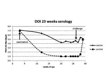
Figure 1 is a graph depicting serology in a DOI study.
Figure 2A, 2B and 20 are bar graphs depicting qPCR results of viral load in
serum (Fig.
2A), feces (Fig. 2B) and organs (Fig. 20).
Date Recue/Date Received 2021-01-22
7
Figure 3 is a graph depicting average body temperature of animals pre- and
post-
vaccination.
Figure 4 is a graph depicting average results of the total PCV2 Ig antibody
response.
Figure 5 is a graph depicting average results of the PCV2 IgM antibody
response.
Figure 6 is a graph depicting anti-PCV2 titre levels in serum.
Figures 7 and 8 are bar graphs depicting viral loads in organs of tested
animals.
Example 1
EXPERIMENTAL DESIGN
Progeny of 10 sows with antibodies against PCV2 were used for this study.
Piglets were
divided across litters into 2 groups of 15 animal animals. At 3 weeks of age,
the piglets
of group 1 were vaccinated intradermally on the right side of the neck with
0.2 ml of a
vaccine comprising recombinantly expressed ORF2 protein of porcine circo virus
type 2
(see WO 2007/028823 for the provision of the protein), using the commercially
available
intradermal vaccination device !DAL (available from MSD Animal Health,
Boxmeer,
The Netherlands), while group 2 was left unvaccinated and served as a control
group.
All study animals were observed daily for clinical signs. Blood samples of all
animals
were taken at time of vaccination, 9,17, 19 and 21 weeks later. Twenty-three
weeks
following vaccination each animal was challenge infected using a wild-type
PCV2
challenge virus strain applied intranasally.
Serum samples and fecal swabs were taken one day before challenge and one, two
and
three weeks after challenge and were examined for PCV2 viral nucleic acid by
quantitative PCR. In addition serum samples were examined for PCV2 antibodies.
Three weeks following challenge, all animals were necropsied and inguinal
lymph node,
tonsil and lung were sampled for determination of PCV2 viral antigen and
nucleic acid.
The vaccine used was given as an oil-in-water emulsion, comprising 5% v/v of
the
mineral oil Marco10 52 (Exxon), 0.30% w/v vitamin E acetate and 0.32%
Polysorbate 80
(TweeInm80; Sigma Aldrich), water for injection and 2000 AU of PCV2 protein
per 0.2 ml.
The AU units are calculated based on an AlphaLISA test of PerkinElmer. For
this test
the wells of a polystyrene microtitre-plate are filled with serial dilutions
of test sample
containing PCV2 ORF2 antigen alongside serial dilutions of a reference
standard.
These dilutions are Incubated with acceptor-beads (coated with monoclonal
antibody
directed against PCV2 ORF2), and biotin-labeled secondary antibody which is
also
Date Recue/Date Received 2021-01-22
CA 02931136 2016-05-19
WO 2015/082457 PCT/EP2014/076223
8
directed against PCV2 ORF2. The amount of bound secondary antibody is then
quantified by incubation with streptavidin coated donor-beads and
chemiluminescent
detection. The reference standard is such that the commercially available
vaccine
Porcilis PCV is set to contain 5000 AU per (2 ml) dose.
EXPERIMENTAL PROCEDURE
Daily observation
All pigs were observed daily for clinical signs of disease. Observations
consisted of
systemic reactions including loss of appetite, tendency to lie down,
listlessness or
drowsiness, shivering, bristling, oedema (especially around the eyes),
vomiting,
diarrhoea and dyspnoea.
Sampling of blood
Blood samples were collected before vaccination, 9, 17, 19 and 21 weeks later.
Blood
samples were collected one day before challenge and 7, 13 and 19 days after
challenge. This was done from all pigs individually.
Fecal swabbing
Fecal swabs were taken from all animals, using one dry swab per animal, one
day
before challenge, 7, 13 and 18 days post challenge. Swabs were taken using
standard
procedures, into medium containing antibiotics. Suspensions of swabbed
material in
medium was clarified by centrifugation, aliquotted and stored at -18 C until
further
use.
Serology
All serum samples were examined for antibodies against PCV2, using standard
ELISA
procedures. In brief, serially diluted serum samples were incubated on
microtiter plates
coated with baculovirus expressed PCV2 ORF2 antigen. After removing the sera,
all
wells were incubated with a fixed amount of biotin-labeled PCV2-specific
monoclonal
antibody. Bound MoAb was then incubated with peroxidase-conjugated
streptavidin
followed by chromophoric detection. Titers were expressed as 10g2 titers.
Postmortem examination
At the end of the experiment all animals were euthanized by bleeding following
CA 02931136 2016-05-19
WO 2015/082457 PCT/EP2014/076223
9
stunning. During necropsy the animal was opened and the viscera are inspected
in-situ,
paying particular attention to the following organs: lungs, inguinal and
mesenteric lymph
nodes, tonsils, thymus, spleen, liver and kidneys. Following this, samples
from tonsil,
lung (lobus accessories), and inguinal lymph node were removed for quick
freezing and
later analysis by quantitative PCR (qPCR).
Quantitative PCR
Quantitative PCR (qPCR) was performed on all sera and fecal swabs, and on 10%
tissue homogenates of tonsil, lung and inguinal lymph nodes. In brief, DNA was
extracted from the samples using a commercial kit. PCV2 genomic DNA in each
sample
was quantified by polymerase chain reaction (PCR), using primers and a dually
labeled
hydrolysis probe specific for PCV2. The cycle number where specific
fluorescence
exceeded the threshold was correlated with the cycle numbers for a set of
samples
containing known amounts of a PCV2-containing plasmid. Results were expressed
as
10g10 copies/p1 of reaction mixture (10g10 c/p1).
RESULTS
At the start of the experiment all animals were found to be healthy. In the
control group
one animal was found dead at 6 weeks post vaccination (wpv). Two vaccinated
animals
had slight local problems, viz, a slight motional dysfunction (stiffness in
one leg). Given
the low problem, these animals were not treated. None of the vaccinated
animals
showed any signs of disease or systemic reactions such as hyperthermia,
reduced feed
intake, anaphylactic shock or vomiting.
The results of the serology are given in Figure 1. It is clear that the
vaccinated animals
keep an anti-PCV2 titer that seems to level out to about 4.0 10g2, whereas in
the control
animals the titer decreases below the detection limit. After challenge (23
wpv, at an age
of 26 weeks), titers slightly rise in the vaccinated group. In the control
group titers rise to
the same level.
The qPCR results are shown in Figures 2A, 2B and 20 ("dpc" = days post
challenge). It
appears that the vaccinated animals, 23 weeks after vaccination were protected
from
challenge infection with PCV2, as shown by the significant reduction of PCV2
nucleic
acid in serum, lymphoid organs and lung. Furthermore, the vaccine was capable
to
CA 02931136 2016-05-19
WO 2015/082457 PCT/EP2014/076223
reduce the viral shedding as demonstrated by a significant reduction of the
viral load in
fecal swabs against PCV2 of at least 23 weeks. This was done in field animals,
having
circulating anti-body titers against PCV2 of approximately 7 log 2, which is
considered a
medium level.
5
Example 2
EXPERIMENTAL DESIGN
A total of 46 piglets from one farrowing batch were allotted to 4 treatment
groups: two
vaccinated groups of 13 piglets each and two control groups of 10 piglets.
Group one
was vaccinated as indicated above under Example 1 when the piglets were
approximately two weeks old, group two was vaccinated when the piglets were
approximately three weeks old. The piglets were intradermally vaccinated in
the right
side of the neck with a single dose of vaccine. Groups 3 (control group 2 week
old
animals) and 4 (control group 3 week old animals) were not vaccinated. Serum
samples
were collected from all animals on the day of vaccination, 2, 3 and 4 weeks
after
vaccination. Temperatures were taken one day before vaccination, at the day of
vaccination and four hours later and at 1, 2, 3, 4 days post vaccination.
EXPERIMENTAL PROCEDURE
Before vaccination, the piglets were observed for general health. Body
temperatures
were taken of all piglets, on day T= -1, day T=0 at 0 and 4 hours after
vaccination, and
on day T=1, 2, 3, 4 post vaccination.
Blood samples were collected on the day of vaccination and 2, 3 and 4 weeks
later. This
was done from all pigs individually according to standard procedures. The
blood
samples were collected without the addition of anti-coagulant. Serum was
prepared
from the clotted blood samples and aliquots were filled and stored at -20 C
until
analysis.
Total PCV2 Ig antibody and PCV2 IgM antibody ELISA were tested as indicated
here
above under Example 1 ("Serology"), except that in the case of IgM antibody
ELISA, the
CA 02931136 2016-05-19
WO 2015/082457 PCT/EP2014/076223
11
plates were coated with IgM antibody and thereafter incubated with PCV2 ORF2
antigen, before incubation with a fixed amount of biotin-labeled PCV2-specific
monoclonal antibody.
RESULTS
At the start of the experiment all animals were found to be healthy. Average
results of
the body temperatures are shown in Figure 3. No difference could be seen in
the
average increase in body temperature between either the two and three week old
animals (maximum average increase was between 0.0 - 0.3 C). Also, the maximum
increase in body temperature of individual animals in group 1 and in group 2
was
comparable to the maximum temperature increase of individual animals in the
two
control groups.
Total PCV2 Ig antibody ELISA
Average results of the total PCV2 Ig antibody response are summarized in
Figure 4.
At the time of vaccination, piglets vaccinated at 2 weeks of age had higher
(most likely
maternally derived) PCV2 antibody titers than piglets vaccinated at 3 weeks of
age. The
vaccinated animals showed an increase in titer considerably higher than the
control
animals.
PCV2 IgM antibody ELISA
Average results of the PCV2 IgM antibody response are shown in Figure 5. At
the time
of vaccination all animals were negative for IgM antibodies. Following
vaccination the
three week old animals had a considerable faster and higher IgM antibody
response
than the two week old animals. The control animals remained negative
throughout the
study.
Based on these results it may be concluded that the one dose intradermal
vaccination of
piglets at 2 and 3 weeks of age resulted in an acceptable safety profile and a
good
serological response. Comparable results were obtained with another experiment
(data
not shown) where the starting level of circulating anti-body titers was even
higher, viz.
up to 9.4 log 2, which is considered to be at the high end of a medium range.
CA 02931136 2016-05-19
WO 2015/082457 PCT/EP2014/076223
12
Example 3
EXPERIMENTAL DESIGN
A total of 30 piglets were allotted to three treatment groups of 30 piglets
each. Piglets
from group 1 were intradermally vaccinated with a single dose of vaccine as
indicated
hereabove under Example 1. Piglets from group 2 were intramuscular vaccinated
with a
single dose of the same vaccine, in the same amount at the same place (in the
neck),
and piglets from group 3 were left untreated. Serum samples were collected
from all
animals on the day of vaccination, three and five weeks after vaccination.
EXPERIMENTAL PROCEDURE
Before vaccination, the piglets were observed for general health, according to
standard
procedures. Sampling of blood and serology of total anti-PCV2 antibodies and
PCV2
ORF2 specific IgM antibodies was done according to the procedure as indicated
here
.. above under Example 2.
RESULTS
At the start of the study all animals were found to be healthy. Results of the
serology are
summarised in Table 1 (titers expressed as 10g2). At the time of vaccination
mean
antibody titers were relatively high. Following vaccination, none of the
animals showed
an increase in PCV2 Ab titer. At 3 and 5 weeks post vaccination, in the ID
group higher
mean PCV2 antibody titers than in the IM group could be observed.
Results of the anti PCV2 IgM serology are summarised in Table 2 (titers
expressed as
10g2). At three weeks post vaccination anti PCV2 IgM antibody titers of the ID
group was
considerably higher than of the IM group and the control group.
CA 02931136 2016-05-19
WO 2015/082457 PCT/EP2014/076223
13
Table 1: Average antibody results
Groups Titer 0 wpv Titer 3 wpv
Titer 5 wpv
1 8.2 7.3 6.2
2 8.6 6.5 5.1
3 8.4 6.2 4.3
Table 2: Average anti PCV2 IgM antibody results
Group IgM titer 3 wpv
1 12.7
2 3.4
3 1.0
When applying the Wilcoxon rank sum test, the p value for the difference in
IgM
.. response for the ID group versus the IM group was 0.0001.
Example 4
Progeny of several sows with antibodies against PCV2 were available for this
study.
Piglets were divided across litters into 3 groups of 18 animals. At 3 weeks of
age, the
piglets of group 1 and 2 were vaccinated intradermally as indicated here above
under
Example 1. The animals in group 2 were vaccinated intradermally at the same
time with
the commercially available inactivated Mhyo vaccine Porch's M Hyo ID Once
.. (containing an Mhyo bacterin) according to manufacturer's instructions at
the other side
of the neck. Animals of group 3 (control group) remained untreated. All study
animals
were observed daily. Serum samples were taken at the time of vaccination and
every
other week until animals were sent to slaughter (23-25 weeks of age). These
samples
were examined for PCV2 antibodies.
Experimental procedures were as indicated here above under Example 1. The
resulting
serology is shown in figure 6. From this figure it becomes clear that the anti-
PCV2 titers
remain well above the level of 4 log 2 as established with the experiments as
described
14
under Example 1 and found to be protective. There is no indication of negative
interference between the vaccines.
This experiment was repeated to check protection against virulent Mycoplasma
hyopneumoniae. For this repeat experiment sixty piglets were used. Forty
animals were
vaccinated at the age of 18-24 days with the Mhyo vaccine and twenty of these
animals
were also vaccinated with the PCV vaccine. Twenty animals were not vaccinated
and
served as challenge controls. Three weeks after vaccination all animals were
infected
with a virulent M. hyopneumoniae strain and three weeks post-challenge all
animals
were post-mortem investigated for lung lesions. Lung lesion scores (LLS) were
compared between the groups.
The LLS for the groups vaccinated with Porcilis0 M Hyo ID Once were
significantly
lower than those of the control group (p<0.05, Dunn's test). There was no
significant
difference between the groups that had been vaccinated with Porcilis0 M Hyo ID
Once
alone or in association with the PCV vaccine. It may thus be concluded that
the
combined vaccination has no negative effect on the immunity obtained with
Porcilis0 M
Hyo ID Once.
Example 5
In total eight vaccines were formulated containing PCV2 ORF2 protein (250 to
6000
AU/0.2 ml), M hyo (at the same level as in Porcilis Mhyo ID Once) and
Lawsonia
antigen (see WO 20089/127684, example 2 for the killed whole cells antigens:
at a level
of approximately 1x10⁹ cells per 0.2 ml). The vaccines contained different
adjuvants.
TM
Some vaccines used the existing biodegrable oil containing adjuvant Diluvac
Forte
(MSD Animal Health, Boxmeer, The Netherlands; called 'OF"). Others used the
adjuvant formulation as described here above under Example 1 (called "X-solve
12"), or
adjuvants formulated with the same constituents as X-solve 12, but at half of
the
concentrations (called "X-solve 6''), or 21/2 times the concentrations as in X-
solve 12
(called "X-solve 30"). The resulting vaccines were as follows (the Mhyo and
Lawsonia
antigens are not recited; content per dose):
Group 1: 2000 AU PCV2/X-solve 30
Group 2: 250 AU PCV2/X-solve 12
Date Recue/Date Received 2021-01-22
CA 02931136 2016-05-19
WO 2015/082457 PCT/EP2014/076223
Group 3: 500 AU PCV2/X-solve 12
Group 4: 2000 AU PCV2/X-solve 12
Group 5: 2000 AU PCV2/X-solve 6
Group 6: 500 AU PCV2/DF
5 Group 7: 2000 AU PCV2/DF
Group 8: 6000 AU PCV2/DF
The progeny of 8 sows were used for this study. The piglets had moderate
(medium
10 level) maternally derived antibodies (MDA) against PCV2 (average: 6.7
10g2). At
three/four weeks of age the piglets from groups one through eight were
vaccinated
intradermally with a single dose, using an !DAL vaccinator. Piglets from
group nine
remained unvaccinated. At seven weeks post vaccination all animals were
transported
to the challenge facilities. One day later all animals were challenge infected
with a
15 challenge strain of PCV2. All piglets were observed daily for clinical
signs.
Local reactions were monitored by palpation, starting on the day of
vaccination and
every two days after vaccination until twenty days post vaccination.
Blood samples were collected from all animals on the day of vaccination and
three
weeks later, one day before challenge, 1 and 2 weeks later and at the time of
necropsy.
Serum samples taken from each animal were tested for antibodies against PCV2
and M
hyo. During necropsy, mesenteric and inguinal lymph node, tonsil and lung were
sampled for quantification of PCV2 nucleic acid.
Experimental procedures were the same as described here above under Example 2.
PCV2 IgM antibody response at time of vaccination was below detection level
for all
groups (below 2.0 log 2). At 3 weeks post vaccination the PVC antibody titer
was the
highest for the 2000AU PCV2/X-solve 30 vaccine, viz. 20 log 2. The X-solve 12
groups,
comprising 250-, 500- and 2000 AU PCV2 antigen per dose had a titer of 9, 16
and 19
log 2 respectively. The group that received the 2000 AU/X-solve 6 vaccine had
a titer of
15 log 2. The DF groups comprising the 500-, 2000- and 6000 AU of PCV2 antigen
per
dose had a titer of 8, 14 and 16 log 2 respectively. The controls had a titer
below
detection level.
In this study, systemic and local reactions were assessed. No systemic
reactions
attributable to vaccination were observed. As far as local effects are
concerned, no
more than three vaccinated animals (having received the X-solve 12 500 and
2000 AU,
CA 02931136 2016-05-19
WO 2015/082457 PCT/EP2014/076223
16
or DF 6000 AU PCV2 per dose vaccine respectively) had slight motility
problems, the
same number as in the control animals. Therefor these reactions may reasonably
be
regarded as unrelated to the vaccination. With regard to other local
reactions, many
animals (between about 60-100%) vaccinated with X-solve showed local
reactions, the
average size of the swellings was less than 3 cm, viz. between 1-2 cm, and the
swellings disappeared within 2-6 days. Using DF, only about 30% of the animals
showed local swellings, the mean size being less than 0.5 cm, and they
disappeared
within a day.
In figures 7 and 8 the viral load in the organs (averaged) is depicted for the
various
vaccines. It appears that all vaccines are able to substantially (in these
cases at least 3
logs) reduce the viral load in the relevant organs.
In this study it appeared that all adjuvants used were safe, induced an anti
PCV2 IgM
.. response and were able to treat an animal against an infection with
pathogenic porcine
circo virus type 2. No negative interference between the different antigens
was found.
Lawsonia serology (not shown) shows a good antibody response, based on which
it is
believed that protection against an infection with Lawsonia intracellularis
was obtained.
In order to confirm this, a next experiment including a challenge with
pathogenic
Lawsonia intracellularis was conducted (see Example 6).
Example 6
In order to confirm that animals are protected against a challenge with
Lawsonia
intracellularis, vaccine formulation with different amounts of the adjuvant X-
solve, as
described in Example 5, were newly formulated for various challenge
experiments. The
basis for these vaccines was a PCV2 vaccine containing PCV2 ORF2 protein. A
first
vaccine was formulated in X-solve 30 as indicated in Example 5. A second
vaccine was
formulated in X-solve 12, wherein Lawsonia antigen was introduced by adding
freeze-
dried killed Lawsonia cells (the same antigen as used in Example 5) to the
ready-to-use
PCV2 vaccine within 30 minutes before administration. The end concentration of
PCV2
ORF2 protein in both vaccines was 2000 AU/0.2 ml. The concentration of
Lawsonia
antigen was the same as used for the experiments as described in Example 5
(about
1x10⁹ cells per 0.2 ml). The resulting vaccines were as follows:
CA 02931136 2016-05-19
WO 2015/082457 PCT/EP2014/076223
17
1: 2000 AU PCV2/Lawsonia/X-solve 30
2: 2000 AU PCV2/Lawsonia/X-solve 12
A first study was performed using vaccine number 1 (X-solve 30). Thirty-nine
pigs were
used, allotted to two groups of 19 and 20 pigs respectively. Both groups were
vaccinated at the age of three weeks with 0.2 ml of the vaccine by intradermal
vaccination in the neck as indicated in Example 1. The first group was
vaccinated with
vaccine number 1 as indicated here above, the second group with the same
vaccine but
without the Lawsonia antigens. This group served as a control for the Lawsonia
challenge. All animals were challenged at the age of 22 weeks. Unacceptable
safety
issues were not seen. The results regarding average daily weight gain (during
days 14-
21 post challenge), ileum scores (3 weeks after challenge; the score is
proportional to
the presence of ileum lesions due to the presence of a Lawsonia infection) and
PPE
incidence are indicated in Table 3. Statistically different values (two-sided
tests, p< 0.05;
ANCOVA test for ADWG, cumulative logit model for the Ileum score and Fischer's
exact
test for PPE incidence) are indicated with an asterisk.
Table 3
Vaccine ADWG in kg Ileum score PPE incidence
Vaccine 1 1.100* 50* 4/19*
Control vaccine 0.886 83.5 11/20
A second study was performed using vaccine number 2 (X-solve 12). Fifty pigs
were
used, allotted to two groups of 25 pigs each. One group was vaccinated at the
age of
three weeks with the vaccine indicated here above as number 2 with 0.2 ml of
this
vaccine by intradermal vaccination in the neck as indicated in Example 1. The
second
group was not vaccinated and served as a control. All animals were challenged
at the
age of 24 weeks. The results regarding average daily weight gain (during days
13-20
post challenge), ileum scores (3 weeks after challenge; the score is
proportional to the
presence of ileum lesions due to the presence of a Lawsonia infection) and PPE
incidence are indicated in Table 4. Statistically different values (two-sided
tests, p< 0.05;
ANCOVAtest for ADWG, cumulative logit model for the Ileum score and Fischer's
exact
test for PPE incidence) are indicated with an asterisk. During the test in
each group 1
animal had to be euthanized due to a-specific, non vaccine related problems.
CA 02931136 2016-05-19
WO 2015/082457 PCT/EP2014/076223
18
Table 4
Vaccine ADWG in kg Ileum score PPE incidence
Vaccine 2 1.001* 129* 11/24*
None -0.053 241 22/24
The results show that the intradermal vaccination of an animal with a combined
vaccine
comprising PCV2 ORF2 protein and killed Lawsonia intracellularis bacteria
provide
protection against an infection with pathogenic Lawsonia intracellularis.
Also, the freeze-
drying of Lawsonia antigen prior to formulation appears to have no negative
effect on
efficacy.