Note: Descriptions are shown in the official language in which they were submitted.
WO 2015/077333 PCT/US2014/066405
SYSTEM AND Method for SPERM SORTING
CROSS-REFERENCE TO RELATED APPLICATIONS
[0001] This application is based on
US Provisional Application Serial No.
61/906,740, filed November 20, 2013, and entitled, "SYSTEM AND METHOD FOR
SPERM SORTING."
STATEMENT REGARDING FEDERALLY SPONSORED RESEARCH
[0002] N/A
BACKGROUND OF THE INVENTION
[0003] The present invention relates generally to systems and methods for
sperm
sorting.
[0004] According to estimates, there are more than 70 million infertile
couples
worldwide. Approximately 1 in every 4 infertile couples seek clinical
treatment, where,
according to sources, male factor may account for about 50 percent of the
infertility
cases. Assisted reproductive technology (ARTs), such as in vitro fertilization
(IVF),
intracytoplasmic sperm injection (ICSI), and intrauterine insemination (IUI),
are
generally utilized in reproductive clinics to treat infertile couples. With an
increasing rate
of male infertility due to environmental and physiological conditions, there
is an ever
growing need for the use of ARTs in reproductive clinics. Isolation of the
most motile
and morphologically normal sperm is an integral process to commonly used
IVF/ICSI
procedures. Selection of healthy sperm from unprocessed semen (stock sperm) is
crucial as it requires selecting sperm that is not only highly motile, but
also has a normal
morphology, mature nuclei, and lesser reactive oxygen species (ROS)
production.
Although current IVF/ICSI procedures results in successful pregnancy
approximately 50
percent of the time, the output can be greatly compromised if the sperm being
selected
are abnormal.
[0005] Currently, the more commonly-known ART techniques use centrifugation
based sperm swim-up, density gradient separation methods, and microfluidic
based
methods with/without the use of chemotaxis to sort sperm. These techniques
have
potential drawbacks and limitations in their use for procedures as delicate as
IVF/ICSI.
-1-
CA 2931201 2019-02-05
CA 02931201 2016-05-19
WO 2015/077333 PCT/US2014/066405
11 is worth noting that the centrifugation based sperm sorting techniques,
such as swim-
up, compromise on sperm quality during the repetitive centrifugation steps.
Quality of a
sperm sample is degraded during swim-up technique due to ROS generation. ROS
exposure can greatly harm the DNA of seemingly motile and healthy sperm.
Furthermore, the centrifugation-based sperm sorting techniques are labor
intensive, and
outcome can vary from technician to technician.
[0006] Sperm sorting technologies based on microfluidics have an advantage
because they can precisely handle small volume of sperm samples. On the other
hand,
microfluidic-based sperm sorting devices have very low throughput and can only
process small semen volumes, such as 2p1 ¨ 50p1, which limits their
application to
reproductive clinics, where normal sperm sample can have volume of ...1.5m1.
[0007] In a clinical ICSI procedure, an embryologist will have on average
20
oocytes that can be handled in four petri dishes, and will need 20 sperm. The
embryologist would like to choose these 20 sperm in an oligospermic sample
among a
few hundred sperm. Such scenario would require real-time monitoring of
individual
sperm and collection from outlet when 20 sperm reach the outlet, which is not
attainable
using current clinical or microfluidic technologies. In a second procedure,
where an
embryologist is handling healthy samples, in vitro fertilization is performed
using 0.5
million healthy sperm suspended in a 5-20 pl suspension to be introduced to an
oocyte.
However, current sorting systems, such as described above, do not provide the
throughput needed to meet these criteria.
[0008] Traditionally, optical microscopes have been used to image sperm
for
computer assisted sperm analysis (CASA) and manual identification of sperm
motility
for ARTs. This classical approach has limitations in tracking a large number
of sperm
simultaneously due to its small field of view (FOV). In addition, sperm
tracking and
motility analyses are performed after sorting. Currently no system exists that
can sort
and analyze sperm simultaneously.
[0009] It would therefore be desirable to provide a system and method for
processing, including as sorting, sperm without damaging the sperm or
subjecting the
sperm to potentially-damaging conditions. Furthermore, it would be desirable
to provide
a system and method that can analyze sperm, but is efficient and able to
scale.
-2-
CA 02931201 2016-05-19
WO 2015/077333 PCMJS2014/066405
SUMMARY OF THE INVENTION
[0010] The
present invention overcomes the aforementioned drawbacks by
providing a system and method that integrates micro- and macro- fluidics to
sort sperm
in a manner that allows efficient selection of sperm that are favorably suited
to
fertilization. In
particular, the present invention recognizes that sperm suited to
fertilization is most desirable and can be selected or sorted using a system
presents
and environment that is akin to that presented in the fertilization process.
In this regard,
a system is provided where macro reservoirs are connected by micropores to
approximate the female genital track. A system and method is provide whereby
the
most motile, morphologically normal, mature, and functional sperm pass
selectively
through the micropores against gravity leaving behind dead or less functional
sperm.
The present invention is a chemical-free, centrifugation-free, and flow-free
technology,
where functional sperm are isolated from unprocessed semen sample with high
retrieval
rate.
[0011] In
accordance with one aspect of the invention, a system for sorting sperm
is provided that includes a housing and a microfluidic system supported by the
housing.
The system also includes an inlet providing access to the microfluidic system
to deliver
sperm to the microfluidic system and an outlet providing access to the
microfluidic
system to harvest sorted sperm from the microfluidic system. The microfluidic
system
provides a flow path for sperm from the inlet to the outlet and includes at
least one
channel extending from the inlet to the outlet to allow sperm delivered to the
microfluidic
system through the inlet to progress along the flow path toward the outlet.
The
microfluidic system also includes a filter including a plurality of micropores
and arranged
in the flow path between the inlet and the outlet to cause sperm traveling
along the flow
path to move against the filter and gravity to reach the outlet.
[0012] In
accordance with another aspect of the invention, a method for sorting
sperm is disclosed that includes delivering a sample of sperm to an inlet
connected to a
microfluidic system and allowing sperm in the sample of sperm to traverse a
flow path
through the microfluidic system toward an outlet providing access to the
microfluidic
system to harvest sorted sperm from the microfluidic system. The method also
includes
filtering the sperm prior to reaching the outlet using a filter having a
plurality of
-3-
CA 02931201 2016-05-19
WO 2015/077333 PCMJS2014/066405
micropores and gravity to restrict movement of the sperm through the filter .
The
method further includes harvesting sperm passing to the outlet after passing
through the
filter and overcoming gravity.
[0013] The foregoing and other aspects and advantages of the invention will
appear from the following description. In the description, reference is made
to the
accompanying drawings which form a part hereof, and in which there is shown by
way
of illustration a preferred embodiment of the invention. Such embodiment does
not
necessarily represent the full scope of the invention, however, and reference
is made
therefore to the claims and herein for interpreting the scope of the
invention.
BRIEF DESCRIPTION OF THE DRAWINGS
[0014] Fig. 1A is a plan view of a sperm sorting system in accordance with
the
present invention.
[0015] Fig. 1B is a cross-sectional view of the system of Fig. 1A.
[0016] Fig. 1C is a schematic view of multichannel system with a collection
chamber to concentrate the sorted sperm.
[0017] Fig. 1D is a schematic view of multi-well system with multiple
channels
connecting the inlet and collection or concentration chamber chamber.
[0018] Fig. 2A is an exploded, cross-sectional view of a sperm sorting and
imaging system in accordance with the present invention.
[0019] Fig. 2B is a detailed, perspective view of a microfluidic system of
Figs. 1A
or 2A.
[0020] Fig. 2C is a schematic view of a multi-channel microfluidic system
in
accordance with the present invention.
[0021] Fig. 2D is a cross-sectional view of overall system for sperm
sorting and
imaging system in accordance with the present invention.
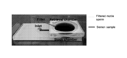
[0022] Fig. 3 is a perspective view of a prototype system for sorting sperm
in
accordance with the present invention.
[0023] Fig. 4 is a series of images of sperm acquired using the present
invention.
[0024] Fig. 5A is graph illustrating motility of human sperm isolated using
different
pore diameter filters and retrieved at different time points
-4-
CA 02931201 2016-05-19
WO 2015/077333 PCMJS2014/066405
[0025] Fig. 5B is a graph illustrating retrieval rate of sorted sperm
using different
chips.
[0026] Figs. 6A, 6B, and 6C are graphs illustrating curvilinear velocity
(VOL),
straight line velocity (VSL), and average path velocity (VAP) of stock and
sorted sperm
using 3, 5, and 8 pm MMSS chips, respectively.
[0027] Fig. 7 is a graph showing normal morphology (%) for stock and
sorted
sperm.
[0028] Fig. 8 is a graph showing mature sperm percentage calculated for
stock
and sorted sperm.
[0029] Fig. 9A is a graph showing sperm sorted using 3, 5, and 8pm filter
devices
showed significantly lesser ROS generation compared to swim-up and washing
methods.
[0030] Figs. 9B through 9G are reactive oxygen species (ROS) generation
graphs for (B) Semen sample, (C) Washed sperm, (D) Sperm sorted using swim-up
method (ROS region is highlighted by circle), (E) Sperm sorted using 3pm MMSS
chip,
(F) Sperm sorted using 5pm MMSS chip, and (G) Sperm sorted using 8pm MMSS
chip.
[0031] Fig. 10A is a graph showing sperm sorted using 5 and 8pm MMSS chips
showed significantly lesser DNA fragmentation compared to swim-up and unsorted
semen sample.
[0032] Figs. 10B through 1OF are DNA fragmentation scatter plots for (B)
Semen
sample, (C) Sperm sorted using swim-up method, (D) Sperm sorted using 3pnn
MMSS
chip, (E) Sperm sorted using 5pnn MMSS chip, and (F) Sperm sorted using 8pm
MMSS
chip.
[0033] Fig. 11 is a flow chart setting forth an example of some steps in
accordance with the present disclosure.
DETAILED DESCRIPTION OF THE INVENTION
[0034] The present invention recognizes that the vaginal mucus becomes
watery
and forms tiny nnicrochannels that help guide sperm through to the egg. The
present
invention recognizes exhaustion as a mechanism for sorting sperm and has been
experimentally and theoretically demonstrated to leverage exhaustion to sort
healthy
-5-
CA 02931201 2016-05-19
WO 2015/077333 PCMJS2014/066405
sperm using coarse-grained multi-scale simulation. Specifically, the present
invention
provides a macro-micro fluidic sperm sorting (MMSS) system to efficiently,
reliably, and
successfully sort sperm. As will be described, healthy motile sperm is fully
collected at
the outlets post-sorting. This system improves the efficiency of sperm
selection process
with minimal perturbation, thereby controlling against DNA fragmentation,
accumulation
of debris, and generation of ROS.
[0035] In addition, the present invention can simultaneously sort,
monitor, and
evaluate sperm. Specifically, the present system enables evaluation of each
sperm
individually, for example, based on velocity response, using a wide field-of-
view (FOV)
lensless imaging technology. The system provides a microchip-based, wide-FOV,
lensless technology utilizing shadow imaging. Additionally, the present
invention can be
used to harvest morphometrical information, which is a reliable indicator of
male fertility.
[0036] Referring to Fig. 1A, a sperm sorting system 10 is illustrated. The
system
may be a polydimethylsiloxane- (PDMS) based, polymethylmethacrylate- (PMMA)
based, or other microfluidic system. The system 10 includes a housing 12
having an
inlet 14 and a collection chamber 16 having a filter 18 arranged therein. The
filter 18
may be a polycarbonate filter or other filter having suitable materials
properties, such as
pore or passage size, as will be described. Referring to Fig. 1B, the inlet 14
and
collection chamber 16 are connected through a passage or flow path 20
extending
along a microfluidic chip 22. As will be described, the microfluidic chip 22
may include a
microchip that may be disposable and that handles unprocessed semen samples
(either
fresh or frozen, processed or raw), for example of 10p1-3m1, and sorts sperm
rapidly,
such as in less than 30 minutes, without the need for complex instrumentation
or trained
operators.
[0037] The flow path 20 extends from the inlet 14 to the collection
chamber 16.
At the collection chamber 16 a first or bottom chamber 24 is located proximate
to the
microfluidic chip 22 and a second or top chamber 26 is located distally with
respect to
the microfluidic chip 22, above the first or bottom chamber 24. As will be
described, the
first chamber 24 is designed to collect the semen of a sample, whether fresh
or frozen,
processed or raw, presented to the inlet 14 and the second chamber 26 is
designed to
filter the motile sperms.
-6-
CA 02931201 2016-05-19
WO 2015/077333 PCMJS2014/066405
[0038] Referring to Fig. 1C, the system described above with respect to
Fig. 1B
may be modified to include an additional collection or "concentration" chamber
25 that is
connected to the top chamber by a fluid connection 27. That is, in this
regard, the
sperm may be concentrated in the collection chamber 25 to facilitate easier
harvesting.
[0039] In another configuration, as illustrated in Fig. 1D, the collection
chamber
25 may be connected through a plurality of channels 28 each having an inlet 29
opposite the concentration chamber 25. In this regard, sperm from multiple
flow paths
20 of Fig. 1C or from multiple collection chambers 16 may be delivered to a
common
concentration chamber 25. Such variations on the above-described design can be
used
to facilitate the use of multiple filters and multiple channels to handle even
larger
volumes or for higher throughput applications.
[0040] Referring to Fig. 2A, an exploded view of one optional
configuration of the
system 10 that includes an integrated imaging system is illustrated. The
imaging
system may form a lensless, wide-FOV imaging platform. In this view,
components of
the integrated imaging system, such as a light 30, an imaging sensor 31, and a
glass
protection layer 32 combined with the above-described system 10. In function,
the light
30 illuminates sperm 34 introduced to the microfluidic chip 22 through the
inlet 14. The
illuminated sperm 34 can be imaged by the imaging sensor 31, which may be a
charge-
coupled device (CCD), complementary metal¨oxide¨semiconductor (CMOS), or other
imaging device. More, specifically, referring to Fig. 2B, in function, sperm
and semen 34
may be introduced into the outlet 14 using, for example, a pipette 36. The
sperm
traverse across a media 38 along the microfluidic chip 22, which may include
the
aforementioned glass 32, as well as a PMMA or other material layer 40, with a
double-
sided adhesive (DSA) layer 42 arranged there between to affix the glass 32 and
PMMA
layer 40 together. Ultimately, the sperm 34 traverse the microfluidic chip 22
to the outlet
16, where a mineral oil 44 may be found. Specifically, a thin layer of
sterile, embryo-
tested mineral oil may be placed on top of the media 38 in the inlet 14 and
outlet 16 to
avoid medium evaporation.
[0041] As illustrated, different channel lengths may be used or selected
for
effective sperm sorting. Furthermore, referring to Fig. 2C, a multichannel
design may
be utilized where the inlet 14 and collection chamber 16 are connected by
multiple
-7-
CA 02931201 2016-05-19
WO 2015/077333 PCMJS2014/066405
channels 46. As illustrated, a PBS collection buffer 48 may also be included,
for
example, to use in washings. Furthermore, the chip substrate housing the
channels
may be disposable.
[0042] Referring to Figs. 2A and 2B, if included, lensless imaging can be
used to
record the shadow image of each individual sperm 34 onto an optoelectronic
sensor
array plane 31. This system 10 targets detecting/counting cells or monitoring
in real-
time the dynamic location of hundreds of thousands of individual cells on-chip
over an
ultra-wide FOV, for example, an FOV that is a few centimeters by a few
centimeters.
This technology provides these features with reduced complexity and ease of
miniaturization.
[0043] One particular example of the system 10 including the imaging
capabilities
is illustrated in Fig. 20. A standard microscope cannot monitor a whole
microfluidic
sorting chip and analyze sperm in real time. This challenge can be addressed
by
integrating lensless imaging with microchannels providing parallel on-chip
monitoring
and counting of sperm. The design permits miniaturization of this technology
to make it
suitable for an embryology/clinical lab and point-of-care settings.
[0044] The system 10, in the example in Fig. 2D, includes the light source
30 that
is directed through an aperture 50 in the housing 12, such as a 50 pm
aperture, to focus
monochromatic light 52 toward a the microfluidic chip 22, across which the
sperm 34
traverse, as described above. The system 10 may be coupled with a computer
system
54 connected through a data connection 56, which may be wired or wireless, and
a
rechargeable battery or other power source 58 coupled through a power
connection 60
to provide operational power for the imaging capabilities.
[0045] In one configuration, a combination of polymethyl-methacrylate
(PMMA) of
1.5 mm thickness and double-sided adhesive (DSA) film of 50 pm thickness could
be
used to create microchannels. The DSA film can be cut to create microchannels
of
different lengths ranging from 5 mm to 40 mm using a laser cutter. Inlet and
outlet ports
extend through the PMMA with a diameter of 0.65 mm and 2 mm, respectively. The
DSA film is then placed directly onto the PMMA in effect joining the two. A
glass slide
is placed onto the other side of the DSA film, such that the height of the
channel is
determined by the adhesive layer thickness. The larger outlet size is
particularly
-8-
CA 02931201 2016-05-19
WO 2015/077333 PCMJS2014/066405
designed to extract sorted sperm out of the channel easily accessible by a
pipette. The
distance between inlet and outlet determined the channel length. The length of
the
channel is defined as the distance between inlet and outlet.
[0046] To increase percentage of motile cells at the outlet and for high
volume
processing, a polycarbonate filter can be integrated into these microchips.
This filter-
based device can be designed using 3 mm thick PMMA cut to an area of 50 mm by
30
mm and another cut to an area of 30mm by 30mm. Cylinders of 20 mm diameter can
be cut into both PMMA components and align vertically onto one another using
150 pm
DSA. A 0.6 mm semen injection inlet is also cut into the larger device
component at a
mm distance. The system can be assembled using a whatman nucleopore filter
located between the two PMMA components.
[0047] Referring to Figs. 1A through 2D, the system 10 can be used in large-
scale semen processing. To do so, the sperm 34 is introduced through the inlet
14 to
be place in the microfluidic chip 22. During this movement, the sperm 34 can
be
imaged using the light 30 and imaging sensor 31. The sperm 34 move toward the
outlet
16. This outlet/collection chamber presents two chambers 24, 26. The first
chamber 24
includes a filter presenting micropores and the second chamber 26 includes
another
filter including micropores. In this regard, the system 10 presents macro
reservoirs 14,
16 connected by micropores to approximate the female genital track. Therein,
the
sperm 34 move collectively, influenced by each other, such as would naturally
occur,
along the medium 38 toward the outlet 16. The most motile, morphologically
normal,
mature, and functional sperm pass selectively through the micropores against
gravity
leaving behind dead or less functional sperm in the first chamber 24. That is,
the sperm
head is of spherical shape and has size of about 3pm x 4.5pm. Sperm tails are
about
45-50pm long. If a filter having micropores of diameter larger than sperm head
is
placed in the first and second chambers 24, 26, only sperm that are motile can
make
their way through the micropores, whereas dead, dying, or damaged sperm cannot
pass
through the micropores because of their long tails. Instead, these dead,
dying, and/or
damaged sperm succumb to gravity and remain in the first chamber 24.
[0048] Thus, a microchip-based system is provided that is designed such
that it
does not require any centrifugation steps to retrieve healthy, motile, and
morphologically
-9-
CA 02931201 2016-05-19
WO 2015/077333 PCMJS2014/066405
normal sperm with minimal ROS generation. The device design makes sperm
sorting
procedure less labor intensive and inexpensive. The system incorporates
utilizes
exhaustion in space-constrained channels as a mechanism for sperm sorting. The
system can isolate motile and morphologically normal sperm without any
centrifugation
step. Thus, a current coarse-grained model of sperm motility is used to model
filter-
based microfluidic devices in three dimensions, incorporating effects of
cooperatively
rising from hydrodynamic interactions between sperm, with channel walls, and
with the
filter surfaces and holes. This model allows the design of device parameters
such as
micropore size and incubation times.
[0049] The design and operation of the above-described system can be
further
appreciated from the following discussion of one example of a system,
configuration for
such system, and testing results of such system. This is but one example and
is non-
limiting in nature to the variety of configurations, designs, and operations
that may be
employed and fall within the scope of the present invention.
[0050] Example
[0051] Assembly of MMSS Chip
[0052] The poly (methyl methacrylate) (PMMA, 3mm thick; McMaster Carr,
Atlanta, GA) and double side adhesive (DSA, 1201.1m thick, St. Paul, MN) were
cut using
a laser cutter (Versa LaserTM, Scottsdale, AZ). The design for the chip was
generated
on Coral Draw4 and implemented onto USLE Engrave software for cutting. Primary
components of the MMSS chip included one 3mm PMMA cut to an area of 50mm x
30mm (bottom chamber) and another cut to an area of 30mm x 30mm (top chamber).
A
0.6mm injection point was also cut into the bottom PMMA sheet at a 5mm
distance from
the chambers. Cylinders of 20mm diameter were cut into both PMMA components.
The bottom PMMA chamber was first attached to glass slide using DSA. Top PMMA
chamber was aligned and attached with bottom chamber using DSA. The
NucleporeTM
track-etched polycarbonate membrane filters (Whatman Ltd, 25mm diameter, 3 m,
51.im, 811m) were sandwiched between two PMMA chambers during chip assembly.
Thus, it was considered that at least 1 urn and less than 10 um may be a range
of
advantageous pore sizes. A perspective view of the assembled chip is shown in
Fig. 3.
[0053] Sperm Sorting using MMSS Chip
-10-
CA 02931201 2016-05-19
WO 2015/077333 PCMJS2014/066405
[0054] Thawed, unprocessed semen sample (stock sperm) was injected into the
inlet of MMSS chip until it filled the first/bottom chamber. The first/bottom
chamber was
designed to hold up to 560111 of the semen sample. In another set of
experiments, the
stock semen sample was diluted 4 times with 1 percent bovine serum albumin
(BSA) in
human tubal fluid (HTF) before injection into MMSS chip. Following injection,
the
first/upper chamber was topped off with 560[11 of 1 percent BSA in HTF. Chips
were
then stored at 37 degrees C in incubator for 15, 30, 45, and 60 min intervals
before fluid
from top chamber was collected into eppendorph tubes for analysis.
[0055] Concentration and Motility Analysis
[0056] A standard Makler Haemocytometer was used to analyze the sperm
samples for concentration and motility using optical microscope. Briefly, 1p1
of sperm
sample was pipetted onto Makler Haemocytometer and covered with cover-lid
provided
with Haemocytometer. Sperm were counted by personnel familiar with method
using a
click-counter for at least three times. The sperm that were moving forward
were
considered motile.
[0057] Viability Analysis
[0058] The sperm samples were analyzed for viability using LIVE/DEADO Sperm
Viability Kit (L-7011, Molecular Probes ). SYBR 14 dye was used to stain live
whereas
Propidium Iodide (P1) was used to stain dead sperm. Samples were stained
according
to manufacturer's protocol. Briefly, first SYBR 14 dye was added into sperm
sample to
the final concentration of 100nM. The sample was incubated for 5 min at 37 C.
To
stain the dead sperm, PI dye was added to the sample to the final
concentration of
10kiM and allowed to incubate for 5 additional min. The sperm samples were
smeared
on a glass cover slip and imaged using fluorescent microscope Zeiss Axio
Observer.Z1.
Green and red emission filters were used for SYBR 14 and PI, respectively.
[0059] Velocity Measurement
[0060] Sperm samples were analyzed using the method described by WHO
laboratory manual for sperm analysis. Briefly, sperm was retrieved from the
MMSS
chips (3pm, 5pm, 8pm) after 30 min. Slides were prepared by putting 6p1 of
sperm
sample onto a glass slide and covered by using a 18x18mm cover slip to give
the
sample a depth of 20.7pm. To avoid drying up of samples, slides were made
-11-
CA 02931201 2016-05-19
WO 2015/077333 PCMJS2014/066405
periodically, not simultaneously. Each slide was analyzed under 20x (Carl
Zeiss) using
light microscopy with live images of the sample being projected onto a
computer
monitor. Using a video capturing software (Snagit, TechSmith), movement of
sperm
samples were captured at random locations for 5 secs. Videos were converted to
image
sequences using VideotoJpeg software at 100fps. The image sequence was input
into
ImageJ (National Institute of Health, http://rsbweb.nih.gov/iy) for analysis
using the
CASA plugin to monitor sperm velocity parameters, i.e. straight line velocity
(VSL),
curvilinear velocity (VCL), and average path velocity (VAP).
[0061] Sperm Morphology Assessment
[0062] Recovered sperm suspension from 5pnn, and 8pm MMSS chips were
collected after 30 mins. Sperm retrieved from 3pm MMSS chip were not analyzed
for
sperm morphology, as sperm concentration is too low for morphology analysis. A
10pL
sperm suspension was then taken and placed on a clean and sterile microscope
slide
and feathered smears were prepared. Smears were air dried and prepared for
fixation.
Spermac staining protocol similar to the one provided by FertiPro was followed
to
stain sperm for morphology assessments. Briefly, dried smears were submerged
into
Spermac fixative solution for at least 5 min and then rinsed with DI water.
Stain A was
pipetted at one edge of the slides and allowed to flow over the smear. Slides
were then
placed on a flat surface and allowed to soak with stain for 1 min. The slides
were then
rinsed with DI water twice. Next, stain B was applied similarly to Stain A and
allowed to
penetrate sperm for 1 min. This was followed by a single rinse with DI water.
Finally,
stain C was pipetted over the smear and allowed to sit for 1 min before
rinsing with DI
water. At this point, at least 100 sperm were imaged using oil immersion and
100X
objective (N (no. of repeats) = 3). The sperm was considered morphologically
normal if
it falls within WHO morphology criteria (Head: spherical head; acrosome
covering 40-
70% of head area; head length 3.7-4.7pm; head width 2.5-3.2pm; length-to-width
ratio
1.3-1.8; no more than 2 small vacuoles; post-acrosome region should not
contain any
vacuole. Midpiece: no residual cytoplasm in midpiece; length of midpiece
should be
approximately same as head length; no broken neck. Principal piece: no sharp
angles
or bends indicative of tail break; thinner than midpiece, length of principal
piece should
be approximately 10 times the head length).
-12-
CA 02931201 2016-05-19
WO 2015/077333 PCMJS2014/066405
[0063] Sperm Maturity Assessment
[0064] Recovered sperm suspension from 5, and 8pm MMSS chips were
collected after 30 mins. Sperm retrieved from 3pm MMSS chip were not analyzed
for
nuclear maturity, as sperm concentration is too low for this analysis. Dried
smears were
fixed with the Spermac fixative solution for 5 min and subsequently rinsed
with DI water.
A 5% aniline blue in 4% acetic acid solution was prepared and was poured over
smears. Smears were soaked for 5 min in staining solution and then rinsed with
DI
water. At least 100 sperm were assessed using oil immersion 100X objective (N
(no. of
repeats) = 3). Sperm heads that stained dark blue were declared immature,
while those
that remained unstained were considered mature.
[0065] ROS Detection
[0066] Sperm Washing: lml of semen was removed from a cryopreservation tank
and thawed for 15 min in a 37 C warm bath. Washed semen sample was prepared by
adding 9m1 of HTF+1%BSA media to 1m1 of semen, centrifuging for 500Xg for 5
min
and removing supernatant while leaving sperm pellet at the bottom of tube.
This
procedure was repeated three times. HTF media was added to sperm pellet and
samples were stained with ROS studies.
[0067] Swim-up Method: 1m1 of semen was removed from a cryopreservation
tank and thawed for 15 mins in a 37 desires C warm bath. The semen was diluted
with
9 mL of HTF+1%BSA. The diluted sperm suspension was then centrifuged at 500Xg
for
mins. Following, the supernatant was removed and disposed. The remaining
pellet
was washed again by centrifuging sample at 500Xg for 5 min. The supernatant
was
removed and disposed again. Finally, 500pL of medium was added along the side
wall
of centrifuge tube while avoiding the disruption of the pellet. The sample was
then
placed in the incubator and motile sperm were allowed to swim up out of pellet
for 30
min. The motile sperm were collected by leaving pellet behind. MMSS chips were
incubated for a 30 mins period and sperm suspension was recovered for ROS
studies.
[0068] Staining for ROS detection: ROS generation was examined by using
flow
cytometry in conjunction with two fluorescent dyes, dyhydroethidium (DHE) and
SYTOX
green. DHE reacts with the superoxide anion which produces two fluorochromes
which
bind to sperm DNA and produces a red fluorescence. While SYTOX green is
indicative
-13-
CA 02931201 2016-05-19
WO 2015/077333 PCMJS2014/066405
of cell viability, it produces a green fluorescence when the cell is dead. For
this
experiment, four control samples were prepared in which all consisted of 200pL
of
recovered sperm suspension mixed with 20pL of hydrogen peroxide. This was
followed
by an incubation at 37 degrees C for 30 mins. The dyes were added to the
samples; no
dye for negative control, DHE at 5pM was added to the second sample, SYTOX
green
at 50nM was added to the third sample, and the fourth sample contained both
DHE and
SYTOX at 5pM and 50nM respectively. Dyes were incubated for 15 mins and then
transferred to the flow cytometer for measurement 15 min prior to test
samples.
FACSCalibur flow cytometer (Becton Becton Dickinson, San Jose, CA) was used
during
experiments. Argon laser excitation at 488nm was coupled with emission
measurements using 530/30 band pass (green) and 585/42 band pass (red) filters
for
FL1 and FL2, respectively. Non-sperm events were gated out, and at least
10,000 cells
were examined. For test samples, 500pL sample from thawed semen, the swim up
suspension, 3, 5, and 8pm filter pore size microchips were collected. DHE and
SYTOX
at 5pM and 50nM respectively were added to each sample and allowed to incubate
for
15 min. Samples were taken to the flow cytometer for measurement.
[0069] DNA Fragmentation
[0070] TUNEL assay kit (In Situ Cell Death Detection Kit, Fluorescein by
Roche
Applied Science) was used to quantify DNA fragmentation for raw semen, swim-
up, and
retrieved sperm population from microchip devices with filters of 3, 5 and
8pnn pore size.
All these samples were attained as previously mentioned in ROS Detection
section.
Initially, all the sperm suspensions were washed twice by centrifuging at
500Xg for 5
min with PBS and 1% BSA. Once washed, the concentrations of sperm cells were
adjusted to 2 X 10⁶ cells/ml. Sperm suspensions were then fixed with 4%
paraformaldehyde in PBS (200 pL for every 100 pL of cell suspension) for 30
min at
room temperature. Sperm cells were washed twice at 500Xg for 6 min with PBS
and 1
% BSA and permeabilized with 0.1% TritonX in 0.1% sodium citrate for 2 min
in/on ice.
Sperm were washed twice followed by 1 hour incubation at 37 C with 5pL of
enzyme
(TdT) solution and 45pL of label (dUTP-Flourescein) solution. Similarly, a
negative and
positive control sample was prepared. However, prior to staining, the positive
control
was incubated with DNase for 40 min at 37 'C. During staining, the negative
control was
-14-
CA 02931201 2016-05-19
WO 2015/077333 PCMJS2014/066405
only incubated with label solution (without enzyme solution). After staining,
samples
were washed twice with PBS and 1% BSA and resuspended in PBS (Muratori et al,
2000). Fluorescence emission of DNA fragmented cells were assessed with flow
cytometer and detected by the FL-1 detector (521 nm). A total of 5000 events
were
acquired. Sperm population was gated out from data to eliminate any signal
from debris.
Experiments are repeated 6 times (N=6).
[0071] Results and Discussion
[0072] To develop a chemical-free and centrifugation-free, high-
throughput,
vertical sperm sorting device, the MMSS chips were fabricated and assembled as
described above. Briefly, it is a two-chamber chip separated by polycarbonate
filters of
various diameters, such as, for example, 3, 5, 8pm. The sperm sample was
injected
into the bottom chamber and sorted motile/healthy sperm were collected from
the top
retrieval chamber. The presence of the filters with, for example, uniform
sized pores
between two chambers was designed such that the most motile and healthy sperm
could translocate through the filter pores. Scanning electron microscope (SEM)
images
of polycarbonate filters used for sperm sorting showed uniform pore diameters
as
shown in Fig. 4. SEM images of polycarbonate nuclepore track-etched membrane
filters of different micropore diameters, i) 3pm ii) 5pm and iii) 8pm. The
scale bar is
lOpm. These images shows the comparative size of various filter pores and
sperm.
[0073] The sperm head is of spherical shape and has size of about 3pm x
4.5pm.
Sperm tail is about 45-50pm long. If a filter of diameter larger than sperm
head is
placed between this two-chamber chip, only sperm which are motile can make
their way
through the micropores whereas dead/dying sperm cannot pass through the
micropores
because of their long tails.
[0074] Sperm Motility and Retrieval Rate
[0075] To investigate the motility of the sorted sperm, we analyzed the
sperm
collected from the top retrieval chamber of all three MMSS chips (3, 5, and
8pm
diameter filter chips). Results showed that the sperm sorted with MMSS chips
showed
significantly higher motility as compared to stock sperm sample, such as
illustrated in
Fig. 5A. Specifically, the 3, 5, and 8pm filter chips showed sperm motility of
greater-
than-or-equal-to 95 percent 10, greater-than-or-equal-to 90.4 percent 1.8,
greater-
-15-
CA 02931201 2016-05-19
WO 2015/077333 PCMJS2014/066405
than-or-equal-to 85.9 percent 1.5, respectively, which was significantly
higher than the
stock sperm motility (39.8 percent 1.9). We further investigated the effect
of incubation
time on sperm motility. Sperm were collected after 15, 30, 45 and 60 mins. We
found
that the motility of the retrieved sperm increased when sperm sample was
collected
after a longer period of time; motility in the case of 60 mins time point was
highest
whereas it was lowest for 15 minutes time points, such as illustrated in Fig.
5A. This
increased motility is noticed in all three chips. When HTF+1 percent BSA was
pipetted
to the top chamber of the MMSS chip at the start of each experiment, slight
turbulence
would produce in the sperm sample due to mixing of the two liquids; stock
sperm
sample and HTF+1 percent BSA media. This turbulence in sperm sample is the
possible reason for the lesser sperm motility at the start of the experiment
(after 15
mins) as compared to latter time points (after 30, 45, and 60 mins). In
addition, we
calculated the sperm retrieval rate at various time points, that is,
percentage (%) of
healthy sperm retrieved out of stock sample. Retrieval rate is an important
parameter
for any sperm sorting device especially for the situation where sperm samples
have low
sperm count (oligospermic and azoospermic specimens). In the MMSS chip, the
sperm
retrieval rate was analyzed over a period of time; 15, 30, 45, and 60 min time
points is
illustrated in Fig. 5B. Sperm retrieval rate was maximum for samples collected
after 30
mins time points (3.08 percent 0.42, 23.75 percent 3.96, and 28.58 percent
2.81 for
3, 5, and 8pnn MMSS chips, respectively). We call this 30 minutes time point
as a
saturation time point as sperm retrieval rate was reduced if the sample was
incubated
for more than 30 minutes, such as illustrated in Fig. 5B. We believe that some
of the
sperm might be travelling back through the filter into bottom chamber after 30
minutes.
[0076] Sperm Viability
[0077] Motile sperm are considered viable. To substantiate our finding that
the
sorted sperm are viable, we performed the live/dead staining for sorted sperm
for 30
min time point. The viability of sorted sperm was significantly higher than
stock sperm
sample; 41.0 percent 0.45 (stock sperm), 91.32 percent 3.43 (3pm MMSS chip),
89.83 percent 5.82 (5pm MMSS chip), 91.59 percent 4.44 (3pm MMSS chip).
[0078] Effect of Sample Dilution on Sperm Motility and Retrieval
-16-
CA 02931201 2016-05-19
WO 2015/077333 PCMJS2014/066405
[0079] To investigate the effect of sperm sample dilution on motility and
retrieval
rate, we diluted the stock sperm sample with HTF+1 percent BSA at the ratio of
1:4
before processing using MMSS chips. The motility of the sorted sperm was
significantly
higher than stock sperm sample at all 4 time points (15, 30, 45, and 60 mins);
45.8
percent 1.5 (stock sperm), 95.0 percent 5.0 (3pm MMSS chip), 93.7 percent
4.7
(5pm MMSS chip), 90.7 percent 2.5 (8pm MMSS chip), whereas it was not
different
than if undiluted sperm sample was used, as shown in Fig. 5A. However, the
sperm
retrieval rate increased if diluted sperm sample is used instead of undiluted
stock
sperm, as shown in Fig. 5B. Maximum retrieval rate was found to be 52.68
percent
4.97 for 8pm chip after 30 minutes time point. In diluted sample, sperm has
increased
mean free path before hitting another sperm. This phenomena might has helped
sperm
in reaching and crossing the filter micropore faster. Secondly, the filter has
fixed
number of pores (<14 percent porosity). As lesser number of sperm were trying
to
cross the filter pores in diluted sample, it was more probable for each sperm
to find an
empty pore and translocate through it.
[0080] Sperm Velocity Analysis
[0081] Various sperm velocity parameters were analyzed, i.e. curvilinear
velocity
(VCL), straight line velocity (VSL), and average path velocity (VAP). A
representative
image of sperm track showing these velocity definitions is shown in
Supplementary
Figure 3. The original sperm video from which Figure 3 track is generated is
given as
Supplementary Movie 1. The sorted sperm using MMSS chips showed significantly
higher sperm velocities than stock sperm sample, as illustrated in Fig. 6.
Specifically,
average sperm VCL was increased from 52.7 6.0 pm/sec (stock sperm) to 59.9 3.5
pm/sec, 75.3 3.1 pm/sec, and 75.6 4.5pm/sec for 3, 5, and 8pm MMSS chips,
respectively, as illustrated in Fig. 6A. Average sperm VSL increased from 44.4
5.6
pm/sec (stock sperm) to 52.1 3.5 pm/sec, 63.4 3.5 pm/sec, and 64.1 3.9 pm/sec
for 3,
and 8pm chips, respectively, as illustrated in Fig. 6B. Average sperm VAP
increased
from 48.4 5.8 pm/sec (stock sperm) to 54.1 3.4 pm/sec, 68.0 2.9 pm/sec, and
67.5 4.1 pm/sec for 3, 5, and 8pm chips, respectively, as illustrated in Fig.
6C. Higher
sperm velocities indicate that the sorted sperm are healthier than stock
sample. When
we compared velocities among the sperm sorted using three different MMSS
chips, it
-17-
CA 02931201 2016-05-19
WO 2015/077333 PCMJS2014/066405
was noticed that sperm sorted using 5 and 8pm MMSS chips gave higher VCL, VSL,
and VAP velocities than 3pm filter chip. This is probably due the fact that
mostly
immature motile sperm having head sizes smaller than 3pm could pass through
the
3pm micropores. Only exception to this was the filter areas where two or more
3pm
pores were joined together to make up a larger pore.
[0082] Sperm Morphological Analysis
[0083] For morphological analysis, sperm were stained with Spermac Stain.
Sperm were considered morphologically normal based on the strict criteria
defined by
WHO. Any sperm sample having >4 percent morphologically normal sperm is
considered normal. We found that sperm sorted using 5pm MMSS chips did not
improve the sperm quality in term of overall morphology, though the sorted
sperm were
motile. Sperm sorted using 8pm MMSS chips showed significantly improved
morphology over stock and sperm sorted using 5pm MMSS chip; 30.0 percent 7.6
(8pm MMSS chip), 17.0 percent 3.2 (5pm MMSS chip), and 17.6 percent 0.5
(stock
sperm).
[0084] Sperm Nuclear Maturity Analysis
[0085] Sperm were stained with aniline blue and analyzed for nuclear
maturity.
Aniline blue staining can discriminate the lysine-rich nuclei of immature
sperm and
arginine/cysteine-rich nuclei of mature sperm. The nuclei of immature sperm
were
stained with aniline blue and showed a color contrast between nuclei and
acrosonne.
Representative images of sperm stained with aniline blue and their assessment
criteria
is shown in Fig. 7. Sperm sorted using 5pm filter chip did not show any
improvement
over stock sperm in terms of nuclei maturity. Whereas, sperm sorted using 8pm
filter
chip showed higher nuclear maturity than stock sperm sample, as shown in Fig.
8.; 40.8
percent 5.1 (8pm MMSS chip), 25 percent 4.6 (5pm MMSS chip), and 26.9
percent
5.8 (stock sperm).
[0086] ROS Generation Analysis
[0087] Sorted sperm was analyzed for ROS generation. We have compared the
ROS generation in the sperm after washing method, conventional swim-up method
and
MMSS chips. We found that sperm sorted by MMSS chips produced significantly
lesser
ROS than swim-up and washing method (Fig. 9). Sperm washing and swim-up method
-18-
CA 02931201 2016-05-19
WO 2015/077333 PCMJS2014/066405
produced ROS in 10.1% 0.3% and 10.6% 1.1% of the sperm respectively, whereas
sperm sorted using MMSS chips showed ROS production in only 0.8% 0.4% (3pm
MMSS chip), 0.7% 0.1% (5pm MMSS chip) and 1.0% 0.1% (8pm MMSS chip) of the
sperm. Unsorted semen sample showed ROS generation in 1.8% 0.6% of the sperm,
which clearly indicated that the increased generation of ROS in swim-up and
washing
methods came from centrifugation steps.
[0088] DNA Fragmentation Analysis
[0089] The analysis of sperm DNA fragmentation can differentiate fertile
and
infertile men, and sperm samples showing higher level of DNA fragmentation
results
lower fertilization rates in IVF/ICSI, impaired embryo progression and lower
pregnancy
rates. Sperm sorted using MMSS chips were analyzed for DNA fragmentation. DNA
fragmentation CYO was 1.1% 0.3% (8pm MMSS), 2.1% 0.7% (5pm MMSS chip),
3.4% 0.8% (3pm MMSS chip), 3.7% 1.2% (swim-up method), and 31.2% 1.2%
(unsorted semen). The sorted sperm using 5 pm and 8pm chips showed
significantly
lower DNA fragmentation (%) than unsorted semen sample and sperm sorted using
swim-up method (Fig. 10).
[0090] Discussion
[0091] The ideal sperm sorting technique should (i) be rapid and cost-
effective,
(ii) be less labor intensive, (iii) process larger sperm volumes, (iv) have
higher retrieval
efficiency to isolate motile sperm from dead/non-motile sperm, (v) isolate
sperm with
higher velocity, (vi) isolate morphologically normal and mature sperm, (vii)
reduce ROS
generation and morphological damage by eliminating centrifugation steps,
(viii) reduce
the percentage of sperm DNA fragmentation. These parameters are generally
desirable
features for any sperm-sorting device and the system of the present invention
offers a
platform providing these features.
[0092] In the particular example provided herein, the total material cost
to
fabricate one chip is less than a dollar (50 cents for filter, <50 cents for
PMMA and
DSA). The MMSS chip rapidly (approximately 30 minutes) isolated motile sperm
from
non-motile ones with the higher retrieval rate (28.58 percent 2.81 percent
retrieval from
stock sperm) than swim-up technique (<20 percent). The retrieval was further
increased to 52.68 percent 4.97 (8pm filter) by using diluted sample.
Although sperm
-19-
CA 02931201 2016-05-19
WO 2015/077333 PCT/US2014/066405
dilution gave higher retrieval of healthy sperm, it reduced the actual stock
sperm volume
that could be processed at a time. The stock sperm may be desirably diluted
before
processing for (i) low volume ejaculates, and (ii) ejaculated with very low
sperm count.
MMSS chip design is highly scalable and can process large semen volumes by
using
larger filters (for example, 1.5 ml). Processing a large semen sample is
needed to
retrieve enough sperm for IVF procedures. Furthermore, high volume processing
is
very important for the samples having low sperm count or low sperm motility.
[0093] Sperm having higher velocity parameters can increase the ICSI
fertilization rates. Sperm sorted by MMSS chip showed significantly enhanced
velocity
parameters (VCL, VSL and VAP) compared to stock sperm that clearly
demonstrated
that sorted sperm were of higher quality. Sperm morphology is another
important
indicator for a successful fertilization. Morphologically normal sperm
increase the
fertilization rate during IVF procedures. Sorting sperm using 8pm MMSS chip
improved
sperm morphology by 1.7 folds, which is a significant improvement, as
illustrated in Fig.
7. It is also interesting to note down the association of sperm motility and
morphology.
We found that morphologically normal sperm also showed better velocities,
which
demonstrated that these two functional parameters (sperm velocity and
morphology)
are associated.
[0094] Sperm nuclear maturity has shown an association with male
infertility.
Chromatin condensation as described by nuclear maturity is another predictor
for IVF
outcome. Sperm sorted using 8pm MMSS chips showed significantly improved sperm
maturity compared to stock sample, as illustrated in Fig. 8. We also looked
into the ROS
generation by human sperm. ROS generation is an important investigative tool
to
assess the sperm quality and its apoptosis status. There are many pathways and
reasons leading to sperm ROS generation such as poor differentiation during
spermiogenesis, poor chromatin compactness, exposure to heavy metals, heat or
electromagnetic radiations, prolonged in vitro culture, and presence of sperm
in the
vicinity ROS generating cells. Conventional techniques utilizing
centrifugation steps to
sort healthy sperm is another reason for ROS generation as these techniques
centrifuge
sperm with ROS generating cells such as leukocytes. We found that sperm sorted
-20-
CA 02931201 2016-05-19
WO 2015/077333 PCMJS2014/066405
using all three MMSS chips showed significantly low ROS generation compared to
stock
sperm.
[0095] DNA fragmentation is another very important indicator for male
infertility.
According to some reports, sperm DNA integrity can be considered as an
independent
marker for fertilization. Sperm sorted using MMSS chips showed a significant
improvement in DNA fragmentation compared to unsorted semen sample, as shown
in
Fig. 10. Currently, sperm swim-up method is considered standard to sort sperm
with
lower DNA fragmentation. It is interesting to note here that the sperm sorted
with 5 and
8pm MMSS chips showed ever lower DNA fragmentation than swim-up method. We
believe based on these functional assays that the sperm sorted using 8pm MMSS
chip
are of better quality compared to conventional methods. The sorting of
morphologically
normal, mature, motile and functional sperm would potentially improve IVF/ICSI
outcomes.
[0096] Referring now to Fig. 11, some example steps 100 in a process for
sorting
sperm are provided. The steps 100, beginning at process block 102, include
receiving a
sample of sperm to an inlet of a microfluidic system, such as described above.
Thereafter, at process block 104 the sperm of the sample are allowed to
traverse a flow
path through the microfluidic system toward an outlet providing access to the
microfluidic system for harvesting of sorted sperm from the microfluidic
system. At
process block 106, the sperm are subjected to a filter prior to reaching the
outlet. As
described, the filter has a plurality of micropores and is oriented restrict
movement of
the sperm through the filter using gravity. Thus, at process block 108, sorted
sperm is
provided at the outlet. The sorted sperm includes sperm passing to the outlet
after
passing through the filter and overcoming gravity.
[0097] Thus, the present disclosure provides system and methods for (i)
development of a chemical free and flow free system to sort healthy sperm,
analyze
motility, speed and morphology, (ii) isolation of the sorted healthy sperm,
and (iii)
developing a better understanding of exhaustion and collective motion of
sperm. This
platform is an innovation beyond the existing clinical procedures such as the
swim-up
and microdrop techniques. It is also novel beyond the reported microfluidic
based
sperm sorting devices, as it uses a new ground-breaking knowledge of
exhaustion in
-21-
CA 02931201 2016-05-19
WO 2015/077333 PCT/1JS2014/066405
space-constrained channels for sorting and analyzing sperm. Given that
clinical
reproductive medicine has been a challenging field that is labor intensive,
such an easy-
to-use microchip can lead to improved selection of healthy sperm and decreased
dependence on operator skills, facilitating repeatable, and reliable
operational steps.
[0098] The present invention has been described in terms of one or more
preferred embodiments, and it should be appreciated that many equivalents,
alternatives, variations, and modifications, aside from those expressly
stated, are
possible and within the scope of the invention.
-22-