Note: Descriptions are shown in the official language in which they were submitted.
81797115
ENDODONTIC INSTRUMENTS FORMED FROM OR COATED WITH
A POROUS MATERIAL
Related Applications
[0001] This patent application claims the benefit of and priority to US
Provisional Patent
Application Ser. No. 61/906,688, filed on November 21, 2013.
Field of Invention
[0002] The present invention relates to endodontic instruments, and more
particularly,
instruments and/or coatings formed from a porous material.
Background of the Invention
[0003] Endodontic instruments may be used for cleaning and enlarging the
endodontic cavity
space ("ECS"), also known as the root canal system of a human tooth. The
unprepared root
canal is usually a narrow channel that runs through the central portion of the
root of the tooth.
Cleaning and enlargement of the ECS may be necessitated by the death or
necrosis of the
dental pulp, which is the tissue that occupies that space in a healthy tooth.
This tissue may
degenerate for a multitude of reasons, which include tooth decay, deep dental
restorations,
complete and incomplete dental fractures, traumatic injuries or spontaneous
necrosis due to the
calcification and ischemia of the tissue, which usually accompanies the ageing
process. Similar
to a necrotic or gangrenous appendix, the complete removal of this tissue is
paramount, if not
urgent, because of the subsequent development of infections or dental
abscesses, septicemia,
and/or otherwise.
[0004] The root canal system of a human tooth is often narrow, curved and
calcified, and may
be extremely difficult to negotiate or clean. Indeed, the conventional
endodontic or root canal
instruments currently available are frequently inadequate in the complete
removal of the pulp
and the efficient enlargement of the ECS. Furthermore, they are usually
predisposed to
breakage, causing further destruction to the tooth. Broken instruments are
usually difficult, if not
impossible to remove, often necessitating the removal of the tooth. Injury to
the tooth, which
occurs as the result of a frank perforation or alteration of the natural
anatomy of the ECS, may
also lead to faiiure of the root canal and tooth loss.
[0005] The unprepared root canal of the tooth usually begins as a narrow and
relatively parallel
channel. The portal of entry or the orifice and the portal of exit or foramen
are relatively equal in
diameter. To accommodate complete cleaning and filling of the canal and to
prevent further
1
Date Recue/Date Received 2021-02-02
CA 02931240 2016-05-19
WO 2015/108621 PCT/US2014/066780
infection, the canal must usually be prepared. The endodontic cavity
preparation ("ECP")
generally includes progressively enlarging the orifice and the body of the
canal, while leaving
the foramen relatively small. The result is usually a continuous cone-shaped
preparation.
[0006] In general, endodontic instruments are used to prepare the endodontic
cavity space as
described above. Endodontic instruments may include hand instruments and
engine driven
instruments. The latter may but need not be a rotary instrument. Combinations
of both
conventional hand and engine-driven rotary instruments are usually required to
perform an ECP
successfully and safely.
[0007] An endodontic instrument includes a shaft that includes a tip and a
shank. The
endodontic instrument also includes grooves that generally spiral around the
shaft. The grooves
are referred to in the instant specification as flutes as shown in Figure 1
and Figure 2, which is a
cross-section of the endodontic instrument of Figure 1.
[0008] With reference to FIGS. 1-2, an endodontic instrument (e.g., endodontic
file), generally
denoted as 10, has a shaft 12 tapered along at least a portion of its length
15 and terminating at
a point 14. A portion of the shank above the tapered portion is illustrated as
being substantially
cylindrical. Helical flutes 16 are formed in the tapered portion 15 of the
shaft 12 and define
helical cutting edges 20.
[0009] The flutes are generally the spacing on both sides of a helical
structure (or helix) that
spirals around the shaft. The bottom portion of a flute¨seen as a line or
curve is referred to in
the instant specification as a spline 22. The portion of a spline that comes
into contact with the
surface being cut during cutting will be referred to in the instant
specification as a radial land 24.
Generally, an instrument having right-handed cutting edges is one that will
cut or remove
material when rotated clockwise, as viewed from shank to tip 14. In this
specification, a direction
of rotation will be specified as viewed from the shank to the tip of the
instrument. The cut
direction of rotation for a right-handed endodontic instrument is clockwise.
An instrument having
left-handed cutting edges is one that will cut or remove material when rotated
counter-
clockwise. The cut direction of rotation, in this case, is counter-clockwise.
An instrument may
also reciprocate, or move forward and reverse and have either a right handed
or left handed
flute. In general, a reciprocating endodontic instrument will move in one
direction further than
the other with the handedness of the endodontic instrument being associated
with the larger
angle of rotation.
[0010] An endodontic instrument includes a working portion 26, which is the
portion that may
cut or remove material. The working portion is typically the portion along the
shaft that is
between the tip 14 of the instrument and the proximal end portion 28 of the
flutes. The working
2
CA 02931240 2016-05-19
WO 2015/108621 PCT/US2014/066780
portion is also referred to in this specification as the cutting portion, and
the working length as
the cutting or working length.
[0011] Hand instruments are typically manufactured from metal wire blanks of
varying sizes.
The metallurgical properties of these wires, in general, have been engineered
to produce a wide
range of physical properties. These wires are usually then twisted or cut to
produce specific
shapes and styles. Examples of hand instruments include K-type, H-type, and R-
type hand
instruments. The barbed broach is manufactured from soft iron wire that is
tapered and notched
to form barbs or rasps along its surface. These instruments are generally used
in the gross
removal of pulp tissue or debris from the root canal system. Another R-type
file is a rat-tail file.
[0012] K-type instruments in current usage include reamers and K-files. K
files are generally
available in carbon steel, stainless steel, and more recently, an alloy of
nickel-titanium. To
fabricate a K-type instrument, a round wire of varying diameters is usually
grounded into three
or four-sided pyramidal blanks and then rotated or twisted into the
appropriate shapes. These
shapes are specified and controlled by the American National Standards
Institute ("ANSI") and
the International Standards Organization ("ISO"). The manufacturing processes
for reamers and
files are similar; except however, files usually have a greater number of
flutes per unit length
than reamers. Reamers are used in a rotational direction only, whereas files
may be used in a
rotational or push-pull fashion. Files made from three-sided or triangular
blanks have smaller
cross sectional areas than files made from four-sided blanks. Thus, these
instruments are
usually more flexible and less likely to fracture. They also may display
larger clearance angles
and are more efficient during debridement. Triangular files, therefore, are
generally considered
more desirable for hand instrumentation.
[0013] H-type files are usually manufactured by grinding flutes into tapered
round metal blanks
to form a series of intersecting cones. H-type files may usually cut only in
the pull direction (i.e.,
a pull stroke). Primarily because they have positive cutting angles, H-type
files may be
extremely efficient cutting instruments.
[0014] Hand instruments are usually manufactured according to guidelines of
the ANSI and the
ISO, which specified that a working portion of an instrument be 16 mm in
length. ANSI and ISO
further specified that a first diameter or D1 of the instrument, be 1 mm
from the tip or
DO. Other ANSI and ISO specifications require that: instruments have a
standard taper of
0.02 mm per mm along the working portion; the tip maintain a pyramidal shape
no greater than
75 degree in linear cross section; and hand instruments are available in 21,
25, and 31 mm
lengths.
3
CA 02931240 2016-05-19
WO 2015/108621 PCT/US2014/066780
[0015] In addition to the hand instruments described above, there are rotary
instruments that
are usually motor driven. G-type drills are usually available in carbon or
stainless steel. As is
typical, the G-type drill 300 shown includes a short flame-shaped head
attached to a long
shank. The flutes, in this instance, have U-shaped splines. The instrument
includes cutting
edges that have negative rake-angles. In general, a rake angle is the angle
between the leading
edge of a cutting tool and a perpendicular to the surface being cut. Rake
angle is further
described below. The flame-shaped head includes a non-cutting surface to
prevent perforation.
The instrument may be used as a side-cutting instrument only. The instrument
is relatively rigid
and, therefore, cannot usually be used in a curved space, for example, the
ECS.
Summary of Invention
[0016] The present invention seeks to improve upon prior endodontic
instruments by providing
improved endodontic instruments and/or process for manufacturing the
endodontic instruments.
In one aspect, the present invention provides an endodontic instrument for
cleaning/shaping a
tooth root canal, comprising: an elongated shaft composed of a porous
material, the shaft
having a proximal end portion, a distal end and a tapered working portion
having an external
surface and a rotational axis, the working portion extending from the proximal
end portion to the
distal end.
[0017] In another aspect, the present invention contemplates an endodontic
instrument for
cleaning/shaping a tooth root canal comprising: an elongated shaft having a
proximal end
portion, a distal end and a tapered working portion having a rotational axis,
the working portion
extending from the proximal end portion to the distal end; the external
surface of the shaft
working portion having a plurality of at least two flutes and a geometric
cross section wherein
the instrument is coated with a porous metal.
[0018] In another aspect, the present invention contemplates A method for
forming an
endodontic instrument comprising the steps of: providing a porous material
have a porosity
ranging from about 15% to about 90%; shaping the porous material to form the
endodontic
instrument, the endodontic instrument having a proximal end portion, a distal
end and a tapered
working portion having an external surface and a rotational axis, the working
portion extending
from the proximal end portion to the distal end.
[0019] In yet another aspect, any of the aspects of the present invention may
be further
characterized by one or any combination of the following features: wherein the
porous material
is a porous metal is selected from the group consisting of a Nitinol based
material, a Copper
based material, a titanium based material and a stainless steel based
material; wherein the
4
81797115
instrument has an axis of rotation that is centered such that the cross
section center
of mass (centroid) is located at the axis of rotation; wherein the instrument
has an
axis of rotation that is asymmetric such that the center of mass (centroid) is
not
located at the axis of rotation; wherein the external surface of the working
portion
includes a plurality flutes; wherein the external surface of the working
portion is free
of flutes; wherein the instrument is coated with a porous material; wherein
the coated
porous material is selected from the group consisting of a Nitinol based
material, a
Copper based material, a titanium based material and a stainless steel based
material; wherein the endodontic instrument is a rotatable endodontic
instrument;
wherein the endodontic instrument is a reciprocating endodontic instrument;
wherein
the porous material is processed by high temperature, cold temperatures and/or
strain; wherein the shaping step is selected from the group consisting of a
grinding
step, an additive manufacturing step, a three-dimensional printing step, an
etching
step, and combinations thereof; wherein the working portion includes a
plurality of
flutes; wherein the plurality of flutes are continual helical flutes; wherein
the working
portion is free of a flute; further comprising the step of coating at least a
portion of the
external surface with a porous coating; wherein the porous coating is a porous
metal
selected from the group consisting of a Nitinol based material, a Copper based
material, a titanium based material and a stainless steel based material;
wherein the
shaping step including working the porous material under a strain to form the
endodontic instrument; further comprising the step of heating treating and/or
quenching the shaped endodontic instrument; or any combination thereof.
[0020] It should be appreciated that the above referenced aspects and examples
are non-limiting as others exist with the present invention, as shown and
described
herein. For example, any of the above mentioned aspects or features of the
invention
may be combined to form other unique configurations, as described herein,
demonstrated in the drawings, or otherwise
[0020a] In an embodiment, there is provided an endodontic instrument for
cleaning/shaping a tooth root canal, comprising: an elongated shaft composed
of a
porous material, the shaft having a proximal end portion, a distal end and a
tapered
working portion having an external surface and a rotational axis, the working
portion
Date Recue/Date Received 2021-02-02
81797115
extending from the proximal end portion to the distal end, wherein the porous
material
has a porosity of from about 20% to about 65%.
[0020b] In an embodiment, there is provided an endodontic instrument for
cleaning/shaping a tooth root canal comprising: an elongated shaft having a
proximal
end portion, a distal end and a tapered working portion having an external
surface
and a rotational axis, the working portion extending from the proximal end
portion to
the distal end; the external surface of the shaft working portion having a
plurality of at
least two flutes and a geometric cross section wherein the instrument is
coated with a
porous material, wherein the porous material has a porosity of about 20% to
about
65%.
[0020c] In an embodiment, there is provided a method for forming an endodontic
instrument comprising the steps of: providing a porous material having a
porosity of
about 20% to about 65%; shaping the porous material to form the endodontic
instrument, the endodontic instrument having a proximal end portion, a distal
end and
a tapered working portion having an external surface and a rotational axis,
the
working portion extending from the proximal end portion to the distal end.
Brief Description of the Drawings
[0021] The novel features of the invention are set forth with particularity in
the
appended claims. The invention itself, however, both as to organization and
methods
of operation, together with further objects and advantages thereof, may best
be
understood by reference to the following description, taken in conjunction
with the
accompanying drawings in which:
[0022] FIG. us an perspective view of a typical endodontic instrument;
[0023] FIG. 2 is a cross-sectional perspective view of the endodontic
instrument
shown in FIG.1;
5a
Date Recue/Date Received 2021-02-02
CA 02931240 2016-05-19
WO 2015/108621 PCT/US2014/066780
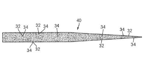
[0024] FIG. 3 is a top view of a porous starting structure according to at
least one example
embodiment of the present invention;
[0025] FIG. 4 is a zoomed-in perspective view in cross-section taken across A-
A of the at least
one example embodiment shown in FIG. 3;
[0026] FIG. 5 is a top view of an endodontic instrument shaped from the porous
starting pre-
structure of FIG. 3 according to the least one example embodiment of the
present invention;
[0027] FIG. 6 is a perspective view of an endodontic instrument according to
at least one
example embodiment of the present invention;
[0028] FIG. 7 is zoomed-in perspective view of the at least one example
embodiment shown in
FIG. 6;
[0029] FIG. 8 is a perspective view of an endodontic instrument according to
at least another
example embodiment of the present invention;
[0030] FIG. 9 is a zoomed-in perspective view of the at least another example
embodiment
shown in FIG. 8;
[0031] FIG. 10 is a perspective view of a starting material according to at
least yet another
example embodiment of the present invention;
[0032] FIG, 11 is a zoomed-in perspective view of the at least yet another
example embodiment
shown in FIG. 10
[0033] FIG. 12 is top view of at least two other example embodiments of the
present invention.
Detailed Description of the Invention
[0034] The standard of care for endodontic instruments may be either a motor
driven rotary file
or a reciprocating motor driven file. When designing an endodontic instrument
(e.g., endodontic
file), many limitation need to be overcome, In particular, the movement of
biological debris from
the apex of the endodontic instrument to the coronal aspect of the endodontic
instrument during
RCT. If the transport of debris is limited, the endodontic instrument may
become less efficient,
become lodged, ledge or fracture. If biological debris is not transported
efficiently, it could lead
to greater amounts of smear layer. Smear layer is biologic debris that is
"coated" on the dentinal
walls via the endodontic instrument during RCT due to forces between the
endodontic
instrument and the canal wall. Smear layer is difficult to remove. If smear
layer is not properly
removed, it could lead to an unsuccessful RCT and/or a retreatment RCT.
Current rotary
endodontic instruments are primarily composed of nitinol. Typical endodontic
files have helical
features that scrape and remove the biological tissue during RCT. The design
and file efficiency
are limited by the surface area that the helixes contact. Although Nitinol is
relatively robust,
6
CA 02931240 2016-05-19
WO 2015/108621 PCT/US2014/066780
there are limits relative to design and material properties that result in
endodontic file properties
like cyclic fatigue, flexibility or peak torque that could be improved.
Finally, when an endodontic
File is fractured in the canal and cannot be removed, the apex of the root
will contain
biocompatible nitinol but the apex could remain open to the surrounding root
area.
[0035] This invention discloses an endodontic instrument comprised of at least
one porous
metal, for example nitinol or nitinol derivatives (NiTiCr, NiTiCo, NiTiFe or
NiTiX wherein X may
be a third or more elements), copper based materials (e.g., CuZnAl or CuAlNi),
stainless steel
and other titanium derivatives. The endodontic instrument may be composed of
porous Nitinol
throughout the entire bulk of the endodontic instrument. It is appreciated
that the endodontic
instrument may be formed of a porous or non-porous material having a
structure/design that
includes a plurality of apertures of similar or various depths, wherein one or
more of the plurality
of apertures may include one or more second materials that may be a porous
and/or non-
porous material. When included, it is believed that weight distribution of a
generally symmetrical
endodontic instrument design along the axis of rotation may be varied such
that during the
rotation or reciprocation of the endodontic instrument the center of mass
(centroid) is not
located at the axis of rotation about a cross-section of a generally
symmetrical instrument.
Additionally, flexibility may be optimized by including multiple porous, non-
porous, or
combination of both materials in the endodontic instrument throughout the
working portion or
within one or more portions thereof to alter the flexibility of the endodontic
instrument along the
entire working portion or variably throughout the working portion at
predetermined
positions/locations radially and/or longitudinally about the endodontic
instrument.
[0036] The endodontic instrument may have a helical design and/or may be
conical without a
helical design and/or have a variable taper. The porous nitinol may be a thin
film coating on the
surface of an endodontic instrument. The endodontic instrument may contain
helixes and/or
may be conical is design with no helixes and/or have a variable taper.
Additionally, the
10037] The endodontic instrument may also be composed of micron thick porous
nitinol coating.
The porous material (e.g., nitinol material) may be at least about 0.05 micron
(e.g., 0.25
microns), typically at least about 0.5 micron, and preferably at least about 1
micron thick film
coating (e.g., on the surface of an endodontic file). Furthermore, it is
appreciated that the porous
material may be less than about 500 microns (e.g., 250 microns, typically less
than about 100
microns, and preferably less than about 50 microns, and more preferably less
than 150 microns
thick film coating. For example, the porous material may be provided in a
range from about
0.05 microns to about 500 microns (e.g., about 0.25 microns to about 250
microns), typically
7
CA 02931240 2016-05-19
WO 2015/108621 PCT/US2014/066780
from about 0.5 microns to about 100 microns, and preferably from about 1
micron to about 50
microns thick film coating.
[0038j It is appreciated that the porous coated may be formed and/or applied
to the porous
staffing structure and/or the endodontic instrument using various procedures
known in the art.
Examples of procedures for forming and/or applying a porous coating to the
porous starting
structure and/or the endodontic instrument may include but are not limited to
medical grade
epoxy/glue, conventional sintering (CS), laser welding, laser melting,
selective laser melting
(SLM), laser sintering, selective laser sintering (SLS), self-propagating high-
temperature
synthesis (SHS), spark plasma sintering (SPS), hot isostatic pressing (HIP),
capsule free HIP
(CF-HIP), laser micro-holes punch, and otherwise.
[0039] Desirably, the porous material (e.g, nitinol) has a porosity of at
least about 5O/0, typically
at least about 15%, preferably at least about 25%, and more preferably at
least about 35% (e.g.,
at least about 45%). Furthermore, the porous material may have a porosity of
less than about
95%, typically less than about 90%, preferably less than 85%, and more
preferably less than
about 80% (e.g., less than about 75%). For example, the porous material may
have a porosity
ranging from about 5% to about 95%, typically from about 15% to about 90%,
preferably from
about 25% to about 85%, and more preferably from about 35% to about 80% (e.g.,
from about
45% to about 75%). Put a different way, the porous material preferably has a
metal surface
area or percentage of metal between about 20% and about 65% and more
preferably between
25% and 55%.
[0040] The endodontic instrument may contain helixes and/or may be conical is
design with no
helixes and /or have a variable taper. The cross sections of the endodontic
instrument
containing or comprised of porous nitinol may be circular, oval, square,
rectangular, rhombi,
parallelogram, star design or contain concavities. The cross section may have
an axis of
rotation on the center of mass or have an axis of rotation that is off-
centered with respect to the
center of mass.
[0041] The invention disclosed above may solve many of the problems associated
with
endodontic instrument design and RCT. First, relative to the amount of surface
that a porous
surface provides relative to a traditional helical design is much higher,
resulting in increased
cutting efficiency. The increase in surface area or contact with the canal
wall will also result in
less smear layer formation. A porous endodontic instrument (e.g., file) or
surface coated porous
endodontic instrument will have to rely less on biological transport of
material because the
material may accumulate in the pores during RCT. If a porous nitinol
instrument becomes
8
CA 02931240 2016-05-19
WO 2015/108621 PCT/US2014/066780
lodged in the canal and cannot be removed, porous nitinol is known to form
hydroxyapatite or
bone. This will seal the apical region of the root canal.
[0042] Finally, an endodontic file composed of porous nitinol or coated with
porous nitinol will
contain less mass than an endodontic file designed similarly from tradition
nitinol. This will result
in improved cyclic fatigue and flexibility.
[0043] The present invention contemplates forming endodontic instruments from
porous starting
(raw) structures. In one specific example as shown in Figure 3, a porous wire
30 is provided.
Desirably, the wire 30 may be shaped as an elongated cylindrical structure;
however, other
starting structures are contemplated. The starting porous structure includes
one or more
apertures 32 provided at various locations thereabout as shown in Figures 4-5.
The apertures
32 may be located at similarly spaced or variable spaced positions about the
structure.
Furthermore, it is appreciated that the apertures 32 may be provided in shapes
and/or sizes that
are similar or different. Desirably, the starting porous structure may include
a generally
homogenous dispersion of apertures 32 throughout, though not required. As
shown in Figure 5,
an endodontic instrument 40 is provided, which has been shaped by a
manufacturing process
(described herein) from the porous wire 30. In this specific example, the
edges (e.g., along the
external surface) of the apertures 32 act as cutting edges 34 for
cleaning/shaping a tooth (e.g.,
root canal).
[0044] Figures 6-7 show at least one example of another embodiment of the
present invention,
which provides for a hybrid endodontic instrument 50. The hybrid instrument 50
may include a
proximal end 52, a proximal end portion 54, a distal end (e.g., tip) 56, a
tapered working portion
58 that extends from the proximal end portion 54 to the distal end 56, and an
instrument axis 57.
The working portion 58 includes an intermediate portion 60 and a distal end
portion 62. In one
specific example, the intermediate portion 60 may include at least two
(helical) cutting edges 64
that define apertures 66 and a generally lattice type structure. Desirably,
the intermediate
portion is substantially hollow and further includes internal supports 68
(Figure 7) extending
between the at least two cutting edges 64. Extending from the intermediate
portion 60, the distal
end portion may include at least two helical flutes 70, which may be formed as
tapered portions
and define at least two second-cutting edges 72. The second cutting edges 72
may extend
from the cutting edges 64 or may be separate therefrom. Desirably, the distal
end portion 62
may be substantially free of apertures 66 thereby forming a substantially
solid (cutting) portion
of porous material or non-porous material.
[0045] The cutting edges 64 and/or the internal supports 68 may include a
width/thickness of at
least about 1 microns (e.g., 25 microns), typically at least about 50 micron,
and preferably at
9
CA 02931240 2016-05-19
WO 2015/108621 PCT/US2014/066780
least about 100 microns (e.g., diameter or otherwise). Furthermore, it is
appreciated that the
cutting edges 64 and/or the internal supports 68 may be less than about 1000
microns (e.g.,
800 microns), typically less than about 600 microns, and preferably less than
about 500
microns, and more preferably less than 150 microns. For example, the cutting
edges 64 and/or
the internal supports 68 may include a width/thickness in a range from about 1
micron to about
1000 microns (e.g., about 26 microns to about 800 microns), typically from
about 50 microns to
about 600 microns, and preferably from about 100 micron to about 500 microns.
[0046] Figures 8-9 show another example of the present invention, which
provides for a tapered
endodontic instrument 50b. In this specific example, the cutting edges 64b of
the intermediate
portion 60b continue into and through the distal end portion 62b to the tip
56b. Desirably, both
the intermediate portion and the distal end portion are substantially hollow
with apertures 66b
throughout. The cutting edges 64b extend along the working portion tapering
towards an
instrument axis 57b to the tip 56b. The tip 56b may be a substantially free of
apertures thereby
forming a substantially solid (cutting) portion of porous material or non-
porous material
[0047] Figures 10-11 show another embodiment of the present invention, which
provide a
porous (raw material) structure 74 for forming an endodontic instrument 50c
(having a generally
similar shape as endodontic instrument 40. In one specific example, the porous
structure 74
may be a porous wire for forming an endodontic instrument such as an
endodontic file. The
porous structure may be formed of a porous material and/or a non-porous
material. Apertures
66c may be provided as any various geometric shape, which may be the same
shapes or
different shapes of the same or different size throughout. In one specific
embodiment, the
apertures 66c may be of a hexagonal shape and of generally the same size to
define a
generally honeycomb lattice-type structure (e.g., honeycomb structure). In is
appreciated that
one or more of the 66c extend radially (at least partially or completely) to
the instrument axis
57c. Additionally, it is appreciated that the porous structure 74 may include
one or more layers
separated by one or more internal supports 68c thereby defining longitudinal
and/or radial
apertures. When included, the apertures may be in alignment (radially) or
offset (longitudinally
parallel to the instrument axis) from layer to layer. It is appreciated that
the porous (starting)
structure 74 may thereafter be shaped to form an endodontic instrument by
manufacturing
process described herein.
[0048] Figure 12 shows another embodiment of the present invention, which
provides an
endodontic instrument 50d and endodontic instrument 50e. Endodontic
instruments 50d and
50e may be formed from a porous material and/or coated with a porous coating
as described
herein. It is appreciated that the structure of the endodontic instrument 50d
may be generally
CA 02931240 2016-05-19
WO 2015/108621 PCT/US2014/066780
symmetrical thereby having an axis of rotation 57d that is centered such that
a first cross-
section 76d (e.g., A-A, B-B, and/or C-C) has a center of mass 78d (centroid)
that may be
generally located about the axis of rotation 57d as compared to the structure
of the endodontic
instrument 50e that may be generally asymmetrical thereby having a second
center of mass
78e (centroid) that may not be located about an axis of rotation 57e in a
second cross-section
76e (e.g., D-D, E-E, and/or F-F). More particularly, it is believed that this
first geometry (57d)
can be symmetrical and the second geometry can be asymmetrical such that the
first cross
section 76d can be closer to the shank end than the tip end. The first cross
section can have a
different number of working surfaces than the second cross section. At the
second cross section
76e, a second centroid 78e can be offset from the axis of rotation. The first
geometry and the
second geometry can include different numbers of working surfaces. The body
can be flexible.
The body can be sufficiently flexible such that when a tip of the body is
bound at a fixed position
as the body rotates or reciprocates, a portion of the body that intersects the
second cross
section bends away from the axis of rotation a substantially equal amount at a
first angle of
rotation and at a second angle of rotation. The first angle of rotation can be
180 from the
second angle of rotation. The second cross section 76e can bend away from the
axis of rotation
57e a substantially equally amount at each angle of rotation. A non-swaggering
portion of the
body can have a centroid 78d that lies substantially on the axis of rotation
57d and intersects
the at least one working surface as the tip of the body is bound at a fixed
position and the body
rotates. The at least one working surface can include a cutting flute. A tip
of the body may not
have cutting surfaces. At a cross section that intersects the axis of
rotation, a center of mass
may be offset from the axis of rotation. The working surface can be configured
to remove
material when the body is rotated or reciprocated within a canal of the
material. When the tip of
the body is held in place and the body is rotated or reciprocated, at least a
portion of the body
may form helical waves.
[0049] Manufacturing
[0050] The porous metal file may be manufactured via traditional nitinol files
where a metal wire
is ground with the appropriate file design. In this case, a porous metal based
wire would be
ground. Also, traditional Nitinol wire could be etched to form a porous
material. The porous file
could also be manufactured by utilizing additive manufacturing techniques, for
example, metal
3D printing or surface coatings.
[0051] Examples of porous materials and/or fabrication methods of porous
materials may
include, but are not limited to those described in Porous NiTi for bone
implants: A review,
11
CA 02931240 2016-05-19
WO 2015/108621 PCT/US2014/066780
(Bansiddhi, Sargeant, Stupp, and Dunand, which is herein incorporated by
reference for all
purposes.
100521 In another embodiment, the present invention may provide various rotary
file designs
and/or raw materials that have posed difficulties while using traditional
manufacturing
techniques such as milling, turning, grinding, laser cutting, photochemical
machining, etc.
Advantageously, these difficulties may be substantially reduced or eliminated
using an additive
manufacturing technology otherwise known as 3D Printing, Rapid Prototyping,
etc, and/or
otherwise known additive technologies.
[0053] Manufacturing and/or shaping of the porous starting structure and/or
the endodontic
instrument may be achieved by known forming processes. Examples of such
forming process
include, but are not limited to 3D printing, additive manufacturing, and metal
injection molding.
3D Printing and additive manufacturing are processes of making three-
dimensional solid objects
out of virtually any shape from a digital model. 3D printing is achieved using
an additive
process, where successive layers of material are laid down in different
shapes. 3D printing is
also considered distinct from traditional machining techniques, which mostly
rely on the removal
of material by methods such as milling, drilling, grinding, etc (substractive
processes). There
are different types of additive processes where some melt or soften material
to produce the
layers such as selective laser melting, direct metal laser sintering,
selective laser sintering, and
fused deposition molding while other processes cure liquid materials such as
stereolithography.
[0054] It is believed that one advantage to having a 3D printed rotary file
may be that it allows
for the design freedom in creating a product that is capable of achieving
different functions while
in the anatomy. For instance, Figure 6 shows a hybrid design where the
endodontic instrument
may shape the root canal apically to allow irrigants to thoroughly clean the
canal apically and
provide for a shape for obturation while in the mid-root and coronal aspects,
it is allowed to
expand and collapse in order to allow the endodontic instrument to adapt to
the natural
anatomy. In this design, the 3D printer may print the solid metal blank
apically and the stent
type designs the remainder of the design. Conventional grinding processes may
then be used
to create the sharp edges and flutes which allows for a traditional file
design apically.
[0055] Another way of using the 3D printing technology is to allow for the
support of structures
internally. For instance, in Figure 7, it shows an example of internal struts
to support the
structure of the stent design. Other alternatives to support structurally
design would be to vary
the width and thickness of the different stent pieces to allow for more
support or increased
flexibility (thicker supports increase the stiffness while thinner supports
provides for more
flexibility).
12
CA 02931240 2016-05-19
WO 2015/108621 PCT/US2014/066780
[0056] Figure 8 shows an example of being able to print a tapered endodontic
instrument (e.g,,
stent file). In one specific example, a tubular stent design may be shaped by
laser cut. By
having a tube design, this prevents the design from being tapered initially
and creates more
stress on the stent design apically since it has to compress more to adapt to
the apical aspect of
the canal. By having a 3D printed tapered stent type design, it allows the
endodontic instrument
to have reduced stresses apically since there is less of a diameter and less
compression
required. Figure 9 shows a zoomed-in view of this type of design such that the
cutting tip may
be closed/joined and allow the endodontic instrument to have more structure
support apically
versus an opened-ended/hollow tip design as described in US patent 7,713,059,
which are
herein incorporated by reference for all purposes.
[0057] Figures 10 and 11 show how the raw material may be created using 3D
printing and
have the blank wire with porous surfaces. The porous raw material may then be
ground to a
desired tip and taper as a finished rotary file. The advantage to this type of
design is that now
the file has less core mass which increases its flexibility and cyclical
fatigue resistance. It also
provides for a rough surface as well as allows space for the cut material to
go. Alternative types
of designs for raw material embodiments are numerous but may include internal
corrugated
wire, random porosity of wire, a hollow tapered wire, etc.
[0058] There are many additive manufacturing techniques and many different
types of materials
used within these additive manufacturing techniques. Preferably, these designs
are produced
out of a metal material but they could be printed out of plastics as the
technology develops.
There are several types of metal 3D printing technologies including Direct
Metal Laser Sintering,
Electron Beam Melting, etc. The materials available with these technologies
include: Cobalt
Chrome, Titanium, Inconel, Nickel, Aluminum, Stainless Steel, Steel, etc. To
our knowledge,
Nickel Titanium is not a material currently available with these technologies
but does not seem
to be an issue in producing parts with these technologies.
[0059] It can be seen that the invention can also be described with reference
to one or more of
the following combinations.
[0060] A rotatable endodontic file for cleaning/shaping a tooth root canal,
comprising: an
elongated shaft having a proximal end portion, a distal end and a tapered
working portion
having a rotational axis, the working portion extending from said proximal
portion to said distal
end; the external surface of said shaft working portion having a plurality of
at least two spirals, a
geometric cross section where the file is composed of porous metal.
[0061] A rotatable endodontic file for cleaning/shaping a tooth root canal,
comprising: an
elongated shaft having a proximal end portion, a distal end and a tapered
working portion
13
CA 02931240 2016-05-19
WO 2015/108621 PCT/US2014/066780
having a rotational axis, the working portion extending from said proximal
portion to said distal
end; the external surface of said shaft working portion having no spirals, a
geometric cross
section where the file is composed of porous metal.
[0062] A rotatable endodontic file for cleaning/shaping a tooth root canal,
comprising: an
elongated shaft having a proximal end portion, a distal end and a tapered
working portion
having a rotational axis, the working portion extending from said proximal
portion to said distal
end; the external surface of said shaft working portion having a plurality of
at least two spirals, a
geometric cross section where the file is coated with a porous metal.
[0063] A rotatable endodontic file for cleaning/shaping a tooth root canal,
comprising: an
elongated shaft having a proximal end portion, a distal end and a tapered
working portion
L a rotational axis, the working portion extending frorn said proximal
potlion to said distal
end; the external surface of said shaft working portion having no spirals, a
geometric cross
section where the file is coated with porous metal.
[0064] A rotatable endodontic file for cleaning/shaping a tooth root canal,
comprising: an
elongated shaft having a proximal end portion, a distal end and a tapered
working portion
having a rotational axis, the working portion extending from said proximal
portion to said distal
end; the external surface of said shaft working portion having a plurality of
at least two spirals, a
geometric cross section where the file is etched to form a porous metal.
[0065] A rotatable endodontic file for cleaning/shaping a tooth root canal,
comprising: an
elongated shaft having a proximal end portion, a distal end and a tapered
working portion
having a rotational axis, the working portion extending from said proximal
portion to said distal
end; the external surface of said shaft working portion having no spirals, a
geometric cross
section where the file is etched to form a porous metal.
[0066] An endodontic file, wherein the porous metal is composed of either a
Nitinoi based
material, Cu based material, titanium based material or a stainless steel
based material.
[0067] An endodontic file, wherein the material is processed by high
temperature, cold
temperatures and/or strain.
[0068] An endodontic file that has an axis of rotation that is centered such
that the cross section
center of mass (centroid) is located at the axis of rotation.
[0069] An endodontic file that has an axis of rotation that is asymmetric such
that the center of
mass (centroid) is not located at the axis of rotation.
[0070] An endodontic file wherein the material is processed by high
temperature, cold
temperatures and/or strain.
[0071] An endodontic file wherein the file is manufactured through a grinding
process.
14
CA 02931240 2016-05-19
WO 2015/108621 PCT/US2014/066780
[0072] An endodontic file wherein the file is manufactured through an additive
manufacturing
process
[0073] An endodontic file wherein the file is manufactured through an additive
manufacturing
process such as 3D printing or a coating technique.
[0074] An endodontic file wherein the file is manufactured through an etching
manufacturing
process
[0075] The invention described herein has many other advantages. The
endodontic instrument
may have a single continuous flow path, which eliminates potential leak paths.
Inherent stress
concentrations may be reduced or substantially eliminated, thereby allowing
the tip and/or the
distal end portions to be reliable during vibration. The configuration of the
tip and/or the distal
end portions guide and transfer the ultrasonic vibration and energy in the
planes of motion,
which provides proper agitation to the irrigants. The tip assembly can also be
disposable,
thereby requiring that a new tip assembly be used for each patient and
insuring that the tip
assembly will be sterile prior to use.
[0076] Each feature disclosed in this specification (including any
accompanying claims,
abstract, and drawings), may be replaced by alternative features having the
same, equivalent or
similar purpose, unless expressly stated otherwise. Thus, unless expressly
stated otherwise,
each feature disclosed is one example only of a generic series of equivalent
or similar features.
[0077] While preferred embodiments of the present invention have been shown
and described
herein, it will be obvious to those skilled in the art that such embodiments
are provided by way
of example only. Numerous variations, changes, and substitutions will be
apparent to those
skilled in the art without departing from the invention. Other foreseen
embodiments or uses for
the present invention include the use of the invention in the field of
phacoemulsification, where a
tip assembly such as the present invention may offer many advantages.
Accordingly, it is
intended that the invention be limited only by the scope of the appended
claims.