Note: Descriptions are shown in the official language in which they were submitted.
CA 02931334 2016-05-20
WO 2015/077648 PCT/US2014/066952
SPECIFICATION OF FUNCTIONAL CRANIAL PLACODE DERIVATIVES
FROM HUMAN PLURIPOTENT STEM CELLS
CROSS-REFERENCE TO RELATED APPLICATION
This application claims priority to United States Provisional Application No.
61/907,302, filed November 21, 2013, the contents of which is hereby
incorporated by
reference in its entirety herein.
FIELD OF THE INVENTION
The present invention relates generally to the field of cell biology of stem
cells, more specifically the directed differentiation of pluripotent or
multipotent stem
cells, including human embryonic stem cells (hESC), somatic stem cells, and
induced
human pluripotent stem cells (hiPSC) using novel culture conditions. In
particular,
cranial placodes are derived from human pluripotent stem cells by a dual-SMAD
inhibition strategy of neural induction coupled with further fate
specification at the
pre-placode stage. The method generates placode-derived trigeminal ganglia,
mature
lens fibers and anterior pituitary hormone-producing cells. Applications of
these cells
include, but are not limited to, human cell-based therapies in sensory and
endocrine
disease.
BACKGROUND OF THE INVENTION
The differentiation capacity of embryonic and somatic stem cells have opened
possibilities for cell replacement therapies for genetic, malignant, and
degenerative
diseases. Neurodegenerative disorders, conditions, and diseases, and their
treatment
by cell-based therapies represent a promising means of preventing, reducing or
eliminating the symptoms. Such disorders include Huntington's disease,
Alzheimer's,
Parkinson's, and amyotrophic lateral sclerosis. They also provide a source of
cells for
screening for critical small molecules (i) that could be useful in for
treatment of
disease; or (ii) for determining the cell fate of neural tissue. Further,
these cells were
studied in order to characterize key genes, mRNA transcripts, and proteins
relevant in
normal or pathological lineages.
1
CA 02931334 2016-05-20
WO 2015/077648 PCT/US2014/066952
Neural development is dictated in time and space by a complex set of signals
that instruct neural precursor identity. While significant progress was made
in animal
models, human neural development remains much less understood.
Previous studies reported directed differentiation of mouse (Wichterle et al.,
2002; Barberi et al., 2003; Watanabe et al., 2005) and human (Perrier et al.,
2004; Li
et al., 2008; Eiraku et al., 2008) ESCs into specific neuron types in response
to
patterning factors defining anterior/posterior (ALP) and dorso-/ventral (D/V)
CNS
identity. These studies demonstrate evolutionary conservation of signaling
systems
that specify the major CNS regions. In mammals, sonic hedgehog (SHH) is a
ventralizing factor acting in a dose-dependent manner to specify the various
ventral
cell types including cells expressing floor plate (FP) in primary neural
explants
(Briscoe and Ericson, 1999) and in mouse ES cells (Mizuseki et al., 2003).
While
application of SHH to hESC-derived neural cells was shown to induce various
ventral
neuron types, the derivation of floor plate (FP) tissue itself was not
reported. As FP is
one signaling center for inducing differentiation pathways and subsequent
committed
cell linage, the ability to produce FP from human ES cells would be a major
step
forward in furthering studies of early human neural development. Furthermore,
little
is known about FP development in humans, due to lack of accessibility to
tissue.
In animals, the FP is a major site of SHH production and several human
developmental disorders are related to alterations in midline SHH signaling
(Mullor et
al., 2002) including certain forms of holoprosencephaly and microphthalmia,
skeletal
disorders including various cleft plate syndromes, and tumor conditions such
as
Gorlin's syndrome; a rare genetic disorder caused by a mutation in the SHH
receptor
Patched 1. However it is not known whether similar alterations in midline SHH
signaling would induce these diseases in humans.
Therefore there is a critical need for inducing human floor plate tissue from
human embryonic stem cells (hESCs) for providing a source of human floor plate
cells. These human floor plate cells are necessary for use in medical research
for
determining causes and treatments of developmental diseases in humans and for
comparative developmental studies of human neural patterning and axonal
pathfinding.
2
CA 02931334 2016-05-20
WO 2015/077648 PCT/US2014/066952
SUMMARY OF THE INVENTION
The present invention relates generally to the field of cell biology of stem
cells, more specifically the directed differentiation of pluripotent or
multipotent stem
cells, including human embryonic stem cells (hESC), somatic stem cells, and
induced
human pluripotent stem cells (hiPSC) using novel culture conditions. In
particular,
cranial placodes are derived from human pluripotent stem cells by a dual-SMAD
inhibition strategy of neural induction coupled with further fate
specification at the
pre-placode stage. The method generates placode-derived trigeminal ganglia,
mature
lens fibers and anterior pituitary hormone-producing cells. Applications of
these cells
include, but are not limited to, human cell-based therapies in sensory and
endocrine
disease.
In one embodiment, the present invention contemplates a method, comprising:
a) providing or plating a plurality of cranial placodal precursor cells in an
N2 cell
culture medium; b) contacting said plurality of placodal precursor cells with
a
composition comprising brain-derived neurotrophic factor, wherein a plurality
of
trigeminal placode cells are created. In one embodiment, the method further
comprises incubating the trigeminal placode cells under conditions such that
trigeminal neurons are created. In one embodiment, the cranial placodal
precursor
cell expresses SIX1, PAX6 and TFAP2A. In one embodiment, the trigeminal
placode
cells express SIX1 and PAX3. In one embodiment, the trigeminal placodal
precursor
cells may be used to identify pharmacological agents to inhibit sensory neuron
infectious agents. In one embodiment, the sensory neuron infectious agent
comprises
Varicella zoster virus (e.g., whose infection results in chicken pox). In one
embodiment, the infected sensory neuron is a trigeminal neuron.
In one embodiment, the present invention contemplates a method, comprising:
a) providing or plating a plurality of cranial placodal precursor cells in a
cell culture
medium; and b) contacting said plurality of placodal precursor cells with a
composition comprising sonic hedgehog, purmorphamine and a 7-secretase
inhibitor,
wherein a plurality of pituitary placode cells are created. In one embodiment,
the
method further comprises incubating said pituitary placode cells under
conditions
such that a plurality of pituitary cells including, but not limited to,
gonadotrophs,
corticotrophs and somatotrophs are created that are capable of secreting
homones. In
one embodiment, the secreted hormones include, but are not limited to, steroid
3
CA 02931334 2016-05-20
WO 2015/077648
PCT/US2014/066952
hormones, adrenocorticotropic hormone and/or growth hormone. In one
embodiment,
the cranial placodal precursor cell expresses SIX1, PAX6 and TFAP2A. In one
embodiment, the pituitary placode cells express SIX1 and PITX1.
In one embodiment, the present invention contemplates a method, comprising:
a) providing or plating a plurality of cranial placodal precursor cells in a
cell culture
medium; and b) contacting said plurality of placodal precursor cells with a
composition comprising sonic hedgehog, purmorphamine, a 7-secretase inhibitor
and
an FGF-inhibitor, wherein a plurality of lens placode cells are created. In
one
embodiment, the method further comprises incubating the lens placode cells
under
conditions such that lens fibers are created. In one embodiment, the cranial
placodal
precursor cell expresses SIX1, PAX6 and TFAP2A. In one embodiment, the lens
placode cells express SIX1 and PITX3.
In one embodiment, the present invention contemplates a method, comprising
grafting a trigeminal placode cell to a subject expressing at least one
symptom of a
pain syndrome, wherein said at least one symptom of the pain syndrome is
reduced.
In one embodiment, the pain syndrome includes, but is not limited to
trigeminal nerve
palsy, trigeminal neuralgia and/or migraine pain. In one embodiment, the
grafting is
performed on the pons. In one embodiment, the trigeminal placode cells migrate
to
the trigeminal nuclei.
In one embodiment, the present invention contemplates a method comprising
grafting a pituitary placode cell to a subject expression at least one symptom
of a
neuroendocrine disorder, wherein said at least one symptom of the
neuroendocrine
disorder is reduced. In one embodiment, the grafted pituitary placode cells
secrete
growth hormone. In one embodiment, the grafted pituitary placode cells secrete
adrenocortiotrophic hormone. In one embodiment, the engraftment is on a leg
muscle. In one embodiment, the engraftment is on the hypothalamic-pituitary
axis.
In one embodiment, the engraftment is on the hypothalamus. In one embodiment,
the
engraftment is on the sella. In one embodiment, the neuroendocrine disorder
comprises diabetes. In one embodiment, the neuroendocrine disorder comprises
hypopituitarism. In one embodiment, the neuroendocrine disorder comprises a
pituitary tumor. In one embodiment, the neuroendocrine disorder comprises
radiation
therapy.
4
CA 02931334 2016-05-20
WO 2015/077648 PCT/US2014/066952
In one embodiment, the present invention contemplates a method comprising
grafting a lens placode cell to a subject expression at least one symptom of
an ocular
disorder, wherein said at least one symptom of the ocular disorder is reduced.
In one
embodiment, the at least one symptom of an ocular disorder comprises
blindness. In
one embodiment, the at least one symptom of an ocular disorder comprises
macular
degeneration. In one embodiment, the at least one symptom of an ocular
disorder
comprises a cataract. In one embodiment, the at least one symptom of an ocular
disorder comprises astigmatism. In one embodiment, the at least one symptom of
an
ocular disorder comprises near sightedness. In one embodiment, the at least
one
symptom of an ocular disorder comprises far sightedness. In one embodiment,
the
grafted lens placode cell secretes a plurality of lens fiber protein. In one
embodiment,
the plurality of lens fiber protein comprises an c3-crystalline lens fiber
protein. In
one embodiment, the plurality of lens fiber protein is layered.
In one embodiment, the present invention contemplates a method, comprising:
a) providing or plating a cell culture comprising human pluripotent stem cells
in a
culture medium; b) contacting said cell culture with a first inhibitor of
Small Mothers
Against Decapentaplegic (SMAD) protein signaling and 444-(1,3-benzodioxo1-5-
y1)-
5-(2-pyridiny1)-1H-imidazol-2-yll benzamide (SB431542); c) removing said first
SMAD inhibitor under conditions such that cranial placode precursor cells are
formed. In one embodiment, the first SMAD inhibitor is selected from the group
consisting of Noggin, a disulfide-linked homodimer of Noggin, Dorsomorphin,
LDN-
193189, and mixtures thereof. In one embodiment, the cranial placode precursor
cells
express SIX1, PAX6 and TFAP2A. In one embodiment, the culture medium
comprises an N2 medium. In one embodiment, the culture medium comprises a
knockout serum replacement medium. In one embodiment, the removing is
performed at least 48 hours after the contacting.
The present inventions provide a method of producing a human neural cell
(neural stem cells, neuronal subtypes, mature neurons, cells of a neural
lineage) by (i)
obtaining stem cells (hESCs, hiPSCs, somatic stem cells, cancer stem cells,
human or
mammalian pluripotent or multipotent cells); and (ii) culturing the human stem
cell
under conditions that block SMAD signaling. In a preferred embodiment, the
methods
for culture include conditions in a feeder-free system. In a preferred
embodiment, the
stem cells are cultured in a monolayer. A preferred embodiment contemplated
the use
5
CA 02931334 2016-05-20
WO 2015/077648
PCT/US2014/066952
of media that is supplemented with compounds Noggin and/or Dorsomorphin and
SB431542.
In one embodiment the inventions provide a kit comprising a first inhibitor of
Small Mothers Against Decapentaplegic (SMAD) protein signaling and a second
inhibitor of Small Mothers Against Decapentaplegic (SMAD) protein signaling.
In
one embodiment, said first inhibitor is selected from the group consisting of
a
disulfide-linked homodimer of Noggin (SEQ ID NO:50), Dorsomorphin, LDN-
193189, combination thereof and mixture thereof. In one embodiment, said
Noggin is
selected from mouse, human, rat, and xenopus. In one embodiment, said is
Noggin is
(SEQ ID NO:50) In one embodiment, said second inhibitor inhibits an anaplastic
lymphoma kinase signaling pathway. In one embodiment, said second inhibitor
inhibits a signaling pathway selected from the group consisting of Lefty,
Activin, and
TGFbeta. In one embodiment, said second inhibitor inhibits both activins and
nodal
signaling. In one embodiment, said second inhibitor is 4-[4-(1,3-benzodioxo1-5-
y1)-5-
(2-pyridiny1)-1H-imidazol -2-yllbenzamide (SB431542) and derivatives thereof.
In
one embodiment, said kit further comprises a human stem cell. In one
embodiment,
the kit further comprises instructions.
In one embodiment the inventions provide a composition comprising a stem
cell, a first inhibitor of Small Mothers Against Decapentaplegic (SMAD)
protein
signaling and a second inhibitor of Small Mothers Against Decapentaplegic
(SMAD)
protein signaling. In one embodiment, said first inhibitor is selected from
the group
consisting of a disulfide-linked homodimer of Noggin, Dorsomorphin, LDN-
193189,
combination thereof and mixture thereof. In one embodiment, said is Noggin is
selected from mouse, human, rat, and xenopus. In one embodiment, said is
Noggin is
(SEQ ID NO:50) In one embodiment, said second inhibitor inhibits the
Lefty/Activin/TGFbeta pathways by blocking phosphorylation of the ALK4, ALK5
and ALK7 receptors. In one embodiment, said second inhibitor inhibits
activin/nodal
signaling. In one embodiment, said second inhibitor is 4-[4-(1,3-benzodioxo1-5-
y1)-5-
(2-pyridiny1)-1H-imidazol -2-yllbenzamide (SB431542) and derivatives thereof.
In
one embodiment, said stem cell is selected from the group consisting of human
embryonic stem cells (hESC), somatic stem cells, and induced human pluripotent
stem cells (hiPSC).
In one embodiment the inventions provide a method for inducing
differentiation in stem cell, comprising, a) providing: i) a cell culture
comprising
6
CA 02931334 2016-05-20
WO 2015/077648 PCT/US2014/066952
human stem cells, ii) a first inhibitor of Small Mothers Against
Decapentaplegic
(SMAD) protein signaling, iii) a second inhibitor of Small Mothers Against
Decapentaplegic (SMAD) protein signaling, and b) contacting said stem cells
with
said first inhibitor of Small Mothers Against Decapentaplegic (SMAD) protein
signaling and said test compound under conditions for inducing differentiation
in a
stem cell into a non-default differentiated cell. In one embodiment, said
first inhibitor
is selected from the group consisting of a disulfide-linked homodimer of
Noggin,
Dorsomorphin, LDN-193189, combination thereof and mixture thereof. In one
embodiment, said is Noggin is selected from mouse, human, rat, and xenopus. In
one
embodiment, said is Noggin is (SEQ ID NO:50) In one embodiment, said second
inhibitor is a ALK4 receptor inhibitor. In one embodiment, said second
inhibitor is 4-
[4-(1,3-benzodioxo1-5-y1)-5-(2-pyridiny1)-1H-imidazol -2-yllbenzamide
(SB431542)
and derivatives thereof. In one embodiment, said non-default differentiated
cell is a
neural progenitor cell. In one embodiment, said non-default differentiated
cell is a part
of a population of cultured cells. In one embodiment, said non-default
differentiated
cell is at least 10% up to 100% of said population of cultured cells. In one
embodiment, said non-default differentiated cell in a population of cultured
cells
expresses paired box gene 6 protein. In one embodiment, said paired box gene 6
protein is expressed in at least 10% of said population of cultured cells. In
one
embodiment, said stem cell is selected from the group consisting of human
embryonic
stem cells (hESC), human somatic stem cells, and induced human pluripotent
stem
cells (hiPSC). In one embodiment, said non-default differentiated cell is a
neural cell.
In one embodiment, said neural cell is selected from the group consisting of
dopamine
positive neurons and floor plate cells.
In one embodiment the inventions provide a composition comprising isolated
human embryonic neural cells. In one embodiment, said isolated human embryonic
neural cells were derived from human embryonic cells. In one embodiment, said
human embryonic neural cells are cultured in vitro. In one embodiment, said
human
embryonic neural cells are attached cells. In one embodiment, said composition
is a
co-culture further comprising a second cell type.
In one embodiment the inventions provide a method for screening biological
agents, comprising, a) providing: i) a cell culture comprising human embryonic
stem
cells (hESCs), and ii) a test compound, and b) contacting said stem cells with
said test
compound under conditions for inducing neural floor plate cells. In one
embodiment,
7
CA 02931334 2016-05-20
WO 2015/077648 PCT/US2014/066952
said test compound is sonic hedgehog or fragment thereof. In one embodiment,
said
human embryonic stem cells are rosette-stage neural cells.
In one embodiment the inventions provide a method for providing
differentiated cells, comprising, a) providing: i) a cell culture of human
embryonic
stem cells (hESCs), and ii) a compound for inducing differentiation, and b)
contacting
said stem cells with said test compound under conditions for inducing neural
floor
plate cells. In one embodiment, said compound is sonic hedgehog protein or
fragment
thereof. In one embodiment, said inducing consists of increasing a
characteristic
selected from the group consisting of flat cellular morphology, expressing
sonic
hedgehog, expressing forkhead box protein A2 (FOXA2), expressing Netrin-1, and
expressing F-Spondin compared to said characteristic expressed in said human
embryonic stems cells cultured without said test compound. In one embodiment,
said
inducing consists of decreasing a characteristic selected from the group
consisting of
rosette structures, BF1 expression, paired box homeotic gene-6 (PAX6)
expression,
NK6 homeobox 1 (NKX6.1), homeobox protein SIX6 expression compared to said
characteristic in said human embryonic stems cells cultured without said test
compound. In one embodiment, the method further provides and comprises a
Noggin
protein and an agent for blocking phosphorylation of a receptor selected from
the
group consisting of activin receptor-like kinase 4 (ALK4), activin receptor-
like kinase
5 (ALK5) and activin receptor-like kinase 7 (ALK7) receptors and contacting
said
human stem cells with said noggin and said agent to human stem cells before
adding
said compound. In one embodiment, the method further provides an antibody,
wherein said antibody is dickkopf homolog 1 (DKK-1) antibody, and contacting
said
stem cells with said antibody for reducing DKK-1 protein function. In one
embodiment, the method further provides and comprises a caudalizing factor
selected
from the group consisting of wingless-type MMTV integration site family,
member 1
(Wnt-1), and Retinoic Acid (RA). In one embodiment, the method further
provides
and comprises a neuron inducing compound and step c) adding said neuron
inducing
compound for inducing progenitor neurons. In one embodiment, said dopamine
neurons express a marker selected from the group consisting of corin, serine
peptidase
(CORN) and nephroblastoma overexpressed gene (NOV). In one embodiment, said
progenitor neurons are dopamine neurons express a marker selected from the
group
consisting of LIM homeobox transcription factor 1, beta (LMX1B) and neurogenin
2
(NGN2). In one embodiment, the method further provides and comprises a stem
cell,
8
CA 02931334 2016-05-20
WO 2015/077648 PCT/US2014/066952
and step d) co-culturing said human neural floor plate cells with said stem
cells for
producing neurite outgrowth from said stem cells.
In one embodiment the inventions provide a neural floor plate cell produced
by the methods described herein. In one embodiment the inventions provide a
placode
cell produced by the methods described herein. In one embodiment the
inventions
provide a lens cell produced by the methods described herein.
The invention contemplates methods for assessing the neural identity of the
derived neural cells. This method may be through morphological means,
functional
assessment, and measurement of expression or downregulation of proteins
associated
with certain lineages. In a preferred method, dopaminergic activity or
functional
assays for motor neurons are utilized.
The present method can by employed to deliver agents or neural cells to the
brain in an effective amount for diagnosis, prevention, treatment of disease,
disorders,
or for patients suffering from nerve damage form stroke. Such cells were co-
committed towards a neural fate.
In one embodiment, the present invention contemplates a composition
comprising isolated human embryonic floor plate cells. In one embodiment, the
isolated human embryonic floor plate cells were derived from human embryonic
cells.
In one embodiment, the human embryonic floor plate cells are cultured in
vitro. In
one embodiment, the human embryonic floor plate cells are attached cells. In
one
embodiment, the composition is a co-culture further comprising a second cell
type.
In one embodiment, the present invention contemplates a method for
screening biological agents, comprising, a) providing: i) a cell culture
comprising
human embryonic stem cells (hESCs), and ii) a test compound, and b) contacting
said
stem cells with said test compound under conditions for inducing neural floor
plate
cells. In one embodiment, the test compound is sonic hedgehog or fragment
thereof.
In one embodiment, the human embryonic stem cells are rosette-stage neural
cells.
In one embodiment, the present invention contemplates a method for
providing differentiated cells, comprising, a) providing: i) a cell culture of
human
embryonic stem cells (hESCs), and ii) a compound for inducing differentiation,
and b)
contacting said stem cells with said test compound under conditions for
inducing
neural floor plate cells. In one embodiment, the compound is sonic hedgehog
protein
or fragment thereof. In one embodiment, the inducing consists of increasing a
characteristic selected from the group consisting of flat cellular morphology,
9
CA 02931334 2016-05-20
WO 2015/077648 PCT/US2014/066952
expressing sonic hedgehog, expressing forkhead box protein A2 (FOXA2),
expressing
Netrin-1, and expressing F-Spondin compared to said characteristic expressed
in said
human embryonic stems cells cultured without said test compound. In one
embodiment, the inducing consists of decreasing a characteristic selected from
the
group consisting of rosette structures, BF1 expression, paired box homeotic
gene-6
(PAX6) expression, NK6 homeobox 1 (NKX6.1), homeobox protein SIX6 expression
compared to said characteristic in said human embryonic stems cells cultured
without
said test compound. In one embodiment, the method further provides Noggin
protein
and an agent for blocking phosphorylation of a receptor selected from the
group
consisting of activin receptor-like kinase 4 (ALK4), activin receptor-like
kinase 5
(ALK5) and activin receptor-like kinase 7 (ALK7) receptors and contacting said
human stem cells with said noggin and said agent to human stem cells before
adding
said compound. In one embodiment, the method further provides an antibody,
wherein said antibody is dickkopf homolog 1 (DKK-1) antibody, and contacting
said
stem cells with said antibody for reducing DKK-1 protein function. In one
embodiment, the method further provides a caudalizing factor selected from the
group
consisting of wingless-type MMTV integration site family, member 1 (Wnt-1),
and
Retinoic Acid (RA). In one embodiment, the further comprises, providing, a
neuron
inducing compound and step c) adding said neuron inducing compound for
inducing
progenitor neurons. In one embodiment, the dopamine neurons express a marker
selected from the group consisting of corin, serine peptidase (CORIN) and
nephroblastoma overexpressed gene (NOV). In one embodiment, the progenitor
neurons are dopamine neurons express a marker selected from the group
consisting of
LIM homeobox transcription factor 1, beta (LMX1B) and neurogenin 2 (NGN2). In
one embodiment, the method further comprises, providing, stem cells, and step
d) co-
culturing said human neural floor plate cells with said stem cells for
producing neurite
outgrowth from said stem cells.
BRIEF DESCRIPTION OF THE FIGURES
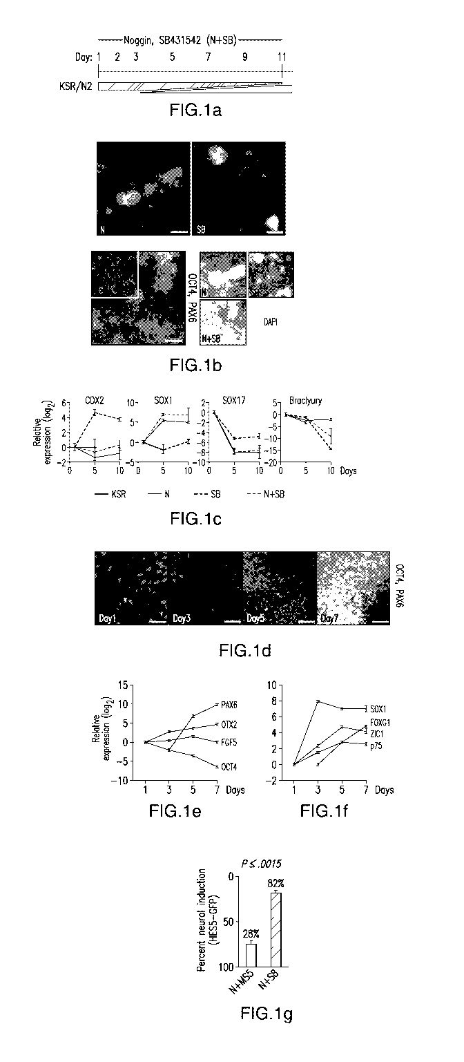
Figure 1 shows exemplary dual SMAD inhibition that allowed for a highly
efficient feeder-free neural induction in adherent cultures within seven days.
(a)
Differentiation scheme used for achieving neural induction was achieved with
the
combination of SB431542, an ALK inhibitor, and Noggin, a BMP inhibitor. (b)
The
dual SMAD inhibition greatly improves neural differentiation (PAX6 expression,
CA 02931334 2016-05-20
WO 2015/077648 PCT/US2014/066952
green) to greater than 80%. Infrequent neural differentiation (< 10% PAX6 +
cells)
were observed when the single factors are used. (c) Real-Time PCR for early
germ
layer markers CDX2, SOX1, SOX17 and Brachyury. (d) Immunoflouresence for
OCT4 (red) and PAX6 (green) expression indicates rapid neutralization occurs
by day
7. (e) Real-Time PCR for PAX6, OTX2, FGF5, OCT4 during dual SMAD inhibition
reveals an epistem cell intermediate at day 5. (f) Real-Time PCR for neural
and
neuronal markers during dual SMAD inhibition differentiation towards
neuroectoderm. (g) A BAC reporter line (HES5-GFP) was used to quantify the
percentage of neural induction for the method using MS5 stromal cells (with
Noggin)
or dual SMAD inhibition (SB431542 and Noggin). Error bars represent S.E.M. and
the p-value was determined using Student's T-test. Abbreviations: N, Noggin;
SB,
SB431542; KSR, knock-out serum replacement medium; N2, N2 medium. Scale bars:
(b) ¨ 200 pm; (d) ¨ 50 pm.
Figure 2 shows exemplary nuclear localization of SMAD4 that diminishes when
hESC cells are treated with Noggin and SB43152 for 24 hours. A proportion of
SMAD4 redistributes to a perinuclear localization resulting in a less defined
cytoplasmic-to-nuclear border.
Figure 3 shows an exemplary model of proposed mechanisms that contribute to
the
action of Noggin and SB431542. These include, but are not limited to,
destabilizing
the TGF/activin- and Nanog-mediated pluripotency network, suppression of
mesendodermal fates by inhibiting endogenous activin and nodal signals, and/or
promoting neuralization of primitive ectoderm through BMP inhibition. (a) At
high
density, primarily CNS cells that are PAX6+ are formed, which are capable of
giving
rise to R-NS cells and patternable neuronal populations of motoneurons and
dopaminergic neurons within 19 d of differentiation. (b) At lower densities,
both CNS
fates with the properties described in (a) and neural crest fates are
observed. Neural
crest lineages include melanocytes and neural crest precursor cells amenable
to
patterning and subtype specification responses. In addition to cell density,
it is likely
that further manipulation of signaling pathways, including BMP pathways, may
skew
that ratio of CNS versus neural crest fates. Solid arrows indicate
demonstrated cell
fate potential; dashed arrows indicate proposed cell fates on the basis of
current
literature.
11
CA 02931334 2016-05-20
WO 2015/077648 PCT/US2014/066952
Figure 4 shows exemplary nanog Real-Time gene expression. hESC treated with
knock-out serum (KSR), Noggin (N), SB431542 (SB), or Noggin and SB431542
(N+SM) in KSR were examined for Nanog expression. The most dramatic
downregulation was observed with the addition of SB431542. The error bars
represent
S.E.M.
Figure 5 shows exemplary GFP expression of HES5-GFP BAC reporter hESC line.
GFP were observed under both conditions for neural induction (Noggin on M55s
or
Noggin with SB431542 at day 13 of differentiation.
Figure 6 shows exemplary endoglin (CD105) expression on M55 feeder cells. M55
cells used to differentiate hESC are uniformly positive for Endoglin (CD105)
expression based on FACS analysis compared to hESC differentiated on day 13
using
combined SMAD suppression. Endoglin expression was used to discriminate and
remover M55 cells from HES5-BAC hESC.
Figure 7 shows exemplary neuralization of hESC by dual SMAD inhibition that
permits a pre-rosette, neural stem cell with dopaminergic and motor neuronal
potential. The PAX6 positive neural tissue (green) expressed rosette markers
(red) (a)
+
Nestin, (b) PLZF, (c) Z01. (d) Rosettes are formed when PAX6 tissue is
passaged to
conditions promoting rosettes (BASF) confirmed by K167 (green) and luminal
phospho-Histone H3 (red) expression, evidence of interkinetic nuclear
migration. In
+
the absence of factors that confer regional neuronal specificity, the PAX6
neural
tissue (green) expressed (e) OTX2, and (f) BF1, indicating that the tissue
defaults to
forebrain specification. Neural crest could be identified on the periphery of
the PAX6
positive tissue (green) based on (g) AP2, (h) HNK1, (i) PAX7, and (j) p75
expression
(red). Upon passage, the neural crest cells gave rise to (k) pigmented cells
(1) that
expressed HMB45 (green), indicating melanosome synthesis. (m) Dopaminergic
neuronal patterning was initiated with the addition of super sonic on day 5-9,
followed
by the addition of brain-derived neurotrophic factor (BDNF), ascorbic acid,
sonic
hedgehog, and FGF8 on day 9-12. Dopaminergic cells were maturated on days 12-
19
with BDNF, ascorbic acid, GDNF, TGFb3, and cAMP. Motor neuronal patterning
12
CA 02931334 2016-05-20
WO 2015/077648 PCT/US2014/066952
was initiated at day 5 with the addition of BDNF, ascorbic acid, sonic
hedgehog, and
retinoic acid. Cells were passaged on day 11. (n-p) Without passage, tyrosine
hydroxylase (TH) positive cells could be observed by day 19. (p) When passaged
en
bloc on day 12, more mature processes from TH positive cells were observed.
For
motoneuron induction, nuclear expression of the motor neuron markers (q) ISL1
and
(r) HB9 were observed within a total of 19 days of differentiation from hESC.
Scale
bars: (a, b, c, e, f, g, h, i, j, o, p, q, and r) ¨ 100 i.tm; (c,d) ¨ 50 i.tm;
(k,l,n) ¨ 200 pm.
Figure 8 shows exemplary plating density that influences PNS vs. CNS cell
generation. Initial hESC plating density determines the ratio of neural-crest
(HNK1,
p75; red) to neural tissue (PAX6; green) present at day 11 of differentiation,
with
higher densities favoring neural differentiation.
Figure 9 shows exemplary induced pluripotent stem cells (IPS) that were
differentiated to neural tissue using dual SMAD inhibition and are patternable
to
C14 C27
dopaminergic neurons and motor neurons. (a-i, ii) Two IPS clones (IPS , IPS )
were generated and screened for OCT4 (red) as well as additional pluripotency
factors
(Tra-1-81, Tra-1-60, SSEA-4 and Nanog). (b-i,ii) the two clones were
neuralized by
dual SMAD inhibition (PAX6 expression, green), and neural crest could be
observed
by HNK1 staining (c-i,ii). Neural tissue from the IPS clones could be induced
to form
rosette-NSCs (d-i,ii) based on KI-67 (red) and phospho-histone H3 (green)
expression, motor neurons (e-i,ii) based on HB9 expression (green), and
dopaminergic neurons (f-i,ii) based on TUJ1 (green) and TH (red) co-
expression.
Scale bars: 200 i.tm ¨ (a); 50 i.tm ¨ (b,c,d,e,f).
Figure 10 shows exemplary combined SMAD inhibition during the first 5 days of
neural-induction. Homogeneous PAX6 expression was observed on day 11 when
SB431542 and Noggin, supplemented in the media, were withdrawn on day 5.
Figure 11 shows exemplary derivation of Six1+ placodal precursors using a
modified
N-SB protocol.
13
CA 02931334 2016-05-20
WO 2015/077648 PCT/US2014/066952
A) Schematic illustration of timed noggin withdrawal paradigm to determine
temporal requirement for endogenous BMP signaling in placode cell
specification.
B) Relative induction of placodal markers (D1x3, Eyal, and Six 1) comparing
modified N-SB protocol as described in (A) to N-SB treatment maintained
throughout differentiation (SBN condition). Optimal co-expression of D1x3,
Eyal, and Six 1 was observed when the cells are treated with noggin for 48
hours. Data represent fold changes of mRNA expression measured by qRT-
PCR at day 11. C) Immunocytochemical analyses showing Sixl (placodal
marker) and Pax6 (anterior neuroectoderm marker) expression at day 11 of
differentiation. Cells treated with the modified N-SB protocol (noggin
withdrawal after 2 days of differentiation) show high percentages of Six1+
cells.D) Approximately seventy percent of cells generated using modified N-
SB conditions (2 days of noggin) are Six1+ compared to standard (anterior
neurectoderm-inducing) 11 days of noggin treatment.
Figure 12 shows exemplary temporal global gene expression profiles during
human
ES cell derived placode specification. A-D) Pair-wise comparison at day 5, day
7, day
9 and day 11 of differentiation of most the differentially expressed genes in
hESC
progeny subjected to the modified (placode-inducing) versus standard (anterior
neuroectoderm-inducing) N-SB protocol. E) Unsupervised clustering of
microarray
data segregates data according to replicates, temporal sampling and treatment
conditions.F) Principal component analysis of data confirms close temporal
correlation of samples during human ES cell differentiation with increasing
separation
of modified versus standard N-SB treated cells at later differentiation stages
Figure 13 shows exemplary derivation of hESC placode derived sensory neurons.
A-
C) Immunocytochemical analysis at day 20 of differentiation demonstrates that
placodal precursor cells efficiently yield neurons that initially retain Sixl
expression.
D, E) Sensory neuron identity is confirmed by expression of Brn3A and Is11 in
the
majority of neurons derived from Six 1+ clusters. F) At day 40 differentiation
neurons
show increased expression of peripherin and decreased levels of Tujl staining
characteristic of a mature peripheral neuron fate. G) Schematic illustration
of marker
expression during sensory neuron specification from hESC derived placodal
cells.
14
CA 02931334 2016-05-20
WO 2015/077648 PCT/US2014/066952
Figure 14 shows exemplary prospective isolation of hESC derived placodal
precursors. A) At day 11 of differentiation hESC derived cells are segregated
into
mutually exclusive p75+ and a Forsel+ precursor cell domains. B) FACS analysis
at
day 11 of differentiation for expression ofp75 and HNK1.C) qRT-PCR data for
Sixl
mRNA expression following separation of cells based on the expression of p75
and
HNK1. Cells single positive for p75 but negative for HNK1 (prospective
placodal
precursors) showed a dramatic increase in Sixl mRNA expression compared to
other
groups.D) An increase in the fraction of cells that are positive for p75 and
negative for
HNK1 is observed when precursors are derived under modified (placode-inducing)
compared to the standard (anterior neuroectoderm-inducing) N-SB induction
conditions.
Figure 15 shows exemplary high SHH levels, increased FOXA2 and decreased BF1
expression. (A) Passage 1, Day 21 of neural differentiation shows no effect of
SHH
treatment when added at Day 15. Results quantified on right, *p<0.01 N=3.
Scale bar,
200um. (B) Day 21 of neural differentiation shows a reduction of rosette like
structures after Sonic C2511 treatment Day 9. Loss of rosettes quantified on
right,
*p<0.01 N=4. Scale bar, 100um. (C) Sonic C2511 treatment results in a decrease
of
BF1 and an increase in Foxa2 at Day 21. Quantified on right, *p<0.05 N=4.
Scale bar,
200um. (D) Day 21 of neural differentiation reveals a decrease in Z01/BF1+
rosette
structures. This decrease is quantified, *p<0.01 N=4. Scale bar, 50um. (E)
Decrease in
PAX6 expression at Day 21 after Sonic C2511 treatment. This decrease is
quantified,
*p<0.01 N=4. Scale bar, 200um. (F) Dose response curve comparing Sonic and
Sonic
C2511 efficacy on FOXA2 induction. (E) Dose response curve of Sonic C2511
comparing the induction of FP markers (FOXA2 and Netrin-1) to another SHH
responsive gene NKX6.1.
Figure 16 shows exemplary floor plate induction that has an early, short
temporal
patterning window (A) Schematic showing different time points of Sonic C2511
additions during neural induction protocol. (B-C) Heading on the left
delineates the
day Sonic C2511 was added, heading on the top delineates when the assay was
stopped. The earlier Sonic C2511 is added, and the longer the cells are
exposed to it,
leads to very high percentages of FOXA2. (C) This result is quantified,
*p<0.01 N=3.
CA 02931334 2016-05-20
WO 2015/077648 PCT/US2014/066952
Scale bars, 200um, high magnification, 50um. (D) Extended treatment with Sonic
C25II (9 days of exposure) does not yield increased FOXA2 induction. (E)
Schematic
of optimal protocol for FOXA2 induction to be used for the rest of the study.
-- Figure 17 shows exemplary hESC derived FP that is functional (A) Schematic
showing when conditioned media was collected. (B) ELISA showing an increase in
levels of Netrin-1 secreted into the media at Days 9 and 11 when Sonic C25II
is added
early to the neural induction, *p<0.01 N=3. (C) Conditioned media from NSB and
NSB+Sonic C25II was collected and placed on cultures containing NSB derived
-- neural precursor cells qRT-PCR showing an induction of ventral genes
(NKX6.1 and
NKX2.1) as well as the SHH responsive gene (GLI2). These inductions are
repressed
in the presence of the SHH antagonist cyclopamine. (C') The induction of
NKX6.1 is
shown at the level of the protein using a GFP expressing line. *p<0.01
compared to
NSB CM, #p<0.05 compared to FP CM, N=3. Scale bar, 200um. (D and E) Neural
-- explants isolated from E8.5 neurectoderm co-cultured with NSB+Sonic C25II
tissue
show ectopic FOXA2 staining. Inset shows co-localization of M6 (Green) and
FOXA2 (Red). (E) This data is quantified, *p<0.001 N=4 explants. Scale bar,
50um.
Figure 18 shows exemplary transcriptional analysis that revealed novel genes
-- involved in FP development. (A-J) qRT-PCR data showing time course of
expression
over the length of the 11 day protocol. The genes looked at represented
different
populations including FP markers (A-D), SHH responsive genes (E-G), neural
markers (H), AN markers (I and J), and genes involved in mesodermal and
endodermal commitment (K and L). (M-R) Detailed time course microarray
analysis
-- (M-N) GO terms for Day 7 (M) and Day 11(N) showing increase or decrease
compared to NSB control. FP condition shows enrichment in genes associated
with
axon guidance and secreted proteins, while showing a decrease in genes
associated
with anterior neurectoderm development. (0-R) Pair wise comparisons showing
genes
up and down regulated compared to NSB control condition at Day 3 (0), Day 5
(P),
-- Day 7 (Q), and Day 11(R).
Figure 19 shows exemplary DKK-1 inhibition of FP induction. (A) qPCR for DKK-1
expression in control NSB condition over time. (B) ELISA measuring DKK-1
protein
levels in the media at Day 5, 7, and 11 showing a decrease in Dkk-1 levels
after Sonic
16
CA 02931334 2016-05-20
WO 2015/077648 PCT/US2014/066952
C25II treatment, *p<0.05 N=3. (C) qPCR for DKK-1 expression in NSB+Sonic C25II
condition over time. (D and E) qPCR for BF1 (D) and FOXA2 (E) showing an
increase in BF1 and decrease of FOXA2 after DKK-1 addition, and an increase in
FOXA2 when DKK-1 antibody is added. (F) Immunostaining for FOXA2 showing a
decrease in FOXA2+ cells when DKK-1 is added. Scale bar, 200um. (G) qPCR for
BF1 expression showing that DKK-1 antibody treatment leads to a decreased
expression at earlier time points (Day 3-Day 5). (H and I) Early addition of
DKK-1
antibody leads to an increase of FOXA2 expression, but has no effect when
added at
later timepoints. (I) Immunocytochemical data demonstrating that Dkk-1
treatment
starting at day 5 of differentiation (or later) does not enhance SHH-mediated
FOXA2
expression.
(J-K") hESC transduced with either control or BF1 shRNA (J and K), GFP is a
marker
of
transduction (A' and B'). When differentiated to neural tissue, a reduction of
BF1 is
seen at the level of the protein compared to control (J" and K"). Scale bars,
A and B
100um, J" and K" 200um. (L) qRT-PCR analysis at Day 11 showed an increase in
FP markers (FOXA2, SHH, Netrin-1 and F-Spondin) in the BF1 shRNA line
compared to the control, p<0.01 N=3. (M) BF1 shRNA leads to an upregulation of
FOXA2 seen at the level of the protein. Scale bar, 200um.
Figure 20 shows exemplary hESC derived FP was shifted along the A/P axis (A)
Immunostaining reveals an increase in FOXA2 in response to FGF8, Wnt-1, and
Retinoic Acid. Scale bar, 200um. (B) qPCR showing caudilizing agents such as
FGF8, Wnt-1, and Retinoic Acid (RA) lead to an increase in FOXA2 and a
reduction
in 5IX6 compared to NSB+Sonic C25II. (C) qPCR for a panel of midbrain FP
markers (CORIN and NOV) and midbrain DA progenitor markers (LMX1B, NGN2,
and EN1). In particular, Wnt-1 treatment causes an upregulation of both
midbrain FP
markers as well as midbrain DA progenitor markers. (D) FP cells were
transfected
with Shh enhancer that drives expression to the anterior ventral axis (SBE2)
or
midbrain ventral axis (SBE1). The default FP exhibits SBE2 activity indicating
an
anterior location. This is abolished upon Wntl and FGF8 addition and SBE1
activity
is now seen suggesting a shift from anterior identity to midbrain. Scale bar,
200um.
(E) Schematic of FP versus AN specification during hESC differentiation.
Neural
differentiation is initiated upon exposure to Noggin and SB431542. SHH
exposure,
17
CA 02931334 2016-05-20
WO 2015/077648
PCT/US2014/066952
starting at day 1 of differentiation, induces FP differentiation and via
inhibition of
DKK-1 and BF1 suppresses AN specification. The regional identity of the
resulting
FP cells is anterior by default but posterior FP tissue were induced in the
presence of
caudalizing factors such as Wnt-1, FGFF8 or RA.
Figure 21 shows exemplary hESC derived FP that expresses appropriate markers
(A)
qRT-PCR data at Day 11 showing an increase in floor plate markers FOXA2, SHH,
Netrin-1, and F-Spondin relative to control NSB conditions. (B) Table
quantifying
results of immunostaining experiments. (C-F) Immunostaining of FOXA2+ cells
reveals co-labelling with few markers such as (C) Nestin, (D) SOX2, (E)
Nkx2.2, and
(F) Tuj 1. Scale bar, (D and F, 50um) (E and G, 100um). (G-H) qRT-PCR data at
Day
11 showing levels of FOXA2 and 50X17 cells differentiated with NSB+Sonic C25II
treatment and cells differentiated towards an endodermal lineage. 50X17 is not
expressed in Sonic C25II conditions but is highly expressed in the endoderm.
This is
shown at the level of the protein by immunostaining (H). Scale bar, 200um.
Figure 22 shows exemplary co-culture of cerebellar plate explants on FP cells
that
induces neurite outgrowth. Cerebellar explants from E8.5 mouse were plated on
NSB
neural cells or NSB + Sonic (FP) cells. After 3 days considerable neurite
outgrowth
was observed in the NSB+Sonic (FP) condition compared to control.
Figure 23 shows exemplary qPCR that validates genes changing in microarray.
(A)
qPCR for FP genes showing an enrichment in Sonic C25II condition compared to
NSB control. (B-D) qPCR validating novel genes that changes in the Sonic C25II
condition compared
to NSB control condition.
Figure 24 shows exemplary BF1 expression that inhibits FP induction (A) qRT-
PCR
at two points during neural differentiation showing a decrease in BF1 levels
in the BF
shRNA hESC line compared to control, *p<0.01 N=3. (B) Cell cycle analysis
revealed no differences in the cell cycle kinetics of the two lines. (C) hESC
expressing BF1 visualized by GFP. Scale bar, 20um. (D) Cells overexpressing BF
lack FOXA2+ expression. Scale bar, 200um. (E) qRT-PCR data at Day 11 showing a
lack of FP induction in BF1 expressing hESC after Sonic C25II treatment.
18
CA 02931334 2016-05-20
WO 2015/077648 PCT/US2014/066952
Figure 25 shows exemplary early WNT1 addition along FP differentiation that
can
cause DA neuron differentiation (A) Adding WNTs or GSK3I3-Inhibitor (BIO
100nM) early can increase FOXA2 expression. (B) Addition of WNT1 to later
stage
neural rosette cells has no effect on FOXA2 induction, scale bar 200um. (C)
WNT1
treated FP cultures can give rise to DA Neurons expressing FOXA2, scale bar
50um.
Figure 26 presents an illustrative Gray's Anatomy plate A: series of
transverse
sections through an embryo of the dog, anterior to posterior, I ¨ V. Section I
is the
most anterior. In V the neural plate is spread out nearly flat. Gray's Anatomy
by
Henry Gray.
Figure 27 presents exemplary data of placode induction, characterization and
validation of protocol across various human ESC and iPSC lines. Error bar
represents
SEM. (*) P < 0.05; (**) P < 0.01; (***) P < 0.001 compared with control N-SB
condition (n = 3 independent experiments). Scale bars correspond to 50 pm..
A) Schematic illustration of the dorsal view of a human neural plate stage
embryo (based on (O'Rahilly, 1987)). Fate studies in model organisms have
identified a unique horseshoe-shaped territory in the head ectoderm that
contains all placode precursors, called the pre-placodal region (PPR; marked
here in red). The neuroectoderm forms the neural plate (marked light blue).
The most lateral aspects of the neural plate form the neural folds (marked in
pink) that give rise to the future neural crest cells.
B) BMP4 treatment induces trophectoderm¨like lineage by morphology
C) CDX2 expression at day 11 following BMP4 treatment or SB + BMP4
treatment. Data represent fold changes of mRNA expression by qRT-PCR as
compared to N-SB condition.
D) SIX1 (red) and PAX6 (green) expression at day 11 of differentiation.
E) Immunocytochemistry for DACH1 (red) expression in N-SB and PIP
conditions.
F) Flow analysis for SOX10 expression at day 11 of PIP using a SOX10::GFP
hESC reporter line.
G, H) The placode induction protocol (PIP) was robust across multiple hESC
(H9, 16 and Hs293) and hiPSC lines (C14, M3X, and C27). Representative
19
CA 02931334 2016-05-20
WO 2015/077648
PCT/US2014/066952
images of SIX1/PAX6 immunocytochemistry are shown in (G) for each line
with higher magnification insets and with quantification of the percentage of
SIX1+ cells under PIP condition in (H).
I) Scheme of experimental design to test specificity of EYA1::GFP enhancer
in the mammalian system. The primary cultures olfactory (OLF) area and
midbrain (MB) area from E11.5 mouse embryo were isolated and nucleofected
with placode specific EYA1::GFP enhancer
J) Only the primary olfactory culture (OLF) showed activation of EYA1
enhancer, while midbrain primary cultures showed no GFP expression.
K) The GFP expression is quantified by FACS analysis under PIP and N-SB
condition.
Figure 28 shows exemplary data of the derivation of Six 1+ placodal precursors
using
a modified dual-SMAD inhibition protocol Error bar represents SD. (*) P <
0.05; (**)
P < 0.01; (***) P < 0.001 compared with control N-SB condition (n = 3
independent
experiments).
A) Schematic illustration of timed Noggin withdrawal paradigm to determine
temporal requirement for endogenous BMP signaling during placode
specification. The protocol is based on modifying the Noggin + SB431542
(NSB) protocol developed for CNS induction (Chambers et al., 2009).
B) Relative induction of placodal markers comparing modified NSB protocol
(various time points of Noggin withdrawal) to N-SB treatment maintained
throughout differentiation (NSB condition). Data represent fold changes of
mRNA expression measured by qRT-PCR at day 11.
C) Immunocytochemical analyses of SIX1 and PAX6 expression at day 11 of
differentiation. Inset shows a confocal section to demonstrate SIX1 expression
within clusters. Scale bars correspond to 50 pm.
D) Quantification of the percentage of Six1+ cells generated under modified
N-SB (5B3 = placode induction (PIP) protocol) versus N-SB condition.
E) Immunocytochemical analysis of placodal markers, EYA1, DACH1, and
FOXG1 in placodal clusters. Insets show higher magnification images for
respective marker. Scale bars correspond to 50 pm. F-H) Temporal analysis of
gene expression in PIP versus N-SB protocol. Values are normalized to the
expression observed in undifferentiated hESCs.
CA 02931334 2016-05-20
WO 2015/077648
PCT/US2014/066952
F) Loss of expression of pluripotency (NANOG, OCT4/POU5F1) and
trophectodermal (TE) markers;
G) Lack of expression of mesodermal marker T; H) of non-neural fates and
induction of placodal fates by monitoring time course expression of
pluripotency markers (NANOG, OCT4), trophectoderm (CDX2), mesoderm
(T), endoderm (S0X17), and placodal markers (DLX3, SIX1, FOXG1).
Figure 29 shows exemplary data of a temporal gene expression analysis of
placodal
(PIP condition) versus CNS (NSB condition) fates:
A) Principal component analysis of data confirms close temporal correlation
of samples during hESC differentiation with increasing separation of PIP
versus N-SB treated cells at later differentiation stages.
B) Confirmation of the microarray data by qRT-PCR.
C) Gene ontology analysis of genes that are upregulated at day 7 of PIP
protocol.
D) Venn Diagram of comparison of the genes that are upregulated at day 5, 7,
and 9 in PIP.
E) Venn Diagram of comparison of the genes that are upregulated at day 7, 9,
and 11 in PIP.
Figure 30 shows exemplary data of a temporal global gene expression profiles
during
hESC-derived placode specification.
A) Clustering of the differentially regulated genes during PIP versus N-SB
protocol. B-E) The top twenty most significant up (red) and downregulated
(blue) genes in PIP versus N-SB protocol by fold change at day 5, 7, 9 and 11.
F-G) Confirmation of ISL1 and TFAP2A expression at the protein level under
PIP conditions (day 11).
H) Confirmation of OVOL2 expression by immunocytochemistry (day 11).
I) Time course analysis of OVOL2 gene expression during PIP, N-SB and
neural crest (NC) protocol. Scale bars in F, G and H correspond to 50 pm.
Figure 31 shows exemplary data of the induction of epidermal versus placodal
fates
upon modulating FGF signaling and the role of BMP and WNT signaling during
21
CA 02931334 2016-05-20
WO 2015/077648 PCT/US2014/066952
human placode induction: Error bar represents SEM. (*) P < 0.05; (**) P <
0.01;
(***) P < 0.001 (n = 3 independent experiments).
A) Schematic summary of differentiation condition used for early keratinocyte
induction.
B) Patches of keratinocyte co-express E-CADHERIN (green) and KRT14
(red) at day 42.
C) The center of the patches expresses KI67 at day 42, while KI67 positive
cells diminish by day 60. KRT14 positive cells are negative for KI67. Scale
bars correspond to 50 pm.
D) Analysis of SIX1 and SOX10 expression at day 11 following treatment
with pharmacological inhibitor (XAV939) and activator (CHIR99021) of
WNT signaling starting at day 3 of PIP.
E) Analysis of CDX2 and SIX1 expression at day 11 following treatment with
inhibitor (Noggin) and activator (BMP4) of BMP signaling starting at day 3 of
PIP.
Figure 32 shows exemplary data where FGF signaling determines placodal versus
non-neural ectoderm/epidermal fate. Error bar represents SD. Error bar
represents SD.
(*) P < 0.05; (**) P < 0.01; (***) P < 0.001 compared with control PIP
condition (n =
3 independent experiments). Scale bars in C and G correspond to 50 pm.
A-B) Time course of the induction of TFAP2A and SIX1 under PIP
conditions.
C) TFAP2A (green) is expressed in placodes and surface ectoderm, while
SIX1 (red) is only expressed in placode cells. Blocking PIP via
pharmacological inhibition of endogenous FGF signaling, blocks the
formation of SIX1+ cells.
D) Quantification of the loss of SIX1 gene expression following treatment
with the FGF inhibitor 5U5402.
E) Expression of early epidermal marker KRT8. Data represent fold changes
of mRNA expression by qRT-PCR at day 11 compared to PIP condition.
F) Expression of late epidermal marker KRT14 during keratinocyte
differentiation. Data represent fold changes of mRNA expression by qRT-PCR
at day 11 and day 42 compared to hESC.
22
CA 02931334 2016-05-20
WO 2015/077648 PCT/US2014/066952
G) Long-term SU5402 treated cultures are expressing KRT 14 protein
suggesting epidermal/keratinocyte fate.
Figure 33 shows exemplary data of a characterization of placode-derived
trigeminal
sensory neuron lineage across multiple human ESC and iPSC lineages: Error bar
represents SEM (n = 3 independent experiments)
A) Immunocytochemical analysis at day 20 of differentiation demonstrates
that SIX1+ placodal clusters efficiently yield large numbers of TUJ1 positive
neurons that initially retain SIX1 expression.
B) At day 42 differentiated neurons express Peripherin (green).
C-E) Short (day 23) and long term (day 55) cultures of trigeminal neuron
mRNA expression of C) RUNX1 D) RET1 E) TRK receptors, Data represent
fold changes (FC) of mRNA expression, normalized to hESC.
F) Trigeminal neurons stain for TRKA (live stains)
G) Schematic representation of differentiation protocol for trigeminal sensory
neurons. The clusters are manually passaged at day 13-17
H) Trigeminal-type sensory neurons can be obtained under the same PIP
conditions at high efficiencies from various hESC (I6) and hiPSC (C27, M3X,
J1-5) lines.
Figure 34 shows exemplary data of a derivation, characterization and
transplantation
of hESC-derived trigeminal type sensory neurons. All scale bars correspond to
50 pm.
A) Schematic representation of various placodes derived during development
for pre-placode cells including hormone producing cells (pituitary placode),
structural cells such as lens fibers (lens placode) and sensory neurons
(trigeminal placode). Trigeminal placode fate (highlighted in orange) appears
to be a default under PIP conditions.
B) PAX3, a trigeminal placode marker is expressed in SIX1+ placode clusters
at the placode stage at day 11.
C) Placode clusters by default rapidly yield cells expressing sensory neuron
makers such as ISL1 and HNK1.
D) Immunocytochemical analysis at day 20 of differentiation demonstrates
that SIX1+ placodal clusters efficiently yield large numbers of TUJ1 positive
neurons that initially retain SIX1 expression.
23
CA 02931334 2016-05-20
WO 2015/077648
PCT/US2014/066952
E) Sensory neuron identity is further confirmed by expression of BRN3A in
the majority of neurons derived from SIX1+ clusters.
F) At day 42 of differentiation, neurons show increased expression of
Peripherin and decreased levels of TUJ1 staining suggesting adoption of a
more mature peripheral neuron fate.
G) Trigeminal neurons stain for glutamate.
H) Expression of RUNX factors,
I) TRK receptors,
J, K) nociceptor-specific channels and receptors in trigeminal neurons
responsible for hot (TRPV1) and cold (TRPM8) sensation and for
inflammatory pain (P2X3; n = 3 independent experiments).
L, M) Examples of single cell patch clamp electrophysiological analysis in
hESC-derived trigeminal-type neurons at day 53 of differentiation.
N) The intensity of the stimulus was started from -125 pA and increased by 25
pA until a single action potential was observed. Steps shown correspond to 25
pA increments.
0) Summary of quantitative electrophysiological parameters showed
comparable patterns for both bipolar and tripolar type neurons.
P-S) Transplantation into trigeminal anlage in chick embryo. P) GFP labeled
cells and graft morphology 2 days after transplantation.
Q) Low power image shows GFP+ fiber bundles: GFP (green) and DAPI
(blue). Insert, section at midbrain level shows GFP+ fiber bundles adjacent to
trigeminal anlage.
R) The human fiber bundles express human cytoplasmic marker (green) and
the sensory neuron marker, Peripherin (red).
S) GFP+/PERIPHERIN+ cells bodies are arranged in ganglia-like clusters in
vivo.
Figure 35 shows exemplary data of a paradigm for assessing the in vivo
properties of
hESC-derived trigeminal neuron precursors:
A) hESC-derived trigeminal neurons form bundles in vitro during
differentiation (day 30) as shown by white circles.
B) Schematic of chick and mouse transplantation experiments.
C) Transplantation time and collection time of chick embryo experiments
24
CA 02931334 2016-05-20
WO 2015/077648
PCT/US2014/066952
D) The negative staining control for transplantation in the chick sections at
neural plate region.
E-H) Transplantation of trigeminal neuron precursors into the adult mouse
pons to test the ability of grafted hESC-derived trigeminal neuron axonal
arbors to reach targets in trigeminal nuclei. E) Location and morphology of
GFP (green) labeled human trigeminal neurons grafted within the pontine
nuclei (Pn) after 1 month transplantation F) human GFP neuronal processes
traveling towards the trigeminal nuclei Aqueduct (Aq). G)
Immunohistochemistry for co-expression of BRN3A (red), GFP (green) and
DAPI (blue) in grafted cells. H) Human neuronal fiber bundles co-express
hNCAM and GFP.
Figure 36 shows exemplary data of an identification of putative pre-placode
and
directed differentiation towards human lens placode lineage. Scale bars in (G)
correspond to 50 pm. Error bar represents SD. (*) P < 0.05; (**) P < 0.01;
(***) P <
0.001 compared with control PIP condition (n = 3 independent experiments).
A) Immunocytochemical analysis for TFAP2A and PAX6 at day 3 of
differentiation. B) Time course analysis at day 5, 7, 9 and 11 of PIP
differentiation show co-expression of TFAP2A and PAX6 at days 7 and 9 of
differentiation.
C) Time course analysis at day 5, 7, 9 and 11 of N-SB differentiation shows
lack of TFAP2A expression but expression of PAX6 in CNS neuroectodermal
cells. Scale bars in A-C correspond to 25 1..tm.
D) Temporal analysis of PAX3 gene expression during PIP versus NSB
protocol.
E) PAX3 expression levels following treatment (days 7-11) with activators or
inhibitor of FGF (FGF8, 5U5402) and WNT signaling (CHIR99021,
XAV939) during PIP. F) PAX6 expression levels using same treatment as in
(E).
F) Results of a four signaling pathway screen (modulators of BMP, FGF,
WNT and SHH signaling added at days 7-11 of PIP). Induction of the lens
placode marker PITX3 by qRT-PCR was observed upon treatment with
activators of BMP signaling (BMP4) or inhibitors of FGF signaling (5U5402).
CA 02931334 2016-05-20
WO 2015/077648 PCT/US2014/066952
G) Modified placode cultures differentiating into Crystalline+ cells with
mature lens fiber morphologies by day 57 of differentiation.
Figure 37 shows exemplary data that pre-placodal cells can be further
differentiated
into lens placode upon treatment with BMPs. Scale bars correspond to 50 pm.
A) TFAP2A, PAX6 and SIX1 co-expression mark a transient putative pre-
placode stage under PIP conditions. Immunocytochemical analysis for SIX1
(red), TFAP2A (blue) and PAX6 (green) at day 7 of differentiation under PIP
condition. Scale bars correspond to 50 pm.
B) Schematic representation of the differentiation condition used for early
lens
induction.
Figure 38 shows exemplary data of a specification and functional
characterization of
hormone-producing pituitary placode derivatives. Scale bars are: 100 i.tm in
(C, P), 50
i.tm in (D, H, I, J, Q, R, S) and 10 i.tm in (K). Error bar represents SEM (*)
P < 0.05;
(**) P < 0.01; (***) P < 0.001 compared with controls PIP condition in (B),
hESC in
(E, F, G; n = 3 independent experiments), HBSS without cells in (L), and
plasma
samples from matrigel-only injected (Sham) animals in (N, 0; n=3 and n=5
animals).
A) Schematic illustration of normal pituitary lineage development in vivo
(Tabar, 2011). Examples of hormone producing cells generated using our
modified PIP are listed in bold (ACTH, GH, FSH).
B) Treatment with SHH from day 7-11 of PIP differentiation induced PITX1
and 5IX6 expression as assessed by qRT-PCR. PITX1 and 5IX6 mark the
pituitary anlage. Low SHH: 2Ong/m1 C25II SHH; high SHH: 10Ong/m1 C25II
SHH + li.tM purmorphamine.
C) Immunocytochemical analysis showed expression of 5IX6 at the protein
level in a subset of clusters in the presence of SHH treatment.
D) Immunocytochemical analysis for expression of LHX3 at day 16 (upon
SHH treatment).
E-G) Induction of defined endocrine precursor lineages: E) TBX19 expression
was highly induced by day 20. F) PIT1 expression at day 20 and 32 of
differentiation (see Figure 57A for treatment paradigm). G) GATA2
expression at day 20 and 32 of differentiation.
26
CA 02931334 2016-05-20
WO 2015/077648
PCT/US2014/066952
H-L) Immunocytochemical evidence of hormone production H) CGA
expression was readily detected by day 16 in SHH-treated PIP cultures. I)
FSH, was expressed by day 27.
J) ACTH expression was most abundant in SHH-treated PIP culture by day 30
of differentiation.
K) GH expression at day 30 of differentiation.
L) ELISA measurement of in vitro hormone production after 10 min exposure
in HBSS.
M) Schematic illustration of transplantation paradigm in nude rat host.
N) ACTH plasma levels in grafted adult nude rats and sham-grafted controls at
4 and 6 weeks after transplantation.
0) GH plasma levels using a human specific ELISA (6 weeks after
transplantation). P-R) Histological analysis 6 weeks after transplantation: P)
Robust survival of hNCAM+ human cells. Q) ACTH expressing and (R) GH
expressing cells in vivo.
Figure 39 shows exemplary data of a protocol to generate anterior pituitary
placode
and hormone producing cells for in vivo transplantation studies.
A) Schematic representation of the differentiation condition used for early
pituitary induction.
B, C) Short-term in vivo analysis upon subcutaneous injections of hESC-
derived GFP+ early pituitary cells in NOD-SCID mice.
D) Coexpression of GSU (CGA) and GFP labeled cells in mouse grafts.
E) Co-expression of FSU and GFP labeled cells in mouse grafts.
Figure 40 shows exemplary data of a proposed model for the derivation of human
placodes from hPSCs. Placode induction is dependent on de-repression of BMP
signaling using dSMADi protocol. Continuous BMP repression induced CNS fates,
de-repression of BMP in combination with the inhibition of FGF signaling by
5U5402
triggers epidermal fates. Pre-placodal cells can be further patterned towards
specific
placode fates by modulating FGF, BMP or SHH signaling at day 7 of
differentiation
leading to functional lens, trigeminal neuron and anterior pituitary
derivatives.
27
CA 02931334 2016-05-20
WO 2015/077648 PCT/US2014/066952
Figure 41A-E shows (A-C) a schematic depicting the protocol used to generate a
SIX1::H2B-GFP reporter cell line; and (D-E) placode cell expressing the
SIX1::H2B-
GFP reporter construct.
Figure 42A-B shows (A) the PIP protocol used to culture cells in E8/E6 cell
culture
media, wherein the culture media was supplemented with SMAD inhibitor SB431542
for culture days 0-11, and SMAD inhibitor LD193189 for days 0-2. (B) shows the
expression of placode precursor markers SIX1 and PAX6 in the cells induced
using
the PIP protocol and E8/E6 media.
Figure 43A-C shows (A) the protocol for the modified PIP-E6 protocol which
supplements the E8/E6 media with BMP4 and the single SMAD inhibitor SB431542
during days 0-3 of the culture; (B-C) shows AP2 expression in cells cultured
according to the PIP-E6 protocol with 0, 1, 5, 10, 15 and 20 ng/ml BMP4.
Figure 44A-C shows (A) the PIP-E6 protocol used for culturing SIX1::H2B-GFP
cells; (B) expression of AP2 and SIX1 in cells induced when BMP4 was present
in
the culture media for days 0-3 at a concentration of 5 ng/ml BMP4 and (C)
expression
of AP2 and SIX1 in cells induced when BMP4 was present in the culture media
for
days 0-3 at a concentration of 20 ng/ml BMP4.
Figure 45A- B shows (A) the PIP-E6 protocol used for culturing SIX1::H2B-GFP
cells using (B) 5 ng/ml BMP4 for 0-1, 0-2 and 0-3 days of culture days of
culture, and
the resulting expression of SIX1 and AP2. The highest level of SIX1 and AP2
expression occurred after the 0-3 day protocol.
Figure 46 shows the gene expression level of various placode markers over time
during culture of SIX::H2B-GFP cells according to the PIP-E6 protocol (BMP4
withdrawn after culture day 3). Also shown are the loss of pluripotency
(OCT4), lack
of muscle (MyoD) or general mesoderm (Brachyury) induction, and the lack of
endoderm (S0X17) or neural crest (S0X10) induction.
28
CA 02931334 2016-05-20
WO 2015/077648
PCT/US2014/066952
Figure 47 shows staining of various proteins confirming placode identity in
cells
cultured in E8/E6 media using the PIP-E6 protocol (BMP4 withdrawn after
culture
day 3).
Figure 48 shows the yield and variability of placode precursor cells generated
using
the PIP-E6 (E8/E6 media) and PIP (KSR media) protocols. PIP-E6 yielded fewer
placode precursor cells, but with less variability compared to PIP.
Figure 49A-B shows (A) the PIP-E6 protocol supplemented with CHIR (Wnt
activator) to induce trigeminal placode as the default placode (supplemented
during
culture days 2-4); and (B) trigeminal placode markers SIX1 and PAX3 expressed
by
cells cultured according to the modified PIP-E6 protocol after 11 and 12 days
of
culture.
Figure 50 shows the pituitary, lens and trigeminal placodes that can be
induced using
the PIP-E6 culture protocol.
Figure 51 shows the cell surface marker GD2 which is enriched in trigeminal
placode
cells induced using the PIP-E6 protocol supplemented with CHIR during culture
days
2-4.
Figure 52 shows the chemical structure of GD2.
Figure 53A-B shows the co-expression of GD2 and the placode marker SIX1::GFP
in
trigeminal placode cells induced using the PIP-E6 protocol supplemented with
CHIR
during culture days 2-4.
Figure 54A-B shows positive control cells in which CD24 and SIX1::GFP are
expressed in all neural cells, including trigeminal placode cells induced
using the PIP-
E6 protocol supplemented with CHIR during culture days 2-4.
Figure 55 shows the cell surface marker CD57 (HNK1) which is enriched in
trigeminal placode cells induced using the PIP-E6 protocol.
29
CA 02931334 2016-05-20
WO 2015/077648 PCT/US2014/066952
Figure 56A-B shows the co-expression of CD57 (HNK1) and SIX1::GFP in
trigeminal placode cells induced using the PIP-E6 protocol supplemented with
CHIR
during culture days 2-4.
Figure 57 shows a PIP-E6 protocol that can be used to generate trigeminal
placode as
the default placode fate, wherein placodes are isolated and sorted based on
their
expression of the GD2 marker.
Figure 58 shows the morphology of GD2 positive trigeminal cells induced using
the
modified PIP-E6 protocol (supplemented with CHIR) after 28 days of
differentiation
(14 days after GD2 sorting) compared to GD2 negative cells.
Figure 59 shows the trigeminal neurons induced using the modified PIP-E6
protocol
(supplemented with CHIR) after 48 days of differentiation (33 days after GD2
sorting).
Figure 60 shows a schematic of a high throughput screen for compounds that
promote
placode induction using the PIP-E6 protocol wherein test compounds are added
to the
culture media at day 3 of the PIP-E6 protocol.
Figure 61 shows results of the high throughput screen for compounds that
promote
placode induction. Three candidate compounds were identified in the screen as
induction enhancers: BRL-54443, Phenanthroline monohydrate, and Parthenolide.
Figure 62 shows the structures of the candidate placode induction enhancers
BRL-
54443, Phenanthroline monohydrate, and Parthenolide.
Figure 63A-B shows (A) the PIP-E-6 protocol used to induce pituitary placode
cells
wherein pituitary patterning factors were supplemented to the PIP-E6 media at
days 6-
15 of the culture; and (B) pituitary placode was induced as evidenced by
expression of
the pituitary placode markers Pitxl, Pitx2, Lhx3 and Lhx4.
CA 02931334 2016-05-20
WO 2015/077648 PCT/US2014/066952
Figure 64 shows that after differentiation to day 30 using the modified PIP-E6
protocol, pituitary placode cells differentiated into hormone expressing cells
corresponding to derivatives of all three pituitary precursor lineages.
-- Figure 65 shows that the proportion of each of the three pituitary
precursor lineages
was modulated by treating the cells with an inhibitor of Notch signaling
(DAPT), and
to a lesser extent by CHIR (activator of Wnt).
Figure 66 shows that pituitary hormone expressing cells induced according to
the PIP-
-- E6 protocol modified with the addition of pituitary patterning factors at
culture day 6
were responsive to external stimuli. In response to somatocrinin the cells
release
growth hormone (GH). In response to nafarelin the cells released follicle
stimulating
hormone (FSH).
-- Figure 67 shows that culturing cells according to the PIP-E6 protocol
supplemented
with BMP2 was able to increase the yield of several pituitary subtype specific
markers.
Figure 68 shows a modified PIP-E6 protocol for the induction of anterior
pituitary
-- gland cells wherein SIX1::GFP hPSCs were cultured according to the PIP-E6
protocol, wherein pituitary patterning factors such as FGF8, FGF10 or SHH,
etc.,
were included in the culture media from days 6-14.
Figure 69 shows that culturing SIX1::GFP hPSCs according to PIP-E6
supplemented
-- with FGF2 or FGF8 at culture days 6-14 may increase the number of putative
pituitary stem cells, as evidenced by expression of Ki67 and Sox2.
Figure 70 shows that cells induced to pituitary hormone expressing fates
through use
of the modified PIP-E6 (supplemented with pituitary patterning factors)
protocol
-- survived when grafted into non-lesioned adult rat brain (adjacent to
pituitary/hypothalamus), and increased the level of detectable ACTH in the
grafted
animals compared to control.
31
CA 02931334 2016-05-20
WO 2015/077648 PCT/US2014/066952
Figure 71 shows that when a mixture of induced pituitary hormone releasing
cells was
grafted into hypophysectomized rats, ACTH levels appeared to increase in 2 out
of 3
grafted animals.
DEFINITIONS
As used herein, the term "inhibitor" in reference to inhibiting a signaling
target
or a signaling target pathway refers to a compound that interferes with (i.e.
reduces or
eliminates or suppresses) a resulting target molecule or target compound or
target
process, such as a particular differentiation outcome, (for example,
suppresses an
active signaling pathway promoting a default cell type differentiation,
thereby
inducing differentiation into a non-default cell type) when compared to an
untreated
cell or a cell treated with a compound that does not inhibit a treated cell or
tissue.
As used herein, the term "Small Mothers Against Decapentaplegic" or
"SMAD" refers to a signaling molecule.
As used herein, the term "neural cell" or "neuronal cell" refers to a cell
that in
vivo would become part of the nervous system and in culture is obtained by
methods
of the present inventions, for example, CNS progenitor cells, patternable
(i.e. a cell
capable of undergoing further differentiation) neuronal populations of
motorneurons
and dopaminergic neurons, placodal precursor cells, high efficiency motor
neuron
cells, etc.
As used herein, the term "high efficiency motor neuron cell" refers to a
neuronal cell capable of conducting an electric current.
As used herein, the term "fate" in reference to a cell, such as "cell fate
determination" in general refers to a cell with a genetically determined
lineage whose
progeny cells are capable of becoming a variety of cell types or a few
specific cell
types depending upon in vivo or in vitro culture conditions. In other words, a
cell's
predetermined fate is determined by it's environment to be destined for a
particular
differentiation pathway such that a cell becomes one cell type instead of
another cell
type, for example, a stem cell's progeny cells whose "neural fate" is to
become a nerve
cell instead of a muscle cell or a skin cell. Typically, a cell's "fate" is
irreversible
except under highly specific conditions. In another example, a "CNS fate"
refers to a
cell capable of becoming a cell associated with the central nervous system.
Conversely, a cell fated to become a neural cell can be called a "neural
progenitor
cell."
32
CA 02931334 2016-05-20
WO 2015/077648 PCT/US2014/066952
As used herein, the term "neural progenitor cell" refers to a cell capable of
forming a part of the nervous system, such as a nerve cell, a glial cell, etc.
As used herein, the term "neuronal subtype" refers to any cell of the neuronal
system, such as a dopamine expression neuron, a peripherin+ neuron, a motor
neuron
cell, etc.
As used herein, the term "cell of a neural lineage" refers to a cell that
differentiated along a nerual precursor pathway.
As used herein, the term "placode" in reference to a cell refers to a cell
capable
of becoming a cell associated with the sensory nervous system. In one
embodiment, a
placode cell is positive for Sixl+, positive for p75 while negative for HNK1.
In one
embodiment, a placode cell obtained using methods of the present inventions is
capable of forming a lens cell.
As used herein, the term "adenohypophyseal precursor" in reference to a cell
refers to a cell whose in vivo progeny cells would be or become a part of the
pituitary
gland. An adenohypophyseal precursor cell of the present inventions refers to
a cell
capable of expressing Lhx3 and CGA.
As used herein, the term "expressing" in relation to a gene or protein refers
to
making an mRNA or protein which can be observed using assays such as
microarray
assays, antibody staining assays, and the like.
As used herein, the term "SMAD inhibitor" in reference to inhibiting a
signaling molecule or a signaling molecule's pathway, such as an inhibitor of
SMAD
signaling, refers to a compound that interferes with (i.e. reduces or
eliminates or
suppresses) the signaling function of the molecule or pathway. In one
embodiment,
an inhibitor of the present inventions induces (changes) or alters
differentiation from a
default to a non-default cell type, for example, one of the methods of the
present
inventions comprising two inhibitors of SMAD signaling produces a non-default
neural progenitor cell.
As used herein, the term "Small Mothers Against Decapentaplegic" or
"SMAD" refers to a signaling molecule.
As used herein, the term "cell differentiation" refers to a pathway by which a
less specialized cell (i.e. stem cell) develops or matures to possess a more
distinct
form and function (i.e. neural plate).
As used herein, the term "differentiation" as used with respect to cells in a
differentiating cell system refers to the process by which cells differentiate
from one
33
CA 02931334 2016-05-20
WO 2015/077648 PCT/US2014/066952
cell type (e.g., a multipotent, totipotent or pluripotent differentiable cell)
to another
cell type such as a target differentiated cell.
As used herein, the term "cell differentiation" in reference to a pathway
refers
to a process by which a less specialized cell (i.e. stem cell) develops or
matures or
differentiates to possess a more distinct form and/or function into a more
specialized
cell or differentiated cell, (i.e. neural cell, neural plate cell, pituitary
cell, adrenal cell,
etc.).
As used herein, the term "neural stem cell" or "NSC" refers to a cell that is
capable of becoming neurons, astrocytes, oligodendrocytes, glial cells, etc.,
in vivo,
and neuronal cell progeny and glial progeny in culture however their in vitro
differentiation potential toward multiple region-specific neuron types is low.
As used herein, the term "default" or "passive" in reference to a cell
differentiation pathway refers to a pathway where a less specialized cell
becomes a
certain differentiated cell type in culture, when not treating with certain
compounds,
i.e., normal cell cultures conditions. In other words, a default cell results
when a cell
is not contacted by a molecule capable of changing the differentiated cell
type (i.e. a
morphogen). In contrast, "non-default" in reference to a cell refers to a
differentiated
cell type that results that is different from a default cell, i.e., a non-
default cell is a
differentiated cell type resulting from a non-default conditions, such as cell
of the
present inventions, including a dopamine positive nerve cell, a floor plate
cell,
posterior FP tissue, etc. A default cell may also be a default cell after a
cell has
contact with a morphogen to become a non-default cell without a subsequent
morphogenic compound, such as a non-default floor plate cell that subsequently
becomes a default posterior FP cell of the non-default cell of the present
inventions.
As used herein, the term "homodimer" in reference to a SMAD molecule
refers to at least two molecules of SMAD linked together, such as by disulfide
linkages.
As used herein, the tern "Noggin" refers a secreted homodimeric glycoprotein
that binds to and inactivates members of the transforming growth factor-beta
(TGF-I3)
superfamily of signaling proteins, such as bone morphogenetic protein-4
(BMP4).
Noggin is typically a 65 kDa protein expressed in human cells as a
glycosylated,
disulfide-linked dimer. (Groppe, et al., (2002). Nature 420, 636-642; Xu, et
al., (2005)
Nat Methods 2, 185-190; Wang, et al., (2005) Biochem Biophys Res Commun 330,
934-942). One example of a Noggin amino acid sequence is: Accession # U79163
34
CA 02931334 2016-05-20
WO 2015/077648 PCT/US2014/066952
single amino acid mouse Noggin (SEQ ID NO:50):
MERCPSLGVTLYALVVVLGLRAAPAGGQHYLHIRPAPSDNLPLVDFTLIEHPD
PIFDPKEKDLNETLLRSLLGGHYDPGFMATSPPEDRPGGGGGPAGGAEDLAEL
FTDQLLRQRPSGAMPSEIKGLEFSEGLAQGKKQRLSKKLRRKLQMWLWSQTF
CPVLYAWNDFTLGSRFWPRYVKVGSCFSKRSCSVPEGMVCKPSKSVHLTVLR
WRCQRRGGQRCGWIPIQYFTPIISECKCSC.
As used herein, the term "SB431542" refers to a molecule with a number CAS
301836-41-9, a molecular formula of C22H18N403, and a name of 44441,3-
benzodioxo1-5-y1)-5-(2-pyridiny1)-1H-imidazol-2-yll-benzamide, for example,
see
structure below:
1-0
=
N 0
\
NH2
...............................................................................
...............................................................................
.................... =
As used herein, the term "Dorsomorphin" refers to a molecule with a number
CAS 866405-64-3, a molecular formula C24H25N50 and a name of 6444241-
Piperidinyl)ethoxy]pheny1]-3-(4-pyridiny1)-p yrazolo[1,5-a]pyrimidine
dihydrochloride, for example see structure below.
N
2HCI
N
As used herein, the term "Lefty" refers to a novel member of the transforming
growth factor beta superfamily that inhibits TGF-beta, including but not
limited to
LEFTY1, LEFTY2, LEFTYA, etc., also known as "EBAF" or "endometrial bleeding
associated factor" or "left-right determination, factor A" transforming growth
factor
beta superfamily)). A Lefty protein is required for left-right asymmetry
determination
of organ systems in mammals.
As used herein, the term "Activin" refers to a member of the transforming
growth factor-beta (TGF-I3) superfamily, such as Activin A, Activin B, etc.
CA 02931334 2016-05-20
WO 2015/077648 PCT/US2014/066952
As used herein, the term "transforming growth factor beta" or "TGF-I3" refers
to a cytokine that regulates growth and differentiation of diverse types of
cells.
As used herein, the term "nodal" refers to a member of the TGF-I3 family of
signaling molecules. Nodal signaling inhibits differentiation of human
embryonic
stem cells along the neuroectodermal default pathway (Vallier, et al., Dev.
Biol. 275,
403-421.
As used herein, the term "ALK" or "anaplastic lymphoma kinase" or
"anaplastic lymphoma receptor tyrosine kinase" or "Ki-1" refers to a membrane
associated tyrosine kinase receptor.
As used herein, the term "ALK4" in reference to a type I serine/threonine
kinase receptor referas to an anaplastic lymphoma receptor tyrosine kinase 4
receptor
that binds to activin to function as an activin receptor.
As used herein, the term "ALK5" in reference to a type I serine/threonine
kinase receptor referas to an anaplastic lymphoma receptor tyrosine kinase 5
receptor
that binds to TGF-I31 to function as a TGF-I31 receptor.
As used herein, the term "ALK7" in reference to a type I serine/threonine
kinase receptor referas to an anaplastic lymphoma receptor tyrosine kinase 7
receptor
that binds to Nodal and Nodal-related proteins to function as a Nodal and
Nodal-
related protein receptor.
As used herein, the term "paired box gene 6" or "PAX6" refers to a marker of
a nondefault neuroprogenitor cell.
As used herein, the term "kit" refers to any delivery system for delivering
materials. In the context of cell differentiation, a kit may refer to a
combination of
materials for contacting stem cells, such delivery systems include systems
that allow
for the storage, transport, or delivery of reaction reagents (e.g., compounds,
proteins,
detection agents (such as PAX6 antibodies), etc. in the appropriate containers
(such as
tubes, etc.) and/or supporting materials (e.g., buffers, written instructions
for
performing cell differentiation, etc.) from one location to another. For
example, kits
include one or more enclosures (e.g., boxes, or bags, and the like) containing
the
relevant reaction reagents (such as Noggin (or a Noggin substitute) and
SB431542 (or
a SB431542 replacement), etc.) and/or supporting materials.
As used herein, the term "inducing differentiation" in reference to a cell
refers
to changing the default cell type (genotype and/or phenotype) to a non-default
cell
type (genotype and/or phenotype). Thus "inducing differentiation in a stem
cell"
36
CA 02931334 2016-05-20
WO 2015/077648 PCT/US2014/066952
refers to inducing the cell to divide into progeny cells with characteristics
that are
different from the stem cell, such as genotype (i.e. change in gene expression
as
determined by genetic analysis such as a microarray) and/or phenotype (i.e.
change in
expression of a protein, such as PAX6 or a set of proteins, such as HMB45
positive
(+) while negative (-) for SOX10.
As used herein, the term "contacting" cells with a compound of the present
inventions refers to placing the compound in a location that will allow it to
touch the
cell in order to produce "contacted" cells. The contacting may be accomplished
using
any suitable method. For example, in one embodiment, contacting is by adding
the
compound to a tube of cells. Contacting may also be accomplished by adding the
compound to a culture of the cells.
As used herein, the term "stem cell" refers to a cell that is totipotent or
pluripotent or multipotent and are capable of differentiating into one or more
different
cell types, such as embryonic stems cells, stem cells isolated from organs,
for
example, skin stem cells.
As used herein, the term "embryonic stem cell" refers to a cell of a stem cell
line, such as WA-09, or a cell isolated from an embryo or placenta or
umbilical cord.
As used herein, the term "adult stem cell" refers to a stem cell derived from
an
organism after birth.
As used herein, the term "neural stem cell" or "NSC" refers to a cell that is
capable of becoming neurons, astrocytes, oligodendrocytes, and glial cells in
vivo,
and neuronal cell progeny and glial progeny in culture however their in vitro
differentiation potential toward multiple region-specific neuron types is low.
As used herein, the term "mesodermal cell line" refers to a cell line
displaying
characteristics associated with mesodermal cells.
As used herein, the term "endodermal cell line" refers to a cell line
displaying
characteristics normally associated with endodermal cells.
As used herein, the term "neural cell line" refers to a cell line displaying
characteristics normally associated with a neural cell. Examples of such
characteristics include, but are not limited to, expression of FOXA2, SHH,
Netrin-1,
F-Spondin, and the like.
37
CA 02931334 2016-05-20
WO 2015/077648
PCT/US2014/066952
As used herein, the term "totipotent" refers to an ability of a cell to
differentiate into any type of cell in a differentiated organism, as well as a
cell of extra
embryonic materials, such as placenta, etc.
As used herein, the tern "pluripotent" refers to a cell line capable of
differentiating into any differentiated cell type.
As used herein, the term "multipotent" refers to a cell line capable of
differentiating into at least two differentiated cell types.
As used herein, the term "differentiation" as used with respect to cells in a
differentiating cell system refers to the process by which cells differentiate
from one
cell type (e.g., a multipotent, totipotent or pluripotent differentiable cell)
to another
cell type such as a target differentiated cell.
As used herein, the term "cell culture" refers to any in vitro culture of
cells.
Included within this term are continuous cell lines (e.g., with an immortal
phenotype),
primary cell cultures, finite cell lines (e.g., non-transformed cells), and
any other cell
population maintained in vitro, including oocytes and embryos.
As used herein, the term "in vitro" refers to an artificial environment and to
processes or reactions that occur within an artificial environment. In vitro
environments can consist of, but are not limited to, test tubes and cell
cultures. The
term "in vivo" refers to the natural environment (e.g., an animal or a cell)
and to
processes or reaction that occur within a natural environment.
As used herein, the term "neural plate" or "medullary plate" refers to a
thickened band of ectoderm (an unpaired ventral longitudinal zone of the
neural tube)
in the midbody region of the developing embryo, which develops
(differentiates) into
the neural tube and neural crest; see, Fig. 12. During embryonic development
neural
plate cells undergo a series of developmental stages and subsequently develop
into
cells forming a brain, spinal cord, and other tissues of the central nervous
system.
As used herein, the term "floor plate" or "FP" or "ventral plate" or "basal
plate" or "neural floor plate" refers to an area of cells that develops at the
midline of
the neural plate and is located at the ventral midline of the neural tube,
see, Fig. 12.
As used herein, the term "neural floor plate cell" "or "FP cell" or "floor
plate
cell" in reference to a cell refers to a cell group also called "specialized
neuroepithelial cells" found in a developing embryo in the neural floor plate.
FP cell
in vitro are cells expressing certain cell markers also found in FP cells in
vivo that are
not found in other cells.
38
CA 02931334 2016-05-20
WO 2015/077648 PCT/US2014/066952
As used herein, the term "roof plate" or "alar plate" or "dorsal roof plate"
refers to a cell group located in at the dorsal region of the forming and
formed neural
tube areas the unpaired dorsal longitudinal zone of the neural tube.
As used herein, the term "neural tube" refers to a hollow cylindrical
structure
of neuroepithelial cells formed from the neuroectoderm cells of an early
embryo by
the closure of the neural groove such that neuroectoderm cells can
differentiate into
brain cells and spinal cord cells.
As used herein, the term "presumptive" or "progenitor" in reference to a cell
or
an area of cells refers to the type of cell or area of cells that would
develop
(differentiate into) under the appropriate conditions, i.e. when contacted
with a proper
growth factor, compound, extracellular signal, intracellular signal, etc. For
example,
"progenitor neuron" refers to a cell that has the capability to develop into a
neuron.
As used herein, the term "dopamine neuron" or "dopaminergic neuron" in
general refers to a cell capable of expressing dopamine. "Midbrain dopamine
neurons" or "mDA" refer to presumptive dopamine expressing cells in forebrain
structures and dopamine expressing cells in forebrain structures.
As used herein, the term "default" in reference to a cell differentiation
pathway
refers to a pathway where a less specialized cell becomes a certain
differentiated cell
type when not contacted by a molecule which changes the differentiated cell
type.
As used herein, the term "cell differentiation" refers to a pathway by which a
less specialized cell (i.e. stem cell) develops or matures to possess a more
distinct
form and function (i.e. neural plate).
As used herein, the term "neurite outgrowth refers to observation of
elongated,
membrane-enclosed protrusions of cytoplasm from cells.
As used herein, the term "attached cell" refers to a cell growing in vitro
wherein the cell contacts the bottom or side of the culture dish, an attached
cell may
contact the dish via extracelluar matrix molecules and the like. As opposed to
a cell in
a suspension culture.
As used herein, the term "marker" or "cell marker" refers to gene or protein
that identifies a particular cell or cell type. A marker for a cell may not be
limited to
one marker, markers may refer to a "pattern" of markers such that a designated
group
of markers may identity a cell or cell type from another cell or cell type.
For
example, FP cells of the present inventions express one or more markers that
distinguish a FP cell, i.e. FOXA2 positive and BF1 negative, from a non-FP
cell, i.e.
39
CA 02931334 2016-05-20
WO 2015/077648 PCT/US2014/066952
FOXA2 negative and BF1 positive.
As used herein, the term "test compound" refers to any chemical entity,
pharmaceutical, drug, and the like that were used to provide cells of the
present
inventions.
As used herein, the term "rosette-stage neural cell" or "R-NSC" refers to a
neural stem cell type in vivtro with broad differentiation potential capable
of forming
central nervous system (CNS) and peripheral nervous system (PNS) cells (fates)
and
capable of in vivo engraftment. In other words, a rosette-stage neural cell is
capable of
forming a rosette structure and rosette-stage neural cell populations have
characteristics of neuronal differentiation.
As used herein, the term "rosette structure" or "rosette" in reference to a
cell
refers to a halo or spoke-wheel arrangement of cells.
As used herein, the term "increasing" in reference to a characteristic refers
to a
larger amount of a characteristic when compared to said characteristic in a
control,
such as when comparing an amount of a marker in human embryonic stems cells
cultured with and without a test compound.
As used herein, the term "decreasing" in reference to a characteristic refers
to
a smaller amount of a characteristic when compared to said characteristic in a
control,
such as when comparing an amount of a marker in human embryonic stems cells
cultured with and without a test compound.
As used herein, the term "reducing protein function" or "loss of function"
refers to interfering with or blocking a function in order to lower that
function, for
example, lowering the function of DKK-1 by blocking antibodies.
As used herein, the term "neuron inducing compound" refers to a substance for
causing differentiation along a cellular pathway leading to neuronal cell.
As used herein, the term "agent for blocking phosphorylation of a receptor"
refers to a substance for reducing receptor function, i.e. by reducing
phosphorylation.
As used herein, the term "response," when used in reference to an assay,
refers
to the generation of a detectable signal (e.g., accumulation of reporter
protein,
increase in ion concentration, accumulation of a detectable chemical product).
The term "sample" is used in its broadest sense. In one sense it can refer to
a
cell or tissue. In another sense, it is meant to include a specimen or culture
obtained
from any source and encompasses fluids, solids and tissues. Environmental
samples
include environmental material such as surface matter, soil, water, and
industrial
CA 02931334 2016-05-20
WO 2015/077648
PCT/US2014/066952
samples. These examples are not to be construed as limiting the sample types
applicable to the present invention.
The terms "purified," "to purify," "purification," "isolated," "to isolate,"
"isolation," and grammatical equivalents thereof as used herein, refer to the
reduction
in the amount of at least one contaminant from a sample. For example, a cell
type is
purified by at least a 10%, preferably by at least 30%, more preferably by at
least
50%, yet more preferably by at least 75%, and most preferably by at least 90%,
reduction in the amount of undesirable cell types, such as isolated
differentiated FP
cells from nonFP cells, such as cells present in a mixed cell culture. Thus
purification
of a cell type results in an "enrichment," i.e., an increase in the amount, of
the
nucleotide sequence in the sample.
The term "naturally occurring" as used herein when applied to an object (such
as cell, tissue, etc.) and/or chemical (such as a protein, amino acid
sequence, nucleic
acid sequence, codon, etc.) means that the object and/or compound are/were
found in
nature. For example, a naturally occurring cell refers to a cell that is
present in an
organism that can be isolated from a source in nature, such as an embryonic
cell,
wherein the cell has not been intentionally modified by man in the laboratory.
As used herein the term, "in vitro" refers to an artificial environment and to
processes or reactions that occur within an artificial environment. In vitro
environments exemplified, but are not limited to, test tubes and cell
cultures.
As used herein the term, "in vivo" refers to the natural environment (e.g., an
animal or a cell) and to processes or reactions that occur within a natural
environment,
such as embryonic development, cell differentiation, neural tube formation,
etc.
As used herein, the term "proliferation" refers to an increase in cell number.
As used herein, the term "ligand" refers to a molecule that binds to a second
molecule. A particular molecule may be referred to as either, or both, a
ligand and
second molecule. Examples of second molecules include a receptor of the
ligand, and
an antibody that binds to the ligand.
The term "derived from" or "established from" or "differentiated from" when
made in reference to any cell disclosed herein refers to a cell that was
obtained from
(e.g., isolated, purified, etc.) a parent cell in a cell line, tissue (such as
a dissociated
embryo, or fluids using any manipulation, such as, without limitation, single
cell
isolation, cultured in vivo, treatment and/or mutagenesis using for example
proteins,
chemicals, radiation, infection with virus, transfection with DNA sequences,
such as
41
CA 02931334 2016-05-20
WO 2015/077648 PCT/US2014/066952
with a morphogen, etc., selection (such as by serial culture) of any cell that
is
contained in cultured parent cells. A derived cell can be selected from a
mixed
population by virtue of response to a growth factor, cytokine, selected
progression of
cytokine treatments, adhesiveness, lack of adhesiveness, sorting procedure,
and the
like.
As used herein, the term "biologically active," refers to a molecule (e.g.
peptide, nucleic acid sequence, carbohydrate molecule, organic or inorganic
molecule,
and the like) having structured, regulatory, and/or biochemical functions.
As used herein, the term "primary cell" is a cell that is directly obtained
from a
tissue (e.g. blood) or organ of an animal in the absence of culture.
Typically, though
not necessarily, a primary cell is capable of undergoing ten or fewer passages
in vitro
before senescence and/or cessation of proliferation. In contrast, a "cultured
cell" is a
cell that has been maintained and/or propagated in vitro for ten or more
passages.
As used herein, the term "cultured cells" refer to cells that are capable of a
greater number of passages in vitro before cessation of proliferation and/or
senescence when compared to primary cells from the same source. Cultured cells
include "cell lines" and "primary cultured cells."
As used herein, the term "cell culture" refers to any in vitro culture of
cells.
Included within this term are continuous cell lines (e.g. with an immortal
phenotype),
primary cell cultures, finite cell lines (e.g., non-transformed cells), and
any other cell
population maintained in vitro, including embryos and embryonic cells.
As used herein, the term "cell line," refers to cells that are cultured in
vitro,
including primary cell lines, finite cell lines, continuous cell lines, and
transformed
cell lines, but does not require, that the cells be capable of an infinite
number of
passages in culture. Cell lines may be generated spontaneously or by
transformation.
As used herein, the terms "primary cell culture," and "primary culture," refer
to cell cultures that have been directly obtained from cells in vivo, such as
from
animal tissue. These cultures may be derived from adults as well as fetal
tissue.
As used herein, the terms "monolayer," "monolayer culture," and "monolayer
cell culture," refers to a cell that has adhered to a substrate and grow as a
layer that is
one cell in thickness, in other words, an "attached cell." Monolayers may be
grown in
any format, including but not limited to flasks, tubes, coverslips (e.g.,
shell vials),
roller bottles, et cetera.
As used herein, the terms "feeder cell layer" or "feeder cell population"
refers
42
CA 02931334 2016-05-20
WO 2015/077648 PCT/US2014/066952
to a monolayer of cells used to provide attachment molecules and/or growth
factors
for an adjacent cell, for example, used in co-culture to maintain pluripotent
stem cells.
As used herein, the terms "suspension" and "suspension culture" refer to cells
that survive and proliferate without being attached to a substrate. Suspension
cultures
are typically produced using hematopoietic cells, transformed cell lines, and
cells
from malignant tumors.
As used herein, the terms "culture media," and "cell culture media," refer to
media that are suitable to support the growth of cells in vitro (i.e., cell
cultures, cell
lines, etc.). It is not intended that the term be limited to any particular
culture medium.
For example, it is intended that the definition encompass outgrowth as well as
maintenance media. Indeed, it is intended that the term encompass any culture
medium suitable for the growth of the cell cultures and cells of interest.
The term, "cell biology" or "cellular biology" refers to the study of a live
cell,
such as anatomy and function of a cell, for example, a cell's physiological
properties,
structure, organelles, interactions with their environment, their life cycle,
division and
death.
As used herein, the term "cell" refers to a single cell as well as to a
population
of (i.e., more than one) cells. The population may be a pure population
comprising
one cell type, such as a population of neuronal cells or a population of
undifferentiated embryonic cells. Alternatively, the population may comprise
more
than one cell type, for example a mixed cell population. It is not meant to
limit the
number of cells in a population, for example, a mixed population of cells may
comprise at least one differentiated cell. In one embodiment a mixed
population may
comprise at least one differentiated. In the present inventions, there is no
limit on the
number of cell types that a cell population may comprise.
As used herein, the term "positive cell" in relation to a stain refers to a
cell that
expresses a marker and thus "stains" for that marker in a detectable
quantitative and/or
qualitative amount above a control or comparative cell. A positive cell may
also refer
to a cell that stains for a molecule such as FOXA2, et cetera.
As used herein, the term "negative cell," refers to a cell absent detectable
signal for a marker, such as a cell failing to stain following contacting with
a FOXA2
antibody detection method, et cetera.
As used herein, the term "caudalization" refers to initiation of posterior
pathways of neural development in the dorsalized ectoderm during embronic
43
CA 02931334 2016-05-20
WO 2015/077648 PCT/US2014/066952
development, for example, dorsalized ectorderm develops various levels of
posterior
neural tissues, depending on the extent of caudalization.
As used herein, the term "caudalizing agent" or "caudalizing factor" refers to
a
compound that induces caudalization, such as Wnt-1 and Retinoic Acid (RA)
As used herein, the terms "reporter gene" or "reporter construct" refer to
genetic constructs comprising a nucleic acid encoding a protein that is easily
detectable or easily assayable, such as a colored protein, fluorescent protein
or
enzyme such as beta-galactosidase (lacZ gene).
As used herein, the term "gene targeting" refers the integration of exogenous
DNA into the genome of a cell at sites where its expression can be suitably
controlled.
This integration occurs as a result of homologous recombination.
A "knock-in" approach as used herein refers to the procedure of inserting a
desired nucleic acid sequence, such as a sequence encoding a reporter gene,
into a
specific locus in a host cell via homologous recombination.
As used herein, the term "genome" refers to the genetic material (e.g.,
chromosomes) of an organism.
The term "nucleotide sequence of interest" refers to any nucleotide sequence
(e.g., RNA or DNA), the manipulation of which may be deemed desirable for any
reason (e.g., treat disease, confer improved qualities, expression of a
protein of
interest in a host cell, expression of a ribozyme, etc.), by one of ordinary
skill in the
art. Such nucleotide sequences include, but are not limited to, coding
sequences of
structural genes (e.g., reporter genes, selection marker genes, oncogenes,
drug
resistance genes, growth factors, etc.), and non-coding regulatory sequences
which do
not encode an mRNA or protein product (e.g., promoter sequence,
polyadenylation
sequence, termination sequence, enhancer sequence, etc.).
As used herein, the term "protein of interest" refers to a protein encoded by
a
nucleic acid of interest.
As used herein, the term "exogenous gene" refers to a gene that is not
naturally
present in a host organism or cell, or is artificially introduced into a host
organism or
cell.
The term "gene" refers to a nucleic acid (e.g., DNA or RNA) sequence that
comprises coding sequences necessary for the production of a polypeptide or
precursor (e.g., proinsulin). The polypeptide can be encoded by a full length
coding
sequence or by any portion of the coding sequence so long as the desired
activity or
44
CA 02931334 2016-05-20
WO 2015/077648 PCT/US2014/066952
functional properties (e.g., enzymatic activity, ligand binding, signal
transduction,
etc.) of the full-length or fragment are retained. The term also encompasses
the coding
region of a structural gene and includes sequences located adjacent to the
coding
region on both the 5' and 3' ends for a distance of about 1 kb or more on
either end
such that the gene corresponds to the length of the full-length mRNA. The
sequences
that are located 5' of the coding region and which are present on the mRNA are
referred to as 5' untranslated sequences. The sequences that are located 3' or
downstream of the coding region and which are present on the mRNA are referred
to
as 3' untranslated sequences. The term "gene" encompasses both cDNA and
genomic
forms of a gene. A genomic form or clone of a gene contains the coding region
interrupted with non-coding sequences termed "introns" or "intervening
regions" or
"intervening sequences." Introns are segments of a gene that are transcribed
into
nuclear RNA (hnRNA); introns may contain regulatory elements such as
enhancers.
Introns are removed or "spliced out" from the nuclear or primary transcript;
introns
therefore are absent in the messenger RNA (mRNA) transcript. The mRNA
functions
during translation to specify the sequence or order of amino acids in a
nascent
polypeptide.
As used herein, the term "gene expression" refers to the process of converting
genetic information encoded in a gene into RNA (e.g., mRNA, rRNA, tRNA, or
snRNA) through "transcription" of the gene (i.e., via the enzymatic action of
an RNA
polymerase), and for protein encoding genes, into protein through
"translation" of
mRNA. Gene expression can be regulated at many stages in the process. "Up-
regulation" or "activation" refers to regulation that increases the production
of gene
expression products (i.e., RNA or protein), while "down-regulation" or
"repression"
refers to regulation that decrease production. Molecules (e.g., transcription
factors)
that are involved in up-regulation or down-regulation are often called
"activators" and
"repressors," respectively.
As used herein, the terms "nucleic acid molecule encoding," "DNA sequence
encoding," "DNA encoding," "RNA sequence encoding," and "RNA encoding" refer
to the order or sequence of deoxyribonucleotides or ribonucleotides along a
strand of
deoxyribonucleic acid or ribonucleic acid. The order of these
deoxyribonucleotides or
ribonucleotides determines the order of amino acids along the polypeptide
(protein)
chain. The DNA or RNA sequence thus codes for the amino acid sequence.
CA 02931334 2016-05-20
WO 2015/077648 PCT/US2014/066952
The term "suspected of having", as used herein, refers a medical condition or
set of
medical conditions (e.g., preliminary symptoms) exhibited by a patient that is
insufficent to provide a differential diagnosis. Nonetheless, the exhibited
condition(s)
would justify further testing (e.g., autoantibody testing) to obtain further
information
on which to base a diagnosis.
The term "at risk for" as used herein, refers to a medical condition or set of
medical conditions exhibited by a patient which may predispose the patient to
a
particular disease or affliction. For example, these conditions may result
from
influences that include, but are not limited to, behavioral, emotional,
chemical,
biochemical, or environmental influences.
The term "effective amount" as used herein, refers to a particular amount of a
pharmaceutical composition comprising a therapeutic agent that achieves a
clinically
beneficial result (i.e., for example, a reduction of symptoms). Toxicity and
therapeutic efficacy of such compositions can be determined by standard
pharmaceutical procedures in cell cultures or experimental animals, e.g., for
determining the LD50 (the dose lethal to 50% of the population) and the ED50
(the
dose therapeutically effective in 50% of the population). The dose ratio
between toxic
and therapeutic effects is the therapeutic index, and it can be expressed as
the ratio
LD50/ED50. Compounds that exhibit large therapeutic indices are preferred. The
data
obtained from these cell culture assays and additional animal studies can be
used in
formulating a range of dosage for human use. The dosage of such compounds lies
preferably within a range of circulating concentrations that include the ED50
with
little or no toxicity. The dosage varies within this range depending upon the
dosage
form employed, sensitivity of the patient, and the route of administration.
The term "symptom", as used herein, refers to any subjective or objective
evidence of disease or physical disturbance observed by the patient. For
example,
subjective evidence is usually based upon patient self-reporting and may
include, but
is not limited to, pain, headache, visual disturbances, nausea and/or
vomiting.
Alternatively, objective evidence is usually a result of medical testing
including, but
not limited to, body temperature, complete blood count, lipid panels, thyroid
panels,
blood pressure, heart rate, electrocardiogram, tissue and/or body imaging
scans.
The term "disease" or "medical condition", as used herein, refers to any
impairment of the normal state of the living animal or plant body or one of
its parts
that interrupts or modifies the performance of the vital functions. Typically
46
CA 02931334 2016-05-20
WO 2015/077648
PCT/US2014/066952
manifested by distinguishing signs and symptoms, it is usually a response to:
i)
environmental factors (as malnutrition, industrial hazards, or climate); ii)
specific
infective agents (as worms, bacteria, or viruses); iii) inherent defects of
the organism
(as genetic anomalies); and/or iv) combinations of these factors.
The terms "reduce," "inhibit," "diminish," "suppress," "decrease," "prevent"
and grammatical equivalents (including "lower," "smaller," etc.) when in
reference to
the expression of any symptom in an untreated subject relative to a treated
subject,
mean that the quantity and/or magnitude of the symptoms in the treated subject
is
lower than in the untreated subject by any amount that is recognized as
clinically
relevant by any medically trained personnel. In one embodiment, the quantity
and/or
magnitude of the symptoms in the treated subject is at least 10% lower than,
at least
25% lower than, at least 50% lower than, at least 75% lower than, and/or at
least 90%
lower than the quantity and/or magnitude of the symptoms in the untreated
subject.
The term "patient" or "subject", as used herein, is a human or animal and need
not be hospitalized. For example, out-patients, persons in nursing homes are
"patients." A patient may comprise any age of a human or non-human animal and
therefore includes both adult and juveniles (i.e., children). It is not
intended that the
term "patient" connote a need for medical treatment, therefore, a patient may
voluntarily or involuntarily be part of experimentation whether clinical or in
support
of basic science studies.
The term "graft", "engraft", "engraftment", or "transplant" refers to any
method that places a donor living tissue onto a reciepient living tissue. For
example,
a plurality of living placode cells may be placed onto a recipient living
tissue under
conditions such that the donor cells become viably attached and develop
normally.
DETAILED DESCRIPTION OF THE INVENTION
The present invention relates generally to the field of cell biology of stem
cells, more specifically the directed differentiation of pluripotent or
multipotent stem
cells, including human embryonic stem cells (hESC), somatic stem cells, and
induced
human pluripotent stem cells (hiPSC) using novel culture conditions. In
particular,
cranial placodes are derived from human pluripotent stem cells by a dual-SMAD
inhibition strategy of neural induction coupled with further fate
specification at the
pre-placode stage. The method generates placode-derived trigeminal ganglia,
mature
lens fibers and anterior pituitary hormone-producing cells. Applications of
these cells
47
CA 02931334 2016-05-20
WO 2015/077648
PCT/US2014/066952
include, but are not limited to, human cell-based therapies in sensory and
endocrine
disease.
I. Neuronal Differentiation Pathways
Transforming growth factor (TGF-beta) and their family members including,
but not limited to, bone morphogenetic proteins (BMPs), Nodal, and activins,
may be
involved in the development and maintenance of various organs, in which stem
cells
play useful roles. The ectoderm germ layer of the embryo is believed to give
rise to
the neuroectoderm (the central and peripheral nervous system, neural crest
cells, and
derivatives). Several lines of evidence suggest a role for Smad signaling
during
neural induction. Generally, Smad proteins are downstream of TGF-beta
superfamily
ligands, and when their specific receptors are activated they stimulate the
phosphorylation of the receptor-regulated Smads (R-Smads: Smadl, Smad2, Smad3,
Smad5 and Smad 8, with Smads 2 and 3 specifically activated by activin/nodal
and
TGF-beta type I receptors and Smads 1, 5, and 8 activated by BMP type I
receptors).
Two distinct pathways converse on Smad 4.
Studies in frog identified bone morphogenic protein (BMP) inhibitors
including chordin (Sasai, et al., Cell 79(5):779 (1994)), follistatin (Hemmati-
Brivanlou,
et al., Cell 77(2):283 (1994)), and noggin (Smith, et al., Cell 70(5):829
(1992)) as the
critical neural inducing factors in the Spearman organizer. Mammalian noggin
(Valenzuela, et al., J Neurosci 15(9):6077 (1995)) has comparable neural
inducing
properties, and treatment with recombinant Noggin has been used in several
hESC
neural induction protocols (Lee, et al., Stem Cells 25(8):1931 (2007);
Elkabetz, et al.,
Genes Dev 22(2):152 (2008)). More recently, the drug SB431542 was shown to
enhance neural induction in an embryoid body (EB) based hESC neural induction
protocol (Smith, et al., Dev Biol 313(1):107 (2008)). SB431542 inhibits the
Lefty/Activin/TGFI3 pathways by blocking phosphorylation of ALK4, ALK5, ALK7
receptors. While Noggin or SB431542 treatment improve the efficiency of neural
induction, neither treatment alone is sufficient to neurally convert hESCs
under
defined or adherent conditions.
48
CA 02931334 2016-05-20
WO 2015/077648 PCT/US2014/066952
II. Stem Cell Culture Techniques
Efforts to culture stem cells under conditions that are robust and highly
repeatable, and minimize opportunities of cross contamination have not been
completely successful. Efforts towards establishing well-defined media and
methods
may allow for repeatability and accuracy for use of stem cells as a
therapeutic agent,
and there has been a move to establish media and culture conditions that are
free of
non-human additives and undefined factors. Modifications of the cell culture
system
have been the focus of a number of recent patents. United States Patent No.
7,005,252, herein incorporated by reference, discusses the growth of primate
embryonic stem cells in the present of Fibroblast Growth Factor (FGF) and a
feeder
cell layer, but in the absence of any animal serum. United States Patent No.
7,297,539, herein incorporated by reference, discusses the growth of
pluripotent stem
cells utilizing a system containing an extracellular matrix in a Fibroblast
Growth
Factor containing medium, but without a feeder layer. United States Patent No.
7,211,434, herein incorporated by reference, describes a method for culturing
mammalian embryonic stem cells in a serum for free and feeder-layer free media
containing leukemia inhibitory factor, another cytokine used to maintain
pluripotency.
Identification of specific and defined compounds as additives to media to
control the
fate of embryonic stem cells are the focus of a number of used patents,
including
United States Patent Nos. 7,332,336, 7,294,510 and 7,252,995, each of which
are
herein incorporated by reference.
Human stem cells have been suggested for use in cell-replacement therapies,
and recent advances in somatic cell reprogramming to induced pluripotent stem
cells
(hiPSCs) has opened the door to generating patient-specific cells for
regenerative
medicine and disease modeling (Takahasi et al, 2007; Kim, et al., Cell,
136(3):411-
419 (2009)). However, to realize the full potential of these approaches
improved
differentiation protocols are required that eliminate the use of undefined
factors such
as neural-inducing stoma (PAX6 or M55 cells (Kawasaki et al., 2000; Lee et al
2007)), the heterogeneous nature of EB differentiation of the poor yield of
protocols
based on selective survival of neural progeny. Understanding and selectively
triggering the signaling pathways necessary and sufficient for neural
indication in
hESCs is a goal in this effort.
Neural stem cell progenitors and neural subtypes as derived from stem cells
have been the focus of numerous scientific publications and patent
applications, but
49
CA 02931334 2016-05-20
WO 2015/077648 PCT/US2014/066952
these disclosures lack the most desirable conditions for controlling stem cell
fate
including the ability to start wit a large number of cells, achieve highly
homogenous
desired cell fates, and use a feeder-free protocol and under adherent
conditions. Shang
et al., and Reubinoff, et al., Nature Biotechnology 19, 1134 - 1140 (2001)
allow for
passive development of neural cell types, but cannot control the neural
differentiation.
III. Stem Cell Differentiation
United States Patent Application Publication No. 2009/0035285, herein
incorporated by reference, teaches methods of producing neural cells, relying
on EB
and rosette formation. United States Patent number 6,833,269, herein
incorporated by
reference, provides differentiation of cells rely on the use of feeder cells
and EB
formation. United States Patent numbers 7,011,828, herein incorporated by
reference,
and Application Publication No. 2005026747, herein incorporated by reference,
teaches and 20060078543, herein incorporated by reference, teach the
proliferation of
an enrich population of hESCs, which are differentiated to neural progenitor
cells,
neurons, or glial cells. United States Patent number 6,887,706, herein
incorporated by
reference, teaches a method of differentiating heESCs intor neural precursor
cells
using FGF2, whereby in vitro differentiation was induced by withdrawal of FGF2
and
plating on ornithine and laminin substrate. United States Patent number
7,368,115
teaches differentiation of neural stem cells or neural stem cell progeny with
pituitary
adenylate cyclase-activating polypeptide.
In a review by Erceg et al., Stem Cells. January; 27(1): 78-87 (2009), herein
incorporated by reference, the author noted that the most important concern of
the
recently published protocols of stem cell differentiation towards neural
lineages is (i)
the risk of non-neural cell contamination; (ii) that the use of stem cell
lines, Matrigel
or conditioned media, including procedures relying on EB formation bears the
risk of
pathogen cross-transfer. None of the foregoing patents or patent applications
teaches
the derivatization of homogenous population of neural cell lineage from stem
cells
under the conditions present in this invention.
The present invention further relates to methods of obtaining populations of
neural progenitor cells derived from human embryonic stem cells (hESCs), in
particular for obtaining neural plate floor tissue. Specifically, some methods
of the
present invention induce neural plate floor development in hESCs for obtaining
dopamine (DA) nerve cells. Further, neural plate floor tissue obtained using
some
CA 02931334 2016-05-20
WO 2015/077648 PCT/US2014/066952
methods of the present invention are contemplated for use in co-cultures with
other
tissues as inducers for shifting differentiation pathways, i.e. patterning.
Current neural induction protocols in human ES cells (hESs) rely embryoid
body formation, stromal feeder co-culture, or selective survival conditions;
each
strategy displaying significant drawbacks such as poorly defined culture
conditions,
protracted differentiation and low yield.
In one embodiment, the present invention contemplates a method comprising a
synergistic action of two inhibitors of SMAD signaling. For example, Noggin
and
SB431542 were discovered to be sufficient for inducing rapid and completed
neural
conversion of hESCs under adherent culture conditions (dual SMAD inhibition
protocol). Temporal fate analysis reveals a transient FGF5+ epiblast-like
stage
followed by PAX6+ neural cells competent of rosette formation. Initial cell
density
determines the ratio of CNS versus neural crest progeny. Directed
differentiation of
hiPSC into midbrain dopamine and spinal motor neurons confirm robustness and
general applicability of the novel induction protocol. Noggin/SB4315242 based
neural induction should greatly facilitate the use of hESC and hiPSCs in
regenerative
medicine and disease modeling and obviate the need for stromal feeder or EB-
based
protocols. Further, this method was adapted to culture systems which may
enhance
the ease, yield efficiency, speed at which neural cells are derived. This
should not be
considered limiting and culture with additional molecules or growth factors,
or
incorporating other methods in the art were considered.
Several lines of evidence demonstrate a role for SMAD signaling during
neural induction. While Noggin or SB431542 co-treatment improve the efficiency
of
neural induction, neither treatment alone is sufficient to neurally convert
hESCs under
defined or adherent conditions. The data presented herein tests whether
combined
blockade of SMAD signaling using Noggin and SB431542 is sufficient to achieve
full
neural conversion and to obviate the need for EB-or stromal-feeder based
protocols.
In one embodiment, the present invention contemplates a method establishing
an even cell distribution for inducing homogenous neural differentiation of
hESCs.
To this end, undifferentiated hESC were dissociated into single cells and re-
plated
onto Matrigel coated dishes in conditioned medium supplement with ROCK
inhibitor,
Y-27632 (Wananabe et al., 2007). After 72 hours, cells were switched from hESC
conditions to knock-out serum replacement medium (KSR) containing either
Noggin,
5B4315242, or both factors and allowed to differentiate for a total of 11 days
(Figure
51
CA 02931334 2016-05-20
WO 2015/077648
PCT/US2014/066952
la.). The reduction in nuclear localization of the oblige co-Smad, Smad 4, was
observed after 24 hours when both Noggin and SB431542 were present (Figure 2).
Neural induction was monitored by expression of PAX6, an early marker of
neuroectodermal differentiation (Callaerts, et al.,. Annu Rev Neurosci 20:483
(1997).
Combined treatment with Noggin and SB431542 dramatically increased the
efficiency of neural induction to greater than 80% of total cells, compared
with less
+
than 10% PAX6 cells when Noggin or SB431542 were used alone (Figure lb). The
synergistic action is unexpected, and there are several potential mechanisms
that
could contribute to the synergistic action of noggin and SB431542. These
include, but
are not limited to, destabilizing the activin- and Nanog-mediated pluripotency
network (Xu, et al., Cell Stem Cell 3(2):196 (2008)), suppression of BMP
induced
differentiation towards trophoblast lineage (Xu, et al., Nat Biotechnol
20(12):1261
(2002)), suppression of mes-/endodermal fates by inhibiting endogenous activin
and
BMP signals (D'Amou, et al., Nat Biotechnol 23(12):1534 (2005)) and promoting
neuralization of primitive ectoderm by BMP inhibition (Laflamme, et al., Nat
Biotechnol 25(9):1015 (2007)).
Temporal analysis of gene expression revealed that treatment with SB431542
induced a rapid loss of Nanog expression (Figure 4) and a dramatic increase in
the
expression of CDX2 (Figure lc). These data suggested SB431542-mediated loss of
pluripotency is associated with differentiation towards trophoblast lineage.
Suppression of CDX2 in the presence of Noggin or Noggin/SB431542 demonstrates
that one role of Noggin is the repression of endogenous BMP signals that drive
trophoblast fates upon differentiation. The pronounced induction of SOX1 in
Noggin/SB431542 treated cultures confirmed a strong bias towards
neurectodermal
lineage in the dual SMAD inhibition protocol. There is also evidence for
suppression
of alternative embryonic germ layers
such as Noggin-mediated suppression of 50X17 (endodermal lineage) and
SB431542mediated suppression of Brachyury (mesodermal lineage) (Figure lc).
Taken together, these results indicate that SB431542 and Noggin work
synergistically
at multiple stages of differentiation to achieve efficient neural conversion
of hESCs.
Lineage progression of hESC progeny after the addition of the two inhibitors
was characterized. Immunocytochemical analysis showed loss of OCT4 expression
by day 5 and strong expression of PAX 6 by day 7 (Figure 1d). These data
pointed to
52
CA 02931334 2016-05-20
WO 2015/077648
PCT/US2014/066952
the presence of an intermediate cell type at day 5 of differentiation that was
negative
for both OCT4 and PAX6. Gene expression analysis revealed peak expression of
the
epiblast marker FGF5 at day 5 of differentiation concomitant with high
expression of
Otx2, another epiblast marker whose expression is maintained during neural
fate
commitment (Figure le). The earliest neural marker expressed was SOX1 (Figure
10, preceding induction of other neurepithelial markers such as ZIC1 or PAX6,
and
preceding expression of anterior CNS (FOXG1) and neural crest (p75) markers.
Early
induction of SOX1 is distinct from previous studies that had suggested PAX6
expression preceding SOX1 induction (Munoz-Sanjuan, et al., Nat Rev Neurosci
3(4):271 (2002)). One possibility to explain this discrepancy could be a
direct
modulation of SOX1 transcription by SMAD signaling. Recently, methods for
were described for establishing stable mouse (Munoz-Sanjuan, et al., Nat Rev
Neurosci
3(4):271 (2002)) and hESC (Placantonakis, et al., Stem Cells 27(3):521-532
(2009))
transgenic reporter lines carrying bacterial artificial chromosomes (BACs)
engineered
to express GFP under control of cell type specific promoters. The data
presented
herein used the HES5::eGFP BAC transgenic hESC reporter line, marking neural
stem and precursor cell progeny to measure the efficiency of neural induction
(Tesar,
Nature 448:196-199 (2007);(Placantonakis, et al., Stem Cells 27(3):521-532
(2009)).
The dual SMAD inhibition protocol was compared to the standard MS5
protocol in the presence of Noggin (Perrier, et al., Proc Nail Acad Sci USA
101(34):12543 (2004)). To this end HES5::eGFP cells were plated in media
supplemented with Noggin either in the presence of MS5 feeder cells or
SB431542
and allowed to differentiate for 13 days, a stage when the GFP cells were
readily
observed under both conditions (Figure 4). GFP expression was quantified by
flow
cytometry. Non-modified H9 cells were used as negative controls. M55 cells
were
excluded from the analysis based on negative selection for the cell surface
molecule
CD105 (Figure 5). Dual SMAD inhibition yielded 82% GFP cells at day 13, a
more
than 3 fold increase compared with the M55/Noggin protocol (Figure 10.
In contrast to the M55 protocol which requires plating of hESC colonies at
low density (Li, et al., Nat Biotechnol 23(2):215 (2005)), the Noggin/SB431542
condition allowed for high plating densities. Therefore, in addition to higher
percentages, the dual SMAD inhibition protocol also resulted in larger
absolute
numbers of Hes5::eGFP cells per each culture plate.
53
CA 02931334 2016-05-20
WO 2015/077648
PCT/US2014/066952
Isolation of rosette neural stem cells was reported (Elkabetz, et al., Genes
Dev.
22,152-165 (2008)) (R-NSCs) in addition to development of neural crest stem
cells
(Lee, et al., Stem Cells 25 (8), 1931 (2007)) (NCSCs) from hESCs. The
presently
disclosed data determines a lineage relationship of the early PAX6+
neuroectodermal
cells observed in the dual SMAD inhibition protocol to the R-NSCs and NCSCs
populations as described previously. Immunocytochemical analysis showed that,
similar to R-NSCs, PAX6+ neuroectodermal cells express general NSC markers
such
as Nestin and R-NSC markers including PLZF (Figure 7 a and b; day 11 of
differentiation).
However, cytoarchitecture and ZO1 expression indicated that
neuroepithelial cells, under these conditions, were non-polarized and
exhibited an
ESC-like cytoarchitecture. These non-polarized areas were interspersed with R-
NSC
like areas composed of polarized columnar epithelial cells (Figure 7c). The
developmental hierarchy of these two cell populations was further explored
upon
subsequent passage. Under these conditions early neuroepithelial cells
spontaneously
converted into rosette structures with apical ZO1 expression and evidence of
interkinetic nuclear migration (Figure 7c, 7d). These data suggested that the
Noggin/SB431542 protocol yields an early PAX6+ neuroepithelial population
capable
of rosette formation. The early PAX6+ cells may therefore represent the most
primitive hESC derived neural precursor stage isolated to date. R-NSCs have
been
shown to acquire anterior CNS marker by default (Elkabetz, et al., Genes Dev.
22:152-165 (2008)). PAX6+ neuroepithelial cells generated via the dual SMAD
inhibition protocol exhibited an anterior CNS character as evidenced by
expression of
Otx2 and FoxG1B (Figure 7 e, 0 similar to R-NSCs (Elkabetz, et al., Genes Dev.
22:152-165 (2008)). PAX6 negative cells under these conditions co-expressed
markers of neural crest including AP2, HNK1, PAX7, and p75 (NGFR) (Figure 2g-
j).
Manipulations of the initial hESC plating density skewed the ratio of PAX6+
CNS
versus PAX6- neural crest-like cells. High plating densities resulted in a
biased
differentiation towards PAX6+ cells, while low densities promoted neural crest-
like
differentiation (Figure 8). The presence of large numbers of neural crest-like
cells
prior to rosette formation suggested that dual SMAD inhibition yields an early
neural
crest population distinct from R-NSC derived NCSCs (Lee, et al., Stem Cells
25(8):1931 (2007)). Supporting the notion of an early neural crest population
with
distinct lineage potential cells could be readily enriched for pigmented cells
co-
expresssing the melanosome marker, HMB45 (Figure 7 k and 1, see Examples for
54
CA 02931334 2016-05-20
WO 2015/077648 PCT/US2014/066952
details). In contrast, R-NSC derived NCSCs typically do not yield pigmented
cells
under comparable conditions (Lee, et al., Stem Cells 25(8):1931 (2007)).
However,
some HMB45+ cells did not coexpress the neural crest marker SOX10 suggesting
the
presence of other pigmented cell populations including PAX6+ retinal pigment
epithelial cells.
Anterior-posterior (AP) and dorso-ventral (DV) identity and neuronal subtype
potential is dependent on early exposure to morphogenic factors such as
retinoic acid,
FGF8, and SHH. The patterning potential of cells generated via the dual SMAD
inhibition protocol was also assesed. For example, day 5 of differentiation
may
present an appropriate developmental window for neural patterning since Oct4
expression is silenced between day 3 and 5 and the neural marker PAX6 is
activated
in the majority of cells between day 5 and 7 (Figure id, e). Derivation of
cells
expressing markers of dopamine neurons was observed following exposure to SHH
and FGF8 (Tomishima, et al., Stem Cells 25 (1), 39 (2007)) starting at day 5
and day 9
of differentiation respectively (Figure 2m). One week after SHH exposure, both
FGF8
and SHH were withdrawn and further differentiated in medium containing BDNF,
ascorbic acid, GDNF, TGF-I33, and cyclic-AMP (BAGTC (Tomishima, et al., Stem
Cells 25 (1), 39 (2007)), see Figure 7m). At day 19 of differentiation neurons
a large
proportion of Tuj1+ neurons co-expressed tyrosine hydroxylase (TH) (Figure 7n,
o),
the rate-limiting enzyme in the synthesis of dopamine. TH neurons emerged
under
these conditions spontaneously even in the absence of cell passaging. However,
derivation of more mature TH cells with long neural processes was promoted
following mechanical isolation and en bloc passage at day 12 of
differentiation.
Nuclear expression of the motor neuron markers ISL1 and HB9 was observed
two weeks upon exposure to BDNF, ascorbic acid, SHH, and retinoic acid (BASR;
day 19 of differentiation) confirming the derivation of somatic type motor
neurons
(Figure 7q,r). Motor neuron derivation was limited to cultures passaged at
about day
11 of differentiation suggesting reduced patterning response at very high cell
densities
as observed for hESC derived R-NSCs (Elkabetz, et al., Genes Dev 22 (2), 152
(2008)). These data demonstrate robust patterning response in Noggin/SB431542
treated neural progeny and derivation of relevant neuron subtypes after short
differentiation periods (approximately 19 days) compared to 30 ¨ 50 days when
using
stromal feeder mediated induction protocols (Lee, et al., Stem Cells 25 (8),
1931
(2007); Tomishima, et al., Stem Cells 25 (1), 39 (2007)).
CA 02931334 2016-05-20
WO 2015/077648 PCT/US2014/066952
As an alternative to the specific Smad inhibitors used here, it is possible to
block both distinct Smad pathways using alternative inhibitors or mechanisms.
Dorsomorphin is a small molecule alternative to Noggin, targeting the same
pathway.
Concentrations ranged from 10 uM to 30 nM, each individual amount added to
10uM
of SB431542. The efficiency was not as high as used with Noggin/ SB431542
based
on the percentage of PAX6+ cells, but combinations of dorsomorphin with
SB431542
allowed for a 15 fold reduction in the concentration of Noggin necessary to
obtain
equivalent efficiency and cell viability. It is possible to utilize other
molecules as
well, although there is currently no known alternative small molecule to
SB431542
that blocks the entire range of targets, but this example demonstrates that
total Smad
cloaked the two known pathways and can result in robust and synergistic
effects that
yield a highly homogenous population of neural cells. These alternative
methods
could also include Smad blockade through mechanism including, but not limited
to,
interfering DNA (to include antisense, siRNA, shRNA, and standard methods
known
to the art), or overexpression of a protein that can block, compete or
otherwise present
Smad 4 function (such as overexpression of Smad 7).
Specific cell fates were tested for their ability to survive, migrate, and
function
as desired in mammals. Transplantation of neurogenic tissue from hiPSCs that
are
differentiated using Noggin and SB431542 protocol followed by a dopamine
neuron
induction protocol can be made into the brains of recipient mice
(specifically,
Nod/SCID) and assessment of the engraftment potential of the cells is
assessed.
Further, it is possible to observe pigmented cells when the cells are further
differentiated from the Noggin and SB431542 protocol towards more mature
neurons
(both motor neurons and dopamine neurons). This data suggests that both
melanocytes and retinal-pigmented epithelium are being produced. Additionally,
PAX6+ central nervous system progenitor cells and a PAX6-HNK1+ peripheral
nervous system progenitor cell were observed. Recent publications have
reported the
reprogramming of human somatic cells into induced pluripotency stem cells
(hiPSCs)
(Takahashi, et al., Cell 131 (5), 861 (2007); Suter, et al., Stem Cells 27, 49-
58
(2008)).
Next it was determined if dual SMAD inhibition could be used to reliably
generate a broad repertoire of hiPSC derived neural cell types. Given the
expected
intrinsic variability among hiPSC clones, reproducible differentiation results
would
confirm the robustness of the presently disclosed differentiation protocol.
For
56
CA 02931334 2016-05-20
WO 2015/077648 PCT/US2014/066952
example, two hiPS clones (IPSc14,sip C17;
Figure 9a-i and a-ii) were generated using
lentiviral transduction of human fetal lung fibroblasts with cMYC, KLF4, OCT4,
and
SOX2. Both clones express the pluripotency markers including Nanog, Tra-1-60,
and
SSEA-3 at the undifferentiated state and are capable of differentiating into
derivatives
of the three germ layers. Upon neural induction via the noggin/SB431542
protocol,
both clones yielded nearly homogenous populations of PAX6+ cells by day 11 of
differentiation (Figure 9 b-i, b-u). Using the strategies described above
manipulating,
cell density, passage, and patterning factors both hiPSC clones could be
readily biased
towards generating HNK1+ putative neural crest progeny (Figure 9c-i,c-ii),
hiPSC
derived R-NSCs (Figure 9d-i,d-ii), and specific hiPSC derived neuron subtypes
including somatic motor neurons (Figure 9e-i,e-ii) and dopamine neurons
(Figure 9f-
i,f-ii). These data demonstrate robustness and modularity of the dual SMAD
inhibition strategy beyond hESC differentiation. In one embodiment, the
presently
disclosed method offers an efficient, defined, and robust platform for the
rapid
generation of hiPSC derived neural cell types.
Thus a novel method of neural differentiation was discovered by combining at
least two signaling inhibitors, i.e. SB431542 and Noggin. While for most of
the
studies presented herein used a 11-day treatment period, subsequent studies
showed
that comparable levels of neural induction were achieved when the treatment is
shortened to the first 5 days of differentiation (Figure 10). This reduced
time of
treatment should further reduce complexity and cost, particularly in the case
of
recombinant Noggin. In some embodiments, an inhibitor of SMAD signaling is
replaced by an inhibitor of a Bone morphogenetic protein (BMP signaling)
pathway.
Small molecule inhibitors of the BMP pathway are also available that could
potentially substitute for noggin function and further reduce costs. Thus, in
some
embodiments, noggin is replaced by Dorshomophin. In other embodiments, noggin
is
replaced by LDN-193189, for an exemplary structure see
WIAM
N
''-'s------ am
N
N
,N
(another example, 'StemoleculeTM BMP Inhibitor
LDN-193189' StemGent, Cambridge, Massachusetts).
57
CA 02931334 2016-05-20
WO 2015/077648 PCT/US2014/066952
Cranial placodes are transient developmental structures critical for the
formation of the lens, nasal epithelium, otic structures, cells of the
adenohypophysis,
and multiple cranial nerves including the trigeminal ganglion. Little is known
about
human placode biology due to the inaccessibility of the tissue during
development and
the lack of validated markers. The art is limited to extrapolating from other
species
such as xenopus, zebrafish, chick, and to lesser extent mouse development.
In one embodiment, the present invention contemplates a derivation of cranial
placodes and placode derived sensory neurons from human embryonic stem cells
(hESCs). Sixl+ hESC derived placode precursors are obtained at high yield (71%
of
total cells) within 11 days of differentiation using a modified dual SMAD
inhibitor
protocol. Six 1+ cells co-express other putative placode markers such as eyes
absent
homolog 1 (Drosophila) (Eya 1), Dachshund homolog 1 (Dachl), eyes absent
homolog
4 (Drosophila) (Eya4), and SIX homeobox 3 (Six3; sine oculis homeobox homolog
3
(Drosophila)) and temporal transcriptome analysis identifies additional
placode and
sub-placode specific markers. Prospective pan-placode precursor cells were
isolated
based on the expression of p75 in the absence of HNK1 expression. Specific
enhancer
GFP constructs enable marking placodal cells with putative specificity to a
subset of
placodal regions. Human ESC derived placodal cells were highly efficiently
converted into pure populations of sensory neurons expressing insulin gene
enhancer
protein ISL-1 (Is11), brain-specific homeobox/POU domain protein 3A (Brn3a), p-
m-
tubulin (Tujl) and peripherin. The isolation of hESC-derived placodal
represents a
novel model system to study human placode development and enable the
derivation of
unlimited numbers of previously inaccessible sensory neuron population for the
study
of sensory function and pain.
Cranial placodes are transient developmental structures that give rise to the
peripheral olfactory system, the lens, the anterior pituitary, otic
structures, and
sensory ganglia including trigeminal neurons. Defects in placode development
are
involved in a range of human congenital malformations, including blindness,
deafness
and loss of the sense of smell (Baker, et al., Dev Biol, 232(1):1-61 (2001);
Bailey, et
al., Curr Top Dev Biol, 72:167-204 (2001), herein incorporated by reference).
Cranial
placode development has been well characterized in various model organisms
including Xenopus, chick and zebrafish (Baker, et al., Dev Biol, 232(1):1-61
(2001);
Bailey, et al., Curr Top Dev Biol, 72:167-204 (2006); Bhattacharyya, et al.,
Curr Opin
Genet Dev. 14(5): 520-6 (2004); and Baker, et al., Development, 2000. 127(14):
p.
58
CA 02931334 2016-05-20
WO 2015/077648 PCT/US2014/066952
3045-56, all of which are herein incorporated by reference). Despite the
importance of
placode biology in development and disease, however, human placode development
has remained unexplored. This is largely due to inaccessibility of early human
placode tissue and the associated lack of appropriate markers and techniques.
Embryonic stem cells have the unique ability to self-renew in a nearly
unlimited fashion while retaining the ability to differentiate into all the
various cell
types that make up an adult organism. During human development, pluripotent
cells
of the inner cell mass (ICM) and epiblast from which human ES cells are
derived
gives rise to the three germ layers and all subsequent derivatives, including
placode
cells. One question is whether the in vivo differentiation potential of the
human ICM
were harnessed using human ES cell-based culture systems in vitro.
Over the last few years a number of protocols have been developed for the
directed differentiation of human ES cells into various tissue specific cell
types, such
as midbrain dopamine neurons, Perrier, et al., Proc Natl Acad Sci USA,
101(34):12543-8 (2004), herein incorporated by reference, spinal motoneurons
Li, et
al., Nat Biotechnol, 23(2): 215-21 (2005), herein incorporated by reference,
multipotent mesenchymal precursors, Barberi, et al., PLoS Med, 2(6):e161
(2005);
Barberi, et al., Nat Med, 2007. 13(5):642-8, herein incorporated by reference,
cardiac
cells, Laflamme, et al., Nat Biotechnol, 25(9):1015-24 (2007), herein
incorporated by
reference, and hepatocyte-like cells Agarwal, et al., Stem Cells, 26(5):1117-
27 (2008),
herein incorporated by reference. Directed differentiation into cells of
peripheral
neuron identity has been achieved via a neural crest precursor intermediate.
The initial
protocols on generating human ES cell-derived neural crest cells, Lee, et al.,
Nat
Biotechnol, 25(12):1468-75 (2007), herein incorporated by reference were based
on a
M55 co-culture system promoting neural induction, Perrier, et al., Proc Natl
Acad Sci
USA, 101(34):12543-8 (2004); Barberi, et al., Nat Biotechnol, 21(10):1200-7
(2003),
herein incorporated by reference. The M55 culture system was used successfully
for
deriving and isolating various neural crest fates from human ES cells and
human IPS
cells, and for modeling a familial dysautonomia (FD), a rare human genetic
disorder
affecting neural crest-derived neurons, Lee, et al., Nature, 461(7262):402-6
(Epub
2009 Aug 19) herein incorporated by reference.
Recently, a defined neural induction strategy that is based on the concomitant
inhibition of the BMP and TGFb/Activin/Nodal signaling pathways was reported.
Chambers, et al., Nat Biotechnol, 27(3):275-80 (2009), herein incorporated by
59
CA 02931334 2016-05-20
WO 2015/077648 PCT/US2014/066952
reference. Exposure to Noggin (N) and SB431542 (SB) leads to a synchronized
and
rapid differentiation of human ES cells or IPS cells towards neural fates
under
adherent culture conditions and therefore obviates the need for both co-
culture and
embryoid body formation during the induction process. The more rapid and
synchronized differentiation response using the N-SB protocol enables testing
of the
precise relationship of specific morphogens in biasing developmental fate in
vitro.
In some embodiments, a modified N-SB protocol allow the efficient derivation
of highly enriched populations of placodal precursors. Placodal fate is
induced at the
expense of neuroectodermal cells upon withdrawal of noggin treatment 48 hours
after
N-SB induction. These data illustrated the use of the N-SB induction system to
optimize the generation of non-CNS derivatives, demonstrate the importance of
endogenous BMP signaling during hESC differentiation and enable the derivation
of
unlimited numbers of placode derivates such as cranial sensory neurons for the
study
of sensory function and pain.
Several observations from the studies described herein are presented as
follows for use in some of the methods of the present invention.
A. BMP Dependent Specification of Placodal Fates During HESC
Differentiation
BMPs exert wide-ranging effects on early embryonic fate specification in vivo
and are involved in the specification of various extra-embryonic structures,
determination of definitive mesodermal cells, specification of non-neural
ectoderm,
placode and neural crest tissues. The use of the N-SB culture system enables a
highly
synchronized and efficient differentiation of human ES cells. The data herein
studies
the N-SB system reveals both BMP dose and timing of application can modulate
the
specification of placodal fates. Important questions remain as to whether the
system
was used similarly to optimize differentiation towards non-neural ectoderm
fates and
the generation of primitive skin precursor cells. Data indicated that a subset
of Sixl
negative cells under the modified N-SB culture conditions express p63, a known
marker of early epidermal precursors during development.
CA 02931334 2016-05-20
WO 2015/077648
PCT/US2014/066952
B. The Emergence of Putative Pan-Placodal Fates During Human
Embryonic Stem (ES) Cell Differentiation
Highly efficient differentiation towards Sixl+ fates and the rapid emergence
of insulin gene enhancer protein ISL-1 , also known as ISL LIM homeobox 1,
(Is11)+
cells during human ES cell differentiation suggest that hESC derived cells may
initially adopt a pre-placodal precursor fate. The emergence of a pre-placodal
region
has been described during xenopus and zebrafish development marking a
horseshoe
shaped area in the most anterior region of the embryo surrounding the anterior
neuroectodermal cells. The data presented herein reports on the generation of
sensory
neuron precursors from the Sixl+ placodal regions. However, future studies may
address the plasticity of these placodal cells upon exposure to alternative
differentiation regimens. Of particular interest may be the derivation of
adenohypophyseal cells to study specification of various hormone producing
cell
types. Such cells are of interest for developmental studies and for studies
aimed at
defining pharmacological control of hormone release. Approximately, 15% of the
cells at the Sixl+ stage express Lhx3, a marker of adenohypophyseal precursor
cells.
Expression of CGA, the precursor protein in the production of adenohypophyseal
hormones, was observed during human ES cell differentiation in the modified N-
SB
protocol at day 11 of differentiation (Figure 12D). The presence of Lhx3+
putative
adenohypophyseal precursor cells and the expression of CGA showed that cells
of
adenohypophyseal lineage were readily induced using the modified N-SB
protocol.
C. Sensory Neuron Specification From Sixl-F Placodal Precursors
The presently disclosed findings indicate a placodal origin of the sensory
neuron populations generated in the modified N-SB protocol. This is based on
the
expression of Sixl in the precursor clusters isolated for subsequent sensory
neuron
generation and the co-expression of Six-1 in early stage sensory neurons.
Placode
derived sensory neurons share various markers with sensory neurons derived
from
neural crest lineages such as Brn3A, Is11 and Peripherin. However, the
modified N-
SB protocol shows highly efficient induction of FoxG1B and other anterior
markers
expressed in placodal precursor and not expressed in early neural crest
lineages.
Initial cell density (Chambers et al., Nature Biotechnology 27(3) 275-280,
2009,
Corrigendum: in Nature Biotechnology 27(4):1) and modulation of Wnt signaling
(reference) may enable specification of placodal versus neural crest derived
sensory
neuron populations. Access to highly purified populations of cranial sensory
neurons
61
CA 02931334 2016-05-20
WO 2015/077648
PCT/US2014/066952
represent a novel tool for the future development high throughput drug
discovery
assays. For example, compounds modulating placode derived trigeminal neurons
may
be of particular interest given the well known clinical syndromes associated
with
trigeminal nerve dysfunction.
E. Specification of functional floor plate tissue from human
embryonic stem cells occurs at the expense of anterior neurectoderm
The floor plate (FP) is a critical signaling center during neural development
located along the ventral midline of the embryo. Little is known about FP
development in humans, due the lack of tissue accessibility. This disclosure
describes
the derivation of human embryonic stem cells (hESC-) and subsequently derived
FP
tissue capable of secreting Netrin-1 and SHH and influencing patterning of
primary
and hESC derived tissues. Induction of FP in hESCs is dependent on early SHH
exposure and occurs at the expense of anterior neurectoderm (AN). Global gene
expression and functional studies identify SHH-mediated inhibition of DKK-1 as
key
factor in AN repression. hESC derived FP tissue is shown to be of anterior
SIX6+
character but responsive to caudalizing factors suppressing SIX6 expression
and
inducing a shift in expression of region-specific SHH enhancers. These data
established hESC derived FP as an experimental model system and define early
signaling events that modulate FP versus AN specification.
Neural development is dictated in time and space by a complex set of signals
that instruct neural precursor identity. While significant progress has been
made in
animal models, human neural development remains much less understood. Human
embryonic stem cells (hESCs) offer an accessible and manipulatable cell
platform to
model the early stages of human development.
Previous studies have reported the directed differentiation of mouse
(Wichterle et al., 2002; Barberi et al., 2003; Watanabe et al., 2005) and
human
(Perrier et al., 2004; Li et al., 2008; Eiraku et al., 2008) ESCs into
specific neuron
types in response to patterning factors defining anterior/posterior (A/P) and
dorso-
/ventral (D/V) CNS identity. These studies demonstrate evolutionary
conservation of
signaling systems that specify the major CNS regions. In mammals, sonic
hedgehog
(SHH) is a ventralizing factor acting in a dose-dependent manner to specify
the
various ventral cell types including cells expressing floor plate (FP) in
primary neural
explants (Briscoe and Ericson, 1999) and in mouse ES cells (Mizuseki et al.,
2003).
While application of SHH to hESC-derived neural cells has been shown to induce
62
CA 02931334 2016-05-20
WO 2015/077648 PCT/US2014/066952
various ventral neuron types, the derivation of floor plate (FP) tissue itself
has not yet
been reported. As FP is one signaling center, the ability to produce FP from
human
ES cells will be a step forward in furthering an understanding of early human
neural
development.
The FP runs along the most medial aspect of the ventral neural tube extending
most caudally from the spinal cord, through the midbrain, up to the
diencephalon with
its anterior limit being just below the zona limitans intrathalamica (Jesse11
et al.,
1989). At the most anterior aspect the FP stops where the anterior
neurectoderm (AN)
begins studies have shown that AN commitment renders cells incapable of
responding
to FP inductive signals (Placzek et al., 2003). Classic studies have shown FP
cells to
exhibit a unique, flat morphology, and to express FP specific markers
including SHH,
FOXA2, F-Spondin, and Netrin-1 (Placzek, 1995). Studies in mouse and chick
embryos have identified two major organizer functions for the FP: the
secretion of the
morphogen SHH patterning the ventral neural tube (Placzek and Briscoe, 2005),
and
the expression of Netrin-1 guiding commissural axons across the midline
(Charron et
al., 2003). The FP is generally considered a non-neurogenic region. However,
genetic
lineage mapping studies in the mouse have recently reported that the midbrain
FP
selectively exhibits neurogenic potential and is the source of ventral
midbrain
dopamine neurons (Kittappa et al, 2007; Ono et al., 2007; Joksimovic et al.,
2009).
To date, little is known about FP development in humans, due to lack of
accessibility to tissue. In animals, the FP is a major site of SHH production
and
several human developmental disorders are related to alterations in midline
SHH
signaling (Mullor et al., 2002) including certain forms of holoprosencephaly
and
microphthalmia, skeletal disorders including various cleft plate syndromes,
and tumor
conditions such as Gorlin's syndrome; a rare genetic disorder caused by a
mutation in
the SHH receptor Patched 1. Thus, understanding how human FP is generated will
assist comparative developmental studies of human neural patterning and axonal
pathfinding and the resulting cells could potentially serve as a source of
specific
neuron types that have a FP origin.
The data provided herein demonstrate exemplary directed differentiation of
hESCs into FP tissue, as the first example of generating a human developmental
organizer structure in vitro. For example, human FP specification may be
dependent
on early high-dose SHH signaling that represses DKK1-mediated specification of
AN.
Functionality of the FP is demonstrated by secretion of Netrin-1 and SHH and
the
63
CA 02931334 2016-05-20
WO 2015/077648 PCT/US2014/066952
ability to induce ectopic FP tissue and neurite outgrowth in primary mouse and
rat
explants.
Human ESC-derived FP adopts anterior identity by default but were specified
to posterior fates in response to caudalizing cues providing access to region-
specific
FP tissue. The experimental system presented here, and in combination with
compositions and methods described for highly efficient neural conversion of
human
ES and iPS cells by dual inhibition of SMAD signaling, Nat. Biotechnol. 26,
275-280
(2009); published online 1 March 2009; corrected after print 16 March 2009,
Corrigendum: Chambers, et al., should facilitate studies on FP-mediated
signaling
events critical during early human neural development.
IV. Stem Cell Lines
The present invention is not limited to the use of any particular type of
human
stem cells. Indeed, the use of a variety of types of human stem cells is
contemplated.
Methods for obtaining totipotent or pluripotent cells from humans, monkeys,
mice,
rats, pigs, cattle and sheep have been previously described. See, e.g., U.S.
Pat. Nos.
5,453,357; 5,523,226; 5,589,376; 5,340,740; and 5,166,065 (all of which are
specifically incorporated herein by reference); as well as, Evans, et al.,
Theriogenology 33(1):125-128, 1990; Evans, et al., Theriogenology 33(1):125-
128,
1990; Notarianni, et al., J. Reprod. Feral. 41(Suppl.):51-56, 1990; Giles, et
al., Mol.
Reprod. Dev. 36:130-138, 1993; Graves, et al., Mol. Reprod. Dev. 36:424-433,
1993;
Sukoyan, et al., Mol. Reprod. Dev. 33:418-431, 1992; Sukoyan, et al., Mol.
Reprod.
Dev. 36:148-158, 1993; Iannaccone, et al., Dev. Biol. 163:288-292, 1994; Evans
&
Kaufman, Nature 292:154-156, 1981; Martin, Proc Natl Acad Sci USA 78:7634-
7638,
1981; Doetschmanet al. Dev Biol 127:224-227, 1988); Gileset al. Mol Reprod Dev
36:130-138, 1993; Graves & Moreadith, Mol Reprod Dev 36:424-433, 1993 and
Bradley, et al., Nature 309:255-256, 1984.
In some embodiments, undifferentiated human embryonic cells lines are
contemplated for use, for examples, cell line WA09, and the like.
V. Dual SMAD Differentiation
One finding of the studies described herein, is the "default" nature and
anterior
bias of the embryonic cell derived FP tissue. The lack of any obvious
mesodermal
64
CA 02931334 2016-05-20
WO 2015/077648 PCT/US2014/066952
intermediates in both NSB (Chambers et al., 2009; Figure 18) and the FP
(Figure 18)
induction protocol presented here, suggests that the in vitro derivation of
neuroectoderm and FP tissue is not dependent on any additional mesoderm
derived
signals. FP induction occurs readily even in the presence of SB431542, an
inhibitor of
TGFb/Activin/Nodal signaling in contrast to data in zebrafish where nodal is
thought
to be essential for FP induction (reviewed Placzek and Briscoe, 2005). The
anterior
default of the FP tissue reported here is reminiscent of the anterior default
observed in
hESC derived neuroectodermal cells. Studies in chick development have proposed
two distinct origins of anterior versus posterior FP namely progenitors in the
epiblast
and axial mesoderm (Placzek et al., 2003). This data indicated that human FP
precursor cells, similar to neuroectodermal cells, are capable of being
respecified
towards posterior FP identity in response to caudalizing factors including
FGF8 and
Wnt-1.
To date, it has not been clearly shown whether expression of AN markers
inhibits the ability of cells to yield certain lineages. In hESC derived
neural rosette
cells, expression of BF1 does not preclude patterning towards posterior CNS
fates
including HB9+ somatic motoneurons.
However, the efficiency of generating caudal neuron fates is significantly
reduced as compared to BF1-negative rosettes (Elkabetz, et al., 2008).
Previous
studies in primary mouse explants showed that AN cells are not competent to
differentiate into FP cells in response to SHH alone (Placzek, et al., 1993).
The
methods and results described herein during the development of the present
inventions showed an AN commitment that was not capable of FP specification.
An additional key finding described herein is a dramatic (strong) induction of
DKK-1 during NSB-mediated neural differentiation. During neural
differentiation of
mouse ESCs exposed to extrinsic DKK-1 enhanced AN induction under serum-free
embryoid body (SFEB) conditions (Watanabe et al., 2005). Thus, some
embodiments
of the present invention contemplate that early induction of endogenous DKK-1
during neural differentiation is least in part responsible for the AN default
phenotype
observed in hESCs. Functional studies demonstrated herein that inhibition of
DKK-1
using blocking antibodies significantly improved FP yield. Further, addition
of DKK-
1 antibodies along with SHH at day 5 or later time-points was not sufficient
to extend
the temporal window of FP competency (Figure 19H and 191). Similarly, the
addition
of WNTs (Wnt-1, Wnt3A or GSK inhibitor (BIO)) to neural rosette stage cells
was
CA 02931334 2016-05-20
WO 2015/077648 PCT/US2014/066952
not sufficient to induced FP competency (Figure 25). These data demonstrated
that
very early DKK1 mediated AN bias suppresses FP potential of hESC derived
precursors.
Additional data in BF1 knockdown hESC lines showed enhanced FP yield
with improved yield of posterior CNS cell types such as HB9+ motoneurons. Thus
a
model system presented herein is contemplated to be suitable to assess the
contribution of other molecules with WNT inhibitory function such as Cerberus
(Bouwmeester, et al., 1996) to AN specification and repression of FP fate.
Finally, the
system and methods described herein are contemplated to allow the
identification of
potential upstream regulators of DKK-1 expression regulating AN default in
hESCs.
Suppression of DKK-1 and subsequently AN fates via early exposure to high
levels of extrinsic SHH results was a surprising result, as SHH is a classic
ventralizing
factor and not known to exert effects on AP specification during neural
development.
Our studies did not address whether regulation of DKK-1 by SHH is direct or
caused
by inducing an alternative precursor population devoid of DKK-1 expression.
While
DKK-1 inhibited FP induction, exposure to WNT1 enhanced the derivation of FP
tissue from hESCs. These data raised the question whether RA that induce a
similar
increase in FP yield would affect WNT signaling or whether induction of
posterior
fates enhances FP yield independently of Wnt signaling.
The availability of an unlimited number of FP cells of defined regional
identity provides a valuable tool for studying human neural development.
Recent
studies in the mouse suggest that some regions of the FP, beyond roles in
neural
patterning and axonal path finding, may serve as a source of specific neuron
types
including midbrain dopamine neurons. Re-specification of regional identity of
hESC
derived FP was demonstrated towards midbrain character based on the expression
of
midbrain specific markers and the activation of midbrain specific SHH enhancer
elements. Evidence was found that hESC derived FP tissue is capable of
yielding
TH+/FOXA2+ putative midbrain DA neurons (Figure 25). The results shown herein
provided insights into the induction and regional specification of human FP
versus
AN fates and established hESCs as a powerful model system to create a
functional
organizer tissue suitable for modeling more complex interactions during human
development.
In conclusion, exemplary data shown herein showed that neural differentiation
hESCs default towards an AN fate by upregulating DKK-1 and subsequently BF1,
66
CA 02931334 2016-05-20
WO 2015/077648 PCT/US2014/066952
while AN commitment actively repressed FP competency in hESC progeny.
However, an early high level of SHH reduced DKK-1 levels enabling FP induction
at
the expense of AN while loss-of-function of DKK-1 or BF1 increased FP
production.
Thus human ESC derived FP is anterior by default but was posteriorized in
response
to caudalizing factors. This is summarized in Figure 6E.
Thus, numerous embodiments of the present inventions are summarized in the
following Tables.
Table A. Exemplary ranges of amounts of compounds for obtaining neural cells
of the
present inventions.
Noggin Dorsomorphin SB431542 Sonic C25H
Concentration Concentration Concentration (SHH)
Concentration
Noggin with 125-500 ng/ml NA 0.001 to 1000 NA
SB431542 microM
Noggin with 500 ng/ml NA 0.001 to 1000 200-2000
5B431542 and microM ng/ml
SHH
Dorsomorphin NA 100-5000 nM, 0.001 to 1000 NA
with best results 600 microM
SB431542 nM
Noggin with 25-500 ng/ml, 100-5000 nM, 0.001 tO 1000 NA
Dorsomorphin high efficiency best results 600 microM
with to 30 ng/ml nM
SB431542
67
CA 02931334 2016-05-20
WO 2015/077648 PCT/US2014/066952
Table B. Exemplary time of addition of compounds of the present inventions for
producing neural cell types of the present inventions.
Start Cell Type and Stem cells Stem cells Stem cells
Modified N-SB N-SB
conditions including iPS including iPS and including
iPS and treated hESC or treated
and hESC. hESC. hESC. iPS: KSR medium hESC
or
Low density of High density of Low or high or
conditioned iPS in
cells: KSR cells: KSR medium density of cells: medium
non-
medium or or conditioned KSR medium or adherent
conditioned medium conditioned embryoid
medium medium bodies
N-SB: Noggin Add both day 0 Add both day 0 of Add both
day 0 of NA Add both
and/or of culture of culture and culture
and day 0 of
Dorsomorphin culture and continue adding continue
adding culture
with 5B431542 continue adding fresh
aliquots when fresh aliquots and
fresh aliquots feeding cells when feeding cells continue
when feeding adding
cells fresh
aliquots
when
feeding
cells; SB
withdraw
at or
around
Day 7
Modified N-SB NA NA NA Add both day 0 of NA
Noggin/ culture and replace
Dorsomorphin with cell media
withdrawal 2 days without Noggin
(1 ¨ 3) after N-SB and/or
induction Dorsomorphin day
1 (ranging from 6
hours to 4 days
after day 0)
SHH or C2511 NA NA Add day 1 after N- NA NA
SB additions
(ranging 0-5 days)
Gradually
replacing KSR
media with N2
media between
Day 5 and 11.
Resulting cells CNS progenitor CNS progenitor FOXA2+ (BF1
Sixl+ placodal High
cells (PAX6+) cells (PAX6+) (R- reduced) 50X17-
precursors leading efficiency
and PNS NS cells and neural cells to motor
progenitor cells patternable i.e. FP Brn3a+ progenitor
neuron
(p75+, HNK-1+) neuronal differentiation cells,
leading to cells
populations of with FP anterior immature
neuronal
motoneurons and cells as a default cells, Tujl+,
dopaminergic type but posterior peripherin+
and
neurons within FP tissue can be mature neurons.
19 d of initiating induced in the
differentiation) presence of
caudalizing factors
such as Wnt-1,
FGFF8 or RA.
68
CA 02931334 2016-05-20
WO 2015/077648 PCT/US2014/066952
VI. Speciation Of Cranial Placode Cells
In one embodiment, the present invention contemplates a method comprising
the specification of trigeminal, lens and anterior pituitary placode lineages.
In one
embodiment, the method further comprises specification of precursor placode
cells
using a modified PIP condition. Although it is not necessary to understand the
mechanism of an invention, it is believed that other placode fates are also
accessible
as well using modified PIP conditions. For example, exposure of pre-placode
cells to
FGF8 enriches for ASCL1 expression compatible with olfactory placode fate
(Balmer
and LaMantia, 2005). Exposure to caudalizing cues such as WNT3A leads to the
induction of a population of cells coexpres sing SIX1 and SOX10 compatible of
otic
placode fates. Those conditions, while requiring further optimization, support
the
notion that PIP represents a universal platform for cranial placode fate
specification.
Cranial placodes are embryonic structures essential for sensory and endocrine
organ development. Human placode development has remained largely inaccessible
despite the serious medical conditions caused by the dysfunction of placode-
derived
tissues. In some embodiments, the efficient derivation of cranial placodes
from
human pluripotent stem cells is disclosed. For example, a timed removal of the
BMP
inhibitor Noggin, a component of the dual-SMAD inhibition strategy of neural
induction, triggers placode induction at the expense of other CNS fates.
Alternatively,
a concomitant inhibition of FGF signaling may disrupt placode derivation and
induces
surface ectoderm. Further fate specification at the pre-placode stage enables
the
selective generation of cranial placode cells including, but not limited to,
placode-
derived trigeminal ganglia capable of in vivo engraftment, mature lens fibers
and
anterior pituitary hormone-producing cells that upon transplantation produce
human
GH and ACTH in vivo. The data disclosed herein establish a powerful
experimental
platform to study human cranial placode development and set the stage for the
development of human cell-based therapies in sensory and endocrine disease.
An understanding of human cranial placode development has been challenging
due to the lack of a tractable experimental system, the inaccessibility of
this transient
structure during early human development and the absence of validated human
placode markers. The presently disclosed data resolve those major challenges
and
establish a versatile platform for the study of human placode and ectoderm
development (Figure 40). While previous studies have observed the emergence of
69
CA 02931334 2016-05-20
WO 2015/077648 PCT/US2014/066952
certain placode derivatives such as lens (Ooto et al., 2003; Zhang et al.,
2010) or otic
placode derived cells (Chen et al., 2012; Oshima et al., 2010), the mechanisms
of
specific placode induction were not addressed, and no general model of placode
specification were suggested in those studies.
Very recent studies have reported an ability to direct pluripotent stem cells
towards placode fates (Leung et al., 2013; Mengarelli and Barberi, 2013; Shi
et al.,
2007). However, no functional human placode derivatives have been generated
under
those conditions. In contrast, studies in mouse ESCs have successfully derived
functional otic (Koehler et al., 2013) and pituitary (Suga et al., 2011)
placode
derivatives.
A developmental question of particular interest is the origin of placode
cells.
The prevalent hypothesis in the art is that placode tissue originates from non-
neural
ectoderm upon response to inductive signals from the adjacent neural tissue
(Schlosser, 2006). The presently disclosed data indicate that human placode
development similarly originates from non-neural ectoderm based on the result
that
TFAP2A expression precedes the induction of SIX1 while TFAP2A is suppressed
during N-SB induction. BMP-based induction of TFAP2A and GATA3 may be
useful in establishing non-neural ectoderm lineage competent for placode
induction
(Kwon et al., 2010).
The ability to block placode induction at the expense of non-neural ectoderm
by inhibition of FGF signaling further supports a non-neural ectoderm origin
and
suggests that endogenous FGF signals may be the neural signal responsible for
the
placode default in PIP. In one embodiment, a method delineates a time point of
developmental commitment to placodal fate by day 7, given that treatment of
cells at
day 7 of differentiation (pre-placode) induced a switch between various
placode fates
but did not affect the ratio of cells of placode versus non-neural ectoderm
fate.
Previous work described FOXG1 and DACH1 as markers of anterior
neuroectoderm and neural rosette stage cells during hPSC differentiation
(Chambers
et al., 2009; Elkabetz et al., 2008). The data presented herein demonstrates
that a
small percentage of SIX1+ cells emerge spontaneously under NSB conditions.
Therefore, differentiation studies aimed at generating CNS lineages should
address
whether contaminating placodal tissues are present in hPSC derived neural
cultures.
Although it is not necessary to understand the mechanism of an invention, it
is
believed that those spontaneously emerging placodal cells are likely the
source of
CA 02931334 2016-05-20
WO 2015/077648 PCT/US2014/066952
lentoid bodies and other placode derivatives observed in past neural
differentiation
studies. The gene expression data presented herein define a broad set of
placode
markers. For example, OVOL2 has been previously reported to be expressed in
the
mouse epiblast and surface ectoderm, and loss of OVOL2 leads to early
embryonic
lethality (Mackay et al., 2006).
In one embodiment, the present invention contemplates that OVOL2 as a
human pre-placode marker. In other embodiment, FOXCl and ISL1 are additional
transcription factors specifically expressed at the pre-placode stage. The
presently
disclosed data provide a framework for defining transcriptional networks that
distinguish cranial placode identity from early CNS, neural crest and surface
ectoderm
identity.
In one embodiment, the present invention contemplates a method comprising a
transient pre-placode population competent to adopt various specific placode
identities. The emergence of a pre-placodal region has been described during
Xenopus and zebrafish development (Bailey et al., 2006; Martin and Groves,
2006;
Schlosser, 2006). The data described herein show that PIP initially yields a
PAX6+/SIX1+ anterior pre-placode population that spontaneously adopts a more
posterior, PAX3+ ophthalmic trigeminal fate upon further differentiation,
likely due
to the caudalizing effects of endogenous FGF and WNT signals.
In other embodiment, exposure to SHH or suppression of FGF signaling at day
7 of differentiation directs the putative pre-placode precursors into anterior
pituitary
and lens placodes fates. Lens placode has been suggested as the default state
during
chick placode development with FGF8 being necessary and sufficient to specify
olfactory placode fate (Bailey et al., 2006). Other studies in mouse ESCs
(Koehler et
al., 2013; Suga et al., 2011) have shown the feasibility of generating hormone-
producing pituitary cells and otic sensory neurons respectively using
sophisticated 3D
culture systems.
The presently disclosed data demonstrate that modified PIP conditions yield
human pituitary precursors efficiently without the need for complex 3D culture
conditions. While a bias was observed in generating preferentially TBX19-
related
pituitary lineages including ACTH producing cells, all three major precursor
lineages
(GATA-2, TBX19, and PIT1 lineages, Figure 38A) could be derived. Therefore it
is
likely that further optimization of the protocol will provide selective access
to
individual pituitary hormone lineages.
71
CA 02931334 2016-05-20
WO 2015/077648 PCT/US2014/066952
In one embodiment, the present invention contemplates a method comprising
engrafting cranial placode cells in vivo. In one embodiment, the cranial
placode cells
comprise anterior pituitary cells. In one embodiment, the engraftment is on a
leg
muscle. In one embodiment, the engraftment is on the hypothalamic-pituitary
axis.
In one embodiment, the engraftment is on the hypothalamus. In one embodiment,
the
engraftment is on the sella. In one embodiment, the engrafted pituitary
placode cells
produce and secrete pituitary hormones.
In one embodiment, the present invention contemplated a method comprising
a PIP-based differentiation capable of deriving trigeminal sensory neurons. It
has
been reported that trigeminal neurons are involved in several pain syndromes
such as
trigeminal nerve palsy, trigeminal neuralgia and migraine pain (Love and
Coakham,
2001). Therefore, an ability to generate large numbers of trigeminal neurons
will be
particularly useful for modeling human nociception and for the development of
cell-
based drug screens in pain research. Another important application will be
modeling
Herpes simplex encephalitis using human iPSCs (Lafaille et al., 2012). HSV-1
is a
virus that specifically persists in a latent form within the trigeminal
ganglia (Barnett et
al., 1994).
The ability to derive trigeminal neurons from patient-specific iPSCs should
address whether defects in the control of viral latency contributes to Herpes
simplex
encephalitis. The robust in vivo survival of trigeminal placode precursors
raises the
possibility for developing future regenerative approaches with the goal of
nerve repair
following mechanical, radiation or chemotherapy-induced damage.
Although it is not necessary to understand the mechanism of an invention it is
believed that a potential large impact on regenerative medicine may come from
deriving functional hormone producing cells. For example, hypopituitarism is a
common consequence of congenital defects, head injury or therapeutic
intervention in
patients with pituitary tumors or patients receiving radiation therapy (Tabar,
2011).
While replacement hormones can be given to normalize resting serum levels in
patients, the financial, logistic and medical costs for such life-long
treatments are
considerable. Furthermore, hormone replacement therapy does not allow for
dynamic
release in response to circadian rhythms, or rapid adjustments to
physiological
changes in the environment or stressful challenges. Therefore, an ability to
generate
large numbers of functional ACTH and GH producing cells launches the
possibility of
72
CA 02931334 2016-05-20
WO 2015/077648 PCT/US2014/066952
long-term therapeutic cell replacement strategies in pediatric and adult
patients for
restoring endocrine function.
A. Cranial Placode Development
Cranial placodes are believed to give rise to cells of the sensory organs,
including, but not limited to, the optic lens, the nasal epithelium, otic
structures, the
adenohypophysis, and a subset of cranial nerves such as the trigeminal
ganglia.
During development, sensory placodes are formed at the interface of the non-
neural ectoderm and neural plate, surrounding the anterior portion of the
future central
nervous system (CNS) (Figure 27A).
Defects in placode development may cause a wide spectrum of human
congenital malformations ranging from blindness and deafness to hormone
imbalance
or loss of smell (Abdelhak et al., 1997; Baker and Bronner-Fraser, 2001; Ruf
et al.,
2004). To date, cranial placode development has been characterized in model
organisms, including the frog, zebrafish, chicken and to a lesser extent, the
mouse
(Baker and Bronner-Fraser, 2001; Bhattacharyya and Bronner-Fraser, 2004;
Schlosser, 2006). However, human placode development has remained largely
unexplored due to lack of access to early human tissue and specific placode
markers.
Human pluripotent stem cells (hPSCs), including human embryonic (hESCs)
and human induced pluripotent stem cells (hiPSCs) have the potential to self-
renew,
while retaining a very broad differentiation potential. Over the last few
years,
protocols have been developed for directing the fate of hESCs into specific
cell
lineages. For example, the derivation of CNS cells was among the first hESC
differentiation protocols developed in the field (Reubinoff et al., 2001;
Zhang et al.,
2001). The differentiation of hESCs into cells of the peripheral nervous
system has
also been achieved (Lee et al., 2007; Menendez et al., 2011).
In contrast to the successful derivation and application of defined CNS and
NC derived cell types, there has been limited success on modeling cranial
placode
development in hPSCs. Recently, a neural induction strategy based on the
concomitant inhibition of the Bone Morphogenetic Protein (BMP) and
TGFP/Activin/Nodal signaling pathways (dual- SMAD inhibition (dSMADi) was
reported (Chambers et al., 2009)). This strategy exposes hPSCs to Noggin (N)
and
SB431542 (SB) that leads to a synchronized, rapid and efficient
differentiation of
hPSCs into a variety of CNS cell fates.
73
CA 02931334 2016-05-20
WO 2015/077648 PCT/US2014/066952
In one embodiment, the present invention contemplates a method comprising a
de-repression of endogenous BMP signaling during dSMADi induced
differentiation
that is sufficient for the selective induction of human cranial placodes. In
one
embodiment, a novel placode induction protocol (PIP) induces > 70% of all
cells to
adopt a SIX1+ cranial placode precursor fate by day 11 of differentiation.
In one embodiment, the present invention contemplates a method to identify a
pre-placodal lineage competent to differentiate into selective placode fates
including,
but not limited to, trigeminal sensory neurons, mature lens fibers and/or
hormone-
producing anterior pituitary cells. In one embodiment, trigeminal sensory
neurons are
characterized by techniques including, but not limited to, marker expression,
electrophysiology and transplantation into the developing chick embryo and/or
the
adult mouse CNS. In other embodiments, the method is capable of deriving human
pituitary cells producing growth hormone (GH) and/or adrenocorticotropic
hormone
(ACTH) either in vitro and/or in vivo.
B. De-Repression Of Endogenous BMP Signaling Induces Placode At
The Expense Of Neuroectoderm
To address whether the previously reported dSMADi protocol (supra) is
suitable for derivation of placodal cells, a set of appropriate placode
markers was
defined. Based on studies in model organisms, members of the SIX, EYA, and DLX
family of transcription factors have been reported to mark human placode fate
(Baker
and Bronner-Fraser, 2001; Schlosser, 2006). Within the ectodermal lineage,
SIX1 is
placode-specific, marking both the early pre-placodal region and the various
specific
placodes (Schlosser, 2006). Based on studies in the chick embryo, placode
induction
relies on a complex interplay of FGF, BMP and WNT signals during early
ectodermal
patterning in vivo (Litsiou et al., 2005). Activity of BMPs within the
ectoderm is also
thought to be particularly useful in allocating fates. For example, a model
has been
proposed initially whereby high levels of signaling promote an epidermal fate,
moderate levels induce placodes, intermediate levels specify NC and a complete
absence of BMP activity is required for neural plate formation (Wilson et al.,
1997).
More recent studies have revised the above model by confirming an early role
for
BMP signaling in establishing placode competence (Kwon et al., 2010) while the
subsequent stage was shown to require BMP-inhibition rather than BMP
activation
(Ahrens and Schlosser, 2005; Kwon et al., 2010; Litsiou et al., 2005).
74
CA 02931334 2016-05-20
WO 2015/077648 PCT/US2014/066952
To test whether early BMP exposure promotes the derivation of SIX1+
placodal cells, SB (the TGFI3 inhibitor) treated hESCs were exposed to various
concentrations of BMP4. However, addition of BMP4 in the presence of SB caused
a
dramatic morphological change and triggered induction of CDX2 (Figures 27B,
27C),
similar to the BMP-mediated induction of trophectoderm-like lineages reported
previously (Xu et al., 2002).
A timed withdrawal of the BMP inhibitor Noggin during N-SB differentiation
was also tested to determine if placodal fates could be induced via derepres
sing
endogenous BMP signaling. A time course analysis was performed during which
Noggin was removed at different time points of the N-SB protocol (Figure 28A).
Gene expression analysis at day 11 revealed a robust induction of DLX3, SIX1
and
EYA1 (Figure 28B) upon withdrawal of Noggin at day 2 or 3 of differentiation.
In
contrast, Noggin withdrawal at day 1 of differentiation led to the induction
of EYA1
in the absence of SIX1 expression and triggered morphological changes as well
as
CDX2 expression, suggesting trophectodermal differentiation (though CDX2 and
EYA1 can also be expressed in hESC-derived mesodermal lineages (Bernardo et
al.,
2011)).
These data indicate that EYA1 is expressed in both trophectodermal and
placodal lineages, and that co-expression with SIX1 can define a placodal
lineage.
Immunocytochemical analysis of hESC progeny at day 11 of differentiation
demonstrated that Noggin withdrawal at day 3 (PIP conditions) induced a switch
from
82% PAX6+ neuroectodermal cells under N-SB conditions to 71% SIX1+ putative
placode precursor cells under PIP (Figures 28C, 28D, 27D). SIX1+ clusters
expressed other placodal markers such as EYA1, DACH1 and FOXG1 (BF1) (Figure
28E).
DACH1 is also expressed in anterior neuroectodermal cells (Elkabetz et al.,
2008) marking neural rosettes while in PIP treated cultures DACH1 marks
placodal
clusters (Figure 27E). Temporal analysis of gene expression under PIP
conditions
revealed rapid downregulation of pluripotency markers (OCT4, NANOG), as well
as
markers of trophectoderm (CDX2) (Figure 28F), mesoderm (T) and endoderm
(50X17) (Figure 28G). SIX1 expression in placode was confirmed in human
primary
tissue (Carnegie Stage 15, ¨ 5.5 weeks p.c.; data not shown). SIX1 is also
expressed
in precursors of skeletal muscle, thymus and kidney cells.
CA 02931334 2016-05-20
WO 2015/077648 PCT/US2014/066952
However, expression of skeletal muscle (MYOD), endoderm (S0X17), or
mesoderm (Brachyury (T)) markers were not detected during PIP confirming that
the
hESC-derived SIX1+ cells are of placode identity. Very few (< 1%) NC lineage
cells
were observed under PIP conditions based on SOX10 expression by
immunocytochemistry and SOX10::GFP (Chambers et al., 2012; Mica et al., 2013)
reporter line expression (Figure 27F).
Induction of cranial placode markers was observed by day 5 with FOXG1
preceding expression of SIX1 and DLX3 (Figure 28H). The PIP protocol was
validated in multiple hESC and hiPSC lines (Figures 27G, 27H). A conserved Eya
1
enhancer element (Ishihara et al., 2008) was used to further validate the
identity of
hESC-derived placodal precursors. GFP expression was readily observed
following
nucleofection of the enhancer in olfactory placode but not in age-matched
midbrain
cultures (Figures 27I-J) confirming specificity. In hESC-derived placode an 8-
fold
increase was observed in the percentage of cells with enhancer activity in PIP
versus
N-SB (Figure 27K).
C. Microarray Analysis Reveals Novel Human Placode Progenitor
Gene Expression
A temporal transcriptome analysis was performed to establish an unbiased
molecular assessment of the in vitro placode induction process. RNA was
collected at
five time points in triplicate (day 1, 3, 5, 7, and 11) in control N-SB versus
PIP treated
cultures (Figures 30A-E; all raw data are available on GEO:
(ncbi.nlm.nih.gov/geo/:
Accession # pending). Prior to microarray analysis, the quality of each sample
was
verified for expression of a panel of placode markers (SIX1, DLX3, EYA1) and
the
absence of other lineage markers (FOXA2, (endoderm), 50X17 (endoderm), MYOD
(skeletal muscle), CDX2 (trophoblast), and T (mesoderm).
Cluster and principal component analyses showed a temporal segregation of
the transcriptome data in PIP versus N-SB treated cells by day 7 of
differentiation
(Figures 30A, 29A). Transcriptome data also defined a set of genes that
distinguish
placodal fate from neuroectodermal fate (Table Sl, 2). To gain insight into
specific
genes differentially expressed during placode induction, pairwise comparisons
were
performed for each differentiation time point (Figures 30B-E). Among the most
highly enriched transcripts under PIP condition were known placode markers
such as
GATA3, DLX5, DLX3, TFAP2A, and TFAP2C. SIX1 was significantly upregulated
in the microarray analysis by day 9 of differentiation, though it was not
among the
76
CA 02931334 2016-05-20
WO 2015/077648
PCT/US2014/066952
most differentially regulated genes. Differential expression for these and
additional
genes was verified by qRT-PCR (Figure 29B).
A significant transcriptional change was observed in WNT and BMP pathway
components such as an increase in the WNT pathway inhibitor DKK-1 and BMP
antagonists, such as GREMLIN-1 and BAMBI (Figures 30B-D), which are known
transcriptional targets of BMP signaling (Grotewold et al., 2001).
Interestingly, ISL1,
a reported marker for sensory neurons, motoneurons, heart progenitors and
pancreatic
islet cells was also induced (Hunter and Rhodes, 2005). Based on the early
onset of
ISL1 expression and the absence of mesodermal fates, it was surmised that,
under PIP
conditions, ISL1 represents an early human placode marker. ISL1 was one of the
first
makers induced during PIP differentiation. Similar to TFAP2A and SIX1 (Figure
30F), ISL1 protein remained expressed in the majority of cells by day 11 of
differentiation partially co-localizing with SIX1 (Figure 30G).
Another novel placode marker identified in the presently disclosed microarray
expression data is OVOL2, a member of the Ovo family of zinc-finger
transcription
factors. OVOL2 was enriched during PIP by day 5 of differentiation and placode
clusters showed strong immunoreactivity for OVOL2 (Figure 30H). In contrast,
OVOL2 was downregulated under conditions promoting CNS (N-SB) or neural crest
(N-SB/CHIR (Chambers et al., 2012; Mica et al., 2013)) fates (Figure 301).
Also identified were additional differentially expressed genes during anterior
placode specification, including FOXCl and HAPNL1 (also known as CRTL1).
FOXCl marks the lens placode during chick development (Bailey et al., 2006)
but
may also be expressed at the pre-placode stage (Sasaki and Hogan, 1993).
HAPNL1
was one of the most differentially expressed genes in PIP and was previously
shown
to be expressed in the surface ectoderm and chick neural plate border (Colas
and
Schoenwolf, 2003). Gene ontology (GO) analysis, using DAVID
(david.abcc.ncifcrf.gov/; (Dennis et al., 2003)) indicated that BMP and WNT
pathways transcripts are highly enriched in PIP versus N-SB conditions at day
7 and
day 9 of differentiation that are associated with sensory organ, ectoderm and
epidermis development (Figure 29C). The total number of differentially
expressed
genes increased by day 7 of differentiation (Figures 29D, 29E) matching the
proposed
time frame for placode commitment. In summary, the presently performed
molecular
analyses provide a preliminary roadmap of human placode development and an
important resource for future functional studies.
77
CA 02931334 2016-05-20
WO 2015/077648 PCT/US2014/066952
D. Early Treatment With FGF-Inhibitor Suppresses Placode And
Induces Surface Ectoderm Fate
Lineage studies in several vertebrate species indicate that cranial placode
precursors originate from the surface ectoderm and are induced by signals
emanating
from the adjacent neuroectoderm (Pieper et al., 2012), rather than being of
neural
origin. However, no such data are available for human development.
The putative timing of human placode induction (¨ day 20 p.c.) (O'Rahilly,
1987)) makes this a stage largely inaccessible to experimental manipulations.
Gene
expression data during PIP indicate that TFAP2A is one of the earliest
upregulated
and differentially expressed genes (Figures 30B-E). During mouse development
early
expression of the orthologous gene, Tcfap2a (E7.5) is restricted to the future
surface
ectoderm (Arkell and Beddington, 1997).
The data presented herein demonstrates an initial detection of TFAP2A
induction at day 5 of PIP while absent in N-SB protocol (Figure 32A). This
observation suggests that the period between day 3 and 5 of PIP corresponds to
a
commitment towards surface ectoderm development. SIX1 was induced two days
later than TFAP2A (Figures 32A, 32B) further pointing to a surface ectoderm
intermediate.
In vivo genetic studies indicate that high levels of BMPs, in the absence of
FGF signaling, promote epidermal fate whereas placodal cells are established
at
reduced levels of BMPs upon activation of FGFs (Kudoh et al., 2004; Litsiou et
al.,
2005). Although it is not necessary to understand the mechanism of an
invention, it is
believed that blocking endogenous FGF signaling under PIP conditions may
disrupt
placode induction and trigger epidermal fate. For example, the present data
show that
5U5402, a small molecule inhibiting FGF signaling, from day 3-11 of
differentiation
(Figure 32A) suppressed the emergence of SIX1+ clusters while maintaining
TFAP2A expression (Figure 32C).
Quantification of SIX1 and TFAP2A gene expression at day 11 confirmed a
complete loss of placode marker expression (Figure 32D), while TFAP2A
expression
was maintained. The ectodermal precursor identity of TFAP2A+ cells was
supported
by the expression of the epidermal precursor marker KRT8 in PIP + 5U5402
treated
cultures (Figure 32E). Furthermore, long-term cultures (day 42-60) showed
robust
induction of the mature keratinocyte marker KRT14 (Figure 32F) and formed E-
CADHERIN positive patches with KRT14-immunoreactive cells at the periphery
78
CA 02931334 2016-05-20
WO 2015/077648 PCT/US2014/066952
(Figure 31B). Analysis of the proliferative capacity of the epidermal
precursors
showed KI67 expression primarily in E-CADHERIN+/KRT14- cells. Cell
proliferation decreased by day 60 concomitant with an increased percentage of
cells
expressing KRT14 (Figures 32G, 31C). In addition to the requirement for
endogenous FGF signaling, low WNT and BMP levels were observed as other useful
parameters for the transition from surface ectoderm (day3) to early placode
fate.
Exposure to high concentrations of CHIR or BMP4 suppressed placode at the
expense
of NC or putative trophectoderm, respectively (Figures 31D, 31E).
E. Placode Precursors Efficiently Differentiate Into Trigeminal-
Type
Sensory Neurons
Cranial placodes are believed give rise to a broad range of specialized cell
types including, but not limited to, hormone producing cells of the anterior
pituitary
gland, structural cells such as lens fibers in the eye and sensory neurons
including, but
not limited to, trigeminal neurons (Figure 34A). Placodes can be characterized
by the
expression of specific PAX genes (McCauley and Bronner-Fraser, 2002). The data
presented herein show that under standard PIP conditions most SIX1+ clusters
co-
expressed PAX3 (Figure 34B) suggesting an ophthalmic trigeminal placode
identity
(McCabe et al., 2004; Stark et al., 1997). The spontaneous generation of HNK1+
(Metcalfe et al., 1990) cells with neuronal morphologies and co-expression of
ISL1
confirmed peripheral sensory neuron identity (Figure 34C). To further
ascertain a
placode origin of the sensory neurons under PIP conditions, coexpression of
neuronal
markers with SIX1 was assessed (Figures 34D, 33A). A lack of SOX10 expression
during PIP-based sensory neuron differentiation ruled out a neural crest
origin. By
day 20 of differentiation (7 days after replating), the cells formed ganglia-
like
structures with neurons extending long, radial processes and with nuclear
expression
of BRN3A (Figures 34D, 34E), a sensory neuron marker. Most neurons retained
ISL1 expression by day 42 of differentiation and acquired expression of the
peripheral
neuron marker peripherin (Figures 34F, 33B).
Immunocytochemical analysis for
neurotransmitter phenotypes revealed expression of glutamate (Figure 34G) but
lack
of expression of TH and GABA (data not shown). These data are compatible with
the
generation of glutamatergic trigeminal sensory neurons. Gene expression
analysis
showed induction of RUNX1 (Figures 34H, 33C) and RET (Figure 33D) indicating
sensory/nociceptive lineage. Expression of TRK receptors (Figures 341, 33E)
79
CA 02931334 2016-05-20
WO 2015/077648 PCT/US2014/066952
including NTRK1 and NTRK2 points to the presence of both nociceptive and non-
nociceptive sensory neurons (Figures 33E-G).
Diagnostic markers of nociceptive neuron identity include expression of
specific sodium channels (SCN9A, SCN10A, SCN11A; Figure 34J) as well as
classic
pain receptors such as the capsaicin receptor (TRPV1), the receptor for cold
sensation
(TRPM8) and the P2X3 receptor critical for sensation of inflammatory pain
mediated
by ATP (Figure 34K). Gene expression analysis in long-term trigeminal neuronal
cultures (day 55) showed sustained expression of RET1 and RUNX1 (Figures 33C,
33D).
A functional analysis of placode-derived sensory neurons was performed by
whole-cell patch-clamp recordings (Figure 33G). Neurons were filled with
Lucifer
yellow from the recording pipette exhibiting either bipolar or tripolar
morphologies
(Figures 34L, 34M). Responses were measured to a series of hyperpolarizing and
depolarizing pulses (Figure 34N). The hESC- derived neurons produced single
action
potentials at a threshold depolarization matching the functional properties
reported for
primary embryonic trigeminal neurons (Grigaliunas et al., 2002). The average
resting
membrane potential (RMP) was -65.6 6.7 (Figures 34L, 34M). Passive membrane
and action potential properties were comparable between bipolar and tripolar
sensory
neurons as summarized in (Figure 340) suggesting that two morphologically
distinct
populations do not reflect functionally distinct subgroups. The robustness of
the
trigeminal sensory neuron induction protocol was further assesed across
multiple
hESC and hiPSC lines where comparable percentages of ISL1 and BRN3A
expressing neurons were observed (Figure 33H).
F. In Vivo Analysis Of hESC-Derived Trigeminal Neurons In The
Developing Chick Embryo And Adult Mouse CNS
To assess the in vivo properties of hESC-derived trigeminal placode
precursors, PIP-induced neuronal clusters, derived from a constitutively GFP
expressing hESC line (Figures 35A, 35B), were injected into the developing
chick
embryo targeting the early trigeminal anlage at H&H stage 10-12 (Figure 35C).
Human cells were identified based on GFP expression and use of human specific
antibodies against cytoplasmic antigen (hCA). Two days after in ovo
transplantation
surviving GFP+ cells were found dispersed in the area of the endogenous chick
trigeminal ganglion (Figure 34P). Extensive GFP+ human fiber bundles
coexpressing
hCA and peripherin were observed (Figures 34Q, 34R). In contrast, no hCA or
CA 02931334 2016-05-20
WO 2015/077648 PCT/US2014/066952
peripherin expression was detected in the neural tube of the embryo (Figure
35D).
The in vivo fiber outgrowth 2 days after transplantation was reminiscent of
the
extensive in vitro fiber outgrowth of replated trigeminal neuron clusters
(Figure 35A).
Peripherin expression in vivo (Figure 34S) confirmed the peripheral neuron
identity
of the grafted cells.
hESC-derived trigeminal neurons were assessed as to whether they can engraft
in the adult mouse CNS and project towards a physiological target. The
trigeminal
nuclei in the brainstem receive afferent innervation from the trigeminal
sensory
ganglion that is relayed to the contralateral thalamus. The pons was selected
as site
for transplantation, as it is surgically accessible and located within
proximity of the
trigeminal brain stem nuclei that receive afferent input from the trigeminal
ganglia.
Hence, GFP+ human trigeminal neuron clusters were injected into adult NOD/SCID
mice via stereotactic surgery. Histological analysis 4 weeks after
transplantation
showed survival of GFP+ human cell graft in the ventral pons (Figure 35E).
While
GFP+ cell bodies remained tightly clustered at injection site, GFP+ fibers
showed
extensive projections into the host brain (N= 6) including the endogenous
trigeminal
nuclei (Figure 35F). Expression of BRN3A confirmed the sensory neuron identity
of
the cells (Figure 35G). Graft-derived human fiber bundles (hNCAM+ and GFP+)
were observed emanating from the graft core (Figure 35H).
These data demonstrate in vivo survival of trigeminal placode derivatives,
differentiation along sensory neuron lineage and the establishment of axonal
projections towards relevant endogenous targets in the embryonic chick and
adult
mouse brain.
The data suggests that trigeminal sensory neurons may be useful for drug
screening
for pain syndromes and migraines. For example, a read out could be done by in
vitro
electrophysiology on trigeminal neurons generated from trigeminal placode
cells. The
data presented herein shows that placodal-trigeminal sensory neurons has
single
action potentials matching the functional properties recorded for primary
trigeminal
sensory neurons. Moreover, these cells express diagnostic markers of
noniceptive
neuron identity include expression of specific sodium channels.
G. Identification Of A Putative Pre-Placode Stage
The data presented herein indicate that PIP conditions efficiently induce
ophthalmic trigeminal placode fates. To investigate whether other placodal
fates can
81
CA 02931334 2016-05-20
WO 2015/077648 PCT/US2014/066952
be generated using modified PIP conditions, the presence of putative pre-
placode cells
in the culture system was addressed.
During vertebrate development, the pre-placode is characterized as the
developmental anlage containing precursor cells competent to respond to
signals
determining placode identity (Martin and Groves, 2006). Pre-placodal cells in
various
model organisms have been shown to express Six 1 and to co-express markers of
both
ectodermal and neural fate. However, the development of a human pre-placode
remains unexplored.
A time-course co-expression analysis was performed for TFAP2A (early
ectodermal marker) and PAX6 (early neuroectoderm marker (Zhang et al., 2010)).
During the first three days of differentiation only a few sparse patches of
PAX6 or
TFAP2A expressing cells were observed without evidence of co-expression
(Figure
36A). At day 5, two days following Noggin withdrawal (PIP), there was an
increase
in the number of TFAP2A+ cells (Figure 36B) under PIP conditions and a
concomitant loss of TFAP2A+ cells in N-SB (Figure 36C). At day 7, N-SB
conditions yielded PAX6+ cells devoid of TFAP2A expression (Figure 36C) while
PIP treated cultures showed extensive co-expression of TFAP2A and PAX6,
indicating pre-placode identity (Figure 36B). The emergence of TFAP2A/PAX6
double positive cells coincided with the onset of SIX1 gene expression and the
emergence of SIX1+ clusters around day 7 of differentiation (Figures 32B,
37A). By
day 11, placode clusters were negative for PAX6 but retained expression of
TFAP2A
(Figure 36B) suggesting that early anterior PAX6+ pre-placode cells give rise
to
PAX6-negative posterior placode populations enriched for PAX3+. This
differentiation model was further supported by gene expression data showing a
robust
PAX3 increase from day 7-11 of PIP (Figure 36D). Treatment with inhibitors of
WNT or FGF signaling from day 7-11 of PIP suppressed PAX3 induction (Figure
36E) while maintaining PAX6 (Figure 36F).
These results indicate that endogenous signals contribute to the transition
from
an anterior PAX6+ pre-placode to a posterior PAX3+ placode lineage. The small
number of TFAP2A cells in day 11 N-SB cultures (Figure 36C) likely represents
neural crest precursors (Chambers et al., 2009) that lack PAX6 expression.
Under PIP
conditions the percentage of contaminating SOX10+ NC cells at day 11 was < 1%
(Figure 27F).
82
CA 02931334 2016-05-20
WO 2015/077648 PCT/US2014/066952
H. Treatment With FGF-Inhibitor SU5402 At Pre-Placode Stage
Induces Lens Fates
Putative pre-placode cells were tested as to whether they can be
differentiated
into specific placode fates other then trigeminal neurons. The spontaneous
appearance of lens precursors (lentoid bodies) from primate and hESCs has been
previously reported (Ooto et al., 2003; Zhang et al., 2010), although the
lineage origin
and inducing signals remained unexplored in those studies.
The impact of four developmental signaling pathways on lens placode
specification was tested by using activators and inhibitors of BMP, FGF, WNT,
and
Hedgehog signaling. To quantify the induction of lens placode fate, the lens
precursor
marker PITX3 expression was monitored at day 16 of differentiation (Figure
37B).
PITX3 expression was significantly induced in the presence of recombinant BMP4
or
upon exposure to the FGF-inhibitory molecule, SU5402 (Figure 36F). A role for
BMPs and FGFs has been previously proposed in developmental studies in the
chick
(Sjodal et al., 2007). Further differentiation revealed strong induction of c3-
crystalline and the formation of mature lens fiber structures by day 57
(Figure 36G,
left panel). The characteristic layering of lens fibers (Figure 36G, right
panel)
mimicked the structural properties of developing lens in vivo.
I. Treatment With SHH At Pre-Placode Stage Induces Anterior
Pituitary Cells
The data presented herein show that pre-placodal cells can be differentiated
into anterior pituitary placode and recreate the various pituitary precursors
and
hormone producing cell types (Figure 38A). Further treatment with agonists for
SHH
signaling (Figure 39A) at the pre-placode stage (day 7-11) induced expression
of the
oral ectoderm marker SIX6 and PITX1 (Figure 38B), master regulators of
pituitary
gland development (Tremblay et al., 1998). Induction of PITX1 and SIX6 at
transcript and protein levels was dependent on SHH dose (Figures 38B, 38 C).
Furthermore, SHH treatment triggered the expression of the definitive
pituitary
precursor marker LHX3 (Figure 38D).
Endocrine cells of the anterior pituitary gland are derived from three main
precursors lineages (Scully and Rosenfeld, 2002). Following PIP + SHH
treatment, a
robust induction of TBX19 was observed (Figure 38E), that was specific to
precursors
83
CA 02931334 2016-05-20
WO 2015/077648 PCT/US2014/066952
giving rise to ACTH and MSH producing cells. Induction of PIT1 and GATA2
precursor lineages was less efficient but was increased following treatment
with the 7-
secretase inhibitor DAPT (Figures 38F, 38G).
Immunocytochemistry for pituitary hormones showed expression of CGA by
16 days while protein expression of FSH, ACTH and GH was first observed by day
25-30 of differentiation (Figures 38H-K). The induction of ACTH+ cells was
particularly efficient (Figure 38J) and in vitro release of ACTH hormone could
readily
be detected by ELISA (Figure 38L).
Furthermore, these hESC-derived pituitary cells are capable of in vivo
survival
and function in mouse and rat xenograft models (Figures 38M, 39B, 39C).
Subcutaneous injection of GFP-marked, hESC-derived pituitary precursors (day
16 of
differentiation) into adult male NOD/SCID mice demonstrated survival of GSU
and
FSH cells in vivo (Figures 39D, 39E).
Longer-term survival studies in nude male rats (n=8; 4-6 weeks post grafting)
showed significant increases in serum ACTH levels (Figure 38N) as measured
during
early morning hours (low point of endogenous ACTH expression during diurnal
cycle). Human GH levels were measured in vivo using an ELISA assay that
selectively detects human but not mouse GH (Figure 380).
Transplantation into rat hosts allowed for repeated blood draws and showed a
consistent increase of ACTH and GH levels in grafted as compared to sham
injected
(matrigel-only) animals. Finally, histological analysis demonstrated an
average 0.46
0.015 million surviving hNCAM+ cells (Figure 38P) at 6 weeks after
transplantation.
About 10% of the surviving human cells expressed ACTH (Figure 38Q) and 6% of
the cells were immunoreactive for GH (Figure 38R).
The ability to derive functional hormone producing pituitary cells from hESCs
via modified PIP conditions is particularly intriguing given previous work in
mouse
ESCs suggesting the need for complex co-culture systems to induce pituitary
lineages
(Suga et al., 2011). In vivo survival and production of graft-derived ACTH and
GH
suggest translational potential for patients suffering from genetic, surgical
or radiation
induced hypopituitarism (Tabar, 2011).
ACTH producing in vitro pituitary cells derived from pituitary placode cells
could be used to generate ACTH in vitro. ACTH is very important for treating
the
infantile spasms. Furthermore, one commercially available composition (Acthar
Gel)
84
CA 02931334 2016-05-20
WO 2015/077648 PCT/US2014/066952
is a long lasting animal product used to treat infantile spasms. Acthar Gel
has an
disadvantage in that it is currently an extremely expensive pharmaceutical
product.
Prices per vial have been as high as $36,000. Growth hormone could be also
used for
metabolic disorders such as growth hormone insufficiency.
VII. Induction of Placode Cells from Non-placode Cells
In certain embodiments, the present invention provides for compositions, kits
and methods for inducing placode precursor cells from non-placode cells, such
as, for
example, embryonic stem cells (ESCs) including human embryonic stem cells
(hESCs), or induced pluripotent stem cells (iPSCs), including human iPSCs. In
certain embodiments, the placode precursor cells are induced by culturing the
non-
placode cells with an inhibitor of SMAD, as described herein. In certain
embodiments, the SMAD inhibitor is an ALK inhibitor. In certain embodiments,
the
ALK inhibitor inhibits the Lefty/Activin/TGFbeta pathways by blocking
phosphorylation of the ALK4, ALK5 and/or ALK7 receptors. In certain
embodiments, the SMAD inhibitor is SB431542. In certain embodiments, the
compositions, kits and methods further include a BMP active agent, for
example,
BMP4. In certain embodiments, the non-placode cells are cultured in media
comprising the SMAD inhibitor and the BMP active agent, wherein the BMP active
agent is withdrawn from the media after 1, or 2, or 3, or 4, or 5, or 6, or 7,
or 8, or 9,
or 10 days of culture. In certain embodiments, the BMP active agent is
withdrawn
from the media after 3 days of culture. In certain embodiments, the BMP active
agent
is present in the culture media at a concentration of between about 0.5 and
about 20
ng/mL, or between about 1 and about 15 ng/ml, or between about 2 and about 10
ng/ml, or between about 3 and about 5 ng/ ml. In certain embodiments the BMP
active agent is present in the culture media at a concentration of about 5
ng/ml.
In certain embodiments, culturing the non-placode cells according to the
methods described herein induces differentiation of cells expressing
detectable levels
of SIX1 and PAX6. In certain embodiments the cells also express a detectable
level
of TFAP2A. In certain embodiments, the cells expressing detectable levels of
SIX1
and PAX6 are placode precursor cells.
In certain embodiments, culturing the non-placode cells according to the
methods described herein induces cells expressing detectable levels of SIX1
and
PAX3. In certain embodiments, the cells expressing detectable levels of SIX1
and
CA 02931334 2016-05-20
WO 2015/077648 PCT/US2014/066952
PAX3 are trigeminal placode cells, in certain embodiments, the cells
expressing
detectable levels of SIX1 and PAX3 are induced by culturing cells expressing
detectable levels of SIX1 and PAX6 (for example, placode precursor cells) with
a
trigeminal placode inducing agent such as brain-derived neurotrophic factor
(BDNF)
and/or a Wnt. In certain embodiments, the BDNF and Wnt are human BDNF and
Wnt. In certain embodiments, the trigeminal placode inducing agents are added
to the
culture media at day 3, or day 4, or day 5, or day 6, or day 7, or day 8, or
day 9, or day
of the placode inducing cell culture protocol described herein.
In certain embodiments, the induced cells expressing SIX1 and PAX3 also
10 express detectable levels of GD2. In certain embodiments, the cells
expressing SIX1,
PAX3 and GD2 are trigeminal placode cells. In certain embodiments, the present
invention provides for methods of isolating or sorting cells expressing
detectable
levels of GD2 from a mixture of cells not expressing detectable levels of GD2.
In
certain embodiments, said methods include FACS sorting or any other cell
sorting
method known in the art.
In certain embodiments, the induced cells expressing SIX1 and PAX3 also
express detectable levels of CD57 (HNK1). In certain embodiments, the cells
expressing SIX1, PAX3 and CD57 (HNK1) are trigeminal placode cells. In certain
embodiments, the present invention provides for methods of isolating or
sorting cells
expressing detectable levels of CD57 (HNK1) from a mixture of cells not
expressing
detectable levels of CD57 (HNK1). In certain embodiments, said methods include
FACS sorting or any other cell sorting method known in the art.
In certain embodiments, culturing the non-placode cells according to the
methods described herein induces cells expressing detectable levels of SIX1
and
PITX3. In certain embodiments, the cells expressing detectable levels of SIX1
and
PITX3 are lens placode cells. In certain embodiments, the cells expressing
detectable
levels of SIX1 and PITX3 are induced by culturing cells expressing detectable
levels
of SIX1 and PAX6 (for example, placode precursor cells) with a lens placode
inducing agent such as sonic hedgehog (SHH), purmorphamine, a y-secretase
inhibitor and/or an FGF inhibitor. In certain embodiments, the SHH is human
SHH.
In certain embodiments, the lens placode inducing agents are added to the
culture
media at day 3, or day 4, or day 5, or day 6, or day 7, or day 8, or day 9, or
day 10 of
the placode inducing cell culture protocol described herein.
86
CA 02931334 2016-05-20
WO 2015/077648 PCT/US2014/066952
In certain embodiments, culturing the non-placode cells according to the
methods described herein induces cells expressing detectable levels of SIX1
and
PITX1. In certain embodiments, the cells expressing detectable levels of SIX1
and
PITX1 are pituitary placode cells. In certain embodiments, the cells
expressing
detectable levels of SIX1 and PITX1 are induced by culturing cells expressing
detectable levels of SIX1 and PAX6 (for example, placode precursor cells) with
a
pituitary patterning factor, for example, sonic hedgehog (SHH), purmorphamine,
a y-
secretase inhibitor, FGF8, FGF10, BMP2, and/or hypothalamus CM. In certain
embodiments, the SHH, FGF8, FGF10, BMP2 and hypothalamus CM are human
SHH, FGF8, FGF10, BMP2, and hypothalamus CM. In certain embodiments, the
pituitary patterning factors are added to the culture media at day 3, or day
4, or day 5,
or day 6, or day 7, or day 8, or day 9, or day 10 of the placode inducing
culture
protocol described herein. In certain embodiments, the pituitary placodes
cultured
under the conditions described herein further differentiate into pituitary
hormone
expressing cells.
In certain embodiments, the present invention provides for methods of grafting
pituitary placode cells and/or pituitary hormone expressing cells induced
according to
the methods described herein into a subject in need thereof, for example, in a
subject
diagnosed or at risk for hypopituitarism. In certain embodiments, the cells
are grafted
into the brain or pituitary gland of the subject.
In certain embodiments, the invention provides for cells prepared according to
the methods described herein.
In certain embodiments, "detectable" levels of proteins means that the
proteins
are detectable by immunocytochemical techniques known in the art.
In certain embodiments, an "induced pluripotent stem cell" refers to a
pluripotent stem cell that is generated from a non-pluripotent cell, such as
from an
adult cell, for example, and adult somatic cell, or from any other
differentiated or
mature cell.
In certain embodiments, the cells prepared according to the methods described
herein, and administered or grafted to a recipient subject for therapeutic
purposes, as
described herein, are heterologous, autologous, allogenic, xenogeneic, or
syngeneic to
the recipient of the cells.
87
CA 02931334 2016-05-20
WO 2015/077648 PCT/US2014/066952
VIII. Screen for Placode Inducing Agents
In certain embodiments, the present invention provides for methods of
screening for compounds that enhance the induction of placode and placode
precursor
cells induced from non-placode cells cultured according to the methods
described
herein.
In certain embodiments the screening method comprises culturing non-placode
cells according to the methods described herein, wherein a test compound is
added to
the culture media at day 1, or day 2, or day 3, or day 4, or day 5, or day 6,
or day 7. In
certain embodiments, the test compound is added to the culture media at day 3.
In
certain embodiments, the screening method comprises determining the level of
detectable expression of placode precursor markers, such as SIX1 and/or PAX6
and/or TFAP2A, in cells cultured with the test compound and comparing said
levels
to the level of expression of the same markers in cells cultured in media
without the
test compound, wherein an increase in the level of marker expression in the
cells
cultured with the test compound indicates that the test compound enhances
placode
precursor induction.
In certain embodiments, the present invention provides for compounds that
enhance placode precursor induction as described herein. In certain
embodiments, the
compound is BRL-54443, or a salt thereof, such as BRL-54443 maleate.
In certain embodiments, the compound is parthenolide.
In certain embodiments, the compound is phenantroline.
In certain embodiments, the invention provides for methods of enhancing the
induction of placode precursor cells, wherein non-placode cells are cultured
according
to the methods described herein, and wherein the cell culture media is
supplemented
with a compound that enhances placode precursor induction at a concentration
of
between about 0.001 and about 20 mM, or between about 0.01 and about 15 mm, or
between about 0.1 and about 10 mM, or between about 1 and about 10 mM, or
between about 2 and 5 mM.
88
CA 02931334 2016-05-20
WO 2015/077648 PCT/US2014/066952
REFERENCES
Reference List 1
The following references are herein incorporated in their entirety. Barberi,
et al.,
(2003). Nat Biotechnol. 10:1200-1207; Bouwmeester, et al., (1996). Nature
382:595-
601; Briscoe, J., and Ericson, J. (1999). Semin Cell Dev Biol. 3:353-62;
Chambers, et
al., (2009). Nat Biotechnol 27, 275-280; Charrier, et al., (2002). Development
129:4785-4796; Charron, et al., (2003). Cell 113:11-23; D'Amour, et al.,
(2005). Nat
Biotechnol 23, 1534-1541; Dennis, et al., (2003). DAVID: Database for
Annotation,
Visualization, and Integrated Discovery. Genome Biol 4, P3; Eiraku, et al.,
(2008).
Cell Stem Cell 3, 519-53; Elkabetz, et al., (2008). Genes Dev 22:152-165;
Ericson,
et al., (1996). Cell 87, 661-673. ; Fasano, et al., (2007). Cell Stem Cell 1,
87-99;
Fasano, et al., (2009). Genes Dev 23, 561-574; Glinka, et al., (1998). Nature
391, 357-
362; Haung, et al., (2009). Nat Protoc 4, 44-57; Hunter, et al., (1991). Proc
Natl Acad
Sci USA 88, 3666-3670; Ivanova, et al., (2006). Nature 442, 533-538; Jeong, et
al.,
(2003). Development 130, 3891-3902; Jeong, et al., (2005). Development. 133,
7761-
7772; Jeong, et al., (2008) Nat Genet 40, 1348-1353; Jesse11, (2000). Nat Rev
Genet
1, 20-29; Jesse11, et al., (1989). Ciba Found Symp 144, 255-276; discussion
276-280,
290-255; Joksimovic, et al., (2009). Nat Neurosci 12, 125-131; Kimura-Yoshida,
et
al., (2006). PNAS 104, 5919-59249; Kittappa, et al., (2007). PLoS Biol 5,
e325; Li, et
al., (2008). Stem Cells 4, 886-89399; Lois, et al., (2002). Science 295, 868-
872;
Lyuksyutova, et al., (2003). Science 302, 1903-1904; Matise, et al., (1998).
Development 125, 2759-2770; Mizuseki, et al., (2003). Proc Natl Acad Sci U S A
100, 5828-5833; Mukhopadhyay, et al., (2001). Dev Cell 3, 423-434; Mullor, et
al.,
(2002).Trends Cell Biol 12, 562-569; Ono, et al., (2007). Development 134,
3213-
3225; Perrier, et al., (2004). Proc Natl Acad Sci U S A 101, 12543-12548;
Placzek, et
al., (1993). Development 117, 205-218; Placzek, M. (1995). Curr Opin Genet
Dev 5, 499-506; Placzek, et al., (2003). Development 130, 4809-4821; Placzek,
et al.,
(2005). Nat Rev Neurosci 6, 230-240; Roelink, et al., (1994). Cell 76, 761-
775; Shen,
et al., (2006). Nat Neurosci 9, 743-751; Shirasaki, et al., (1995). Neuron 14,
961-972;
Suter, et al., Stem Cells, 27(1):49-58 (2009); Venezia, et al., (2003). PLoS
Biol 10,
e301; Watanabe, et al., (2005). Nat Neuro 3, 288-296; Weinstein, et al.,
(1999). Annu
89
CA 02931334 2016-05-20
WO 2015/077648
PCT/US2014/066952
Rev Cell Dev Biol 15, 411-433; Wichterle, et al., (2002). Cell 110, 385-397;
Zhang,
et al., Nature Biotechnology 19, 1129 - 1133 (2001); and Zoltewicz, et al.,
(1999).
Development 126, 5085-5095.
Reference List 2
Abdelhak, S., Kalatzis, V., Heilig, R., Compain, S., Samson, D., Vincent, C.,
Weil,
D., Cruaud, C., Sahly, I., Leibovici, M., et al. (1997). A human homologue of
the
Drosophila eyes absent gene underlies branchio-oto-renal (BOR) syndrome and
identifies a novel gene family. Nature genetics 15, 157-164.
Ahrens, K., and Schlosser, G. (2005). Tissues and signals involved in the
induction of
placodal Sixl expression in Xenopus laevis. Developmental biology 288, 40-59.
Arkell, R., and Beddington, R. S. (1997). BMP-7 influences pattern and growth
of the
developing hindbrain of mouse embryos. Development 124, 1-12.
Bailey, A. P., Bhattacharyya, S., Bronner-Fraser, M., and Streit, A. (2006).
Lens
specification is the ground state of all sensory placodes, from which FGF
promotes
olfactory identity. Dev Cell 11, 505-517.
Baker, C. V., and Bronner-Fraser, M. (2001). Vertebrate cranial placodes I.
Embryonic induction. Dev Biol 232, 1-61.
Balmer, C. W., and LaMantia, A. S. (2005). Noses and neurons: induction,
morphogenesis, and neuronal differentiation in the peripheral olfactory
pathway.
Developmental dynamics : an official publication of the American Association
of
Anatomists 234, 464-481.
Barnett, E. M., Jacobsen, G., Evans, G., Cassell, M., and Perlman, S. (1994).
Herpes
simplex encephalitis in the temporal cortex and limbic system after trigeminal
nerve
inoculation. J Infect Dis 169, 782-786.
CA 02931334 2016-05-20
WO 2015/077648
PCT/US2014/066952
Bernardo, A. S., Faial, T., Gardner, L., Niakan, K. K., Ortmann, D., Senner,
C. E.,
Callery, E. M., Trotter, M. W., Hemberger, M., Smith, J. C., et al. (2011).
BRACHYURY and CDX2 mediate BMP-induced differentiation of human and
mouse pluripotent stem cells into embryonic and extraembryonic lineages. Cell
Stem
Cell 9, 144-155.
Bhattacharyya, S., and Bronner-Fraser, M. (2004). Hierarchy of regulatory
events in
sensory placode development. Curr Opin Genet Dev 14, 520-526.
Chambers, S. M., Fasano, C. A., Papapetrou, E. P., Tomishima, M., Sadelain,
M., and
Studer, L. (2009). Highly efficient neural conversion of human ES and iPS
cells by
dual inhibition of SMAD signaling. Nat Biotechnol 27, 275-280.
Chambers, S. M., Qi, Y., Mica, Y., Lee, G., Zhang, X. J., Niu, L., Bilsland,
J., Cao,
L., Stevens, E., Whiting, P., et al. (2012). Combined small-molecule
inhibition
accelerates developmental timing and converts human pluripotent stem cells
into
nociceptors. Nat Biotechnol 30, 715-720.
Chen, W., Jongkamonwiwat, N., Abbas, L., Eshtan, S. J., Johnson, S. L., Kuhn,
S.,
Milo, M., Thurlow, J. K., Andrews, P. W., Marcotti, W., et al. (2012).
Restoration of
auditory evoked responses by human EScell- derived otic progenitors. Nature
490,
278-282.
Colas, J. F., and Schoenwolf, G. C. (2003). Localization of cartilage linking
protein 1
during primary neurulation in the chick embryo. Brain Res Dev Brain Res 141,
141-
148.
Dennis, G., Sherman, B. T., Hosack, D. A., Yang, J., Gao, W., Lane, H. C., and
Lempicki, R. A. (2003). DAVID: Database for Annotation, Visualization, and
Integrated Discovery. Genome Biology 2003 4:P3 4, P3.
Elkabetz, Y., Panagiotakos, G., Al Shamy, G., Socci, N. D., Tabar, V., and
Studer, L.
(2008). Human ES cell-derived neural rosettes reveal a functionally distinct
early
neural stem cell stage. Genes Dev 22, 152- 165.
91
CA 02931334 2016-05-20
WO 2015/077648
PCT/US2014/066952
Grigaliunas, A., Bradley, R. M., MacCallum, D. K., and Mistretta, C. M.
(2002).
Distinctive neurophysiological properties of embryonic trigeminal and
geniculate
neurons in culture. Journal of Neurophysiology 88, 2058-2074.
Grotewold, L., Plum, M., Dildrop, R., Peters, T., and Riither, U. (2001).
Bambi is
coexpressed with Bmp-4 during mouse embryogenesis. Mech Dev 100, 327-330.
Hunter, C. S., and Rhodes, S. J. (2005). LIM-homeodomain genes in mammalian
development andhuman disease. Mol Biol Rep 32, 67-77.
Ishihara, T., Sato, S., Ikeda, K., Yajima, H., and Kawakami, K. (2008).
Multiple
evolutionarily conservedenhancers control expression of Eyal. Dev Dyn 237,
3142-
3156.
Koehler, K. R., Mikosz, A. M., Molosh, A. I., Patel, D., and Hashino, E.
(2013).
Generation of inner ear sensory epithelia from pluripotent stem cells in 3D
culture.
Nature doi: 10.1038/nature12298.
Kudoh, T., Concha, M. L., Houart, C., Dawid, I. B., and Wilson, S. W. (2004).
Combinatorial Fgf andBmp signalling patterns the gastrula ectoderm into
prospective
neural and epidermal domains. Development 131, 3581-3592.
Kwon, H. J., Bhat, N., Sweet, E. M., Cornell, R. A., and Riley, B. B. (2010).
Identification of early requirements for preplacodal ectoderm and sensory
organ
development. PLoS Genet 6, e1001133.
Lafaille, F. G., Pessach, I. M., Zhang, S.-Y., Ciancanelli, M. J., Herman, M.,
Abhyankar, A., Ying, S.-Y., Keros, S., Goldstein, P. A., Mostoslavsky, G., et
al.
(2012). Impaired intrinsic immunity to HSV-1 in human iPSC-derived TLR3-
deficient CNS cells. Nature 491, 769-773.
92
CA 02931334 2016-05-20
WO 2015/077648
PCT/US2014/066952
Lee, G., Kim, H., Elkabetz, Y., Al Shamy, G., Panagiotakos, G., Barberi, T.,
Tabar,
V., and Studer, L. (2007). Isolation and directed differentiation of neural
crest stem
cells derived from human embryonic stem cells. Nat Biotechnol 25, 1468-1475.
Leung, A. W., Kent Morest, D., and Li, J. Y. (2013). Differential BMP
signaling
controls formation and differentiation of multipotent preplacodal ectoderm
progenitors from human embryonic stem cells. Dev Biol 379, 208-220.
Litsiou, A., Hanson, S., and Streit, A. (2005). A balance of FGF, BMP and WNT
signalling positions the future placode territory in the head. Development
132, 4051-
4062.
Love, S., and Coakham, H. B. (2001). Trigeminal neuralgia: pathology and
pathogenesis. Brain 124, 2347-2360.
Mackay, D. R., Hu, M., Li, B., Rheaume, C., and Dai, X. (2006). The mouse
Ovol2
gene is required for cranial neural tube development. Dev Biol 291, 38-52.
Martin, K., and Groves, A. K. (2006). Competence of cranial ectoderm to
respond to
Fgf signaling suggests a two-step model of otic placode induction. Development
133,
877-887.
McCabe, K. L., Manzo, A., Gammill, L. S., and Bronner-Fraser, M. (2004).
Discovery of genes implicated in placode formation. Dev Biol 274, 462-477.
McCauley, D. W., and Bronner-Fraser, M. (2002). Conservation of Pax gene
expression in ectodermal mplacodes of the lamprey. Gene 287, 129-139.
Menendez, L., Yatskievych, T. A., Antin, P. B., and Dalton, S. (2011). Wnt
signaling
and a Smad pathway blockade direct the differentiation of human pluripotent
stem
cells to multipotent neural crest cells. Proceedings of the National Academy
of
Sciences of the United States of America 108, 19240-19245.
93
CA 02931334 2016-05-20
WO 2015/077648
PCT/US2014/066952
Mengarelli, I., and Barberi, T. (2013). Derivation of multiple cranial tissues
and
isolation of lens epithelium-like cells from human embryonic stem cells. Stem
Cells
Transl Med 2, 94-106.
Metcalfe, W. K., Myers, P. Z., Trevarrow, B., Bass, M. B., and Kimmel, C. B.
(1990).
Primary neurons that express the L2/HNK-1 carbohydrate during early
development
in the zebrafish. Development 110, 491-504.
Mica, Y., Lee, G., Chambers, S. M., Tomishima, M. J., and Studer, L. (2013).
Modeling neural crest induction, melanocyte specification, and disease-related
pigmentation defects in hESCs and patient specific iPSCs. Cell Rep 3, 1140-
1152.
O'Rahilly, R. M., F. (1987). Developmental stages in human embryos, Vol 637,
(Washington, D.C.: Carnegie Institution of Washington).
Ooto, S., Haruta, M., Honda, Y., Kawasaki, H., Sasai, Y., and Takahashi, M.
(2003).
Induction of the differentiation of lentoids from primate embryonic stem
cells. Invest
Ophthalmol Vis Sci 44, 2689-2693.
Oshima, K., Shin, K., Diensthuber, M., Peng, A. W., Ricci, A. J., and Heller,
S.
(2010). Mechanosensitive hair cell-like cells from embryonic and induced
pluripotent
stem cells. Cell 141, 704-716.
Pieper, M., Ahrens, K., Rink, E., Peter, A., and Schlosser, G. (2012).
Differential
distribution of competence for panplacodal and neural crest induction to non-
neural
and neural ectoderm. Development 139, 1175-1187.
Reubinoff, B. E., Itsykson, P., Turetsky, T., Pera, M. F., Reinhartz, E.,
Itzik, A., and
Ben-Hur, T. (2001). Neural progenitors from human embryonic stem cells. Nature
biotechnology 19, 1134-1140.
Ruf, R. G., Xu, P. X., Silvius, D., Otto, E. A., Beekmann, F., Muerb, U. T.,
Kumar,
S., Neuhaus, T. J., Kemper, M. J., Raymond, R. M., Jr., et al. (2004). SIX1
mutations
cause branchio-oto-renal syndrome by disruption of EYA1-SIX1-DNA complexes.
94
CA 02931334 2016-05-20
WO 2015/077648
PCT/US2014/066952
Proceedings of the National Academy of Sciences of the United States of
America
101, 8090-8095.
Sasaki, H., and Hogan, B. L. (1993). Differential expression of multiple fork
head
related genes during gastrulation and axial pattern formation in the mouse
embryo.
Development 118, 47-59.
Schlosser, G. (2006). Induction and specification of cranial placodes. Dev
Biol 294,
303-351. Scully, K. M., and Rosenfeld, M. G. (2002). Pituitary development:
regulatory codes in mammalian organogenesis. Science 295, 2231-2235.
Shi, F., Corrales, C. E., Liberman, M. C., and Edge, A. S. (2007). BMP4
induction of
sensory neurons from human embryonic stem cells and reinnervation of sensory
epithelium. Eur J Neurosci 26, 3016-3023.
Sjodal, M., Edlund, T., and Gunhaga, L. (2007). Time of exposure to BMP
signals
plays a key role in the specification of the olfactory and lens placodes ex
vivo. Dev
Cell 13, 141-149.
Stark, M. R., Sechrist, J., Bronner-Fraser, M., and Marcelle, C. (1997).
Neural tube-
ectoderm interactions are required for trigeminal placode formation.
Development
124, 4287-4295.
Suga, H., Kadoshima, T., Minaguchi, M., Ohgushi, M., Soen, M., Nakano, T.,
Takata,
N., Wataya, T., Muguruma, K., Miyoshi, H., et al. (2011). Self-formation of
functional adenohypophysis in three-dimensional culture. Nature 480, 57-62.
Tabar, V. (2011). Making a pituitary gland in a dish. Cell Stem Cell 9,490-
491.
Tremblay, J. J., Lanctot, C., and Drouin, J. (1998). The pan-pituitary
activator of
transcription, Ptxl (pituitary homeobox 1), acts in synergy with SF-1 and Pitl
and is
an upstream regulator of the Limhomeodomain gene Lim3/Lhx3. Mol Endocrinol 12,
428-441.
CA 02931334 2016-05-20
WO 2015/077648 PCT/US2014/066952
Wilson, P. A., Lagna, G., Suzuki, A., and Hemmati-Brivanlou, A. (1997).
Concentration-dependent patterning of the Xenopus ectoderm by BMP4 and its
signal
transducer Smadl. Development 124, 3177-3184.
Xu, R. H., Chen, X., Li, D. S., Li, R., Addicks, G. C., Glennon, C., Zwaka, T.
P., and
Thomson, J. A. (2002). BMP4 initiates human embryonic stem cell
differentiation to
trophoblast. Nat Biotechnol 20, 1261-1264.
Zhang, S. C., Wernig, M., Duncan, I. D., Brustle, 0., and Thomson, J. A.
(2001). In
vitro differentiation of transplantable neural precursors from human embryonic
stem
cells. Nat Biotechnol 19, 1129-1133.
Zhang, X., Huang, C. T., Chen, J., Pankratz, M. T., Xi, J., Li, J., Yang, Y.,
Lavaute, T.
M., Li, X., Ayala, M., et al. (2010). Pax6 is a human neuroectoderm cell fate
determinant. Cell Stem Cell 7, 90-100.
Reference List 3
Amit, M., and Itskovitz-Eldor, J. (2002). Derivation and spontaneous
differentiation
of human embryonic stem cells. Journal of Anatomy 200, 225-232.
Chambers, S. M., Fasano, C. A., Papapetrou, E. P., Tomishima, M., Sadelain,
M., and
Studer, L. (2009). Highly efficient neural conversion of human ES and iPS
cells by
dual inhibition of SMAD signaling. Nat Biotechnol 27, 275-280.
Chen, G., Gulbranson, D. R., Hou, Z., Bolin, J. M., Ruotti, V., Probasco, M.
D.,
Smuga-Otto, K., Howden, S. E., Diol, N. R., Propson, N. E., et al. (2011).
Chemically
defined conditions for human iPSC derivation and culture. Nat Methods 8, 424-
429.
Dennis, G., Sherman, B. T., Hosack, D. A., Yang, J., Gao, W., Lane, H. C., and
Lempicki, R. A. (2003). DAVID: Database for Annotation, Visualization, and
Integrated Discovery. Genome Biology 2003 4:P3 4, P3.
96
CA 02931334 2016-05-20
WO 2015/077648 PCT/US2014/066952
Downey, T. (2006). Analysis of a multifactor microarray study using Partek
genomics
solution. Methods Enzymol 411, 256-270.
Huang, D. W., Sherman, B. T., and Lempicki, R. A. (2009). Systematic and
integrative analysis of large gene lists using DAVID bioinformatics resources.
Nature
Protocols 4, 44-57.
O'Rahilly, R. M., F. (1987). Developmental stages in human embryos, Vol 637,
(Washington, D.C.: Carnegie Institution of Washington).
EXPERIMENTAL
The following examples serve to illustrate certain embodiments and aspects of
the present invention and are not to be construed as limiting the scope
thereof. In the
experimental disclosures which follow, the following abbreviations apply: N
(normal); M (molar); mM (millimolar); 1AM (micromolar); mol (moles); mmol
(millimoles); i.tmol (micromoles); nmol (nanomoles); pmol (picomoles); g
(grams);
mg (milligrams); i_tg (micrograms); ng (nanograms); pg (picograms); L and
(liters); ml
(milliliters); i.il (microliters); cm (centimeters); mm (millimeters); i.tm
(micrometers);
nm (nanometers); U (units); min (minute); s and sec (second); deg (degree);
and C
(degrees Centigrade/Celsius).
The following are general cell culture formulations:
hESC medium for maintenance (1 liter):
800 mL DMEM/F12, 200 mL of Knockout Serum Replacement, 5 mL of 200 mM L-
Glutamine, 5 mL of Pen/Strep, 10 mL of 10 mM MEM minimum non-essential amino
acids solution, 10001AL of13-mercaptoethanol, bFGF (final concentration is 4
ng/mL)
KSR medium for hESC differentiation (1 liter):
820 mL of Knock out DMEM, 150 mL of Knock out Serum Replacement, 10 mL of
200 mM L-Glutamine, 10 mL of Pen/Strep, 10 mL of 10 mM MEM, 1 mL of 0-
mercaptoethanol
N2 medium for hESC differentiation (1 liter):
985 ml dist. H20 with DMEM/F12 powder, 1.55 g Glucose, 2.00 g NaHCO3, 25 mg
insulin, 0.1 g apotransferrin, 30 nM sodium selenite, 1001AM putrescine, 20 nM
97
CA 02931334 2016-05-20
WO 2015/077648
PCT/US2014/066952
progesterone
DMEM with 10% FBS for preparing PMEF (1 liter):
885 mL of DMEM, 100 mL of FBS, 10 mL of Pen/Strep, 5 mL of L-Glutamine
Alpha MEM with 10% FBS for preparing MS-5 feeder (1 liter):
890 mL of Alpha MEM, 100 mL of FBS, 10 mL of Pen/Strep
Gelatin solution (500 ml):
Dissolve 0.5 g of gelatin in 500 ml of warm (50-60 C) Milli-Q water. Cool to
room
temperature.
Noggin was purchased from R&D system as Catalog Number:719-NG,
Recombinant Mouse Noggin Fc Chimera.
EXAMPLE I.
Bone morphogenetic protein (BMP) Levels Determine Placode Fate Identity:
Sensory placodes are developmental structures formed at the interface of early
neuroectodermal and non-neural ectoderm tissue. To address whether the N-SB
culture system is suitable for the derivation of placodal cells, a set of
markers was
established to identify placode identity in human embryonic stem cells (hESCs)-
derived cultures. From studies in other model organisms, a number of candidate
markers to identify human placodal precursor during hESC differentiation
include,
but are not limited to, members of the Six, Eya, and Dlx family of
transcription
factors. Six 1 marks a pre-placodal region and placodal cells in model
organisms but
is also expressed in skeletal muscle precursors.
Immunocytochemical analyses
revealed 6% 4% Six 1+ cells at day 11 of N-SB differentiation. The absence of
the
expression of skeletal muscle markers in Six 1+ cells suggested placodal
precursor cell
identity.
A large number of developmental studies have demonstrated a role for BMP
signaling during early ectodermal patterning in vivo. One model suggests that
a
gradient of BMP activity within the ectoderm allocates different cell fates,
with high
levels of signaling promoting epidermis, moderate levels inducing placodes,
intermediate levels specifying neural crest and complete absence of BMP
activity
being required for neural plate formation in vivo Streit, et al., Dev Biol,
2004. 276(1):
p. 1-15, herein incorporated by reference. To test whether addition of
exogenous
BMPs enhances the derivation of Six 1+ cells, SB431542 (1 M) treated hESCs
were
98
CA 02931334 2016-05-20
WO 2015/077648 PCT/US2014/066952
exposed to various concentration of BMP4. However, early addition of BMP4
caused
a dramatic morphological chance of the cells and induction of cdx2 suggesting
differentiation towards trophoectodermal fates. The absence of SB431542 has
demonstrated that early exposure to BMPs can drive trophoectodermal fates
during
hESC differentiation. Chambers, et al., Nat Biotechnol, 2009. 27(3): p. 275-
80; Xu,
et al., Nat Biotechnol, 2002. 20(12): p. 1261-4, herein incorporated by
reference.
Withdrawal of the BMP inhibitor noggin during N-SB differentiation may
enhance the emergence of placodal fates by de-repressing endogenous BMP
signaling. To this end, a time course study was performed by removing noggin
at
different time points of the NSB protocol (Fig. 11A) while monitoring the
induction
of placodal marker (Fig. 11B) trophoectodermal, neurectodermal by qRT-PCR
analysis at day 11 of differentiation. Withdrawal of noggin at day 2 or 3 of
differentiation yielded efficient induction of Sixl while noggin withdrawal at
day lof
differentiation lead to the induction of Eyal in the absence of Six 1
expression. The
induction of morphological changes and expression of Cdx2 in cultures
subjected to
day 1 noggin withdrawal indicated differentiation towards trophoectodermal
fates and
suggested that Eyal is expressed in trophoectodermal precursors in addition to
placodal cells. Immunocytochemical analysis demonstrated that that noggin
withdrawal at day 3 of differentiation induced a switch from primarily Pax6+
neurectodermal cells obtained under standard N-SB conditions to populations
composed of 71% Sixl+ putative placode precursor cells (Fig. 11C, D). Co-
labeling
studies demonstrated co-expression of other placodal markers in Six1+ cells
such as
Eyal in the absence of markers of skeletal muscle fates.
Microarray analysis reveals novel human placode progenitor gene expression:
To obtain an unbiased measure of placode induction of placode precursor cell
identity, the inventors performed a time course analysis of global gene
expression
using the Illumina bead array platform. RNA was collected at five time points
during
differentiation (Dayl, 3, 5, 7, and 11) in control N-SB cultures (yielding
anterior
neural plate cells; noggin / SB431542 treatment for day 1-11) and under
conditions
promoting placodal fates (anterior placode cells; SB431542 treatment for 11
days;
Noggin treatment from day 1-3). Prior to a microarray analysis the quality of
each
sample was verified for expression of a panel of placode markers (Six 1, D1x3,
Eyal)
and the absence of other lineages such as Foxa2 (endoderm), Sox17 (endoderm),
MyoD (skeletal muscle), cdx2 (trophoblast), and T (Brachyury)(mesoderm).
Global
99
CA 02931334 2016-05-20
WO 2015/077648 PCT/US2014/066952
gene expression studies were carried out in three independent samples for each
time
point and culture condition. Data were converted into log2 ratios comparing
levels of
gene expression in placode versus N-SB protocol during differentiation (Fig.
12A-D).
The time course data were subjected to gene ontology (GO) enrichment
analysis using DAVID (http://david.abcc.ncifcrf.gov/; Dennis, et al., Genome
Biology
2003 4:P3, 2003. 4(5): p. P3, herein incorporated by reference) as unbiased
assessment of placode transcription profile. Among the transcripts highly
enriched in
placode conditions versus NSB control cultures at day 5 and day 7 of
differentiation
were genes associated with the sensory organ development, BMP and Wnt
pathways,
inner ear development and neural patterning. Enrichment for sensory organ
development and neural patterning factors further confirm anterior placode
identity of
cultures derived using the modified N-SB protocol. Neural plate markers and
neuronal
markers were also down-regulated in the modified N-SB (noggin withdrawal after
2
days of differentiation) versus NSB (i.e. treatment of cells with at least 2
SMAD or
BMP signaling compounds) protocol.
To gain insight into specific genes differentially expressed during placode
specification, the inventors performed pair-wise comparisons at for each
differentiation stage. While the majority of genes significantly regulated at
day 5 and
day 7 of differentiation (as compared to day 1) were shared in NSB and
modified N-
SB protocol, a subset of transcripts was differentially regulated. In
particular, an
increase in Islet-1, a very well known marker for sensory neuron development
was
observed in the modified N-SB protocol. Is11 is also expressed in other
lineages such
as motoneuron, heart progenitors and pancreatic islet cells. The early
expression onset
of expression during hESC differentiation suggests that Islet-1 marks early
placode
precursor cells similar to its expression during zebrafish development where
it marks
the horseshoe shaped at the anterior pre-placodal region. Thus, Islet-1 is one
of the
first placode markers expressed during differentiation of human pluripotent
stem
cells. The modified N-SB protocol induced highly enriched expression for Islet-
1 at
day 5, day 7, day 9 and day 11 when compared to NSB conditions. In microarray
data,
placode genes, GATA3, D1x5, TFAP2a, and TFAP2c are enriched. Significant
changes in Wnt pathway and BMP pathway components were abs observed. For
example, as early as Day5, there was a significant increase of the Wnt pathway
inhibitor DKK-1 and this increase was sustained over the course of the
protocol. A
significant down-regulation of several Wnt receptors was observed in response
to
100
CA 02931334 2016-05-20
WO 2015/077648 PCT/US2014/066952
removal of Noggin. Induction of BMP antagonists such as gremlin-land BAMBI
that
are activated in response to BMP signaling confirm that noggin withdrawal
causes
changes in endogenous BMP signaling during hESC differentiation with a
corresponding increase of downstream genes.
Furthermore, a number of additional genes were identified that were
differentially expressed during anterior placode specification including
Shisa2, Ovol2
and Foxcl. Differential expression for these and additional genes was verified
by
qRT-PCR. Ovol2 is a known marker of surface ectoderm and placodal fates in
various model organisms.In mice, the Ovol2 knockout is lethal and Ovol2-/-
mouse
embryos do not develop placode-derived tissues such as optic cup, and otic
vesicle.
A gene cluster analysis showed when genes were expressed the highest; time of
maximum (TOM) and the lowest; time of minimum expression (TIM). GO ontology
terms were mapped into this analysis and were able to identify precise
developmental
windows during placode precursor cell specification. These data additionally
confirmed the identity of hESC derived placodal tissue and revealed markers
and
pathways involved in placode versus anterior neural plate (AN) specification.
Clustering of differentially expressed transcripts (Fig. 12E) revealed correct
matching of all replicate samples. These data also revealed a close temporal
matching
of samples independent of treatment while pinpointing to a subset of genes
that
distinguish neuroectodermal from placodal precursors (boxed area in Fig. 12E).
Principle component analysis confirmed high temporal correlation of samples
with
increasing divergence between neuroectodermal and placodal precursor cells at
later
differentiation stages (Fig. 12F).
Placode progenitors are beleived give rise to sensory neurons. Isolation of
Six 1+
placodal precursors followed by culture under serum free conditions revealed
efficient
differentiation into neurons that retain Six 1 expression (Fig. 13A-C). The
sensory
neuron identity of these cells was confirmed by the expression of Brn3A, Is1-1
measured at day 20 of differentiation (Fig. 13D, E). Longer-term
differentiation
studies (day 40) resulted in cells with strong expression of the peripheral
neuron
marker Peripherin (Fig. 13F) and reduced expression of Tujl compatible with in
vitro
maturation of sensory neuron progeny. In vitro developmental progression from
placode precursor identity to mature sensory neuron fates is illustrated
schematically
in Fig. 13G.
101
CA 02931334 2016-05-20
WO 2015/077648 PCT/US2014/066952
Results showed that simple modifications in the N-SB protocol induce a
switch in differentiation from neuroectoderm to placodal precursors. Using
timed-
noggin withdrawal, the inventors obtained a yield of 71% of total cells
expressing
Six 1. The isolation of pure placodal precursors requires markers that
prospectively
identify placode fate. The identification of prospective markers is also
useful to
reliably distinguish placodal cells from other alternative lineages such as
CNS
precursor, neural crest lineages and non-neural ectoderm. Of particular
interest is the
separation of placode derived from neural crest derived precursors to reliably
separate
neural crest from placode derived neuronal populations. Previously, it was
demonstrated that isolation of p75+/HNK1+ cells during neural early neural
differentiation marks a population of cells fated towards neural crest
identity (Lee et
al., Nature Biotechnology, 25(12):1468-75 (2007), herein incorporated by
reference).
Here, a relationship of those markers within the placodal lineages was tested.
It was
observed that NGFR efficiently marks placodal cultures in a modified N-SB
protocol
(Fig. 14A) separated by populations of precursors expressing Forsel, a marker
previously associated with anterior neuroectodermal fates (Elkabetz, G&D,
2008,
herein incorporated by reference). Double sorting for p75 and human natural
killer-1
(HNK1) epitope (also known as CD57) expression revealed that p75-single
positive
cells, negative for HNK1, are dramatically enriched in Sixl expression based
on qRT-
PCR analysis (Fig. 14B, C). The placodal identity of the cells is further
supported by
the increase in the number of p75+/ HNK1- cells in the placode-inducing
modified N-
SB protocol as compared to the neuroectoderm-inducing classic N-SB protocol
(Fig.
14D).
EXAMPLE II.
IPS cell generation:
The cDNAs encoding hOct4, hSox2, hK1f4 and c-myc (purchased from Open
Biosystems) were subcloned into self-inactivating lentiviral vectors driven by
the
human phosphoglycerate kinase (PGK) promoter. Lentiviral vector supernatants
were
produced by triple co-transfection of the plasmid DNA encoding the vector,
pCMVAR8.91 and pUCMD.G into 293T cells. Human fetal lung fibroblasts (MRC-5)
4 2
purchased from ATCC (CCL-171) were seeded at 1.5x10 cells/cm in Eagle's
Minimum Essential Medium supplemented with 10% fetal bovine serum (FBS). The
following day the fibroblasts were transduced with equal amounts of
supernatants of
102
CA 02931334 2016-05-20
WO 2015/077648
PCT/US2014/066952
the four lentiviral vectors in the presence of 4 ug/ml polybrene for ¨16
hours. Six
days after transduction, fibroblasts were harvested by trypsinization and
plated at
4
2x10 cells per 60 mm dish on a feeder layer of mytomycin C-treated mouse
embryonic fibroblasts (CF-1). The next day, the medium was switched to hESC
medium. The hiPS lines were confirmed positive for Tra-1-81, Tra-1-60, SSEA-4
and
Nanog by immunoflouresence and flow cytometry. In both hips clones the 4
vector-
encoded transgenes were found to be silenced.
Materials and Methods:
Cells and Culture Conditions (dual SMAD and floor plate). hESCs (WA-09;
passages
35-45) were cultured on mouse embryonic fibroblasts plated at 12-15,000
cells/cm2
(MEFs, Global Stem). A medium of DMEM/F12, 20% knockout serum replacement
(GIBCO), 0.1 mM b-mercaptoethanol, 6ng/mL FGF-2 was changed daily. Cells were
passaged using 6 U/mL of dispase in hESCs media, washed and re-plated at a
dilution
of 1:5 to 1:10.
Neural Induction (dual SMAD).
hESC cultures were disaggregated using accutase for 20 minutes, washed using
hESC
media and pre-plated on gelatin for 1 hour at 37 C in the presence of ROCK
inhibitor
to remove MEFs. The nonadherent hESC were washed and plated on matrigel at a
2
density of 10,000-25,000 cells/cm on matrigel (BD) coated dishes in MEF
conditioned hESC media (CM) spiked with 10 ng/mL of FGF-2 and ROCK-inhibitor.
2
Ideal cell density was found to be 18,000 cells/cm . The ROCK inhibitor was
withdrawn, and hESC were allowed to expand in CM for 3 days or until they were
nearly confluent. The initial differentiation media conditions included knock
out
serum replacement (KSR) media with 10 nM TGF-beta inhibitor (SB431542, Tocris)
and 500 ng/mL of Noggin (R&D). Upon day 5 of differentiation, the TGF-b
inhibitor
was withdrawn and increasing amounts of N2 media (25%, 50%, 75%) was added to
the KSR media every two days while maintaining 500 ng/mL of Noggin. For M55
18
induction, established methods previously reported were used.
Quantitative Real-time (dual SMAD).
Total RNA was extracted using an RNeasy kit (Qiagen). For each sample, 1 ug of
total RNA was treated for DNA contamination and reverse transcribed using the
Quantitect RT kit (Qiagen). Amplified material was detected using Quantitect
SYBR
103
CA 02931334 2016-05-20
WO 2015/077648 PCT/US2014/066952
green probes and PCR kit (Qiagen) on a Mastercycler RealPlex2 (Eppendorf).
Results were normalized to a HPRT control and are from 4-6 technical
replicates of 2-
3 independent biological samples at each data point.
Neuronal patterning and differentiation (dual SMAD).
Dopaminergic patterning was initiated using BDNF, ascorbic acid, sonic
hedgehog,
18
and FGF8 in N2 media as previously reported, and maturation was performed in
the
presence of BDNF, ascorbic acid, GDNF, TGFb-1, and cyclic-AMP. Motor neuron
patterning was performed using BDNF, ascorbic acid, sonic hedgehog, and
retinoic
16
acid in N2 media as previously reported.
Microscopy, antibodies, and flow cytometry (dual SMAD).
Tissue was fixed using 4% paraformaldehyde for 20 minutes, washed with PBS,
permeablized using .5% Triton X in PBS, and blocked using 1% BSA in PBS.
Primary antibodies used for microscopy included PAX6 (Covance), Oct4
(Biovision),
AP2 (Novus Biologicals), GBX2 (Sigma), HNK1 (Sigma), HOXB4 (Developmental
Studies Hybridoma Bank (DSHB)), Nestin (R&D), NKX6.1 (DSHB), OTX2 (gift),
p75 (Advanced Target Systems.), PAX7 (DSHB), PLZF (Calbiochem), TUJ1
(Covance), ZO1 (Zymed), BF1 (FOXG1, gift Esseng Lai), TH (Sigma), HB9
(DSHB), ISL1 (DSHB). CD105-PE (eBioscience) was used for excluding M55
stromal cells for flow cytometery on a FACScan (BD).
Floor Plate: Neural Induction.
For M55 induction, established methods previously reported were used
(Perrier et al., 2004). Feeder free neural induction was carried out as
previously
described (Chambers et al., 2009). Briefly, hESCs cultures were disaggregated
using
accutase for 20 minutes, washed using hESCs media and pre-plated on gelatin
for 1
hour at 37 C in the presence of ROCK inhibitor to remove MEFs. The nonadherent
hESCs were washed and plated on matrigel at a density of 20,000 cells/cm2 on
matrigel (BD) coated dishes in MEF conditioned hESCs media (CM) spiked with
lOng/mL of FGF-2 and ROCK-inhibitor. The ROCK inhibitor was withdrawn, and
hESCs were allowed to expand in CM for 3 days or until they were nearly
confluent.
The initial differentiation media conditions included knock out serum
replacement
(KSR) media with lOnM TGF-b inhibitor (SB431542, Tocris) and 50Ong/mL of
Noggin (R&D). Upon day 5 of differentiation, increasing amounts of N2 media
(25%,
50%, 75%) was added to the KSR media every two days while maintaining 50Ong/mL
104
CA 02931334 2016-05-20
WO 2015/077648 PCT/US2014/066952
of Noggin and TGF-b inhibitor. For FP induction, Sonic C25II was added at
200ng/ml. In some experiments, DKK-1 (R&D 10Ong/m1) FGF8 (R&D 50ng/m1),
Wnt-1 (Peprotech 50ng/m1) and Retinoic Acid (R&D luM) were added.
Quantitative Real-time PCR.
Total RNA was extracted using an RNeasy kit (Qiagen). For each sample, 1
ug of total RNA was treated for DNA contamination and reverse transcribed
using the
Superscript III (Invitrogen). Amplified material was detected using Taqman
probes
and PCR mix (ABI) on a Mastercycler RealPlex2 (Eppendorf). All results were
normalized to a HPRT control and are from 3 technical replicates of 3
independent
biological samples at each data point.
Micorarray Analysis.
Total RNA was isolated at Days 2, 3, 5, 7, and 11 of differentiation from both
control (NSB) and FP (NSB+Shh C25I1) using Trizol (Invitrogen). Three
biological
replicates per time point were used. All samples were processed by the MSKCC
Genomics Core Facility and hybridized on 11lumina human 6 oligonucleotide
arrays.
Normalization and model-based expression measurements were performed with
using
the 11lumina analysis package (LUMI) available through open-source
Bioconductor
project(www.bioconductor.org) with in the statistical programming language R
(http://cran.r-project.org/). A pairwise comparison between NSB and
NSB+Sonic was performed using the Linear Models for Microarray Data
package (LIMMA) available through Bioconductor. Genes found to have an
adjusted
p-value < .05 and a fold change greater than 2 were considered significant.
Expression
differences are reported as the log2 of the fold change. Gene Ontology
enrichment
was determined by entering gene lists into the Database for Annotation,
Visualization,
and Integrated Discovery (DAVID; http://www.david.niaid.nih.gov) (Huang et
al.,
2009 and Dennis et al., 2003). Timing of maximal and minimal expression was
calculated as previously reported (Venezia et al., 2004).
Briefly, a regression line was fit to both the NSB+Sonic C25II and NSB
conditions. From these trend lines, genes were categorized based on at which
time
point its maximal and minimal expression occurred.
Microscopy, Antibodies, And Flow Cytometery.
Tissue was fixed using 4% paraformaldehyde and Picric acid for 15 minutes,
washed with PBS, permeablized using 0.3% Triton X in PBS, and blocked using
10%
Donkey Serum. Primary antibodies used for microscopy included PAX6 (Covance),
105
CA 02931334 2016-05-20
WO 2015/077648 PCT/US2014/066952
TUJ1 (Covance), ZO1 (Zymed), BF1 (FOXG1, gift E.Lai), TH (Pelfreez), NKX6.1
(DSHB) and FOXA2 (SantaCruz).
Vector design and Lentiviral Production.
A third generation lentiviral vector (Lois et al., 2002) was modified to
express
a BF1 ORF from the Ub-C promoter (Fasano et al., 2009) and a BF1 shRNA from
the
H1 promoter as described (Fasano et al., 2007; Ivanova et al., 2006). Foxgl
shRNA
constructs were used as previously described (Shen et al., 2006). The shRNA
expressing lentiviral plasmid was co-transfected with plasmids pVSV-G and
pCMVd8.9 into 293FT cells. Viral containing media were collected, filtered,
and
concentrated by ultracentrifugation. Viral titers were measured by serial
dilution on
NIH 3T3 cells followed by flow cytometric analysis after 72 hours.
Generation of BF1 shRNA and over-expressing human ES lines.
hESCs (WA-09; passages 35) were dissociated and plated on Matrigel with
the ROCK inhibitor as singles cells. 24hrs post plating the ES cells were
transduced
with either control (empty vector), BF1 shRNA, or BF-1 ORF containing vectors.
1
week later, GFP expressing colonies were manually picked and plated on MEFs.
Cells
were then expanded, tested for mycoplasma, and a normal karyotype.
Dissection Of Primary Explants.
E8.5, TP Taconic Swiss Webster females were dissected and embryos were
removed. Neurectodermal tissues were dissected and left as chunks plated on
top of
FP cells. For neurite growth assay E12.5 Sprague-Dawley rat cerebellar plate
tissue
was dissected and plated on top of hESC derived FP cells or control
neuroectodermal
cells (NSB protocol). Outgrowth from rat explants tissue was analyzed at day 3
of co-
culture.
Conditioned Media and ELISA.
hESCs were differentiated to neural or FP cells, Shh was removed a day 6, and
the media was harvested at both day 9 and day 11 of cultures. Using a human
Netrin-1
ELISA kit (Axxora) according to the manufactures protocol, Netrin-1 protein
levels
were detected. For co-culture experiments, the media was filtered and added to
cultures straight or a 1:2 dilution in fresh media.
Statistical Analysis.
Results shown are mean + s.e.m. Asterisks and pound signs identify
experimental groups that were significantly different from control groups by a
t-test,
106
CA 02931334 2016-05-20
WO 2015/077648
PCT/US2014/066952
one way ANOVA, or two way ANOVA with a Bonferroni correction for multiple
comparisons (p-value, 0.05), where applicable.
EXAMPLE III.
Early High-Dose SHH Exposure Induces FOXA2 And Represses BF1.
hESC derived neural cells at the rosette stage were differentiated into both
CNS and PNS progeny and patterned towards multiple cells fates along the A/P
and
D/V axis (Elkabetz et al., 2008). These results demonstrated that rosette
stage cells
were highly plastic and responsive to patterning cues including SHH.
Specification of
progenitor cells into FP tissue and cells during mouse development was thought
to
depend on SHH signaling within early neural lineages. The inventors tested
whether
rosette-stage neural cells were competent to undergo FP specification in
response to
SHH. High concentrations of SHH were needed to induce FP during mouse
development (Roelink et al., 1994; Ericson et al., 1996). Recombinant N-
terminal
SHH has a limited activity range due to the lack of posttranslational
modifications
required for full SHH action. Recently, a modified version of recombinant SHH
became available where SHH was tethered to two Isoleucines (Sonic C25II, R&D
Systems), mimicking more closely the potency of mammalian SHH protein. In most
functional assays C25II was ¨ 10 times more potent than non-modified N-
terminal
SHH.
However, dose-response studies done during the development of the present
inventions with both conventional SHH and SHH-C2511 on established rosette-
stage
neural cells did not yield cells expressing FP markers such as FOXA2 (FP
marker)
under any of the conditions tested. The majority of cells retained rosette
cytoarchitecture and staining for the AN marker BF1 as described previously
(Figure
1A) (Elkabetz et al., 2008). These results were a surprise, i.e. exposure to
high SHH
was not sufficient to convert established rosette-stage cells into FP.
Based on the hypothesis that FP specification in the mouse occurs at early
developmental stages, at the time of or prior to neural induction, the
inventors
repeated SHH induction studies at Day 9, the time of rosette specification,
using
classic stromal-feeder mediated neural induction protocols (Elkabetz et al.,
2008).
Under this paradigm the inventors noticed a drastic change in cell morphology
restricted to the cells treated with SHH-C2511 (Figure 1B). Cells exhibited a
flat
morphology devoid of rosette structures. Furthermore, the inventors' observed
a
107
CA 02931334 2016-05-20
WO 2015/077648
PCT/US2014/066952
robust upregulation of FOXA2+ and a concomitant decrease in BF1+ cells (Figure
1C) and the decrease in the total number of Z01+ rosettes (Figure 1D). In
addition to
decreased expression of BF1 the inventors also observed decreased expression
of
PAX6, another maker expressed in the AN (Figure 1E). Dose-response studies
demonstrated that induction of FOXA2+ cells and the concomitant decrease in
BF1
and PAX6 expression were achieved at concentrations of 125 - 500 ng/ml of SHH
C25II (Figure 1F and not shown). No efficient induction of FOXA2+ cells was
observed with any of the concentrations tested using non-modified N-terminal
SHH.
The inventors performed dose response studies to understand how FP marker
induction compared to that of NKX6.1 expression; a gene known to respond to
lower
concentrations of SHH. At low concentrations of Sonic C25II there is no
expression
of FP markers FOXA2 and Netrin-1 but a robust increase in NKX6.1 expression.
At
higher concentrations FP markers rapidly rise, while NKX6.1 expression tapers
off
(Figure 1G). These data demonstrated that early exposure to high levels of SHH
decreases anterior AN markers and induces the FP marker FOXA2.
EXAMPLE IV.
The competency for FP induction is restricted to a narrow window of
differentiation.
While a robust (strong observed signal, such as staining) upregulation of
FOXA2 was induced with the initial procedures, merely around 30% of the cells
were
positive after about 21 days of culture using classic stromal-feeder mediated
neural
induction. Therefore the inventors tested several types of culture
compositions and
methods for increasing the total number of cultured cells expressing FOXA2.
Recently the inventors developed and described a rapid and defined neural
induction paradigm yielding significantly higher numbers of neural cells based
on
inhibiting SMAD signaling via exposure to noggin and SB431542 (NSB protocol;
(Chambers et al., 2009)). Using this protocol, in combination with
compositions and
methods of the present inventions, the inventors aimed to optimize FP
differentiation
by adding Sonic C25II at different time points during neural induction and
assaying
for FOXA2 expression. Differentiation was initiated upon NSB exposure, and
Sonic
C25II was added at Day 1, Day 3, Day 5, or Day 7 (Figure 2A). The most
efficient
108
CA 02931334 2016-05-20
WO 2015/077648 PCT/US2014/066952
FOXA2 induction was observed in cultures treated with SHH starting at day 1 of
differentiation with FOXA2+ cells representing about 65% of total cells
(Figure 2B
and 2C). Extended SHH treatment beyond Day 11 of differentiation did not
increase
FOXA2 yield (Figure 2D). These data demonstrated that an early high SHH signal
is
needed to establish FP identity and suggest a critical window of competency
for FP
specification. Furthermore, the differentiation conditions establish a robust
platform
for inducing human FOXA2+ cells in vitro.
FOXA2 is a key marker of FP development. However, FOXA2 is also highly
expressed in the endoderm. To further characterize the hESC derived putative
FP
tissue, the inventors performed qRT-PCR analyses for candidate markers at Day
11.
Using the NSB protocol as a control, the inventors confirmed a dramatic
increase in
the expression of FOXA2 and other FP markers including SHH, F-Spondin, and
Netrin-1 (Figure 7). The inventors further characterized the nature of FOXA2+
putative FP cells using a panel of neural precursor, glial, neuronal and non-
neural
markers (Figure 7). FOXA2+ cells co-labelled with only a limited subset of
these
markers including Nestin (86%) and SOX2 (17%). To distinguish FOXA2 expression
in hESC derived FP versus endoderm tissue, the inventors differentiated hESCs
to
endoderm (D'Amour et al., 2005). As expected, under both FP and endoderm
differentiation conditions, the inventors observed an increase in FOXA2
expression
compared with NSB treated control cells. However, induction of the endoderm
marker SOX17 was limited to the endoderm condition and no SOX17 was present in
hESC derived FP cells (Figure 7). The inventors also did not observe
expression of
other endodermal markers such as AFP and Albumin expression in hESC derived FP
cells. These data demonstrated that hESC derived FOXA2+ cells in the NSB+SHH
protocol express FP and early neural precursor markers and lack expression of
endodermal markers.
EXAMPLE V.
hESCs derived FP cells are functional.
The FP has important functional roles during development in neural patterning
and axonal path finding (Jes sell, 2000). To assess the functional properties
of hESCs
derived FP conditioned media was isolated at days 9 and 11 and tested for
expression
of Netrin-1 in the medium using ELISA (Figure 3A). Under normal NSB
conditions,
Netrin-1 is detectable at Day 9 and decreases at Day 11 while in the NSB+SHH
109
CA 02931334 2016-05-20
WO 2015/077648 PCT/US2014/066952
condition, there is a 3.5 fold increase in Netrin-1 levels, increasing at Day
11 (Figure
3B). SHH is a critical patterning factor secreted by FP cells and specifying
ventral cell
types in a dose-dependent manner. To test if hESCs derived FP secretes factors
that
can specify ventral precursor domains, conditioned media (CM) was isolated at
Days
9 and 11 of the differentiation. At Day 6, exogenous SHH was removed and
cultures
were washed to eliminate any exogenously added SHH from the medium. Naïve
neural progenitor cells were isolated at Day 11 of the control (NSB) protocol
and
cultured with either NSB CM or FP CM. After Day 5 of culture in the presence
of
CM, the inventors probed for the expression of ventral precursor markers and
expression of the SHH responsive gene GLI2. The inventors found that compared
with CM obtained from NSB control cultures, CM from hESC derived FP tissue
efficiently induced expression of ventral genes including NKX2.1 and NKX6.1
(Figure 3C). Increase in the expression of ventral markers was confirmed at
the level
of protein (Figure 3C').
To see if this result was SHH-mediated, the inventors demonstrated increased
expression of GLI2 upon exposure to CM from hESC derived FP. When this
experiment was repeated in the presence of the SHH antagonist cyclopamine, all
three
genes, NKX2.1, NKX6.1, and GLI2 were significantly reduced (Figure 3C)
demonstrating dependence of patterning response on SHH signaling.
Classical studies demonstrated that FP explants can induce an ectopic FP in
early neuroectodermal tissue (Placzek et al., 1993). To test if the hESCs
derived FP is
capable of inducing FP markers in primary mouse explants, neuroectodermal
tissue
was isolated from an E8.5 mouse embryo and placed it in direct contact with
hESC
derived FP cells. After 3 days of co-culture explants were identified based on
expression of the mouse specific M6 marker, rinsed and mounted on slides to be
stained for FOXA2. As a control condition, mouse explants were co-cultured
with
hESC derived neural tissue using the NSB protocol. While co-culture with
control
hESC derived neural tissue did not yield FOXA2+ cells, explants co-cultured
with
hESC derived FP cells showed robust induction of FOXA2+ cells, particularly at
the
periphery of the explant (Figure 4D and 4E). Neurite growth promoting effects
of
hESC derived FP cells was observed in primary rat E12.5 rat cerebellar plate
explants
(Figure 8), an assay used previously to demonsrate axonal growth promoting
effects
of primary rodent FP tissue (Shirasaki et al., 1995). These experiments
demonstrated
that hESCs derived FP can mimic the functional properties of primary FP tissue
as an
110
CA 02931334 2016-05-20
WO 2015/077648 PCT/US2014/066952
organizer by secreting Netrin-1 and SHH capable of ventralizing naïve hESC
derived
and primary mouse neural precursor cells.
EXAMPLE VI.
Temporal transcriptome analysis reveals that FP specification occurs at the
expense of AN.
To gain further insight into the factors critical for human FP specification,
high resolution temporal gene expression profiles of candidate markers were
performed at 6 time points during the 11 day protocol. FOXA2 expression was
observed as steady increase in transcript levels starting at day 3 of
differentiation
(compared to NSB) consistent with immunostaining data (Figure 4A).
Interestingly,
other FP markers; SHH, Netrin-1, and F-Spondin followed a different expression
pattern (Figure 4B-4D). All three markers showed a more delayed induction with
a
dramatic increase in expression (compared to NSB condition) at day 7 of
differentiation.
PTCH1 expression is used commonly as a transcriptional readout of SHH
activity.
Dramatic increase in PTCH1 expression was observed as early as day 3 of
differentiation with levels further increasing by a factor of 3 over the next
2 days
(Figure 4E). It has been shown previously that the SHH downstream effector
GLI2 is
essential for FP induction but decreases at later stages of FP development
(Matise et
al., 1998) and that GLI2 can directly activate FOXA2 expression (Jeong and
Epstein,
2003). An early increase in both GLI2 and FOXA2 expression (Figure 4A and 4F)
was observed followed by a decrease in GLI2 at Day 11 consistent with a role
of
GLI2 specifically during FP induction. A similar trend albeit at much lower
induction
levels is observed for GLI1 (Figure 4G).
SOX1 is an early neural marker and is not expressed in the medial FP
(Charrier et al., 2002). Consistent with a rapid neural induction, SOX1 was
rapidly up
regulated in NSB conditions and continued to increase with time. Upon addition
of
SHH a much smaller increase in SOX1 levels is observed at day 3 compared with
control NSB conditions (Figure 4H). NSB conditions yield neural cells with a
AN
bias expressing BF1 at high levels (Chambers et al., 2009). However, when SHH
is
added to the culture, there is a drastic reduction in PAX6 and BF1 at day 7
(Figures 41
and 4J). FInduction of the endoderm marker SOX17 and mesoderm marker Brachury
was not observed (Figures 4K and 4L) suggesting that FP induction, similar to
AN
111
CA 02931334 2016-05-20
WO 2015/077648 PCT/US2014/066952
induction using the NSB protocol, occurs without contribution of an obvious
mesodermal or endodermal intermediate. These data demonstrated appropriate
marker
expression in hESC derived FP, initiated by GLI2 and FOXA2 expression and
followed by expression of functional FP markers such as Netrin-1, SHH, and F-
Spondin. The drop in PAX6 and BF1 expression at the time of FP specification
suggests that induction of FP occurs at the expense of AN.
EXAMPLE VII.
Global Transcriptome analysis during hESC derived FP specification.
Temporal profiles of global gene expression at 5 time points during
differentiation was established during the development of the present
inventions (Day
1, 3, 5, 7 and 11) in control NSB cultures (yielding AN) and in Sonic C25II
treated
cultures (yielding FP; see Figure 2E). Prior to microarray analysis the
quality of each
sample was verified for expression of a panel of FP markers (Figure 9). Global
gene
expression studies were carried out in three independent samples for each time
point
and culture condition. Data were converted into log2 ratios comparing levels
of gene
expression in FP versus NSB protocol during differentiation (Figures 41-4Q).
Raw
data are available in GEO database (http://www.ncbi.nlm.nih.gov/geo/)
accession
number: GSEXXX (number available at time of publication).
The time course data were subjected to gene ontology (GO) enrichment
analysis using DAVID (http://david.abcc.ncifcrf.gov/; Dennis et al., 2003) as
unbiased
assessment of the FP transcriptional profile. Among the transcripts highly
enriched in
SHH treated versus NSB control cultures at day 7 and 11 of differentiation
were genes
associated with the Wnt and hedgehog pathways, axon guidance, and secreted
proteins (Figures 4L and 4M). Enrichment for patterning and axonal guidance
factors
further confirm FP identity of SHH treated cultures. Further, SHH-mediated
suppression of AN was demonstrated when transcripts that included genes
involved in
forebrain development showed a larger amount of downregulation in the FP
culture
methods for producing floor plate cells than when compared to cells cultured
with the
NSB protocol (Figure 4L, M).
Pairwise comparisons at for each differentiation stage was done to gain
insight
into specific genes differentially expressed during FP specification. While
the
majority of genes significantly regulated at day 3 and day 5 of
differentiation (as
compared to day 1) were shared in NSB and FP protocol, a subset of transcripts
was
112
CA 02931334 2016-05-20
WO 2015/077648
PCT/US2014/066952
differentially regulated (Figures 4N-4Q). In particular, an increase in
Patched-1
(PTCH1), a component and known transcriptional downstream target of the SHH
signaling was noticed. In this protocol, PTCH1 is highly enriched at all time
points,
except Dll where it starts to decrease (Figure 4N-4Q).
The inventors observed significant changes in the Wnt pathway components.
As early as Day 5 there was a significant decrease of the Wnt pathway
inhibitor
DKK-1 and this decrease was sustained over the course of the protocol (Figure
4N-4Q
and Figure 7). Significant upregulation of several Frizzled genes that have
been
previously shown to be involved in midline axon guidance during mouse
development
(Lyuksyutova et al., 2003) in the midline (Figure 4P and 4Q) was also
observed.
Additionally, a number of additional genes were identified that were
differentially
expressed during FP specification including 5IX6, CAPN6, IGFBP3 and FIBLN1
(Figures 4N-4Q). Differential expression for these and additional genes was
verified
by qRT-PCR (Figure 9). While systematic in situ hybridization screens in mouse
and
human embryonic tissue will be required to validate putative human FP markers,
based on the literature and MGI (Mouse gene expression database), many of the
genes
identified have compatible expression patterns in the anterior midline and
floor plate
tissue such as HESX1 (Zoltewicz et al., 1999) or RBP1 (CRBP1 ¨ Hunter et al.,
1991)
respectively.
EXAMPLE VIII.
A gene cluster analysis was also done that showed when genes are expressed
the highest; time of maximum (TOM) and the lowest; time of minimum (TIM)
expression. GO ontology terms were mapped during this analysis and were able
to
identify precise developmental windows during the FP specification process.
These
data further confirmed the identity of hESC derived FP tissue and provides
insight
into genes differentially expressed during FP versus neuroectodermal fate
specification.
EXAMPLE IX.
Suppression Of DKK-1 Blocks AN Commitment And Enhances FP Generation.
The inventors observation that FP commitment occurs at the expense of AN
was strengthened by the global gene expression profiles obtained herein that
revealed
a rapid down regulation of the Wnt signaling inhibitor DKK-1. DKK-1 was
initially
113
CA 02931334 2016-05-20
WO 2015/077648 PCT/US2014/066952
identified as a factor expressed in the xenopus head organizer that was
necessary and
sufficient to induce head development (Glinka et al., 1998). DKK-1-mediated
inhibition of Wnt signaling during mouse development is essential for anterior
brain
development (Mukhopadhyay et al., 2001), and FOXA2 knockout embryos show
increased expression of DKK-1 in the ectoderm at E7.5 (Kimura-Yoshida et al.,
2006). During NSB induction it was observed that DKK-1 transcript levels rise
sharply at day 5 from 200 to 5000 fold and then drop back down consistent with
the
role of DKK-1 as an AN inducer. ELISA assays were done to measure DKK-1
protein
levels in the medium and found levels as high as 12ng/m1 (Figures 5A, B). A
drastic
reduction of DKK-1 at both mRNA and protein levels was observed as early as 2
days
post Sonic C25II treatment (Figure 5B,C). The decrease in DKK-1 expression was
sustained and accompanied by decreases in AN markers including PAX6, BF1,
OTX1, OTX2, and EMX1 (Figure 4).
To test whether DKK-1 is functionally involved during hESC differentiation
in FP specification, the inventors added recombinant DKK-1 in combination with
Sonic C25II and assessed FP marker expression. While treatment with Sonic
C25II
alone resulted in a decrease of the AN marker BF1 and an upregulation of FOXA2
(Figure 5D-F), the addition of DKK-1 caused a decrease in FOXA2 message and
protein and a more rapid rise in BF1 transcript (Figure 5D-F). Conversely,
addition of
DKK-1 antibody to cells in the NSB protocol caused a significant delay and
decrease
in the levels of BF1 expression. These data indicated that endogenous DKK-1
levels
are critical for AN specification. Next, hESCs were differentiated in the
presence of
both Sonic C25II and DKK-1 neutralizing antibody. Under these conditions,
early
transient induction of BF1 transcript at day 5 is suppressed and accompanied
by an
increase in FOXA2 levels (Figure 5E and 5G).
The data obtained during the development of the present inventions revealed a
critical window for FP specification during neural induction. With the
observation
that DKK-1 expression can inhibit FOXA2 expression, the following test was
designed to demonstrate whether the addition of DKK-1 blocking antibody
extends
the window of competency for SHH mediated FP induction. DKKK-1 antibody was
added at Dayl, Day 5, and Day 9 of differentiation. Exposure to SHH was
initiated at
the same time points and the expression of FOXA2 was assayed following 9 days
of
SHH exposure. When Dkk-1 was added along with SHH at Dayl an increase in
FOXA2+ cells was observed. However, when added at Day 5 or Day 9, DKKK-1
114
CA 02931334 2016-05-20
WO 2015/077648
PCT/US2014/066952
antibody FOXA2+ cells were not observed (Figure 5H and 51). These data
indicate
that high, early endogenous levels of DKK-1 in the NSB protocol initiated AN
commitment and suppressed FP competency. Early treatment with SHH repressed
DKK-1 mediated AN specification and enabled differentiation towards FP
lineage.
However, inhibition of DKK-1 at day 5 of later stages did not extend the
temporal
window for FP induction.
EXAMPLE X.
Bfl Expression Represses Fp Commitment.
The inventors discovered that SHH addition to stem cell cultures caused FP
differentiation at the expense of AN, mediated at least in part, through
inhibition of
DKK-1. DKK-1 was shown to specify BF1+ neurectoderm BF1 (Mukhopadhyay et
al., 2001), and BF1 is expressed in most neural cells upon NSB induction
(Figures 4
and 5). To test whether expression of the forkhead factor BF1 directly
represses FP
competency during neural induction, hESCs were transduced with a BF1 shRNA
construct (Fasano et al., 2009; Shen et al., 2006) and clonal lines were
derived. BF1 is
not highly expressed in hESCs and there was no difference in cell morphology
or
colony size (Figure10)).
However, upon neural differentiation of hESCs there was a decrease in BF1
protein expression (Figures 5J" and 5K") and an 80% decrease in BF1 transcript
(Figure 10). While BF1 loss of function has been associated with deficits in
proliferation and cell cycle progression at the neural precursor stage, BF1
knockdown
lines at the hESC stage showed cell cycle kinetics comparable to control
vector
transduced lines (Figure 10). BF1 knock-down and control hESCs were then
differentiated to FP and subjected to qRT-PCR analysis for a panel of FP
markers.
After 11 days of differentiation, there was as significant increase in
expression of all
FP markers in the BF1 shRNA condition (Figure 5L). Furthermore,
immunocytochemical analyses revealed a significant increase in the number of
FOXA2+ cells, representing greater than 90% of total cells in the BF1
knockdown
hESC line (Figure 5M).
hESC lines were generated by overexpression of BF1 using a previous
described vector (Fasano et al., 2009). These transgenic cells were then
cultured
under conditions that induced differentiation towards FP lineage. At Day 11,
compared to a control GFP expressing clones, there was a reduction of FOXA2+
cells
115
CA 02931334 2016-05-20
WO 2015/077648 PCT/US2014/066952
and a decrease in FP marker expression (Figure 10). These data demonstrated
that
BF1 expression inhibited the derivation of hESC derived FP.
EXAMPLE XI.
The A/P Axis Of The FP Were Altered By Caudalizing Agents.
While certain characteristics are shared among all FP cells, such as FOXA2
and Netrin-1 expression, differences have been reported between different
regions of
the floor plate along the A/P axis (Placzek and Briscoe, 2005). In particular,
recent
studies have shown that the midbrain FP expresses markers such as CORIN (Ono
et
al., 2007) and NOV (Placzek and Briscoe, 2005).
Additionally, the midbrain FP was shown to be neurogenic giving rise to
midbrain DA neurons and expressing markers of DA progenitors such as LMX1B and
NGN2 (Joksimovic et al., 2009). In contrast both the hindbrain and spinal cord
FP
appear to be non-neurogenic. To better understand the A/P identity of the FP
cells
generated from hESCs qRT-PCR analysis for the midbrain FP markers CORIN and
NOV was done, as well as analysis of DA progenitor markers LMX1B and EN1.
However expression of these markers was not detected. Next gene expression
data
sets were used to identify differentially regulated transcripts markers that
could shed
light onto the positional identity of the FP cells.
Elegant studies in the mouse showed that specific enhancer elements direct
Shh expression in different regions along the A/P axis of the FP (Jeong et
al., 2005).
SIX6 were dramatically increased during FP induction compared with NSB control
conditions (About 50,000-fold increase in mRNA levels at Day 5 of
differentiation;
Figure 9). SIX6 has been shown to bind to the SHH gene at an enhancer region
known as SBE2 that directs SHH expression to the most anterior aspect of the
ventral
brain (Jeong et al., 2008). The inventors contemplated the use of SIX6 as a
putative
marker of anterior FP identity. Thus SIX6 status of the hESC derived FP was
used to
mark respecification in response to known caudalizing agents such as FGF8, Wnt-
1,
and Retinoic Acid. Each of these caudalizing factors was added in combination
with
SHH and the resulting tissue was assessed for expression of the FP markers
FOXA2
and NETRIN-1, the AN marker BF1, and putative anterior FP marker SIX6. FP
116
CA 02931334 2016-05-20
WO 2015/077648 PCT/US2014/066952
generation was found not compromised in the presence of caudalizing factors.
In fact,
the addition of Wnt-1 or RA significantly potentiated FP production based on
FOXA2
and Netrin-1 expression. (Figure 6A and 6B). Enhanced expression of FP markers
in
the RA and Wnt-1 group was correlated with a dramatic reduction in BF1
expression
further supporting the notion that AN commitment counteracts FP induction
(Figure
6B). Strikingly, in all conditions, there was a significant reduction in 5IX6
expression,
with the Wnt-1 and RA conditions being the most effective at suppressing
anterior FP
identity.
EXAMPLE XII.
This example shows exemplary experiments designed to determine whether
any of the methods (conditions) used herein would lead to an upregulation of
midbrain FP and DA progenitor markers. The inventors discovered that different
factors had varied effects on marker expression (Figure 6B). In particular,
exposure to
Wnt-1 resulted in a significant increase in the midbrain FP markers CORIN and
NOV,
as well as increases in the DA progenitor markers LMX1B, EN1, and NGN2.
Previous studies showed that Wnt signaling was critical in the neurogenic
response of
the midbrain FP (Joksimovic et al., 2008).
As mentioned above, studies had identified different enhancers that directed
SHH expression to different A/P region along ventral axis (Jeong et al.,
2008). To
further demonstrate that the addition of caudilizing factors re-specifies A/P
identity of
the resulting FP tissue, hESCs derived FP were generated in the presence or
absence
of Wnt-1 or FGF8, and transfected the resulting tissue with two SHH enhancer
constructs driving LacZ expression in different A/P domains of the FP. The
SBE1
construct directs SHH expression to the midbrain region of the floor plate
while the
SBE2 enhancer directs SHH expression to the most anterior region of the FP
where
Six6 has been shown to bind. In the absence of caudalizing factors (SHH C25II
alone), LacZ expression was observed herein following transfection with the
SBE2
but not the SBE1 enhancer supporting the hypothesis that hESC derived FP is
anterior
by default (Figure 6C). In contrast SBE2 activity was abolished upon treatment
with
Wntl or FGF8 while SBE1 activity was induced under these conditions. These
data
indicated that FGF8 or Wntl treatment induces a shift in FP identity towards a
more
caudal, midbrain-like identity (Figure 6C).
117
CA 02931334 2016-05-20
WO 2015/077648 PCT/US2014/066952
In conclusion, our data demonstrated that upon neural differentiation hESCs
default towards an AN fate by upregulating DKK-1 and subsequently BF1, and
that
AN commitment actively represses FP competency in hESC progeny. However, an
early high level of SHH reduces DKK-1 levels enabling FP induction at the
expense
of AN while loss-of-function of DKK-1 or BF1 increases FP production. Human
ESC
derived FP is anterior by default but were posteriorized in response to
caudalizing
factors. This is summarized in Figure 6E.
EXAMPLE XIII
Cell culture: hESCs (WA-09; passages 35-45), hiPSC lines (iPS-14, iPS-27;
passages
20-30), and 16 were maintained at undifferentiated state and differentiated
towards
CNS lineages using dual-SMAD inhibition protocol described previously. For
placode
induction protocol (PIP), Noggin was removed at day 3 of differentiation. In
some
experiments, BMP-4, Noggin, DKK-1, FGF8, SU5402, Wnt-3a, DAPT, CHIR99021,
Cyclopamine, Sonic Hedgehog (SHH) and Purmorphamine were added. For
differentiation towards trigeminal sensory fate placode clusters were
maintained in N2
medium supplemented with ascorbic acid and BDNF. Pituitary fate was induced by
exposure to SHH and Purmorphamine from day 7-11 of PIP followed by treatment
with DAPT to promote PIT1+ fate.
EXAMPLE IVX.
Cell characterization: qRT-PCR data were normalized to HPRT and are based on 4-
6
technical replicates from at least 3 independent experiments. Global gene
expression
analysis was performed by the MSKCC genomics core according to the
specification
of the manufacturer (IIlumina Human-6 oligonucleotide arrays). Detailed
information
on the use of primary antibodies for immunocytochemistry and flow analysis and
on
the electrophysiological analyses is presented in extended supplementary
methods.
EXAMPLE XV.
Animal studies: Animal studies were done in accordance with protocols approved
by
our institutional Animal Care and Use Committee and following NIH guidelines.
Hormone producing cells were injected subcutaneously into adult male NOD-SCID
IL2Rgc mice and adult 8 male nude rats. Blood was collected at 4 ¨ 6 weeks
after the
transplantation followed by ELISA analysis for determining hormone levels.
Chick
118
CA 02931334 2016-05-20
WO 2015/077648 PCT/US2014/066952
transplantation studies were performed at HH Stage 9-10 and embryos were
harvested
at HH Stage 20. Injections into pons of adult NOD-SCID IL2Rgc mice were
performed by stereotactic surgery.
EXAMPLE XVI.
Statistical analysis: Statistical analysis was performed using GraphPad Prism
version
5.0b (GraphPad Software). All data were derived from at least 3 independent
experiments. Asterisks mark experimental groups that were significantly
different
from control groups by a two-tailed Students t-test, or by ANOVA followed by
Dunnett test to compare control against multiple independent treatment groups.
Data
are presented as mean SEM unless indicated otherwise.
EXAMPLE XVII
Cells and culture conditions: hESCs (WA-09; XX, passages 35-45), hiPSC lines
(iPS-
14, iPS- 27; passages 20-30) (Chambers et al., 2009), and 16 (Amit and
Itskovitz-
Eldor, 2002) were cultured on mouse embryonic fibroblasts plated at 12- 15,000
cells/cm2 (MEFs, Global Stem) and maintained in medium consisting of DMEM/F12,
20% knockout serum replacement (GIBCO), 0.1 mM b-mercaptoethanol, 6 ng/mL
FGF-2 changed daily or in mTeSRTml (StemCell Technologies, Inc., Vancouver,
Canada) on hESC-qualified MatrigelTM (BD Biosciences, San Jose, CA) coated
plates.
Cells were passaged using 6 U/mL of dispase in hESCs media, washed and re-
plated
at a dilution of 1:10 to 1:15.
EXAMPLE XVIII
Neural Induction and placode induction: Feeder-free neural induction was
carried out
as previously described (Chambers et al., 2009). Briefly, hESCs cultures were
disaggregated using accutase for 20 minutes, washed using hESCs media and pre-
plated on gelatin for 1 hour at 37 C in the presence of ROCK inhibitor to
remove
MEFs. The non-adherent hESCs were washed and plated on matrigel at a density
of
60,000 cells/cm2 on matrigel (BD) coated dishes in MEF conditioned hESCs media
(CM) spiked with 10 ng/mL of FGF-2 and ROCK-inhibitor. The ROCK inhibitor was
withdrawn after 24 hours, and hESCs were allowed to expand in CM for 2 days or
until they were 95% confluent. The initial differentiation media conditions
included
knock out serum replacement (KSR) media with 101AM TGF-13 inhibitor (SB431542,
119
CA 02931334 2016-05-20
WO 2015/077648 PCT/US2014/066952
Tocris) and 250 ng/mL of Noggin (R&D). Upon day 5 of differentiation,
increasing
amounts of N2 media (25%, 50%, and 75%) were added to the KSR media every two
days while maintaining 500 ng/mL of Noggin and TGF-I3 inhibitor. Similar
results
could be obtained when using KSR-free conditions (E6 medium (Chen et al.,
2011))
following adjustments in BMP signaling; data not shown). For placode
induction,
Noggin was removed at day3 of differentiation. In some experiments, BMP-4 (R&D
50 ng/ml), Noggin (R&D 250 ng/ml), DKK-1 (R&D 100 ng/ml), FGF8 (R&D 50
ng/ml), 5U5402 (Tocris 101AM), Wnt-3a (R&D 50 ng/ml), DAPT (Tocris, 101AM),
CHIR99021 (Stemgent, 3 1AM), Cyclopamine (Tocris, 101AM), Sonic Hedgehog
(C2511-R&D 100 ng/ml), and Purmorphamine (Stemgent, li.tM), were added.
EXAMPLE IXX
Terminal differentiation of trigeminal sensory neurons: The placode clusters
were
isolated manually at day 13-17. Clusters were replated onto culture dishes pre-
coated
with 15 i_tg/mL polyornithine, li_tg/mL laminin (Po/Lam) and maintained in N2
medium supplemented with ascorbic acid (AA, 0.2 mM), and BDNF (20 ng/mL). For
electrophysiology experiments, NGF and ROCK inhibitor was used in the ACSF,
which increases the survival of cells within the chamber.
EXAMPLE XX
Differentiation of hormone producing cells: During placode induction protocol,
Sonic
Hedgehog (C2511-R&D 100 ng/ml) and Purmorphamine (Stemgent, li.tM) were added
between days 7-11 of PIP to promote differentiation towards pituitary placode
anlage.
The cells were maintained without passaging in N2 medium and treated with
additional factors such as DAPT (Tocris, 101AM), between day 13 and day 17 of
differentiation to induce PIT1+ and GATA2+ precursor cells (see Figure 39A).
EXAMPLE XXI
Quantitative Real-time PCR: Total RNA was extracted using an RNeasy kit
(Qiagen)
or Trizol. For each sample, li_tg of total RNA was DNAase treated and reverse
transcribed using the Quantitect RT kit (Qiagen). Amplified material was
detected
using Taqman probes and PCR mix (ABI) on a Mastercycler RealPlex2 (Eppendorf).
120
CA 02931334 2016-05-20
WO 2015/077648 PCT/US2014/066952
All results were normalized to a HPRT housekeeping gene control and are based
on
4-6 technical replicates from each of at least 3 independent experiments.
EXAMPLE XXII
Microarray Analysis: Total RNA was isolated at day 1, 3, 5,7, 9 and 11 of
differentiation from both control (NSB) and PIP using Trizol (Invitrogen).
Three
biological replicates per time point were used. All samples were processed by
the
MSKCC Genomics Core Facility and hybridized on IIlumina Human-6
oligonucleotide arrays. Quantile Normalization and model-based expression
measurements were performed by using the Partek Genomic Suite (Partek GS)
(Downey, 2006). A pair-wise comparison between NSB and PIP was performed.
Genes found to have an adjusted p-value < .001 and a fold change greater than
2 were
considered significant. Expression differences are reported as the log2 of the
fold
change. Gene Ontology enrichment was determined by entering gene lists into
the
Database for Annotation, Visualization, and Integrated Discovery (DAVID;
david.niaid.nih.gov) (Dennis et al., 2003; Huang et al., 2009).
EXAMPLE XXIII
Microscopy, antibodies, and flow cytometry: Cells were fixed using 4%
paraformaldehyde for 15 minutes, washed with PBS, permeabilized using 0.3%
Triton
X in PBS, and blocked using 1% BSA. Primary antibodies used for microscopy
included PAX6 (Covance, DSHB), TUJ1 (Covance), BRN3A (Chemicon), AP2a
(DSHB), HNK1 (Sigma), PAX3 (DSHB), SIX1 (ABR, Atlas), ISL1 (DSHB),
PERIPHERIN (Santa Cruz), GSU (gift A. McNeilly), CRYAB (Chemicon), DACH1
(Proteintech), EYA1 (gift Kawakami), E-CADHERIN (Abcam), FSH (gift A.
McNeilly), GATA2 (Abcam), chick GFP (Abcam), hNCAM (Santa Cruz),
GLUTAMATE (Sigma), KRT14 (Labvision), 5IX6 (Atlas), LHX3 (Abcam), OVOL2
(Aviva), TFAP2A (DSHB), FOXG1 (gift E. Lai), hCA (Stem Cells), and Ki67
(Sigma). Appropriate Alexa 488, Alexa 568, Alexa 647 secondary antibodies
(Molecular Probes) and/or DAPI counterstaining was used for visualization. For
Flow
Cytometry, cells were mechanically dissociated after exposure to accutase for
20 min
at 25 C. To eliminate dead cell populations in FACS analysis, we used DAPI or
7-
AAD according to manufacturer's recommendation. Cells were analyzed using
FACScan (Becton Dickinson) and FlowJo software (Tree Star, Inc.).
121
CA 02931334 2016-05-20
WO 2015/077648 PCT/US2014/066952
EXAMPLE IVXX
Electrophysiology: The clusters were seeded on glass slides. Slides are
recovered in
artificial CSF (ACSF) containing the following: 119 mM NaC1, 2.5 mM KC1, 26.2
mM NaHCO3, 2.5 mM CaC12, 1.3 mM MgC12, 1 mM NaH2PO4, and 20 mM
glucose (pH 7.4, osmolarity 300 mOsm), bubbled with 5% CO2/95% 02 at room
temperature for a minimum of 1 h before recording. Slices were constantly
perfused
with ACSF during recordings. Whole-cell recordings were made from the neurons
migrating from the clusters, which tended to be more mature than cells in the
center of
the clusters. Patch electrodes (5-8 M) were filled with intracellular solution
containing the following: 130 mM K-gluconate, 16 mM KC1, 2 mM MgC12, 10 mM
HEPES, 0.2 mM EGTA, 4 mM Na2-ATP, and 0.4 mM Na3-GTP (pH 7.25,
osmolarity 290 mOsm). The membrane potential of each cell was identified
shortly
after rupturing the patch and periodically during the course of the experiment
to
ensure there was no significant deterioration of the health of the cell.
Spontaneous
miniature synaptic currents were recorded in voltage-clamp mode held at ¨60
mV.
Depolarizing and hyperpolarizing current steps (0.2 Hz; duration 500 ms) were
applied to the cells to help characterize their electrophysiological profile.
All
parameters were measured for a minimum of three trials for each cell, and the
average
value was calculated.
EXAMPLE XXV
In vivo transplantation: All animal experiments were done in accordance with
protocols approved by our institutional Animal Care and Use Committee and
following NIH guidelines for animal welfare. Murine subcutaneous injections of
hormone producing cells: For subcutaneous transplantations, 1x10⁶ hESC-derived
pituitary cells (at day 16 and day 32 of differentiation) in 0.5 ml of
matrigel or
matrigel alone (as controls) were injected into adult 8-weeks-old male NOD-
SCID
IL2Rgc null strain mice and adult 8-weeks-old male nude rats (Taconic). The
whole
blood collections or retrorbital blood collection per RARC guidelines were
performed
a week, 4 weeks and 6 weeks after the transplantations for hormone
measurements
from the plasma. Animals were sacrificed at 2 days, 1 week, and 6 weeks after
transplantation and processed for histology. Matrigel plugs from adult mice
were
fixed in 4% paraformaldehyde and cryosectioned for immunohistochemical
analysis.
122
CA 02931334 2016-05-20
WO 2015/077648 PCT/US2014/066952
EXAMPLE XXVI
Transplantation into chick trigeminal ganglion anlage: For in ovo
transplantation,
fertile eggs (Charles River) were incubated at 37 C in a humidified incubator.
The
GFP hESC-placode derived neurons were transplanted into the prospective
trigeminal
ganglion of HH Stage 9-10 chick embryos. Eggs are incubated until HH Stage 20.
The
chick embryo was sectioned transverse at the level of the midbrain neural tube
to
visualize the trigeminal ganglia.
EXAMPLE XXVII
Transplantation into mouse pons region: The GFP expressing hESC derived
trigeminal neuron/progenitor cells at day 22 of differentiation were
transplanted into
the right pons of nine NOD-SCID IL2Rgc null strain mouse by using stereotactic
surgery; the coordinates are Lambda: -0.77, Bregma: -4.16, D/V: 4.65 and M/L:
+0.5(Right). For each animal 200,000 cells were transplanted in a 100K4t1
density.
Tissues from chick embryos and adult mice were fixed in 4% paraformaldehyde
and
cryosectioned for immunohistochemical analysis.
EXAMPLE XXVIII
In vitro and in vivo analysis of ACTH and GH release: Hormone producing hESC-
derived cells were analyzed at day 36 of differentiation. The cells were
rinsed with
HBSS and subsequently exposed to fresh HBSS (500 i.il at 37 C for 10 min). The
supernatant was subsequently collected and subjected to ELISA using the ACTH
LumELISA kit (Calbiotech). ACTH was also readily detected from conditioned
medium subjected to ELISA. For in vivo studies, blood samples from mice and
rats
engrafted with hESCderived anterior pituitary cells and matrigel-only controls
were
collected into K2 EDTA-treated BD Microtainer MAP (BD) at 8 a.m. under
conditions to minimize stress. Plasma was isolated by centrifugation for 20
min at
2,000 g using a refrigerated centrifuge. The supernatant was collected for
ELISA to
measure hormone levels. GH levels were measured similarly both in vitro and in
vivo
after transplantation using the Human Growth Hormone ELISA kit (Calbiotech).
123
CA 02931334 2016-05-20
WO 2015/077648 PCT/US2014/066952
EXAMPLE XXIX
PIP protocol using E8/E6 cell culture media: E8/E6 cell culture media
(Essential8Tm/Essential6Tm, Nat Methods. 2011 May;8(5):424-9) comprises the
ingredients DMEM/F12, ascorbic acid, selenum, insulin, NaHCO3, transferrin,
FGF2
and TGFI3. The media differs from KSR media described herein in that E8/E6
does
not include an active BMP or Wnt ingredient. The present example describes a
modified PIP protocol that utilizes the BMP deficient E8/E6 media.
A Sixl knock in GFP reporter cell line (SIX1::H2B-GFP) was developed to
monitor when cells have been transformed to a placode fate. (Figure 41A-E).
Culturing the reporter cells in E8/E6 media according to the KSR PIP protocol
described herein, in which cells were cultured in media comprising the SMAD
inhibitors SB431542 and LDN193189, wherein LDN193189 was withdrawn after 2
days of culture, yielded low levels of placode cells after 11 days of culture.
(Figure
42A-B). BMP4, which is not present in E8/E6 media, is necessary for cells to
differentiate into non-neural ectoderm and placode, as evidenced by expression
of the
marker AP2. (Figure 43A-C). Culturing the SIX1::H2B-GFP reporter cells in
E8/E6
comprising SB431542 and BMP4, wherein BMP4 was withdrawn after 3 days of
culture, resulted in placode development when BMP4 was present in the media at
a
concentration of 5 ng/ml. However, at a concentration of 20 ng/ml, or culture
with 5
ng/ml of BMP4 for more than 5 days, resulted in the induction of non-neural
ectoderm (skin precursors) instead of placode precursors. (Figure 44A-C).
Culturing
the SIX1::H2B-GFP cells in E8/E6 with SB431542 and 5 ng/ml BMP4, wherein
BMP4 was withdrawn after 3 days of culture (compared to withdrawal after 1 or
2
days of culture), produced the highest yield of placode precursors after 6
total days of
culture in the E8/E6 media. (Figure 45A-B). Culturing the cells according to
the
modified PIP protocol (PIP-E6, wherein BMP4 is present at a concentration of 5
ng/ml and is withdrawn after day 3) resulted in the induction of various
placode
markers after 11 days of culture. It also resulted in the loss of pluripotency
(OCT4),
lack of muscle (MyoD) or general mesoderm (Brachyury) induction, and the lack
of
endoderm (S0X17) or neural crest (S0X10) induction. (Figure 46 and 47).
Although
the yield of induced placode cells was low using the PIP-E6 protocol, the
induction
was highly consistent compared to the KSR PIP protocol. (Figure 48).
124
CA 02931334 2016-05-20
WO 2015/077648
PCT/US2014/066952
EXAMPLE XXX
Modified PIP-E6 to induce trigeminal placode as a default: Unlike KSR, E8/E6
does
not include any Wnt active compounds. In order to make the trigeminal placode
fate
the default fate when culturing using the PIP-E6 protocol, PIP-E6 was modified
by
adding CHIR (Wnt activator) to the culture protocol. Including CHIR in the
media
during days 2-4 of the PIP-E6 protocol resulted in the induction of trigeminal
placodes as the default placode. (Figure 49A-B). Figure 50 depicts the
pituitary, lens
and trigeminal placodes that can be induced using the PIP-E6 culture protocol.
EXAMPLE XXXI
Identification of trigeminal placode markers GD2 and CD57 (HNK1): Six1::GFP
marker cells were cultured according to the PIP-E6 protocol, as described by
Figure
49. After 11 days of culture, the following steps were performed:
= Cells were replated at a concentration of 50,000 cells/96 well
= Cells were allowed to attach to plates during a 4h attachment phase
= Live cell staining was conducted using BD Lyoplate
= 242 human cell surface antigens were detected (mostly CD proteins)
= Cells were fixed after 2nd antibody staining
= Cells were imaged using Operetta the following day
= The % of Six1::GFP+/Marker+ (A1exa647+) and Marker+ (A1exa647+) was
quantified
Surface markers that enrich for trigeminal placode are shown in Figure 51.
Marker GD2 was identified as a marker for trigeminal placode and subsequently
used
for isolation of trigeminal placode from human ESC/ipSC cells cultured under
conditions described herein. GD2 is a disialoganglioside expressed on neural
tissue
including tumors of neuroectodermal origin such as neuroblastoma. The
structure of
GD2 is shown in Figure 52. Figure 53A-B shows the co-expression of GD2 and the
placode marker SIX1::GFP in trigeminal placode cells. Figure 54A-B shows
positive
control cells in which CD24 and SIX1::GFP are expressed in all neural cells,
including trigeminal placode.
CD57 (HNK1) was also identified as a marker that may be useful for
identifying and isolating trigeminal placode. (Figure 55). Co-expression of
CD57
(HNK1) and SIX1::GFP in placode cells is shown in Figure 56A-B.
125
CA 02931334 2016-05-20
WO 2015/077648 PCT/US2014/066952
Figure 57 describes a PIP-E6 protocol that can be used to generate trigeminal
placode as the default placode fate, wherein placodes are isolated and sorted
based on
their expression of the GD2 marker. Under these culture conditions, cells are
cultured
in E8/E6 media supplemented with SB431542, BMP4 and CHIR99021 (Wnt),
wherein BMP is withdrawn after 3 days of culture, and CHIR99021 (Wnt) is
included
in the media from about days 1.5-3.5. After sorting, the cells were cultured
in a media
to support trigeminal differentiation (in NBM/B27 media supplemented with
BDNF,
GDNF, NGF, and DAPT (a Notch inhibitor)). Figure 58 shows the morphology of
GD2 positive trigeminal cells after 28 days of differentiation (14 days after
GD2
sorting) compared to GD2 negative cells. Figure 59 shows the trigeminal
neurons
after 48 days of differentiation (33 days after GD2 sorting).
EXAMPLE XXXII
High throughput screen for compounds that promote placode induction: SIX1::GFP
cells were cultured according to the PIP-E6 protocol. 1280 test compounds were
screened for their ability to enhance placode induction. Each test compound
was
added to the cell culture media at day 3, and cells were imaged to determine
the level
of placode induction at day 6. (Figure 60). Figure 61 shows that results of
the screen.
Three candidate compounds were identified as promoting placode induction: BRL-
54443, Phenanthroline monohydrate, and Parthenolide, (Figure 61 and Figure
62).
EXAMPLE XXXIII
Induction of anterior pituitary gland cells: Pituitary placode cells were
induced by
culturing SIX1::GFP cells according to the PIP-E6 protocol, wherein pituitary
factors
SHH, FGF8, FGF10 and/or hypothalamus CM were supplemented in the culture
media from days 6-15. (Figure 63A), As shown in Figure 63B, pituitary placode
was
induced as evidenced by expression of the markers Pitx 1, Pitx2, Lhx3 and
Lhx4. The
pituitary markers were expressed 10 to 100 fold greater than in placodes
induced
using the PIP-E6 protocol alone without supplementation. After differentiation
to day
30 using the modified PIP-E6 protocol, pituitary placode cells differentiated
into
hormone expressing cells corresponding to derivatives of all three pituitary
precursor
lineages. (Figure 64 and Figure 65). The proportion of each of the three
precursor
lineages were modulated by treating the cells with an inhibitor of Notch
signaling
(DAPT), and to a lesser extent by CHIR (activator of Wnt). (Figure 65).
126
CA 02931334 2016-05-20
WO 2015/077648
PCT/US2014/066952
The hormone expressing cells were responsive to external stimuli. In
particular, in response to stimulation with somatocrinin, the cells released
growth
hormone (GH), and in response to nafarelin the cells released follicle
stimulating
hormone (FSH). (Figure 66). The pituitary cells induced according to the
modified
PIP-E6 protocol were cultured with BMP2 and/or FGF8 to determine whether such
agents could manipulate the yield of pituitary cells expressing the subtype
specific
markers Tbx19, Pitl, GATA2, POMC1, GH1, FDHM, and LHB. BMP2 was able to
increase the yield of several subtype specific markers. (Figure 67).
EXAMPLE XXXIV
Induction of anterior pituitary gland cells: SIX1::GFP hPSCs were cultured
according
to a modified PIP-E6 protocol, wherein pituitary factors such as FGF8, FGF10
or
SHH, etc., were included in the culture media from days 6-14. Cells were
sorted
based on expression of SIX1::GFP, and were replated at a concentration of
10,000
cells/cm2 on PO/L/F, and were incubated with small molecule/growth factors for
2
weeks. (Figure 68). As shown in Figure 69, FGF2 and FGF8 may increase the
number of putative pituitary stem cells, as evidenced by expression of Ki67
and Sox2.
(Figure 69).
EXAMPLE XXXV
Grafting in vitro differentiated mixtures of pituitary hormone releasing cells
into rat:
Mixtures of pituitary hormone releasing cells derived in vitro from human PSCs
using
the modified PIP-E6 protocol described in the preceding two examples were
grafted
into unlesioned adult rat brain adjacent to the pituitary / hypothalamic site.
The
grafted cells survived and increased the level of detectable ACTH in the
grafted
animals compared to control. (Figure 70). When the mixture of induced
pituitary
hormone releasing cells was grafted into hypophysectomized rats, ACTH levels
appeared to increase in 2 out of 3 grafted animals. (Figure 71).
All publications and patents mentioned in the above specification are herein
incorporated by reference. Various modifications and variations of the
described
method and system of the invention will be apparent to those skilled in the
art without
departing from the scope and spirit of the invention. Although the invention
has been
described in connection with specific preferred embodiments, it should be
understood
127
CA 02931334 2016-05-20
WO 2015/077648
PCT/US2014/066952
that the invention as claimed should not be unduly limited to such specific
embodiments. Indeed, various modifications of the described modes for carrying
out
the invention that are obvious to those skilled in cellular biology,
neurobiology,
cancer cell biology, molecular biology, biochemistry, chemistry, organic
synthesis, or
related fields are intended to be within the scope of the following claims.
128