Note: Descriptions are shown in the official language in which they were submitted.
CA 02931377 2016-05-24
WO 2015/078735 PCT/EP2014/074950
1
Device and method for obtaining pulse transit time and/or pulse wave velocity
information of
a subject
FIELD OF THE INVENTION
The present invention relates to a device and a method for obtaining pulse
transit time and/or pulse wave velocity information of a subject, such as a
person or animal.
BACKGROUND OF THE INVENTION
Currently, as leading cause of mortality in western countries cardiovascular
diseases (CVD) are largely responsible for the ever increasing costs of
healthcare systems.
Research studies on hypertension have, so far, generally focused on vascular
resistance and small arteries. The high prevalence of systolic hypertension in
patients older
than 50 years and the development of noninvasive Doppler and echo tracking
techniques
have made it possible to determine large-artery stiffness with a high degree
of reproducibility.
Increased arterial stiffness and disturbed wave reflections are the basis for
understanding
reduced aortic elasticity and systolic hypertension, particularly in older
people. This
hemodynamic pattern results from mechanical factors and other pressure-
independent risk
factors, such as diabetes mellitus, renal failure, obesity and severe
atherosclerosis.
The roles of arterial stiffness and wave reflections in hypertension have been
elucidated by modern interpretations of the blood-pressure curve in relation
to its propagation,
mechanisms of systolic-blood-pressure amplification, and the pulse-pressure
amplitude. New
predictors of cardiovascular risk have been identified, such as increased
pulse pressure and
pulse-wave velocity as well as disturbed wave reflections, all of which are
independent
predictors of cardiovascular risk that are more powerful than either systolic
or diastolic blood
pressure alone. Therapeutic trials are investigating ways to reduce stiffness,
and thereby
allow the selective reduction of systolic and pulse pressure in hypertensive
patients with or
without advanced renal failure.
Because several studies have recently highlighted the important role that
arterial stiffness plays in the development of CVD, and since central
stiffness has been shown
to be the best independent predictor of both cardiovascular and all-cause
mortality, stiffness
might be considered to be the missing vascular-related parameter in ambulatory
CA 02931377 2016-05-24
WO 2015/078735 PCT/EP2014/074950
2
cardiovascular monitoring. However, the only available technique for measuring
arterial
stiffness non-invasively so far is the so-called pulse wave velocity (PWV).
EP 2 000 084 Al discloses an apparatus for obtaining pulse wave velocity
information including a light-emitting unit, an image sensor configured to
capture images, in
time sequence, relating to a living body, a lens, an extreme-occurrence-time
obtaining unit
configured to obtain times T1 and T2 at which extremes occur in time sequence
with respect
to brightness values of a first region and a second region of each of the
captured images, the
time T1 being obtained for one of the first regions and the time T2 being
obtained for one of
the second regions, and a PWV calculation unit configured to calculate a pulse
wave velocity
according to expression P = (Y L / f) / (T2 - T1), where Y represents a
distance on the image
sensor, the distance corresponding to a distance between the first region and
the second
region, f represents the focal length of the lens, and L represents a distance
between the lens
and the living body.
EP 2 631 874 Al discloses a system and method for determining an arterial
pulse transit time of a subject of interest in a remote sensing environment. A
video imaging
system is used to capture a time varying source images of a proximal and
distal region of a
subject intended to be analyzed for arterial pulse transit time. A time series
signal for each of
the proximal and distal regions is extracted from the source images and a
phase of each of the
extracted time series signals is computed. A difference is then computed
between these
phases. This phase difference is a monotonic function of frequencies in the
signals. From the
monotonic function, an arterial pulse transit time of the subject is
extracted. The subject's
arterial pulse transit time is then communicated to a computer system. The
computer system
determines blood pressure, blood vessel blockage, blood flow velocity, or a
peripheral
neuropathy.
US 2010/0195473, WO 2012/093320 A2 and the article of W. Verkruijsse et
al.: "A novel biometric signature: multi-site, remote (> 100 m) photo-
plethysmography using
ambient light", Technical Note PR-TN 2010/00097, 03/2010, disclose further
developments
of the applicant regarding a device and method for remote photo-
plethysmography.
WO 2013/1663341 Al discloses physiological characteristic detection based
on reflected components of light.
DE 197 41 982 discloses a system for local non-invasive functional indicating
of dermal blood perfusion.
US 2013/0046192 Al discloses an image-based PWV measurement device
and method.
CA 02931377 2016-05-24
WO 2015/078735 PCT/EP2014/074950
3
SUMMARY OF THE INVENTION
It is an object of the present invention to provide a device and a method for
unobtrusively, reliably and efficiently obtaining pulse transit time and/or
pulse wave velocity
information of a subject that enable a fast but reliable determination and/or
monitoring of the
subject.s health condition and a better prediction of the subject.s health
status deteriorations.
In a first aspect of the present invention a device for obtaining pulse
transit
time and/or pulse wave velocity information of a subject is presented, said
device comprising
- an interface for receiving a set of image frames of a subject acquired by
an
imaging unit,
- a motion detection unit for detecting motion of different body parts of
the
subject,
- an ROT selection unit for selecting at least two regions of interest at
body parts
of the subject within said set of image frames,
- a signal extraction unit for extracting at least two
photoplethysmographic, PPG,
signals from at least two selected region of interest from said set of image
frames,
- a motion correction unit for controlling said ROT selection unit to
select only
regions of interest at substantially unmoved body parts and/or for controlling
said signal
extraction unit to extract a PPG signal only from regions of interest at
substantially unmoved
body parts or to correct PPG signals extracted from regions of interest at
moving body parts,
- a distance determination unit for determining the physical distance
between
selected regions of interest within an image frame, and
- a calculation unit for determining pulse transit time and/or pulse wave
velocity
information from the PPG signals extracted from different regions of interest
and the
respective determined physical distance between the respective regions of
interest.
In a second aspect of the present invention a corresponding method of
obtaining pulse transit time and/or pulse wave velocity information of a
subject is presented.
In yet further aspects of the present invention, there are provided a computer
program which comprises program code means for causing a computer to perform
the steps
of the method disclosed herein when said computer program is carried out on a
computer as
well as a non-transitory computer-readable recording medium that stores
therein a computer
program product, which, when executed by a processor, causes the method
disclosed herein
to be performed.
Preferred embodiments of the invention are defined in the dependent claims. It
shall be understood that the claimed method and computer program have similar
and/or
CA 02931377 2016-05-24
WO 2015/078735 PCT/EP2014/074950
4
identical preferred embodiments as the claimed device and as defined in the
dependent
claims.
The present invention provides a reliable and efficient device and method that
provide PWV measurement automatically, continuously, and in a non-obtrusive
way, while
remaining unaffected by movements of the subject=s body or body portions or
changes in
body position or being automatically adjusted to body pose. Further, it
enables a continuous
measurement of transit time of a pressure pulse when travelling through the
body, e.g. when
travelling from the Aortic Valve to the Strenum (the so-called av2sPTT).
Further, pulse
transit time (PTT) can be determined and PWV values can be calculated, e.g. in
the following
way:
PWV = D / PTT,
where D is the length of an arterial segment and the pulse transit time is
defined as:
PTT = PATd = PATp,
where PATp is the arrival time of the pressure pulse at the point closer to
the heart and PATd
is the arrival time of the pressure pulse at extremity.
Thus, the present invention substantially provides a signal processing chain
to
acquire PTT and/or PWV information from image data by combing an automatic
detection of
several non-moving ROIs on skin, determination (e.g. estimation) of the
physical distance
between those ROT, calculation of phase shift between those PPG signals.
Contrary to known
systems using several contact PPG sensors placed on body parts (e.g. legs,
arms, forehead),
synchronized with each other or/and with ECG, all the information used
according to the
present invention comes from one single optical sensor, namely an imaging unit
such as a
video camera.
EP 2000084 Al discloses a specific hardware setup for transmissive or
reflective PPG on a finger, but does not address the aspects of measurement of
PWV on more
than one part of a body, which is neither desired nor possible using the
disclosed hardware
setup. In contrast, the present invention discloses a multi-spot measurement
of PPG signals
and an analysis of changes in PPG morphology depending on motion on different
body
locations.
In a preferred embodiment said signal extraction unit is configured to select
a
plurality of regions of interest from a plurality of different body parts of
the subject, wherein
said signal extraction unit is configured to extract a plurality of PPG
signals from said
plurality of selected regions of interest and wherein said calculation unit is
configured to
determine pulse transit time and/or pulse wave velocity information from the
PPG signals
CA 02931377 2016-05-24
WO 2015/078735
PCT/EP2014/074950
extracted from a plurality of different regions of interest and the respective
determined
physical distance between the respective regions of interest. By this multi-
site PPG
measurement, i.e. by obtaining multiple PPG signals from multiple ROIs from
different body
parts, the reliability and accuracy of the obtained pulse transit time and/or
pulse wave
5 velocity information of a subject can be increased.
In another embodiment said calculation unit is configured to determine a first
body map indicating the determined pulse transit time and/or pulse wave
velocity information
for the respective body parts. This body map provides the caregiver with a
good and quick
overview of healthy and potentially unhealthy region of the subject.s body.
Preferably, the device further comprises a vital signs determination unit for
determining vital sign information from the PPG signals extracted from one or
more selected
regions of interest. Several vital signs of different physiological origin
(e.g. PPG, breathing
motion) may be acquired from multiple locations of the subject.s body,
simultaneously with
context information (e.g. body motion, distance between ROIs). Signal
processing methods
are applied to extract derivative vital signs based on combined analysis of
measured
physiological signals and context information.
Vital signs of a person, for example the heart rate (HR), the respiration rate
(RR) or the blood oxygen saturation, serve as indicators of the current state
of a person and
can be used as predictors of medical events. For this reason, vital signs are
extensively
monitored in inpatient and outpatient care settings, at home or in further
health, leisure and
fitness settings.
One way of measuring vital signs is plethysmography. Plethysmography
generally refers to the measurement of volume changes of an organ or a body
part and in
particular to the detection of volume changes due to a cardio-vascular pulse
wave traveling
through the body of a subject with every heart beat. Photoplethysmography
(PPG) is an
optical measurement technique that evaluates a time-variant change of light
reflectance or
transmission of an area or volume of interest. PPG is based on the principle
that blood
absorbs more light than surrounding tissue, so variations in blood volume with
every heart
beat affect transmission or reflectance correspondingly. Besides information
about the heart
rate, a PPG waveform (also referred to as PPG signal) can comprise information
attributable
to further physiological phenomena such as the respiration. By evaluating the
transmissivity
and/or reflectivity at different wavelengths (typically red and infrared), the
blood oxygen
saturation can be determined. Conventional pulse oximeters are often attached
to the skin of
the subject. Therefore, they are referred to as ,contact= PPG devices.
CA 02931377 2016-05-24
WO 2015/078735 PCT/EP2014/074950
6
Recently, non-contact, remote PPG (RPPG) devices for unobtrusive
measurements have been introduced. Remote PPG utilizes light sources or, in
general
radiation sources, disposed remotely from the subject of interest. Similarly,
also a detector,
e.g. a camera or a photo detector, can be disposed remotely from the subject
of interest.
Therefore, remote PPG systems and devices are considered unobtrusive and well
suited for
medical as well as non-medical everyday applications.
Verkruysse et al., "Remote plethysmographic imaging using ambient light",
Optics Express, 16(26), 22 December 2008, pp. 21434-21445 demonstrate that
photoplethysmographic signals can be measured remotely using ambient light and
a
conventional consumer level video camera. One of the main advantages of camera-
based
vital signs monitoring over on-body sensors is the high ease-of-use: there is
no need to attach
a sensor, just aiming the camera at the skin/chest of the subject is
sufficient. Another
advantage of camera-based vital signs monitoring over on-body sensors is the
potential for
achieving motion robustness: cameras have a significant spatial resolution
while contact
sensors mostly consist of a single element detector.
Preferably, said vital signs determination unit is configured to determine the
(changes of) arterial blood oxygen saturation at different body parts and
determine a second
body map indicating the determined arterial blood oxygen saturation for the
respective body
parts. A caregiver can thus easily see if the subject has any health problem,
which is
particularly useful in baby care and monitoring of premature and newborn
babies.
In an advantageous embodiment the device further comprises a respiration
determination unit for determining respiratory information, in particular
respiration rate
and/or changes of respiration volume, of the subject from said set of image
frames at selected
regions of interest. The respiration rate is one of the most important vital
signs in healthcare
which can be reliably obtained by the proposed device and method.
Further, in an embodiment said ROT selection unit is configured to select
regions of interest from which the strongest and/or most reliable PPG signals
can be extracted.
For instance, regions of interest, from which the PPG signal showing the
highest SNR, or
regions of interest showing no or only a small amount of motion of the
respective body part,
may be selected. This increases the reliability and accuracy of the obtained
information.
Advantageously, said calculation unit is configured to determine phase shifts
between PPG signals extracted from different regions of interest and to
determine pulse
transit time and/or pulse wave velocity information from said phase shifts and
the determined
CA 02931377 2016-05-24
WO 2015/078735 PCT/EP2014/074950
7
physical distance between the respective regions of interest. This provides a
reliable way of
determining pulse transit time and/or pulse wave velocity information.
Advantageously, said calculation unit is configured to determine differences
in
pulse shapes between PPG signals extracted from different regions of interest.
This
information may be used to facilitate the diagnosis and assessment of various
vascular
diseases, for instance lower limb peripheral arterial occlusion disease
(PAOD).
In still another embodiment the device further comprises a body posture
detection unit for detecting the body posture of the subject, wherein said
calculation unit is
configured to take the body posture into account in the determination of the
pulse transit time
and/or pulse wave velocity information. The body posture can be quite easily
determined
from image data of the subject, e.g. by pattern recognition or image detection
algorithms.
Knowing the body posture during the determination of the pulse transit time
and/or pulse
wave velocity information and/or characteristics of pulse signals this
determination becomes
reproducible and the information obtained at different times becomes
comparable.
Preferably, said calculation unit is configured to monitor said pulse transit
time and/or pulse wave velocity information over time. Hence, a subject, e.g.
a patient in a
hospital or a premature baby, can be safely and unobtrusively monitored all
the time so that
any critical change of the subject.s health status can be quickly and reliably
detected so that
an alarm can be issued immediately.
In still another embodiment said calculation unit is configured to determine
changes in blood pressure from the determined pulse transit time and/or pulse
wave velocity
information. Thus, another piece of valuable information can be obtained
indicating the
subject.s health state.
In yet another embodiment the device further comprises an imaging unit for
acquiring image frames of the subject. The device may then correspond to a
camera device
including the above described elements for obtaining pulse transit time and/or
pulse wave
velocity information of a subject.
According to another aspect the present invention provides a device for
obtaining physiological information of a subject, said device comprising
- an interface for receiving a set of image frames of a subject acquired by
an
imaging unit,
- a motion detection unit for detecting motion of different body
parts of the
subject,
CA 02931377 2016-05-24
WO 2015/078735 PCT/EP2014/074950
8
- an ROT selection unit for selecting at least two regions of interest at
body parts
of the subject within said set of image frames,
- a signal extraction unit for extracting at least two
photoplethysmographic, PPG,
signals from at least two selected regions of interest from said set of image
frames,
- a motion correction unit for controlling said ROT selection unit to
select only
regions of interest at substantially unmoved body parts and/or for controlling
said signal
extraction unit to extract a PPG signal only from regions of interest at
substantially unmoved
body parts or to correct PPG signals extracted from regions of interest at
moving body parts,
- a calculation unit for determining physiological information of the
subject
including one or more of diagnosis of diabetes, evaluation of local blood
microcirculation,
analysis of changes of local blood perfusion by analyzing PPG signals acquired
from
different body parts of the subject.
According to this aspect information from the PPG signals extracted from
different regions of interest is evaluated. For instance, diagnosis of
diabetes can be performed
by analyzing the difference in phase and shape of two PPG signals acquired
from both feet or
both legs. Further, the local blood microcirculation and local blood perfusion
acquired from
different body parts can be analyzed simultaneously. Thus, multiple PPG
signals from
various body parts may be used for other applications apart from PTT and PWV
analysis. In
another embodiment the above described calculation unit for determining PTT
and/or PWV
information may be configured further to obtain such additional physiological
information
(i.e. regarding diabetes, local blood microcirculation, changes of local blood
perfusion).
BRIEF DESCRIPTION OF THE DRAWINGS
These and other aspects of the invention will be apparent from and elucidated
with reference to the embodiment(s) described hereinafter. In the following
drawings
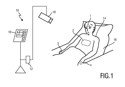
Fig. 1 shows an exemplary subject monitoring setup including an embodiment
of a device according to the present invention;
Fig. 2 shows a schematic illustration of a first embodiment of a device
according to the present invention;
Fig. 3 shows an electrocardiogram and a photoplethysmogram for measuring a
pulse arrival time according to the state of the art;
Fig. 4 shows an electrocardiogram and two PPG signals obtained at different
ROIs for illustrating the determination of PTT and PWV;
CA 02931377 2016-05-24
WO 2015/078735 PCT/EP2014/074950
9
Fig. 5 shows a schematic illustration of a second embodiment of a device
according to the present invention; and
Fig. 6 shows a flowchart of an embodiment of a method according to the
present invention.
DETAILED DESCRIPTION OF THE INVENTION
Fig. 1 shows an exemplary embodiment of a monitoring system 10 including a
device 12 for obtaining pulse transit time and/or pulse wave velocity
information of a
subject 14 according to the present invention. The subject 14, in this example
a patient, lies in
a bed 16, e.g. in a hospital or other healthcare facility. Image frames of the
subject 14 are
captured by means of a camera 18 including a suitable photosensor. The camera
18 forwards
the recorded image frames to the device 12. The device 12 is further connected
to an
interface 20 for displaying the determined information and/or for providing
medical
personnel with an interface to change settings of the device 12, the camera 18
or the
monitoring system 10. Such an interface 20 may comprise different displays,
buttons,
touchscreens, keyboards or other human machine interface means.
A monitoring system 10 as illustrated in Fig. 1 may, e.g., be located in a
hospital, healthcare facility, elderly care facility or the like. Apart from
the monitoring of
patients, the present invention may also be applied in other fields such as
neonate monitoring,
general surveillance applications, security monitoring or so-called live style
environments,
such as fitness equipment, or the like. The uni- or bidirectional
communication between the
device 12, the camera 18 and the interface 20 may work via a wireless or wired
communication interface. Other embodiments of the present invention may
include a
device 12, which is not provided stand-alone, but integrated into the camera
18 or the
interface 20.
Fig. 2 shows a more detailed schematic illustration of a first embodiment 12a
of the device 12 according to the present invention. The device 12a comprises
an interface 22
for receiving a set of image frames of a subject. Thereby, the interface 22
may correspond to
a wired or wireless network connection, any kind of serial connection or
another standard or
non-standard communication interface. The received image frames 19 may
particularly
correspond to a video sequence captured by means of an analog or digital
photosensor, e.g. in
a (digital) camera. Such a camera usually includes a photosensor, such as a
CMOS or CCD
sensor, which may also operate in a specific spectral range (visible, IR) or
provide
information for different spectral ranges. The camera may provide an analog or
digital signal.
CA 02931377 2016-05-24
WO 2015/078735 PCT/EP2014/074950
The image frames 19 include a plurality of image pixels having associated
pixel values.
Particularly, the image frames include pixels representing light intensity
values captured with
different photosensitive elements of a photosensor. These photosensitive
elements may be
sensitive in a specific spectral range (i.e. representing a specific color).
The image frames
5 include at least some image pixels being representative of a skin portion
of the subject.
Thereby, an image pixel may correspond to one photosensitive element of a
photodetector
and its (analog or digital) output or may be determined based on a combination
(binning) of a
plurality of the photosensitive elements.
The device 12a further comprises a motion detection unit 24 for detecting
10 motion of different body parts of the subject 14. Motion of a body part
may be detected by
comparing the current image with a reference image and simply counting the
number of
different pixels or by any other conventional motion detection algorithm.
The device 12a further comprises an ROT selection unit 26 for selecting at
least two regions of interest at body parts of the subject 14 within said set
of image frames 19.
In Fig. 1 such different ROIs 1, 2, 3, 4 are schematically indicated.
Selecting a region of
interest may be made by detecting a skin area from which light is reflected
that is received by
the imaging unit. Advantageous methods for selecting a region of interest in
order to derive
PPG signals from the image frames obtained from the region of interest are
generally known
in the art, e.g. from Georg Lempe, Sebastian Zaunseder, Tm Wirthgen, et al.
fROI selection
for Remote Photoplethysmographyõ, Informatik aktuell, Bildverarbeitung f. r.
die Medizin,
2013.
The device 12a further comprises a signal extraction unit 28 for extracting at
least two photoplethysmographic (PPG) signals from at least two selected
regions of interest
from said set of image frames 19. The extraction of PPG signals from an
imaging unit is
widely known in the art of vital signs monitoring and remote PPG. The
principle is e.g.
described in the above mentioned paper of Verkruysse et al. Such a signal
extraction unit 26
may particularly correspond to an analog or digital signal processor. A PPG
signal may
particularly correspond to a signal representing fluctuations in the light
intensity determined
based on a time series of image frames 19. Such a PPG signal may be
representative of a vital
sign of a subject such as a heart rate, the respiratory rate or the (arterial)
blood oxygen
saturation. The signal extraction unit 26 may particularly extract the PPG
signal based on
multiple image pixels and/or based on a series of time-consecutive image
frames.
The device 12a further comprises a motion correction unit 30 for controlling
said ROT selection unit 26 to select only regions of interest at substantially
unmoved body
CA 02931377 2016-05-24
WO 2015/078735 PCT/EP2014/074950
11
parts and/or for controlling said signal extraction unit 28 to extract a PPG
signal only from
regions of interest at substantially unmoved body parts or to correct PPG
signals extracted
from regions of interest at moving body parts. In this way, the effect of
motion shall be
cancelled or excluded as much as possible in order to increase the accuracy
and reliability of
finally obtained information.
The device 12a further comprises a distance determination unit 32 for
determining the physical distance between selected regions of interest. This
distance can be
easily determined within an image frame, e.g. by measuring the distance
between the centers
of the respective regions of interest. This can be done either by measuring
the distance
between body parts directly on a body, or by measuring the distance in pixels
between
coordinates of the centrums of ROIs and normalizing it to the size of the
entire body in pixels.
Finally, the device 12a further comprises a calculation unit 34 for
determining
pulse transit time and/or pulse wave velocity information 35 from the PPG
signals extracted
from different regions of interest and the respective determined physical
distance between the
respective regions of interest.
The various units of the device 12a may be comprised in one or multiple
digital or analog processors depending on how and where the invention is
applied. The
different units may completely or partly be implemented in software and
carried out on a
personal computer connected to a device for obtaining image frames of a
subject, such as a
camera device. Some or all of the required functionality may also be
implemented in
hardware, e.g. in an application specific integrated circuit (ASIC) or in a
field programmable
gate array (FPGA).
Arterial stiffness and Pulse Wave Velocity are generally estimated by
measuring PTT, which in its turn requires synchronized measurement of PPG
signals at
several sites of a body. Currently, multi-site PPG measurement is performed by
means of
placing several contact PPG sensors on body parts (legs, arms, forehead),
synchronized with
each other or/and with ECG. There are several disadvantages associated with
such set-up.
The set-up with several synchronized PPG and ECG sensors is cumbersome, takes
time to
install and therefore prone to errors. The shape and arrival time of pulse at
different body
locations is influenced by gravitation and therefore dependent on body
posture. Therefore,
the exact body pose of a subject should be carefully recorded and taken into
account during
measurements. For sensors placed on the forehead, positioning of the sensor is
crucial, since
the direction of blood flow affects the pulse delays measured by the detector.
Dependence of
CA 02931377 2016-05-24
WO 2015/078735 PCT/EP2014/074950
12
the shape of the pulse signal on placement of contact PPG sensor and the
sensor construction
makes accurate and reproducible measurement of PTT difficult.
In general, reproducibility of multi-site PPG measurement by means of probe
attachment to a body is affected by several factors, such as probe-tissue
interface pressure,
motion artifacts, subject posture and relaxation, breathing, etc. Moreover,
the measurement of
PPG signals on limited number of body spots (e.g. only legs, hands) might be
sufficient to
estimate PWV, but not enough to provide other information related to
monitoring of cardio
vascular system. For instance, analysis of the difference of phase and shape
of PPG signals
between foots provides an indication of diabetes, spatial distribution of PPG
amplitudes gives
the information about the local condition of micro vascular blood flow and
tissue viability,
etc.
The proposed device and method, in contrast, can unobtrusively, reliably and
synchronously measure spatial PPG information from multiple body sites
simultaneously,
automatically adjust to body position, respiration, body motion, and provide a
set of
parameters to compare shapes, phase, arrival times, amplitudes of PPG signals
from multiple
sites. Optionally, in an embodiment a multispectral high frame rate camera,
optionally
synchronized with ECG, is used for acquisition of the image data. This device
can optionally
contain a source of structured illumination emitted towards the chest of a
subject.
In this context fspatial PPG informationõ means a 2D array, where each pixel
represents an amplitude of extracted PPG signal. In other words, spatial PPG,
breathing, or
Sp02 information is generally a 2D map, where each pixel corresponds to 1D
signal of PPG,
breathing, or Sp02 signal extracted from either that pixel on a skin, or from
an ROT around
that pixel.
Fig. 3 shows, for illustration purposes, an electrocardiogram and a
photoplethysmogram for evaluating the pulse arrival time according to the
state of the art.
The electrocardiogram and the photoplethysmogram are detected at different
positions on the
human body in order to measure the pulse transit time and to detect trends in
the blood
pressure from the pulse arrival time.
The pulse arrival time is usually determined as a time frame from a maximum
peak R of the electrocardiogram to a certain point in time of the
photoplethysmogram. The
pulse arrival time may be detected as a time frame from the maximum R of the
electrocardiogram to a minimum value F of the photoplethysmogram as a foot
pulse arrival
time PATfoot or to a maximum value T of the photoplethysmogram as a top pulse
arrival time
CA 02931377 2016-05-24
WO 2015/078735 PCT/EP2014/074950
13
PATtop or as a time to the maximum slope of the photoplethysmogram between the
maximum
and the minimum value of the photoplethysmogram.
Fig. 4 shows a diagram of an ECG signal and two PPG signals obtained at the
hand (PPGhand) and at the foot (PPGfoot) of a subject. Therein the pulse
transit time at the hand
(PTThand) and at the foot (PTTfoot) are indicated as well as their difference
PTTdiff. The pulse
wave velocity PWV is obtained by calculating PWV = D / PTTdiff, where D is the
distance
between the hand and the foot, i.e. the positions where the PPG signals were
measured.
Fig. 5 shows another embodiment of a device 12b according to the present
invention comprising some additional elements compared to the embodiment 12a
shown in
Fig. 2. It shall be noted however that not all of these additional elements
need to be provided,
in further embodiments of the device only one or more of these additional
elements are
provided.
In particular, the device 12b comprises a vital signs determination unit 36
for
determining vital sign information 37 from the PPG signals extracted by the
signal extraction
unit 28 from one or more selected regions of interest. The term fvital signõ
as used in the
context of the present invention refers to a physiological parameter of a
subject (i.e. a living
being) and derivative parameters. In particular, the term "vital sign"
comprises heart rate (HR)
(sometimes also called pulse rate), heart rate variability (pulse rate
variability), pulsatility
strength, perfusion, perfusion variability, PPG pulsatility, Traube Hering
Mayer waves,
respiratory rate (RR), body skin temperature, blood pressure, pulse transit
time (PTT), a
concentration of a substance in blood and/or tissue, such as (arterial) blood
oxygen saturation
or glucose level. The term fvital sign informationõ as used in the context of
the present
invention comprises the one or more measured vital signs as defined above.
Furthermore, it
comprises data referring to a physiological parameter, corresponding waveform
traces or data
referring to a physiological parameter of a time that can serve for subsequent
analysis.
For instance, the changes of (arterial) blood oxygen saturation at different
body parts can thus be quickly determined, from which a body map indicating
the determined
oxygen saturation for the respective body parts can be quickly obtained. How
to determine
the blood oxygen saturation from PPG signals is generally known in the art and
e.g.
described in Wieringa, et al., "Contactless Multiple Wavelength
Photoplethysmographic
Imaging: A First Step Toward "Sp02 Camera" Technology," Ann. Biomed. Eng. 33,
1034-
1041 (2005).
The device 12b further comprises a respiration determination unit 38 for
determining respiratory information 39, in particular respiration rate and/or
changes of
CA 02931377 2016-05-24
WO 2015/078735 PCT/EP2014/074950
14
respiration volume, of the subject 14 from said set of image frames at
selected regions of
interest. Respiration information is a very valuable and essential information
quickly
providing information about sudden changes of the subject.s health condition.
This
respiration monitoring may e.g. be realized by detecting the subtle breathing
motion in the
subject.s chest (or belly) area.
A usable method for determining respiratory information from image data are
e.g. described in WO 2012/140531 Al according to which electromagnetic
radiation emitted
and/or reflected of a person is detected, wherein this electromagnetic
radiation comprises a
continuous or discrete characteristic motion signal related to the respiratory
rate of the person
and other motion artifacts related to the movement of the person or related to
ambient
conditions. This method increases the reliability of the respiratory rate
measurement by
taking into account data processing means adapted to separate the respiratory
rate signal from
overall disturbances by taking into account a predefined frequency band,
common predefined
direction or an expected amplitude band and/or amplitude profile to
distinguish the different
signals.
Another usable method for inferring the respiration rate from PPG signals,
which are modulated in amplitude, frequency and baseline is described in
Addison et. al. J.,
fDeveloping an algorithm for pulse oximetry derived respiratory rate (RRoxi):
a healthy
volunteer study,õ Journal of Clinical Monitoring and Computation, 26:45-51
(2012). Further
usable methods are also known in the art.
The device 12b further comprises a body posture detection unit 40 for
detecting the body posture of the subject 14. The calculation unit 34 takes
the body posture
into account in the determination of the pulse transit time and/or pulse wave
velocity
information. The body posture, e.g. lying on the back, on the side, sitting,
standing, etc., can
be determined from the image data 19 by conventional image processing methods,
such as
pattern recognition or other algorithms. Usable methods are e.g. described in
L. Panini, R.
Cucchiara fA Machine learning approach for human posture detection in domotics
applicationsõ, Proceedings of the 12th International Conference on Image
analysis and
Processing (ICIAP=03) and Humberto Souto Junior, Soraia Raupp Musse,
fAutomatic
Detection of 2D Human Posture based on Single Images,õ Proceedings of
Graphics, Patters
and Images (Sibgraphi), 2011, Aug, 2011.
The information about the posture of the body may be used in several ways for
proper calculation of PTT, PWV and evaluation of the pulse shape properties at
various body
locations. First of all, the posture information allows an accurate
calculation of distances
CA 02931377 2016-05-24
WO 2015/078735 PCT/EP2014/074950
between ROIs on various body parts. For that, the system should know the
position of a body
(e.g. siting, laying, etc.) and adjust the direct distance between ROIs
accordingly. Moreover,
the body position influences a pulse shape of extracted PPG signals. For
instance, the shape
of PPG signal extracted from a palm will be very different depending whether a
hand is
5 below a heart level or above. Therefore, for proper analysis of pulse
shape, the positions of
body parts in relation to each other are very useful.
Preferred embodiments of the proposed device thus have one or more of the
following monitoring functionalities:
- Automatic estimation of body posture and/or continuous tracking of motion
of
10 body parts. This is important to correctly calculate the distances
between ROIs on various
body parts and to make a proper analysis of pulse shape signal.
- Estimation of biometrical body parameters (length of arms, legs, distance
from
a palm to a heart, etc.). PPG signals extracted from different body parts
would have different
shape. Therefore, in order to accurately estimate the PTT/PWV and analyze
(changes of)
15 pulse shape information, estimation of biometrical body parameters are
useful. However, in a
basic embodiment, just a detection of body peripherals, and an analysis of PPG
signals
extracted from body peripherals might be sufficient.
- Measurement of respiratory motion and/or estimation of respiratory rate.
Breathing influences the shape of PPG signal, as well as inter peak distance
of pulse signals
and their amplitude. Therefore, in order to accurately analyze the differences
in PPG signals
extracted from various body parts, removal of the variability in PPG signals
caused by
respiration might be useful, as proposed in an additional embodiment of the
present invention.
Moreover, the breathing signal (rate, shape of respiratory signal) contains
important
information about the health condition of a person by itself.
- Measurement of relative changes of respiratory volume (e.g. by means of
analysis of structured light pattern changes during breathing). Regularity of
breathing and
type of breathing (belly or chest) provides an important information about the
health
condition of a person.
- Measurement of PPG signals in different wavelengths, including at least
green,
red, infra-red. Monitoring of PPG signals in at least two wavelengths is
required to provide
robustness of PPG measurement to motion and ambient illumination, and to
provide Sp02
measurements.
- Analysis of PPG imaging (spatial map of PPG amplitude) of visible skin
areas
of a body in at least green and infra-red color channels. Changes of PPG
imaging per spatial
CA 02931377 2016-05-24
WO 2015/078735 PCT/EP2014/074950
16
skin location can be used for evaluation of blood microcirculation, as e.g.
described in U.
Rubins, V. Upmalis, et al. fReal-time Photoplethysmography Imaging System,õ
IFMBE
proceedings 34, pp. 183-186, 2011. This paper describes the use of PPG imaging
for
monitoring of blood perfusion changes during local anesthesia. Moreover, PPG
imaging can
be used as a tool to automatically detect ROIs on a body with the strongest
PPG signal, which
will serve as reliable ROIs for PTT and PWV measurement.
- Monitoring of changes of Sp02 values at different body sites.
Oxygenation of
arterial blood is changing over a body with different dynamics. Spatial
dynamics of Sp02
changes may be used for estimation of local microcirculation in a way similar
to PPG
imaging.
In preferred embodiments of the device body posture and/or body motion are
determined, and/or control for adaptive acquisition of vital signs is
provided. In particular,
based on PPG imaging skin segments are defined, which have the strongest and
most reliable
PPG signal (using PPG imaging, as described above), which segments are used as
virtual
sensors, particularly for PTT and PWV measurement. Further, an objective
estimation of
exact body posture is made to provide reproducibility of PTT, PWV
measurements.
Estimation of body motion is performed to control the acquisition of PPG
signals (e.g. to stop
acquisition from a particular body part, if motion of this part is detected)
or to provide motion
robust acquisition of PPG signals. Estimation of the respiratory motion (in
particular both
respiration rate and relative changes of volume), which information is used to
control the
acquisition and adaptive analysis of PPG signals, which would be required for
accurate
calculation of PTT and PWV. Further, an ECG sensor can be optionally provided
for more
accurate calculation and/or confirmation of PTT and PWV. In the embodiment
with ECG
sensor, PTT and PWV are calculated based on a time difference between peaks of
ECG
(reference time stamps) and peaks of pulse PPG signal from one or several body
parts. In an
embodiment without ECG, PTT and PWV are calculated based on time distance
between
beats of PPG signals acquired from different body parts.
In this context, a fvirtual sensorõ means an ROT on skin, wherein all pixels
are
preferably averaged to extract a physiological signal. For instance, if an ROT
(fvirtual sensorõ)
is selected on a forehead, all pixels within this forehead ROT are averaged to
extract one PPG
signal. The proposed device and method can have either thousands of such ROIs
/ virtual
sensors, or only one virtual sensor, which includes all pixels of the visible
skin.
CA 02931377 2016-05-24
WO 2015/078735
PCT/EP2014/074950
17
Moreover, in preferred embodiments of the device one or more of the
following functionalities are provided (which are preferably carried out by
the calculation
unit 34 or by separate additional units):
- Analyze the time difference between beats of PPG signals acquired from
fvirtual sensorsõ (i.e. the selected ROIs) located at legs, hands, and around
a heart area of a
person. In another embodiment of the invention, the time differences between
beats of PPG
signals acquired from selected ROIs are calculated with reference to beats of
an ECG signal
(if available)
- Calculate the distance between fvirtual sensorsõ.
- Calculate PTT and PWV between several pairs of fvirtual sensors,õ taking
into account the information from above two steps.
- Analyze the difference in Sp02 trending between virtual sensor on a
forehead
and body peripherals.
- Analyze the phase shift of PPG signal between two feet from fvirtual
sensorsõ
located at the same distance from a heart.
- Analyze the relation between respiratory volume, respiratory rate, and
changes
of PPG amplitude. For example, the method described in Lena Nilsson, Tomas
Goscinski, et
al. fRespiratory variations in the photoplethysmographic waveform: acute
hypovolaemia
during spontaneous breathing is not detectedõ, 2010 Physiol. Meas. Volume 31,
Number 7 or
in Nilsson L, Johansson A, Kalman S., fRespiratory variations in the
reflection mode
photoplethysmographic signal. Relationships to peripheral venous pressure,õ
Medical and
Biological Engineering and Computing 2003 May; 41(3):249-54 can be used for
this purpose.
The imaging unit 18, which may also be part of the device 12, is preferably a
video camera for acquiring PPG signals in several color channels from multiple
fvirtual
sensorsõ (ROIs), from which various PPG-related information, in particular
vital signs, such
as Sp02, pulse shape, pulse amplitude etc. are derived. Further, respiratory
rate and changes
of respiratory volume can be derived from the acquired image data, e.g. by
analyzing motion
of a chest and/or belly area.
By analyzing he differences in PPG-related information between fvirtual
sensorsõ PTT, PWV, speed of Sp02 changes etc. can be estimated, and the
dependency
between respiratory efforts, respiratory volume and changes in shape,
amplitude and inter-
peak distances of extracted PPG signals can be analyzed.
Fig. 6 shows a flowchart of an embodiment of a method according to the
present invention. In a first step S10 the image data (video data) are
obtained, e.g. of the
CA 02931377 2016-05-24
WO 2015/078735 PCT/EP2014/074950
18
entire body of the subject, in different color channels. In step S12 visible
skin areas are
detected in the image data. In step S14 PPG imaging is performed for the
visible skin areas.
For instance, a spatial map of PPG amplitudes for some or each pixel of a skin
ROT is
obtained from the image data. In step S16 ROIs with the strongest PPG
pulsatility are
detected, which ROIs represent fvirtual sensors,õ i.e. locations from which
the signals will be
used for further processing. In this step S16 information obtained from step
S18, in which
non-moving ROIs are detected, is additionally used, i.e. only non-moving ROIs
are generally
used as fvirtual sensorsõ.
In step S20 PPG signals are acquired from all detected ROIs (fvirtual
sensorsõ). In step S22 the PPG phase shift between two or more virtual sensors
are analyzed.
The phase shift between PPG signals acquired from various body parts will be
used for
calculation of PTT, PWV and eventually for arterial stiffness estimation.
In step S24 the respiratory rate, spatial breathing map and/or changes of the
volume are analyzed. Based on this information and the PPG signals obtained in
step S20
changes in the PPG morphology and Sp02 are analyzed in step S26 depending on
the
respiration.
In step S28 the distance between detected ROIs is estimated. Based on the
information from steps S20 and S30 PTT, PWV and the speed of changes of Sp02
between
the detected ROIs are calculated. Finally, in step S32 blood pressure changes
are estimated
based on the calculated PTT and PWV, for instance according to a method as
disclosed in J.
Sola, St. Rimoldi, and Yves Allemann, fAmbulatory monitoring of the
cardiovascular system:
the role of Pulse Wave Velocity,õ in New Developments in Biomedical
Engineering, I-Tech
Education and Publishing, Vienna, Austria, ISBN 978-953-7619-57-1.
According to another aspect a device is proposed for obtaining physiological
information of the subject. Said device generally comprises all elements of
the device 12a
shown in Fig. 2, except for the distance determination unit 32. Further, the
calculation unit 34
is configured differently, namely to determine physiological information of
the subject
including one or more of diagnosis of diabetes, evaluation of local blood
microcirculation,
analysis of changes of local blood perfusion by analyzing PPG signals acquired
from
different body parts of the subject. Multiple PPG signals extracted from
different regions of
interest are evaluated to perform a diagnosis of diabetes (e.g. by analyzing
the difference in
phase and shape of two PPG signals acquired from both feet or both legs), to
monitor the
local blood microcirculation and local blood perfusion acquired.
CA 02931377 2016-05-24
WO 2015/078735 PCT/EP2014/074950
19
In summary, the proposed device and method allow estimating several vital
signs from one video stream, analyzing the differences in morphology and
temporal changes
of those vital signs between several parts of a body and estimating the local
vascular
characteristics of different body parts at the same time. PTT and PWV are
estimated from
PPG signals acquired from several body sites, selected preferably based on the
strength of
PPG imaging and local motion information. Further, changes of blood pressure
can be
evaluated based on the estimated PWV.
Instead of combining various contact sensors (ECG, PPG, respiration, etc.) as
conventionally done, the proposed device and method provide the same or even
more
functionalities. The proposed device and method thus do not just replace the
functionalities of
ECG, PPG, etc. sensors, but provide a functionality achieved currently only by
a particular
way of combination of those known sensors. For instance, currently, in order
to acquire PWV,
two contact PPG sensors should measure PPG signals synchronously and the
analysis system
must know exactly the positions of those sensors, measure the physical
distance between
sensors, etc. All this is replaced by the signal processing provided in the
proposed device and
method. By use of a (single) imaging device (e.g. camera) several vital signs
of different
physiological origin (PPG, breathing motion) can thus be measured from
multiple locations
of a body, simultaneously with context information (body motion, distance
between ROIs),
and signal processing can be applied to extract derivative vital signs based
on combined
analysis of measured physiological signals and context information.
The proposed method and device can particularly be used for quick evaluation
(scan) of a cardiovascular condition of a person by measuring multiple vital
signs from
different parts of the body without a hassle to attach several contact sensors
and provide for
their synchronization. They can further be used either for a periodic scan
during ambulatory
cardiovascular monitoring, or for continuous monitoring to detect early
deteriorations and to
reliably detect severe deteriorations of the person.s condition (e.g.
centralization).
While the invention has been illustrated and described in detail in the
drawings
and foregoing description, such illustration and description are to be
considered illustrative or
exemplary and not restrictive; the invention is not limited to the disclosed
embodiments.
Other variations to the disclosed embodiments can be understood and effected
by those
skilled in the art in practicing the claimed invention, from a study of the
drawings, the
disclosure, and the appended claims.
In the claims, the word "comprising" does not exclude other elements or steps,
and the indefinite article "a" or "an" does not exclude a plurality. A single
element or other
CA 02931377 2016-05-24
WO 2015/078735 PCT/EP2014/074950
unit may fulfill the functions of several items recited in the claims. The
mere fact that certain
measures are recited in mutually different dependent claims does not indicate
that a
combination of these measures cannot be used to advantage.
A computer program may be stored or distributed on a suitable non-transitory
5 medium, such as an optical storage medium or a solid-state medium
supplied together with or
as part of other hardware, but may also be distributed in other forms, such as
via the Internet
or other wired or wireless telecommunication systems.
Any reference signs in the claims should not be construed as limiting the
scope.